Review

Mineral Surface-Templated Self-Assembling Systems: Case Studies from Nanoscience and Surface Science towards Origins of Life Research

by
Richard J. Gillams
1,2,† and
Tony Z. Jia
1,*,†
1
Earth-Life Science Institute, Tokyo Institute of Technology, 2-12-1-IE-1 Ookayama, Meguro-ku, Tokyo 152-8550, Japan
2
Structural Genomics Consortium, Nuffield Department of Medicine, University of Oxford, Old Road Campus Research Building, Oxford OX3 7DQ, UK
*
Author to whom correspondence should be addressed.
These authors contributed equally to this work.
Submission received: 31 March 2018 / Revised: 26 April 2018 / Accepted: 3 May 2018 / Published: 8 May 2018
(This article belongs to the Special Issue Water–Rock Interactions and Life)

Abstract

:
An increasing body of evidence relates the wide range of benefits mineral surfaces offer for the development of early living systems, including adsorption of small molecules from the aqueous phase, formation of monomeric subunits and their subsequent polymerization, and supramolecular assembly of biopolymers and other biomolecules. Each of these processes was likely a necessary stage in the emergence of life on Earth. Here, we compile evidence that templating and enhancement of prebiotically-relevant self-assembling systems by mineral surfaces offers a route to increased structural, functional, and/or chemical complexity. This increase in complexity could have been achieved by early living systems before the advent of evolvable systems and would not have required the generally energetically unfavorable formation of covalent bonds such as phosphodiester or peptide bonds. In this review we will focus on various case studies of prebiotically-relevant mineral-templated self-assembling systems, including supramolecular assemblies of peptides and nucleic acids, from nanoscience and surface science. These fields contain valuable information that is not yet fully being utilized by the origins of life and astrobiology research communities. Some of the self-assemblies that we present can promote the formation of new mineral surfaces, similar to biomineralization, which can then catalyze more essential prebiotic reactions; this could have resulted in a symbiotic feedback loop by which geology and primitive pre-living systems were closely linked to one another even before life’s origin. We hope that the ideas presented herein will seed some interesting discussions and new collaborations between nanoscience/surface science researchers and origins of life/astrobiology researchers.

Graphical Abstract

1. Introduction

What fundamental requirements does Earth or any planet need to produce and sustain life? This is a fundamental question related to our own genesis and history, and the study of origins of life seeks answers and plausible explanations to this and related questions regarding how life on Earth came into existence and how life could exist or arise elsewhere in the universe. The exploration of space is a natural extension of this line of inquiry and provides an array of technical challenges that has driven innovation and continues to inspire scientists to understand their environment, both on this planet and beyond. This overarching aim disguises an inventory of scientific understanding that bridges a broad range of disciplines from astrophysics to microbiology, from chemistry to geology, and requires collaborative efforts where the boundaries of research fields become blurred or new fields must be created. Specifically, there are many research fields which lie at the periphery of origins of life research, including nanoscience and surface science, that are willing and able to contribute to origins of life research, yet much of the research remains unknown to those in the field. How do we engage and collaborate with such research communities in order to incorporate their ideas and answer fundamental questions related to life’s origins?
Recent studies in origins of life research have focused on how the basic building blocks of life (i.e., amino acids, nucleotides, lipids, etc.) could have been formed on early Earth or on other planets. As the origin of life on Earth likely involved aqueous chemistry [1], much prebiotic chemistry research has focused on this. Yet interfacial and supramolecular chemistry should also be considered in a prebiotic context, as these systems result in phenomena that can catalyze reactions and facilitate novel reaction pathways inaccessible in solution [2]. This increased structural, functional, and/or chemical complexity is achieved through, for example, increased local concentrations of adsorbed reactants (pushing the equilibrium towards product formation) [3,4], orientational specificity of adsorbed reactants (which would result in a preferred face or moiety of an adsorbed molecule to be accessible (or inaccessible) to catalysts in solution) resulting in catalytic selectivity [5,6], or through the provision or accession of electrochemical driving forces such as in mineral semiconductors [7,8]. The most common interfaces available on primitive Earth or other extraterrestrial bodies are mineral surfaces. Minerals are produced during the formation of terrestrial planets [9], and various minerals, including apatites (Ca5(PO4)3(F,Cl,OH)) [10], phosphate minerals such as schreibersite ((Fe,Ni)3P) [11,12], and borates such as colemanite (Ca2B6O11·5H2O) [13,14] (although boron is fairly common in different sedimentary rocks, a mechanism by which high concentrations of specific borate minerals could have accumulated on early Earth is unclear [15]), can catalyze the synthesis of nucleotides and amino acids from simple chemical precursors like phosphates, nucleosides, or formamide. These nucleotides and amino acids can adsorb onto clay minerals such as montmorillonite ((Na,Ca)0.33(Al,Mg)2(Si4O10)(OH)2·nH2O), illite ((K,H3O)(Al,Mg,Fe)2(Si,Al)4O10[(OH)2,(H2O)]), and kaolinite (Al2Si2O5(OH)4) [16,17,18], which can catalyze their polymerization into more complex biopolymers such as nucleic acids [19,20,21,22] and peptides [23,24,25,26,27]. Various mineral surfaces such as montmorillonite [28], silicates (such as aluminum silicate, Al2SiO5), carbonates (such as hydrotalcite, Mg6Al2CO3(OH)16·4(H2O)) [29], and sulfides (such as pyrite, FeS2), among others [30] have also been shown to promote fatty acid vesicle formation, an important process in the initial development of primitive cells by which biopolymers become stably encapsulated within a compartment, i.e., a protocell. Thus, mineral–water interfaces are clearly important for prebiotic chemistries leading to primitive life both on Earth [31] as well as across the entire cosmos [32]. As mineral surfaces are likely to be similar in structure universally, understanding the catalytic properties of terrestrial mineral surfaces in the laboratory would give us more information on how life could have emerged on other rocky planets.
In this review, we examine how mineral surfaces can template and promote prebiotic chemical and biomolecular self-assemblies that were likely an essential stage in the origin of life on Earth by presenting case studies from nanoscience and surface science, such as organization of small organic monolayers on mineral surfaces, secondary structure changes of nucleic acids upon mineral surface binding, assembly of peptide amyloids by mineral surfaces, and mineralization of new mineral surfaces by self-assembled chemical systems. Self-assemblies such as folded proteins or DNA double helices [33] are important in extant life as they provide structures and functions that non-assembled systems are simply unable to provide. Similar assemblies may have been especially important for early living systems long before the emergence of genetic evolution due to their ability to achieve higher complexity compared to simpler structural systems. Self-assembly is also often spontaneous and energetically favorable, taking advantage of non-covalent bonds (covalent bonds such as phosphodiester or peptide bonds in biopolymers produced by condensation were generally thermodynamically unfavorable on early Earth [34]), including H-bonding, aromatic stacking, electrostatics, and hydrophobic interactions [35,36,37] and can be perturbed through simple processes available on early Earth such as heating [38], hydration/drying [39], pH changes [40], and magnetic [41] and electric fields [42], allowing for dynamic assembly and disassembly processes. We present some case studies from surface science and nanotechnology that may be unfamiliar to the origins of life/astrobiology research communities to highlight the breadth of assembly-promoting functions that different mineral surfaces may provide, and the increase in structural, functional, and/or chemical complexity arising from these self-assemblies. It is clear from these examples that the origins of life research field can be enhanced through closer collaboration with colleagues in the nanoscience and surface science research fields.

2. Case Studies

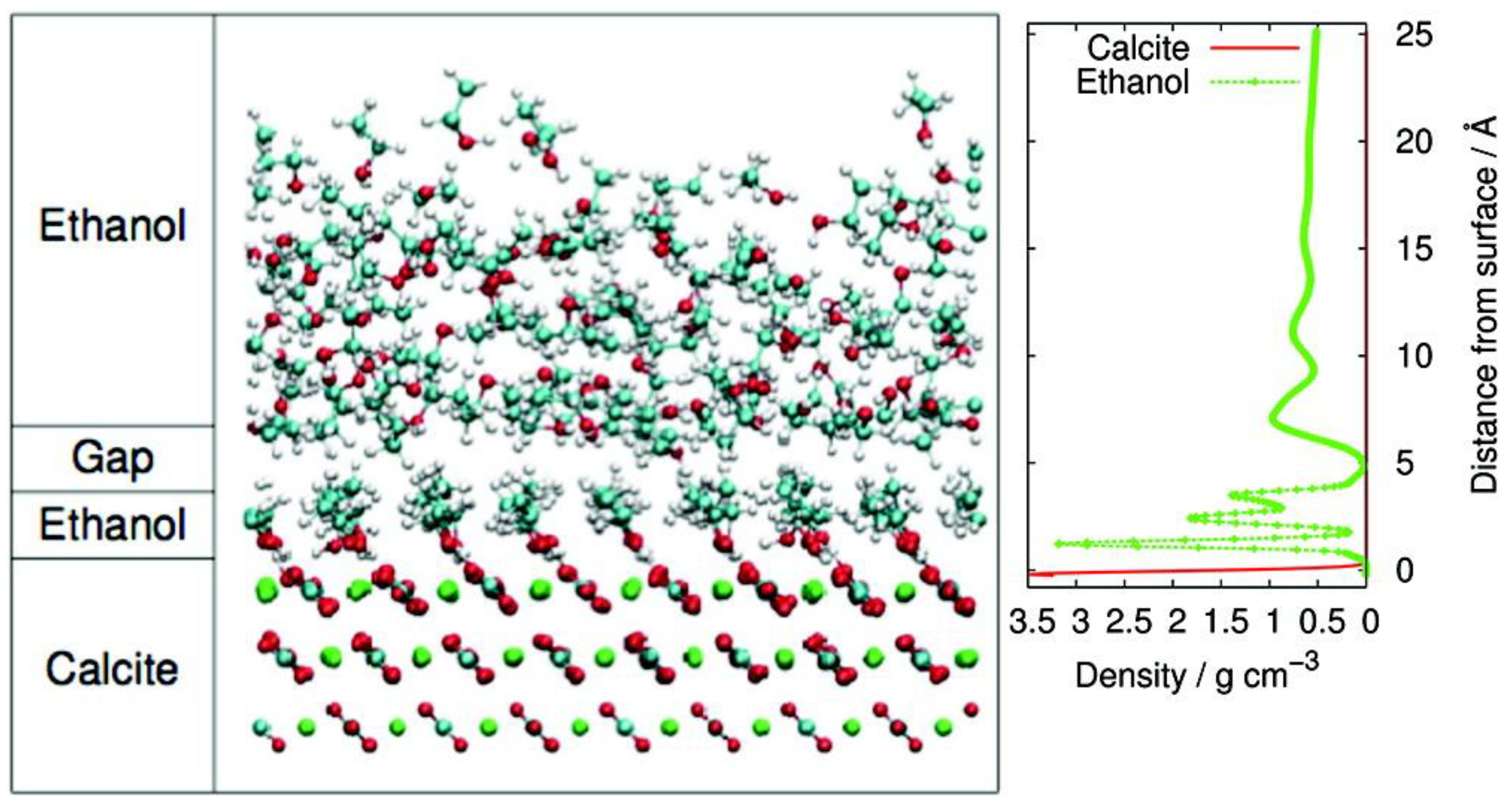
2.1. Simple Organic Molecules

In addition to adsorption of biomonomers such as nucleotides and amino acids, mineral surfaces are able to adsorb or interact with other various small organics including small carboxylic acids like salicylic acid onto kaolinite (Al2Si2O5(OH)4) and aluminum oxide (Al2O3) [43] and simple alcohols such as methanol or isopropanol onto calcite (CaCO3) [44]. The adsorption of these small organic molecules is often governed by the functional groups on a given molecule, the structure and charge of the mineral surface, and also the solvent [43], often generating mono- or poly-layers of the small organic molecule on the mineral surface [44,45,46]. Ordered surface organic layers (in this case, stearic acid) have even been studied in the context of calcite crystal nucleation [47].
Stipp and coworkers studied the microstructure of ethanol layers adsorbed to the surface of calcite after incubation in liquid ethanol or water/ethanol mixtures and determined that ethanol monolayers form readily [45,48,49]. In the case of incubation with pure ethanol, two organic layers formed, with the layer closest to the calcite surface having a thickness equal to the molecular length of a single ethanol molecule [45] (Figure 1). These ethanol molecules adsorb perpendicular to the mineral surface in a highly ordered manner, while the second ethanol layer is much thicker, slightly less ordered, and is separated from the first layer by a gap. All of the polar hydroxyl groups in the first ethanol layer are oriented towards the ionic calcite surface, while the non-polar ends of the ethanol are oriented away from the mineral surface; this was also observed in studies with other alcohols like pentanol and propanol [44]. If the upper layer is removed, then the assembly of single alcohol monolayers onto calcite or any other mineral would change the surface to be hydrophobic. Where initially more hydrophilic groups would interact preferentially with and be adsorbed directly by the mineral surface, after a mineral surface is treated with small organic amphiphiles, this new hydrophobic surface could then restrict the ability of water or hydrophilic molecules such as metal ions from accessing the mineral surface [48].
One may also imagine a scenario where only part of a surface is populated by an organic monolayer, such as on a heterogeneous rock surface with multiple mineral types that have different adsorption propensities of organics such as shales, which are sedimentary rocks that contain multiple different mineral species [50,51]. Consider a shale which has been eroded slightly by weathering such that the exposed sections contain the surfaces of multiple minerals including clays, quartzes, carbonates, calcites, and others. Amino acids have been shown to have specific preferences for binding to certain minerals, such as aspartic acid preferentially adsorbing onto calcite (CaCO3) and glycine, serine, isoleucine, leucine and arginine preferentially adsorbing onto quartz (SiO2) [52]. Different clays have also shown different propensities for adsorption of various simple organics, including small polar organics like formamide or urea strongly adsorbing onto kaolinite (Al2Si2O5(OH)4) and various aromatic compounds like benzene and phenol interacting with Cu-Montmorillonite (copper (II) with (Na,Ca)0.33(Al,Mg)2(Si4O10)(OH)2·nH2O) [53]. A diverse heterogeneous organic microenvironment could form on such shale surfaces, providing localized reactions on each specific mineral. This microenvironment could have also facilitated interactions or reactions between the different heterogeneous organic pockets due to the adjacent proximity of these reactions. If so, such heterogeneous environments could assist early chemical systems in diversifying and incorporation or coupling of different types of reactions together to form larger, more complex chemical networks.

2.2. Nucleic Acids

Nucleic acids are the genetic materials for all known modern life, and thus it is important to understand how these materials emerged, evolved, and what governs their function [33]. The structures assembled by nucleic acids are not merely one-dimensional primary sequence strings of nitrogenous bases, but rather are due to complex folding into secondary, tertiary, and quaternary structures that result in geometries and assemblies essential for cellular function [54]. Within a cell, DNA molecules are typically assembled into helices, which allow for complementary strands to hybridize. Major and minor grooves are generated in the phosphate backbones of these helices that allow for binding of specific proteins [55]. With the help of certain enzymes, these assemblies temporarily come apart to allow access to the bases during important cellular processes including transcription [33]. Specific, complex assemblies are also required for the function of various ribozymes, primitive RNA-based catalysts that may have been important during the initial emergence and evolution of primitive genetic systems through their potential catalysis of RNA polymerization [56,57]. Although nucleotides can oligomerize somewhat under prebiotically plausible conditions, for example through high-energy chemical activation in solution [58] (in the absence of an activated group, nucleotides have so far not been shown to be able to polymerize at all) or on clay mineral surfaces [19], there is still no known mechanism by which nucleotides can efficiently polymerize into nucleic acid polymers and subsequently replicate and evolve in the absence of enzymes [59]. Despite this limitation in knowledge, there has still been plenty of work probing nucleic acid adsorption onto mineral surfaces. The assembly of nucleotide monomers adsorbed onto mineral surfaces into monolayers has been postulated by some as an important process that promoted interactions between nucleotides and other important biomolecules including amino acids and carbohydrates [60]. Both single- and double-stranded nucleic acids have also been shown to adsorb onto various mineral surfaces, including mica (general) [61], clay minerals [62], calcites, carbonates, and metal oxides [63].
Typically, double-stranded DNA preferentially assembles into a B-form helical conformation in standard aqueous physiological conditions [64]. However, upon binding to the clay mineral kaolinite (Al2Si2O5(OH)4), DNA changes to a Z-form helical conformation [62], a unique left-handed helical form apparent (though rare) in nature [65]. Although some functions for Z-form DNA have been discovered, including increasing of mutagenesis and deletion rate near Z-form DNA loci [66,67], no specific biological function has yet been determined other than their apparent role in the function of certain poxviruses [68]. Binding to montmorillonite ((Na,Ca)0.33(Al,Mg)2(Si4O10)(OH)2·nH2O), however, does not change the helical conformation at all [62]. In an early Earth scenario, it is possible that binding to clay mineral surfaces such as kaolinite (Al2Si2O5(OH)4) provided access to novel helical structures for nucleic acids, which normally are only possible in non-standard conditions, for example A-form transitions which normally require high relative humidity or Z-form transitions which normally require high salt concentrations [68,69].
Although it is clear, due to its dual role as a genetic information carrier/proliferator and as a catalyst, that RNA would have likely been more important in the initial emergence of life than DNA, there is a current lack of research on effects of mineral surface adsorption on RNA conformations, save for some computational studies [70]. However, given the fact that, similar to DNA, RNA can also transition between various helical forms depending on environmental factors [71], future studies of mineral surface-effected changes in adsorbed RNA conformations would be useful and of interest to the origins of life research community. For example, normally nucleotides within a standard A-form helical RNA exists in the C3′-endo anti sugar pucker conformation, while Z-form RNA is composed of repeating nucleotides with alternating C2′-endo anti and C3′-endo syn sugar puckers, often with alternating cytidine-guanosine bases [72]. However, some small functional RNAs such as riboswitches or aptamers contain Z-like steps, which are localized areas of a nucleic acid strand generated when two adjacent nucleotides exist in the anti (5′) and syn (3′) conformation [73]. Z-form RNA perhaps composed of cytidine-guanine repeats (which may have been more abundant than adenine and uridine on the primitive earth as cytidine and guanine are less error prone and polymerize orders of magnitude faster in a nonenzymatic replicating RNA system than adenine and uridine [74]), RNA containing Z-like steps, or any other novel RNA structures that could have been induced in a primitive replicating genetic system, perhaps by mineral adsorption, could have potentially allowed primitive nucleic acid systems to explore additional structure space during evolution in a search for novel or more efficient functions or catalytic activity, respectively.
Additionally, DNA molecules have been shown to adsorb to mica (general) and form specific structures under different ionic conditions. Salts like magnesium chloride and cobalt chloride supply divalent cations, which bridge the negatively charged mica surface with the negatively charged nucleic acid phosphate backbone [75], resulting in DNA adsorbing onto a mica surface; a salt like sodium chloride, which only provides a monovalent cation, does not promote DNA adsorption at all [61]. Different divalent counterions can result in different structural moieties of DNA upon mica surface binding: magnesium cations resulted in condensed and coiled DNA (with some topography) on the surface, while cobalt cations induced formation of flat two-dimensional monolayers. This is due to the fact that cobalt cations have a greater affinity for mica than magnesium cations [76], resulting in tighter binding between the DNA and the mica surface. This binding could lead to a monolayer structure where most of the DNA is adhered to the surface, as opposed to magnesium-induced coiled DNA structures where much less of the DNA is adhered (but it is still immobilized) to the surface. In addition to cobalt cations, which could have been introduced (although perhaps not in high enough concentration to effect the structural changes described above) into prebiotic aqueous systems through weathering of cobalt-containing minerals [77], other more abundant prebiotic divalent cations that show a strong ability to promote DNA adsorption on mica include nickel and zinc. The ionic radii of these two divalent cations (similar to cobalt and magnesium; although the ionic radii of cobalt and magnesium cations are similar, magnesium cations do not contain any d electrons and are thus unable to form as many complexes as cobalt, explaining why cobalt has a greater affinity for mica than magnesium) are small enough to fit into mica cavities, resulting in a stronger adhesion of DNA to the mica surface [76]. Ferrous iron could have also been a strong promoter of DNA-mica surface adsorption on early Earth, as its ionic radius is similar to that of cobalt and zinc cations [78]. It would be of great interest to study the structure of adsorbed DNA and RNA on mica surfaces in the presence of these additional divalent cations rather than cobalt due to their higher concentration than cobalt on the early Earth (in particular ferrous iron, which was very abundant in early Earth oceans [79]) to determine whether mica surfaces coupled with prebiotically available divalent cation solutions could have contributed to additional structural order and complexity for primitive nucleic acids.
The structuring of nucleic acids, in particular RNA, onto mineral “scaffolds” in different ionic conditions could have also provided an anchoring surface for easy and rapid access to a complex library of different sequences that could be probed for catalytic activity or binding through flowing substrates. Such an arrayed nucleic acid system would be structurally similar to DNA nano-arrays assembled on mica (general) surfaces that have been developed by materials scientists [80] (Figure 2). This idea could also be extended to include the possibility of assembly on mineral surfaces of primitive nucleic acid origamis [81], modern synthetically-produced structures that rely on specifically-designed base–base interactions to result in assembly into specific shapes. Although much nucleic acid origami research has focused only on the novel structural aspects within DNA-only systems, recently researchers have made designed functional DNA origamis, including nanocages for encapsulation of small molecules in drug delivery [82,83] or lipid membrane extensions that promote cell–cell adhesion [84]. In addition to DNA origamis, more relevant to early Earth systems, RNA origamis have been developed [85,86,87]. One such system involved using modified versions of the Tetrahymena GI ribozyme [88] and adding short, specifically-designed recognition sites, i.e., short runs of a few bases, to some regions of the modified ribozyme. These modules were then allowed to assemble, and due to the designed interactions between certain recognition sites, modular trimers and tetramers assembled into triangular and square-like structures, respectively. Although the structure is by itself already novel, both the assembled triangle and square structures resulted in greater catalytic activity than just its separate components [86]. This demonstration of structural assemblies of catalytic RNA resulting in greater catalytic activity suggests that in a primitive genetic system on the early Earth, modular self-assemblies combining multiple short catalytic RNA could have also resulted in catalytic enhancements (or even novel functions). However, one must keep in mind that the examples provided from the nucleic acid origami fields all require very specific base-designs, a feature that an early Earth system would not have had access to. Nevertheless, at least in [86], each of the recognition sites contained only 4–8 nucleotides, suggesting that even in a primitive system, the required recognition site sequences are still reasonably short (such that there is a possibility that they could have formed through random sequence polymerization, especially if there was a much greater abundance of cytidines and guanines in the available nucleotide pool for the same reasons as described above [74]) and thus it still could have been possible for simpler, shorter self-assembled modular catalytic structures to have formed.

2.3. Peptides

One of the most important findings in the field of degenerative disease medicine (and also, as it turns out, peptide nanoscience) was the discovery that a short peptide of around 40 residues, amyloid beta, assembles into filamentous plaques in the brain of Alzheimer’s disease patients [89]. This physiologically produced peptide, which misfolds and aggregates into oligomers, is generally the cause of this deadly neurodegenerative disease [90] likely through inhibition of certain tissue functions, although it is not well understood and other hypotheses exist [91,92]. Since this discovery, many other proteins and short peptides have been implicated in diseases arising from the assembly of amyloid fibrils [93], and at the same time these discoveries have inspired much new research in small peptide self-assemblies. In an attempt to develop a nanotubular material relevant to nanowire nanofabrication, Gazit and coworkers discovered that within the amyloid beta peptide, a short diphenylalanine dimer was able to self-assemble into distinct nanotubes, giving the first demonstration that even peptides with as little as two subunits can self-assemble into robust nanostructures [94]. Since then, many other small, self-assembling peptides have been discovered through experimental and computational methods [95,96]. These assemblies contain varied architectures including fibers, films, and hydrogels [97], and have a wide range of engineering applications including as antibacterial agents [98], semiconductors [99], and much more [100].
Although these examples are all of modern peptide self-assemblies in applicational settings, peptide self-assemblies, including those templated by mineral surfaces, are still relevant to prebiotic chemistry as they are structures that can provide structural and functional complexity to early living systems. Although amino acids produced on early Earth through atmospheric discharge [101,102], from extraterrestrial sources including meteorites [103] and in undersea hydrothermal systems [104,105] can then polymerize into simple peptides through volcanic-gas activation of amino acid monomers [106,107] or on clay mineral surfaces coupled with wet–dry cycles [23,24,25,108], the ability of mineral surfaces to catalyze the polymerization of very long peptides with catalytic functions remains poorly studied and understood. Thus, self-assembly of short peptides may be one energetically favorable way for an early Earth system to access emergent architectures or functional properties. As mineral surfaces present sites capable of interaction with the various functional groups on peptides, these surfaces could have been anchors for promoting short peptide self-assembly.
Many studies in the nanoscience field have probed the role of mineral surfaces in self-assembling peptide systems. For example, 16-mers of polyglycine can aggregate into ordered helical and β-sheet secondary structures on oxide surfaces including anatase (TiO2) and amorphous SiO2 [109]. This demonstration is important as secondary structures (as well as tertiary and quaternary structures) are necessary for proteins to achieve catalytic function [69], suggesting that mineral surfaces could have assisted in developing primitive peptide function by promoting the formation of more complex structures not possible in solution, similar to what was observed above in RNA upon kaolinite (Al2Si2O5(OH)4) binding. The assembly of other peptides, such as longer amyloids (around 100 residues) or short amyloid-derived peptides (eight to nine residues), into filaments or fibrils can also be templated by mica (general and muscovite, (KAl2(Si3Al)O10(OH)2)) surfaces [110,111,112] (Figure 3). On primitive Earth, these filamentous nanostructures could have aided in increasing the structural or functional complexity of early chemical systems, for example by scaffolding and/or catalyzing the polymerization of biopolymers like other peptides [113]. Nanofibers also could have been more efficient, compared to mineral surfaces, at promoting the self-assembly of additional peptide nanostructures [114]. Finally, the physical appearance of self-assembled peptide nanofibers, such as in Figure 3, appears to be fibers branching out from a central nucleation site. This bears a striking physical similarity to peptide dendrimers, which are branched polymers containing a core from which various functional peptide groups emanate [115]. Although the branching of peptide nanofibers takes place at the supramolecular scale, as opposed to the branching of peptide dendrimers taking place on the molecular scale, given the similarity in overall dendrimer-like structure perhaps branched peptide nanofibers on early Earth could have had similar functions as peptide dendrimers. For example, if peptide nanofibers contained many catalytic residues like serine, histidine, or aspartic acid [116], perhaps the supramolecular ordering afforded by the fibrils could allow the formation of primitive catalytic triads [117], just like those found in some peptide dendrimer enzymes [118].
In addition to amyloids, molecules like peptide amphiphiles [119] or amphiphilic peptides [120] are also able to assemble into nanostructures on mineral surfaces such as mica (general). Although these are the only two examples given of mineral-templated nanofiber structures, there is a wide variety of other structures and functions that self-assembled peptide amphiphiles [121] and amphiphilic peptides [122] are able to achieve in solution, each of which would be an interesting case study for origins of life researchers in the context of mineral-surfaces. For example, simple amphiphilic peptides like A6D or A6K (A: alanine, D: aspartic acid, K: lysine) have surfactant-like properties and can form structures such as nanotubes and nanovesicles [123,124]. In particular, surfactant-like peptide nanovesicles, which are about 50 nm in diameter and similar in size to oleic acid vesicles used in many origins of life studies [125], could have been an encapsulation alternative to oleic acid vesicles, which can be unstable depending on the ionic conditions (e.g., in high-magnesium ion conditions and without a magnesium ion chelator like citrate present [126]). Peptide amphiphiles (although it is unclear exactly how a long aliphatic tail could have attached itself onto a peptide), on the other hand, can form nanofibers which have the ability to affect local geology by promoting mineralization of new hydroxyapatite (Ca5(PO4)3(OH)) mineral surfaces [127].
Given the appropriate conditions and peptides (for example, short tripeptides like lysine-tyrosine-phenylalanine, i.e., KYF, [95]) a dense network of peptide nanofilaments assembled on a mineral surface in a confined space on the early earth, such as in mineral pores [128], could have formed a peptide hydrogel [129] with the potential to incorporate mineral particles like amorphous calcium carbonate [130]. A primitive hydrogel structure could have decreased diffusion rates [131] or slowed down the degradation of important and/or labile prebiotic nutrients [132] including amino acids or other small organics. Diffusion rate decreases in particular are very relevant to primitive RNA replication systems, as inhibition of complementary RNA strand re-annealing in viscous solutions (through slowing down RNA diffusion) is one of the only known ways by which RNA systems can overcome the “strand inhibition” problem and be able to undergo sustained replication [133]. These self-assembled peptide gels could have then also stabilized or localized primitive cells through their adhesive properties [134] or even directly affected local geology by driving the formation of hydroxyapatite (Ca5(PO4)3(OH)) [135] (similar to what has been seen in peptide amphiphile nanofibers) or other minerals. There are many other examples of self-assembled peptide structures independent of mineral surfaces composed of short, prebiotically available peptides with emergent properties important for the initial emergence of life including binding [136], segregation [137,138], encapsulation [139], or even catalysis [140,141]; these structures are worth further study in the context of co-assembly with mineral surfaces to determine whether additional unique structures or functions could have emerged.

2.4. Biomineralization

While much has been discussed about minerals promoting and templating biomolecular self-assemblies, many of which are relevant to the origin of life on early Earth, converse processes of self-assemblies promoting mineral formation also exist. These processes and reactions are important in the context of biomineralization, an important biological process by which organisms produce minerals for their own use through biocatalysis [142]. Some common biogenic minerals include structures like teeth, bones, shells, and pearls [143,144], as well as magnetite (Fe3O4) [145], coral [146], and otoliths (“ear stones”) [147]. What is very clear from the immense number of examples of biomineralization is that living organisms on Earth have the ability to dramatically alter Earth’s geology. Given that microbes [148] and viruses [149] (both of which were amongst the earliest residents on Earth) have the ability to affect local mineral compositions and that certain microbial biomineralization processes like magnetite formation potentially evolved early in Earth’s history [150], Earth’s geology has likely been affected directly by its inhabitants since (or even before) the origin of life. Previously, we mentioned that self-assemblies of specific peptides [135] and peptide amphiphiles [127] are able to effect the growth of hydroxyapatite (Ca5(PO4)3(OH)). Another major class of biopolymers that can also template mineral growth is polysaccharides (although there is not yet a reasonable prebiotic pathway to efficient polysaccharide synthesis), which are important in biological assemblies such as cellulose as structures in plants [151] or glycogen energy stores in animals [152]. Polysaccharides such as chitosan, normally derived from chitin in crustacean shells [153], are not only able to adsorb onto mineral surfaces like mica [154], but also self-assemble into a wide variety of nanostructures, e.g., fibers or films, on these surfaces. These nanostructures can actually act as supramolecular scaffolds or templates for the production of minerals such as hematite (Fe2O3) [155], one of the minerals found on Mars [156]. In addition to these examples, there is a large list of other self-assembled structures that promote mineralization and mineral growth that would be interesting to study in the context of the emergence of primitive “bio-” or “pre-bio-” mineralization on the early Earth [157,158,159,160,161,162] (Figure 4).

3. Prospective

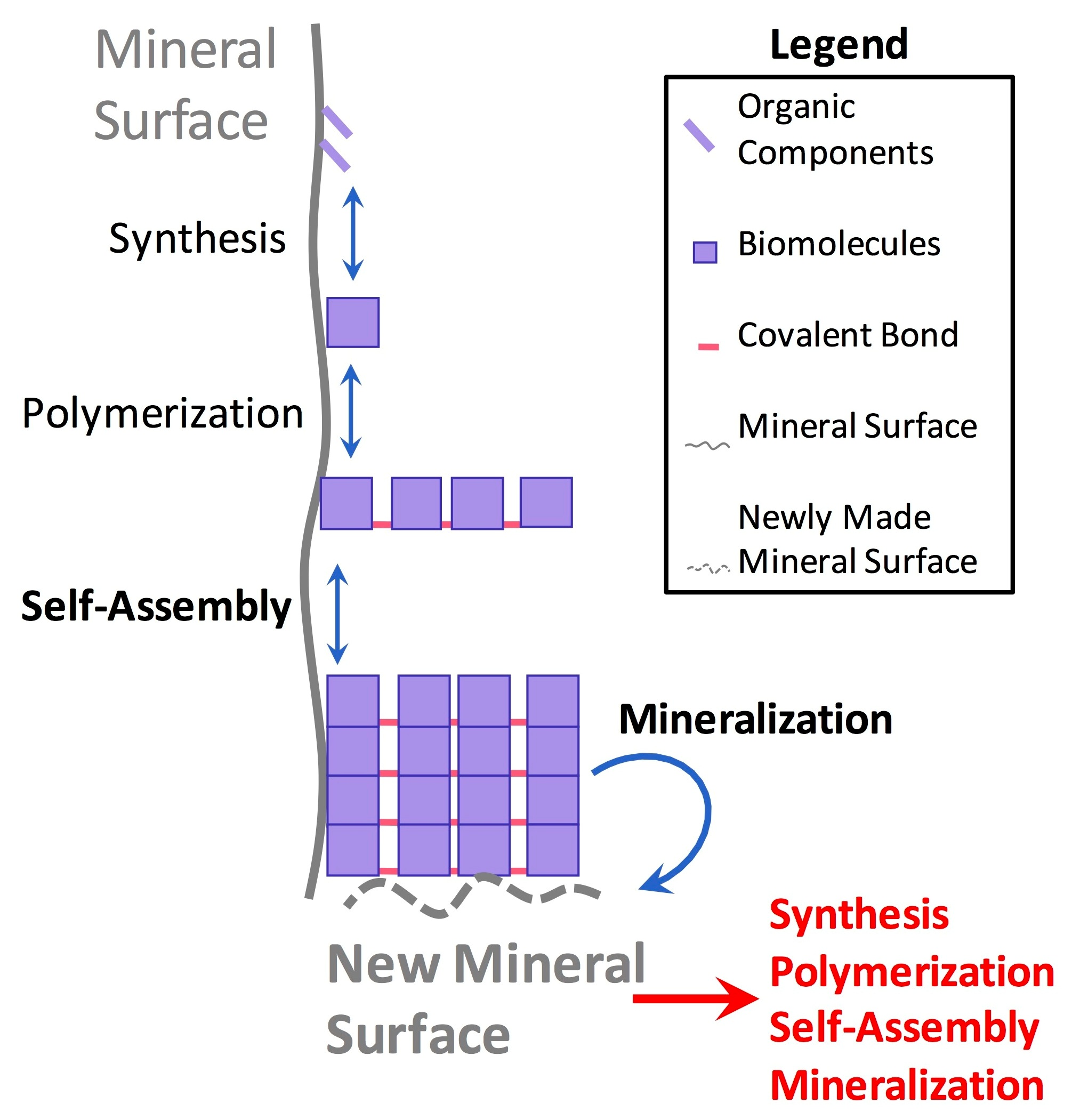
3.1. Synergistic Cyclical Model of Mineral-Templated Self-Assembling Systems Promoting Mineralization

We envision a model by which mineral surfaces on primitive Earth catalyzed the synthesis and/or adsorption of simple biomolecules (such as nucleotides [10]), polymerization of monomers into polymers (such as peptides [23] or nucleic acids [19]), and eventual self-assembly (such as in peptide amyloid structures [110,112]) of these molecules into supramolecular structures (Figure 5). As we have shown in this review, these self-assemblies can afford a primitive system novel structures or catalytic activities such as DNA changing to a Z-form helical conformation upon kaolinite (Al2Si2O5(OH)4) binding [62] or amyloid self-assemblies catalyzing the synthesis of short peptides [113]. Some of the supramolecular structures assembled on mineral surfaces could have then, in turn, affected the local mineral composition through the catalysis of new mineral formation (for example, peptide hydrogels promoting hydroxyapatite formation (Ca5(PO4)3(OH) [135] (Figure 5), which then would have affected primitive chemistry in a circular autocatalytic fashion. It is very clear that the geology of our planet was affected directly by various non-living chemical processes while the Earth-life system was moving beyond primitive chemistries and into a living system early in its history, resulting in an interactive and symbiotic feedback loop in which geology and early pre-living systems were closely linked to one another even before life’s origin.

3.2. Incorporating Recent Discoveries in Nanoscience and Surface Science into Origins of Life Research

The systems presented in this review only offer a small glimpse into the realm of recent discoveries within nanoscience and surface science. While these examples are heavily application focused, the analytical tools used, mechanisms elucidated, and systems studied in these fields are still able to contribute to basic science research, similar to how basic science is able to do the same for applicational engineering research. Origins of life research is inherently an interdisciplinary field, and often involves researchers in chemistry, biology, geology, planetary science, physics, complex systems, and many other fields. Nanoscience and surface science research both certainly have a place in such an interdisciplinary field as well, as evidenced by the various examples that we have presented. We hope that through these various case studies of mineral-templated self-assemblies, researchers in the origins of life and astrobiology fields see the merit of incorporating ideas from these fields into their own research, specifically in order to study questions on how complementary mineral–chemical systems can interact, co-assemble, and co-evolve over time. However, there are other relevant prebiotic systems in which ideas from nanoscience or other materials science- or engineering-related fields would be able to contribute to, and thus we will not limit the scope of interaction between these fields just to mineral surfaces. As evidenced by the 2018 Gordon Research Conference on the Origins of Life, there has been a steady recent increase in participation in our research field by researchers from traditionally only peripherally origins of life-related fields, including nanoscientists and engineers, which is highly encouraging. This is because only through a truly interdisciplinary effort can we come to understand more about both our own origins and whether life could have possibly emerged elsewhere in the universe.

Author Contributions

R.J.G. and T.Z.J. conceived and designed the paper. T.Z.J. wrote the paper with assistance from R.J.G.

Acknowledgments

The authors would like to thank K. N. Johnson, H. J. Cleaves, and G. Helffrich from the Earth-Life Science Institute (ELSI) at Tokyo Institute of Technology and A. Jain from the Advanced Science Research Center at the City University of New York for critical reading of the manuscript and helpful discussions. Tony Z. Jia is a research fellow from ELSI at Tokyo Institute of Technology, and is supported by a Tokyo Institute of Technology Seed Grant “Tane” 1798 and by funding from the ELSI Director’s Fund. ELSI is a member of the World Premier International Research Center Initiative and is supported in part by the Japanese Ministry of Education, Culture, Sports, Science and Technology. Richard J. Gillams is a postdoctoral fellow of the ELSI Origins Network (EON), which is supported by a grant from the John Templeton Foundation. The opinions expressed in this publication are those of the authors and do not necessarily reflect the views of the John Templeton Foundation. The authors would also especially like to thank the organizers and participants of the EON workshop held at ELSI in September 2017 titled “Cosmic Perspective of Earth: A Planet Permeated and Shaped by Life—Implications for Astrobiology” (P. Boston, M. Chan, and H. J. Cleaves) for providing the environment for the initial seeding of this idea, along with the many great discussions that arose [163].

Conflicts of Interest

The authors declare no conflict of interest.

References

  1. Damineli, A.; Damineli, D.S.C. Origens da vida. Estudos Avançados 2007, 21, 263–284. [Google Scholar] [CrossRef]
  2. Kawai, S.; Haapasilta, V.; Lindner, B.D.; Tahara, K.; Spijker, P.; Buitendijk, J.A.; Pawlak, R.; Meier, T.; Tobe, Y.; Foster, A.S.; et al. Thermal control of sequential on-surface transformation of a hydrocarbon molecule on a copper surface. Nat. Commun. 2016, 7, 12711. [Google Scholar] [CrossRef] [PubMed]
  3. Johlin, J.M. Interfacial adsorption as a Function of the Concentration of Colloidal Solutions. J. Biol. Chem. 1929, 84, 543–551. [Google Scholar]
  4. Nič, M.; Jirát, J.; Košata, B.; Jenkins, A.; McNaught, A. Adsorption. In IUPAC Compendium of Chemical Terminology; Nič, M., Jirát, J., Košata, B., Jenkins, A., McNaught, A., Eds.; IUPAC: Zurich, Switzerland, 2009; ISBN 9780967855097. [Google Scholar]
  5. Pang, S.H.; Medlin, J.W. Controlling Catalytic Selectivity via Adsorbate Orientation on the Surface: From Furfural Deoxygenation to Reactions of Epoxides. J. Phys. Chem. Lett. 2015, 6, 1348–1356. [Google Scholar] [CrossRef] [PubMed]
  6. Chatterjee, D.; Akash, R.; Kamalnath, K.; Ahmad, R.; Singh, A.K.; Ravishankar, N. Orientation Selection during Heterogeneous Nucleation: Implications for Heterogeneous Catalysis. J. Phys. Chem. C 2017, 121, 10027–10037. [Google Scholar] [CrossRef]
  7. Nørskov, J.K. Electronic factors in catalysis. Prog. Surf. Sci. 1991, 38, 103–144. [Google Scholar] [CrossRef]
  8. Xu, Y.; Schoonen, M.A.A. The absolute energy positions of conduction and valence bands of selected semiconducting minerals. Am. Mineral. 2000, 85, 543–556. [Google Scholar] [CrossRef]
  9. Hazen, R.M.; Grew, E.S.; Downs, R.T.; Golden, J.; Hystad, G. Mineral Ecology: Chance and Necessity in the Mineral Diversity of Terrestrial Planets. Can. Mineral. 2015, 53, 295–324. [Google Scholar] [CrossRef]
  10. Schwartz, A.W.; Veen, M.; Bisseling, T.; Chittenden, G.J.F. Prebiotic nucleotide synthesis-demonstration of a geologically plausible pathway. Orig. Life 1975, 6, 163–168. [Google Scholar] [CrossRef] [PubMed]
  11. Pasek, M.A. Schreibersite on the early Earth: Scenarios for prebiotic phosphorylation. Geosci. Front. 2017, 8, 329–335. [Google Scholar] [CrossRef]
  12. Gull, M.; Mojica, M.A.; Fernández, F.M.; Gaul, D.A.; Orlando, T.M.; Liotta, C.L.; Pasek, M.A. Nucleoside phosphorylation by the mineral schreibersite. Sci. Rep. 2015, 5, 17198. [Google Scholar] [CrossRef] [PubMed]
  13. Saladino, R.; Barontini, M.; Cossetti, C.; Di Mauro, E.; Crestini, C. The effects of borate minerals on the synthesis of nucleic acid bases, amino acids and biogenic carboxylic acids from formamide. Orig. Life Evol. Biosph. 2011, 41, 317–330. [Google Scholar] [CrossRef] [PubMed]
  14. Ricardo, A.; Carrigan, M.A.; Olcott, A.N.; Benner, S.A. Borate minerals stabilize ribose. Science 2004, 303, 196. [Google Scholar] [CrossRef] [PubMed]
  15. Grew, E.S.; Bada, J.L.; Hazen, R.M. Borate minerals and origin of the RNA world. Orig. Life Evol. Biosph. 2011, 41, 307–316. [Google Scholar] [CrossRef] [PubMed]
  16. Lambert, J.-F. Adsorption and polymerization of amino acids on mineral surfaces: A review. Orig. Life Evol. Biosph. 2008, 38, 211–242. [Google Scholar] [CrossRef] [PubMed]
  17. Hashizume, H. Adsorption of Nucleic Acid Bases, Ribose, and Phosphate by Some Clay Minerals. Life 2015, 5, 637–650. [Google Scholar] [CrossRef] [PubMed]
  18. Hedges, J.I.; Hare, P.E. Amino acid adsorption by clay minerals in distilled water. Geochim. Cosmochim. Acta 1987, 51, 255–259. [Google Scholar] [CrossRef]
  19. Ferris, J.P. Montmorillonite-catalysed formation of RNA oligomers: The possible role of catalysis in the origins of life. Philos. Trans. Royal Soc. Lond. B Biol. Sci. 2006, 361, 1777–1786. [Google Scholar] [CrossRef] [PubMed]
  20. Ferris, J.P.; Hill, A.R., Jr.; Liu, R.; Orgel, L.E. Synthesis of long prebiotic oligomers on mineral surfaces. Nature 1996, 381, 59–61. [Google Scholar] [CrossRef] [PubMed]
  21. Ferris, J.; Ertem, G. Oligomerization of ribonucleotides on montmorillonite: Reaction of the 5′-phosphorimidazolide of adenosine. Science 1992, 257, 1387–1389. [Google Scholar] [CrossRef] [PubMed]
  22. Kawamura, K.; Ferris, J.P. Kinetic and Mechanistic Analysis of Dinucleotide and Oligonucleotide Formation from the 5′-Phosphorimidazolide of Adenosine on Na-Montmorillonite. J. Am. Chem. Soc. 1994, 116, 7564–7572. [Google Scholar] [CrossRef] [PubMed]
  23. Orgel, L.E. Polymerization on the rocks: Theoretical introduction. Orig. Life Evol. Biosph. 1998, 28, 227–234. [Google Scholar] [CrossRef] [PubMed]
  24. Hill, A.R., Jr.; Böhler, C.; Orgel, L.E. Polymerization on the rocks: Negatively-charged alpha-amino acids. Orig. Life Evol. Biosph. 1998, 28, 235–243. [Google Scholar] [CrossRef] [PubMed]
  25. Liu, R.; Orgel, L.E. Polymerization on the rocks: Beta-amino acids and arginine. Orig. Life Evol. Biosph. 1998, 28, 245–257. [Google Scholar] [CrossRef] [PubMed]
  26. Lahav, N.; White, D.; Chang, S. Peptide formation in the prebiotic era: Thermal condensation of glycine in fluctuating clay environments. Science 1978, 201, 67–69. [Google Scholar] [CrossRef] [PubMed]
  27. Kitadai, N.; Oonishi, H.; Umemoto, K.; Usui, T.; Fukushi, K.; Nakashima, S. Glycine Polymerization on Oxide Minerals. Orig. Life Evol. Biosph. 2017, 47, 123–143. [Google Scholar] [CrossRef] [PubMed]
  28. Hanczyc, M.M.; Fujikawa, S.M.; Szostak, J.W. Experimental models of primitive cellular compartments: Encapsulation, growth, and division. Science 2003, 302, 618–622. [Google Scholar] [CrossRef] [PubMed]
  29. Hanczyc, M.M.; Mansy, S.S.; Szostak, J.W. Mineral surface directed membrane assembly. Orig. Life Evol. Biosph. 2007, 37, 67–82. [Google Scholar] [CrossRef] [PubMed]
  30. Sahai, N.; Kaddour, H.; Dalai, P.; Wang, Z.; Bass, G.; Gao, M. Mineral Surface Chemistry and Nanoparticle-aggregation Control Membrane Self-Assembly. Sci. Rep. 2017, 7, 43418. [Google Scholar] [CrossRef] [PubMed]
  31. Ebisuzaki, T.; Maruyama, S. Nuclear geyser model of the origin of life: Driving force to promote the synthesis of building blocks of life. Geosci. Front. 2017, 8, 275–298. [Google Scholar] [CrossRef]
  32. Westall, F.; Brack, A. The Importance of Water for Life. Space Sci. Rev. 2018, 214, 50. [Google Scholar] [CrossRef]
  33. Alberts, B.; Johnson, A.; Lewis, J.; Morgan, D.; Raff, M.; Roberts, K.; Walter, P. Molecular Biology of the Cell; Garland Science: Oxford, UK, 2014; ISBN 9780815344322. [Google Scholar]
  34. Grover, M.; He, C.; Hsieh, M.-C.; Yu, S.-S. A Chemical Engineering Perspective on the Origins of Life. Processes 2015, 3, 309–338. [Google Scholar] [CrossRef]
  35. Mandal, D.; Nasrolahi Shirazi, A.; Parang, K. Self-assembly of peptides to nanostructures. Org. Biomol. Chem. 2014, 12, 3544–3561. [Google Scholar] [CrossRef] [PubMed]
  36. Zhao, Y.; Yang, W.; Chen, C.; Wang, J.; Zhang, L.; Xu, H. Rational design and self-assembly of short amphiphilic peptides and applications. Curr. Opin. Colloid Interface Sci. 2018, 35, 112–123. [Google Scholar] [CrossRef]
  37. Singh, N.; Kumar, M.; Miravet, J.F.; Ulijn, R.V.; Escuder, B. Peptide-Based Molecular Hydrogels as Supramolecular Protein Mimics. Chemistry 2017, 23, 981–993. [Google Scholar] [CrossRef] [PubMed]
  38. Aumiller, W.M., Jr.; Pir Cakmak, F.; Davis, B.W.; Keating, C.D. RNA-Based Coacervates as a Model for Membraneless Organelles: Formation, Properties, and Interfacial Liposome Assembly. Langmuir 2016, 32, 10042–10053. [Google Scholar] [CrossRef] [PubMed]
  39. Fung, S.Y.; Keyes, C.; Duhamel, J.; Chen, P. Concentration Effect on the Aggregation of a Self-Assembling Oligopeptide. Biophys. J. 2003, 85, 537–548. [Google Scholar] [CrossRef]
  40. Koga, S.; Williams, D.S.; Perriman, A.W.; Mann, S. Peptide-nucleotide microdroplets as a step towards a membrane-free protocell model. Nat. Chem. 2011, 3, 720–724. [Google Scholar] [CrossRef] [PubMed]
  41. Wang, M.; He, L.; Yin, Y. Magnetic field guided colloidal assembly. Mater. Today 2013, 16, 110–116. [Google Scholar] [CrossRef]
  42. Yin, Y.; Niu, L.; Zhu, X.; Zhao, M.; Zhang, Z.; Mann, S.; Liang, D. Non-equilibrium behaviour in coacervate-based protocells under electric-field-induced excitation. Nat. Commun. 2016, 7, 10658. [Google Scholar] [CrossRef] [PubMed]
  43. Thomas, J.E.; Kelley, M.J. Interaction of mineral surfaces with simple organic molecules by diffuse reflectance IR spectroscopy (DRIFT). J. Colloid Interface Sci. 2008, 322, 516–526. [Google Scholar] [CrossRef] [PubMed]
  44. Hakim, S.S.; Olsson, M.H.M.; Sørensen, H.O.; Bovet, N.; Bohr, J.; Feidenhans’l, R.; Stipp, S.L.S. Interactions of the Calcite {10.4} Surface with Organic Compounds: Structure and Behaviour at Mineral—Organic Interfaces. Sci. Rep. 2017, 7, 7592. [Google Scholar] [CrossRef] [PubMed]
  45. Pasarín, I.S.; Yang, M.; Bovet, N.; Glyvradal, M.; Nielsen, M.M.; Bohr, J.; Feidenhans’l, R.; Stipp, S.L.S. Molecular ordering of ethanol at the calcite surface. Langmuir 2012, 28, 2545–2550. [Google Scholar] [CrossRef] [PubMed]
  46. Osman, M.A.; Ernst, M.; Meier, B.H.; Suter, U.W. Structure and Molecular Dynamics of Alkane Monolayers Self-Assembled on Mica Platelets. J. Phys. Chem. B 2002, 106, 653–662. [Google Scholar] [CrossRef]
  47. Duffy, D.M.; Harding, J.H. Simulation of organic monolayers as templates for the nucleation of calcite crystals. Langmuir 2004, 20, 7630–7636. [Google Scholar] [CrossRef] [PubMed]
  48. Sand, K.K.; Yang, M.; Makovicky, E.; Cooke, D.J.; Hassenkam, T.; Bechgaard, K.; Stipp, S.L.S. Binding of ethanol on calcite: The role of the OH bond and its relevance to biomineralization. Langmuir 2010, 26, 15239–15247. [Google Scholar] [CrossRef] [PubMed]
  49. Cooke, D.J.; Gray, R.J.; Sand, K.K.; Stipp, S.L.S.; Elliott, J.A. Interaction of ethanol and water with the {1014} surface of calcite. Langmuir 2010, 26, 14520–14529. [Google Scholar] [CrossRef] [PubMed]
  50. Yaalon, D.H. Mineral Composition of the Average Shale. Clay Miner. 1962, 5, 31–36. [Google Scholar] [CrossRef]
  51. Banerjee, S.; Abdulsattar, Z.R.; Agim, K.; Lane, R.H.; Hascakir, B. Mechanism of polymer adsorption on shale surfaces: Effect of polymer type and presence of monovalent and divalent salts. Petroleum 2017, 3, 384–390. [Google Scholar] [CrossRef]
  52. Carter, P.W. Adsorption of amino acid-containing organic matter by calcite and quartz. Geochim. Cosmochim. Acta 1978, 42, 1239–1242. [Google Scholar] [CrossRef]
  53. Lagaly, G.; Barrer, R.M.; Goulding, K. Clay-Organic Interactions. Philos. Trans. Royal Soc. A Math. Phys. Eng. Sci. 1984, 311, 315–332. [Google Scholar] [CrossRef]
  54. Mao, C. The emergence of complexity: Lessons from DNA. PLoS Biol. 2004, 2, e431. [Google Scholar] [CrossRef] [PubMed] [Green Version]
  55. Privalov, P.L.; Dragan, A.I.; Crane-Robinson, C.; Breslauer, K.J.; Remeta, D.P.; Minetti, C.A. What drives proteins into the major or minor grooves of DNA? J. Mol. Biol. 2007, 365, 1–9. [Google Scholar] [CrossRef] [PubMed]
  56. Walter, N.G.; Engelke, D.R. Ribozymes: Catalytic RNAs that cut things, make things, and do odd and useful jobs. Biologist 2002, 49, 199–203. [Google Scholar] [PubMed]
  57. Robertson, M.P.; Joyce, G.F. The origins of the RNA world. Cold Spring Harb. Perspect. Biol. 2012, 4, a003608. [Google Scholar] [CrossRef] [PubMed]
  58. Li, L.; Prywes, N.; Tam, C.P.; O’Flaherty, D.K.; Lelyveld, V.S.; Izgu, E.C.; Pal, A.; Szostak, J.W. Enhanced Nonenzymatic RNA Copying with 2-Aminoimidazole Activated Nucleotides. J. Am. Chem. Soc. 2017, 139, 1810–1813. [Google Scholar] [CrossRef] [PubMed]
  59. Szostak, J.W. The eightfold path to non-enzymatic RNA replication. J. Syst. Chem. 2012, 3, 2. [Google Scholar] [CrossRef]
  60. Sowerby, S.J.; Heckl, W.M. The role of self-assembled monolayers of the purine and pyrimidine bases in the emergence of life. Orig. Life Evol. Biosph. 1998, 28, 283–310. [Google Scholar] [CrossRef] [PubMed]
  61. Kan, Y.; Tan, Q.; Wu, G.; Si, W.; Chen, Y. Study of DNA adsorption on mica surfaces using a surface force apparatus. Sci. Rep. 2015, 5, 8442. [Google Scholar] [CrossRef] [PubMed]
  62. Cai, P.; Huang, Q.-Y.; Zhang, X.-W. Interactions of DNA with Clay Minerals and Soil Colloidal Particles and Protection against Degradation by DNase. Environ. Sci. Technol. 2006, 40, 2971–2976. [Google Scholar] [CrossRef] [PubMed]
  63. Cleaves, H.J., II; Crapster-Pregont, E.; Jonsson, C.M.; Jonsson, C.L.; Sverjensky, D.A.; Hazen, R.A. The adsorption of short single-stranded DNA oligomers to mineral surfaces. Chemosphere 2011, 83, 1560–1567. [Google Scholar] [CrossRef] [PubMed]
  64. Perona, J.J. DNA Structure. In Encyclopedia of Genetics; Elsevier: New York, NY, USA, 2001; pp. 572–574. ISBN 9780122270802. [Google Scholar]
  65. Herbert, A.; Rich, A. The Biology of Left-handed Z.-DNA. J. Biol. Chem. 1996, 271, 11595–11598. [Google Scholar] [CrossRef] [PubMed]
  66. Freund, A.M.; Bichara, M.; Fuchs, R.P. Z-DNA-forming sequences are spontaneous deletion hot spots. Proc. Natl. Acad. Sci. USA 1989, 86, 7465–7469. [Google Scholar] [CrossRef] [PubMed]
  67. Wang, G.; Christensen, L.A.; Vasquez, K.M. Z-DNA-forming sequences generate large-scale deletions in mammalian cells. Proc. Natl. Acad. Sci. USA 2006, 103, 2677–2682. [Google Scholar] [CrossRef] [PubMed]
  68. Rich, A.; Zhang, S. Timeline: Z-DNA: The long road to biological function. Nat. Rev. Genet. 2003, 4, 566–572. [Google Scholar] [CrossRef] [PubMed]
  69. Berg, J.M.; Tymoczko, J.L.; Stryer, L. Biochemistry, Fifth Edition; W. H. Freeman: New York, NY, USA, 2002; ISBN 9780716730514. [Google Scholar]
  70. Swadling, J.B.; Coveney, P.V.; Christopher Greenwell, H. Stability of free and mineral-protected nucleic acids: Implications for the RNA world. Geochim. Cosmochim. Acta 2012, 83, 360–378. [Google Scholar] [CrossRef]
  71. Riazance, J.H.; Baase, W.A.; Johnson, W.C., Jr.; Hall, K.; Cruz, P.; Tinoco, I., Jr. Evidence for Z-form RNA by vacuum UV circular dichroism. Nucleic Acids Res. 1985, 13, 4983–4989. [Google Scholar] [CrossRef] [PubMed]
  72. Davis, P.W.; Adamiak, R.W.; Tinoco, I., Jr. Z-RNA: The solution NMR structure of r(CGCGCG). Biopolymers 1990, 29, 109–122. [Google Scholar] [CrossRef] [PubMed]
  73. D’Ascenzo, L.; Leonarski, F.; Vicens, Q.; Auffinger, P. “Z-DNA like” fragments in RNA: A recurring structural motif with implications for folding, RNA/protein recognition and immune response. Nucleic Acids Res. 2016, 44, 5944–5956. [Google Scholar] [CrossRef] [PubMed]
  74. Heuberger, B.D.; Pal, A.; Del Frate, F.; Topkar, V.V.; Szostak, J.W. Replacing uridine with 2-thiouridine enhances the rate and fidelity of nonenzymatic RNA primer extension. J. Am. Chem. Soc. 2015, 137, 2769–2775. [Google Scholar] [CrossRef] [PubMed]
  75. Franchi, M.; Ferris, J.P.; Gallori, E. Cations as mediators of the adsorption of nucleic acids on clay surfaces in prebiotic environments. Orig. Life Evol. Biosph. 2003, 33, 1–16. [Google Scholar] [CrossRef] [PubMed]
  76. Hansma, H.G.; Laney, D.E. DNA binding to mica correlates with cationic radius: Assay by atomic force microscopy. Biophys. J. 1996, 70, 1933–1939. [Google Scholar] [CrossRef]
  77. Moore, E.K.; Hao, J.; Prabhu, A.; Zhong, H.; Jelen, B.I.; Meyer, M.; Hazen, R.M.; Falkowski, P.G. Geological and Chemical Factors that Impacted the Biological Utilization of Cobalt in the Archean Eon. J. Geophys. Res. Biogeosci. 2018, 123, 743–759. [Google Scholar] [CrossRef]
  78. Shannon, R.D. Revised effective ionic radii and systematic studies of interatomic distances in halides and chalcogenides. Acta Crystallogr. A 1976, 32, 751–767. [Google Scholar] [CrossRef]
  79. Crowe, S.A.; Jones, C.; Katsev, S.; Magen, C.; O’Neill, A.H.; Sturm, A.; Canfield, D.E.; Haffner, G.D.; Mucci, A.; Sundby, B.; et al. Photoferrotrophs thrive in an Archean Ocean analogue. Proc. Natl. Acad. Sci. USA 2008, 105, 15938–15943. [Google Scholar] [CrossRef] [PubMed]
  80. Sun, X.; Hyeon Ko, S.; Zhang, C.; Ribbe, A.E.; Mao, C. Surface-mediated DNA self-assembly. J. Am. Chem. Soc. 2009, 131, 13248–13249. [Google Scholar] [CrossRef] [PubMed]
  81. Woo, S.; Rothemund, P.W.K. Self-assembly of two-dimensional DNA origami lattices using cation-controlled surface diffusion. Nat. Commun. 2014, 5, 4889. [Google Scholar] [CrossRef] [PubMed]
  82. Perrault, S.D.; Shih, W.M. Virus-inspired membrane encapsulation of DNA nanostructures to achieve in vivo stability. ACS Nano 2014, 8, 5132–5140. [Google Scholar] [CrossRef] [PubMed] [Green Version]
  83. Wang, P.; Meyer, T.A.; Pan, V.; Dutta, P.K.; Ke, Y. The Beauty and Utility of DNA Origami. Chem 2017, 2, 359–382. [Google Scholar] [CrossRef]
  84. Akbari, E.; Mollica, M.Y.; Lucas, C.R.; Bushman, S.M.; Patton, R.A.; Shahhosseini, M.; Song, J.W.; Castro, C.E. Engineering Cell Surface Function with DNA Origami. Adv. Mater. 2017, 29. [Google Scholar] [CrossRef] [PubMed]
  85. Han, D.; Qi, X.; Myhrvold, C.; Wang, B.; Dai, M.; Jiang, S.; Bates, M.; Liu, Y.; An, B.; Zhang, F.; et al. Single-stranded DNA and RNA origami. Science 2017, 358, eaao2648. [Google Scholar] [CrossRef] [PubMed]
  86. Oi, H.; Fujita, D.; Suzuki, Y.; Sugiyama, H.; Endo, M.; Matsumura, S.; Ikawa, Y. Programmable formation of catalytic RNA triangles and squares by assembling modular RNA enzymes. J. Biochem. 2017, 161, 451–462. [Google Scholar] [CrossRef] [PubMed]
  87. Rahman, M.M.; Matsumura, S.; Ikawa, Y. Artificial RNA Motifs Expand the Programmable Assembly between RNA Modules of a Bimolecular Ribozyme Leading to Application to RNA Nanostructure Design. Biology 2017, 6, 37. [Google Scholar] [CrossRef] [PubMed]
  88. Van der Horst, G.; Christian, A.; Inoue, T. Reconstitution of a group I intron self-splicing reaction with an activator RNA. Proc. Natl. Acad. Sci. USA 1991, 88, 184–188. [Google Scholar] [CrossRef] [PubMed]
  89. Goedert, M.; Spillantini, M.G. A century of Alzheimer’s disease. Science 2006, 314, 777–781. [Google Scholar] [CrossRef] [PubMed]
  90. Hamley, I.W. The Amyloid Beta Peptide: A Chemist’s Perspective. Role in Alzheimer’s and Fibrillization. Chem. Rev. 2012, 112, 5147–5192. [Google Scholar] [CrossRef] [PubMed]
  91. Ferreira, S.T.; Vieira, M.N.N.; De Felice, F.G. Soluble protein oligomers as emerging toxins in Alzheimer’s and other amyloid diseases. IUBMB Life 2007, 59, 332–345. [Google Scholar] [CrossRef] [PubMed]
  92. Eisele, Y.S.; Monteiro, C.; Fearns, C.; Encalada, S.E.; Wiseman, R.L.; Powers, E.T.; Kelly, J.W. Targeting protein aggregation for the treatment of degenerative diseases. Nat. Rev. Drug Discov. 2015, 14, 759–780. [Google Scholar] [CrossRef] [PubMed]
  93. Ow, S.-Y.; Dunstan, D.E. A brief overview of amyloids and Alzheimer’s disease. Protein Sci. 2014, 23, 1315–1331. [Google Scholar] [CrossRef] [PubMed]
  94. Reches, M.; Gazit, E. Casting metal nanowires within discrete self-assembled peptide nanotubes. Science 2003, 300, 625–627. [Google Scholar] [CrossRef] [PubMed]
  95. Frederix, P.W.J.; Scott, G.G.; Abul-Haija, Y.M.; Kalafatovic, D.; Pappas, C.G.; Javid, N.; Hunt, N.T.; Ulijn, R.V.; Tuttle, T. Exploring the sequence space for (tri-)peptide self-assembly to design and discover new hydrogels. Nat. Chem. 2014, 7, 30–37. [Google Scholar] [CrossRef] [PubMed] [Green Version]
  96. Pappas, C.G.; Shafi, R.; Sasselli, I.R.; Siccardi, H.; Wang, T.; Narang, V.; Abzalimov, R.; Wijerathne, N.; Ulijn, R.V. Dynamic peptide libraries for the discovery of supramolecular nanomaterials. Nat. Nanotechnol. 2016, 11, 960–967. [Google Scholar] [CrossRef] [PubMed]
  97. Adler-Abramovich, L.; Gazit, E. The physical properties of supramolecular peptide assemblies: From building block association to technological applications. Chem. Soc. Rev. 2014, 43, 6881–6893. [Google Scholar] [CrossRef] [PubMed]
  98. Schnaider, L.; Brahmachari, S.; Schmidt, N.W.; Mensa, B.; Shaham-Niv, S.; Bychenko, D.; Adler-Abramovich, L.; Shimon, L.J.W.; Kolusheva, S.; DeGrado, W.F.; et al. Self-assembling dipeptide antibacterial nanostructures with membrane disrupting activity. Nat. Commun. 2017, 8, 1365. [Google Scholar] [CrossRef] [PubMed]
  99. Tao, K.; Makam, P.; Aizen, R.; Gazit, E. Self-assembling peptide semiconductors. Science 2017, 358, eaam9756. [Google Scholar] [CrossRef] [PubMed]
  100. Galit Fichman, E.G. Self-assembly of short peptides to form hydrogels: Design of building blocks, physical properties and technological applications. Acta Biomater. 2014, 10, 1671–1682. [Google Scholar] [CrossRef] [PubMed]
  101. Plankensteiner, K.; Reiner, H.; Schranz, B.; Rode, B.M. Prebiotic Formation of Amino Acids in a Neutral Atmosphere by Electric Discharge. Angew. Chem. Int. Ed. 2004, 43, 1886–1888. [Google Scholar] [CrossRef] [PubMed]
  102. Miller, S.L. The atmosphere of the primitive Earth and the prebiotic synthesis of amino acids. Orig. Life 1974, 5, 139–151. [Google Scholar] [CrossRef] [PubMed]
  103. Sephton, M.A. Organic compounds in carbonaceous meteorites. Nat. Prod. Rep. 2002, 19, 292–311. [Google Scholar] [CrossRef] [PubMed]
  104. Hennet, R.J.-C.; Holm, N.G.; Engel, M.H. Abiotic synthesis of amino acids under hydrothermal conditions and the origin of life: A perpetual phenomenon? Naturwissenschaften 1992, 79, 361–365. [Google Scholar] [CrossRef] [PubMed]
  105. Marshall, W.L. Hydrothermal synthesis of amino acids. Geochim. Cosmochim. Acta 1994, 58, 2099–2106. [Google Scholar] [CrossRef]
  106. Leman, L.J.; Huang, Z.-Z.; Reza Ghadiri, M. Peptide Bond Formation in Water Mediated by Carbon Disulfide. Astrobiology 2015, 15, 709–716. [Google Scholar] [CrossRef] [PubMed]
  107. Leman, L. Carbonyl Sulfide-Mediated Prebiotic Formation of Peptides. Science 2004, 306, 283–286. [Google Scholar] [CrossRef] [PubMed]
  108. Erastova, V.; Degiacomi, M.T.; Fraser, D.G.; Greenwell, H.C. Mineral surface chemistry control for origin of prebiotic peptides. Nat. Commun. 2017, 8, 2033. [Google Scholar] [CrossRef] [PubMed]
  109. Martra, G.; Deiana, C.; Sakhno, Y.; Barberis, I.; Fabbiani, M.; Pazzi, M.; Vincenti, M. The formation and self-assembly of long prebiotic oligomers produced by the condensation of unactivated amino acids on oxide surfaces. Angew. Chem. Int. Ed. Engl. 2014, 53, 4671–4674. [Google Scholar] [CrossRef] [PubMed]
  110. Kang, S.-G.; Li, H.; Huynh, T.; Zhang, F.; Xia, Z.; Zhang, Y.; Zhou, R. Molecular mechanism of surface-assisted epitaxial self-assembly of amyloid-like peptides. ACS Nano 2012, 6, 9276–9282. [Google Scholar] [CrossRef] [PubMed]
  111. Li, H.; Zhang, F.; Zhang, Y.; Ye, M.; Zhou, B.; Tang, Y.-Z.; Yang, H.-J.; Xie, M.-Y.; Chen, S.-F.; He, J.-H.; et al. Peptide diffusion and self-assembly in ambient water nanofilm on mica surface. J. Phys. Chem. B 2009, 113, 8795–8799. [Google Scholar] [CrossRef] [PubMed]
  112. Zhu, M.; Souillac, P.O.; Ionescu-Zanetti, C.; Carter, S.A.; Fink, A.L. Surface-catalyzed amyloid fibril formation. J. Biol. Chem. 2002, 277, 50914–50922. [Google Scholar] [CrossRef] [PubMed]
  113. Rout, S.K.; Friedmann, M.P.; Riek, R.; Greenwald, J. A prebiotic template-directed peptide synthesis based on amyloids. Nat. Commun. 2018, 9, 234. [Google Scholar] [CrossRef] [PubMed]
  114. Sun, L.; Narimatsu, T.; Tsuchiya, S.; Tanaka, T.; Li, P.; Hayamizu, Y. Water stability of self-assembled peptide nanostructures for sequential formation of two-dimensional interstitial patterns on layered materials. RSC Adv. 2016, 6, 96889–96897. [Google Scholar] [CrossRef]
  115. Sadler, K.; Tam, J.P. Peptide dendrimers: Applications and synthesis. Rev. Mol. Biotechnol. 2002, 90, 195–229. [Google Scholar] [CrossRef]
  116. Holliday, G.L.; Mitchell, J.B.O.; Thornton, J.M. Understanding the functional roles of amino acid residues in enzyme catalysis. J. Mol. Biol. 2009, 390, 560–577. [Google Scholar] [CrossRef] [PubMed]
  117. Dodson, G. Catalytic triads and their relatives. Trends Biochem. Sci. 1998, 23, 347–352. [Google Scholar] [CrossRef]
  118. Lagnoux, D.; Delort, E.; Douat-Casassus, C.; Esposito, A.; Reymond, J.-L. Synthesis and esterolytic activity of catalytic peptide dendrimers. Chemistry 2004, 10, 1215–1226. [Google Scholar] [CrossRef] [PubMed]
  119. Liao, H.-S.; Lin, J.; Liu, Y.; Huang, P.; Jin, A.; Chen, X. Self-assembly mechanisms of nanofibers from peptide amphiphiles in solution and on substrate surfaces. Nanoscale 2016, 8, 14814–14820. [Google Scholar] [CrossRef] [PubMed]
  120. Lin, J.; Luo, J.-B.; Yang, S.-T.; Zhou, Q.-H. Template-directed self-assembly of a designed amphiphilic hexapeptide on mica surface. Colloid Polym. Sci. 2013, 291, 2263–2270. [Google Scholar] [CrossRef]
  121. Cui, H.; Webber, M.J.; Stupp, S.I. Self-Assembly of Peptide Amphiphiles: From Molecules to Nanostructures to Biomaterials. Biopolymers 2010, 94, 1–18. [Google Scholar] [CrossRef] [PubMed]
  122. Mikhalevich, V.; Craciun, I.; Kyropoulou, M.; Palivan, C.G.; Meier, W. Amphiphilic Peptide Self-Assembly: Expansion to Hybrid Materials. Biomacromolecules 2017, 18, 3471–3480. [Google Scholar] [CrossRef] [PubMed]
  123. Hamley, I.W. Self-assembly of amphiphilic peptides. Soft Matter 2011, 7, 4122–4138. [Google Scholar] [CrossRef]
  124. Vauthey, S.; Santoso, S.; Gong, H.; Watson, N.; Zhang, S. Molecular self-assembly of surfactant-like peptides to form nanotubes and nanovesicles. Proc. Natl. Acad. Sci. USA 2002, 99, 5355–5360. [Google Scholar] [CrossRef] [PubMed]
  125. Hentrich, C.; Szostak, J.W. Controlled growth of filamentous fatty acid vesicles under flow. Langmuir 2014, 30, 14916–14925. [Google Scholar] [CrossRef] [PubMed]
  126. Adamala, K.; Szostak, J.W. Nonenzymatic template-directed RNA synthesis inside model protocells. Science 2013, 342, 1098–1100. [Google Scholar] [CrossRef] [PubMed]
  127. Hartgerink, J.D.; Beniash, E.; Stupp, S.I. Self-assembly and mineralization of peptide-amphiphile nanofibers. Science 2001, 294, 1684–1688. [Google Scholar] [CrossRef] [PubMed]
  128. Lai, P.; Moulton, K.; Krevor, S. Pore-scale heterogeneity in the mineral distribution and reactive surface area of porous rocks. Chem. Geol. 2015, 411, 260–273. [Google Scholar] [CrossRef]
  129. Jonker, A.M.; Dennis, W.P.; van Hest, J.C.M. Peptide- and Protein-Based Hydrogels. Chem. Mater. 2012, 24, 759–773. [Google Scholar] [CrossRef]
  130. Lei, Z.; Wang, Q.; Sun, S.; Zhu, W.; Wu, P. A Bioinspired Mineral Hydrogel as a Self-Healable, Mechanically Adaptable Ionic Skin for Highly Sensitive Pressure Sensing. Adv. Mater. 2017, 29, 1700321. [Google Scholar] [CrossRef] [PubMed]
  131. Feng, Y.; Lee, M.; Taraban, M.; Yu, Y.B. Diffusion of small molecules inside a peptide hydrogel. Chem. Commun. 2011, 47, 10455–10457. [Google Scholar] [CrossRef] [PubMed]
  132. Li, J.; Mooney, D.J. Designing hydrogels for controlled drug delivery. Nat. Rev. Mater. 2016, 1, 16071. [Google Scholar] [CrossRef] [PubMed]
  133. He, C.; Gállego, I.; Laughlin, B.; Grover, M.A.; Hud, N.V. A viscous solvent enables information transfer from gene-length nucleic acids in a model prebiotic replication cycle. Nat. Chem. 2016, 9, 318–324. [Google Scholar] [CrossRef] [PubMed]
  134. Caliari, S.R.; Burdick, J.A. A practical guide to hydrogels for cell culture. Nat. Methods 2016, 13, 405–414. [Google Scholar] [CrossRef] [PubMed]
  135. Gungormus, M.; Branco, M.; Fong, H.; Schneider, J.P.; Tamerler, C.; Sarikaya, M. Self assembled bi-functional peptide hydrogels with biomineralization-directing peptides. Biomaterials 2010, 31, 7266–7274. [Google Scholar] [CrossRef] [PubMed]
  136. Kasotakis, E.; Mossou, E.; Adler-Abramovich, L.; Mitchell, E.P.; Forsyth, V.T.; Gazit, E.; Mitraki, A. Design of metal-binding sites onto self-assembled peptide fibrils. Biopolymers 2009, 92, 164–172. [Google Scholar] [CrossRef] [PubMed]
  137. Jia, T.Z.; Hentrich, C.; Szostak, J.W. Rapid RNA exchange in aqueous two-phase system and coacervate droplets. Orig. Life Evol. Biosph. 2014, 44, 1–12. [Google Scholar] [CrossRef] [PubMed] [Green Version]
  138. Aumiller, W.M., Jr.; Keating, C.D. Phosphorylation-mediated RNA/peptide complex coacervation as a model for intracellular liquid organelles. Nat. Chem. 2016, 8, 129–137. [Google Scholar] [CrossRef] [PubMed]
  139. Matsuura, K.; Nakamura, T.; Watanabe, K.; Noguchi, T.; Minamihata, K.; Kamiya, N.; Kimizuka, N. Self-assembly of Ni-NTA-modified β-annulus peptides into artificial viral capsids and encapsulation of His-tagged proteins. Org. Biomol. Chem. 2016, 14, 7869–7874. [Google Scholar] [CrossRef] [PubMed]
  140. Rufo, C.M.; Moroz, Y.S.; Moroz, O.V.; Stöhr, J.; Smith, T.A.; Hu, X.; DeGrado, W.F.; Korendovych, I.V. Short peptides self-assemble to produce catalytic amyloids. Nat. Chem. 2014, 6, 303–309. [Google Scholar] [CrossRef] [PubMed]
  141. Al-Garawi, Z.S.; McIntosh, B.A.; Neill-Hall, D.; Hatimy, A.A.; Sweet, S.M.; Bagley, M.C.; Serpell, L.C. The amyloid architecture provides a scaffold for enzyme-like catalysts. Nanoscale 2017, 9, 10773–10783. [Google Scholar] [CrossRef] [PubMed]
  142. Heuer, A.; Fink, D.; Laraia, V.; Arias, J.; Calvert, P.; Kendall, K.; Messing, G.; Blackwell, J.; Rieke, P.; Thompson, D.; et al. Innovative materials processing strategies: A biomimetic approach. Science 1992, 255, 1098–1105. [Google Scholar] [CrossRef] [PubMed]
  143. Addadi, L.; Weiner, S. Biomineralization: Mineral formation by organisms. Phys. Scr. 2014, 89, 098003. [Google Scholar] [CrossRef]
  144. Dhami, N.K.; Reddy, M.S.; Mukherjee, A. Biomineralization of calcium carbonates and their engineered applications: A review. Front. Microbiol. 2013, 4, 314. [Google Scholar] [CrossRef] [PubMed]
  145. Kirschvink, J.L. Magnetite biomineralization and geomagnetic sensitivity in higher animals: An update and recommendations for future study. Bioelectromagnetics 1989, 10, 239–259. [Google Scholar] [CrossRef] [PubMed]
  146. Tambutté, S.; Holcomb, M.; Ferrier-Pagès, C.; Reynaud, S.; Tambutté, É.; Zoccola, D.; Allemand, D. Coral biomineralization: From the gene to the environment. J. Exp. Mar. Biol. Ecol. 2011, 408, 58–78. [Google Scholar] [CrossRef]
  147. Fablet, R.; Pecquerie, L.; de Pontual, H.; Høie, H.; Millner, R.; Mosegaard, H.; Kooijman, S.A.L.M. Shedding light on fish otolith biomineralization using a bioenergetic approach. PLoS ONE 2011, 6, e27055. [Google Scholar] [CrossRef] [PubMed] [Green Version]
  148. Selenska-Pobell, S.; Merroun, M. Accumulation of Heavy Metals by Micro-organisms: Biomineralization and Nanocluster Formation. In Prokaryotic Cell Wall Compounds; Springer: Berlin, Germany, 2010; pp. 483–500. [Google Scholar]
  149. Pacton, M.; Wacey, D.; Corinaldesi, C.; Tangherlini, M.; Kilburn, M.R.; Gorin, G.E.; Danovaro, R.; Vasconcelos, C. Viruses as new agents of organomineralization in the geological record. Nat. Commun. 2014, 5, 4298. [Google Scholar] [CrossRef] [PubMed]
  150. Lin, W.; Paterson, G.A.; Zhu, Q.; Wang, Y.; Kopylova, E.; Li, Y.; Knight, R.; Bazylinski, D.A.; Zhu, R.; Kirschvink, J.L.; et al. Origin of microbial biomineralization and magnetotaxis during the Archean. Proc. Natl. Acad. Sci. USA 2017, 114, 2171–2176. [Google Scholar] [CrossRef] [PubMed]
  151. Butardo, V.M.; Sreenivasulu, N. Tailoring Grain Storage Reserves for a Healthier Rice Diet and its Comparative Status with Other Cereals. Int. Rev. Cell Mol. Biol. 2016, 323, 31–70. [Google Scholar] [PubMed]
  152. Adeva-Andany, M.M.; González-Lucán, M.; Donapetry-García, C.; Fernández-Fernández, C.; Ameneiros-Rodríguez, E. Glycogen metabolism in humans. BBA Clin. 2016, 5, 85–100. [Google Scholar] [CrossRef] [PubMed]
  153. Elieh-Ali-Komi, D.; Hamblin, M.R. Chitin and Chitosan: Production and Application of Versatile Biomedical Nanomaterials. Int. J. Adv. Res. (Indore) 2016, 4, 411–427. [Google Scholar]
  154. Hansma, H.G. Possible origin of life between mica sheets: Does life imitate mica? J. Biomol. Struct. Dyn. 2013, 31, 888–895. [Google Scholar] [CrossRef] [PubMed]
  155. Nidhin, M.; Sreeram, K.J.; Nair, B.U. Polysaccharide films as templates in the synthesis of hematite nanostructures with special properties. Appl. Surf. Sci. 2012, 258, 5179–5184. [Google Scholar] [CrossRef]
  156. Allen, C.C.; Westall, F.; Schelble, R.T. Importance of a martian hematite site for astrobiology. Astrobiology 2001, 1, 111–123. [Google Scholar] [CrossRef] [PubMed]
  157. Murai, K.; Kinoshita, T.; Nagata, K.; Higuchi, M. Mineralization of Calcium Carbonate on Multifunctional Peptide Assembly Acting as Mineral Source Supplier and Template. Langmuir 2016, 32, 9351–9359. [Google Scholar] [CrossRef] [PubMed]
  158. Nonoyama, T.; Tanaka, M.; Kinoshita, T.; Nagata, F.; Sato, K.; Kato, K. Morphology control of calcium phosphate by mineralization on the β-sheet peptide template. Chem. Commun. 2010, 46, 6983. [Google Scholar] [CrossRef] [PubMed]
  159. Ribeiro, S.; Radvar, E.; Shi, Y.; Borges, J.; Pirraco, R.P.; Leonor, I.B.; Mano, J.F.; Reis, R.L.; Mata, Á.; Azevedo, H.S. Nanostructured interfacial self-assembled peptide–polymer membranes for enhanced mineralization and cell adhesion. Nanoscale 2017, 9, 13670–13682. [Google Scholar] [CrossRef] [PubMed]
  160. Elham, R.; Remzi, B.C.; Helena, A. Self-assembled peptide-polymer hydrogels as a 3D nanofiber substrate for biomimetic hydroxyapatite mineralization. Front. Bioeng. Biotechnol. 2016, 4. [Google Scholar] [CrossRef]
  161. Li, Q.-L.; Ning, T.-Y.; Cao, Y.; Zhang, W.-B.; Mei, M.L.; Chu, C.H. A novel self-assembled oligopeptide amphiphile for biomimetic mineralization of enamel. BMC Biotechnol. 2014, 14, 32. [Google Scholar] [CrossRef] [PubMed] [Green Version]
  162. Wang, J.; Ouyang, Z.; Ren, Z.; Li, J.; Zhang, P.; Wei, G.; Su, Z. Self-assembled peptide nanofibers on graphene oxide as a novel nanohybrid for biomimetic mineralization of hydroxyapatite. Carbon N. Y. 2015, 89, 20–30. [Google Scholar] [CrossRef]
  163. Chan, M.; Cleaves, H.J., II; Boston, P. What Would Earth Be Like Without Life? EOS 2018, 99. [Google Scholar] [CrossRef]
Figure 1. The snapshot time step from molecular dynamics simulations of ethanol layer assembly (into two distinct layers: a thin, ordered monolayer with the thickness of exactly one ethanol molecule, and a second larger less-ordered layer above the monolayer, separated by a gap) on a calcite surface, along with the ethanol spatial density profile associated with it. Reprinted with permission from Pasarín, I. S., Yang, M., Bovet, N., Glyvradal, M., Nielsen, M. M., Bohr, J., Feidenhans’l, R., and Stipp, S. L. S. 2012. “Molecular Ordering of Ethanol at the Calcite Surface.” Langmuir 28 (5):2545–50. [45] Copyright 2011 American Chemical Society.
Life 08 00010 g001
Figure 2. Atomic force microscope images of a structured DNA microarray self-assembled on a mica (general) surface. Reprinted with permission from Sun, X., Ko, S. H., Zhang, C., Ribbe, A. E., and Mao, C., 2009. “Surface-Mediated DNA Self-Assembly.” J. Am. Chem. Soc. 131 (37):13248-9. [80] Copyright 2009 American Chemical Society.
Life 08 00010 g002
Figure 3. Atomic force microscope images of the assembly dynamics of SMA (an amyloidogenic light chain variable domain peptide with 114 residues) nanofibrils on a mica surface over time (AE) show 12–16 h of incubation with image size of 1 µm × 1 µm, while (F) shows 6 days of incubation with image size of 5 µm × 5 µm). It appears that longer incubation times result in longer nanofibrils, such as in (F), as compared to shorter incubation times, such as in (AE). Reprinted with permission from Zhu, M., Souillac, P. O., Ionescu-Zanetti, C., Carter, S. A., and Fink, A. L. 2002. “Surface-catalyzed Amyloid Fibril Formation.” J. Biol. Chem. 277(52):50914-22. [112] Copyright 2002 American Society for Biochemistry and Molecular Biology.
Life 08 00010 g003
Figure 4. Transmission electron microscope images of calcium carbonate minerals at different nutrient concentrations and incubation times grown on a self-assembled short peptide nanostructure (Ac-VHVEVS-CONH2), suggesting that mineral growth can be effected by simple peptide self-assemblies. Reprinted with permission from Murai, K., Kinoshita, T., Nagata, K., and Higuchi, M. 2016. “Mineralization of Calcium Carbonate on Multifunctional Peptide Assembly Acting as Mineral Source Supplier and Template.” Langmuir 32 (36):9351–59. [157] Copyright 2016 American Chemical Society.
Life 08 00010 g004
Figure 5. Synergistic cyclical model of mineral-templated self-assembling systems promoting mineralization. Simple precursors of biomolecules adsorbed onto a mineral surface react and produce biomolecules such as nucleotides or amino acids. Then, some of these adsorbed molecules polymerize into polymers (such as peptides or nucleic acids). Given the optimal conditions and mineral surfaces, biopolymers produced then self-assemble into supramolecular structures such as amyloid fibers or primitive nucleic acid “origamis”. Although not shown in the figure, at any stage in the adsorption, polymerization, and self-assembly pathway (which are all reversible reactions), each molecule can also desorb off of the mineral surface. However, certain self-assembled supramolecular structures (whether on or off the mineral surface) have the ability to promote the formation of new minerals, such as peptide nanostructures catalyzing the formation of hydroxyapatite, similar to modern-day biomineralization processes. The newly produced mineral surfaces could then catalyze and template the synthesis, polymerization, and/or assembly of the same structures that allowed its own mineralization, leading to a synergistic cyclical positive feedback loop by which primitive self-assemblies could have affected the mineral composition of early Earth, and vice-versa.
Life 08 00010 g005

