Article

Cerebral Protection in TAVR—Can We Do Without? A Real-World All-Comer Intention-to-Treat Study—Impact on Stroke Rate, Length of Hospital Stay, and Twelve-Month Mortality

1
Department of Internal Medicine II, Division of Cardiology, Medical University of Vienna, 1090 Vienna, Austria
2
Department of Cardiac Surgery, Medical University of Vienna, 1090 Vienna, Austria
3
Cardiovascular Imaging Research Center, Massachusetts General Hospital and Harvard Medical School, Boston, MA 02115, USA
4
Department of Internal Medicine 3, University Hospital St. Poelten, Karl Landsteiner University of Health Sciences, 3500 Krems, Austria
*
Author to whom correspondence should be addressed.
J. Pers. Med. 2022, 12(2), 320; https://doi.org/10.3390/jpm12020320
Submission received: 30 December 2021 / Revised: 25 January 2022 / Accepted: 3 February 2022 / Published: 21 February 2022
(This article belongs to the Section Clinical Medicine, Cell, and Organism Physiology)

Abstract

:
Background: Stroke associated with transcatheter aortic valve replacement (TAVR) is a potentially devastating complication. Until recently, the Sentinel™ Cerebral Protection System (CPS; Boston Scientific, Marlborough, MA, USA) has been the only commercially available device for mechanical prevention of TAVR-related stroke. However, its effectiveness is still undetermined. Objectives: To explore the impact of Sentinel™ on stroke rate, length of hospital stay (LOS), and twelve-month mortality in a single-center, real-world, all-comers TAVR cohort. Material and Methods: Between January 2019 and August 2020 consecutive patients were assigned to TAVR with or without Sentinel™ in a 1:1 fashion according to the treating operator. We defined as primary endpoint clinically detectable cerebrovascular events within 72 h after TAVR and as secondary endpoints LOS and 12-month mortality. Logistic and linear regression analyses were used to assess associations of Sentinel™ use with endpoints. Results: Of 411 patients (80 ± 7 y/o, 47.4% female, EuroSCORE II 6.3 ± 5.9%), Sentinel™ was used in 213 (51.8%), with both filters correctly deployed in 189 (46.0%). Twenty (4.9%) cerebrovascular events were recorded, ten (2.4%) of which were disabling strokes. Patients with Sentinel™ suffered 71% less (univariate analysis; OR 0.29, 95%CI 0.11–0.82; p = 0.02) and, respectively, 76% less (multivariate analysis; OR 0.24, 95%CI 0.08–0.76; p = 0.02) cerebrovascular events compared to patients without Sentinel™. Sentinel™ use was also significantly associated with shorter LOS (Regression coefficient −2.47, 95%CI −4.08, −0.87; p < 0.01) and lower 12-month all-cause mortality (OR 0.45; 95%CI 0.22–0.93; p = 0.03). Conclusion: In the present prospective all-comers TAVR cohort, patients with Sentinel™ use showed (1) lower rates of cerebrovascular events, (2) shortened LOS, and (3) improved 12-month survival. These data promote the use of a CPS when implanting TAVR valves.

1. Background

Almost 4% of individuals over 70 and more than 9% over 80 years of age suffer from severe aortic stenosis (AS) [1]. Transcatheter aortic valve replacement (TAVR) has revolutionized treatment options in this common disease, especially in patients with high surgical risk. The technology has become widely applicable and safe through the availability of various self-expanding and balloon-expandable devices designed to fit almost every anatomy [2]. Recent guideline recommendations have greatly expanded the indication for TAVR, particularly in low-risk and younger age groups [3]. Despite ongoing improvements of the TAVR procedure, significant risks remain. One of the most dangerous complications is stroke, which has been reported to affect up to one in ten patients [4,5,6,7,8]. Factors that have been shown to predispose for TAVR-associated cerebrovascular events include age, atrial fibrillation, history of cerebrovascular events, very severe and calcified AS, and implantation of more than one valve [4,5,7]. However, no widely accepted algorithms currently exist that would allow the prediction of TAVR-related cerebrovascular events. To protect the supra-aortal arteries from embolizing material during the procedure, mechanical cerebral protection systems (CPS) have been developed. Until recently, only the Sentinel™ (Boston Scientific) CPS has been commercially available. However, data on the effectiveness of Sentinel™ are conflicting. While several small studies failed to demonstrate a reduction of clinically relevant stroke in TAVR with Sentinel™ [9,10,11], three larger studies reported significant reductions in cerebrovascular event [12,13,14] and also in-hospital mortality [11,12] rates. The reasons for these discrepancies as well as the limitations of the Sentinel™ CPS have so far not been carefully investigated. In particular, it remains unclear whether Sentinel™ also impacts the length of hospital stay (LOS) and twelve-month mortality. Thus, the effectiveness of Sentinel™ is still undetermined, and it has, as a result, not evolved as standard-of-care.
In this prospective study, we investigated the efficacy of Sentinel™ using clinically detectable cerebrovascular events within 72 h after TAVR as primary and LOS and 12-month mortality as secondary endpoints.

2. Methods

2.1. Study Population

We enrolled consecutive patients with AS scheduled for transfemoral TAVR at the General Hospital Vienna, a university-affiliated tertiary center. The multi-disciplinary heart team confirmed the eligibility and the decision for TAVR according to current guidelines [15]. Patients were assigned to TAVR with or without Sentinel™ according to the first operator as two out of four first operators systematically used the device whenever possible, while the other operators did not. Procedural complications and in-hospital clinical outcomes were assessed according to the Valve Academic Research Consortium-2 (VARC-2) criteria [16]. The patients were followed for at least 12 months. The study was approved by the local ethics committee (EK No. 2218/2016). All patients provided written informed consent.

2.2. Transcatheter Aortic Valve Replacement

Transfemoral TAVR procedures were performed according to institutional standards in local or general anesthesia in a hybrid catheterization laboratory. According to current guideline recommendations, oral anticoagulation was discontinued before the procedure. During TAVR, patients received unfractionated heparin with a target activated clotting time of 250 s. Antiplatelet therapy, if present, was continued. The percentages of antiplatelet therapy used refer to the preprocedural therapy, postinterventionally, DAPT was started according to 2017 ESC Guidelines for the Management of Valvular Heart Disease [15].

2.3. Sentinel™ Cerebral Protection Device
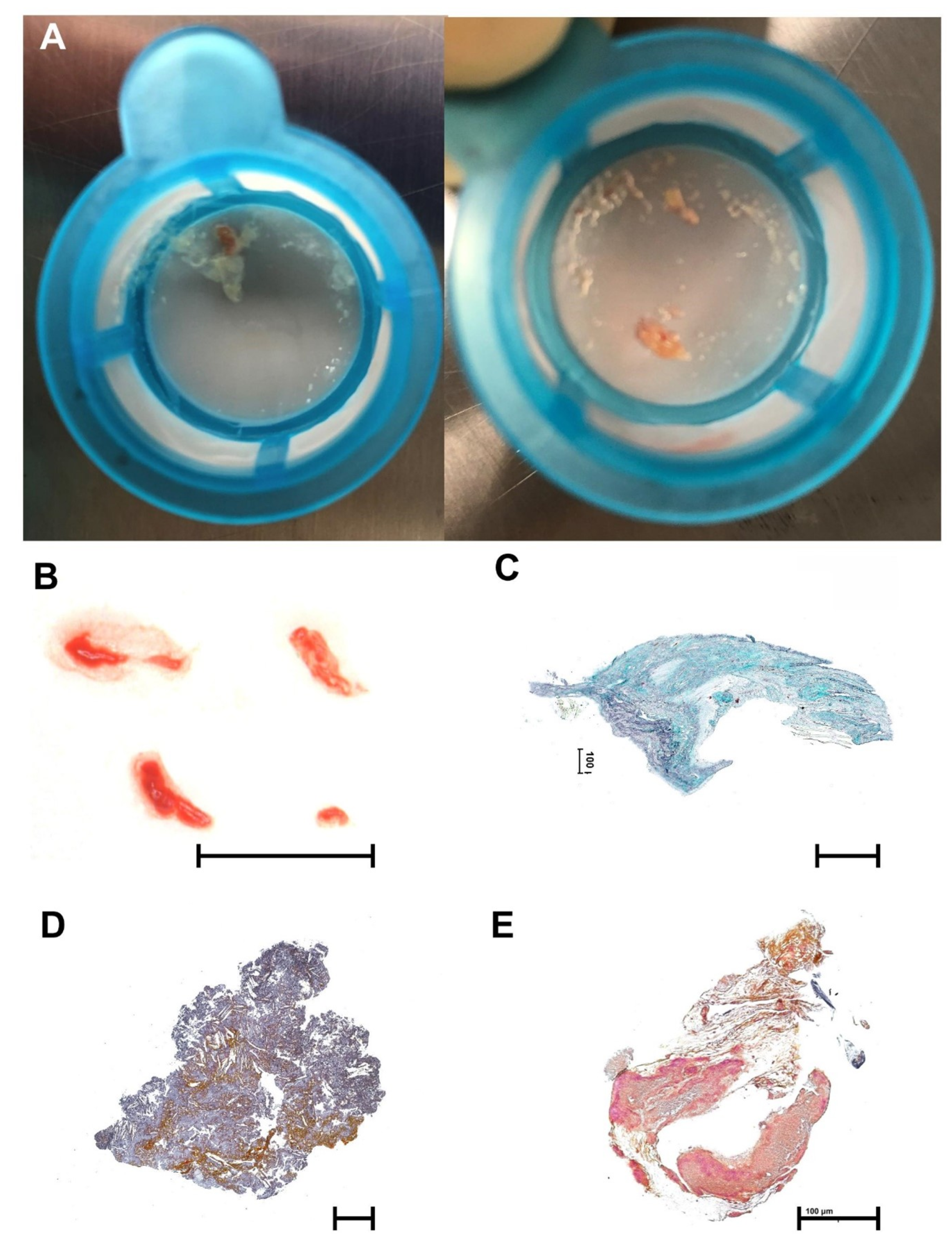
The Sentinel™ device (Boston Scientific©) is a dual-filter-based intra-luminal embolic protection device inserted through a 6-French sheath via the right radial, ulnar or brachial artery. The proximal filter has to be positioned in the brachiocephalic trunk and the distal filter in the left common carotid artery. Both filters consist of a radiopaque nitinol frame with a 140 µm pore polyurethane filter. The two filters cover more than 90% of the cerebral blood flow, excluding only the left vertebral artery’s territory [17]. One study reported that 74% of the brain volume is totally, 24% is partially protected, leaving only 2% of brain volume unprotected [18]. Prior to the procedure, brachiocephalic trunk and carotid artery anatomy were analyzed on computed tomography angiography. Sentinel™ use was not attempted in the presence of severe left carotid artery stenosis, defined as stenosis severity >70%, or if it was known that right radial access was not possible. According to the protocol, ulnar, brachial, and axillar access was not used. The Sentinel™ system was positioned after insertion of the TAVR sheath and removed after valve deployment, prior to femoral closure.

2.4. Histopathology

Immediately after the procedure, debris from the Sentinel™ filters were collected, digitally photographed and stored in 7.5% neutral buffered formalin. Particles with a diameter >2 mm were dehydrated and embedded in paraffin. The paraffin block was cut into 3µm slices, which were stained with modified trichrome [19].

2.5. Definition of Endpoints

Cerebrovascular events were classified according to VARC-2 criteria, including disabling and non-disabling stroke as well as transitory ischemic attack [16]. The primary endpoint was defined as any clinically detectable cerebrovascular event within 72 h of TAVR. [13,20] During these 72 h, patients were evaluated clinically every 6 to 12 h [4,12,21,22] by a board-certified cardiologist. Face palsy, arm weakness, speech changes, eye deviation or denial were assessed according to the FAST-ED scale [23]. In case of any sign of neurological compromise, a comprehensive neurological assessment, including the modified Rankin scale as well as the National Institute of Health Stroke Scale (NIHSS), was performed by a board-certified neurologist, similar to the approach used by Seeger et al. [12]. This assessment also included cerebral computed tomography and/or cerebral magnetic resonance imaging (cMRI), interpreted by board-certified neuroradiologists. The final diagnosis of a cerebrovascular event was adjudicated by a board-certified neurologist. All-cause mortality at 12 months and LOS in days were chosen as secondary endpoints.
LOS was defined as overall length of hospital stay post-procedure, including the stay at the tertiary care center and any immediate subsequent hospital stay. After discharge, patients were followed in our outpatient clinic at three and twelve months.

2.6. Statistical Analysis

Categorical parameters are presented as counts and percentages, continuous variables as mean ± standard deviation. To compare baseline variables between groups, χ2 test or Fisher’s exact test and Wilcoxon ranks sum test were applied as appropriate.
For the primary endpoint, we used logistic regression models to identify baseline variables associated with cerebrovascular events. For the assessment of differences in LOS and 12-month mortality, linear as well as univariate and multivariate Cox regression models were applied. In addition, we used Kaplan–Meier estimates and the log-rank test to report differences in all-cause mortality. Variables with a p-Value < 0.05 were entered in the multivariate analysis using a simultaneous approach.
In a separate step, we performed a propensity score analysis using the psmatch2 command in Stata 15.1 (StataCorp., College Station, TX, USA) using EuroSCORE II, atrial fibrillation (AF), diabetes mellitus, gender, coronary artery disease (CAD) and valve type (self-expanding versus balloon-expandable), similar to that previously used by Seeger et al. [20]. A p-Value < 0.05 was considered statistically significant, and tests were two-sided. Statistical analysis was performed using SPSS 26.0.

3. Results

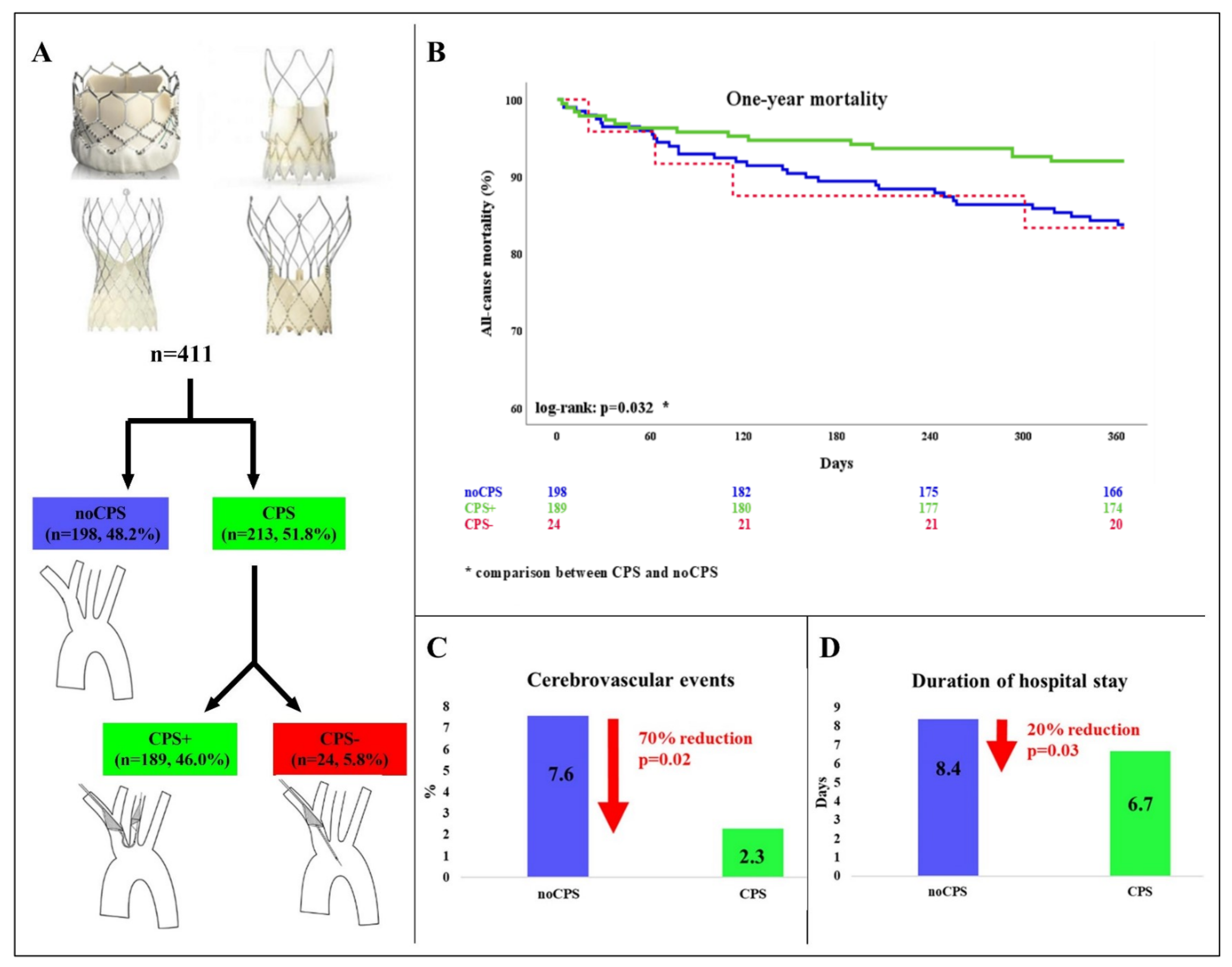
3.1. Patient Population

Between January 2019 and August 2020, 411 consecutive TAVR patients were enrolled. The mean age was 80.4 ± 6.7 years, 47.4% were female. A prior history of cerebrovascular events was present in 7.4% of patients.
Figure 1 and Panel A of Figure 2 depict patient flow. Out of our 411 patients, Sentinel™ was used in 213 (51.8%, CPS). Of these, the device was correctly deployed in 189 individuals (46.0% in total; 88.7% of CPS; CPS+). In 24 patients (5.8% of the total study cohort; 11.3% of CPS; CPS−) only the proximal filter could be positioned correctly in the brachiocephalic trunk while the distal filter remained closed in the ascending aorta. 198 (48.2%) underwent TAVR without CPS (noCPS).
Table 1 displays baseline characteristics, which were well balanced across groups (noCPS vs. CPS). Differences were only observed with regard to mean and peak aortic valve gradients. No differences were found regarding procedural parameters except for fluoroscopy time, which was longest in CPS− patients. No local vascular complications were attributed to CPS use.

3.2. Cerebrovascular Events

In total, 20 (4.9%) cerebrovascular events occurred, of which 10 (2.4%) were disabling strokes with a modified Rankin Scale >2 at 90 days (Table 2). In the Sentinel™ group cerebrovascular events were reduced by 70% (noCPS 7.6% vs. CPS 2.3%, p = 0.02) (Figure 2, Panel C).
In the Sentinel™ group (CPS) five cerebrovascular events were recorded. Of these, two occurred in patients with correctly positioned CPS (CPS+; 2/189, 1.1%). Both comprised cerebral regions supplied by the left vertebral artery. One stroke was disabling with a modified Rankin Scale > 2 at 90 days. In the CPS− group, three events occurred (3/24; 12.5%). In two of them, brain areas supplied by the left carotid or vertebral artery were affected, which were not covered by the distal filter. In one, the event was defined as disabling. In the third patient, who also suffered a disabling stroke, the right mid cerebral artery was occluded, indicating incomplete coverage of the right brachiocephalic trunk/the right carotid artery.
In the multivariate analysis, Sentinel was significantly associated with a decreased stroke rate after 72 h (OR 0.24, 95%CI 0.08–0.76; p = 0.02), whereas implantation of more than one valve was the only factor significantly associated with a higher cerebrovascular event rate (OR 16.7, 95%CI 2.69–103.92; p < 0.01; Table 3).
On propensity score-matched analysis, Sentinel™ remained significantly associated with lower cerebrovascular event rate (OR 0.12, 95%CI 0.03–0.51; p < 0.01, Supplemental Material Table S1). When the analysis was restricted to patients who underwent TAVR with Sentinel™, correct CPS placement remained the only parameter independently associated with cerebrovascular events (OR 0.06, 95%CI 0.01–0.43; p < 0.01).

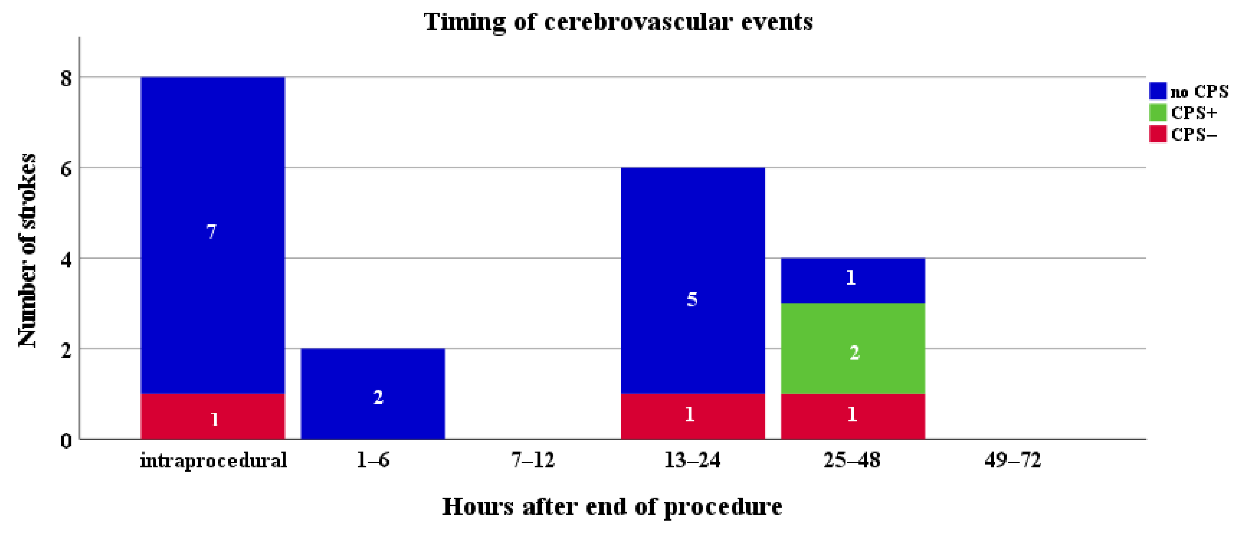
3.3. Timing of Stroke

Of 20 cerebrovascular events, 16 (80%) events were considered acute (within 24 h of TAVR) [5,24,25]. In this group, no CPS+ patients suffered an event. Four strokes occurred 25 to 72 h after TAVR. (Figure 3).
Both patients with a cerebrovascular event in the CPS+ group suffered a sub-acute stroke (at 30 and 48 h after TAVR, respectively). The three events in the CPS− group occurred at 0, 19, and 27 h.
All other events were observed in patients without CPS.

3.4. All-Cause Mortality at 12 Months

At 12 months, 51 patients (12.4%) had died. Sentinel™ use was associated with a 45% reduction of all-cause mortality at twelve months (noCPS 16.2% vs. CPS 8.9%, p = 0.026). In patients with incorrectly/incompletely positioned Sentinel™ (CPS−), mortality at 12 months was similar to TAVR without CPS (16.7%, Figure 2, Panel B; Table 2).
In the univariate Cox regression analysis, Sentinel™ was significantly associated with a reduced twelve-month mortality (OR 0.48, 95%CI 0.26–0.87; p = 0.02). In the multivariate analysis, Sentinel™ remained significantly associated with one-year mortality (OR 0.45, 95%CI 0.22–0.93, p = 0.03) as well as procedure time (OR 1.02, 95%CI 1.01–1.03; p < 0.01; Table 4).
In the propensity score-matched analysis, Sentinel™ remained significantly associated (OR 0.48, 95%CI 0.27–0.88; p = 0.02; Supplemental Material Table S2) with all-cause mortality at 12 months.

3.5. Length of Hospital Stay

After TAVR, patients stayed in hospital for 7.5 ± 8.0 days. Overall, the systematic use of Sentinel™ decreased the LOS after TAVR by 20% (noCPS 8.4 ± 9.6 vs. CPS 6.7 ± 6.1 days; p = 0.03; Figure 2, Panel D). Incorrect/incomplete CPS deployment did not change the LOS (noCPS 8.4 ± 9.6 vs. CPS− 8.6 ± 9.9 days; p = 0.92; Table 5).
Patients with cerebrovascular events required significantly longer in-hospital stays than patients without (no event 7.1 ± 6.7 vs. event 16.4 ± 19.1 days; p < 0.01). In addition, among patients who suffered cerebrovascular events those with SentinelTM required significantly shorter LOS (noCPS 7.7 ± 7.6 vs. CPS 6.4 ± 5.3 days; p = 0.05)
In the multivariate analysis, SentinelTM (Regression coefficient −2.47, 95%CI −4.08, −0.87; p < 0.01) as well as fluoroscopy time (Regression coefficient 0.20, 95%CI 0.10, 0.29; p < 0.01) were the only parameters significantly associated with LOS (Table 5). In the propensity score-matched analysis, Sentinel™ remained significantly associated (adj. Regression coefficient −2.005, 95%CI −3.561, −0.450; p = 0.01; Supplemental Material Table S3) with LOS.

3.6. Learning Curve

To assess a potential impact of a learning curve for the use of Sentinel™, patients were divided into two groups comprising the first and the second half of the study population. No differences with regard to rates of incorrect/incomplete CPS deployment were observed (1st half 11.2%, 2nd half 11.3%). Fluoroscopy time did also not significantly change from the first to the second half of patients (19 ± 9 min vs. 18 ± 9 min, p = 0.256).

3.7. Histopathology

In 91% of filters, debris was captured. In 64%, particles were large enough (>2 mm) to allow comprehensive histopathological work-up. The most commonly isolated materials were valve tissue (62.5%) arterial wall (37.5%) and atherosclerotic plaque (32.5%). Rare findings included myocardium (10.0%), acute thrombus (7.5%), organized thrombus (7.5%) and foreign material (2.5%). Figure 4 shows typical examples of fresh debris, valve tissue, atherosclerotic plaque material, thrombus, and fibrous tissue.

4. Discussion

In the present TAVR population we systematically evaluated the Sentinel™ CPS effectiveness and limitations and report three main findings: patients who underwent TAVR with Sentinel™ CPS showed (1) a 70% lower risk of stroke, (2) significantly shorter LOS, and (3) 42% lower all-cause mortality rates at twelve months. However, in 1 out of 10 patients, Sentinel™ deployment was incomplete/incorrect. In these patients, cerebrovascular event, LOS, and mortality rates were as high/long as in patients receiving TAVR without Sentinel™.
The rapidly growing use of TAVR for the treatment of aortic valve disease, including young and low-risk populations [3], increases the necessity to make the procedure as safe as possible. One of the most devastating TAVR-related complications is stroke. Although rates of clinically overt cerebrovascular events associated with TAVR are reported to be low—particularly in low-risk patients [26]—they remain a major threat. Such events severely impair quality of life [27], increase mortality [4], and are associated with substantial healthcare costs [28]. Reported rates vary from 0.6% [14] to 10.0% [10], depending on patient population, study design, and stroke definition. As we treat more and more patients with TAVR and now seem to have a tool at hand that can potentially help to avoid cerebrovascular events in any age group, we are convinced that such tools should be used more broadly.
The Sentinel™ CPS has, until recently, been the only commercially available device for mechanical prevention of TAVR-related cerebrovascular events. However, conflicting data regarding its value and limitations have been reported [9,10,11,12,13,14]. Three small studies used cMRI to evaluate the impact of Sentinel™ on new brain lesions after TAVR [4,8,9]. In CLEAN-TAVI, such new lesions were reduced in the study arm [9], while in the SENTINEL and the MISTRAL-C trials no difference was found with regard to new lesion volume on cMRI with and without Sentinel™ [10,11]. Moreover, all three studies failed to show a reduction in clinically relevant cerebrovascular event rate with Sentinel™, although a strong trend was shown in the SENTINEL trial (5.6% vs. 9.1%, p = 0.25).
These studies were followed by three more recent larger trials [12,13,14]. In Sentinel-Ulm, including 802 patients, clinically overt stroke within 7 days after TAVR declined from 4.6% to 1.4% in the SentinelTM group (p = 0.03) [12], indicating a significant impact of CPS on cerebrovascular event rate. However, patients without CPS underwent TAVR during an earlier time span, thus representing a “historical cohort”, which causes potential bias (e.g. previous models of valves, experience of centers and operators). Similar results were reported from a large, but retrospective analysis of 1305 TAVR patients. Sentinel™ use was associated with a reduction of cerebrovascular events at 72 h of 65% (p < 0.01) and a reduction of the combined endpoint consisting of all-cause mortality and cerebrovascular events at 72 h of 66% (p < 0.01) [13]. In addition, a recent retrospective propensity-matched analysis by Megaly et al., comprising 1575 TAVR patients, showed a 76% reduction of cerebrovascular events with SentinelTM (p < 0.01). In-hospital mortality decreased from 1.0% to 0.0% (p = 0.04); however, LOS remained unchanged (p = 0.30) [14]. Conversely, Stachon et al. did not report a significant reduction of stroke rate in patients with SentinelTM (no SentinelTM 2.12% vs. SentinelTM 2.81%, p = 0.06) However, in that study patients with SentinelTM required significantly shorter LOS (13.87 ± 9.21 vs. 12.30 ± 7.54 days, p < 0.01) [29]. Both studies were retrospective observational analyses based on administrative data and included few patients that received SentinelTM (1.4% [14] and 3.8% [29] of the patient population, respectively).
The LOS observed in our study (7.5 ± 8.0 days) is consistent with existing data reporting LOS in Europe. Kaier et al retrospectively examined 9345 patients and showed an average LOS of 17.5 days [30], similarly a recent study evaluating patients in the FRANCE-TAVI registry showed a median LOS of 7 (5–9) days [31]. Data from the National Inpatient Sample database showed a mean hospital stay of 5.7 ± 0.1days [32].
The present prospective real-world all-comers intention-to-treat study aimed to add to these previous reports by assessing (1) frequency and effect of incorrect/incomplete Sentinel™ deployment, (2) impact of systematic Sentinel™ use on LOS as well as (3) 12-month all-cause mortality. We showed here that Sentinel™ only offers protection if deployed correctly. A partial deployment was associated with event rates and duration of hospital stay similar to patients treated without CPS. Unfortunately, we were not able to define reliable predictors for successful placement, which was possible in 90% of individuals who were planned CPS-protected TAVRs. The small subgroup, in which correct Sentinel™ deployment was impossible (CPS−), seems to represent a high-risk population, presenting with particularly difficult anatomy due to atherosclerotic alterations such as severe kinking and/or calcification of the supra-aortic arteries. The assumption, that stroke in this subgroup may be caused by maneuvering of the CPS is possible, but, as SentinelÔ in general greatly reduced stroke rate, seems unlikely. However, the CPS− group was small (25 patients) therefore risk assessment is difficult. So far, incomplete deployment of Sentinel™ was only mentioned in the Sentinel™ trial; however, no association with stroke was shown [10].
The present study was the first to evaluate the effect of Sentinel™ CPS on twelve-month mortality, and clearly demonstrated a strong impact with a 45% reduction in all-cause death. It is still unclear whether clinically inapparent TAVR-associated cerebrovascular events may be more frequent than estimated and whether such events impact mid- and long-term mortality. However, our results bring previous TAVR studies on the effectiveness of Sentinel™ that used cMRI for the assessment of new brain lesions back into focus [4,9,10]. Although all of them failed to show an impact of Sentinel™ on clinically detected cerebrovascular events, the volume of new brain lesions on cMRI was reduced in CLEAN-TAVI [9]. In the two other studies, a trend towards a reduction in lesion volume with Sentinel™ was shown, which failed to reach statistical significance–potentially due to limited patient numbers (Sentinel trial: 121 treated with CPS [10]; MISTRAL-C: 32 treated with CPS [11]). Of note, several previous studies [10,11,33] reported the presence of debris in >90% of Sentinel™ filters, which is consistent with our findings. Thus, subclinical cerebral damage may be frequent during unprotected TAVR and may impair outcome. The impact of subclinical stroke on long-term outcome is unclear; however, several studies have shown, that silent embolism can lead to more pronounced cognitive decline as well as increased risk of dementia [34,35]. Therefore, an impact of CPS use on long-term outcome cannot be excluded. Following these results, we also showed here for the first time that the systematic use of Sentinel™ is associated with the duration of hospital stay after TAVR.
Our study also showed limitations of the Sentinel™ device. Firstly, Sentinel™ does not provide complete protection of the entire brain, which limits its beneficial effect. Newly developed cerebral protection devices are currently entering the market, providing complete coverage of the cerebral vessels [36,37]. However, data on their effectiveness is more limited than for Sentinel™, and strokes despite use of these devices have been reported (2.2% to 6.4% of in-hospital stroke [36,37]). Further limitations refer to failed or incomplete Sentinel™ deployment in a significant proportion of our patients (11% deployment incomplete, 7% right radial/brachial access not possible). It furthermore cannot be out ruled that Sentinel™ placement and associated manipulations potentially cause cerebrovascular events.

5. Limitations

Several limitations merit comment. Given the single center nature, a selection bias cannot be excluded. However, advantages of single center studies include (a) adherence to a constant clinical routine, (b) constant quality of work-up, and (c) constant follow-up. Furthermore, patients were not randomized but pseudo-randomly assigned to TAVR with or without Sentinel™, irrespective of baseline characteristics. Cerebral MRI prior and after TAVR would have been the gold standard for the assessment of cerebral lesion volume. However, due to restricted cMRI capacity at our institution, we had to dispense systematic cMRI assessments. These were only performed in patients with clinical signs of cerebral compromise after TAVR. Finally, the small overall number of cerebrovascular events may limit generalizability of our findings, especially regarding patients with incompletely deployed Sentinel™.

6. Conclusions

In the present prospective all-comers TAVR cohort, patients with systematic use of Sentinel™ showed (1) reduced rates of cerebrovascular events, (2) shortened LOS after TAVR, and (3) a reduction of all-cause mortality at 12 months. These data promote the use of a CPS when implanting TAVR valves. Long-term cost-effectiveness and the impact of anatomical constraints, necessitating customized procedural approaches, require further study.

Impact on Daily Practice

What is known: The Sentinel™ Cerebral Protection System has been designed for mechanical prevention of transcatheter aortic valve replacement (TAVR)-related stroke. However, Sentinel™ limitations, as well as impact on twelve-month mortality and duration of hospital stay, are not well explored.
What is new: In the present prospective all-comers cohort patients who underwent TAVR with Sentinel™ not only suffered fewer acute cerebrovascular events but also experienced shorter length of hospital stay and improved twelve-month survival.
What is next: Long-term cost-effectiveness and the impact of anatomical constraints, requiring customized procedural approaches, require further study.

Supplementary Materials

The following are available online at https://www.mdpi.com/article/10.3390/jpm12020320/s1, Table S1: Propensity-score matched analysis—Cerebrovascular events at 72 h; Table S2: Propensity-score matched analysis—Six months mortality; Table S3: Propensity-score matched analysis—Length of hospital stay.

Author Contributions

Conceptualization, C.D., A.A.K., J.M.; methodology, C.D., M.K., C.N., A.A.K., G.G., C.H., J.M.; software, C.D., A.A.K., J.M.; validation, all authors.; formal analysis, C.D., A.A.K., J.M.; investigation, all authors.; resources, all authors.; data curation, C.D., M.K., C.N., A.A.K., I.L., G.G., J.M.; writing—original draft preparation, C.D., A.A.K., J.M.; writing—review and editing, all authors.; visualization, C.D., M.K., C.N., A.A.K., G.G., J.M.; supervision, A.A.K., G.G., C.H., J.M.; project administration, G.G., C.H., J.M. All authors have read and agreed to the published version of the manuscript.

Funding

This research received no external funding.

Data Availability Statement

Data presented in this study are available upon request from the corresponding author.

Conflicts of Interest

J.M. received speaker fees and institutional grant from Edwards Lifesciences; speaker fees from Boston Scientific, Medtronic, and Abbott; C.H. received proctor fees, speakers bureau, and institutional grant from Edwards Lifesciences and Boston Scientific. M.A. received proctor fees from Edwards Lifesciences and Abbott, Advisor fees from Medtronic. The remaining authors declare no conflict of interest.

References

  1. Iung, B.; Vahanian, A. Epidemiology of acquired valvular heart disease. Can. J. Cardiol. 2014, 30, 962–970. [Google Scholar] [CrossRef]
  2. Carroll, J.D.; Mack, M.J.; Vemulapalli, S.; Herrmann, H.C.; Gleason, T.G.; Hanzel, G.; Deeb, G.M.; Thourani, V.H.; Cohen, D.J.; Desai, N.; et al. STS-ACC TVT Registry of Transcatheter Aortic Valve Replacement. J. Am. Coll. Cardiol. 2020, 76, 2492–2516. [Google Scholar] [CrossRef]
  3. Otto, C.M.; Nishimura, R.A.; Bonow, R.O.; Carabello, B.A.; Erwin, J.P.; Gentile, F.; Jneid, H.; Krieger, E.V.; Mack, M.; McLeod, C.; et al. 2020 ACC/AHA Guideline for the Management of Patients With Valvular Heart Disease: A Report of the American College of Cardiology/American Heart Association Joint Committee on Clinical Practice Guidelines. Circulation 2020, 77, 450–500. [Google Scholar] [CrossRef]
  4. Tay, E.L.W.; Gurvitch, R.; Wijesinghe, N.; Nielispach, F.; Wood, D.; Cheung, A.; Ye, J.; Lichtenstein, S.V.; Carere, R.; Thompson, C.; et al. A high-risk period for cerebrovascular events exists after transcatheter aortic valve implantation. JACC Cardiovasc. Interv. 2011, 4, 1290–1297. [Google Scholar] [CrossRef]
  5. Nombela-Franco, L.; Webb, J.G.; de Jaegere, P.P.; Toggweiler, S.; Nuis, R.-J.; Dager, A.E.; Amat-Santos, I.J.; Cheung, A.; Ye, J.; Binder, R.K.; et al. Timing, predictive factors, and prognostic value of cerebrovascular events in a large cohort of patients undergoing transcatheter aortic valve implantation. Circulation 2012, 126, 3041–3053. [Google Scholar] [CrossRef]
  6. Mastoris, I.; Schoos, M.M.; Dangas, G.D.; Mehran, R. Stroke after transcatheter aortic valve replacement: Incidence, risk factors, prognosis, and preventive strategies. Clin. Cardiol. 2014, 37, 756–764. [Google Scholar] [CrossRef]
  7. Kapadia, S.; Agarwal, S.; Miller, D.C.; Webb, J.G.; MacK, M.; Ellis, S.; Herrmann, H.C.; Pichard, A.D.; Tuzcu, E.M.; Svensson, L.G.; et al. Insights into Timing, Risk Factors, and Outcomes of Stroke and Transient Ischemic Attack after Transcatheter Aortic Valve Replacement in the PARTNER Trial (Placement of Aortic Transcatheter Valves). Circ. Cardiovasc. Interv. 2016, 9, 1–10. [Google Scholar] [CrossRef]
  8. Auffret, V.; Regueiro, A.; Del Trigo, M.; Abdul-Jawad Altisent, O.; Campelo-Parada, F.; Chiche, O.; Puri, R.; Rodes-Cabau, J. Predictors of Early Cerebrovascular Events in Patients With Aortic Stenosis Undergoing Transcatheter Aortic Valve Replacement. J. Am. Coll. Cardiol. 2016, 68, 673–684. [Google Scholar] [CrossRef]
  9. Haussig, S.; Mangner, N.; Dwyer, M.G.; Lehmkuhl, L.; Lücke, C.; Woitek, F.; Holzhey, D.M.; Mohr, F.W.; Gutberlet, M.; Zivadinov, R.; et al. Effect of a Cerebral Protection Device on Brain Lesions Following Transcatheter Aortic Valve Implantation in Patients With Severe Aortic Stenosis: The CLEAN-TAVI Randomized Clinical Trial. JAMA 2016, 316, 592–601. [Google Scholar] [CrossRef]
  10. Kapadia, S.R.; Kodali, S.; Makkar, R.; Mehran, R.; Lazar, R.M.; Zivadinov, R.; Dwyer, M.G.; Jilaihawi, H.; Virmani, R.; Anwaruddin, S.; et al. Protection Against Cerebral Embolism During Transcatheter Aortic Valve Replacement. J. Am. Coll. Cardiol. 2017, 69, 367–377. [Google Scholar] [CrossRef]
  11. Van Mieghem, N.M.; van Gils, L.; Ahmad, H.; van Kesteren, F.; van der Werf, H.W.; Brueren, G.; Storm, M.; Lenzen, M.; Daemen, J.; van den Heuvel, A.F.M.; et al. Filter-based cerebral embolic protection with transcatheter aortic valve implantation: The randomised MISTRAL-C trial. EuroIntervention 2016, 12, 499–507. [Google Scholar] [CrossRef]
  12. Seeger, J.; Gonska, B.; Otto, M.; Rottbauer, W.; Wohrle, J. Cerebral Embolic Protection During Transcatheter Aortic Valve Replacement Significantly Reduces Death and Stroke Compared With Unprotected Procedures. JACC. Cardiovasc. Interv. 2017, 10, 2297–2303. [Google Scholar] [CrossRef]
  13. Seeger, J.; Kapadia, S.R.; Kodali, S.; Linke, A.; Wohrle, J.; Haussig, S.; Makkar, R.; Mehran, R.; Rottbauer, W.; Leon, M. Rate of peri-procedural stroke observed with cerebral embolic protection during transcatheter aortic valve replacement: A patient-level propensity-matched analysis. Eur. Heart J. 2019, 40, 1334–1340. [Google Scholar] [CrossRef]
  14. Megaly, M.; Sorajja, P.; Cavalcante, J.L.; Pershad, A.; Gössl, M.; Abraham, B.; Omer, M.; Elbadawi, A.; Garcia, S. Ischemic Stroke With Cerebral Protection System During Transcatheter Aortic Valve Replacement. JACC Cardiovasc. Interv. 2020, 13, 2149–2155. [Google Scholar] [CrossRef]
  15. Baumgartner, H.; Falk, V.; Bax, J.J.; De Bonis, M.; Hamm, C.; Holm, P.J.; Iung, B.; Lancellotti, P.; Lansac, E.; Muñoz, D.R.; et al. 2017 ESC/EACTS Guidelines for the management of valvular heart disease. Eur. Heart J. 2017, 38, 2739–2791. [Google Scholar] [CrossRef]
  16. Kappetein, A.P.; Head, S.J.; Genereux, P.; Piazza, N.; van Mieghem, N.M.; Blackstone, E.H.; Brott, T.G.; Cohen, D.J.; Cutlip, D.E.; van Es, G.-A.; et al. Updated standardized endpoint definitions for transcatheter aortic valve implantation: The Valve Academic Research Consortium-2 consensus document. Eur. Heart J. 2012, 33, 2403–2418. [Google Scholar] [CrossRef]
  17. Zhao, M.; Amin-Hanjani, S.; Ruland, S.; Curcio, A.P.; Ostergren, L.; Charbel, F.T. Regional cerebral blood flow using quantitative MR angiography. AJNR Am. J. Neuroradiol. 2007, 28, 1470–1473. [Google Scholar] [CrossRef]
  18. Vlastra, W.; Vendrik, J.; Koch, K.T.; Baan, J.; Piek, J.J.; Delewi, R. Cerebral protection devices during transcatheter aortic valve implantation. Trends Cardiovasc. Med. 2018, 28, 412–418. [Google Scholar] [CrossRef]
  19. Garvey, W.; Fathi, A.; Bigelow, F.; Capenter, B.J.C. Stain technology—A combined elastic, fibrin and collagen stain. Biotech. Histochem. 1987, 2, 26. [Google Scholar] [CrossRef]
  20. Seeger, J.; Falk, V.; Hildick-Smith, D.; Bleiziffer, S.; Blackman, D.J.; Abdel-Wahab, M.; Allocco, D.J.; Meredith, I.T.; Wöhrle, J.; Van Mieghem, N.M. Insights on Embolic Protection, Repositioning, and Stroke: A Subanalysis of the RESPOND Study. J. Interv. Cardiol. 2020, 2020, 3070427. [Google Scholar] [CrossRef]
  21. Nuis, R.-J.; Van Mieghem, N.M.; Schultz, C.J.; Moelker, A.; van der Boon, R.M.; van Geuns, R.J.; van der Lugt, A.; Serruys, P.W.; Rodes-Cabau, J.; van Domburg, R.T.; et al. Frequency and causes of stroke during or after transcatheter aortic valve implantation. Am. J. Cardiol. 2012, 109, 1637–1643. [Google Scholar] [CrossRef]
  22. Makkar, R.R.; Fontana, G.P.; Jilaihawi, H.; Kapadia, S.; Pichard, A.D.; Douglas, P.S.; Thourani, V.H.; Babaliaros, V.C.; Webb, J.G.; Herrmann, H.C.; et al. Transcatheter aortic-valve replacement for inoperable severe aortic stenosis. N. Engl. J. Med. 2012, 366, 1696–1704. [Google Scholar] [CrossRef]
  23. Dowbiggin, P.L.; Infinger, A.E.; Purick, G.T.; Swanson, D.R.; Asimos, A.; Rhoten, J.B.; VonCannon, S.; Dometrovich, M.; Studnek, J.R. Prehospital Evaluation of the FAST-ED as a Secondary Stroke Screen to Identify Large Vessel Occlusion Strokes. Prehospital Emerg. Care 2021, 1–6. [Google Scholar] [CrossRef]
  24. Nijenhuis, V.J.; Bennaghmouch, N.; van Kuijk, J.-P.; Capodanno, D.; ten Berg, J.M. Antithrombotic treatment in patients undergoing transcatheter aortic valve implantation (TAVI). Thromb. Haemost. 2015, 113, 674–685. [Google Scholar] [CrossRef]
  25. Armijo, G.; Nombela-Franco, L.; Tirado-Conte, G. Cerebrovascular Events After Transcatheter Aortic Valve Implantation. Front. Cardiovasc. Med. 2018, 5, 104. [Google Scholar] [CrossRef]
  26. Mack, M.J.; Leon, M.B.; Thourani, V.H.; Makkar, R.; Kodali, S.K.; Russo, M.; Kapadia, S.R.; Malaisrie, S.C.; Cohen, D.J.; Pibarot, P.; et al. Transcatheter Aortic-Valve Replacement with a Balloon-Expandable Valve in Low-Risk Patients. N. Engl. J. Med. 2019, 380, 1695–1705. [Google Scholar] [CrossRef]
  27. Smith, C.R.; Leon, M.B.; Mack, M.J.; Miller, D.C.; Moses, J.W.; Svensson, L.G.; Tuzcu, E.M.; Webb, J.G.; Fontana, G.P.; Makkar, R.R.; et al. Transcatheter versus surgical aortic-valve replacement in high-risk patients. N. Engl. J. Med. 2011, 364, 2187–2198. [Google Scholar] [CrossRef]
  28. Tam, D.Y.; Qiu, F.; Elbaz-Greener, G.; Henning, K.A.; Humphries, K.H.; Lauck, S.B.; Webb, J.; Fremes, S.E.; Wijeysundera, H.C. Predictors of Cumulative Health Care Costs Associated With Transcatheter Aortic Valve Replacement in Severe Aortic Stenosis. Can. J. Cardiol. 2020, 36, 1244–1251. [Google Scholar] [CrossRef]
  29. Stachon, P.; Kaier, K.; Heidt, T.; Wolf, D.; Duerschmied, D.; Staudacher, D.; Zehender, M.; Bode, C.; von Zur Mühlen, C. The Use and Outcomes of Cerebral Protection Devices for Patients Undergoing Transfemoral Transcatheter Aortic Valve Replacement in Clinical Practice. JACC. Cardiovasc. Interv. 2021, 14, 161–168. [Google Scholar] [CrossRef]
  30. Kaier, K.; Reinecke, H.; Naci, H.; Frankenstein, L.; Bode, M.; Vach, W.; Hehn, P.; Zirlik, A.; Zehender, M.; Reinöhl, J. The impact of post-procedural complications on reimbursement, length of stay and mechanical ventilation among patients undergoing transcatheter aortic valve implantation in Germany. Eur. J. Health Econ. 2018, 19, 223–228. [Google Scholar] [CrossRef]
  31. Durand, E.; Avinée, G.; Gillibert, A.; Tron, C.; Bettinger, N.; Bouhzam, N.; Gilard, M.; Verhoye, J.P.; Koning, R.; Lefevre, T.; et al. Analysis of length of stay after transfemoral transcatheter aortic valve replacement: Results from the FRANCE TAVI registry. Clin. Res. Cardiol. 2021, 110, 40–49. [Google Scholar] [CrossRef]
  32. Elbaz-Greener, G.; Rozen, G.; Kusniec, F.; Marai, I.; Carasso, S.; Ko, D.T.; Wijeysundera, H.C.; Alcalai, R.; Planer, D.; Amir, O. Comparing Trajectory of Surgical Aortic Valve Replacement in the Early vs. Late Transcatheter Aortic Valve Replacement Era. Front. Cardiovasc. Med. 2021, 8, 680123. [Google Scholar] [CrossRef]
  33. Seeger, J.; Virmani, R.; Romero, M.; Gonska, B.; Rottbauer, W.; Wöhrle, J. Significant Differences in Debris Captured by the Sentinel Dual-Filter Cerebral Embolic Protection During Transcatheter Aortic Valve Replacement Among Different Valve Types. JACC. Cardiovasc. Interv. 2018, 11, 1683–1693. [Google Scholar] [CrossRef]
  34. Vermeer, S.E.; Prins, N.D.; den Heijer, T.; Hofman, A.; Koudstaal, P.J.; Breteler, M.M.B. Silent Brain Infarcts and the Risk of Dementia and Cognitive Decline. N. Engl. J. Med. 2003, 348, 1215–1222. [Google Scholar] [CrossRef]
  35. De Carlo, M.; Liga, R.; Migaleddu, G.; Scatturin, M.; Spaccarotella, C.; Fiorina, C.; Orlandi, G.; De Caro, F.; Rossi, M.L.; Chieffo, A.; et al. Evolution, Predictors, and Neurocognitive Effects of Silent Cerebral Embolism During Transcatheter Aortic Valve Replacement. JACC Cardiovasc. Interv. 2020, 13, 1291–1300. [Google Scholar] [CrossRef]
  36. Lansky, A.J.; Schofer, J.; Tchetche, D.; Stella, P.; Pietras, C.G.; Parise, H.; Abrams, K.; Forrest, J.K.; Cleman, M.; Reinöhl, J.; et al. A prospective randomized evaluation of the TriGuardTM HDH embolic DEFLECTion device during transcatheter aortic valve implantation: Results from the DEFLECT III trial. Eur. Heart J. 2015, 36, 2070–2078. [Google Scholar] [CrossRef]
  37. Nazif, T.M.; Moses, J.; Sharma, R.; Dhoble, A.; Rovin, J.; Brown, D.; Horwitz, P.; Makkar, R.; Stoler, R.; Forrest, J.; et al. Randomized Evaluation of TriGuard 3 Cerebral Embolic Protection After Transcatheter Aortic Valve Replacement: REFLECT II. JACC. Cardiovasc. Interv. 2021, 14, 515–527. [Google Scholar] [CrossRef]
Figure 1. Patient flow. NoCPS indicates TAVR without Sentinel™, CPS indicates TAVR with Sentinel™. Patients in whom both filters were correctly deployed are presented as CPS+, patients with incomplete/incorrect CPS deployment are indicated as CPS−.
Jpm 12 00320 g001
Figure 2. Study design and main results. NoCPS indicates TAVR without Sentinel™; CPS, TAVR with Sentinel™; CPS+, both filters deployed correctly; CPS–, incorrect/incomplete CPS deployment. Panel A, Patient flow; Panel B, Kaplan-Meier curves comparing twelve-month all-cause mortality of patients undergoing TAVR with and without Sentinel™; Panel C, Cerebrovascular event rate at 72 h after TAVR comparing noCPS and CPS; Panel D, Length of hospital stay comparing noCPS and CPS.
Jpm 12 00320 g002
Figure 3. Timing of strokes in patients undergoing TAVR. NoCPS indicates TAVR without Sentinel™. Patients in whom both filters were correctly deployed are presented as CPS+, patients with incomplete/incorrect CPS deployment are indicated as CPS−.
Jpm 12 00320 g003
Figure 4. Debris captured in Sentinel™ filters. (A) Macroscopic images of fresh debris as collected immediately after the procedure; (B) Macroscopic image of fresh debris, black scale bar indicates 5 mm; (CE) Tissue stained with trichrome, black scale bar indicates 400 µm; (C) valve tissue; (D) atherosclerotic plaque; (E) thrombus and fibrous tissue.
Jpm 12 00320 g004
Table 1. Baseline characteristics.
All
n = 411
noCPS
n = 198 (48.2%)
CPS
n = 213
(51.8%)
CPS+
n = 189 (46.0%)
CPS−
n = 24
(5.8%)
p-Value *
Clinical and laboratory parameters
Age, y80.4 ± 6.780.4 ± 6.880.4 ± 6.780.3 ± 6.681.2 ± 7.31.000
Sex, female, %195 (47.4)100 (50.5)95 (44.6)88 (46.6)7 (29.2)0.231
BMI, kg/m227.2 ± 5.227.0 ± 5.327.3 ± 5.227.3 ± 5.127.5 ± 5.90.474
Arterial hypertension, %364 (89.2)175 (89.3)189 (89.2)169 (89.9)20 (83.3)0.965
Diabetes mellitus, %136 (33.4)65 (33.2)71 (33.6)66 (35.1)5 (21.7)0.917
Peripheral artery disease, %46 (11.3)19 (9.7)27 (12.8)23 (12.2)4 (17.4)0.323
Coronary artery disease, %260 (63.7)127 (64.8)133 (62.7)116 (61.7)17 (70.8)0.665
Carotid artery stenosis, %55 (13.5)23 (11.7)32 (15.2)29 (15.4)3 (13.0)0.665
Hyperlipidemia, %297 (73.2)136 (69.4)161 (76.7)140 (74.9)21 (91.3)0.098
Prior stroke, %30 (7.4)15 (7.7)15 (7.1)11 (5.9)4 (17.4)0.834
Atrial fibrillation, %169 (41.5)86 (43.9)83 (39.3)71 (37.8)12 (52.2)0.353
COPD, %46 (11.3)20 (10.2)26 (12.3)23 (12.2)3 (12.5)0.500
Prior cardiac surgery, %58 (14.1)27 (13.6)31 (14.6)24 (12.7)7 (29.2)0.789
NYHA functional class 0.187
I, %29 (7.6)13 (7.1)16 (8.0)16 (9.0)0.0
II, %113 (29.4)50 (26.9)63 (31.7)55 (31.1)8 (36.4)
III, %225 (58.4)115 (61.8)110 (55.3)98 (55.4)12 (54.5)
IV, %18 (4.7)8 (4.3)10 (5.0)8 (4.5)2 (9.1)
CCS ≥ III, %36 (8.9)18 (9.1)18 (8.6)15 (8.1)3 (12.5)0.853
Syncope, %65 (16.8)28 (14.9)37 (18.6)34 (19.2)3 (13.6)0.331
NT-proBNP, pg/mL3844 ± 65174139 ± 66703584 ± 64133414 ± 60624898 ± 87210.406
Creatinine, mg/dL1.34 ± 0.911.35 ± 0.821.34 ± 1.001.34 ± 1.031.33 ± 0.630.869
Oral anticoagulation, %139 (34.1)74 (37.4)65 (31.0)55 (29.6)10 (41.7)0.171
SAPT, %136 (33.1)56 (31.5)64 (34.9)56 (34.4)7 (38.9)0.504
DAPT, %101 (28.1)45 (25.3)56 (30.8)52 (31.7)4 (22.2)0.247
EuroSCORE II, %6.3 ± 5.96.4 ± 5.46.4 ± 6.46.2 ± 6.27.9 ± 7.60.997
Echocardiographic parameters
AV mPG, mmHg47 ± 1546 ± 1449 ± 1750 ± 1741 ± 140.035
AV pPG, mmHg75 ± 2373 ± 2178 ± 2580 ± 2566 ± 200.030
AV Vmax, m/s4.3 ± 0.74.2 ± 0.64.4 ± 0.74.4 ± 0.74.0 ± 0.60.111
AVA, cm20.7 ± 0.20.7 ± 0.20.7 ± 0.20.7 ± 0.20.7 ± 0.20.126
AS Stage, % 0.906
High gradient279 (73.6)134 (73.2)145 (74.0)131 (75.7)14 (60.9)
LFLG + LVEF < 50%47 (12.4)22 (12.0)25 (12.8)21 (12.1)4 (17.4)
LFLG + LVEF ≥ 50%53 (14.0)27 (14.8)26 (13.3)21 (12.1)5 (21.7)
AR ≥ II, %28 (7.1)16 (8.1)12 (6.0)11 (6.2)1 (4.3)0.409
MR ≥ II, %67 (16.9)39 (19.9)28 (14.0)22 (12.4)6 (26.1)0.118
TR ≥ II, %69 (17.3)38 (19.2)31 (15.5)26 (14.7)5 (21.7)0.331
LVEF, %50 ± 949 ± 950 ± 950 ± 949 ± 100.315
Procedural characteristics
Self-expanding valves, % †263 (64.0)134 (67.7)129 (60.6)116 (61.4)13 (54.2)0.133
Valve size, mm27 ± 327 ± 326 ± 326 ± 327 ± 30.163
Conscious sedation, %392 (95.4)186 (93.9)206 (96.7)182 (96.3)24 (100)0.181
Procedure time, min59 ± 3057 ± 2460 ± 3560 ± 3661 ± 270.301
Predilation, %198 (49.0)92 (47.2)106 (50.7)95 (51.4)11 (45.8)0.477
Postdilation, %103 (25.6)49 (25.3)54 (25.8)48 (25.9)6 (25.0)0.894
Fluoroscopy time, min17 ± 916 ± 819 ± 919 ± 921 ± 9< 0.001
Contrast, mL144 ± 55149 ± 59139 ± 52139 ± 51138 ± 600.079
Implantation > 1 valve, %7 (1.7)4 (2.0)3 (1.4)2 (1.1)1 (4.2)0.716
Valve-in-valve, %28 (6.9)13 (6.6)15 (7.1)13 (6.9)2 (8.3)0.860
AR indicates aortic regurgitation; AS, aortic stenosis; AV, aortic valve; AVA, aortic valve area; BMI, Body Mass Index; CCS, Canadian Cardiovascular Society; COPD, chronic obstructive pulmonary disease; CPS, cerebral protection device; CPS+, Sentinel™ deployed correctly; CPS −, Sentinel™ deployed incorrectly/incompletely; DAPT, dual antiplatelet therapy; LFLG, low flow low gradient; LVEF, left ventricular ejection fraction; mPG, mean pressure gradient; MR, mitral regurgitation; noCPS, no Sentinel™ used; NT-proBNP, N-terminal pro brain natriuretic peptide; NYHA, New York Heart Association; pPG, peak pressure gradient; SAPT, single antiplatelet therapy; TR, tricuspid regurgitation; Vmax, maximal velocity. *, comparison between CPS and noCPS group. †, Self-expanding valves used: Portico™ (13.6%), Symetis Acurate Neo™ (34.0%), Medtronic Evolut R™ (14.6%), Centera Valve™ (1.0%), Allegra Valve NVT™ (0.7%).
Table 2. Outcome analysis.
All
n = 411
noCPS
n = 198 (48.2%)
CPS
n = 213 (51.8%)
CPS+
n = 189 (46.0%)
CPS−
n = 24
(5.8%)
p-Value *
Cerebrovascular event at 72 h20 (4.9)15 (7.6)5 (2.3)2 (1.1)3 (12.5)0.014
Disabling stroke (%)10 (2.4)7 (3.5)3 (1.4)1 (0.5)2 (8.3)0.162
Non-disabling stroke (%)8 (1.9)6 (3.0)2 (0.9)1 (0.5)1 (4.2)0.162
TIA (%)2 (0.5)2 (1.0)0000.231
All-cause mortality at 72 h (%)2 (0.5)1 (0.5)1 (0.5)1 (0.5)00.939
All-cause mortality at 12 months51 (12.4)32 (16.2)19 (8.9)15 (7.9)4 (16.7)0.026
Hospital stay (days) 7.5 ± 8.08.4 ± 9.66.7 ± 6.16.4 ± 5.48.6 ± 9.90.031
CPS indicates cerebral protection device; CPS+, Sentinel™ deployed correctly; CPS−, Sentinel™ deployed incorrectly/incompletely; noCPS, no Sentinel™ used; TIA, transitory ischemic attack.*, comparison between CPS and noCPS group.
Table 3. Logistic regression analysis - Cerebrovascular events at 72 h.
Univariate RegressionMultivariate Regression
Odds Ratio (95%CI)p-ValueOdds Ratio (95%CI)p-Value
Baseline characteristics
Sex, female0.727 (0.291–1.818)0.496
Age1.007 (0.942–1.078)0.829
Body mass index1.002 (0.916–1.096)0.969
Arterial hypertension0.664 (0.187–2.362)0.527
Diabetes mellitus1.365 (0.544–3.421)0.507
Peripheral artery disease0.404 (0.053–3.087)0.382
Coronary artery disease1.768 (0.630–4.967)0.279
Carotid stenosis > 70%2.267 (0.789–6.508)0.128
Hyperlipidemia1.498 (0.490–4.583)0.479
Previous stroke-
Atrial fibrillation0.938 (0.375–2.347)0.892
COPD1.369 (0.386–4.854)0.627
Previous cardiac surgery1.574 (0.507–4.886)0.432
Previous syncope1.538 (0.486–4.875)0.464
NYHA functional class0.938 (0.515–1.708)0.835
CCS ≥ III1.157 (0.257–5.198)0.849
NT-proBNP (log.)0.680 (0.297–1.560)0.363
Creatinine0.773 (0.375–1.592)0.484
Oral anticoagulation1.618 (0.654–4.001)0.296
SAPT1.898 (0.750–4.805)0.176
DAPT0.676 (0.219–2.089)0.497
EuroSCORE II1.019 (0.955–1.088)0.566
Echocardiographic parameters
AV mPG0.979 (0.946–1.014)0.235
AV pPG0.993 (0.970–1.015)0.519
AVA2.167 (0.165–28.388)0.556
AV Vmax0.772 (0.373–1.598)0.486
LVEF0.987 (0.942–1.035)0.592
Procedural characteristics
Self-expanding valve1.390 (0.563–3.433)0.476
SentinelTM0.293 (0.105–0.823)0.0200.239 (0.075–0.762)0.016
Valve size1.077 (0.927–1.250)0.331
Predilation0.746 (0.294–1.896)0.539
Postdilation1.042 (0.366–2.968)0.938
Procedure time1.010 (1.001–1.020)0.0321.009 (0.969–1.075)0.129
Implantation >1 valve32.083 (6.620–155.493)<0.00116.710 (2.687–103.923)0.003
Fluoroscopy time1.051 (1.010–1.093)0.0141.021 (0.969–1.075)0.436
AV indicates aortic valve; AVA, aortic valve area; CCS, Canadian Cardiovascular Society; COPD, chronic obstructive pulmonary disease; DAPT, dual antiplatelet therapy; log, logarithmized; LVEF, left ventricular ejection fraction; mPG, mean pressure gradient; NYHA, New York Heart Association; pPG, peak pressure gradient; NT-proBNP, N-terminal pro brain natriuretic peptide; SAPT, single antiplatelet therapy; Vmax, maximal velocity.
Table 4. Logistic regression analysis—Twelve-months mortality.
Univariate RegressionMultivariate Regression
Odds Ratio (95%CI)p-ValueOdds Ratio (95%CI)p-Value
Baseline characteristics
Sex, female1.712 (0.964–3.040)0.067
Age0.999 (0.959–1.040)0.956
Body mass index0.988 (0.930–1.049)0.684
Arterial hypertension0.532 (0.259–1.093)0.086
Diabetes mellitus1.563 (0.898–2.720)0.114
Peripheral artery disease2.286 (1.173–4.457)0.0150.935 (0.319–2.744)0.903
Coronary artery disease1.530 (0.827–2.830)0.175
Carotid stenosis > 70%1.653 (0.828–3.300)0.154
Hyperlipidemia0.776 (0.429–1.401)0.400
Previous stroke0.772 (0.240–2.479)0.664
Atrial fibrillation2.335 (1.331–4.097)0.0031.343 (0.662–2.724)0.413
COPD1.066 (0.455–2.499)0.883
Previous cardiac surgery1.926 (1.088–3.678)0.0470.611 (0.185–2.016)0.419
Previous syncope1.159 (0.562–2.393)0.689
NYHA functional class1.882 (1.184–2.993)0.0081.212 (0.727–2.021)0.460
CCS ≥ III0.635 (0.198–2.040)0.446
NT-proBNP (log.)1.369 (1.098–1.708)0.0051.180 (0.885–1.574)0.259
Creatinine1.162 (0.933–1.446)0.180
Oral anticoagulation1.583 (0.906–2.767)0.107
SAPT1.184 (0.638–2.197)0.593
DAPT0.882 (0.444–1.749)0.719
EuroSCORE II1.055 (1.025–1.085)<0.0011.034 (0.984–1.088)0.186
Echocardiographic parameters
AV mPG *0.978 (0.958–0.998)0.0320.986 (0.963–1.011)0.271
AV pPG *0.983 (0.970–0.997)0.018
AVA1.690 (0.342–8.345)0.519
AV Vmax *0.609 (0.397–0.936)0.024
LVEF0.982 (0.954–1.010)0.199
Procedural characteristics
Self-expanding valve0.796 (0.441–1.439)0.451
SentinelTM0.478 (0.262–0.873)0.0160.454 (0.222–0.931)0.031
Valve size1.058 (0.962–1.164)0.246
Predilation0.888 (0.509–1.548)0.674
Postdilation0.831 (0.425–1.622)0.586
Procedure time1.014 (1.008–1.020)<0.0011.016 (1.005–1.027)0.005
Implantation > 1 valve2.755 (0.670–11.330)0.160
Fluoroscopy time1.026 (0.999–1.054)0.059
AV indicates aortic valve; AVA, aortic valve area; CCS, Canadian Cardiovascular Society; COPD, chronic obstructive pulmonary disease; DAPT, dual antiplatelet therapy; log, logarithmized; LVEF, left ventricular ejection fraction; mPG, mean pressure gradient; NYHA, New York Heart Association; pPG, peak pressure gradient; NT-proBNP, N-terminal pro brain natriuretic peptide; SAPT, single antiplatelet therapy; Vmax, maximal velocity. * only AV mPG was entered in the simultaneous multivariate regression.
Table 5. Linear regression analysis—Length of hospital stay.
Univariate RegressionMultivariate Regression
Regression Coefficient (95%CI)p-ValueRegression Coefficient (95%CI)p-Value
Baseline characteristics
Sex, female0.173 (−1.385, 1.731)0.827
Age0.126 (0.911, 0.241)0.031
Body mass index−0.076 (−0.249, 0.098)0.390
Arterial hypertension−0.703 (−3.225, 1.819)0.584
Diabetes mellitus0.558 (−1.105, 2.200)0.510
Peripheral artery disease0.845 (−1.632, 3.322)0.503
Coronary artery disease1.831 (0.213, 3.449)0.0271.332 (−0.327, 2.990)0.115
Carotid stenosis > 70%−0.490 (−2.785, 1.804)0.675
Hyperlipidemia−0.493 (−2.267, 1.282)0.585
Previous stroke0.000 (−3.003, 3.003)1.000
Atrial fibrillation0.515 (−1.076, 2.106)0.525
COPD−0.488 (−2.955, 1.979)0.697
Previous cardiac surgery−0.239 (−2.474, 1.995)0.833
Previous syncope2.227 (0.126, 4.328)0.0381.834 (−0.309, 3.976)0.093
NYHA functional class0.737 (−0.265, 1.739)0.149
CCS ≥ III−0.324 (−3.087, 2.440)0.818
NT-proBNP (log.)2.263 (0.976, 3.550)<0.0010.426 (−0.293, 1.144)0.244
Creatinine0.650 (−0.205, 1.505)0.136
Oral anticoagulation0.057 (−1.584, 1.699)0.945
SAPT0.585 (−1.161, 2.332)0.510
DAPT−0.072 (−1.899, 1.755)0.938
EuroSCORE II0.117 (−0.013, 0.247)0.078
Echocardiographic parameters
AV mPG−0.030 (−0.084, 0.024)0.273
AV pPG−0.003 (−0.040, 0.034)0.871
AVA−5.240 (−9.953, −0.527)0.029−4.355 (−8.985, 0.275)0.065
AV Vmax0.077 (−1.194, 1.348)0.906
LVEF−0.101 (−0.192, −0.011)0.029−0.072 (−0.166, 0.023)0.136
Procedural characteristics
Self-expanding valve1.480 (−0.134, 3.094)0.072
SentinelTM−1.972 (−3.520, −0.423)0.013−2.474 (−4.075, −0.874)0.003
Valve size−0.015 (−0.301, 0.270)0.915
Predilation−0.864 (−2.441, 0.713)0.282
Postdilation−1.064 (−2.875, 0.748)0.249
Procedure time0.023 (−0.003, 0.048)0.088
Implantation > 1 valve3.386 (−2.634, 9.405)0.269
Fluoroscopy time0.127 (0.036, 0.219)0.0060.196 (0.097, 0.294)<0.001
AV indicates aortic valve; AVA, aortic valve area; CCS, Canadian Cardiovascular Society; COPD, chronic obstructive pulmonary disease; DAPT, dual antiplatelet therapy; log, logarithmized; LVEF, left ventricular ejection fraction; mPG, mean pressure gradient; NYHA, New York Heart Association; pPG, peak pressure gradient; NT-proBNP, N-terminal pro brain natriuretic peptide; SAPT, single antiplatelet therapy; Vmax, maximal velocity.
Publisher’s Note: MDPI stays neutral with regard to jurisdictional claims in published maps and institutional affiliations.

