Article

Evidence of Transmission of Plasmodium vivax 210 and Plasmodium vivax 247 by Anopheles gambiae and An. coluzzii, Major Malaria Vectors in Benin/West Africa

1
Ecole de Gestion et d’Exploitation des Systèmes d’Elevage, Université Nationale d’Agriculture, Kétou BP 44, Benin
2
Centre de Recherche Entomologique de Cotonou, Ministère de la Santé, Cotonou 06 BP 2604, Benin
3
Ecole Polytechnique d’Abomey Calavi, Université d’Abomey-Calavi, Abomey-Calavi 01 BP 526, Benin
4
Faculté des Sciences et Techniques, Université d’Abomey-Calavi, Abomey-Calavi 01 BP 526, Benin
5
Faculté d’Agronomie, Université de Parakou, Parakou BP 123, Benin
6
Faculty of Infectious and Tropical Diseases, The London School of Hygiene and Tropical Medicine, London WC1E 7HT, UK
*
Author to whom correspondence should be addressed.
Insects 2023, 14(3), 231; https://doi.org/10.3390/insects14030231
Submission received: 15 November 2022 / Revised: 3 February 2023 / Accepted: 7 February 2023 / Published: 25 February 2023

Abstract

:

Simple Summary

For any country to achieve an effective malaria control program, the information on causative parasite diversity and dynamics in the region is a key factor. This study demonstrates that in Benin, the occurrence of malaria cases is not solely caused by Plasmodium falciparum (common malaria parasite) and involves other non-falciparum species. The aim of this work is to assess the prevalence of various Plasmodium species in locally collected mosquito cohorts, either through human landing catches or pyrethrum spray catches. Thus, the comparative data on the prevalence of circumsporozoite protein (CSP) antibodies of P. falciparum and P. vivax in Anopheles gambiae s.l., the major insect-vector responsible for transmitting the malaria parasite to humans in Benin, were reported. Further, to delineate the possible contribution of various Plasmodium species in malaria infection in collected mosquitoes, a molecular species-specific polymerase chain reaction analysis was performed. The important finding of this study reveals the variation in the distribution of Plasmodium species prevalence in Benin. The information incurred through this study will be very helpful for malaria control stakeholders as well as planning and management agencies designing effective diagnostics and preventive measures and treatments to curb the lethal impact of malaria in Benin.

Abstract

Current diagnostic and surveillance systems in Benin are not designed to accurately identify or report non-Plasmodium falciparum (Pf) human malaria infections. This study aims to assess and compare the prevalence of circumsporozoite protein (CSP) antibodies of Pf and P. vivax (Pv) in Anopheles gambiae s.l. in Benin. For that, mosquito collections were performed through human landing catches (HLC) and pyrethrum spray catches (PSC). The collected mosquitoes were morphologically identified, and Pf, Pv 210, and Pv 247 CSP antibodies were sought in An. gambiae s.l. through the ELISA and polymerase chain reaction (PCR) techniques. Of the 32,773 collected mosquitoes, 20.9% were An. gambiae s.l., 3.9% An. funestus gr., and 0.6% An. nili gr. In An. gambiae s.l., the sporozoite rate was 2.6% (95% CI: 2.1–3.1) for Pf, against 0.30% (95% CI: 0.1–0.5) and 0.2% (95% CI: 0.1–0.4), respectively, for Pv 210 and Pv 247. P. falciparum sporozoite positive mosquitoes were mostly An. gambiae (64.35%), followed by An. coluzzii (34.78%) and An. arabiensis (0.86%). At the opposite, for the Pv 210 sporozoite-positive mosquitoes, An. coluzzii and An. gambiae accounted for 76.92% and 23.08%, respectively. Overall, the present study shows that P. falciparum is not the only Plasmodium species involved in malaria cases in Benin.

1. Introduction

Globally, the number of malaria cases was estimated at 241 million in 2020 versus 227 million in 2019 [1]. WHO African Region shoulders the heaviest burden of the disease, with 95% of all cases recorded in 2020 and 602,000 deaths, 80% of which occurred in children under 5 years old [1]. The intertropical zone to which Benin belongs to is a region with an environmental and health situation that is essentially characterized by various tropical diseases, of which the most important is malaria [2]. According to the National Health Information and Management System of the Ministry of Health, malaria constitutes the main reason for consultation and hospitalization in the general population, particularly in children under five. The incidence of the disease in 2020 was 19% in the general population and 41.6% in children under five [3].
The parasite responsible for malaria infection is a protozoan belonging to the genus Plasmodium. It is mainly transmitted by Anopheles gambiae s.l. (sensu lato), Anopheles funestus, and Anopheles nili groups [4,5]. Of all the known Plasmodium species, 11 (Plasmodium falciparum, Plasmodium vivax, Plasmodium ovale, Plasmodium malariae, Plasmodium knowlesi, P. cynomolgi, P. inui, P. coatneyi, P. inui–like, P. simiovale, and P. simium) are able to infect humans [6,7,8]. The two most widespread species, namely P. falciparum and P. vivax [1], can live in sympatry [9]. However, of the two, P. falciparum is the most virulent one, as it causes 99.7% of malaria cases in the WHO African region [1]. P. vivax is widespread in Southeast and Central Asia, as well as South America, but it is present only in a few African countries, including Uganda and Ethiopia [1,10,11,12]. Two other species, including P. ovale, which is found mainly in West Africa, and P. malariae, which is found in different places globally, are generally very rare [13]. For P. knowlesi, the transmission is zoonotic, and parasitism is asymptomatic with low levels of intensity, causing mild disease in humans [14]. Others zoonotic malaria caused by P. cynomolgi, P. inui, P. inui–like, P. coatneyi, and P. simiovale were detected in Malaysia [7].
Malaria in sub-Saharan Africa has historically been almost exclusively attributed to P. falciparum. A key difference between P. falciparum and P. vivax is the ability of the P. vivax to form dormant liver stages (hypnozoites) that evade the human immune system so that it can trigger relapses of clinical episodes several weeks or months after treatment [15]. In Benin, as in many other African countries, several trials focused only on P. falciparum in mosquito vectors. This allowed a better knowledge of this parasite at the expense of P. vivax, which has particular biological characteristics that allow it to resist prevention methods [16]. However, P. vivax infections have been documented in Africa in Duffy-negative blood group individuals who were considered as naturally protected against P. vivax infection [17]. Indeed, the Duffy antigen has been identified as an obligatory trans-membrane receptor for P. vivax in red blood cells [18]. This implies that the prevalence of P. vivax in Africa is probably underestimated, requiring large-scale epidemiological studies to appreciate the burden of P. vivax infections [19].
The results from the study conducted in Benin show a surprisingly high exposure of populations to P. vivax, resulting in submicroscopic infections [16]. This suggests a likely underestimated and insidious parasite presence in West Africa. While vaccination campaigns and therapeutic efforts are all focused on P. falciparum, it is also essential to consider the epidemiological impact of P. vivax and test mosquitoes for the presence of antibodies of this Plasmodium species. The present study aims to evaluate the prevalence of sporozoite of P. falciparum and P. vivax in Anopheles gambiae s.l., the main malaria vector in different communes throughout Benin.

2. Materials and Methods
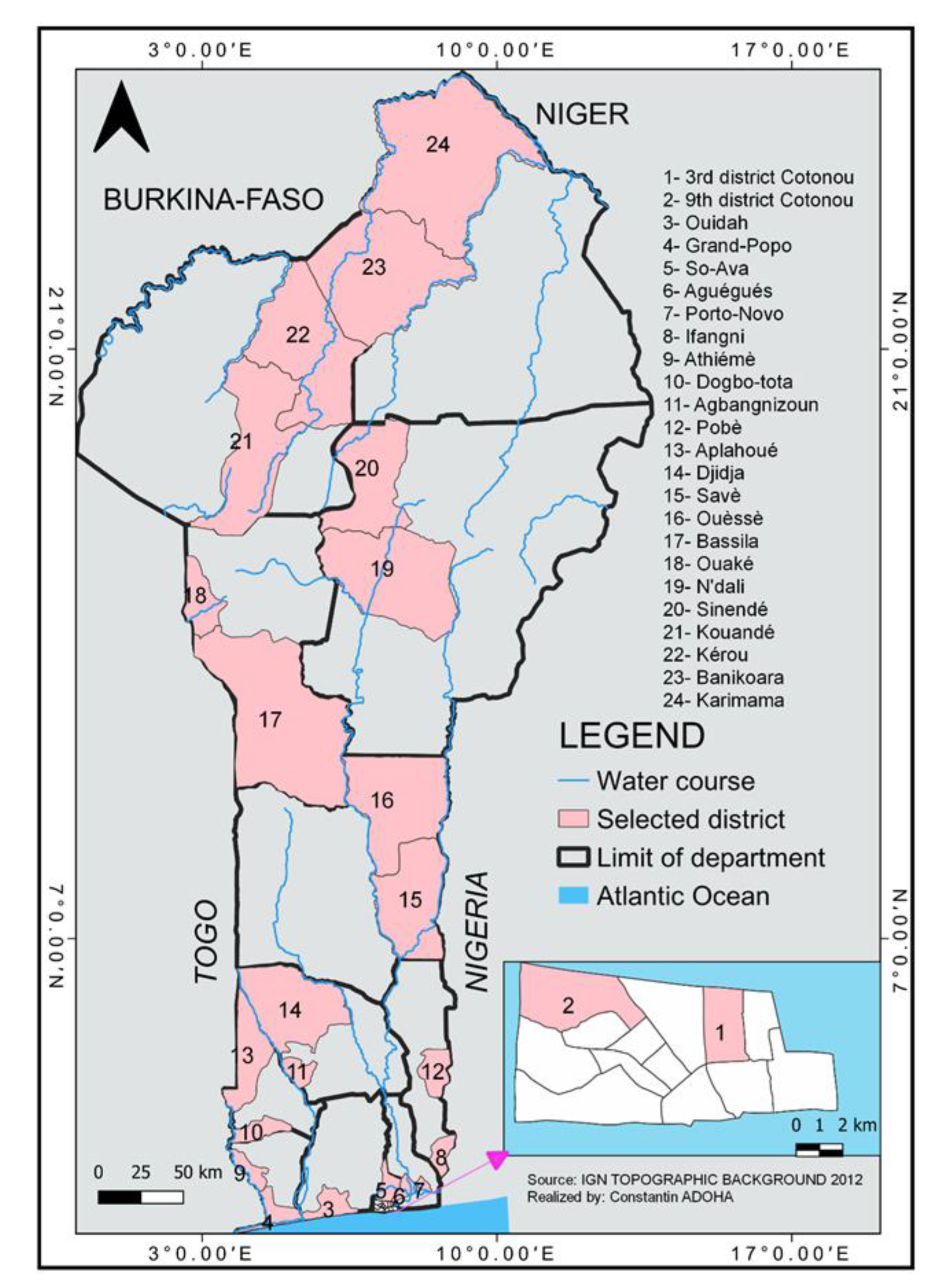
2.1. Study Sites

Mosquito collection was conducted in all 12 regions of Benin from April to December 2019. This collection period coincides with the two rainy seasons of the year, periods characterized by the creation of numerous puddles that favor the proliferation of mosquitoes, including malaria-transmitting ones. In each region, two communes were randomly selected to look for the presence of Plasmodium species in mosquitoes. Thus, 24 communes were selected for the study (Figure 1). In each of them, two sites selected at random were surveyed for the collection of mosquitoes.
In communes located in the southern and central part of the country, the hot and humid climate is of the sub-equatorial type with four seasons, including a long (April to July) and short (October to November) rainy season, as well as a long (December to March) and short (August to September) dry season. In the northern communes, the climate is Sudano-Guinean, with a single rainy season (June to November). The rest of the year (December to May) is the dry season. The average annual rainfall is about 1340 mm in the south and 700 mm in the north [20], while the temperature varies little with an annual average between 26 °C and 30 °C [21].

2.2. Mosquito Collection

In all sites, the collection of adult mosquitoes was performed using both Human Landing Catches (HLCs) and morning Pyrethrum Spray Catches (PSCs). The two methods were used to increase the likelihood to collect a great number of Plasmodium sporozoite-positive mosquitoes.
HLCs were performed between 9:00 p.m. and 5:00 a.m. using eight people, with four (two positioned indoors and two outdoors) used between 9:00 p.m. and 1:00 a.m. and replaced with four others from 1:00 a.m. to 5:00 a.m. Thus, each seated collector used a flashlight as well as a sucking tube to collect the mosquitoes that landed on his/her naked lower legs. Collectors were rotated over collection of hours to limit bias related to individual variations in the attractiveness of Anopheles vectors. They were previously trained to collect the mosquitoes.
PSCs were carried out in 10 bedrooms randomly selected in each commune. The houses that were selected have ceilings and do not have eaves. They also have windows and doors that are kept closed before spraying, which prevents mosquitoes from escaping. This method, which was started at 7:00 a.m., enabled the collection of mosquitoes that entered the bedrooms overnight. Thus, the Rambo aerosol (Gongoni Company Limited, Kano, Nigeria) combining Beta-cypermethrin, tetramethrin, and permethrin was sprayed in the interior of rooms where white sheets were previously laid on the floor. This allowed the collection of indoor-resting mosquitoes.
Adult mosquitoes were then morphologically identified using a binocular loupe, as well as the morphological identification key of Gillies & Coetzee [22]. Anopheles were stored individually in eppendorf tubes containing silica gel and cotton and kept at −20 °C until processing.

2.3. Blood Meal Origin in An. gambiae s.l. Collected by PSC

The origin of blood meal in females An. gambiae s.l. collected through PSC in the different communes were determined using the ELISA test, the protocol of which was described by Beier et al. [23]. Antibodies used for this testing were from human (Sigma, St. Louis, MO, USA, 11 mg/mL), cow (Sigma, 11 mg/mL), sheep (Sigma, 4 mg/mL), and pig (Sigma, 11 mg/mL). The blood meal origin was in Anopheles to ensure that the detected Plasmodium species were from humans living in the commune. Abdomens of blood-fed mosquitoes were ground in 250 μL of Phosphate Buffer Saline (PBS). For each well, 50 μL of each blood eluate were introduced (one mosquito per column) and incubated for 3 h at room temperature. The sensitized plates were washed twice with PBS-Tween 20. For each well and each row, 50 μL of labeled IgG solution were distributed. After a 1 h incubation at room temperature, the plates were emptied and washed with PBS/Tween 20. Then, 100 μL of the peroxidase enzyme substrate were added to each well and incubated in the dark for 30 min. A blue color was obtained, and the reaction was blocked with 50 μL of a 4 N sulfuric acid solution. Results were read at 450–620 nm with the BioTek Elx808 ELISA plate reader (BioTek, Winsooki, VT, USA ). The negative (PBS 1×) and positive controls, in particular the sera of the different hosts sought (human, cow, goat, pig), were placed on each line.

2.4. Circumsporozoite Protein ELISA for the Detection of P. falciparum, P. vivax 210 and P. vivax 247

In this study, the detection of P. falciparum and P. vivax 210 and 247 was done using the heads and thorax of these sampled mosquitoes according to the method described by Burkot et al. [24]. A sampling of the collected Anopheles was done by commune in the laboratory before the tests were performed. The heads–thoraxes of the sampled mosquitoes were individually crushed, after a 1 h incubation in 50 μL of blocking buffer (BB) containing IGEPAL CA-630 (Sigma, Darmstadt, Germany). During the grinding, two times 100 μL of BB was supplemented to each ground material. Then, the 96 wells of a plate were first sensitized with circumsporozoite (CS) protein capture monoclonal antibodies (0.5 mg/mL, BEI Resources). For each well of the plate, 50 μL of this solution were introduced and left overnight on the bench. The following day, the wells were emptied without being washed and received 200 μL of BB for 1 h at room temperature. These wells were again emptied without being washed, and each received 50 μL of mosquito ground material. After a 2 h incubation at room temperature, the wells were emptied and washed twice with PBS/Tween 20. Each well then received 50 µL of peroxidase-conjugated monoclonal antibodies (0.5 mg/mL, BEI Resources) for 1 h on the bench. Then, 7.5 µL of the substrate solution was mixed with 5 mL of BB. After 1 h of incubation at room temperature, the wells were emptied and washed 4 times with PBS/Tween 20. Then, 100 μL of the peroxidase substrate were placed in each well. The reaction occurs in the dark for 30 min. At the end, the positive wells were colored in blue. This reaction was blocked with 4 N sulfuric acid. The positive wells were colored in yellow. Plates were read using the BioTek Elx808 ELISA plate reader at 405 nm and 650 nm (BioTek, Winsooki, VT, USA). Laboratory-susceptible strain served as a negative control. P. falciparum, P. vivax 210, and P. vivax 247 at 2 pg/µL (BEI Resources) were used as positive controls. The testing was simultaneously conducted on 3 plates to detect the presence of the three Plasmodium species (P. falciparum, P. vivax 210, and P. vivax 247).

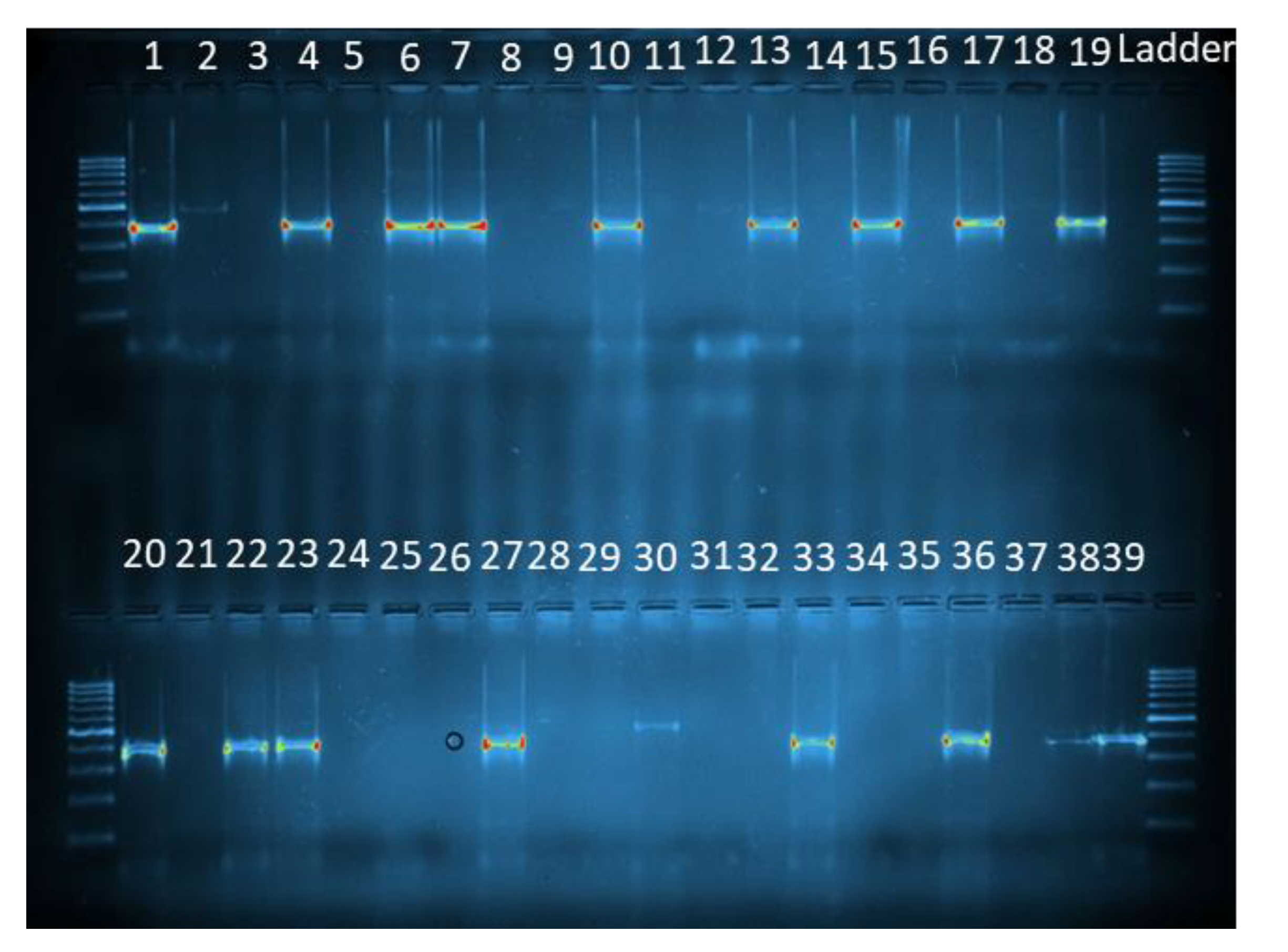
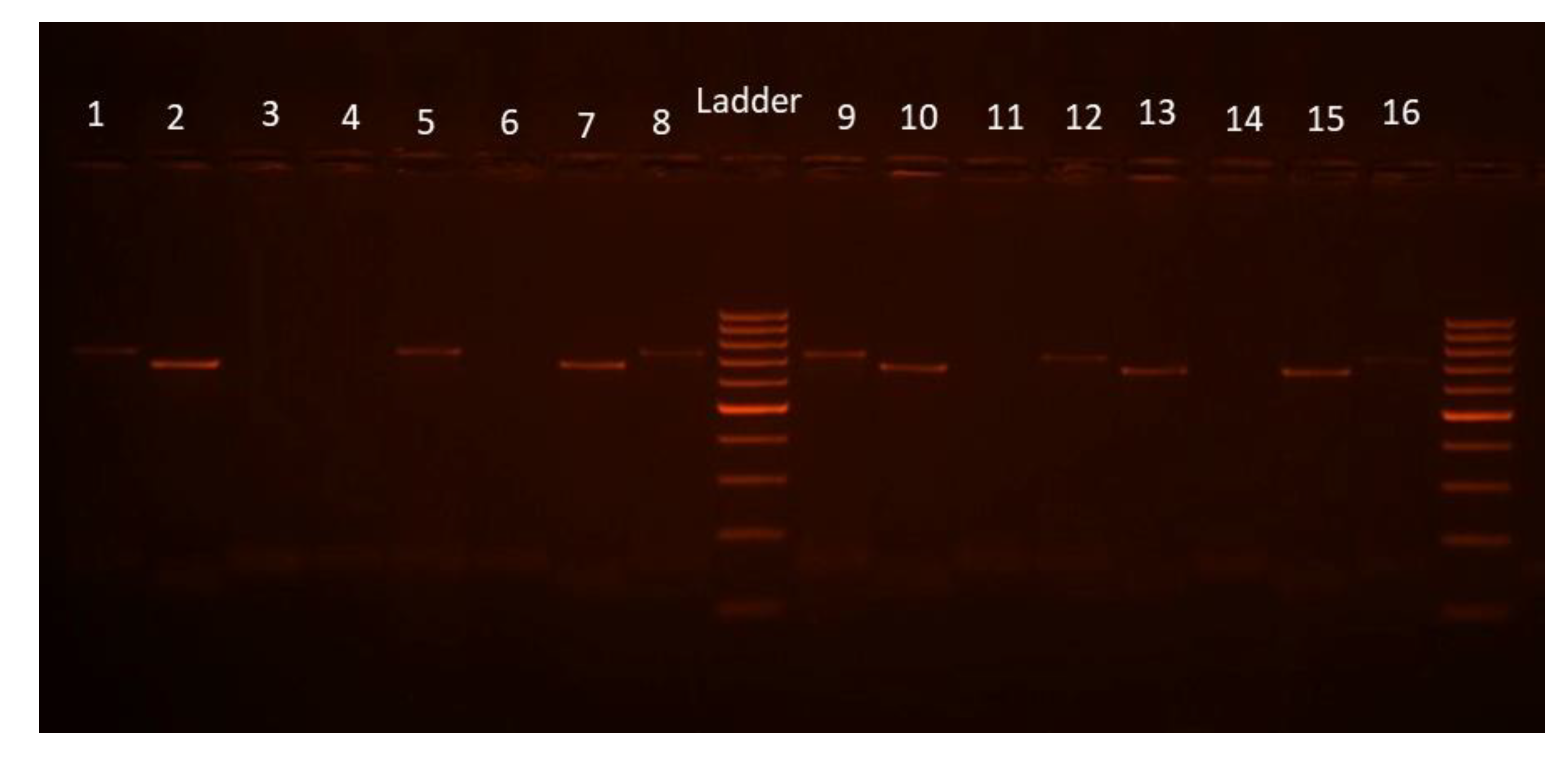
2.5. DNA Extraction of Plasmodium Species and Detection of P. vivax Serotypes

To characterize the Plasmodium species (P. falciparum, P. vivax 210, and 247) in the different communes, Plasmodium DNA was extracted from the grindings of the heads–thoraxes of the mosquitoes used for the ELISA CSP testing. In each commune, a total of 50 mosquitoes, including all Plasmodium sporozoite-positive samples identified through ELISA CSP testing, plus a subset of randomly selected negative ones, were analyzed by PCR according to the protocol described in Methods in Anopheles Research [25].
Plasmodium DNA was extracted with 2% CTAB (Cetyl trimethyl ammonium bromide). The grindings of the heads–thoraxes of the mosquitoes used for the ELISA CSP testing were ground in 200 μL of 2% CTAB. After 5 min in a water bath at 65 °C, the grindings were mixed with 200 μL of chloroform and then centrifuged at 14,000 rpm for 5 min. The supernatant was gently transferred into another tube containing 200 μL of isopropanol then centrifuged at 12,000 rpm for 15 min. The DNA was purified with 200 μL of 70% ethanol. Another centrifugation occurred at 14,000 rpm for 5 min. The content of each tube was gently inverted to preserve the DNA, which will then be dried for at least 3 h on the bench. Finally, 20 μL of bi-distilled water was added to the DNA, which is left on the bench overnight or half a day.
Then, PCR amplification of the Plasmodium species was performed in a thermocycler (Eppendorf, Hamburg, Germany) following PCR conditions described in Methods in Anopheles Research [25].
The identification of Plasmodium species was performed using the primers presented in Table 1. UNR and PLF are universal primers that hybridize to a particular nucleotide sequence in all Plasmodium species. FAR forms with UNR and PLF, the pair of primers that amplify a specific nucleotide fragment in P. falciparum. The same applies for the pair of primers formed by UNR- PLF and VIR, which amplifies a specific nucleotide fragment in P. vivax. Then, the PVCSP1 and PVCSP2 primers were used to identify the P. vivax variants (Table 1). DNA of P. falciparum, P. vivax, P. vivax 210, and P. vivax 247 (BEI Resources) was used as positive controls, while sterilized water was used as a negative control. Total of 18 µL of each amplified product for each reaction was analyzed with electrophoresis gel using a 2% agarose gel (Sigma). When successfully amplified, the gel images were recorded and visualized on transilluminator EBOX 1000 (Vilber, Marne-la-Vallée, France).

2.6. Molecular Species Identification in the Anopheles gambiae Complex

The abdomens, legs, and wings of the same mosquito individuals previously tested through PCR for detection of Plasmodium infection were molecularly speciated using the protocol of Santolamazza et al. [26]. PCR amplification of Anopheles gambiae complex species was carried out in a 25 μL reaction mixture containing 5 μL genomic DNA for An. gambiae s.l., 1 × PCR buffer 5×, 1.5 mM MgCl2, 0.2 mM of each dNTP, 10 pmoles of each primer (Table 1), and 5 U of Taq polymerase (Eurogentec, Liège, Belgium). The amplification was performed in a thermocycler (Eppendorf, Hamburg, Germany) as follows: an initial denaturation of the DNA material at 94 °C for 5 min, followed by 35 cycles of 94 °C for 30 s, 54 °C for 30 s, and 72 °C for 30 s, with a final extension at 72 °C for 10 min. DNA of An. gambiae, An. coluzzii, and An. arabiensis were used as positive controls, while sterilized water was used as a negative control. The amplification product was migrated on a 1.5% agarose gel (Sigma) in the presence of ethidium bromide (Sigma) used as an intercalant. This allows for the assessing of the involvement of each species of the An. gambiae complex in the transmission of the different Plasmodium species.

2.7. Data Analysis

Data were analyzed using R Core Team software (version 4.1.3-2022) and Graph Pad Prism software version 5.0. The graphs were made using Graph Pad software version 5.0. The exact binomial method was used to calculate the prevalence and their confidence intervals with the R Core Team software (version 4.1.3-2022).

2.8. Ethical Consideration

The present study received ethical approval from the Institutional Ethics Committee of Centre de Recherche Entomologique de Cotonou (N° 04/IECC). Mosquito collectors gave their consent prior to being involved in the study. They were trained to collect the mosquito before being bitten and were regularly subjected to medical check-ups. When they had malaria, they were taken care of and received anti-malarial medication like Artemisinin-based Combination Therapy if required. They were also vaccinated against yellow fever.

3. Results

3.1. Mosquito Species Composition

Table 2 shows the species composition of mosquito populations collected through HLCs in the different surveyed regions over the study period. Overall, a great diversity was observed in mosquito fauna collected on human bait, regardless of the commune. In total, 32,773 specimens of mosquitoes belonging to 18 species were collected using HLCs. Overall, the most predominant species in the whole study area were Culex quinquefasciatus (40.48%), followed by Anopheles gambiae s.l. (20.92%), Culex gr decens (10.23%), Mansonia africana (18.27%), Anopheles funestus (3.94%), and Aedes aegypti (2.61%) (Table 1). At the region level, Anopheles gambiae s.l. was the most abundant mosquito species in Zou (63.9%), Donga (56.9%), Atacora (50.8%), Mono (39.1%), and Couffo (35%), while Anopheles funestus was the most abundant one in Collines (60.9%). In the Ouémé, Littoral, Atlantique, and Borgou regions, Cx quinquefasciatus was the major species with frequencies of 51.7%, 78.6%, 43.8%, and 48.3%, respectively. Mansonia africana was predominant in the Plateau (59%) and Alibori (57.4%) regions (Table 2).

3.2. Molecular Species Identification of the Sigbling Species within the Anopheles gambiae Complex

Of the 6857 collected, 940 (13.7%) specimens of An. gambiae s.l. were analyzed. An. gambiae, An. coluzzii, and An. arabiensis were the species recorded and accounted for 53.68%, 45.88%, and 0.44%, respectively (Figure 2). In the Porto-Novo, Aguégués, 3rd district of Cotonou, 9th district of Cotonou, Ouidah, So-Ava, Athiémé, Grand Popo, Aplahoué, and Dogbo regions, An. coluzzii was the predominant species with mean frequencies varying between 71.43% and 100%. Conversely, in the regions of Ifangni, Pobè, Agbangnizoun, Ouèssè, Savè, N’dali, Sinendé, Ouaké, Bassila, Kérou, and Kouandé, An. gambiae was the major species with a mean frequency ranging from 58.62% to 100%. In the commune of Djidja, the two species were collected in similar proportions. An. arabiensis was only found in the commune of Savè (Figure 2).

3.3. Blood Meal Origin in An. gambiae s.l. Collected through PSC

Of the 1392 mosquitoes collected by PSC, 514 were specimens of An. gambiae s.l., of which 311 (60.5%) were blood-fed. Out of the 311 blood-fed specimens of An. gambiae s.l. that were tested through ELISA blood meal in the study area, 95.5% took their blood meal on humans (Table 3).
Approximately 16.67% and 7.14% of the tested individuals of An. gambiae s.l. had successful blood meal intake on cow in the communes of Kouandé and Karimama, respectively. In Athiémé and Karimama, the proportion of of An. gambiae s.l that blood-fed on sheep was 4.87% and 3.57%, respectively. Blood meal intake on pigs occurred in 1.69%, 8.33%, and 1.61% of specimens of An. gambiae s.l analyzed in Aguégués, Sô-Ava, and Athiémé, respectively. In the commune of Sô-Ava, 4.17% of tested individuals of An. gambiae s.l took their blood meal on both humans and pigs (Table 3).

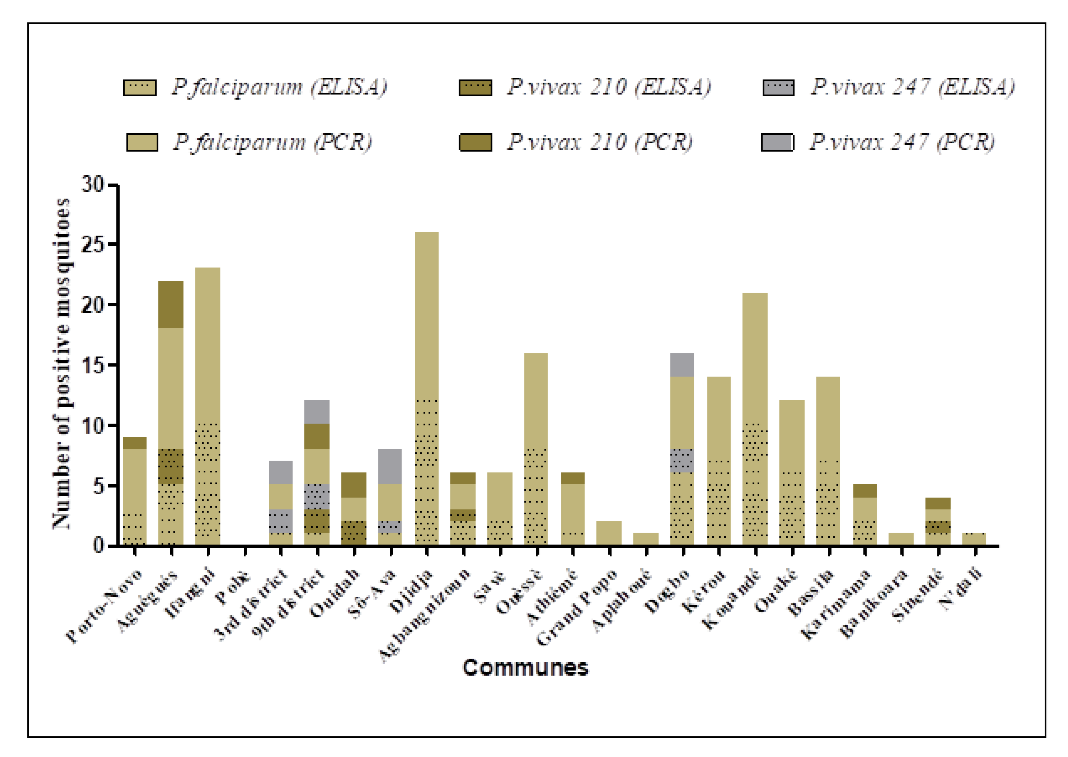
3.4. Prevalence of P. falciparum, P. vivax 210 and 247 CSP Antibodies in An. gambiae s.l.

3.4.1. Plasmodium Detection by ELISA

A total of 3300 head–thoraxes of An. gambiae s.l. out of the 6857 collected were tested through ELISA CSP for each of the three Plasmodium species, equating to a total of 9900 samples analyzed. The number of sporozoite-positive mosquitoes for each Plasmodium species at the commune level is mentioned on Figure 3. The combined data showed that 86 mosquitoes were positive for P. falciparum, 9 for P. vivax 210, and 7 for P. vivax 247. Thus, the average prevalence (sporozoite index: SI) for P. falciparum was 2.6% (CI: 2.1–3.1) compared to 0.3% (CI: 0.1–0.5) for P. vivax 210 and 0.2% (CI: 0.1–0.4) for P. vivax 247 in the whole study area.
Out of the 228 specimens of An. funestus analyzed, only two (one in Ouèssè and one in Savè) were positive for P. falciparum. No specimen of An. funestus was found positive for P. vivax 210 and P. vivax 247.
The three species of Plasmodium-P. falciparum, P. vivax 210, and P. vivax 247-were found in An. gambiae s.l in Cotonou with a respective prevalence of 0.7% (CI: 0–2.1), 0.55% (CI: 0.05–3.5), and 1.45% (CI: 0.15–5). In the communes of Aguégués, Agbangnizoun, and Sinendé, P. falciparum (SI = 2% CI: 0.3–3.7; 2.8% CI: 0–6.6; and 1.5% CI: 0–4.3, respectively) and P. vivax 210 (SI = 1.2% CI: 0.2–3.5; 1.4% CI: 0–7.5 and 1.5% CI: 0–7.9, respectively) were detected in An. gambiae s.l. during the study. In Sô-Ava and Dogbo, P. falciparum (SI = 0.5% CI: 0–1.5 and 6% CI: 1.3–10.7, respectively) and P. vivax 247 (SI = 0.5% CI: 0–2.8 and 2% CI: 0.2–7, respectively) were detected (Figure 3). In Ouidah, only P. vivax 210 (SI = 1.2% CI: 0.2–4.4) was found in An. gambiae s.l., whereas P. falciparum was the only species detected in Porto-Novo (1.5% CI: 0–3.2), Ifangni (5% CI: 2–8.1), Djidja (8% CI: 3.7–12.3), Savè (0.9% CI: 0–2.1), Ouèssè (4% CI: 1.3–6.7), Athiémé (0.3% CI: 0–1), Kérou (4.7% CI: 1.3–8), Kouandé (6.7% CI: 2.7–10.7), Ouaké (4.6% CI: 1–8.2), Bassila (5% CI: 1.4–8.6), Karimama (2.7% CI: 0–6.3), and N’dali (1.5% CI: 0–4.5).
No mosquito was found positive for the three searched Plasmodium species in Pobè, Grand Popo, Aplahoué, and Banikoara. No co-infection was recorded in the Anopheles tested throughout the surveyed communes (Figure 3).

3.4.2. Plasmodium Detection by PCR

All samples positive for any of the Plasmodium species sought by the CSP ELISA test were also positive for the PCR test (Figure 3). Of the 940 samples of An. gambiae s.l. tested through PCR, 115 were positive for P. falciparum, 13 for P. vivax 210, and 9 for P. vivax 247 (Figure 3, Figure 4, Figure 5). P. falciparum was detected in all surveyed communes except Pobè, where no positive specimen was found for the three searched Plasmodium species.
P. vivax 210 was detected in Cotonou (2/100), Ouidah (2/50), Porto-Novo (1/50), Aguégués (4/50), Athiémé (1/50), Agbangnizoun (1/33), Karimama (1/32), and Sinendé (1/32), whereas P. vivax 247 was found only in Cotonou (4/100), Sô-Ava (3/50), and Dogbo (2/38). In Ifangni (13/50) and Grand Popo (2/50), P. falciparum was the sole detected Plasmodium species.
As observed with the CSP ELISA test, no co-infection was recorded. In Grand Popo, Aplahoué, and Banikoara, P. falciparum sporozoite-positive mosquitoes were found through PCR, which was not the case with the ELISA CSP test. The same trend was observed with P. vivax 210 in Karimama.

3.5. Distribution of the Plasmodium Species in the Sibling Species of the Anopheles gambiae Complex

Among the 115 specimens of Anopheles gambiae s.l found positive for P. falciparum through PCR, 64.35% (n = 74) were An. gambiae, 34.78% (n = 40) were An. coluzzii, and 0.86% (n = 1) were An. arabiensis. Similarly, the P. falciparum infectivity rate was significantly higher in An. gambiae than in An. coluzzii (p < 0.001).
At the opposite, most P. vivax 210-positive mosquitoes were An. coluzzii (76.92%). The rest were An. gambiae. The infectivity rates of P. vivax 210 were significantly higher in An. coluzzii than in An. gambiae (p < 0.05).
Concerning P. vivax 247, only An. coluzzii was positive to this variant (Table 4).

4. Discussion

In the current context of malaria elimination, the identification of Plasmodium species in humans and vectors is a prerequisite not only to understand the epidemiology of the disease, but also to implement an effective control strategy against these parasites. The present study shows the presence of antibodies of P. falciparum and P. vivax in the An. gambiae complex, the main malaria vector complex in Benin. This result corroborates the work of Poirier et al. [16], who showed a surprisingly high exposure of Beninese to P. vivax, resulting in submicroscopic infections.
In the present study, 18 mosquito species, including five Anopheles ones, were identified in the 24 surveyed communes. These results are reminiscent to those of Huttel [27] and Salako et al. [28], who recorded 13 and 14 mosquito species in Southeastern and Northwestern Benin, respectively. Of the five collected Anopheles species, three (An. gambiae s.l., An. funestus gr., and An. nili gr) have already been incriminated in malaria transmission in Benin [4,5,29,30,31,32].
The molecular species identification performed in An. gambiae s.l. revealed the presence of three sibling species, namely An. gambiae, An. coluzzii, and An. arabiensis. These species were found in sympatry but in variable proportions. Overall, An. gambiae accounted for 53.68% of all specified mosquitos, followed by An. coluzzii (45.88%) and An. Arabiensis, which was found at a very low frequency. This result is similar to the one of Aikpon et al. [33] that showed the same trend in Atacora department, the cotton zone of Benin. The opposite result, displaying An. coluzzii as the predominant species, was observed by Gnanguenon et al. [30] in Allada, Dassa, and Malanville. Although these species often have different geographical and seasonal distributions, they can also live in sympatry. In the present study, An. arabiensis was found only in Savè, a wet savanna area. This is consistent with works by Gnanguenon et al. [30], who found An. arabiensis in Dassa, a commune that is quite close to Savè. Over past years, An. arabiensis was considered a dry savanna species found in abundance between the extreme north of Benin and Parakou [34]. It is, therefore, necessary to assess if the species migrated from its preferred area to a more southern area. A possible migration could be explained by factors such as climate change, the destruction of the vector’s ecological zones for agricultural and hunting purposes, the harvesting of timber, and pastoralism.
The findings of the present study revealed spatial heterogeneity in the distribution of Plasmodium species prevalent in Benin. The mean prevalence of antibodies of P. falciparum, P. vivax 210, and P. vivax 247 in An. gambiae s.l. were, respectively, 2.6%, 0.30%, and 0.2% in the study area. In Cotonou, the three Plasmodium species were found in An. coluzzii with a high prevalence for P. vivax 247 (4%) compared to P. vivax 210 (2%) and P. falciparum (2%). In the communes of Aguégués, Porto-Novo, Ouidah, Agbangnizoun, Athiémé, Karimama, and Sinendé, P. falciparum and P. vivax 210 were detected with similar frequencies across communes (p > 0.05). In Sô-Ava and Dogbo, only P. falciparum and P. vivax 247 were registered with similar frequencies (p > 0.05). In the rest of the communes, P. falciparum was the sole species found except for Pobè, where no infection to three searched Plasmodium species was observed. The null infection observed in Pobè could be due to the low number of mosquitos tested in this commune. This variation of the prevalence of the Plasmodium species across communes can be explained by the period of collection of the mosquitoes, as well as the ecological and evolutionary factors associated with mosquitoes, and that can promote divergence and restrict gene flow between vector-adapted parasite lineages [35]. Furthermore, contrary to the findings of Sandeu et al. [36], no co-infection with different species of Plasmodium cells was recorded in Anopheles mosquitoes analyzed in the communes surveyed over the present study. In addition, over the trial of Sandeu et al. [36] where the different species of Plasmodium were searched and quantified in malaria vectors, P. vivax was not found in any of the mosquito samples analyzed, contrary to what is shown in the present study. Indeed, Howes et al. [37] previously showed that P. vivax was the most prevalent malaria parasite globally but clearly "rare" in Africa. The majority of African populations do not express the Duffy blood group antigen, which is the only known receptor for the P. vivax infection. Since this discovery in the 1970s, the low clinical incidence of P. vivax in Africa led to a perception that this Plasmodium species is completely absent, and that all apparent cases are from a misdiagnosis. Thus, no public health consideration is made for this parasite in terms of diagnosis, treatment, or surveillance. As more sensitive diagnosis methods become available, P. vivax infection in Africa is increasingly being reported through various types (entomological, serological, community prevalence) of surveys, as well as clinical data on infections from local residents and international travelers.
It would have been interesting to separate the infection data of mosquitoes according to the biting locations (indoor and outdoor), which we did not do during the trial. Thus, it would be important to take into account the microclimatic conditions (ambient temperature and relative humidity) of the indoor and outdoor environment where collections were undertaken as it matters to the vector availability and density. This constitutes a limitation for the study.
In the present study, the distribution of the different Plasmodium species in the sibling species of An. gambiae s.l. was also assessed across the surveyed communes. Of the 115 samples positive for P. falciparum through PCR, An. gambiae was predominant (n = 74), followed by An. coluzzii (n = 40) and An. arabiensis (n = 1). The infectivity of An. coluzzii to P. vivax (variant 210 and 247) was significantly higher than that of An. gambiae. Indeed, most (76.92%) and all mosquitoes that were positive for P. vivax 210 and P. vivax 247, respectively, were An. coluzzii. This result contradicts findings from Goupeyou-Youmsi et al. [38] obtained in two neighboring villages of the rural commune of Andriba in Madagascar. Indeed, they found that An. arabiensis, which was the sole species detected within the An. gambiae complex, carried P. falciparum, P. vivax, and P. malariae. Contrary to the present study, only An. arabiensis was the vector of three Plasmodium species (P. falciparum, P. vivax, and P. malariae). These differences could be explained by weather conditions, the ecological environment, and/or the geographical distribution of vectors that are different between countries [38]. These results suggest that more screening of An. coluzzii has to be undertaken, as P. vivax is difficult to eliminate, and more ultra-sensitive methods are required to detect such infections in the community. This is a valid point that epidemiologists and entomologists need to worry about since the urban malaria vector, An. stephensi, which invaded many parts of sub-Saharan Africa, such as Ethiopia and Sudan [39,40], Somalia, and more recently Nigeria [41], could also be infected with P. vivax. For that, the larval habitats of the malaria vectors in Benin need to be documented as efforts for larval source management, along with adult control interventions (LLINs and targeted IRS) that can be strengthened in order to have positive results.
In the present study, PCR allows for the detection of sporozoites of both P. falciparum and P. vivax in some mosquitoes, while ELISA-CSP gave negative results. A possible explanation would be that the number of sporozoites present in the salivary glands of the mosquito is too low to be detected by the ELISA-CSP test. In addition, several studies have also mentioned the difficulty of ELISA in detecting the antigens of other Plasmodium species such as P. malariae and P. ovale, but also the problem of antigenic variation of the CSP antigen between geographical areas [42,43]. Finally, an extension of this study in all geographical areas of the country and other West African regions could help generate valuable epidemiological data on the two variants of P. vivax and allow for a more elaborated malaria pre-elimination policy. The evaluation of P. vivax variants is a prerequisite to the development of an effective vaccine. Other aspects that are worth evaluation include the geographical distribution the variants, the intensity of their transmission, the ability of the vectors to allow for their development, and the variability of the human immune response towards them.

5. Conclusions

The present study constitutes a first in Benin that detects P. falciparum and P. vivax in An. gambiae s.l. using both CSP ELISA and PCR techniques. The results showed that the risk of exposure of populations to P. falciparum, P. vivax 210, and P. vivax 247 varies from species to species. In addition, both variants of P. vivax are present in Benin. It would, therefore, be necessary to evaluate the impact of currently used antimalarial drugs on this Plasmodium species for a better therapeutic management of malaria in the country. These results will help the National Malaria Control Program to better plan for the therapeutic management strategy of malaria cases to move towards the pre-elimination of the disease.

Author Contributions

Conceptualization, F.T., R.A.O. and M.A.; Data curation, B.A., F.T. and R.A.O.; Formal analysis, M.E.G., R.A.O. and A.S. (Aboubakar Sidick); Investigation, A.F., F.T., R.A.O., M.E.G. and C.Z.K.; Methodology, G.G.P., F.T., R.A.O. and A.S. (Arthur Sovi); Supervision, M.A.; Writing—original draft, R.A.O., F.T. and G.G.P.; Writing—review & editing, R.A.O., A.S. (Arthur Sovi), F.T. and M.A. All authors have read and agreed to the published version of the manuscript.

Funding

This work has been financially supported by the Global Fund to Fight AIDS, Tuberculosis and Malaria Grant BEN-M-PNLP; 2018–2020.

Data Availability Statement

Data is contained within the article.

Acknowledgments

We are grateful to the Centre de Recherche Entomologique de Cotonou (CREC) and National Malaria Control Programme through the Global Fund to Fight AIDS, Tuberculosis and Malaria, which supported financially this study. We thank Louisa Messenger for her technical assistance and the team of CREC for their technical assistance during field work and laboratory. We also thank the people of different communes for their collaboration.

Conflicts of Interest

The authors declare no conflict of interest.

References

  1. WHO. World Malaria Report 2021; World Health Organization: Geneva, Switzerland, 2021. [Google Scholar]
  2. Damien, G.B.; Djènontin, A.; Rogier, C.; Vincent Corbel, V.; Bio Bangana, S.; Chandre, F.; Akogbéto, M.; Kindé-Gazard, D.; Massougbodji, A.; Henry, M.-C. Malaria infection and disease in an area with pyrethroid-resistant vectors in southern Benin. Malar. J. 2010, 9, 380–391. [Google Scholar] [CrossRef] [Green Version]
  3. Ministère de la Santé. Annuaire des Statistiques Sanitaires; Direction de la Programmation et de la Prospective: Cotonou, Benin, 2020; p. 278. [Google Scholar]
  4. Ossè, R.A.; Tokponnon, F.; Padonou, G.G.; Sidick, A.; Aïkpon, R.; Fassinou, A.; Koukpo, C.Z.; Sèwadé, W.; Akinro, B.; Sovi, A.; et al. Involvement of Anopheles nili in Plasmodium falciparum transmission in North Benin. Malar. J. 2019, 18, 152–162. [Google Scholar] [CrossRef] [PubMed]
  5. Djènontin, A.; Bio-Bangana, S.; Moiroux, N.; Henry, M.; Bousari, O.; Chabi, J.; Ossè, R.; Koudenoukpo, S.; Corbel, V.; Akogbéto, M.; et al. Culicidae diversity, malaria transmission and insecticide resistance alleles in malaria vector in Ouidah-Kpomasse-Tori district from Benin (West Africa): A pre-intervention study. Parasites Vectors 2010, 3, 83–84. [Google Scholar] [CrossRef] [Green Version]
  6. Kantele, A.; Jokiranta, T.S. Review of cases with the emerging fifth human malaria parasite, Plasmodium Knowlesi. Clin. Infect. Dis. 2011, 52, 1356–1362. [Google Scholar] [CrossRef]
  7. Yap, N.J.; Hossain, H.; Nada-Raja, T.; Ngui, R.; Muslim, A.; Hoh, B.-P.; Khaw, L.T.; Abdul Kadir, K.; Paul Divis, C.S.; Vythilingam, I.; et al. Natural human infections with Plasmodium cynomolgi, P. inui, and 4 other simian malaria parasites, Malaysia. Emerg. Infect. Dis. 2021, 27, 2187–2191. [Google Scholar] [CrossRef] [PubMed]
  8. Brasil, P.; Zalis, M.G.; de Pina-Costa, A.; Siqueira, A.M.; Júnior, C.B.; Silva, S.; Areas, A.L.L.; Pelajo-Machado, M.; de Alvarenga, D.A.M.; da Silva Santelli, A.C.F.; et al. Outbreak of human malaria caused by Plasmodium simium in the Atlantic Forest in Rio de Janeiro: A molecular epidemiological investigation. Lancet Glob. Health 2017, 5, e1038–e1046. [Google Scholar] [CrossRef] [Green Version]
  9. Loeffel, M.; Ross, A. The relative impact of interventions on sympatric Plasmodium vivax and Plasmodium falciparum malaria: A systematic review. PLoS Negl. Trop. Dis. 2022, 16, e0010541. [Google Scholar] [CrossRef] [PubMed]
  10. Gething, P.; Elyazar, I.R.F.; Moyes, C.; Smith, D.; Battle, K.; Guerra, C.A.; Patil, A.P.; Tatem, A.J.; Howes, R.E.; Myers, M.F.; et al. A Long Neglected World Malaria Map: Plasmodium vivax Endemicity in 2010. PLoS Negl. Trop. Dis. 2012, 6, e1814. [Google Scholar] [CrossRef]
  11. Geleta, G.; Ketema, T. Severe malaria associated with Plasmodium falciparum and P. vivax among children in Pawe Hospital, Northwest Ethiopia. Malar. Res. Treat. 2016, 2016, 1240962. [Google Scholar]
  12. Asua, V.; Mugenyi, L.; Rosenthal, P.J.; Nsobya, S.L.; Nankabirwa, J.I.; Walakira, A.; Kamya, M.R.; Tukwasibwe, S.; Conrad, M. Plasmodium Species Infecting Children Presenting with Malaria in Uganda. Am. J. Trop. Med. Hyg. 2017, 97, 753–757. [Google Scholar] [CrossRef] [Green Version]
  13. Roucher, C.; Rogier, C.; Sokhna, C.; Tall, A.; Trape, J.-F. A 20-Year Longitudinal Study of Plasmodium ovale and Plasmodium malariae Prevalence and Morbidity in a West African Population. PLoS ONE 2014, 9, e87169. [Google Scholar] [CrossRef] [PubMed] [Green Version]
  14. Cox-Singh, J.; Singh, B. Knowlesi malaria: Newly emergent and of public health importance? Trends Parasitol. 2008, 24, 406–410. [Google Scholar] [CrossRef] [PubMed] [Green Version]
  15. Wells, T.N.; Burrows, J.N.; Baird, J.K. Targeting the hypnozoite reservoir of Plasmodium vivax: The hidden obstacle to malaria elimination. Trends Parasitol. 2010, 26, 145–151. [Google Scholar] [CrossRef] [PubMed]
  16. Poirier, P.; Doderer-Lang, C.; Atchade, P.S.; Lemoine, J.P.; Coquelin, M.L.; Abou-bacar, A.; Pfaff, A.W.; Brunet, J.; Arnoux, L.; Haar, E.; et al. The hide and seek of Plasmodium vivax in West Africa: Report from a large-scale study in Beninese asymptomatic subjects. Malar. J. 2016, 15, 570–579. [Google Scholar] [CrossRef] [PubMed] [Green Version]
  17. Mercereau-Puijalon, O.; Ménard, D. Plasmodium vivax and the Duffy antigen: A paradigm revisited. Transfus. Clin. Et Biol. 2010, 17, 176–183. [Google Scholar] [CrossRef]
  18. Miller, L.H.; Mason, S.J.; Clyde, D.F.; McGinniss, M.H. The Resistance Factor to Plasmodium vivax in Blacks. N. Engl. J. Med. 1976, 295, 302–304. [Google Scholar] [CrossRef]
  19. Moreira, C.M.; Abo-Shehada, M.; Price, R.N.; Drakeley, C.J. A systematic review of sub-microscopic Plasmodium vivax infection. Malar. J. 2015, 14, 360. [Google Scholar] [CrossRef] [Green Version]
  20. Badou, D.F.; Afouda, A.A.; Diekkrüger, B.; Kapangaziwiri, E. Investigation on the 1970s and 1980s drought in four tributaries of the Niger River Basin (West Africa). In Proceedings of the 36th IAHR World Congress, The Hague, The Netherlands, 28 June–3 July 2015; pp. 1–5. [Google Scholar]
  21. Bossa, A.Y.; Diekkrüger, B.; Agbossou, E.K. Scenario-Based Impacts of Land Use and Climate Change on Land and Water Degradation from the Meso to Regional Scale. Water 2014, 6, 3152–3181. [Google Scholar] [CrossRef] [Green Version]
  22. Gillies, M.T.; Coetzee, M.A. Supplement to the Anophelinae of Africa South of the Sahara, 2nd ed. S. Afr. Inst. Med. Res. 1987, 55, 143. [Google Scholar]
  23. Beier, J.C.; Vaughan, J.A.; Madani, A.; Noden, B.H. Plasmodium falciparum: Release of circum sporozoïte protein by sporozoïtes in the mosquito vector. Exp. Parasitol. 1992, 75, 248–256. [Google Scholar] [CrossRef]
  24. Burkot, T.R.; Graves, P.M.; Wirtz, R.A.; Gibson, F.D. The efficiency of sporozoïtes transmission in the human malarias Plasmodium falciparum and P. vivax. Bull. World Health Organ. 1987, 65, 375–380. [Google Scholar]
  25. MR4. Methods in Anopheles Research, 2nd ed.; CDC: Atlanta, GA, USA, 2010; p. 343. [Google Scholar]
  26. Santolamazza, F.; Mancini, E.; Simard, F.; Qi, Y.; Tu, Z.; della Torre, A. Insertion polymorphisms of SINE200 retrotransposons within speciation islands of Anopheles gambiae molecular forms. Malar. J. 2008, 7, 163. [Google Scholar] [CrossRef] [PubMed] [Green Version]
  27. Huttel, J. Note sur la répartition des moustiques dans le Bas-Dahomey. Bull. Soc. Path. Exot. 1950, 43, 563–566. [Google Scholar]
  28. Salako, A.S.; Ossè, R.; Padonou, G.G.; Dagnon, F.; Aïkpon, R.; Kpanou, C.; Sagbohan, H.; Sovi, A.; Sèzonlin, M.; Akogbeto, M.C. Population Dynamics of Anopheles gambiae s.l. and Culex quinquefasciatus in Rural and Urban Settings Before an Indoor Residual Spraying Campaign in Northern Benin. Vector Borne Zoonotic Dis. 2019, 19, 674–684. [Google Scholar] [CrossRef] [PubMed]
  29. Aïkpon, R.; Sèzonlin, M.; Tokponon, F.; Okè, M.; Oussou, O.; Oké-Agbo, F.; Beach, R.; Akogbéto, M. Good performances but short lasting efficacy of Actellic 50 EC Indoor Residual Spraying (IRS) on malaria transmission in Benin, West Africa. Parasites Vectors 2014, 7, 256. [Google Scholar] [CrossRef] [PubMed] [Green Version]
  30. Gnanguenon, V.; Govoétchan, R.; Agossa, F.R.; Ossè, R.; Oke-Agbo, F.; Azondekon, R.; Sovi, A.; Attolou, R.; Badirou, K.; Tokponnon, F.T.; et al. Transmission patterns of Plasmodium falciparum by Anopheles gambiae in Benin. Malar. J. 2014, 13, 444. [Google Scholar] [CrossRef] [Green Version]
  31. Salako, A.S.; Ahogni, I.; Kpanou, C.; Sovi, A.; Azondekon, R.; Sominahouin, A.A.; Tokponnon, F.; Gnanguenon, V.; Dagnon, F.; Iyikirenga, L.; et al. Baseline entomologic data on malaria transmission in prelude to an indoor residual spraying intervention in the regions of Alibori and Donga, Northern Benin, West Africa. Malar. J. 2018, 17, 392. [Google Scholar] [CrossRef]
  32. Yovogan, B.; Sovi, A.; Padonou, G.G.; Adoha, C.J.; Akinro, B.; Chitou, S.; Accrombessi, M.; Dangbénon, E.; Akpovi, H.; Messenger, L.A.; et al. Pre-intervention characteristics of the mosquito species in Benin in preparation for a randomized controlled trial assessing the efficacy of dual active-ingredient long-lasting insecticidal nets for controlling insecticide-resistant malaria vectors. PLoS ONE 2021, 16, e0251742. [Google Scholar] [CrossRef]
  33. Aikpon, R.; Missihoun, A.; Lokossou, A.; Aikpon, G.; Salifou, S.; Dansi, A.; Agbangla, C. Hétérogénéité génétique et résistance des vecteurs du paludisme (Anopheles gambiae s.l) aux insecticides en zone Cotonnière au Benin. Int. J. Biol. Chem. Sci. 2020, 14, 2724–2736. [Google Scholar] [CrossRef]
  34. Akogbeto, M. Entomological study on the malaria transmission in coastal and lagoon areas: The case of a village built on a brackish lake. Ann. Soc. Belge Med. Trop. 1995, 75, 219–227. [Google Scholar]
  35. Joy, D.A.; Gonzalez-Ceron, L.; Carlton, J.M.; Gueye, A.; Fay, M.; McCutchan, T.F.; Su, X. Local Adaptation and Vector-Mediated Population Structure in Plasmodium vivax Malaria. Mol. Biol. Evol. 2008, 25, 1245–1252. [Google Scholar] [CrossRef] [PubMed] [Green Version]
  36. Sandeu, M.M.; Moussiliou, A.; Moiroux, N.; Padonou, G.G.; Massougbodji, A.; Corbel, V.; Ndam, N.T. Optimized Pan-species and Speciation Duplex Real-time PCR Assays for Plasmodium Parasites Detection in Malaria Vectors. PLoS ONE 2012, 7, e52719. [Google Scholar] [CrossRef] [Green Version]
  37. Howes, R.E.; Reiner Jr, R.C.; Battle, K.E.; Longbottom, J.; Mappin, B.; Ordanovich, D.; Tatem, A.J.; Drakeley, C.; Gething, P.W.; Zimmerman, P.A.; et al. Plasmodium vivax transmission in Africa. PLoS Negl. Trop. Dis. 2015, 9, e0004222. [Google Scholar] [CrossRef] [Green Version]
  38. Goupeyou-Youmsi, J.; Rakotondranaivo, T.; Puchot, N.; Peterson, I.; Girod, R.; Vigan-Womas, I.; Paul, R.; Ousmane Ndiath, M.; Bourgouin, C. Differential contribution of Anopheles coustani and Anopheles arabiensis to the transmission of Plasmodium falciparum and Plasmodium vivax in two neighbouring villages of Madagascar. Parasites Vectors 2020, 13, 430. [Google Scholar] [CrossRef] [PubMed]
  39. Ahmed, A.; Pignatelli, P.; Elaagip, A.; Abdel Hamid, M.M.; Fateh Alrahman, O.; Weetman, D. Invasive malaria vector Anopheles stephensi mosquitoes in Sudan, 2016–2018. Emerg. Infect. Dis. 2021, 27, 2952–2954. [Google Scholar] [CrossRef]
  40. Carter, T.E.; Yared, S.; Gebresilassie, A.; Bonnell, V.; Damodaran, L.; Lopez, K.; Ibrahim, M.; Mohammed, S.; Janies, D. First detection of Anopheles stephensi Liston, 1901 (Diptera: Culicidae) in Ethiopia using molecular and morphological approaches. Acta Trop. 2018, 188, 180–186. [Google Scholar] [CrossRef] [PubMed]
  41. WHO. WHO Initiative to Stop the Spread of Anopheles stephensi in Africa; World Health Organization: Geneva, Switzerland, 2022. [Google Scholar]
  42. Collins, K.; Hochberg, L.; Ryan, P.; Collins, W.; Wirtz, R.; Ryan, J. Quantification of Plasmodium malariae infection in mosquito vectors. Ann. Trop. Med. Parasitol. 2004, 98, 469–472. [Google Scholar] [CrossRef] [PubMed]
  43. Bassene, H.; Kengne, P.; Ndiath, M.O.; Sokhna, C.; Dupressoir, T.; Fontenille, D.; Trape, J.-F. Comparaison des méthodes de la PCR, d’ELISA-CSP et d’observation microscopique directe pour la détection des sporozoïtes de Plasmodium falciparum chez Anopheles gambiae au Sénégal. Bull. Soc. Pathol. Exot. 2009, 102, 233–237. [Google Scholar] [PubMed]
Figure 1. Map of Benin showing the different study sites.
Insects 14 00231 g001
Figure 2. Proportion of the species of the An. gambiae complex in each commune.
Insects 14 00231 g002
Figure 3. Number of P. falciparum, P. vivax 210 and P. vivax 247 sporozoite positive mosquitoes detected through PCR and CSP ELISA tests in all surveyed communes.
Insects 14 00231 g003
Figure 4. Diagnostic PCR using the primers set for P. falciparum and P. vivax. 1: positive control P. falciparum (395 bp); 2: positive control P. vivax (499 bp); 3: negative control; 4–39: samples; Ladder = 100 bp (Solis BioDyne).
Insects 14 00231 g004
Figure 5. Diagnostic PCR using the primers set for P. vivax variants. 1: positive control P. vivax 210 (789 bp); 2: positive control P. vivax 247 (834 bp); 3: negative control; 4–16: samples; Ladder = 100 bp (Solis BioDyne).
Insects 14 00231 g005
Table 1. Primers used for the detection and identification of Plasmodium and An. gambiae complex species.
SpeciesPrimersSequences (5′-3′)Amplicon Size (bp)
Universal reverse
Plasmodium spp.
UNR
PLF
GACGGTATCTGATCGTCTTC
AGTGTGTATCAATCGAGTTTC
P. falciparumFARAGTTCCCCTAGAATA GTTACA395
P. vivaxVIRAGGACTTCCAAGCCGAAGC499
P. malariaeMARGCCCTCCAA TTGCCT TCT G269
P. ovaleOVRGCATAAGGAATGCAAAGAACAG436
P. vivax 210 PVCSP1CCAGTGCTATGGAAGTTCGTC789
P. vivax 247 PVCSP2CCAATTTTCCTGTTTCCCATAA834
An. coluzzii200X6.1FTCG CCT TAG ACC TTG CGT TA479
An. gambiae200X6.1RCGC TTC AAG AAT TCG AGA TAC249
An. arabiensis 223
Table 2. Culicidae diversity and abundance in the departments of Benin from April to December 2019.
OuéméPlateauLittoralAtlantiqueCollineZouMonoCouffoAtacoraDongaAliboriBorgouTotal
SpeciesPorto-NovoAguéguésIfangniPobè3rd District9th DistrictOuidahSô-AvaSavèOuèssèDjidjaAgbangni
Zoun
AthiéméGrand PopoAplahouéDogboKérouKouandéBassilaOuakéKarimamaBanikoaraN’daliSinendé
Anopheles gambiae s.l.52490949020116577169391992803409314693982169170321174166891978736857
Anopheles funestus gr0000008068229917960286034295540169001292
Anopheles nili gr00000000000000001872000000189
Anopheles pharoensis716020228311011630030005001143
Anopheles ziemanni220280001230001100230221518101129
Aedes aegypti30231687033411311657114012111620311000855
Other Aedes004334001613143011263120000121
Culex quinquefasciatus8172291364806173614191943597175608262242518265111183541118589810513,266
Other Culex241268222222738550801513223093518322401203917
Mansonia africana14911161726071024579311273195998742251620443133495988
Total15534462925352019232090344852099366764352432458139831540346650128930969410019122932,757
Other Aedes: Ae. vitattus, Ae. circumluteolus, Ae. palpalis gr, Ae. Albopictus; Other Culex: Cx. gr decens, Cx. nebulosus, Cx. tigripex, Cx. annulioris. Mansonia uniformis was collected in low numbers (n = 16).
Table 3. Blood meals source in An. gambiae s.l. females collected in Benin departments.
Blood Meal Source in An. gambiae s.l. females
DepartmentsCommunesTotal TestedN. Human (%)N. Cow (%)N. Sheep (%)N. Pig (%)N.
Human + Cow (%)
N.
Human + Sheep (%)
N.
Human + Pig
OuéméPorto-Novo55 (100)
Aguégués5958 (98.31) 1 (1.69)
PlateauIfangni3737 (100)
Pobè0
Littoral3rd district Cotonou11 (100)
9th district Cotonou88 (100)
AtlantiqueOuidah1313 (100)
Sô-Ava2421 (87.5) 2 (8.33) 1 (4.17)
ZouDjidja2626 (100)
Agbangnizoun66 (100)
CollinesSavè22 (100)
Ouèssè11 (100)
MonoAthiémé6258 (93.55) 3 (4.84)1 (1.61)
Grand Popo22 (100)
CouffoAplahoué55 (100)
Dogbo1313 (100)
AtacoraKérou22 (100)
Kouandé65 (83.33)1 (16.67)
DongaOuaké77 (100)
Bassila11 (100)
AliboriKarimama2823 (82.14)2 (7.14)1 (3.57) 2 (7.14)
Banikoara0
BorgouSinendé22 (100)
N’dali11 (100)
Total 311297 (95.5)3 (0.96)4 (1.29)4 (1.29)2 (0.64)01 (0.32)
N.: Number of Anopheles gambiae s.l having taken their blood meal on.
Table 4. Distribution of Plasmodium species in the sibling species of An. gambiae complex.
Anopheles gambiae s.l. SpeciesN. TestedP. falciparumP. vivax 210P. vivax 247
An. gambiae4637430
An. coluzzii47440109
An. arabiensis3100
Total940115139
Disclaimer/Publisher’s Note: The statements, opinions and data contained in all publications are solely those of the individual author(s) and contributor(s) and not of MDPI and/or the editor(s). MDPI and/or the editor(s) disclaim responsibility for any injury to people or property resulting from any ideas, methods, instructions or products referred to in the content.

