Next Issue
Volume 3, March
Previous Issue
Volume 2, September


Epigenomes, Volume 2, Issue 4 (December 2018) – 5 articles

Cover Story (view full-size image): 
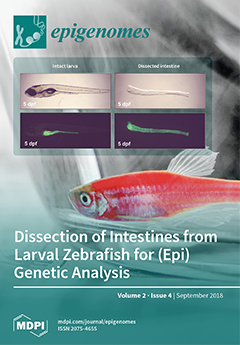
The left panel of this figure shows a five-day-old zebrafish larva that expresses GFP specifically in the intestine. By using tweezers, the intestine can be removed from the body and non-intestinal tissue can be subsequently removed. The right panel of the figure shows the resulting isolated intestine. This isolated tissue is the starting point to study the genetic and epigenetic regulation of intestinal development. View this paper.
  • Issues are regarded as officially published after their release is announced to the table of contents alert mailing list.
  • You may sign up for e-mail alerts to receive table of contents of newly released issues.
  • PDF is the official format for papers published in both, html and pdf forms. To view the papers in pdf format, click on the "PDF Full-text" link, and use the free Adobe Reader to open them.
Order results
Result details
Select all
Export citation of selected articles as:
4 pages, 176 KiB  
Editorial
Non-CpG Methylation Revised
by Andrea Fuso
Epigenomes 2018, 2(4), 22; https://doi.org/10.3390/epigenomes2040022 - 12 Dec 2018
Cited by 8 | Viewed by 4191
Abstract
Textbook and scientific papers addressing DNA methylation usually still cite “DNA methylation occurs at CpG cytosines”. Methylation at cytosines outside the CpG nucleotide, the so-called “non-CpG methylation”, is usually considered a minor and not biologically relevant process. However, the technical improvements and additional [...] Read more.
Textbook and scientific papers addressing DNA methylation usually still cite “DNA methylation occurs at CpG cytosines”. Methylation at cytosines outside the CpG nucleotide, the so-called “non-CpG methylation”, is usually considered a minor and not biologically relevant process. However, the technical improvements and additional studies in epigenetics have demonstrated that non-CpG methylation is present with frequency higher than previously thought and retains biological activity, potentially relevant to the understanding and the treatment of human diseases. Full article
(This article belongs to the Special Issue Epigenetics of the Nervous System)
11 pages, 1450 KiB  
Review
How to Design a Whole-Genome Bisulfite Sequencing Experiment
by Claudius Grehl, Markus Kuhlmann, Claude Becker, Bruno Glaser and Ivo Grosse
Epigenomes 2018, 2(4), 21; https://doi.org/10.3390/epigenomes2040021 - 11 Dec 2018
Cited by 6 | Viewed by 10697
Abstract
Aside from post-translational histone modifications and small RNA populations, the epigenome of an organism is defined by the level and spectrum of DNA methylation. Methyl groups can be covalently bound to the carbon-5 of cytosines or the carbon-6 of adenine bases. DNA methylation [...] Read more.
Aside from post-translational histone modifications and small RNA populations, the epigenome of an organism is defined by the level and spectrum of DNA methylation. Methyl groups can be covalently bound to the carbon-5 of cytosines or the carbon-6 of adenine bases. DNA methylation can be found in both prokaryotes and eukaryotes. In the latter, dynamic variation is shown across species, along development, and by cell type. DNA methylation usually leads to a lower binding affinity of DNA-interacting proteins and often results in a lower expression rate of the subsequent genome region, a process also referred to as transcriptional gene silencing. We give an overview of the current state of research facilitating the planning and implementation of whole-genome bisulfite-sequencing (WGBS) experiments. We refrain from discussing alternative methods for DNA methylation analysis, such as reduced representation bisulfite sequencing (rrBS) and methylated DNA immunoprecipitation sequencing (MeDIPSeq), which have value in specific experimental contexts but are generally disadvantageous compared to WGBS. Full article
(This article belongs to the Special Issue Plant Epigenetics)
Show Figures

Figure 1

18 pages, 4938 KiB  
Article
On the Cooperation between Epigenetics and Transcription Factor Networks in the Specification of Tissue Stem Cells
by Torsten Thalheim, Lydia Hopp, Hans Binder, Gabriela Aust and Joerg Galle
Epigenomes 2018, 2(4), 20; https://doi.org/10.3390/epigenomes2040020 - 23 Oct 2018
Cited by 13 | Viewed by 4590
Abstract
It is generally accepted that epigenetic modifications, such as DNA and histone methylations, affect transcription and that a gene’s transcription feeds back on its epigenetic profile. Depending on the epigenetic modification, positive and negative feedback loops have been described. Here, we study whether [...] Read more.
It is generally accepted that epigenetic modifications, such as DNA and histone methylations, affect transcription and that a gene’s transcription feeds back on its epigenetic profile. Depending on the epigenetic modification, positive and negative feedback loops have been described. Here, we study whether such interrelation are mandatory and how transcription factor networks affect it. We apply self-organizing map machine learning to a published data set on the specification and differentiation of murine intestinal stem cells in order to provide an integrative view of gene transcription and DNA, as well as histone methylation during this process. We show that, although gain/loss of H3K4me3 at a gene promoter is generally considered to be associated with its increased/decreased transcriptional activity, such an interrelation is not mandatory, i.e., changes of the modification level do not necessarily affect transcription. Similar considerations hold for H3K27me3. In addition, even strong changes in the transcription of a gene do not necessarily affect its H3K4me3 and H3K27me3 modification profile. We provide a mechanistic explanation of these phenomena that is based on a model of epigenetic regulation of transcription. Thereby, the analyzed data suggest a broad variance in gene specific regulation of histone methylation and support the assumption of an independent regulation of transcription by histone methylation and transcription factor networks. The results provide insights into basic principles of the specification of tissue stem cells and highlight open questions about a mechanistic modeling of this process. Full article
Show Figures

Graphical abstract

23 pages, 5553 KiB  
Article
Genetic and Epigenetic Regulation of Zebrafish Intestinal Development
by Bilge San, Marco Aben, Dei M. Elurbe, Kai Voeltzke, Marjo J. Den Broeder, Julien Rougeot, Juliette Legler and Leonie M. Kamminga
Epigenomes 2018, 2(4), 19; https://doi.org/10.3390/epigenomes2040019 - 23 Oct 2018
Cited by 13 | Viewed by 6329
Abstract
Many regulatory pathways are conserved in the zebrafish intestine compared to mammals, rendering it a strong model to study intestinal development. However, the (epi)genetic regulation of zebrafish intestinal development remains largely uncharacterized. We performed RNA-sequencing and chromatin immunoprecipitation (ChIP)-sequencing for activating (H3K4me3) and [...] Read more.
Many regulatory pathways are conserved in the zebrafish intestine compared to mammals, rendering it a strong model to study intestinal development. However, the (epi)genetic regulation of zebrafish intestinal development remains largely uncharacterized. We performed RNA-sequencing and chromatin immunoprecipitation (ChIP)-sequencing for activating (H3K4me3) and repressive (H3K27me3) chromatin marks on isolated intestines at 5, 7, and 9 days post-fertilization (dpf), during which zebrafish transit from yolk dependence to external feeding. RNA-sequencing showed the enrichment of metabolic maintenance genes at all time points and a significant increase in lipid metabolism between 5 and 9 dpf. A strong correlation was observed between gene expression and presence of chromatin marks on gene promoters; H3K4me3-marked genes were expressed higher than H3K27m3-marked genes. Next, we studied a key epigenetic player, Enhancer of zeste homolog 2 (Ezh2). Ezh2 places the repressive H3K27me3 mark on the genome and is highly conserved in vertebrates. We used the nonsense mutant allele ezh2(hu5670) to study the effect of ezh2 loss on intestinal development. These mutants survived gastrulation and died around 11 dpf, showing severe morphological defects in the intestine and liver, accompanied by decreased intestinal (fabp2) and hepatic (fabp10a) marker expressions. Our results suggest that Ezh2 is essential for proper intestinal tissue maintenance and overall survival. Full article
(This article belongs to the Special Issue Polycomb and Trithorax Group of Proteins in Development and Disease)
Show Figures

Figure 1

8 pages, 1064 KiB  
Review
The Role of the Prader-Willi Syndrome Critical Interval for Epigenetic Regulation, Transcription and Phenotype
by Simona Zahova and Anthony R. Isles
Epigenomes 2018, 2(4), 18; https://doi.org/10.3390/epigenomes2040018 - 18 Oct 2018
Cited by 3 | Viewed by 5352
Abstract
Prader-Willi Syndrome (PWS) is a neurodevelopmental disorder caused by loss of expression of the paternally inherited genes on chromosome 15q11.2-q13. However, the core features of PWS have been attributed to a critical interval (PWS-cr) within the 15q11.2-q13 imprinted gene cluster, containing the small [...] Read more.
Prader-Willi Syndrome (PWS) is a neurodevelopmental disorder caused by loss of expression of the paternally inherited genes on chromosome 15q11.2-q13. However, the core features of PWS have been attributed to a critical interval (PWS-cr) within the 15q11.2-q13 imprinted gene cluster, containing the small nucleolar RNA (snoRNA) SNORD116 and non-coding RNA IPW (Imprinted in Prader-Willi) exons. SNORD116 affects the transcription profile of hundreds of genes, possibly via DNA methylation or post-transcriptional modification, although the exact mechanism is not completely clear. IPW on the other hand has been shown to specifically modulate histone methylation of a separate imprinted locus, the DLK1-DIO3 cluster, which itself is associated with several neurodevelopmental disorders with similarities to PWS. Here we review what is currently known of the molecular targets of SNORD116 and IPW and begin to disentangle their roles in contributing to the Prader-Willi Syndrome phenotype. Full article
Show Figures

Figure 1

Previous Issue
Next Issue
Back to TopTop