Article

Impact of Isolated Exercise-Induced Small Airway Dysfunction on Exercise Performance in Professional Male Cyclists

by
Konstantinos M. Pigakis
1,2,*,
Vasileios T. Stavrou
2,
Aggeliki K. Kontopodi
1,
Ioannis Pantazopoulos
3,
Zoe Daniil
2,4 and
Konstantinos Gourgoulianis
2,4
1
Department of Respiratory & Critical Care Medicine, Creta Interclinic, 71304 Heraklion, Greece
2
Laboratory of Cardiopulmonary Testing and Pulmonary Rehabilitation, Faculty of Medicine, University of Thessaly, 41110 Larissa, Greece
3
Department of Emergency Medicine, Faculty of Medicine, University of Thessaly, 41110 Larissa, Greece
4
Department of Respiratory Medicine, Faculty of Medicine, University of Thessaly, 41110 Larissa, Greece
*
Author to whom correspondence should be addressed.
Sports 2024, 12(4), 112; https://doi.org/10.3390/sports12040112
Submission received: 21 March 2024 / Revised: 13 April 2024 / Accepted: 17 April 2024 / Published: 19 April 2024

Highlights

What are the main findings?
  • Elite athletes may experience nonbeneficial effects on their respiratory system. This research aimed to examine the impact of exercise on small airways and to establish whether isolated exercise-induced small airway dysfunction hurts exercise performance.
  • It is imperative to recognize that the small airways are affected in isolation or in combination with the reduction in forced expiratory volume at the first second (FEV1). This understand-ing is crucial for the accurate diagnosis and effective treatment of respiratory symptoms of elite athletes.
  • Athletes with exercise-induced isolated small airway dysfunction have a lower exercise ca-pacity (VO2 max).
What is the implication of the main finding?
  • Isolated exercise-induced small airway dysfunction may be considered an indicator of exercise-induced bronchoconstriction in professional athletes.
  • The detection and diagnosis of small airway dysfunction are important because treatment may reverse the remodeling of small airways in professional athletes and improve their exercise capacity.
  • However, there are many questions about nonbeneficial effects on respiratory function in professional endurance athletes. Because, in most cases, elite athletes with affected respiratory function are asymptomatic, consideration should be given to systematic control of the respiratory system, such as through regular cardiological control.

Abstract

:
Background: Professional cycling puts significant demands on the respiratory system. Exercise-induced bronchoconstriction (EIB) is a common problem in professional athletes. Small airways may be affected in isolation or in combination with a reduction in forced expiratory volume at the first second (FEV1). This study aimed to investigate isolated exercise-induced small airway dysfunction (SAD) in professional cyclists and assess the impact of this phenomenon on exercise capacity in this population. Materials and Methods: This research was conducted on professional cyclists with no history of asthma or atopy. Anthropometric characteristics were recorded, the training age was determined, and spirometry and specific markers, such as fractional exhaled nitric oxide (FeNO) and immunoglobulin E (IgE), were measured for all participants. All of the cyclists underwent cardiopulmonary exercise testing (CPET) followed by spirometry. Results: Compared with the controls, 1-FEV3/FVC (the fraction of the FVC that was not expired during the first 3 s of the FVC) was greater in athletes with EIB, but also in those with isolated exercise-induced SAD. The exercise capacity was lower in cyclists with isolated exercise-induced SAD than in the controls, but was similar to that in cyclists with EIB. This phenomenon appeared to be associated with a worse ventilatory reserve (VE/MVV%). Conclusions: According to our data, it appears that professional cyclists may experience no beneficial impacts on their respiratory system. Strenuous endurance exercise can induce airway injury, which is followed by a restorative process. The repeated cycle of injury and repair can trigger the release of pro-inflammatory mediators, the disruption of the airway epithelial barrier, and plasma exudation, which gradually give rise to airway hyper-responsiveness, exercise-induced bronchoconstriction, intrabronchial inflammation, peribronchial fibrosis, and respiratory symptoms. The small airways may be affected in isolation or in combination with a reduction in FEV1. Cyclists with isolated exercise-induced SAD had lower exercise capacity than those in the control group.

1. Introduction

Cycling is considered one of the most challenging endurance sports [1,2,3]. Competitive cycling is extremely demanding, and it is known that significant pressure is exerted on the respiratory system during periods of high-intensity exercise [4,5], routinely exposing one to multiple environmental conditions, variations in temperature and humidity, aeroallergens, and particles resulting from the combustion process that takes place in engines. Competitive cycling may lead to airway alterations, which are prevalent in up to one in five elite endurance athletes [6,7,8].
Many professional cyclists frequently experience a feeling of shortness of breath, end-race coughing, and a sense of having “smaller lungs” during or after competition. Some studies support that lung function can worsen following endurance sporting events [3,9]. These studies aimed to examine exercise-induced bronchoconstriction (EIB) in athletes and determine whether changes in FEV1 are linked with respiratory symptoms, training volume, race time, and exercise capacity. In a first study [9], participants’ pulmonary function was assessed before a race, 8–10 min after the race (post-test 1), and the day after the race (post-test 2). The results showed that 46% of the participants had EIB at post-test 1 and 28% had EIB at post-test 2. The criterion for EIB diagnosis was a decrease in FEV1 of ≥10% after exercise. A second study [3] aimed to examine the impact of systemic hydration on lung function and to determine whether it could reverse dehydration-induced changes in lung function. This research was conducted among professional cyclists without a prior history of asthma or atopy. The lung function of participants was assessed before a cardiopulmonary exercise test (CPET) and at multiple intervals (at the 3rd, 5th, 10th, 15th, and 30th min) following the CPET. According to the criteria for EIB, i.e., a reduction in FEV1 of ≥10% after exercise [10], 30% of the participants presented with EIB in post-exercise spirometry.
Exercise-induced bronchoconstriction refers to transient airway contraction following intense exercise, even in individuals with no prior history of asthma [11,12]. This condition is common among elite athletes and may limit their performance [12,13,14,15,16], suggesting that environmental factors play a more important role than genetic factors. Environmental factors may also have a supplementary impact on the underlying genetic tendency toward bronchoconstriction and may be independent and significant etiological factors [12]. EIB is often classified as either EIB with asthma (EIB in asthmatic individuals) or EIB without asthma (EIB activated by exercise in individuals without other symptoms of asthma) [12]. EIB affects 20% to 50% of professional athletes, especially those who engage in high-intensity aerobic activity [12,17,18]. Hyperventilation [19] and a hyperosmotic environment in the airway are the primary triggers for EIB [19,20]. In professional athletes, hyperventilation can result in airway epithelial damage and the release of special mediators that activate bronchoconstriction [11,21,22,23]. Some researchers have demonstrated that training in endurance sports may not only boost bronchoconstriction, but can also lead to airway hyper-reactivity outside of training and exercise conditions or to persistent airway remodeling [24,25,26].
The epithelium of small airways appears to be the most vulnerable to damage and repair, as observed in mouse studies [16]. Exercise can affect the small airways alone, without the classical diagnosis of EIB, that is, a reduction of ≥10% in FEV1 after exercise [3]. The impact of isolated SAD on exercise performance among athletes without EIB is not yet fully understood, as it is difficult to comprehend due to the rarity of subjects presenting with isolated small airway dysfunction. This has caused some to refer to the small airways as the ‘quiet zone’ [27]. The small airways are peripheral airways that are localized from the seventh or eighth generation of airways to the bronchioles, with an inside diameter of less than 2 mm. Their walls mostly lack cartilage, their entire cross-sectional area is large, and their airflow velocity is slow. As such, small airways are more vulnerable to injury due to toxic particles, which can cause obstruction [28]. Studies investigating the relationship between small airway dysfunction and exercise capacity have focused on older individuals with lung diseases and tobacco or environmental exposure [29,30], where relationships are confused by the existence of gas exchange and large airway defects and are overstated by the loss of elastic recoil inherent to aging [29,30].
There are various techniques for detecting SAD, yet there is no agreement on the gold standard for diagnosis. Spirometry is the most commonly used method for evaluating pulmonary ventilation. The indicators of forced expiratory flow are the most commonly used to estimate the function of small airways. Expiratory flow-volume loops (EFVLs) help detect asthma [31,32]. Imaging studies have shown robust correlations between EFVL-derived spirometric indices and some measures of airway obstruction [33]. An extensive review of EFVL-derived spirometric indices revealed the severity of obstruction and the size of the airways affected [34]. Forced expiratory flows evaluated at the mid-portion of the expiratory flow-volume loop, namely, FEF50%, FEF75%, and FEF25–75%, were effective in measuring airway obstruction and airflow limitation [35]. It is accepted that SAD is present when two of the above three parameters are lower than 65% of the predicted values [36]. However, the American Thoracic Society does not suggest the application of these indicators for the determination of SAD [36]. The FEF25–75 may be used to detect individuals with early COPD who have clinical symptoms, but whose spirometry has not yet detected the obstruction, recommending that it may be used as a marker of SAD [37]. Additionally, there are various other markers of small airway function with better punctuality. The forced expiratory volume in the third second (FEV3), the FEV3/FVC ratio, and the 1-FEV3/FVC are correlated with air trapping and hyperinflation [38]. The 1-FEV3/FVC is estimated to demonstrate the residual un-exhaled vital capacity at the end of the third second [1 − FEV3/FVC = (FVC − FEV3)/FVC].
Plethysmography is a technique used to measure lung volume, and it cannot be measured via spirometry. Air trapping and hyperinflation assessment are often performed using the evaluation of the residual volume (RV) and RV/TLC (total lung capacity) ratio [39]. Impulse oscillometry (IOS) is a noninvasive method for assessing lung function, and it can be used to assess small airways. High-resolution computed tomography (HRCT) is a technique that offers anatomical information for the airways. However, its use has been restricted due to radiation exposure. Magnetic resonance imaging (MRI) is rarely employed for the respiratory system due to technical restrictions, but hyperpolarized MRI is an area of increasing interest. It has no radiation risk, but it is an expensive method and has only been conducted in research centers.
To date, we have not found any studies examining the occurrence of isolated exercise-induced SAD in athletes who have normal pre-exercise spirometry results and do not meet the post-exercise spirometry criteria for EIB. This study is based on data from earlier research [3]. The study design used in the previous research work is still applicable to this investigation, which has different research questions and results that were not the focus of the first study. The data were analyzed differently in the two studies, and the conclusions are also distinct. The protocol of the previous study divided the methodology into two phases: before and after the hydration of the athletes. The findings of the previous study support that systemic hydration has a positive effect on both VO2 max and pulmonary function in professional cyclists, and it is effective in preventing reductions in their pulmonary function after exercise. In the present study, we used the data obtained from the first phase (before hydration). This research aimed to investigate isolated exercise-induced small airway dysfunction in professional cyclists and assess the impact of this phenomenon on exercise performance in this population.

2. Materials and Methods

This study is based on data from earlier research [3], which were analyzed differently. The protocol of the previous study divided the methodology into two phases: before and after hydration of the athletes. In the present study, we used the data obtained from the first phase (before hydration).
A total of one hundred professional male cyclists (100) were called to participate in this research. All of the participants were used to cycling for more than 15 h every week, with sessions lasting at least 3 to 4 h. They were required to refrain from exercise for 48 h before attending a session and to arrive at the laboratory between 9:00 a.m. and 2:00 p.m. The laboratory had an air purification system (Health-Way Deluxe, DFS Technology-VOC Filter, New York, NY, USA) to ensure that the air was clean and safe. The study protocol was approved by the ethics committee of Creta InterClinic Hospital (registration number: 129/16-09-2020), and all subjects provided consent in accordance with the Helsinki Declaration and the regulations of the European Parliament and Council of the European Union for the collection and use of their data.
This research was carried out from October 2020 to January 2021 and involved members of two professional cycling teams in Crete. The inclusion criteria for the study were as follows: age between 18 and 35 years, absence of a history of asthma as confirmed by medical records and spirometry, and a training age of more than 3 years in the sport. The exclusion criteria included female gender, history of smoking, craniofacial and upper airway abnormalities (with a possibility of incorrect application of the ergospirometry mask), injuries sustained within the last 12 months, anemia (Hb < 13.5 g/dL), seasonal allergies affecting the upper and lower respiratory systems, high levels of FeNO (>25 ppb), and high levels of IgE (>100 UI/mL).
Notably, females were excluded from the research due to physiological differences between the sexes, such as differences in reproductive endocrinology, the menstrual cycle, and hormonal contraceptive use. These factors may affect exercise capacity and disturb the objective accuracy of CPET results.
A detailed medical history was taken from all the participants. All cyclists were free from respiratory infection for at least two weeks before entry into the study. Additionally, a clinical examination that included recording demographic and anthropometric parameters, such as height, body mass, and body mass index (BMI), and the determination of the participants’ training age was conducted. All subjects were tested using spirometry, and specific markers, such as FeNO and IgE, were measured. Finally, the participants underwent a cardiopulmonary exercise test (CPET), followed by spirometry at the 3rd, 5th, 10th, 15th, and 30th minutes after the completion of the CPET.
Body mass (kg) and height (cm) were evaluated following the manual reference for anthropometric standardization [40]. All participants wore light clothes and were barefoot (Table 1).
For the needs of the present study, lung function was assessed via spirometry and flow-volume loop analysis. The study protocol required us to evaluate lung function at regular intervals after CPET (at the 3rd, 5th, 10th, 15th, and 30th minutes). Thus, there was limited time to employ additional diagnostic tests other than spirometry. Pulmonary function was evaluated following the ATS/ERS guidelines [36] on a spirometer (Ergocard Clinical with ExpAir E01-10, Medisoft Group, Namur, Belgium). We recorded the following values: forced vital capacity (FVC), FEV1, FEV3 (forced expiratory volume in 3 s), FEF25–75 (forced mid-expiratory flow), FEF50, FEF75, and the FEV1/FVC and FEV3/FVC ratios.
The cardiopulmonary exercise test was conducted using a cycloergometer (Ergocardr clinical with ExpAir Software, Medisoft Group, Namur, Belgium). The study protocol involved a two-minute rest period, followed by one minute of warm-up pedaling against a minimal load of 20 watts, which progressively increased by 50 watts every three minutes. The indoor circumstances during the CPET were constant, with a temperature of 24–26 °C and a humidity of 45–50%. Athletes were required to wear appropriate sports clothing for cycling, and they were free to have their regular breakfast three hours prior to the test. They were also asked to avoid exercising 48 h before the test. The test duration was until exhaustion, with continuous monitoring of the heart rate, electrocardiogram, oxygen saturation, and blood pressure being recorded every three minutes. The indications for early test termination included myocardial ischemia, complex ventricular premature beats, grade 2 or 3 atrioventricular block, a sudden decrease in blood pressure by more than 20 mmHg, or increased blood pressure (>220/120 mmHg) [12,41]. During the test, oxygen consumption (VO2), carbon dioxide production (VCO2), tidal volume (VT), respiratory rate (RR), and minute ventilation (VE) were directly measured.
The statistical analysis was conducted using IBM SPSS Statistics 26.0 (SPSS Inc., version 26, Chicago, IL, USA). A p-value of less than 0.05 was assumed to indicate statistical significance. Categorical variables are expressed as counts and percentages. For continuous variables, data normality was evaluated using the Kolmogorov–Smirnov test. Relationships between continuous variables were measured using Pearson’s correlation coefficients. A paired-sample t-test was used to estimate the differences in spirometry before and after the CPET. An independent-sample t-test was applied to compare the means of two independent samples. One-way analysis of variance or the Kruskal–Wallis test was used to evaluate differences among the control, isolated exercise-induced SAD, and EIB groups adjusted for age, training age, and BMI to assess the robustness of the associations.

3. Results

One hundred participants (n = 100) were recruited from the professional cycling community. All participants had acceptable spirometry and CPET results and were included in the final assessment. The control group was defined as having normal spirometry after the CPET (ΔFEV1 < 10% after the exercise test), the EIB group was defined as having a percentage drop in FEV1 (ΔFEV1) of ≥10% after the exercise test, and the isolated exercise-induced small airway dysfunction group was defined as having a percentage drop in FEF25–75 (ΔFEF25–75) of ≥25% after the exercise test (Figure 1).
All subjects were young cyclists with a mean age of 27.0 ± 5.0 years, while the median age was 30.0 years. The BMI of the athletes ranged from 21.5 to 26.8 kg/m2 and the mean BMI was 23.8 ± 1.4 kg/m2. The baseline demographics and laboratory results obtained during deployment are listed in Table 1.
The results of baseline spirometry are listed in Table 2. There were no abnormalities in the baseline test. In addition, Table 2 presents the demographic characteristics and spirometric results of the participants by study group after the CPET. The control group was defined as having normal spirometry after the CPET, the EIB group was defined as those meeting the EIB criteria with or without small airway dysfunction, and the isolated exercise-induced small airway dysfunction group was defined as those with isolated small airway dysfunction. The baseline characteristics of the participants before the CPET were compared with the results of the participants in each study group after the CPET. Upon separately comparing each group with the control group, the following data were observed. All post-exercise spirometric values, except for the FVC, FEV1, and FEV1/FVC, were significantly lower in the isolated exercise-induced SAD group than in the controls. In the EIB group, all values, except for the FVC, were significantly lower than those in the controls. There were significant differences in the FEV1, FEV3, FEV1/FVC, 1-FEV3/FVC, and MVV between the exercise-induced ISAD and EIB groups.
Table 3 presents the CPET results among the three groups of participants. The VO2 max was significantly lower in the isolated exercise-induced SAD and EIB groups than in the controls. The VO2 max was lower in the EIB group than in the exercise-induced ISAD group, but the difference was not significant. The anaerobic threshold (VO2 AT%) was also lower in the isolated exercise-induced SAD and EIB groups than in the controls, but, again, there was no significant difference between the two groups. There were no significant differences in the O2-pulse (VO2/HR) between the groups. The minute ventilation (VE) was significantly greater in both the SAD and EIB groups than in the controls, with the EIB group having a slightly greater VE than that of the SAD group. The ventilatory equivalent for carbon dioxide (VE/VCO2 ratio) was greater in the EIB group than in the control group. The difference was statistically significant. However, the VE/VCO2 ratio was also greater in the SAD group than in the control group, but the difference was not significant. Between the SAD and EIB groups, the VE/VCO2 ratio was significantly greater in the EIB group. The ventilatory reserve (VE/MVV%) was significantly greater in both the SAD and EIB groups than in the controls. However, the VE/MVV% was significantly greater in the EIB group than in the SAD group. The respiratory exchange ratio (RER) was significantly lower in the SAD and EIB groups than in the controls. Moreover, the RER was significantly lower in the EIB group than in the SAD group, but the difference was not significant (Table 3).
Using the controls as the reference group, the VO2 max was compared between the groups. The data were adjusted for age, BMI, and training age. Multiple linear regression analysis revealed that the VO2 peak was lower in the ISAD and the EIB groups (Table 4).

4. Discussion

This study has two novel findings. Firstly, participants without EIB (who did not meet the spirometric criteria for EIB) but with isolated exercise-induced small airway dysfunction presented lower exercise capacity than those in the controls. However, their exercise performance was similar to that of the EIB group. Secondly, lower exercise capacity in the group with isolated small airway dysfunction appeared to be associated with a poorer ventilatory reserve (VE/MVV%) compared with that of the control group.
Post-exercise spirometry revealed that cyclists in the ΕΙΒ group had significantly lower FEV1 values than those of cyclists in the control and SAD groups. Although their FEV1 values were within the normal range, cyclists with ISAD had poorer exercise capacity and breathing reserves during exercise than those of the controls.
Professional athletes train every day to stay in shape and build endurance. However, they may experience a decline in exercise performance despite their hard work due to overtraining, which occurs when they exercise too much without enough recovery time between workouts. This can lead to overtraining syndrome or burnout, where athletes experience a plateau or decline in athletic performance. Continuing to work while experiencing overtraining syndrome can cause more damage to the body and lengthen the necessary recovery time. Recovery from overtraining requires rest and limiting or even stopping training for a determined period. Prevention of overtraining syndrome should be considered by all who interact with endurance athletes. Unfortunately, there is no definitive biomarker available to differentiate functional overreaching from nonfunctional overreaching or nonfunctional overreaching from overtraining. Healthcare providers must still rule out other medical conditions. The differential diagnosis of underperformance, among other things, should also include a thorough examination of the respiratory system to rule out conditions that lead to exercise underperformance. EIB is a serious condition in professional endurance athletes. The symptoms of EIB in this population are nonspecific, and the diagnosis must be established with specific tests [12]. The differential diagnosis of EIB is wide. Many professional athletes with EIB have no history of asthma, indicating that in these cases, environmental agents are more significant than genetic factors. Many investigators have suggested defining EIB with asthma as the manifestation of bronchoconstriction after exercise in asthmatic athletes and EIB without asthma as the manifestation of bronchoconstriction caused by physical activity in individuals without other symptomatology of asthma [12]. Exercise-induced laryngeal dysfunction (EILD) can often present with respiratory symptoms. EILD can appear in isolation or can co-occur with EIB [12]. Gastroesophageal reflux disease and laryngopharyngeal reflux can also be exacerbated by physical activity and imitate respiratory symptoms [12]. Our study is the first in the literature to address small airway dysfunction caused by functional overtraining and how this dysfunction can negatively impact exercise capacity and athletic performance. The small airways seem to be impacted in isolation or in association with a reduction in FEV1. Individuals with isolated exercise-induced SAD have a lower exercise capacity. Accurate recognition and early identification of SAD are important because treatment may be able to reverse airway remodeling in athletes and improve their exercise capacity. Clinicians should consider, among other things, the possibility that poor physical condition in endurance athletes may be due to a pathology of the peripheral airways to prevent a misdiagnosis of overtraining, which could have negative consequences on the athlete’s career due to the necessity of abstention from the sport for a long time, leading to a loss of training benefits.
In this research, we demonstrated that the fraction of FVC that does not expire at the end of the first three seconds of the FVC (1-FEV3/FVC) was significantly greater in both EIB athletes and those with ISAD than in the controls.
The earliest alteration in airflow limitation is a decrease in the final fragment of the spirogram, while the initial fragment is weakly affected [42]. Later fractions of forced exhalation, i.e., those after FEV1, such as FEV3, were suggested to define declines in terminal expiratory flow [43,44]. The FEV3 and FEV3/FVC have been used in recent years [45,46,47]. Later, the concept of the 1-FEV3/FVC was introduced as an assessment of the late expiratory fraction in smokers and never-smokers [48]. Our research suggests that this parameter can be employed to evaluate the occurrence of hyperinflation and air trapping in cases where lung volumes cannot be estimated. This was demonstrated in our study, where measurements had to be taken in a very short time after one another.
According to one study, the 1-FEV3/FVC is a better measure of airflow limitation than the FEF25–75, as false-negative and false-positive test results might often occur when using the FEF25–75 [44]. However, in our study, we found that the FEF25–75 had a strong correlation with the 1-FEV3/FVC ratio in both the overall population of the research and in the subgroups. A previous study revealed that the FEF25–75 is more strongly correlated with RV/TLC than with the 1-FEV3/FVC, while the 1-FEV3/FVC is more strongly correlated with the RV than with the FEF25–75 [38].
There are no existing studies estimating the impact of ISAD on exercise performance, especially in athletes. Our investigation is the first attempt to research this theme. Previous studies have demonstrated that exercise capacity is lower in smokers who do not have obstructive spirometry than in nonsmokers, and SAD is one of the causes of this finding [49,50]. Our study on cyclists established this finding and showed that athletes with ISAD are at a greater risk for lower exercise performance. Another study on miners revealed that numerous pathological processes in the small airway can be impacted by dust exposure, and this is assumed to be the primary cause of exercise intolerance [29]. Our research expands these results to elite cyclists and shows that athletes without EIB who have ISAD also have lower exercise capacity, regardless of BMI, age, and training age. This finding has implications for the better identification of the respiratory health of athletes and supports the idea that professional endurance athletes may have a nonbeneficial impact on their small airway function. Individuals with small airway dysfunction may experience air trapping during exercise [51,52], which can negatively affect ventilatory reserves and exercise capacity in athletes. Our research confirms this speculation. We found that the ventilatory reserve was lower in athletes with ISAD than in the controls. We also found that the VO2 peak was negatively correlated with the ventilatory reserve in the CPET and with small airway dysfunction in post-exercise spirometry. Consequently, we consider that the lower exercise performance in athletes with ISAD may be related to air trapping during exercise. Our research confirms this speculation. A larger sample size would be needed for the correct estimation of small airway dysfunction in athletes. These ideas require additional study.
We are confident that the lower VO2 peak in the isolated exercise-induced small airway dysfunction group was not the result of decreased motivational trying because the athletes continued to exercise until exhaustion. It is widely recognized that age, BMI [53], and, possibly, training age can affect test performance. Therefore, we corrected for confounding factors after performing a univariable analysis to support our conclusions.
This research has some limitations that should be considered. First, the diagnosis of exercise-induced small airway dysfunction was only based on post-exercise spirometry, which is known to be more variable than other methods. Although the FEF25–75%, FEF50%, and FEF75% are commonly employed to assess SAD, they have some limitations. Other than spirometry, there are several techniques for assessing the function of small airways, including impulse oscillometry, high-resolution computed tomography (HRCT), body plethysmography, and gas washout. Additionally, we must acknowledge that the study included only male participants. This was because the menstrual cycle and fluctuations in female sex hormones can affect athletic performance. Therefore, females were excluded from the study to reduce the variability in the results.

5. Conclusions

The findings of this study indicate that professional cyclists may experience non-beneficial effects on their respiratory system. Strenuous endurance exercise can induce airway injury, which is followed by a restorative process. The repeated cycle of injury and repair can trigger the release of pro-inflammatory mediators, the disruption of the airway epithelial barrier, and plasma exudation, which gradually give rise to airway hyper-responsiveness, exercise-induced bronchoconstriction, intrabronchial inflammation, peribronchial fibrosis, airway remodeling, and respiratory symptoms. The small airways may be affected in isolation or in combination with a reduction in FEV1. Cyclists with isolated exercise-induced SAD had lower exercise capacity than those in the controls. The small airways seem to be impacted independently or in combination with the reduction in FEV1. Individuals with isolated exercise-induced SAD have lower exercise capacity regardless of age, training age, and BMI. The role of small airways in EIB still must be elucidated, and more investigations are required to evaluate whether SAD may be considered a kind of EIB in nonasthmatic and nonatopic professional cyclists. An accurate and early diagnosis of SAD is important because treatment reverses airway remodeling in athletes and improves their exercise capacity. A promising marker of air trapping and hyperinflation is the 1-FEV3/FVC, which can conveniently be determined from spirometric volumes. We recommend using the 1-FEV3/FVC, along with the FEF25–75, FEF50, and FEF75, during spirometry to estimate isolated exercise-induced SAD in athletes. However, future studies involving comparisons among athletes from different endurance sports are warranted to confirm our findings.

Author Contributions

Conceptualization, K.M.P. and K.G.; methodology, K.M.P. and V.T.S.; investigation, K.M.P., V.T.S. and A.K.K.; data curation, K.M.P., V.T.S. and A.K.K.; writing—original draft preparation, K.M.P. and A.K.K.; writing—review and editing, K.M.P. and V.T.S.; supervision, K.G., Z.D. and I.P. All authors contributed to the article and approved the submitted version. All authors have read and agreed to the published version of the manuscript.

Funding

This research received no external funding.

Institutional Review Board Statement

This study was conducted in accordance with the Declaration of Helsinki and was approved by the Ethics Committee of Creta InterClinic General Hospital (registry number: 149/26-09-2022, approval date: 16 September 2020). All participants provided written informed consent for the use of their personal data according to the European Parliament and the Council of the European Union.

Informed Consent Statement

Informed consent was obtained from all subjects involved in the study. Written informed consent has been obtained from the participants to publish this paper.

Data Availability Statement

The data presented in this study are available on request from the corresponding author. The data are not publicly available due to privacy and ethical restrictions.

Acknowledgments

I am overwhelmed, in all humbleness and gratefulness, to acknowledge the depth of my gratitude to all those who have helped me to put these ideas well above the level of simplicity and into something concrete. I would like to express my special thanks and gratitude to Gourgoulianis Konstantinos, who gave me the excellent opportunity to perform this study, which also helped me in performing much research in which I came to learn about so many new things. Any attempt could not be satisfactorily completed without the support of my family, friends, and colleagues. I would like to thank my wife, Kontopodi Aggeliki, who helped me very much in collecting data for this project despite her busy schedule. Secondly, I would like to thank my guides (Vasileios Stavrou, Ioannis Pantazopoulos, and Zoe Daniil), who have always been there by my side. In addition, I would like to thank all of the athletes who participated in the study. Finally, I would like to thank Creta InterClinic General Hospital, a member of the Hellenic Healthcare Group, which helped provide us with the laboratory equipment that we needed.

Conflicts of Interest

The authors declare no conflicts of interest.

References

  1. Allen, H.; Price, O.J.; Greenwell, J.; Hull, J.H. Respiratory impact of a grand tour: Insight from professional cycling. Eur. J. Appl. Physiol. 2021, 121, 1027–1036. [Google Scholar] [CrossRef] [PubMed]
  2. Bell, P.G.; Furber, M.J.; Van Someren, K.A.; Anton-Solanas, A.; Swart, J. The Physiological Profile of a Multiple Tour de France Winning Cyclist. Med. Sci. Sports Exerc. 2017, 49, 115–123. [Google Scholar] [CrossRef] [PubMed]
  3. Pigakis, K.M.; Stavrou, V.T.; Pantazopoulos, I.; Daniil, Z.; Kontopodi-Pigaki, A.K.; Gourgoulianis, K. Effect of Hydration on Pulmonary Function and Development of Exercise-Induced Bronchoconstriction among Professional Male Cyclists. Adv. Respir. Med. 2023, 91, 239–253. [Google Scholar] [CrossRef] [PubMed]
  4. Dominelli, P.B.; Katayama, K.; Vermeulen, T.D.; Stuckless, T.J.R.; Brown, C.V.; Foster, G.E.; Shell, A.W. Work of breathing influences muscle sympathetic nerve activity during semi-recumbent cycle exercise. Acta Physiol. 2019, 225, e13212. [Google Scholar] [CrossRef] [PubMed]
  5. Allen, J.R.; Satiroglu, R.; Fico, B.; Tanaka, H.; Vardarli, E.; Luci, J.; Coyle, E.F. Inertial Load Power Cycling Training Increases Muscle Mass, and Aerobic Power in Older Adults. Med. Sci. Sports Exerc. 2021, 53, 1188–1193. [Google Scholar] [CrossRef] [PubMed]
  6. Price, O.J.; Ansley, L.; Menzies-Gow, A.; Cullinan, P.; Hull, J.H. Airway dysfunction in elite athletes—An occupational lung disease? Allergy 2013, 68, 1343–1352. [Google Scholar] [CrossRef] [PubMed]
  7. Dickinson, J.W.; Whyte, G.P.; McConnell, A.K.; Harries, M.G. Impact of changes in the IOC-MC asthma criteria: A British perspective. Thorax 2005, 60, 629–632. [Google Scholar] [CrossRef] [PubMed]
  8. Dickinson, J.W.; Whyte, G.P.; McConnell, A.K.; Nevill, A.M.; Harries, M.G. Mid-expiratory flow versus FEV1 measurements in the diagnosis of exercise-induced asthma in elite athletes. Thorax 2006, 61, 111–114. [Google Scholar] [CrossRef] [PubMed]
  9. Stensrud, T.; Rossvoll, Ø.; Mathiassen, M.; Melau, J.; Illidi, C.; Ostgaard, H.N.; Hisdal, J.; Stang, J. Lung function and oxygen saturation after participation in Norseman Xtreme Triathlon. Scand. J. Med. Sci. Sports 2020, 30, 1008–1016. [Google Scholar] [CrossRef]
  10. Aggarwal, B.; Mulgirigama, A.; Berend, N. Exercise-induced bronchoconstriction: Prevalence, pathophysiology, patient impact, diagnosis, and management. NPJ Prim. Care Respir. Med. 2018, 28, 31. [Google Scholar] [CrossRef]
  11. Weiler, J.M.; Bonini, S.; Coifman, R.; Craig, T.; Delgado, L.; Capao-Filipe, M.; Passali, D.; Randolph, C.; Storms, W.; Ad Hoc Committee of Sports Medicine Committee of American Academy of Allergy, Asthma & Immunology. American Academy of Allergy, Asthma & Immunology Work Group report: Exercise-induced asthma. J. Allergy Clin. Immunol. 2007, 119, 1349–1358. [Google Scholar] [PubMed]
  12. Pigakis, K.M.; Stavrou, V.T.; Pantazopoulos, I.; Daniil, Z.; Kontopodi, A.K.; Gourgoulianis, K.; Kontopodi, A. Exercise-Induced Bronchospasm in Elite Athletes. Cureus 2022, 14, e20898. [Google Scholar] [CrossRef] [PubMed]
  13. Holzer, K.; Douglass, J.A. Exercise-induced bronchoconstriction in elite athletes: Measuring the fall. Thorax 2006, 61, 94–96. [Google Scholar] [CrossRef] [PubMed]
  14. Greiwe, J.; Cooke, A.; Nanda, A.; Epstein, S.Z.; Wasan, A.N.; Shepard, K.V., 2nd; Capao-Filipe, M.; Nish, A.; Rubin, M.; Gregory, K.L.; et al. Work Group Report: Perspectives in Diagnosis and Management of Exercise-Induced Bronchoconstriction in Athletes. J. Allergy Clin. Immunol. Pract 2020, 8, 2542–2555. [Google Scholar] [CrossRef] [PubMed]
  15. Jones, R.S.; Buston, M.H.; Wharton, M.J. The effect of exercise on ventilatory function in the child with asthma. Br. J. Dis. Chest 1962, 56, 78–86. [Google Scholar] [CrossRef] [PubMed]
  16. Couto, M.; Kurowski, M.; Moreira, A.; Bullens, D.M.A.; Carlsen, K.H.; Delgodo, L.; Kowalski, M.L.; Seys, S.F. Mechanism of exercise-induced bronchoconstriction in athletes: Current perspectives and future challenges. Allergy 2018, 73, 8–16. [Google Scholar] [CrossRef]
  17. Carlsen, K.H.; Anderson, S.D.; Bjermer, L.; Bonini, S.; Brusasco, V.; Canonica, W.; Cummiskey, J.; Delgado, L.; Del Giacco, S.R.; Drobnic, F.; et al. Exercise-induced asthma, respiratory and allergic disorders in elite athletes: Epidemiology, mechanisms, and diagnosis: Part I of the report from the joint task force of the European Respiratory Society (ERS) and the European Academy of Allergy and Clinical Immunology (EAACI) in cooperation with GA2LEN. Allergy 2008, 63, 387–403. [Google Scholar] [PubMed]
  18. Parsons, J.P.; Hallstrand, T.S.; Mastronarde, J.G.; Kaminsky, D.A.; Rundel, K.W.; Hull, J.H.; Storms, W.W.; Welier, J.M.; Cheek, F.M.; Wilson, K.C.; et al. An Official American Thoracic Society Clinical Practice Guideline: Exercise-induced bronchoconstriction. Am. J. Respir. Crit. Care Med. 2013, 197, 1016–1027. [Google Scholar] [CrossRef] [PubMed]
  19. Boulet, L.P.; O’Byrne, P.M. Asthma, and exercise-induced bronchoconstriction in athletes. N. Engl. J. Med. 2015, 372, 641–648. [Google Scholar] [CrossRef]
  20. Messan, F.; Marqueste, T.; Akplogan, B.; Decherchi, P.; Grélot, L. Exercise-Induced Bronchospasm Diagnosis in Sportsmen and Sedentary. Int. Sch. Res. Not. 2012, 2012, 314583. [Google Scholar] [CrossRef]
  21. Bonini, M.; Palange, P. Exercise-induced bronchoconstriction: New evidence in pathogenesis, diagnosis, and treatment. Asthma Res. Pract. 2015, 1, 2. [Google Scholar] [CrossRef] [PubMed]
  22. Fitch, K.D. An overview of asthma and airway hyperresponsiveness on Olympic athletes. Br. J. Sports Med. 2012, 46, 413–416. [Google Scholar] [CrossRef] [PubMed]
  23. Hallstrand, T.S.; Moody, M.W.; Wurfel, M.M.; Schwartz, L.B.; Henderson, W.R., Jr.; Aitken, M.L. Inflammatory basis of exercise-induced bronchoconstriction. Am. J. Respir. Crit. Care Med. 2005, 172, 679–686. [Google Scholar] [CrossRef]
  24. Anderson, S.D.; Holzer, K. Exercise-induced asthma: Is the right diagnosis in elite athletes. J. Allergy Clin. Immunol. 2000, 106, 419–428. [Google Scholar] [CrossRef] [PubMed]
  25. Kippelen, P.; Anderson, S.D. Airway injury during high–level exercise. Br. J. Sports Med. 2012, 46, 385–390. [Google Scholar] [CrossRef] [PubMed]
  26. Zarqa, A.; Norsk, P.; Ulrik, C.S. Mechanism and Management of Exercise Induced Asthma in Elite Athletes. J. Asthma 2012, 49, 480–486. [Google Scholar]
  27. Mead, J. The lungs “quiet zone”. N. Engl. J. Med. 1970, 282, 1318–1319. [Google Scholar] [CrossRef] [PubMed]
  28. Hogg, J.C.; Pare, P.D.; Hackett, T.L. The contribution of small airway obstruction to the pathogenesis of chronic obstructive pulmonary disease. Physiol. Rev. 2017, 97, 529–552. [Google Scholar] [CrossRef]
  29. Petsonk, E.; Stansbury, R.; Beekman-Wagner, L.A.; Long, J.L.; Wang, M.L. Small Airway Dysfunction and Abnormal Exercise Responses. A Study in Coal Miners. Ann. Am. Thorac. Soc. 2016, 13, 1076–1080. [Google Scholar] [CrossRef] [PubMed]
  30. Elbehairy, A.F.; Ciavaglia, C.E.; Webb, K.A.; Guenette, J.A.; Jensen, D.; Mourad, S.M.; Neder, J.A.; O’Donnell, D.E.; Canadian Respiratory Research Network. Pulmonary Gas Exchange Abnormalities in Mild Chronic Obstructive Pulmonary Disease. Implications for Dyspnea and Exercise Intolerance. Am. J. Respir. Crit. Care Med. 2015, 191, 1384–1394. [Google Scholar] [CrossRef]
  31. Sposato, B.; Scalese, M.; Migliorini, M.G.; Di Tomassi, M.; Scala, R. Small airway impairment and bronchial hyperresponsiveness in asthma onset. Allergy Asthma Immunol. Res. 2014, 6, 242–251. [Google Scholar] [CrossRef] [PubMed]
  32. Majak, P.; Cichalewski, L.; Ożarek-Hanc, A.; Stelmach, W.; Jerzyńska, J.; Stelmach, I. Airway response to exercise measured by area under the expiratory flow-volume curve in children with asthma. Ann. Allergy Asthma Immunol. 2013, 111, 512–515. [Google Scholar] [CrossRef] [PubMed]
  33. Schroeder, J.D.; McKenzie, A.S.; Zach, J.A.; Wilson, C.G.; Curran-Everett, D.; Stinson, D.S.; Newell, J.D.; Lynch, D.A. Relationships between airflow obstruction and quantitative CT measurements of emphysema, air trapping, and airways in subjects with and without chronic obstructive pulmonary disease. AJR Am. J. Roentgenol. 2013, 201, W460–W470. [Google Scholar] [CrossRef] [PubMed]
  34. Lutfi, M.F. Review Article Vital capacity derived spirometric measurements. Sudan Med. J. 2012, 48, 86–100. [Google Scholar]
  35. Lutfi, M.F.; Sukkar, M.Y. Reliability of spirometric measurements in assessing asthma severity. Kuwait Med. J. 2010, 3, 433–439. [Google Scholar]
  36. Graham, B.L.; Steenbruggen, I.; Miller, M.R.; Barjaktarevic, I.Z.; Cooper, B.G.; Hall, G.L.; Halstrand, T.S.; Kaminsky, D.A.; McCarthy, K.; McCormack, M.C.; et al. Standardization of Spirometry 2019 Update. An Official American Thoracic Society and European Respiratory Society Technical Statement. Am. J. Respir. Crit. Care Med. 2019, 200, e70–e88. [Google Scholar] [CrossRef] [PubMed]
  37. Morris, Z.Q.; Coz, A.; Starosta, D. An isolated reduction of the FEV3/FVC ratio is an indicator of mild lung injury. Chest 2013, 144, 1117–1123. [Google Scholar] [CrossRef] [PubMed]
  38. Borekci, S.; Demir, T.; Gorek, A.; Uygun, M.; Yildirim, N. A simple measure to assess hyperinflation and air trapping: 1-Forced expiratory volume in three seconds/forced vital capacity. Balkan Med. J. 2017, 34, 113–118. [Google Scholar] [CrossRef]
  39. Li, K.; Gao, Y.; Pan, Z.; Jia, X.; Yan, Y.; Min, X.; Huang, K.; Jiang, T. Influence of emphysema and air trapping heterogeneity on pulmonary function in patients with COPD. Int. J. Chron. Obstruct. Pulmon. Dis. 2019, 14, 2863–2872. [Google Scholar] [CrossRef]
  40. Lohman, T.G.; Roche, A.F.; Martorell, R. Anthropometric Standardization Reference Manual; Human Kinetics Books: Chicago, IL, USA, 1988. [Google Scholar]
  41. Albouaini, K.; Egred, M.; Alahmar, A.; Wright, D.J. Cardiopulmonary exercise testing and its application. Heart 2007, 93, 1285–1292. [Google Scholar] [CrossRef]
  42. Pellegrino, R.; Viegi, G.; Brusasco, V.; Crapo, R.O.; Burgos, F.; Casubari, R.; Coates, A. Interpretative strategies for lung function tests. Eur. Respir. J. 2005, 26, 948–968. [Google Scholar] [CrossRef]
  43. Hansen, J.E. Clinical Function Testing and Interpretation, 1st ed.; Clinical Focus Series; Jaypee Brothers Medical Publishers: London, UK, 2011. [Google Scholar]
  44. Hansen, J.E.; Sun, X.G.; Wasserman, K. Discriminating measures and normal values for expiratory obstruction. Chest 2006, 129, 369–377. [Google Scholar] [CrossRef]
  45. Crapo, R.O.; Morris, A.H.; Gardner, R.M. Reference spirometric values using techniques and equipment that meet ATS recommendations. Am. Respir. Dis. 1981, 123, 659–664. [Google Scholar]
  46. Miller, M.O.; Grove, D.M.; Pinnock, A.C. Time domain spirogram indices: Their variability and reference values in nonsmokers. Am. Rev. Respir. Dis. 1985, 132, 1041–1048. [Google Scholar] [PubMed]
  47. Miller, M.O.; Grove, D.M.; Pinnock, A.C. Patterns of spirometric abnormality in individual smokers. Am. Rev. Respir. Dis. 1985, 132, 1034–1040. [Google Scholar] [PubMed]
  48. US Department of Health and Human Services [DHHS]; National Center for Health Statistics. Third National Health and Nutrition Examination Survey, 1988–1994: NHANES III Raw Spirometry Data File; Centers for Disease Control and Prevention: Hyattsville, MD, USA, 2001. [Google Scholar]
  49. Deng, Z.; Li, X.; Li, C.; Zheng, Y.; Wu, F.; Wanz, Z.; Liu, S.; Tian, H.; Zheng, J.; Peng, J.; et al. Impaired exercise capacity in individuals with non-obstructive small airway dysfunction. J. Thorac. Dis. 2023, 15, 472–483. [Google Scholar] [CrossRef] [PubMed]
  50. Elbehairy, A.; Elbehairy, J.; Guenette, J.; Faisal, A.; Faisal, A.; Casey, E.; Ciavaglia, C.E.; Weeb, K.; Jensen, D.; Ramsook, A.; et al. Mechanisms of exertional dyspnoea in symptomatic smokers without COPD. Eur. Respir. J. 2016, 48, 694–705. [Google Scholar] [CrossRef] [PubMed]
  51. Ofir, D.; Lavenezianna, P.; Webb, K.A.; Lam, Y.M.; O’Donnell, D.E. Mechanisms of dyspnoea during cycle exercise in symptomatic patients with GOLD stage I chronic obstructive pulmonary disease. Am. J. Respir. Crit. Care Med. 2008, 177, 622–629. [Google Scholar] [CrossRef]
  52. Di Marco, F.; Terrano, S.; Job, S.; Rinaldo, R.F.; Sferrazza Papa, G.F.; Roggi, M.A.; Santus, P.; Centanni, S. Cardiopulmonary exercise testing and second-line pulmonary function: Tests to detect obstructive pattern in symptomatic smokers with borderline spirometry. Respir. Med. 2017, 127, 7–13. [Google Scholar] [CrossRef]
  53. American Thoracic Society. American College of Chest Physicians. ATS/ACCP Statement on cardiopulmonary exercise testing. Am. Respir. Crit. Care 2003, 167, 211–277, Erratum in Am. J. Respir. Crit. Care Med. 2003, 1451–1452. [Google Scholar] [CrossRef]
Figure 1. Flowchart illustrating the athlete selection procedure. CPET, cardiopulmonary exercise test; EIB, exercise-induced bronchoconstriction.
Sports 12 00112 g001
Table 1. Demographic, somatometric, and laboratory characteristics of the athletes.
Age, years27.0 ± 5.0
Training age, years12.0 ± 5.0
Body mass index, kg/m223.8 ± 1.4
Hemoglobin, g/dL14.8 ± 1.1
FeNO, ppb11.0 ± 3.0
IgE, UI/mL53.0 ± 7.0
Table 2. Comparison of the demographic, somatometric, and spirometric characteristics of athletes in each study group after the CPET. The data are presented as the mean ± SD. * p < 0.05 vs. the controls.  p < 0.05, EIB group vs. the exercise-induced isolated SAD group.
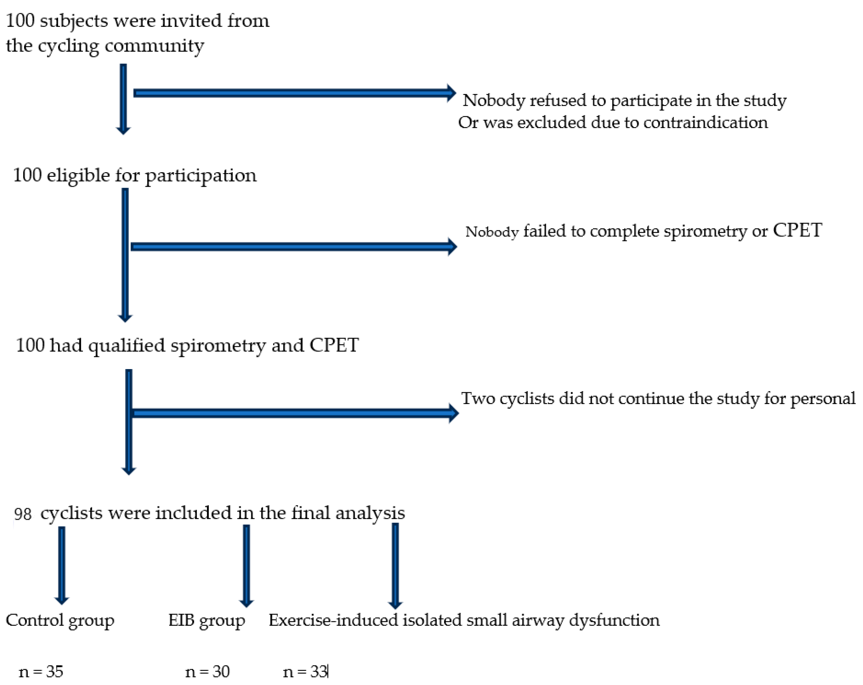
Baseline Characteristics (before the CPET)
n = 100
Control Group (after the CPET)
n = 35
Isolated Exercise-Induced SAD Group (after the CPET)
n = 33
EIB Group (after the CPET)
n = 30
FVC, L6.40 ± 0.66.10 ± 0.76.10 ± 0.706.0 ± 0.72
FVC, %117.1 ± 6.7112.4 ± 8.9112.0 ± 4.4110.0 ± 4.6
FEV1, L5.20 ± 0.44.90 ± 0.54.73 ± 0.474.28 ± 0.47 *
FEV1, %120.0 ± 6.9118.0 ± 12117.0 ± 14109.0 ± 10 *
FEV3, L5.85 ± 0.55.65 ± 0.555.32 ± 0.4 *5.10 ± 0.6 *
FEV3, %120.0 ± 6.5119.0 ± 11.5115.0 ± 8.5 *110.0 ± 9.2 *
FEV1/FVC0.81 ± 0.10.80 ± 0.70.77 ± 0.650.71 ± 0.45 *
FEV3/FVC0.91 ± 0.20.93 ± 0.80.87 ± 0.3 *0.85 ± 0.6 *
1-FEV3/FVC, %8.6 ± 2.37.4 ± 2.512.8 ± 3.2 *15.0 ± 2.8 *
MVV, L182.0 ± 34.0171.5 ± 18.9165.5 ± 16.4 *149.8 ± 15.2 *
FEF25–75, L/s5.0 ± 1.14.40 ± 1.21.96 ± 1.4 *1.76 ± 1.35 *
FEF25–75, %103.1 ± 8.390.8 ± 11.565.0 ± 10 *63.0 ± 9.0 *
FEF50, L/s6.20 ± 0.77.60 ± 0.73.51 ± 0.4 *3.49 ± 0.3 *
FEF50, %112.0 ± 4.5138.0 ± 5.363.0 ± 3.5 *63.0 ± 6.0 *
FEF75, L/s3.30 ± 0.43.47 ± 0.71.62 ± 0.4 *1.58 ± 0.4 *
FEF75, %127.0 ± 8.0134.0 ± 7.063.0 ± 4.0 *61.0 ± 7.0 *
Age, years27.0 ± 5.026.2 ± 5.129.2 ± 6.1 *32.0 ± 4.3 *
BMI, kg/m223.8 ± 1.421.9 ± 3.222.3 ± 3.123.2 ± 3.4
Table 3. CPET results among the three groups of participants. * p < 0.05 vs. the controls.  p < 0.05, EIB group vs. the exercise-induced isolated SAD group.
Overall ControlsSADEIB
VO2 max, mL/kg/min65.0 ± 4.469.0 ± 2.062.0 ± 2.3 *61.0 ± 2.35 *
Respiratory exchange ratio (RER)1.20 ± 0.11.22 ± 0.11.18 ± 0.09 *1.15 ± 0.11 *
VO2 AT% predicted VO2 max, %72.4 ± 5.275.5 ± 6.872.0 ± 2.870.0 ± 6.15
O2-pulse (VO2/HR), mL/beats/min19.2 ± 2.519.5 ± 2.719.2 ± 2.218.9 ± 2.5
VE, L/min120.0 ± 26.0113.0 ± 28122.0 ± 27 *125.0 ± 27.5 *
VE/VCO2 ratio (ventilatory efficiency)27.5 ± 1.326.5 ± 1.027.0 ± 1.529.0 ± 1.5 *
Respiratory reserve (VE/MVV, %)64.0 ± 1.362.0 ± 1.074.0 ± 2.0 *83.0 ± 1.0 *
Table 4. Multivariate linear analysis of exercise capacity across the groups.
VariablesControl GroupISAD GroupEIB Group
Β (95% CI)pΒ (95% CI)p
VO2 peakRef. −2.4 (−6.2 to −1.4)0.025−4.0 (−7.7 to −0.2)0.039
Disclaimer/Publisher’s Note: The statements, opinions and data contained in all publications are solely those of the individual author(s) and contributor(s) and not of MDPI and/or the editor(s). MDPI and/or the editor(s) disclaim responsibility for any injury to people or property resulting from any ideas, methods, instructions or products referred to in the content.

