Article

Zinc Deprivation as a Promising Approach for Combating Methicillin-Resistant Staphylococcus aureus: A Pilot Study

by
Yomna A. Elhakim
1,
Amal E. Ali
2,
Alaa El-Dien M. S. Hosny
1,3 and
Nourtan F. Abdeltawab
1,*
1
Department of Microbiology and Immunology, Faculty of Pharmacy, Cairo University, Cairo 11562, Egypt
2
Department of Microbiology and Immunology, Faculty of Pharmacy, Future University in Egypt, New Cairo 12311, Egypt
3
Department of Microbiology and Immunology, Faculty of Pharmacy, Modern University for Technology and Information (MTI), Cairo 12055, Egypt
*
Author to whom correspondence should be addressed.
Pathogens 2021, 10(10), 1228; https://doi.org/10.3390/pathogens10101228
Submission received: 20 July 2021 / Revised: 6 September 2021 / Accepted: 15 September 2021 / Published: 23 September 2021
(This article belongs to the Collection New Insights into Bacterial Pathogenesis)

Abstract

:
Methicillin-resistant Staphylococcus aureus (MRSA) infections are a global health burden with an urgent need for antimicrobial agents. Studies have shown that host immune responses limit essential metals such as zinc during infection, leading to the limitation of bacterial virulence. Thus, the deprivation of zinc as an important co-factor for the activity of many S. aureus enzymes can be a potential antimicrobial approach. However, the effect of zinc deprivation on S. aureus and MRSA is not fully understood. Therefore, the current study aimed to dissect the effects of zinc deprivation on S. aureus hemolytic activity and biofilm formation through employing biochemical and genetic approaches to study the effect of zinc deprivation on S. aureus growth and virulence. Chemically defined media (CDM) with and without ZnCl2, was used to assess the effect of zinc deprivation on growth, biofilm formation, and hemolytic activity in methicillin-susceptible S. aureus (MSSA) RN6390 and MRSA N315 strains. Zinc deprivation decreased the growth of RN6390 and N315 S. aureus strains significantly by 1.5–2 folds, respectively compared to the zinc physiological range encountered by the bacteria in the human body (7–20 µM) (p < 0.05). Zinc deprivation significantly reduced biofilm formation by 1.5 folds compared to physiological levels (p < 0.05). Moreover, the hemolytic activity of RN6390 and N315 S. aureus strains was significantly decreased by 20 and 30 percent, respectively compared to physiological zinc levels (p < 0.05). Expression of biofilm-associated transcripts levels at late stage of biofilm formation (20 h) murein hydrolase activator A (cidA) and cidB were downregulated by 3 and 5 folds, respectively (p < 0.05) suggested an effect on extracellular DNA production. Expression of hemolysins-associated genes (hld, hlb, hla) was downregulated by 3, 5, and 10 folds, respectively, in absence of zinc (p < 0.001). Collectively the current study showed that zinc deprivation in vitro affected growth, biofilm formation, and hemolytic activity of S. aureus. Our in vitro findings suggested that zinc deprivation can be a potential supportive anti-biofilm formation and antihemolytic approach to contain MRSA topical infections.

1. Introduction

Staphylococcus aureus is implicated in community-acquired and nosocomial infections posing a health care burden [1,2,3]. Since the discovery of penicillin, S. aureus was one of the pathogens that developed resistance against most newly introduced antibiotics. Staphylococcus aureus was remarkably able to resist a number of antibiotics leading to the development of methicillin-resistant S. aureus (MRSA) and vancomycin-resistant S. aureus (VRSA) [4]. The emergence of resistant strains and their high prevalence worldwide has resulted in the difficult eradication of staphylococcal infections in health care facilities [5,6,7]. Enzymes like proteases, lipases, and nucleases enable S. aureus to obtain nutrients from host tissues to help bacterial growth [8]. Besides enzymes, cytotoxins including the four members of the hemolysins family help S. aureus evade the host immune system [9]. Not only does S. aureus produce toxins, but also it can persist in the human body using its ability of biofilm formation [10]. Cells within biofilm have a higher survival chance due to resistance to sweeping by frictional forces and difficulty of phagocytosis of cells within the biofilm. Moreover, the limitation of antibiotic diffusion into the biofilm community hinders the action of antibiotics [11,12,13].
A promising approach to combat staphylococcal infections is by mimicking the human immune responses that disrupt the ability of bacteria to grow. This includes mechanisms of limiting essential nutrients, a process known as nutritional immunity [14]. For instance, neutrophils can restrict the growth of S. aureus by creating an environment devoid of metals [15]. Utilizing metal ions as cofactors in basic metabolic pathways is essential for the survival of all living organisms whether it is the pathogenic organism or its host [16,17]. During evolution, the common need for these important nutrients directed the host–pathogen relationship into a competition over metal ions. To overcome pathogenic microbes, the host has developed several mechanisms to limit the levels of free metals. Meanwhile, pathogens have developed metal ions acquisition and transport mechanisms to overcome such adverse environments [18,19,20].
Impact of iron limitation on bacterial growth and how bacterial pathogens developed mechanisms to overcome such limitation has been studied [21,22,23]. The feasibility of limiting iron in the supportive treatment and prevention of bacterial infections has been explored. For instance, lactoferrin, an iron-binding glycoprotein, is used in the supportive treatment of Helicobacter pylori infections [24]. Zinc starvation is imposed inside the human immune cells by zinc-binding proteins, e.g., calprotectin and metallothionein, limiting infection [25,26,27]. However, a few studies have focused on dissecting the effects of zinc limitation on bacterial growth and virulence. One study showed that in liver abscesses immune cells deplete zinc and manganese to limit bacterial growth [15]. Another study showed that limiting zinc has an important role in controlling H. pylori infection thus modifying bacterial host interaction [28]. Velasco et al. (2018) suggested a possible impact of zinc on hemolysins activity in Escherichia coli, where zinc deprivation and zinc uptake regulator (zur) were involved in the expression of an alpha-hemolysin virulence factor in clinical isolates of uropathogenic E. coli [29]. However, the effect of zinc on host-pathogen interaction especially in gram-positive microorganisms in specific S. aureus remains not well-studied.
Thus, the main goal of this pilot study was to identify the effect of zinc deprivation on the growth and virulence of S. aureus. Staphylococcus aureus growth, biofilm formation, and hemolysis activity were compared under the zinc physiological range encountered by the bacteria in the human body (7 and 20 µM) and in absence of zinc [30,31,32,33,34]. Zinc concentrations were chosen based on previous studies showing physiologically encountered levels in the body [35,36,37,38,39] and those inside immune cells where calprotectin and other proteins subject bacteria to complete deprivation of zinc [15,25]. Both biochemical and genetic approaches were applied to understand how S. aureus is affected by metal ion starvation mimicking that imposed inside the host immune cells. The current study showed zinc deprivation significantly reduced S. aureus growth, biofilm formation, and hemolysis activity. Thus, zinc deprivation has a potential use in the supportive treatment of S. aureus topical infection. Results of this study can help in understanding the role of zinc metal ions in the pathogenesis of S. aureus, hence provide insight towards designing better preventive approaches targeting multiple facets of this process.

2. Results

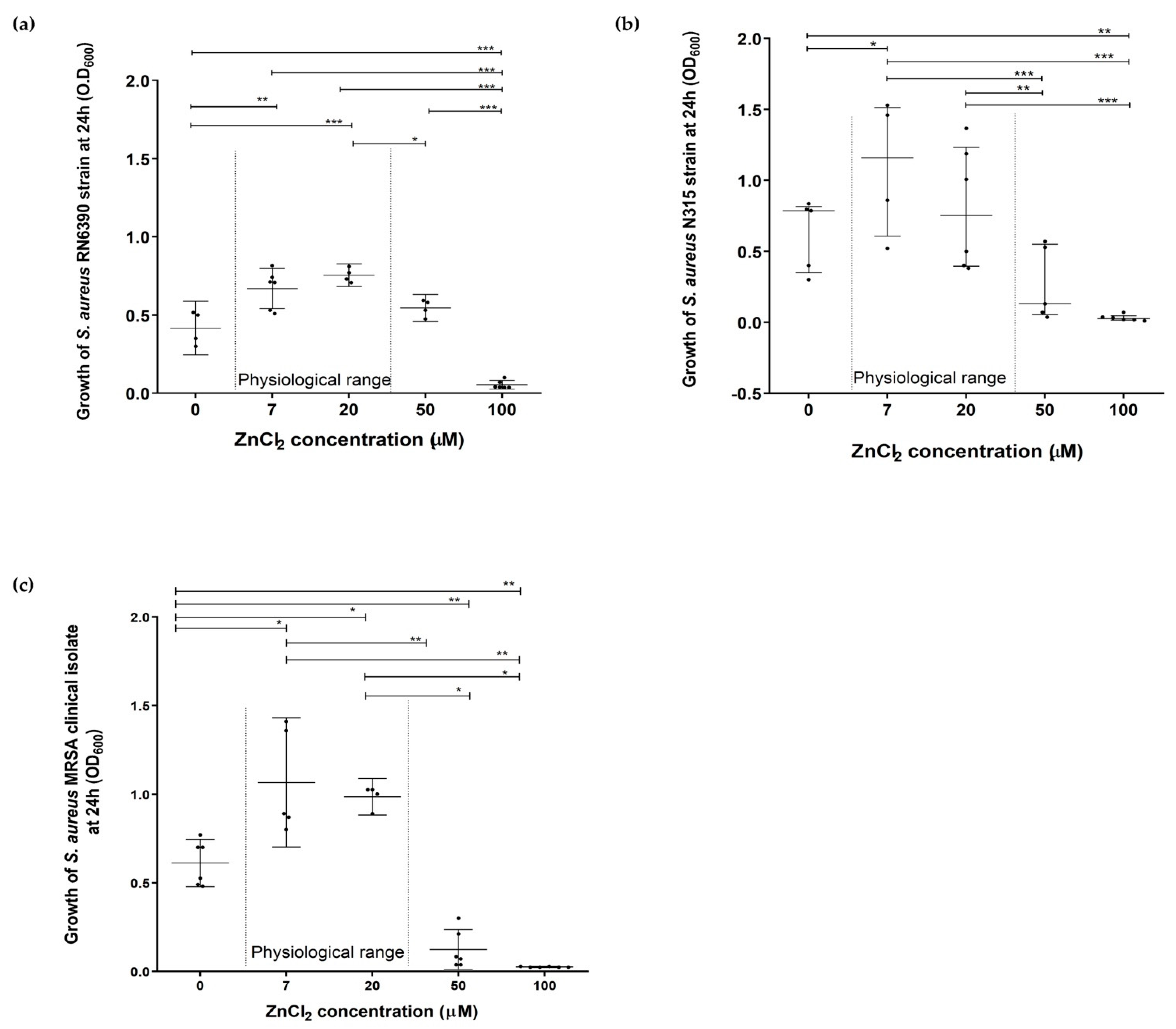
2.1. Assessment of the Effect of Zinc Deprivation on Growth of S. aureus in Chemically Defined Media

The effects of zinc deprivation, high and toxic levels of zinc, 50 and 100 µM, respectively, were compared to the physiological range in humans (7 and 20 µM) on growth patterns of S. aureus RN6390, N315 strains, and MRSA clinical isolate [36,37]. Results showed that zinc deprivation led to a significant decrease in growth of all tested staphylococci at 24 h compared to the physiological range of zinc (7–20 µM) (p < 0.05) (Figure 1 and Supplementary Figure S1). High and toxic zinc levels 50 and 100 µM, respectively also inhibited S. aureus RN6390, N315 and MRSA clinical isolate growth at 24 h where the decrease was significant compared to physiological zinc levels (p < 0.05) (Figure 1 and Supplementary Figure S1). A way the bacterial population responds to adverse conditions to favor the survival of the community is dying and releasing nutrients from lysed cells. To test if this might be occurring, samples of S. aureus N315 strain cultivated in CDM with the corresponding zinc concentration were sampled at 0 and 24 h, respectively. The collected samples from each CDM at these specified time points were analyzed for their zinc content using ICP. The analysis revealed that bacterial inoculation into CDM with no zinc (ICP = 0.007 ppm) showed an increase in zinc content after 24 h incubation to become 4 µM (ICP = 0.564 ppm). Meanwhile, staphylococcal growth in CDM within the physiological range of zinc (ICP = 0.863 ppm equivalent to 6.33 µM) showed no significant change in zinc levels after 24 h incubation becoming 5.91 µM (ICP = 0.806 ppm).

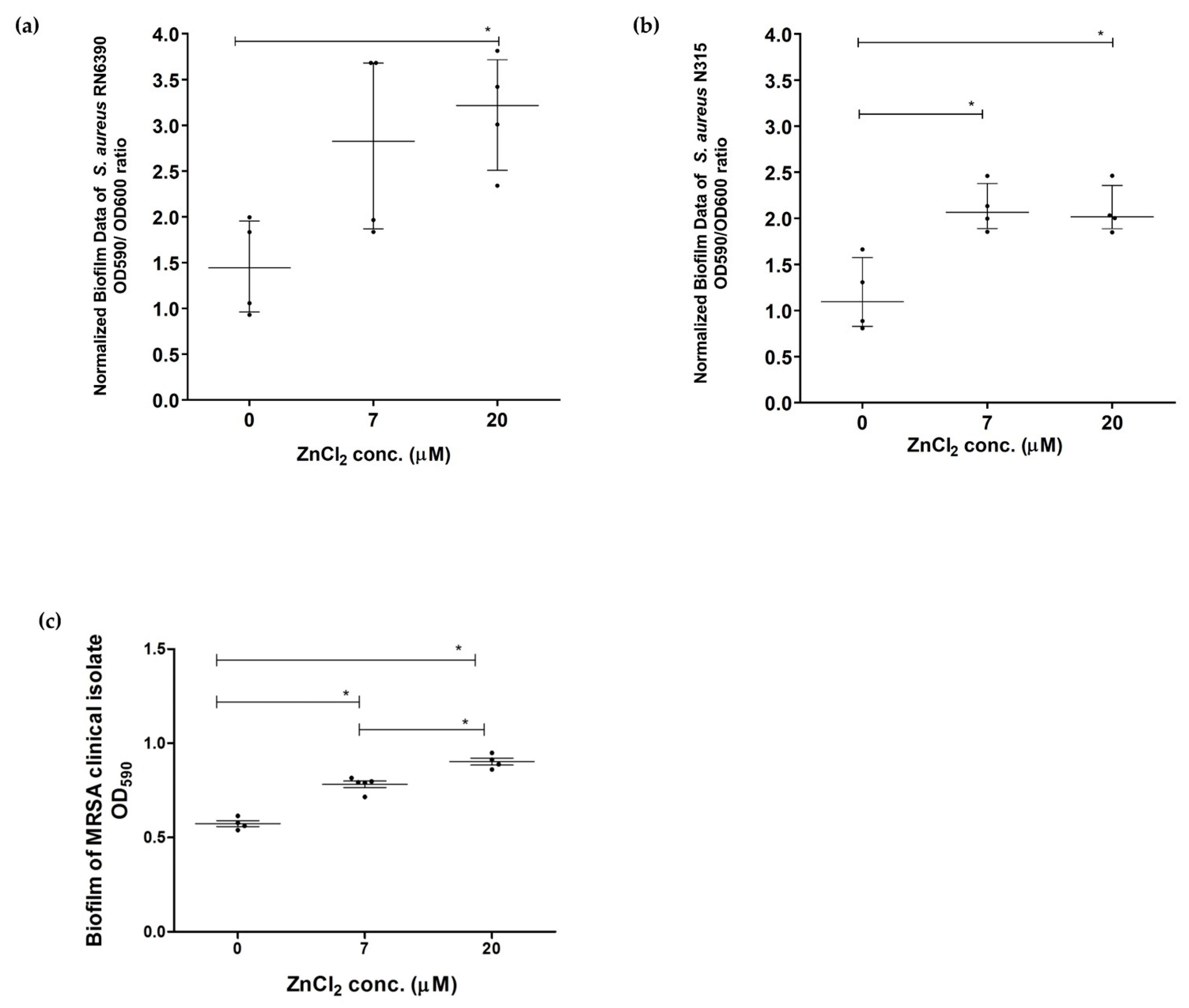
2.2. Assessment of Effect of Zinc Deprivation on Biofilm Formation Ability of S. aureus in Chemically Defined Media Using Crystal Violet Microtiter Plate Method

The ability of S. aureus studied strains to form biofilm was preliminarily tested in CDM, BHI, and tryptone soya broth (TSB) media at different time points representing different biofilm stages. The effect of zinc deprivation was evident at 20 h, thus 20 h was chosen as a time point for further studies. Zinc deprivation significantly inhibited biofilm formation ability in S. aureus RN6390 compared to 20 µM zinc (p < 0.05) normalized to bacterial growth (Figure 2a). Similarly, zinc depletion significantly inhibited biofilm formation in S. aureus N315 compared to physiological range (7–20 µM) normalized to bacterial growth (p < 0.05) (Figure 2b) and MRSA clinical isolate (Figure 2c).

2.3. Assessment of the Effect of Zinc Deprivation on Oxidative Stress and Metalloproteases Activity of S. aureus Exemplified as Catalase Activity and Protease Activity

The effect of zinc on catalase production was examined in overnight cultures of S. aureus N315. Catalase activity was measured as observed effervescence intensity after the addition of 3% hydrogen peroxide (H2O2) to overnight cultures in different zinc concentrations (0, 7, 20, 50, and 100 µM) CDM. Zinc deprivation showed less effervescence than that observed at the physiological range of zinc (7–20 µM) (Figure 3a). While bacterial cultures with zinc concentration 50 µM and higher showed no effervescence (Figure 3a). Next, metalloproteases activity of S. aureus N315 cultivated in CDM with different zinc concentrations on 3% skimmed milk agar was tested. N315 showed bacterial growth on the skimmed milk agar with no effect of zinc deprivation on protease activity (Figure 3b). Similar results were observed for RN6390 strain and MRSA clinical isolate (Figures S2 and S3).

2.4. Determination of the Effect of Zinc Deprivation on S. aureus Hemolytic Activity in Chemically Defined Media

Preliminary hemolytic activity was tested qualitatively on blood agar where S. aureus N315 showed gamma (γ) hemolysis at different zinc concentrations. To precisely examine the effect of zinc deprivation on the hemolytic activity of S. aureus, a quantitative assay for alpha-hemolytic activity was performed according to Bose J. et al. (2014) [40]. The hemolytic activity of S. aureus RN6390 grown under zinc deprivation was reduced even 50% hemolysis was not reached at starting concentration (no dilution) vs. bacteria grown at physiological zinc showed ~70% hemolysis (Table 1). N315 grown in zinc-deprived media showed 50% hemolysis at 2-fold dilution, while in the physiological range of zinc (20 µM), 50% hemolysis was achieved at a dilution of 6-fold (Table 2). This data indicated that S. aureus growing under zinc-deprived conditions showed lower hemolysis activity compared to those growing under physiological zinc range (20 µM) evident by requiring bacteria to be more concentrated for reaching 50% lysis. This might be due to the reduced expression of alpha-hemolysin. At a 16-fold dilution, N315 hemolytic activity was reduced by zinc deprivation (less than 10% hemolysis) compared to ~40% hemolysis for bacteria grown under 20 µM ZnCl2 conditions. In conclusion, the hemolytic activity of S. aureus RN6390 and N315 was reduced by zinc deprivation compared to 20 µM zinc.

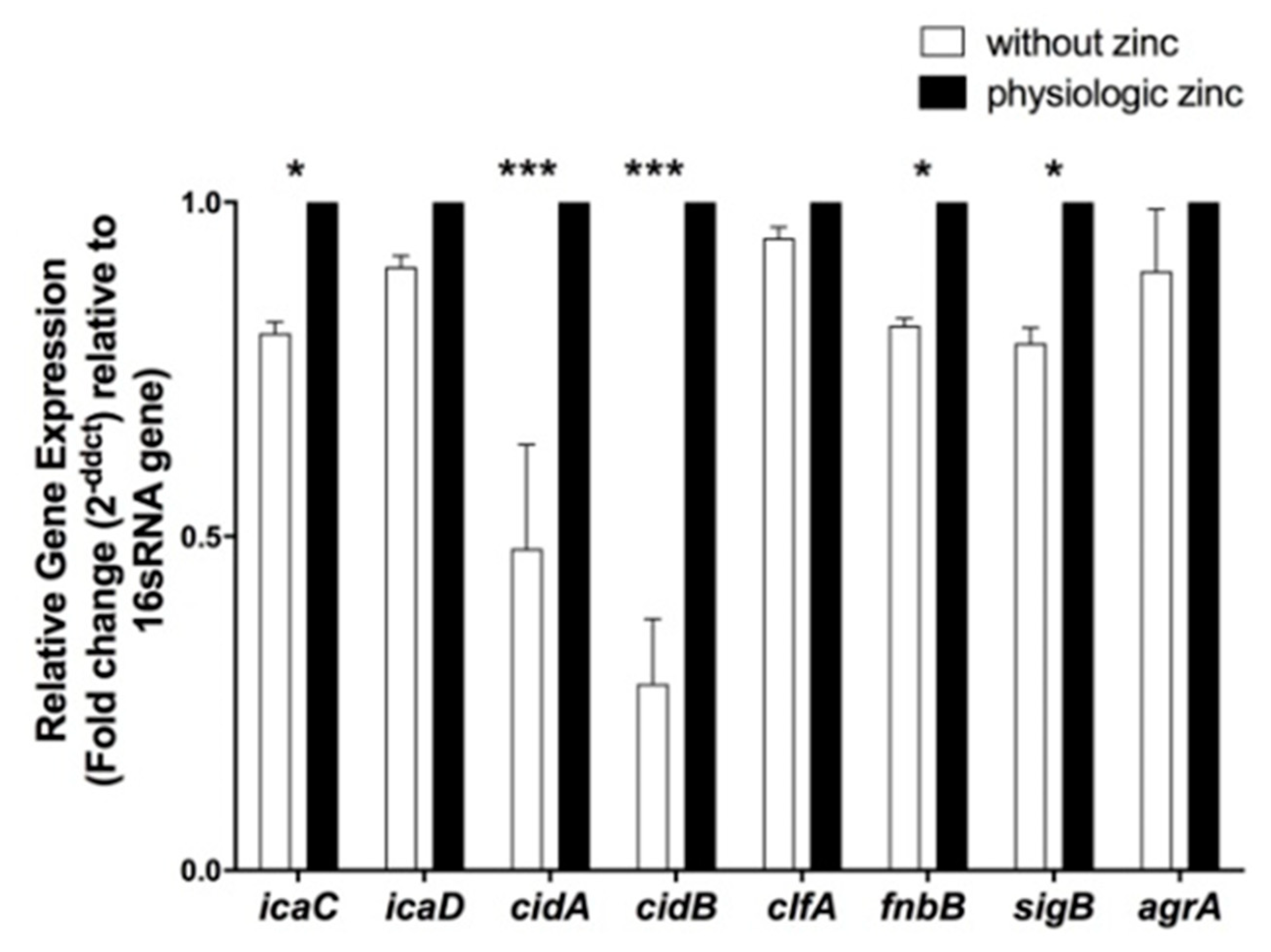
2.5. Determination of the Effect of Zinc Deprivation on Differential Gene Expression of Selected Biofilm Associated Genes in S. aureus N315 under Biofilm Condition

To attempt to understand the genetic basis of biofilm formation was inhibited under zinc-depleted conditions, expression of biofilm-associated genes was examined in biofilm-forming N315 MRSA cells grown under zinc-deprivation at 20 h. Standard MRSA N315 strain was chosen for further molecular studies as it is a representative MRSA strain with complete genome sequence available, unlike MRSA clinical isolate. Moreover, MRSA is currently a bigger health problem than MSSA. Expression of the selected genes was presented as fold change normalized to16s ribosomal RNA (16S rRNA) gene, house-keeping gene normalizer, and relative to the expression to bacteria grown at 20 µM zinc (physiological conditions). Depletion of zinc significantly down-regulated the expression of genes associated with intercellular adhesion revealed that intercellular adhesion molecule C (icaC) by 1.25 folds while icaD expression level showed no significant change (Figure 4). Similarly, the depletion of zinc significantly downregulated the expression of genes associated with extracellular DNA (eDNA) production; murein hydrolase acti-vator A (cidA) and cidB by 3 and 5 folds, respectively (p < 0.05) (Figure 4). Furthermore, the gene associated with surface binding namely fibronectin binding protein B (fnbpB) was significantly downregulated by 1.25 folds, while clumping factor A (clfA) was not affected (Figure 4). The absence of zinc during the late stage of biofilm formation significantly downregulated the positive biofilm regulator sigma factor B (sigB) by 1.25 folds while the negative biofilm regulator accessory gene regulator (agrA) was not affected (Figure 4).

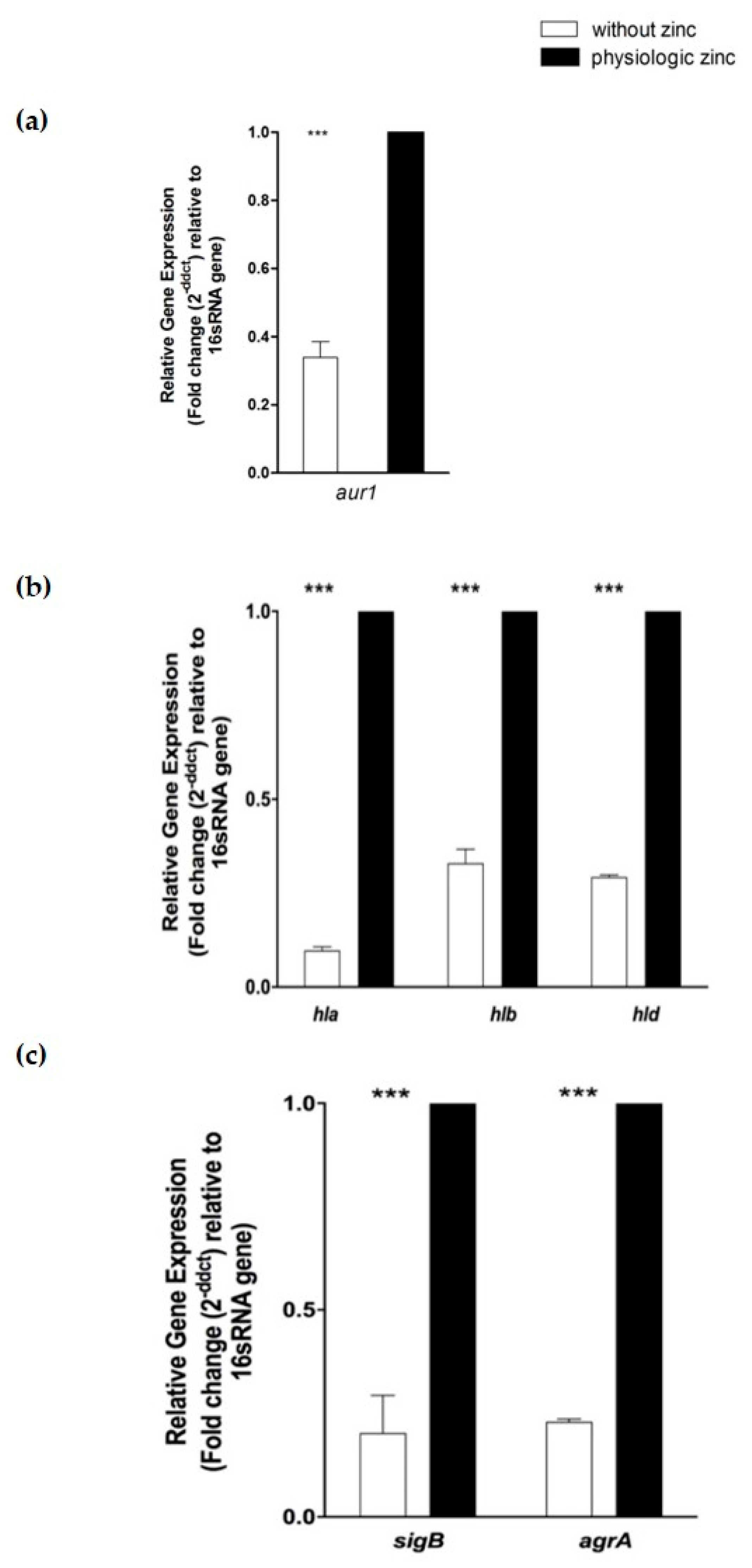
2.6. Determination of the Effect of Zinc Deprivation on Differential Gene Expression of Selected Protease and Hemolysis Associated Genes in S. aureus N315 under Planktonic Condition
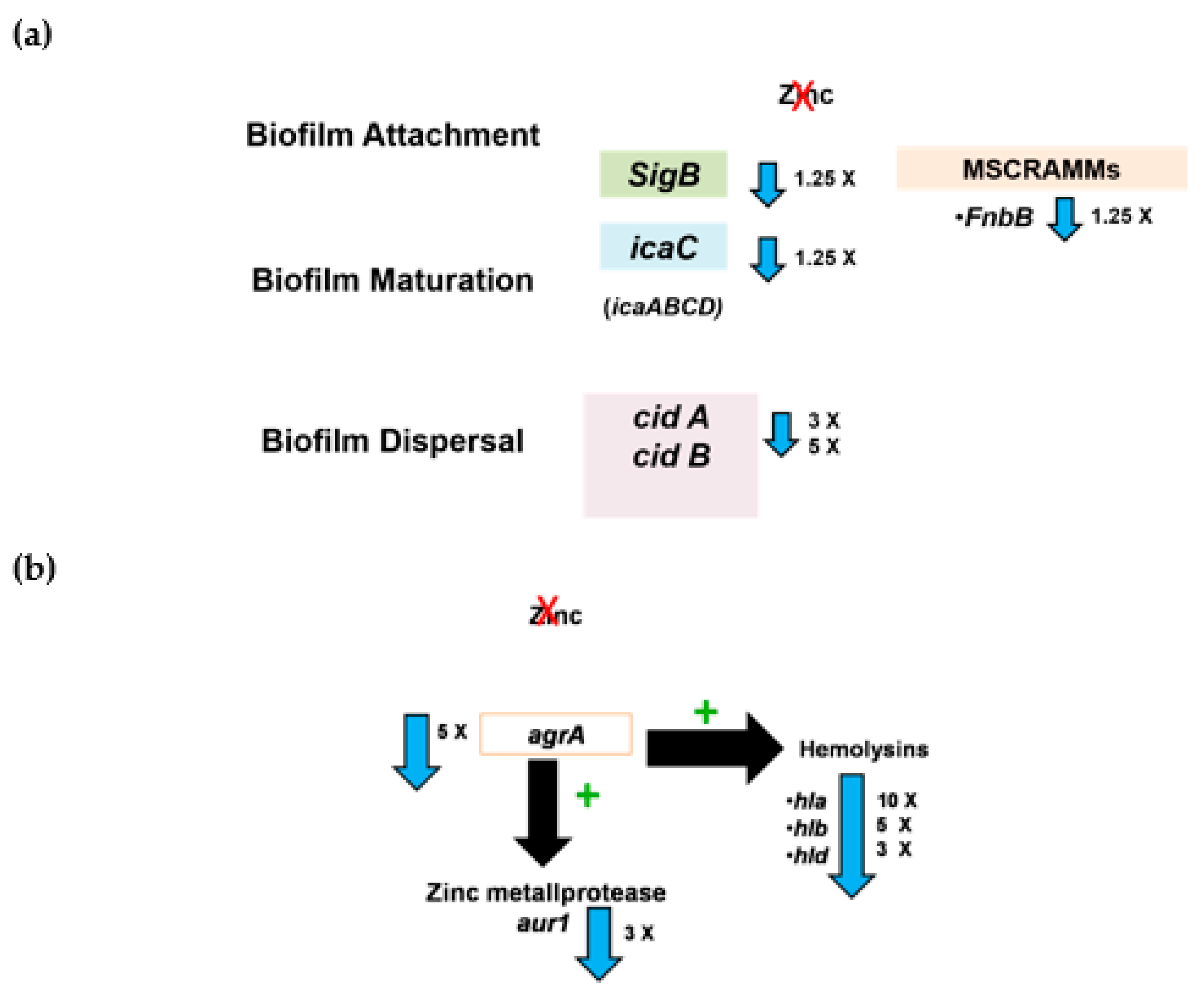
The levels of expression of selected protease- and hemolysis-associated genes were examined without zinc and at 20 µM zinc (physiological conditions) at 24 h under planktonic condition. In addition, genes encoding sigB and agrA, both parts of regulatory systems, were assayed for their expression under the same conditions. Differential gene expression analysis showed that zinc deprivation significantly down-regulated zinc metalloprotease aureolysin (aur1) gene associated with protease activity (p < 0.001) by 3 folds (Figure 5a). In addition, zinc deprivation significantly down-regulated hemolysis associated genes; alpha-hemolysin (hla), beta-hemolysin (hlb), and delta-hemolysin (hld) by 10, 5, and 3 folds respectively (p < 0.001) (Figure 5b). Regulatory genes sigB and agrA were significantly downregulated by 4.3 and 5 folds, respectively, upon zinc deprivation (Figure 5c).

3. Discussion

Staphylococcus aureus remarkably resists many antibiotics from methicillin (MRSA) to vancomycin (VRSA). The emergence of antibiotic-resistant S. aureus strains creates a persistent need for developing new therapeutics for staphylococcal-associated diseases [41]. A promising approach to combat staphylococcal infections is by disrupting its ability to overcome the host immune response, this can be through the use of metal limitation [33]. Among the important metals that play a relevant role during host colonization by bacteria is zinc. Zinc is the second most abundant transition metal cofactor after iron; it is required for many metabolic processes [42]. The importance of zinc metal ion for S. aureus can be targeted as a new weapon for combating staphylococcal infections by limiting its availability. Although it is known that vertebrates sequester zinc, the extent of metal starvation imposed on S. aureus is not well-studied [43].
In the current study, the effect of zinc deprivation on the growth and selected virulence factors of S. aureus N315 and RN6390 strains was studied. To properly study the effect of zinc deprivation, it was crucial to prepare a chemically defined medium (CDM) containing all metals except zinc and another CDM containing the physiologic zinc concentrations. Several studies showed zinc reference ranges needed for homeostasis in the human body in different organs. Studies have shown that physiologically relevant zinc levels encountered by the bacteria in the human body ranged between 7 and 20 µM [35,36,37,38,39]. Studies showed that zinc concentrations below 7 µM had no significant rescuing effect on S. aureus [31,34]. Thus, the used zinc levels in CDM with zinc were chosen to mimic the physiologically relevant concentrations occurring in the human body (7 and 20 µM). CDM without zinc was used to mimic the conditions imposed by the human immune cells on bacteria, where calprotectin and other proteins subject bacteria to complete deprivation of zinc [15,25]. Thus, zinc deprivation was explored in the current study as a possible approach to combating S. aureus infections.
The zinc levels in the prepared CDM without zinc were estimated by zincon colorimetric assay [44]. The zincon assay revealed minute levels of zinc in the prepared CDM using a calibration curve. For accurate determination of zinc concentrations in the prepared CDM with specified zinc concentrations (absence, 7 and 20 µM), the highly sensitive inductively coupled plasma spectrometry (ICP) method was used. ICP is an analytical technique developed to detect elements with atomic mass ranges 7 to 250 available at very low concentrations [35,45]. ICP technology is capable of detecting most of the periodic table of elements at milligram to nanogram levels per liter. Zinc concentration as little as 0.04 ppm could be detected at a wavelength of 481.053 nm [46]. ICP showed that the prepared CDM at the specified zinc concentrations were acceptable (Supplementary Table S1). Based on our results, zincon colorimetric assay was not reliable to assess zinc concentrations, only ICP-MS would be recommended to be used for accurate determination of zinc concentrations.
Bacterial cultures of S. aureus N315 grown at 0 and 24 h in CDM at specified zinc concentrations were tested for changes in zinc content. The results showed that in the case of CDM with no zinc, bacterial growth led to zinc release in the external environment, while bacteria growing in culture media with physiologic zinc concentrations showed no change. This could be due to the released zinc from dead bacteria into the CDM. Our study showed that zinc at 7–20 µM was required for S. aureus growth similar to Lindsay and Foster (2001) study where the addition of 20 µM zinc enhanced the growth of S. aureus 8325-4 in a chemically limited (CL) medium. Lindsay and Foster (2001) showed that growth enhancement is mediated by two ABC transporters, mreA and mreB [31]. Additional studies on other MRSA strains should prove useful as those studies we performed on MRSA isolate showed similar results to those of the N315 strain. Therefore, future studies on other MRSA standard strains would add emphasis to our current findings observed on the widely studied N315 MRSA strain.
Staphylococcus aureus N315 biofilm at later stages (20 h) was significantly inhibited by zinc deprivation compared to physiologic zinc levels (7–20 µM). Gene expression analysis revealed that zinc depletion affected the expression of genes associated with biofilm attachment (fnbpB) and maturation (icaC gene part of icaADBC-encoded polysaccharide intercellular adhesin (PIA)) and extracellular DNA (eDNA) dispersal (cidA and cidB) (summarized in Figure 6a). fnbpB encodes a fibrinogen-binding protein, a prominent member of microbial surface components recognizing adhesive matrix molecules (MSCRAMMs). MSCRAMMs mediate attachment to indwelling devices, bacteria-host adhesion, and bacterial colonization [11,47]. This suggested that zinc depletion affected several steps in biofilm formation, attachment, and maturation in a PIA-dependent manner [48]. Moreover, sigB gene encoding sigma factor B, a regulator of biofilm, was also downregulated. Our finding that zinc deprivation inhibited biofilm formation is similar to previous reports showing that zinc is important for biofilm formation in S. aureus USA300 and SH1000 strains [32,49,50]. Likewise, a derivative of the compound 2-aminobenzimidazole (2-ABI), directly bound to ZnCl2 was found to act as a biofilm inhibitor of MRSA, VRSA, and S. epidermidis biofilms in zinc-dependent mechanism [51]. However, previous studies on zinc deprivation effect on S. aureus biofilm used microbiological media like brain-heart infusion or tryptic soy agar with metal chelators. Here, we used a chemically defined medium to better study the effect of zinc deprivation on biofilm formation in a controlled approach in the presence of other metals. Chemically defined media are of value in studying the minimal nutritional requirements of microorganisms and provide a basis to investigate the requirements of the microbial cell [52]. Therefore, the use of chemically defined media is a must to be able to govern the concentration and hence the availability of zinc in minute amounts mimicking the physiologically relevant concentrations occurring in the human body the organism encounters within the immune system.
Staphylococcus aureus genome encodes three families of proteases: metalloprotease, cysteine, and serine proteases [53]. The metalloproteinase: aureolysin (Aur) is also known as protease III and is the only metalloprotease produced by S. aureus which requires zinc ions for its activity. Aureolysin is activated by auto-proteolytic cleavage initiating a proteolytic cascade of activation of other proteases [54]. The metalloprotease aureolysin plays an important role in staphylococcal inhibition of the complement system and helps S. aureus in immune system evasion through mediating the cleavage of the antimicrobial peptide LL-37 [55,56]. In addition, it contributes to bacterial spread and invasion via activation of the human fibrinolytic system [55]. The important role of zinc metalloproteinase aureolysin in staphylococcal pathogenesis requires further investigation of the impact of zinc sequestering effect on its activity. In the current study, the effect of zinc limitation on protease activity was examined qualitatively on skimmed milk agar plates. This assay could not report the effect of zinc limitation on protease activity in S. aureus. This required further assessment by studying the aureolysin gene expression and the expression of its regulators. Aureolysin is the only metalloprotease produced by S. aureus and requires zinc ions for its activity [54]. This might provide an explanation for the downregulation of aureolysin encoding gene (aur) in the absence of zinc as it has been reported that the activity of aureolysin was inhibited by the metal chelator EDTA, the calcium chelator EGTA and the zinc-specific chelator 1,10-phenanthroline [57]. Two central regulators (accessory gene regulator (agr) and sigB) of metalloproteases operon and cytotoxins were down-regulated suggesting an effect of zinc deprivation on several proteases and hemolysins [58] (summarized in Figure 6b).
Staphylococcus aureus secretes a variety of interacting toxins affecting the plasma membrane of the host cells including human erythrocytes [59]. Our study revealed that zinc deprivation significantly inhibited hemolytic activity of S. aureus N315 and RN6390 and downregulated expression levels of hla, hlb, and hld genes encoding alpha, beta, and delta hemolysins of S. aureus respectively (Figure 6b). Although alpha-hemolysin is one of the most studied of S. aureus cytotoxins [60], there are no studies, to the best of our knowledge, investigating the role of zinc deprivation on S. aureus hemolysins expression and activity. The most relevant study was on the mechanism of Hla-mediated cell lysis via binding to A-disintegrin and metalloprotease 10 (ADAM10); a zinc-dependent metalloprotease trans-membrane protein on human cells. Consequently, it was postulated by Wilke and Wardenburg that the use of zinc metalloprotease inhibitors including ADAM10 might be useful in combating staphylococcal infections [61]. Previous studies suggested a possible impact of zinc on hemolysins activity in different pathogens other than S. aureus. For instance, Clostridium perfringens and E. coli hemolysins induce cell leakage of certain metabolites through stable pore formation in the cells. This leakage was inhibited by the addition of zinc, calcium, and magnesium divalent cations [62,63]. Moreover, Velasco et al. (2018) were the first to describe the involvement of zinc deprivation and zinc uptake regulator (zur) in the expression of an alpha-hemolysin virulence factor in clinical isolates of uropathogenic E. coli [29]. On the contrary, zur has no apparent role in staphylococcal pathogenicity examined using a mouse-abscess model [31].
Studies have shown that zinc starvation can be imposed by the host during infection and is mediated by zinc-binding proteins e.g., calprotectin and metallothionein limiting bacterial viability and virulence [25,26,27]. The feasibility of limiting metal ions in the treatment and prevention of infection has been explored in the use of lactoferrin, which is an iron-binding glycoprotein, in the supportive treatment of H. pylori infections [24], prevention of antibiotic-associated diarrhea and Clostridium difficile infections [64,65] and ulcerative colitis [66]. Moreover, antimicrobial peptides (AMP) as lactoferrin have been used as a coating for dental, bone, and other medically used implants [67]. Therefore, to apply the findings of this study in clinical settings, we propose the use of either zinc-specific chelating agents e.g., 1,10-phenanthroline or diethylenetriaminepentaacetic acid, or recombinant AMP as recombinant calprotectin or metallothionein in the supportive treatment of topical infection. Concerns regarding the effect of zinc deprivation on human cells has been studied previously where human keratinocytes (HaCaT cells) exposed to zinc deficiency induced by TPEN a high-affinity zinc chelator showed no changes in cell viability and growth, or in the cytoskeletal and cell adhesion systems over a period of 7 days [68]. The current study in vitro results suggested that zinc deprivation for 24 to 48 h might be effective as a supportive treatment in topical S. aureus infections. Thus, the duration of zinc deprivation is less than 7 days, after which the induction of apoptosis can be observed [68]. Zinc chelators thus might be for example incorporated in the wound dressings for infected wounds or as a coating for medical devices and implants or as a component of rinses in surgeries around the area of the implant.

4. Materials and Methods

4.1. Bacterial Strains Maintenance and Culturing Conditions

RN6390 and N315 strains were used as standard methicillin-sensitive S. aureus (MSSA) and methicillin-resistant S. aureus (MRSA) strains, respectively, both are well-defined standard strains [69,70]. N315 strain and the used MRSA clinical isolate are both mec-A gene-positive MRSA [70,71]. Moreover, the N315 strain was chosen for further molecular studies as it is a representative MRSA with a complete genome sequence available. Standard strains confirmatory identifications were performed including gram staining, biochemical testing, and antibiotic susceptibility testing by Kirby-Bauer disk diffusion method using methicillin (5 µg), cefoxitin (30 µg), and oxacillin (1 µg) disks [72]. Fresh cultures of RN6390, N315 strains, and MRSA clinical isolate were cultured from −80 °C glycerol stocks by streaking on brain heart infusion (BHI) agar and incubated overnight at 37 °C. Sub-cultures were prepared by transferring three to five colonies into 5 mL of chemically defined medium (CDM) in sterile glass 15 mL tubes and incubated overnight at 37 °C, shaking at 180 rpm. The optical density at 600 nm (OD600nm) of the overnight cultures was adjusted by appropriate dilution with respective medium to 1, followed by 50 times dilution in the media with the corresponding ZnCl2 concentrations (0, 7, 20, 50 µM) in sterile glass test tubes. Then the cultures were incubated at 37 °C and 180 rpm and used for relevant experiments or assay. The clinical MRSA isolate was from a collaborator and was identified by conventional biochemical testing including yellow colonies on mannitol salt agar base, positive coagulase, catalase, and typical S. aureus oxidation-fermentation reactions. Moreover, biochemical testing using an API kit confirmed the isolate to be S. aureus. Antimicrobial susceptibility testing of the MRSA clinical isolate showed that it was resistant to oxacillin, methicillin, and cefoxitin and sensitive to vancomycin and linezolid. Moreover, oxacillin resistance screening agar base (ORSAB) showed blue colonies indicative of oxacillin resistance [71].

4.2. Chemically Defined Medium for Assessment of Zinc Deprivation

Time and effort were put into preparing CDM and troubleshooting the preparation till we finally used modified Vitko and Richardson (2013) [73]. The amounts and order of addition of the medium components in specific the salts and trace elements solutions were according to Taylor and Holland (1989) and Hussain et al. (1991) [30,74] (Table 3). In brief for salt solution preparation, Na2HPO4, K2HPO4, (NH4)2SO4, and MgSO4.7H2O were dissolved in deionized water while MgSO4.7H2O solution was prepared separately as it caused precipitation in the salt solution. For amino acids solutions, each phenylalanine and isoleucine was dissolved separately in 1 M NH4OH. Proline, valine, glycine, threonine, alanine, and serine each was dissolved in deionized water, while the rest of the amino acids were dissolved each separately in 1 N HCl. Each of the bases (U, C, G, A, T) was dissolved separately in deionized water. The vitamins solution was prepared as a stock solution containing thiamine, niacin, biotin, and calcium pantothenate dissolved together in deionized water. All the previous solutions were sterilized individually by autoclaving, while the trace metal element solution was sterilized by filtration. Various trace element solutions were incorporated in the chemically defined medium to test the interactions between nickel, cobalt, and zinc as stated by Remy et al. (2012) [75]. After sterilization, each culture medium was constituted by adding 2 mL of the salt solution, 1 mL of each amino acid, 1 mL of each base, 0.1 mL of the vitamin solution, and 0.1 mL of the trace metal elements solution, 4 mL of 10% glucose solution as a carbon source, and finally MgSO4.7H2O to the beaker while slowly stirring the mixture. The pH of the medium was then adjusted to 7.4 using 10 M NaOH, and the final volume of the medium was brought up to 100 mL using deionized water then the reconstituted medium was filter sterilized.
Zinc concentrations in the CDM containing zinc were chosen to match physiologically relevant levels encountered by bacteria in the human body between 7 and 20 µM [36,37]. In the CDM without zinc, the environment of the immune cells where calprotectin among other proteins completely deprives the bacteria of zinc was mimicked, thus the choice of no zinc in the prepared CDM without zinc.

4.3. Analysis of Zinc Concentrations in the Prepared Chemically Defined Media

Two analytical approaches were used to estimate zinc levels in the prepared CDM; zincon assay and inductively coupled plasma spectrometry (ICP).

4.3.1. Zincon Assay

Colorimetric determination of zinc using zincon assay following Platte and Marcy (1959) method as an analytical estimate of zinc [44]. The assay depends on the use of zincon (2-carboxy-2′-hydroxy-5′-sulfoformazylbenzene) that can form a blue zinc–zincon complex with zinc metal ion. A calibration curve obtained from known amounts of zinc was constructed and it showed the minute amount of zinc in the CDM without zinc (calculated from the calibration curve Supplementary Figure S2).

4.3.2. Inductively Coupled Plasma Spectrometry (ICP)

The second approach for determining zinc levels was the advanced analytical technique of ICP, a highly sensitive analytical technique developed to detect elements available at very low concentrations [35,45]. Samples were analyzed at the Faculty of Agriculture, Ain Shams University using Profile Plus high dispersion ICP, USA. Zinc concentration as little as 0.04 ppm could be detected at a wavelength of 481.053 nm. The amount of ZnCl2 in ppm can be calculated as follows:
1   µ M   ZnCl 2 136.286 × 10 6   g   /   L   ZnCl 2   0.136286   ppm   ZnCl 2
ICP confirmed that each of the prepared CDM was containing the intended amount of zinc (in the form of ZnCl2) as shown in Supplementary Table S1.

4.4. Determination of the Effect of Zinc Deprivation on the Growth of Selected S. aureus Strains Using Chemically Defined Media

Cultures were prepared as detailed under bacterial strains maintenance and culturing conditions and the OD600nm was measured at different time intervals 6, 12, 24, 48 h to choose the suitable time point that better show the effect of zinc limitation on staphylococcal growth.

4.5. Determination of Effect of Zinc Deprivation on the Biofilm Formation of Selected S. aureus Strains Using Crystal Violet Microtiter Plate Assay Method

To test the biofilm-forming ability of S. aureus in CDM, cultures were prepared as detailed under bacterial strains maintenance and culturing conditions then, 200 µL of the diluted samples were added into the wells of a sterile 96 well non-pyrogenic polystyrene culture plate tested as quadrates (four technical replica) for each sample. Chemically defined medium with no zinc represented the negative control. The plates were incubated at 37 °C statically for 12 and 24 h. On the second day, planktonic growth was measured at 600 nm using 96-well plate reader Synergy 2 then the solution was discarded, and the wells were washed with PBS three times and the plate was left to fully dry. Crystal violet was prepared as 0.5% in deionized water and left in a shaker incubator at 37 °C overnight for complete dissolution. For visualizing the adherent cells, 200 µL of 0.5% crystal violet were added to each well and the plate was left static for 30 min. The plate was then washed with sterile deionized water three times then left to completely dry. To measure the optical density of the stained cells, 200 µL of 96% ethanol were added to each well, and the plate was put onto a shaker at 115 rpm for 15 min, then 125 µL were transferred to a new plate. The absorbance of the transferred stained cells was measured at 590 nm using 96-wells plate reader Synergy 2. The obtained data were interpreted using the following formula to determine the extent of biofilm formation ability in each organism, BF = AB − CW, where BF is the biofilm formation, AB is the OD590 nm of stained attached bacteria and CW is the OD590 nm of stained control wells containing bacteria-free medium. The ratio of the OD590/OD600 was used to normalize the amount of biofilm formed to the growth of bacteria in the presence and absence of different ZnCl2 concentrations [76].

4.6. Qualitative Assessment of the Effect of Zinc Deprivation on the Oxidative Stress Exemplified as Catalase Activity of Selected S. aureus Strains in Chemically Defined Media

Cultures were prepared as detailed under bacterial strains maintenance and culturing conditions and the overnight cultures were centrifuged at 13,000 rpm for 5 min then the supernatant was tested for catalase activity by adding 1 mL of 3% H2O2 to 5 mL of bacterial supernatant of the corresponding zinc concentration and the intensity of effervescence formation was observed [77].

4.7. Qualitative Assessment of the Effect of Zinc Deprivation on the Metalloproteases Exemplified as Protease Activity of Selected S. aureus Strains in Chemically Defined Media

Cultures were prepared as detailed under bacterial strains maintenance and culturing conditions and then 10 µL of the adjusted bacterial culture was spotted on 3% Skimmed milk agar, respectively. The spotted cultures were allowed to dry aseptically and incubated at 37 °C, 24 h. After 24 h incubation, the plates were examined for the presence of transparent zones around the spots or the growth of bacteria from the inoculation site or halo formation. Some bacterial cultures needed further 24 h incubation at 4 °C for better visualization of the protease activity [78].

4.8. Quantitative Assessment of the Effect of Zinc Deprivation on the Hemolytic Activity of Selected S. aureus Strains in Chemically Defined Media

Cultures were prepared as detailed under bacterial strains maintenance and culturing conditions and then 10 µL of each diluted bacterial culture were spotted on blood agar to be incubated overnight at 37 °C. Complete hemolysis appeared as a clear zone around the bacterial growth [56,79].
A quantitative assessment of the effect of zinc depletion on the alpha-hemolytic activity of S. aureus assay was performed as described elsewhere with some modifications [40,80]. Briefly, a single colony was used to inoculate 5 mL of CDM with different zinc concentrations. The cultures were incubated at 37 °C with shaking at 180 rpm for 24 h then the samples were placed on ice. The cultures were diluted with the corresponding CDM to equalize the OD600nm to a value within 0.05, pelleted by centrifugation at 4 °C, and sterile filtered through a 0.2 µm filter. Serial 2-fold dilutions of each bacteria-free supernatant in PBS were incubated for 1 h at 37 °C with a ~2% to 4% solution of PBS-washed human red blood corpuscles in Alsever’s solution. The heme-containing supernatant was harvested by centrifugation and was assessed by measuring absorbance at 450 nm. The dilution required to achieve maximal lysis was compared to complete lysis with 0.1% SDS control (positive or total lysis control) and untreated RBCs in buffer solution (blank or negative control) at each CDM with different zinc concentrations. Percent hemolysis was calculated based on the equation by Costabile M. (2010) [81] as follows:
Percent   hemolysis   ( % ) =   OD 450   ( test )   OD 450   ( blank ) OD 450   ( total   lysis )   OD 450   ( blank )   ×   100

4.9. Determination of the Effect of Zinc Deprivation on Differential Gene Expression of Selected Genes in S. aureus N315 under Planktonic and Biofilm Conditions

4.9.1. Total RNA Isolation

Staphylococcus aureus N315 cultures were prepared as detailed under bacterial strains maintenance and culturing conditions however in a 24-well plate instead of 15 mL glass tubes. A volume of 5 mL of each adjusted culture in CDM with its corresponding zinc concentration was incubated at 37 °C and 180 rpm for 24 h for planktonic growth. While a volume of 2 mL of each CDM was added into 24-well plates which were incubated statically for 20 h at 37 °C for biofilm growth. Planktonic cells grown for 24 h were prepared for RNA isolation by being pelleted by centrifugation at 4000 rpm for 20 min. The supernatant was discarded, and the pelleted cells were collected to proceed to the total RNA isolation.
Biofilm forming cells adhering to the surface of each well after 20 h were after the planktonic cells were washed off by 2 mL of PBS (pH = 7.4) for two consecutive times. The biofilm-forming (adherent) cells were collected into 1.5 mL sterile RNase-DNase free tubes by washing cells with PBS after scrapping, followed by three alternating times of sonication for 1 min each and intermittent incubation on ice for 30 s. The cells were pelleted by centrifugation at 13,000 rpm for 2 min and the PBS was discarded.
Total RNA was isolated according to the manufacturer’s protocol of Direct-zol TM RNA miniprep (Zymo Research, Irvine, CA, USA). Briefly, the bacterial culture pellets were resuspended in 600 µL trizol reagent, mixed well by gentle vortexing for 30 s then was transferred into NucleoSpin bead tube type B for cell lysis (Macherey-Nagel, Düren, Germany). Cells were lysed by vortexing the beads columns at a speed of 2700 rpm for 5 min using Vortex-Genie 2. This step was repeated four times with intermittent incubation on ice for 60 s for a total of 20 min. Then the beads column was spined at 13,000 rpm for 2 min followed by transferring 570 µL of the supernatant to a sterile 1.5 mL tube without disturbing the pellet then proceeding according to manufacturer protocol including DNA removal. The concentration and purity of the isolated RNA were evaluated using UV-Visible Spectro Nanophotometer (IMPLEN GmbH, Munich, Germany), the integrity of the RNA was evaluated by running on 0.8% agarose gel, and 16 s and 23 s rRNA bands were visualized.

4.9.2. Complementary DNA (cDNA) Synthesis

Complementary DNA (cDNA) was prepared according to Quantitect reverse transcription kit protocol (Qiagen, Hilden, Germany). The concentration and purity of the resultant cDNA were evaluated using UV-Visible Spectro Nanophotometer. The prepared cDNA was diluted ten times, concentrations were measured again, and then the diluted cDNA was stored at −20 °C. Two negative controls were included a no template control (NTC), a full reaction without RNA template to serve as a general control for extraneous nucleic acid contamination. The second control was a no reverse transcriptase control (NRT) or minus reverse transcriptase control (-RT).

4.9.3. Quantitative Real-Time PCR Analysis of Differentially Expressed Genes

The changes in gene expression due to zinc deprivation were quantified by Real Time-PCR (qPCR) using Sensifast SYBR Green PCR Kit (Bioline, London, UK) on a Rotor-Gene Real-Time PCR machine (Qiagen, Hilden, Germany). Primers were designed using the integrated DNA technologies (IDT) primer quest tool (Integrated DNA Technologies, Coralville, IA, USA) except for the 16s rRNA gene primer that was based on Attia et al. (2010) [82]. Primers sequences, annealing temperatures, and expected amplicon size are shown in Table 4. Primers were manufactured by IDT and Macrogen Inc., (Seoul, Korea). The thermal cycling conditions were optimized as follows: initial activation at 95 °C for 2 min; followed by 35 cycles of denaturation at 95 °C for 5 s; annealing at 55–60 °C for 10 s; extension at 72 °C for 20 s followed by data acquisition after each cycle. All PCR tests were carried out in duplicates of each treatment from three different biological replica experiments. Expression of selected genes was presented as fold change normalized to 16s rRNA gene, house-keeping gene normalizer, and relative to the expression at 20 µM zinc.

4.10. Statistical Analysis

Data were plotted and analyzed using GraphPad Prism 5.01 (GraphPad Software Inc., San Diego, CA, USA). The results of zinc deprivation effect on growth and biofilm formation of S. aureus were graphically presented as medians with an inter-quartile range of at least four independent experiments. One-way ANOVA analysis was used to compare the effect of different concentrations of zinc on the growth of S. aureus, followed by Tukey’s multiple comparison test to compare the replicate means, where p < 0.05 was considered to be statistically significant. Mann–Whitney test was used to compare medians of the effect of zinc deprivation on biofilm formation, where p < 0.05 was considered to be statistically significant. The results of gene expression analysis were presented as means ± standard deviation of means representative of three independent experiments. Two-way ANOVA analysis was used to compare the effect of zinc deprivation on the gene expression levels, followed by Bonferroni post hoc test to compare the replicate means, where p < 0.05 was considered to be statistically significant.

5. Conclusions

There is an urgent need for developing new strategies that can overcome the continuous ability of staphylococcus to acquire resistance against the commonly used antimicrobial agents. An alternative approach is to develop anti-virulence therapies that interfere with bacterial toxins or virulence factors or pathways that regulate toxins or virulence factors production [83]. The current pilot study focused on exploring the effect of zinc deprivation on the expression level of genes involved in hemolysins, protease, and biofilm regulation in S. aureus N315 standard strain. This is a pilot study, therefore, a few numbers of strains were tested to merely explore the possible effect of zinc deprivation on S. aureus. However, testing more S. aureus strains and/or clinical isolates should prove beneficial towards a deeper understanding of the effect of zinc starvation on the pathogenesis of S. aureus. Our study provides evidence that the hemolytic activity of S. aureus is affected by zinc depletion when assayed phenotypically and on the genetic level. To the best of our knowledge, our study is the first to report a direct effect of zinc deprivation on hemolysins gene expression in the S. aureus N315 strain. These results emphasize the importance of targeting S. aureus secreted toxins and the probability of anti-virulence therapies being used as new approaches in combating staphylococcal infections and preventing harmful effects imposed by these toxins to host cells. In addition, this study suggests a new insight into the utilization of zinc as a factor that might help in limiting biofilm-associated infections. Exploring the competition for zinc between S. aureus and the highly zinc adsorbing fungus Rhizopus arrhizus [84] can be applied in thwarting biofilm formation on indwelling devices. Moreover, the use of either zinc-specific chelating agents e.g., 1,10-phenanthroline or recombinant calprotectin or metallothionein (natural antimicrobial peptides that chelate zinc) can prove useful in facing microbial local colonization thus limiting systemic dissemination. These zinc chelators can be used in the form of wound dressings for infected wounds or applied as a coating for medical devices and implants or as an ingredient in surgical rinses to prevent the spread of topical staphylococcal infections.

Supplementary Materials

The following are available online at https://www.mdpi.com/article/10.3390/pathogens10101228/s1. Table S1: Zinc concentrations in CDM using inductively coupled plasma spectrometry (ICP). Figure S1: Effect of different zinc concentrations on the growth of S. aureus standard MSSA RN6390, MRSA N315 strains, and MRSA clinical isolate at different time points. Figure S2: Assessment of catalase activity of S. aureus. Figure S3: Assessment of protease activity of S. aureus. Figure S4: Calibration curve of zinc concentration using Zincon assay.

Author Contributions

Conceptualization, N.F.A., A.E.A. and A.E.-D.M.S.H.; Methodology, Y.A.E. and N.F.A.; Formal Analysis, Y.A.E. and N.F.A.; Resources, N.F.A.; Writing—Original Draft Preparation, Y.A.E., N.F.A. and A.E.A.; Writing—Review and Editing, Y.A.E., N.F.A., A.E.A. and A.E.-D.M.S.H.; Visualization, Y.A.E. and N.F.A.; Supervision, N.F.A., A.E.A. and A.E.-D.M.S.H. All authors have read and agreed to the published version of the manuscript.

Funding

This research received no external funding.

Institutional Review Board Statement

Not applicable.

Informed Consent Statement

Not applicable.

Data Availability Statement

The data presented in this study are available in the main text, figures, tables and Supplementary Material.

Acknowledgments

The authors are grateful to Vijay Pencholi at The Ohio State University College of Medicine, Columbus, OH, USA and Ramy Karam Aziz at the Faculty of Pharmacy, Cairo University, Cairo, Egypt for generously providing the bacterial standard strains studied in this research. In addition, the authors would like to thank Manal Mobarak and Hanaa Moustafa at the Faculty of Agriculture, Ain Shams University, and Abo–ElKhair ElSayed at the National Research Center, who performed the zinc level analysis using ICP-MS analysis.

Conflicts of Interest

The authors declare no conflict of interest.

References

  1. Holden, M.; Lindsay, J.; Bentley, S. The Grapes of Wrath. Nat. Rev. Microbiol. 2006, 4, 806–807. [Google Scholar] [CrossRef]
  2. Schito, G.C. The Importance of the Development of Antibiotic Resistance in Staphylococcus aureus. Clin. Microbiol. Infect. 2006, 12, 3–8. [Google Scholar] [CrossRef] [PubMed] [Green Version]
  3. Costa, A.R.; Batistão, D.W.F.; Ribas, R.M.; Sousa, A.M.; Pereira, O.; Botelho, C.M. Staphylococcus aureus Virulence Factors and Disease. In Microbial Pathogens and Strategies for Combating Them: Science, Technology and Education; Formatex Research Center: Badajoz, Spain, 2013; pp. 702–710. [Google Scholar]
  4. Udo, E.E. Community-Acquired Methicillin-Resistant Staphylococcus aureus: The New Face of an Old Foe? Med. Princ. Pract. 2013, 22 (Suppl. S1), 20–29. [Google Scholar] [CrossRef]
  5. Abouelfetouh, A. The Status of Methicillin Resistance Among Egyptian Staphylococcus aureus Isolates: An Overview. Infect. Disord.-Drug Targets 2017, 17, 67–69. [Google Scholar] [CrossRef]
  6. Kong, E.F.; Johnson, J.K.; Jabra-Rizk, M.A. Community-Associated Methicillin-Resistant Staphylococcus aureus: An Enemy amidst Us. PLoS Pathog. 2016, 12, e1005837. [Google Scholar] [CrossRef] [Green Version]
  7. Lee, A.S.; De Lencastre, H.; Garau, J.; Kluytmans, J.; Malhotra-Kumar, S.; Peschel, A.; Harbarth, S. Methicillin-Resistant Staphylococcus aureus. Nat. Rev. Dis. Primers 2018, 4, 18033. [Google Scholar] [CrossRef]
  8. Dinges, M.M.; Orwin, P.M.; Schlievert, P.M. Exotoxins of Staphylococcus aureus. Clin. Microbiol. Rev. 2000, 13, 16–34. [Google Scholar] [CrossRef]
  9. DeLeo, F.R.; Diep, B.A.; Otto, M. Host Defense and Pathogenesis in Staphylococcus aureus Infections. Infect. Dis. Clin. N. Am. 2009, 23, 17–34. [Google Scholar] [CrossRef] [Green Version]
  10. Fey, P.D.; Olson, M.E. Current Concepts in Biofilm Formation of Staphylococcus epidermidis. Future Microbiol. 2010, 5, 917–933. [Google Scholar] [CrossRef] [Green Version]
  11. Otto, M. Staphylococcal Biofilms. Curr. Top. Microbiol. Immunol. 2008, 322, 207–228. [Google Scholar] [CrossRef]
  12. Soto, S.M. Importance of Biofilms in Urinary Tract Infections: New Therapeutic Approaches. Adv. Biol. 2014, 2014, 1–13. [Google Scholar] [CrossRef]
  13. Trautner, B.; Darouiche, R. Role of Biofilm in Catheter-Associated Urinary Tract Infection. Am. J. Infect. Control 2004, 32, 177–183. [Google Scholar] [CrossRef]
  14. Weinberg, E.D. Iron and Susceptibility to Infectious Disease. Science 1974, 184, 952–956. [Google Scholar] [CrossRef] [Green Version]
  15. Corbin, B.D.; Seeley, E.H.; Raab, A.; Feldmann, J.; Miller, M.R.; Torres, V.J.; Anderson, K.L.; Dattilo, B.M.; Dunman, P.M.; Gerads, R.; et al. Metal Chelation and Inhibition of Bacterial Growth in Tissue Abscesses. Science 2008, 319, 962–965. [Google Scholar] [CrossRef]
  16. Agranoff, D.D.; Krishna, S. Metal Ion Homeostasis and Intracellular Parasitism. Mol. Microbiol. 1998, 28, 403–412. [Google Scholar] [CrossRef] [Green Version]
  17. Hood, M.I.; Skaar, E.P. Nutritional Immunity: Transition Metals at the Pathogen–Host Interface. Nat. Rev. Microbiol. 2012, 10, 525–537. [Google Scholar] [CrossRef]
  18. Mcdevitt, C.A.; Ogunniyi, A.D.; Valkov, E.; Lawrence, M.C.; Kobe, B.; Mcewan, A.G.; Paton, J.C. A Molecular Mechanism for Bacterial Susceptibility to Zinc. PLoS Pathog. 2011, 7, e1002357. [Google Scholar] [CrossRef] [Green Version]
  19. Hammer, N.D.; Skaar, E.P. The Impact of Metal Sequestration on Staphylococcus aureus Metabolism. Curr. Opin. Microbiol. 2012, 15, 10–14. [Google Scholar] [CrossRef] [Green Version]
  20. Festa, R.A.; Thiele, D.J. Copper at the Front Line of the Host-Pathogen Battle. PLoS Pathog. 2012, 8, e1002887. [Google Scholar] [CrossRef] [Green Version]
  21. Cassat, J.E.; Skaar, E.P. Metal Ion Acquisition in Staphylococcus aureus: Overcoming Nutritional Immunity. Semin. Immunopathol. 2012, 34, 215–235. [Google Scholar] [CrossRef] [Green Version]
  22. Beasley, F.C.; Vinés, E.D.; Grigg, J.C.; Zheng, Q.; Liu, S.; Lajoie, G.A.; Murphy, M.E.P.; Heinrichs, D.E. Characterization of Staphyloferrin A Biosynthetic and Transport Mutants in Staphylococcus aureus. Mol. Microbiol. 2009, 72, 947–963. [Google Scholar] [CrossRef]
  23. Skaar, E.P. The Battle for Iron between Bacterial Pathogens and Their Vertebrate Hosts. PLoS Pathog. 2010, 6, e1000949. [Google Scholar] [CrossRef]
  24. Ciccaglione, A.F.; Di Giulio, M.; Di Lodovico, S.; Di Campli, E.; Cellini, L.; Marzio, L. Bovine Lactoferrin Enhances the Efficacy of Levofloxacin-Based Triple Therapy as First-Line Treatment of Helicobacter pylori Infection: An in Vitro and in Vivo Study. J. Antimicrob. Chemother. 2019, 74, 1069–1077. [Google Scholar] [CrossRef] [Green Version]
  25. Kehl-Fie, T.E.; Chitayat, S.; Hood, M.I.; Damo, S.; Restrepo, N.; Garcia, C.; Munro, K.A.; Chazin, W.J.; Skaar, E.P. Nutrient Metal Sequestration by Calprotectin Inhibits Bacterial Superoxide Defense, Enhancing Neutrophil Killing of Staphylococcus aureus. Cell Host Microbe 2011, 10, 158–164. [Google Scholar] [CrossRef] [Green Version]
  26. Rahman, M.T.; Karim, M.M. Metallothionein: A Potential Link in the Regulation of Zinc in Nutritional Immunity. Biol. Trace Elem. Res. 2018, 182, 1–13. [Google Scholar] [CrossRef]
  27. Chowdhury, D.; Alrefai, H.; Landero Figueroa, J.A.; Candor, K.; Porollo, A.; Fecher, R.; Divanovic, S.; Deepe, G.S.; Subramanian Vignesh, K. Metallothionein 3 Controls the Phenotype and Metabolic Programming of Alternatively Activated Macrophages. Cell Rep. 2019, 27, 3873–3886.e7. [Google Scholar] [CrossRef] [Green Version]
  28. Gaddy, J.A.; Radin, J.N.; Loh, J.T.; Piazuelo, M.B.; Kehl-Fie, T.E.; Delgado, A.G.; Ilca, F.T.; Peek, R.M.; Cover, T.L.; Chazin, W.J.; et al. The Host Protein Calprotectin Modulates the Helicobacter pylori Cag Type IV Secretion System via Zinc Sequestration. PLoS Pathog. 2014, 10, e1004450. [Google Scholar] [CrossRef] [Green Version]
  29. Velasco, E.; Wang, S.; Sanet, M.; Fernández-Vázquez, J.; Jové, D.; Glaría, E.; Valledor, A.F.; O’Halloran, T.V.; Balsalobre, C. A New Role for Zinc Limitation in Bacterial Pathogenicity: Modulation of α-Hemolysin from Uropathogenic Escherichia coli. Sci. Rep. 2018, 8, 6535. [Google Scholar] [CrossRef] [PubMed] [Green Version]
  30. Hussain, M.; Hastings, J.G.M.; White, P.J. A Chemically Defined Medium for Slime Production by Coagulase-Negative Staphylococci. J. Med. Microbiol. 1991, 34, 143–147. [Google Scholar] [CrossRef] [Green Version]
  31. Lindsay, J.A.; Foster, S.J. Zur: A Zn2+-Responsive Regulatory Element of Staphylococcus aureus The GenBank Accession Number for the Sequence Reported in This Paper Is AF101263. Microbiology 2001, 47, 1259–1266. [Google Scholar] [CrossRef] [Green Version]
  32. Conrady, D.G.; Brescia, C.C.; Horii, K.; Weiss, A.A.; Hassett, D.J.; Herr, A.B. A Zinc-Dependent Adhesion Module Is Responsible for Intercellular Adhesion in Staphylococcal Biofilms. Proc. Natl. Acad. Sci. USA 2008, 2008, 19456–19461. [Google Scholar] [CrossRef] [Green Version]
  33. Grim, K.P.; San Francisco, B.; Radin, J.N.; Brazel, E.B.; Kelliher, J.L.; Párraga Solórzano, P.K.; Kim, P.C.; McDevitt, C.A.; Kehl-Fie, T.E. The Metallophore Staphylopine Enables Staphylococcus aureus To Compete with the Host for Zinc and Overcome Nutritional Immunity. mBio 2017, 8, e01281-17. [Google Scholar] [CrossRef] [Green Version]
  34. Li, K.; Gifford, A.H.; Hampton, T.H.; O’Toole, G.A. Availability of Zinc Impacts Interactions between Streptococcus Sanguinis and Pseudomonas Aeruginosa in Coculture. J. Bacteriol. 2020, 202, e00618-19. [Google Scholar] [CrossRef]
  35. Taylor, A. Measurement of Zinc in Clinical Samples. Ann. Clin. Biochem. 1997, 34, 142–150. [Google Scholar] [CrossRef] [Green Version]
  36. Hussain, W.; Mumtaz, A.; Yasmeen, F.; Khan, S.Q.; Butt, T. Reference Range of Zinc in Adult Population (20–29 years) of Lahore, Pakistan. Pak. J. Med Sci. 2014, 30, 545. [Google Scholar] [CrossRef]
  37. Kaur, K.; Gupta, R.; Saraf, S.A.; Saraf, S.K. Zinc: The Metal of Life. Compr. Rev. Food Sci. Food Saf. 2014, 13, 358–376. [Google Scholar] [CrossRef]
  38. Kambe, T.; Hashimoto, A.; Fujimoto, S. Current Understanding of ZIP and ZnT Zinc Transporters in Human Health and Diseases. Cell. Mol. Life Sci. 2014, 71, 3281–3295. [Google Scholar] [CrossRef]
  39. Plum, L.M.; Rink, L.; Hajo, H. The Essential Toxin: Impact of Zinc on Human Health. Int. J. Environ. Res. Public Health 2010, 7, 1342–1365. [Google Scholar] [CrossRef] [Green Version]
  40. Bose, J.L.; Daly, S.M.; Hall, P.R.; Bayles, K.W. Identification of the Staphylococcus aureus VfrAB Operon, a Novel Virulence Factor Regulatory Locus. Infect. Immun. 2014, 82, 1813–1822. [Google Scholar] [CrossRef] [Green Version]
  41. Lin, Y.-C.; Peterson, M.L. New Insights into the Prevention of Staphylococcal Infections and Toxic Shock Syndrome. Expert Rev. Clin. Pharmacol. 2010, 3, 753–767. [Google Scholar] [CrossRef] [Green Version]
  42. Palmer, L.D.; Skaar, E.P. Transition Metals and Virulence in Bacteria. Annu. Rev. Genet. 2016, 50, 67–91. [Google Scholar] [CrossRef] [PubMed] [Green Version]
  43. Weiss, G.; Carver, P.L. Role of Divalent Metals in Infectious Disease Susceptibility and Outcome. Clin. Microbiol. Infect. 2018, 24, 16–23. [Google Scholar] [CrossRef] [Green Version]
  44. Platte, J.A.; Marcy, V.M. Photometric Determination of Zinc with Zincon. Application to Water Containing Heavy Metals. Anal. Chem. 1959, 31, 1226–1228. [Google Scholar] [CrossRef]
  45. Olesik, J.W. ICP-OES Capabilities, Developments, Limitations, and Any Potential Challengers? Spectroscopy 2020, 35, 18–21. [Google Scholar]
  46. Soltyk, K.; Lozak, A.; Warowna-Grzeskiewicz, M.; Fijalek, Z. The AAS ICP-MS and Electrochemical Determinations of Zinc in Selected Pharmaceutical Preparations. Acta Pol. Pharm. 2000, 57, 261–266. [Google Scholar]
  47. Zecconi, A.; Scali, F. Staphylococcus aureus Virulence Factors in Evasion from Innate Immune Defenses in Human and Animal Diseases. Immunol. Lett. 2013, 150, 12–22. [Google Scholar] [CrossRef]
  48. Atshan, S.S.; Shamsudin, M.N.; Karunanidhi, A.; van Belkum, A.; Lung, L.T.T.; Sekawi, Z.; Nathan, J.J.; Ling, K.H.; Seng, J.S.C.; Ali, A.M.; et al. Quantitative PCR Analysis of Genes Expressed during Biofilm Development of Methicillin Resistant Staphylococcus aureus (MRSA). Infect. Genet. Evol. 2013, 18, 106–112. [Google Scholar] [CrossRef]
  49. Conrady, D.G.; Wilson, J.J.; Herr, A.B. Structural Basis for Zn2+-Dependent Intercellular Adhesion in Staphylococcal Biofilms. Proc. Natl. Acad. Sci. USA 2013, 110, E202–E211. [Google Scholar] [CrossRef] [Green Version]
  50. Formosa-Dague, C.; Speziale, P.; Foster, T.J.; Geoghegan, J.A.; Dufrêne, Y.F. Zinc-Dependent Mechanical Properties of Staphylococcus aureus Biofilm-Forming Surface Protein SasG. Proc. Natl. Acad. Sci. USA 2016, 113, 410–415. [Google Scholar] [CrossRef] [PubMed] [Green Version]
  51. Rogers, S.A.; Huigens, R.W., III; Melander, C. A 2-Aminobenzimidazole That Inhibits and Disperses Gram-Positive Biofilms through a Zinc-Dependent Mechanism. J. Am. Chem. Soc. 2009, 131, 9868–9869. [Google Scholar] [CrossRef]
  52. Machado, H.; Weng, L.L.; Dillon, N.; Seif, Y.; Holland, M.; Pekar, J.E.; Monk, J.M.; Nizet, V.; Palsson, B.O.; Feist, A.M. Strain-Specific Metabolic Requirements Revealed by a Defined Minimal Medium for Systems Analyses of Staphylococcus aureus. Appl. Environ. Microbiol. 2019, 85, e01773-19. [Google Scholar] [CrossRef] [Green Version]
  53. Tam, K.; Torres, V.J. Staphylococcus aureus Secreted Toxins and Extracellular Enzymes. In Gram-Positive Pathogens; ASM Press: Washington, DC, USA, 2019; pp. 640–668. [Google Scholar] [CrossRef]
  54. Mukherji, R.; Patil, A.; Prabhune, A. Role of Extracellular Proteases in Biofilm Disruption of Gram Positive Bacteria with Special Emphasis on Staphylococcus aureus Biofilms. Enzym. Eng. 2015, 4, 126. [Google Scholar] [CrossRef] [Green Version]
  55. Beaufort, N.; Wojciechowski, P.; Sommerhoff, C.P.; Szmyd, G.; Dubin, G.; Eick, S.; Kellermann, J.; Schmitt, M.; Potempa, J.; Magdolen, V. The Human Fibrinolytic System Is a Target for the Staphylococcal Metalloprotease Aureolysin. Biochem. J. 2008, 410, 157–165. [Google Scholar] [CrossRef] [PubMed] [Green Version]
  56. Laarman, A.J.; Ruyken, M.; Malone, C.L.; van Strijp, J.A.G.; Horswill, A.R.; Rooijakkers, S.H.M. Staphylococcus aureus Metalloprotease Aureolysin Cleaves Complement C3 To Mediate Immune Evasion. J. Immunol. 2011, 186, 6445–6453. [Google Scholar] [CrossRef] [Green Version]
  57. Takeuchi, S.; Saito, M.; Imaizumi, K.; Kaidoh, T.; Higuchi, H.; Inubushi, S. Genetic and Enzymatic Analyses of Metalloprotease (Aureolysin) from Staphylococcus aureus Isolated from Domestic Animals. Vet. Microbiol. 2002, 84, 135–142. [Google Scholar] [CrossRef]
  58. Pané-Farré, J.; Jonas, B.; Förstner, K.; Engelmann, S.; Hecker, M. The SigB Regulon in Staphylococcus aureus and Its Regulation. Int. J. Med Microbiol. 2006, 296, 237–258. [Google Scholar] [CrossRef]
  59. Vandenesch, F.; Lina, G.; Henry, T. Staphylococcus aureus Hemolysins, Bi-Component Leukocidins, and Cytolytic Peptides: A Redundant Arsenal of Membrane-Damaging Virulence Factors? Front. Cell. Infect. Microbiol. 2012, 2, 12. [Google Scholar] [CrossRef] [PubMed] [Green Version]
  60. Berube, B.J.; Wardenburg, J.B. Staphylococcus aureus α-Toxin: Nearly a Century of Intrigue. Toxins 2013, 5, 1140–1166. [Google Scholar] [CrossRef] [PubMed] [Green Version]
  61. Wilke, G.A.; Wardenburg, J.B. Role of a Disintegrin and Metalloprotease 10 in Staphylococcus aureus α-Hemolysin-Mediated Cellular Injury. Proc. Natl. Acad. Sci. USA 2010, 107, 13473–13478. [Google Scholar] [CrossRef] [PubMed] [Green Version]
  62. Menestrina, G.; Bashford, C.L.; Pasternak, C.A. Pore-Forming Toxins: Experiments with S. aureus α-Toxin, C. perfringens θ-Toxin and E. coli Haemolysin in Lipid Bilayers, Liposomes and Intact Cells. Toxicon 1990, 28, 477–491. [Google Scholar] [CrossRef]
  63. Titball, R.W. Bacterial Phospholipases, C. Microbiol. Rev. 1993, 57, 347–366. [Google Scholar] [CrossRef]
  64. Laffan, A.M.; McKenzie, R.; Forti, J.; Conklin, D.; Marcinko, R.; Shrestha, R.; Bellantoni, M.; Greenough, W.B., III. Lactoferrin for the Prevention of Post-Antibiotic Diarrhoea. J. Health Popul. Nutr. 2011, 29, 547–551. [Google Scholar] [CrossRef] [PubMed] [Green Version]
  65. Chilton, C.H.; Crowther, G.S.; Śpiewak, K.; Brindell, M.; Singh, G.; Wilcox, M.H.; Monaghan, T.M. Potential of Lactoferrin to Prevent Antibiotic-Induced Clostridium difficile Infection. J. Antimicrob. Chemother. 2016, 71, 975–985. [Google Scholar] [CrossRef] [PubMed] [Green Version]
  66. MacManus, C.F.; Collins, C.B.; Nguyen, T.T.; Alfano, R.W.; Jedlicka, P.; de Zoeten, E.F. VEN-120, a Recombinant Human Lactoferrin, Promotes a Regulatory T Cell [Treg] Phenotype and Drives Resolution of Inflammation in Distinct Murine Models of Inflammatory Bowel Disease. J. Crohn’s Colitis 2017, 11, 1101–1112. [Google Scholar] [CrossRef]
  67. Pall, E.; Roman, A. Lactoferrin Functionalized Biomaterials: Tools for Prevention of Implant-Associated Infections. Antibiotics 2020, 9, 522. [Google Scholar] [CrossRef]
  68. Wilson, D.; Varigos, G.; Ackland, M.L. Apoptosis May Underlie the Pathology of Zinc-Deficient Skin. Immunol. Cell Biol. 2006, 84, 28–37. [Google Scholar] [CrossRef]
  69. Novick, R.P. Genetic Systems in Staphylococci. Methods Enzymol. 1991, 204, 587–636. [Google Scholar] [CrossRef]
  70. Kuroda, M.; Ohta, T.; Uchiyama, I.; Baba, T.; Yuzawa, H.; Kobayashi, I.; Cui, L.; Oguchi, A.; Aoki, K.; Nagai, Y.; et al. Whole Genome Sequencing of Meticillin-Resistant Staphylococcus aureus. Lancet 2001, 357, 1225–1240. [Google Scholar] [CrossRef]
  71. Nour El-Din, H.T.; Yassin, A.S.; Ragab, Y.M.; Hashem, A.M. Phenotype-Genotype Characterization and Antibiotic-Resistance Correlations Among Colonizing and Infectious Methicillin-Resistant Staphylococcus aureus Recovered from Intensive Care Units. Infect. Drug Resist. 2021, 14, 1557–1571. [Google Scholar] [CrossRef]
  72. CLSI. Performance Standards for Antimicrobial Susceptibility Testing; Twenty-Fourth Informational Supplement, CLSI Document M100-S24; Clinical and Laboratory Standards Institute: Wayne, PA, USA, 2014. [Google Scholar]
  73. Vitko, N.P.; Richardson, A.R. Laboratory Maintenance of Methicillin-Resistant Staphylococcus aureus (MRSA). Curr. Protoc. Microbiol. 2013, 28, 9C-2. [Google Scholar] [CrossRef] [Green Version]
  74. Taylor, D.; Holland, K. Amino Acid Requirements for the Growth and Production of Some Exocellular Products of Staphylococcus aureus. J. Appl. Bacteriol. 1989, 66, 319–329. [Google Scholar] [CrossRef] [PubMed]
  75. Remy, L.; Carrière, M.; Derré-bobillot, A.; Martini, C.; Sanguinetti, M.; Borezée-durant, E.; Yvette, G. The Staphylococcus aureus Opp1 ABC Transporter Imports Nickel and Cobalt in Zinc-Depleted Conditions and Contributes to Virulence. Mol. Microbiol. 2013, 87, 730–743. [Google Scholar] [CrossRef]
  76. Naves, P.; del Prado, G.; Huelves, L.; Gracia, M.; Ruiz, V.; Blanco, J.; Rodrguez-Cerrato, V.; Ponte, M.C.; Soriano, F. Measurement of Biofilm Formation by Clinical Isolates of Escherichia coli Is Method-Dependent. J. Appl. Microbiol. 2008, 105, 585–590. [Google Scholar] [CrossRef]
  77. Reiner, K. Catalase-Test-Protocol. Am. Soc. Microbiol. 2010, 1–9. Available online: https://asm.org/Protocols/Catalase-Test-Protocol (accessed on 23 September 2021).
  78. Kong, C.; Chee, C.-F.; Richter, K.; Thomas, N.; Rahman, N.A.; Nathan, S. Suppression of Staphylococcus aureus Biofilm Formation and Virulence by a Benzimidazole Derivative, UM-C162. Sci. Rep. 2018, 8, 2758. [Google Scholar] [CrossRef]
  79. Kateete, D.P.; Kimani, C.N.; Katabazi, F.A.; Okeng, A.; Okee, M.S.; Nanteza, A.; Joloba, M.L.; Najjuka, F.C. Identification of Staphylococcus aureus: DNase and Mannitol Salt Agar Improve the Efficiency of the Tube Coagulase Test. Ann. Clin. Microbiol. Antimicrob. 2010, 9, 23. [Google Scholar] [CrossRef] [Green Version]
  80. Bernheimer, A.W. [30] Assay of Hemolytic Toxins. In Microbial Toxins: Tools in Enzymology; Academic Press: Cambridge, MA, USA, 1988; Volume 165, pp. 213–217. [Google Scholar] [CrossRef]
  81. Costabile, M. Measuring the 50% Haemolytic Complement (CH50) Activity of Serum. J. Vis. Exp. 2010, 37, e1923. [Google Scholar] [CrossRef] [Green Version]
  82. Attia, A.S.; Benson, M.A.; Stauff, D.L.; Torres, V.J.; Skaar, E.P. Membrane Damage Elicits an Immunomodulatory Program in Staphylococcus aureus. PLoS Pathog. 2010, 6, e1000802. [Google Scholar] [CrossRef] [Green Version]
  83. Kong, C.; Neoh, H.M.; Nathan, S. Targeting Staphylococcus aureus Toxins: A Potential Form of Anti-Virulence Therapy. Toxins 2016, 8, 72. [Google Scholar] [CrossRef] [Green Version]
  84. Zhou, J.L. Zn Biosorption by Rhizopus arrhizus and Other Fungi. Appl. Microbiol. Biotechnol. 1999, 51, 686–693. [Google Scholar] [CrossRef]
Figure 1. The effect of different zinc concentrations on the growth of Staphylococcus aureus at 24 h. The maximal effect of ZnCl2 deprivation was observed at 24 h compared to the physiological concentration range of ZnCl2 (7–20 µM). Zinc concentrations higher than 50 µM inhibited the growth significantly. (a) RN6390 strain, (b) N315 strain, and (c) methicillin-resistant S. aureus (MRSA) clinical isolate. Mann–Whitney test was used for statistical significance of the medians, where * p < 0.05, ** p < 0.01, *** p < 0.001. Data represented as medians with an interquartile range of at least four separate experiments.
Pathogens 10 01228 g001
Figure 2. The ability of S. aureus tested strains to form biofilm in CDM with different zinc concentrations at 20 h. The ability of S. aureus (a) RN6390, (b) N315 strains, and (c) MRSA clinical isolate to form biofilm was significantly reduced due to zinc deprivation compared to 7 and 20 µM ZnCl2 (p < 0.05). Data represented as medians with an inter-quartile range of at least 4 separate experiments, biofilm data presented after normalization to growth. Mann–Whitney test was used to compare medians of the effect of zinc deprivation on biofilm formation, where * p < 0.05 was considered to be statistically significant.
Pathogens 10 01228 g002
Figure 3. Assessment of catalase and protease activity of N315 S. aureus strain. (a) Catalase activity assessed in CDM, tubes were arranged according to zinc concentrations in CDM as follows; 0, 7, 20, and 50 µM. Better effervescence was observed at the physiological range of zinc (7–20 µM), while zinc limitation and high concentrations showed little or no effervescence, respectively. (b) Protease activity on skimmed milk agar of N315 S. aureus strain, cultures were grown in CDM with different zinc concentrations, no differences were observed.
Pathogens 10 01228 g003
Figure 4. Effect of zinc limitation on differential gene expression of selected biofilm-associated genes in S. aureus standard strain N315 at 20 h under biofilm condition. Zinc limitation significantly downregulated adhesion molecule C (icaC) by 1.25 folds, murein hydrolase activator A and B (cidA and B) by 3 and 5 folds, respectively, fibronectin binding protein B (fnbpB) by 1.25 folds. Sigma factor B (sigB) was downregulated by 1.25 folds. Accessory gene regulator (agrA) and clumping factor A (clfA) showed no significant difference. Two-way ANOVA was used for statistical significance at * p < 0.05, *** p < 0.001. Data are represented as the average of at least three separate experiments.
Pathogens 10 01228 g004
Figure 5. Effect of zinc limitation on differential gene expression of selected protease and hemolysis associated genes in S. aureus N315 under planktonic condition. At the late stage of growth (24 h), zinc limitation significantly down-regulated (a) zinc metalloprotease aureolysin (aur1) gene expression by 3 folds (p < 0.001), (b) alpha-hemolysin (hla), beta-hemolysin (hlb), delta-hemolysin (hld) by 10, 5 and 3 folds, respectively (p < 0.001), (c) sigB and agrA regulatory genes by 4.3 and 5 folds, respectively (p < 0.001). Two-way ANOVA was used for statistical significance at *** p < 0.001. Data are represented as the average of at least three separate experiments.
Pathogens 10 01228 g005
Figure 6. Regulatory factors controlling biofilm and hemolysins in S. aureus and how zinc deprivation affected their gene expression. (a) Several regulators control biofilm formation; extracellular matrix adhesive molecules required for adhesion are negatively regulated by the quorum-sensing regulator agrA while PIA encoding genes are upregulated by sig B while agrA has no regulatory role in PIA expression. In the current study, gene expression analysis of selected biofilm-associated genes revealed that fnbpB was downregulated by 1.25 folds. IcaC only (PIA exporter) was down-regulated upon zinc limitation by 1.25 folds. The genes that regulate eDNA formation; cidA and cid B were downregulated by 3 and 5 folds, respectively; (b) Gene expression analysis showed that absence of zinc significantly downregulated expression of agrA; the prototype quorum-sensing regulator gene by 5 folds (p < 0.001). Accordingly, the effect of zinc limitation on the expression level of the Agr-regulated toxins; alpha-hemolysin, delta- hemolysin, and zinc metalloprotease was tested at 24 h at planktonic growth conditions. The absence of zinc significantly downregulated expression of the hemolysins where hla was downregulated by 10 folds, hlb by 5 folds, and hld by 3 folds compared to physiological zinc concentration (p < 0.001). The expression level of the zinc-associated metalloproteinase aureolysin aur1 was significantly reduced by 3 folds (p < 0.001).
Pathogens 10 01228 g006
Table 1. Quantitative assessment of hemolytic activity of Staphylococcus aureus RN6390 in chemically defined media under the effect of zinc deprivation.
DilutionZinc Depleted ConditionsPhysiological Zinc (20 µM zinc)
Mean Absorbance
at 450 nm
Mean Percent
Hemolysis (%) *
Mean Absorbance
at 450 nm
Mean Percent
Hemolysis (%)
1 **0.96944.091.38967.492
(1:2)0.6626.890.7129.565
(1:4)0.5118.3180.6123.94
(1:8)0.3811.190.3911.8
(1:16)0.349.240.359.688
(1:32)0.21.220.3811.19
* positive control using 0.1% sodium dodecyl sulfate (SDS) showed complete hemolysis of human red blood corpuscles and negative control was untreated human red blood corpuscles in buffer solution. ** Zinc deprivation reduced the hemolytic activity of S. aureus RN6390 significantly (p < 0.0001) compared to 20 µM.
Table 2. Quantitative assessment of hemolytic activity of S. aureus N315 in chemically defined media under the effect of zinc deprivation.
DilutionZinc Depleted ConditionsPhysiological Zinc (20 µM zinc)
Mean Absorbance
at 450 nm
Mean Percent
Hemolysis (%) *
Mean Absorbance
at 450 nm
Mean Percent
Hemolysis (%)
11.34277.171.4380.79
(1:2)0.8746.881.1262.639
(1:4)0.842.8170.9552.817
(1:8)0.3412.4160.7637.527
(1:16) **0.247.010.66834.3
(1:32)0.276.060.3513.752
* positive control using 0.1% SDS showed complete hemolysis of human red blood corpuscles and negative control was untreated human red blood corpuscles in buffer solution. ** significantly reduced hemolytic activity (p < 0.0001) compared to 20 µM.
Table 3. Chemically Defined Medium components and concentrations.
ComponentConcentration
Salt solution
Ammonium sulfate ≥ 99%2 g/L
Magnesium sulfate heptahydrate0.5 g/L
Potassium phosphate monobasic4 g/L
Sodium phosphate dibasic4 g/L
Amino acid solution
L (+) Glutamic acid 99%10 g/L
L (+)-Aspartic acid, 98+%9 g/L
L-Alanine6 g/L
L-Arginine7 g/L
L-Cystine2 g/L
L-Histidine3 g/L
L-Isoleucine3 g/L
L-Leucine9 g/L
L-Lysine monohydrochloride1 g/L
L-Methionine7 g/L
L-Phenylalanine5 g/L
L-Proline 99%1 g/L
L-Serine for chemistry3 g/L
L-Threonine3 g/L
L-Tryptophan1 g/L
L-Tyrosine 99+%5 g/L
L-Valine 99%8 g/L
Bases
Adenine0.5 g/L
Cytosine0.5 g/L
Guanine 99%0.5 g/L
Thymine2g/L
Uracil0.5 g/L
Vitamins solution
Biotin5 mg/L
D-Calcium pantothenate 98%0.25 g/L
Nicotinic acid 99.5%1.2 g/L
Thiamine hydrochloride1 g/L
Trace elements
Calcium chloride dihydrate0.5 g/L
Cobalt chloride hexahydrate0.4 g/L
Copper (II) sulfate pentahydrate0.05 g/L
Iron (III) chloride hexahydrate8 g/L
Manganese (II) sulfate monohydrate0.56 g/L
Nickel (II) chloride hexahydrate0.023 g/L
Zinc chloride ≥ 98%0.0695 g/L
Carbon source
Glucose anhydrous4 g/L
Ultrapure double-distilled deionized water to1L
Table 4. The sequence of real-time PCR primers for the selected virulence and regulatory genes, annealing temperatures, and expected amplicons sizes.
Gene NameGene AbbreviationPrimer Sequence 5′–3′
Forward and Reverse
Annealing Temperature
°C
Expected Amplicon Size
Accessory gene regulatoragrACAAAGTTGCAGCGATGGATTT6292
AGCGTGTATGTGCAGTTTCT
Alpha-hemolysinhlaGGCTCTATGAAAGCAGCAGAT6188
CTGTAGCGAAGTCTGGTGAAA
Beta-hemolysinhlbATCCTTACCAAACACCTGTACTC60269
AGCACCACAACGTGAATCT
Delta-hemolysinhldGGAGTGATTTCAATGGCACAAG6083
GTGAATTTGTTCACTGTGTCGATAA
Clumping factor AclfAGAATCAGCTCCACAGAGTACAG62106
CAGCTACTGCCGCTAAACTAA
Fibronectin binding protein BfnbpBAGCTCAACCAAGTAACGTCTC62114
ACATCTGTACCTGTCGCTTTAG
Gyrase enzymegyr ATCCCAACTGCTGGACTTATTT62111
CGCCTCCACGTTCTTCAATA
Intercellular Adhesion molecule CicaCGCGTTAGCAAATGGAGACTATTG6379
GCGTGCAAATACCCAAGATAAC
Intercellular Adhesion molecule DicaDAAGCCCAGACAGAGGGAATA6285
AGACACAAGATATAGCTAAGTGC
Murein hydrolase activator AcidAGTACCGCTAACTTGGGTAGAAG62109
GCGTAATTTCGGAAGCAACAT
Murein hydrolase activator BcidBACGCAACGGTCGTATGTTTAG63105
TCAGCATGACGCCAGTTAATAC
Sigma factor BsigBAAGGACAATCACATCACGAAGA62101
GGCTTCAAACTTCCGTTCAAA
Zinc metalloprotease Aureolysinaur1GGTCGCACATTCACAAGTTTATC6284
CGCCTGACTGGTCCTTATATTC
16s Ribosomal RNA16sTGAGATGTTGGTTAAGTCCCGCA60188
CGGTTTCGCTGCCCTTTGTATTGT
Publisher’s Note: MDPI stays neutral with regard to jurisdictional claims in published maps and institutional affiliations.

