
Genetic Evidence of Multiple Introductions of Lumpy Skin Disease Virus into Saratov Region, Russia

Abstract
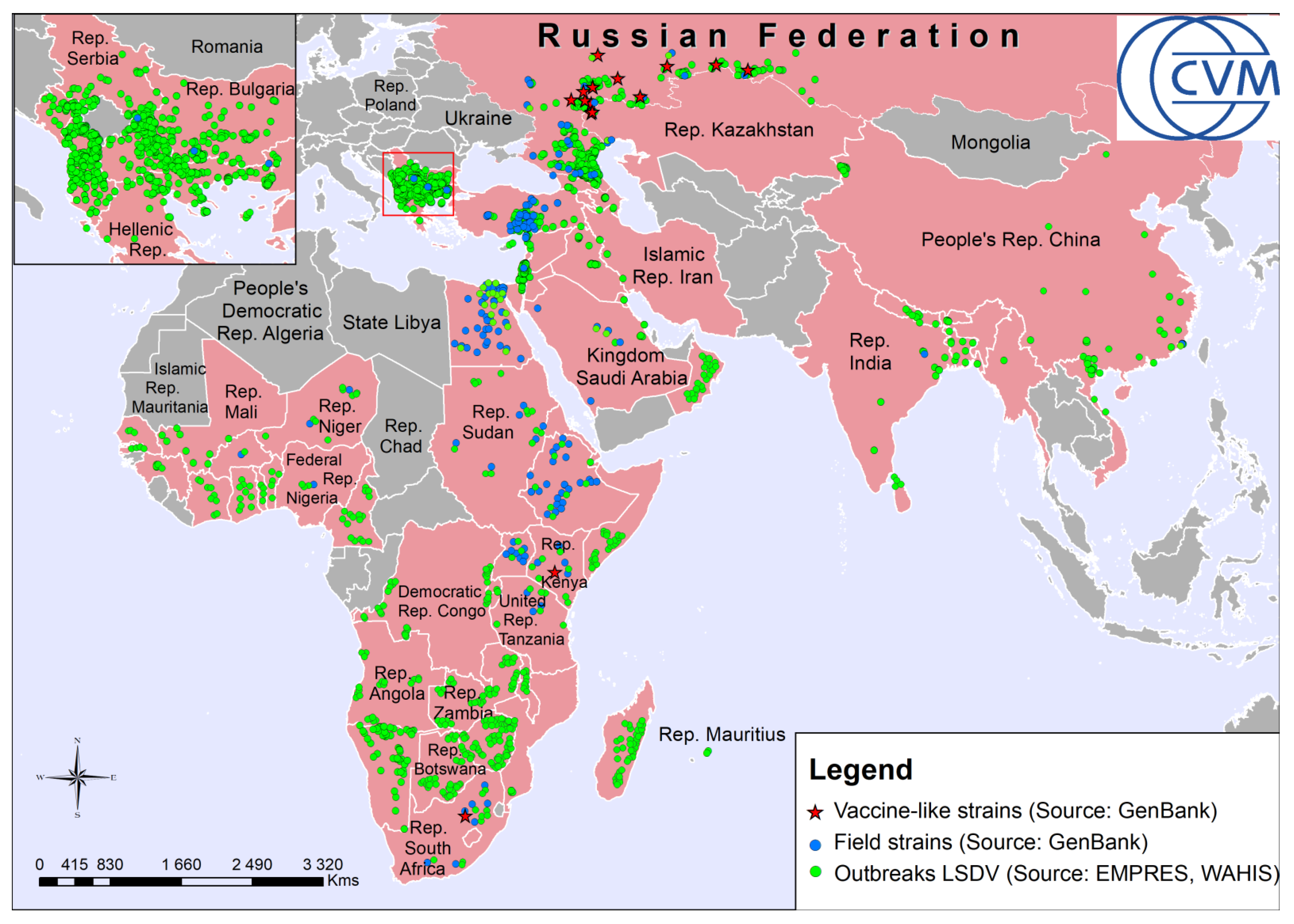
:1. Introduction
2. Results
2.1. Detection of LSDV DNA in Samples from Cattle Vaccinated and Unvaccinated with SPPV-Based Vaccine
2.2. Seroconversion in Cattle Vaccinated with SPPV-Based Vaccine
2.3. Phylogenetic Analysis and Typing and Subtyping
2.4. Phylogeographic Analysis
3. Discussion
4. Materials and Methods
4.1. Setting, Animals and Data Collection
4.2. DNA Extraction, PCR and Sequence Evidence of LSDV
4.3. Detection of Specific Antibodies
4.4. Cartographical and Phylogeographic Analysis
5. Conclusions
Supplementary Materials
Author Contributions
Funding
Institutional Review Board Statement
Informed Consent Statement
Data Availability Statement
Acknowledgments
Conflicts of Interest
References
- Calistri, P.; De Clercq, K.; Gubbins, S.; Klement, E.; Stegeman, A.; Abrahantes, J.C.; Marojevic, D.; Antoniou, S.; Broglia, A. Lumpy skin disease epidemiological report IV: Data collection and analysis. EFSA J. 2020, 18. [Google Scholar] [CrossRef] [Green Version]
- Moller, J.; Moritz, T.; Schlottau, K.; Krstevski, K.; Hoffmann, D.; Beer, M.; Hoffmann, B. Experimental lumpy skin disease virus infection of cattle: Comparison of a field strain and a vaccine strain. Arch. Virol. 2019, 164, 2931–2941. [Google Scholar] [CrossRef]
- OIE (World Organisation for Animal Health). Lumpy skin disease. In Terrestrial Manual; Office International des Epizooties: Paris, France, 2018. [Google Scholar]
- Le Goff, C.; Lamien, C.E.; Fakhfakh, E.; Chadeyras, A.; Aba-Adulugba, E.; Libeau, G.; Tuppurainen, E.; Wallace, D.B.; Adam, T.; Silber, R.; et al. Capripoxvirus G-protein-coupled chemokine receptor: A host-range gene suitable for virus animal origin discrimination. J. Gen. Virol. 2009, 90, 1967–1977. [Google Scholar] [CrossRef]
- Babiuk, S.; Bowden, T.R.; Boyle, D.B.; Wallace, D.B.; Kitching, R.P. Capripoxviruses: An emerging worldwide threat to sheep, goats and cattle. Transbound. Emerg. Dis. 2008, 55, 263–272. [Google Scholar] [CrossRef] [PubMed] [Green Version]
- Tuppurainen, E.S.; Oura, C.A. Review: Lumpy skin disease: An emerging threat to Europe, the Middle East and Asia. Transbound. Emerg. Dis. 2012, 59, 40–48. [Google Scholar] [CrossRef] [PubMed]
- Tuppurainen, E.S.M.; Venter, E.H.; Shisler, J.L.; Gari, G.; Mekonnen, G.A.; Juleff, N.; Lyons, N.A.; De Clercq, K.; Upton, C.; Bowden, T.R.; et al. Review: Capripoxvirus Diseases: Current Status and Opportunities for Control. Transbound. Emerg. Dis. 2017, 64, 729–745. [Google Scholar] [CrossRef] [PubMed]
- Woods, J.A. Lumpy skin disease—A review. Trop. Anim. Health Prod. 1988, 20, 11–17. [Google Scholar] [CrossRef]
- Weiss, K.E. Lumpy skin disease virus. Virol. Monogr. 1968, 3, 111–131. [Google Scholar]
- Beard, P.M. Lumpy skin disease: A direct threat to Europe. Vet. Rec. 2016, 178, 557–558. [Google Scholar] [CrossRef] [Green Version]
- Tulman, E.R.; Afonso, C.L.; Lu, Z.; Zsak, L.; Kutish, G.F.; Rock, D.L. Genome of lumpy skin disease virus. J. Virol. 2001, 75, 7122–7130. [Google Scholar] [CrossRef] [Green Version]
- Tulman, E.R.; Afonso, C.L.; Lu, Z.; Zsak, L.; Sur, J.H.; Sandybaev, N.T.; Kerembekova, U.Z.; Zaitsev, V.L.; Kutish, G.F.; Rock, D.L. The genomes of sheeppox and goatpox viruses. J. Virol. 2002, 76, 6054–6061. [Google Scholar] [CrossRef] [Green Version]
- Thomas, A.D. Mare CVE. Knopvelsiekte. J. S. Afr. Vet. Assoc. 1945, 16, 36–43. [Google Scholar]
- Diesel, A.M. The epizootiology of lumpy skin disease in South Africa. In Proceedings of the 14th International Veterinary Congress, London, UK, 14–17 February 1949; pp. 492–500. [Google Scholar]
- Tuppurainen, E.S.M.; Venter, E.H.; Coetzer, J.A.W. The detection of lumpy skin disease virus in samples of experimentally infected cattle using different diagnostic techniques. Onderstepoort J. Vet. 2005, 72, 153–164. [Google Scholar] [CrossRef] [PubMed]
- Hunter, P.; Wallace, D. Lumpy skin disease in southern Africa: A review of the disease and aspects of control. J. S. Afr. Vet. Assoc. 2001, 72, 68–71. [Google Scholar] [CrossRef] [PubMed] [Green Version]
- MacDonald, R.A.S. Pseudo-Urticaria of Cattle; Government of Northern Rhodesia, Department of Animal Health: Baton Rouge, LA, USA, 1931; pp. 20–21. [Google Scholar]
- Mercier, A.; Arsevska, E.; Bournez, L.; Bronner, A.; Calavas, D.; Cauchard, J.; Falala, S.; Caufour, P.; Tisseuil, C.; Lefrancois, T.; et al. Spread rate of lumpy skin disease in the Balkans, 2015–2016. Transbound. Emerg. Dis. 2018, 65, 240–243. [Google Scholar] [CrossRef] [Green Version]
- Miranda, M.A.; Stegeman, J.A.; Bicout, D.; Botner, A.; Butterworth, A.; Calistri, P.; Winckler, C. Urgent advice on lumpy skin disease EFSA Panel on Animal Health and Welfare. EFSA J. 2016, 14. [Google Scholar] [CrossRef]
- Sevik, M.; Dogan, M. Epidemiological and Molecular Studies on Lumpy Skin Disease Outbreaks in Turkey during 2014–2015. Transbound. Emerg. Dis. 2017, 64, 1268–1279. [Google Scholar] [CrossRef]
- Kitching, R.P. Vaccines for lumpy skin disease, sheep pox and goat pox. Dev. Biol. 2003, 114, 161–167. [Google Scholar]
- Tuppurainen, E.S.; Pearson, C.R.; Bachanek-Bankowska, K.; Knowles, N.J.; Amareen, S.; Frost, L.; Henstock, M.R.; Lamien, C.E.; Diallo, A.; Mertens, P.P. Characterization of sheep pox virus vaccine for cattle against lumpy skin disease virus. Antivir. Res. 2014, 109, 1–6. [Google Scholar] [CrossRef] [Green Version]
- Hamdi, J.; Bamouh, Z.; Jazouli, M.; Boumart, Z.; Tadlaoui, K.O.; Fihri, O.F.; El Harrak, M. Experimental evaluation of the cross-protection between Sheeppox and bovine Lumpy skin vaccines. Sci. Rep. 2020, 10. [Google Scholar] [CrossRef]
- Agianniotaki, E.I.; Tasioudi, K.E.; Chaintoutis, S.C.; Iliadou, P.; Mangana-Vougiouka, O.; Kirtzalidou, A.; Alexandropoulos, T.; Sachpatzidis, A.; Plevraki, E.; Dovas, C.I.; et al. Lumpy skin disease outbreaks in Greece during 2015–16, implementation of emergency immunization and genetic differentiation between field isolates and vaccine virus strains. Vet. Microbiol. 2017, 201, 78–84. [Google Scholar] [CrossRef]
- Bedekovic, T.; Simic, I.; Kresic, N.; Lojkic, I. Detection of lumpy skin disease virus in skin lesions, blood, nasal swabs and milk following preventive vaccination. Transbound. Emerg. Dis. 2018, 65, 491–496. [Google Scholar] [CrossRef]
- Agianniotaki, E.I.; Chaintoutis, S.C.; Haegeman, A.; Tasioudi, K.E.; De Leeuw, I.; Katsoulos, P.D.; Sachpatzidis, A.; De Clercq, K.; Alexandropoulos, T.; Polizopoulou, Z.S.; et al. Development and validation of a TaqMan probe-based real-time PCR method for the differentiation of wild type lumpy skin disease virus from vaccine virus strains. J. Virol. Methods 2017, 249, 48–57. [Google Scholar] [CrossRef] [PubMed]
- Menasherow, S.; Rubinstein-Giuni, M.; Kovtunenko, A.; Eyngor, Y.; Fridgut, O.; Rotenberg, D.; Khinich, Y.; Stram, Y. Development of an assay to differentiate between virulent and vaccine strains of lumpy skin disease virus (LSDV). J. Virol. Methods 2014, 199, 95–101. [Google Scholar] [CrossRef] [PubMed]
- Vidanovic, D.; Sekler, M.; Petrovic, T.; Debeljak, Z.; Vaskovic, N.; Matovic, K.; Hoffmann, B. Real-Time Pcr Assays for the Specific Detection of Field Balkan Strains of Lumpy Skin Disease Virus. Acta Vet. Beogr. 2016, 66, 444–454. [Google Scholar] [CrossRef]
- Lamien, C.E.; Le Goff, C.; Silber, R.; Wallace, D.B.; Gulyaz, V.; Tuppurainen, E.; Madani, H.; Caufour, P.; Adam, T.; El Harrak, M.; et al. Use of the Capripoxvirus homologue of Vaccinia virus 30 kDa RNA polymerase subunit (RPO30) gene as a novel diagnostic and genotyping target: Development of a classical PCR method to differentiate Goat poxvirus from Sheep poxvirus. Vet. Microbiol. 2011, 149, 30–39. [Google Scholar] [CrossRef] [PubMed]
- Erster, O.; Rubinstein, M.G.; Menasherow, S.; Ivanova, E.; Venter, E.; Sekler, M.; Kolarevic, M.; Stram, Y. Importance of the lumpy skin disease virus (LSDV) LSDV126 gene in differential diagnosis and epidemiology and its possible involvement in attenuation. Arch. Virol. 2019, 164, 2285–2295. [Google Scholar] [CrossRef] [PubMed]
- Sprygin, A.; Pestova, Y.; Bjadovskaya, O.; Prutnikov, P.; Zinyakov, N.; Kononova, S.; Ruchnova, O.; Lozovoy, D.; Chvala, I.; Kononov, A. Evidence of recombination of vaccine strains of lumpy skin disease virus with field strains, causing disease. PLoS ONE 2020, 15, e0232584. [Google Scholar] [CrossRef] [PubMed]
- Gelaye, E.; Belay, A.; Ayelet, G.; Jenberie, S.; Yami, M.; Loitsch, A.; Tuppurainen, E.; Grabherr, R.; Diallo, A.; Lamien, C.E. Capripox disease in Ethiopia: Genetic differences between field isolates and vaccine strain, and implications for vaccination failure. Antiviral. Res. 2015, 119, 28–35. [Google Scholar] [CrossRef] [PubMed]
- El-Tholoth, M.; El-Kenawy, A.A. G-Protein-Coupled Chemokine Receptor Gene in Lumpy Skin Disease Virus Isolates from Cattle and Water Buffalo (Bubalus bubalis) in Egypt. Transbound. Emerg. Dis. 2016, 63, e288–e295. [Google Scholar] [CrossRef] [PubMed]
- Biswas, S.; Noyce, R.S.; Babiuk, L.A.; Lung, O.; Bulach, D.M.; Bowden, T.R.; Boyle, D.B.; Babiuk, S.; Evans, D.H. Extended sequencing of vaccine and wild-type capripoxvirus isolates provides insights into genes modulating virulence and host range. Transbound. Emerg. Dis. 2020, 67, 80–97. [Google Scholar] [CrossRef]
- Ochwo, S.; VanderWaal, K.; Ndekezi, C.; Nkamwesiga, J.; Munsey, A.; Witto, S.G.; Nantima, N.; Mayanja, F.; Okurut, A.R.A.; Atuhaire, D.K.; et al. Molecular detection and phylogenetic analysis of lumpy skin disease virus from outbreaks in Uganda 2017–2018. BMC Vet. Res. 2020, 16, 66. [Google Scholar] [CrossRef] [Green Version]
- Kononov, A.; Byadovskaya, O.; Kononova, S.; Yashin, R.; Zinyakov, N.; Mischenko, V.; Perevozchikova, N.; Sprygin, A. Detection of vaccine-like strains of lumpy skin disease virus in outbreaks in Russia in 2017. Arch. Virol. 2019, 164, 1575–1585. [Google Scholar] [CrossRef] [PubMed]
- Sprygin, A.; Van Schalkwyk, A.; Shumilova, I.; Nesterov, A.; Kononova, S.; Prutnikov, P.; Byadovskaya, O.; Kononov, A. Full-length genome characterization of a novel recombinant vaccine-like lumpy skin disease virus strain detected during the climatic winter in Russia, 2019. Arch. Virol. 2020, 165, 2675–2677. [Google Scholar] [CrossRef]
- Van Schalkwyk, A.; Kara, P.; Ebersohn, K.; Mather, A.; Annandale, C.H.; Venter, E.H.; Wallace, D.B. Potential link of single nucleotide polymorphisms to virulence of vaccine-associated field strains of lumpy skin disease virus in South Africa. Transbound. Emerg. Dis. 2020, 67, 2946–2960. [Google Scholar] [CrossRef] [PubMed]
- Aleksandr, K.; Pavel, P.; Olga, B.; Svetlana, K.; Vladimir, R.; Yana, P.; Alexander, S. Emergence of a new lumpy skin disease virus variant in Kurgan Oblast, Russia, in 2018. Arch. Virol. 2020, 165, 1343–1356. [Google Scholar] [CrossRef]
- Sprygin, A.; Pestova, Y.; Prutnikov, P.; Kononov, A. Detection of vaccine-like lumpy skin disease virus in cattle and Musca domestica L. flies in an outbreak of lumpy skin disease in Russia in 2017. Transbound. Emerg. Dis. 2018, 65, 1137–1144. [Google Scholar] [CrossRef] [PubMed]
- Salnikov, N.; Usadov, T.; Kolcov, A.; Zhivoderov, S.; Morgunov, Y.; Gerasimov, V.; Gogin, A.; Titov, I.; Yurkov, S.; Malogolovkin, A.; et al. Identification and characterization of lumpy skin disease virus isolated from cattle in the Republic of North Ossetia-Alania in 2015. Transbound. Emerg. Dis. 2018, 65, 916–920. [Google Scholar] [CrossRef] [PubMed]
- OIE (World Organisation for Animal Health). World Animal Health Information Database (WAHIS) Interface. Available online: https://www.oie.int/wahis_2/public/wahid.php/Diseaseinformation/Immsummary (accessed on 2 June 2021).
- Lojkic, I.; Simic, I.; Kresic, N.; Bedekovic, T. Complete Genome Sequence of a Lumpy Skin Disease Virus Strain Isolated from the Skin of a Vaccinated Animal. Genome Announc. 2018, 6. [Google Scholar] [CrossRef] [PubMed]
- Mathijs, E.; Vandenbussche, F.; Saduakassova, M.; Kabduldanov, T.; Haegeman, A.; Aerts, L.; Kyzaibayev, T.; Sultanov, A.; Van Borm, S.; De Clercq, K. Complete Coding Sequence of a Lumpy Skin Disease Virus Strain Isolated during the 2016 Outbreak in Kazakhstan. Microbiol. Resour. Announc. 2020, 9. [Google Scholar] [CrossRef] [PubMed] [Green Version]
- OIE (Office International des Epizooties). World Organisation for Animal Health. Available online: https://www.oie.int (accessed on 2 June 2021).
- Kumar, S.; Stecher, G.; Tamura, K. MEGA7: Molecular Evolutionary Genetics Analysis Version 7.0 for Bigger Datasets. Mol. Biol. Evol. 2016, 33, 1870–1874. [Google Scholar] [CrossRef] [PubMed] [Green Version]
- Issimov, A.; Rametov, N.; Zhugunissov, K.; Kutumbetov, L.; Zhanabayev, A.; Kazhgaliyev, N.; White, P. Emergence of the First Lumpy Skin Disease Outbreak Among Livestock in the Republic of Kazakhstan in 2016. Preprints 2020, 2020110298. [Google Scholar] [CrossRef]
- Haegeman, A.; De Leeuw, I.; Mostin, L.; Van Campe, W.; Aerts, L.; Vastag, M.; De Clercq, K. An Immunoperoxidase Monolayer Assay (IPMA) for the detection of lumpy skin disease antibodies. J. Virol. Methods 2020, 277, 113800. [Google Scholar] [CrossRef] [PubMed]
- Byadovskaya, O.; Pestova, Y.; Kononov, A.; Shumilova, I.; Kononova, S.; Nesterov, A.; Babiuk, S.; Sprygin, A. Performance of the currently available DIVA real-time PCR assays in classical and recombinant lumpy skin disease viruses. Transbound. Emerg. Dis. 2020. [Google Scholar] [CrossRef]
- Lkjlkj Ireland, D.C.; Binepal, Y.S. Improved detection of capripoxvirus in biopsy samples by PCR. J. Virol. Methods 1998, 74, 1–7. [Google Scholar] [CrossRef]


| Subject | Vaccination Status 1 | PCR Results with Specimens from | Detection of DNA | ELISA | |||
|---|---|---|---|---|---|---|---|
| Blood | Nose | LSDV | SPPV | GTPV | |||
| Case1 | vaccinated | + | + | + | − | − | + |
| Case2 | vaccinated | + | + | + | − | − | + |
| Case3 | unvaccinated | + | + | + | − | − | − |
| Cow4 (negative control) | vaccinated | − | − | − | − | − | + |
| Subtype | LSDV Strain ID | Date of the First Issue of the Strain | Country/Region of Identification | Date of Detection in Saratov Region | Source |
|---|---|---|---|---|---|
| Ia | AF325528 Neethling-2490 | 1958 | Kenya | 2017 | [11] |
| IIa | FJ869376 RSA/54 Haden isolate LSDV17 | 1954 | South Africa | 2019, Ershov District | This study |
| IIc | MH646674 LSDV/ Russia/Saratov/2017 MH753586 Saratov/2017 MT395339 Saratov/2019 | 2017, 2019 | The Russian Federation, Saratov Region | 2017, 2019 | [31,36] |
| IId | MH029290 Dergachevskyi | 2017 | The Russian Federation, Saratov Region, Dergachyovsky District | 2017 | NCBI Acc. No. MH029290 |
| IIg | MK358808 Khvalynsky | 2018 | The Russian Federation, Saratov Region, Khvalynsky District | 2018 | NCBI Acc. No. MK358808 |
Publisher’s Note: MDPI stays neutral with regard to jurisdictional claims in published maps and institutional affiliations. |
© 2021 by the authors. Licensee MDPI, Basel, Switzerland. This article is an open access article distributed under the terms and conditions of the Creative Commons Attribution (CC BY) license (https://creativecommons.org/licenses/by/4.0/).
Share and Cite
Saltykov, Y.V.; Kolosova, A.A.; Filonova, N.N.; Chichkin, A.N.; Feodorova, V.A. Genetic Evidence of Multiple Introductions of Lumpy Skin Disease Virus into Saratov Region, Russia. Pathogens 2021, 10, 716. https://doi.org/10.3390/pathogens10060716
Saltykov YV, Kolosova AA, Filonova NN, Chichkin AN, Feodorova VA. Genetic Evidence of Multiple Introductions of Lumpy Skin Disease Virus into Saratov Region, Russia. Pathogens. 2021; 10(6):716. https://doi.org/10.3390/pathogens10060716
Chicago/Turabian StyleSaltykov, Yuri V., Anna A. Kolosova, Nadezhda N. Filonova, Alexander N. Chichkin, and Valentina A. Feodorova. 2021. "Genetic Evidence of Multiple Introductions of Lumpy Skin Disease Virus into Saratov Region, Russia" Pathogens 10, no. 6: 716. https://doi.org/10.3390/pathogens10060716