Article

High Rates of Multidrug-Resistant Escherichia coli in Great Cormorants (Phalacrocorax carbo) of the German Baltic and North Sea Coasts: Indication of Environmental Contamination and a Potential Public Health Risk

1
Institute for Terrestrial and Aquatic Wildlife Research, University of Veterinary Medicine Hannover (TiHo) Foundation, 25761 Büsum, Germany
2
Institute for Veterinary Food Science, Justus Liebig University Giessen, 35392 Giessen, Germany
3
Office for Veterinary Affairs and Consumer Protection, Ministry of Lower Saxony for Food, Agriculture and Consumer Protection, 29221 Celle, Germany
4
CIISA—Centre for Interdisciplinary Research in Animal Health, Faculty of Veterinary Medicine, University of Lisbon, 1300-477 Lisbon, Portugal
5
Associate Laboratory for Animal and Veterinary Sciences (AL4AnimalS), Avenida da Universidade Técnica, 1300-477 Lisbon, Portugal
6
Fish Disease Research Unit, University of Veterinary Medicine Hannover, 30559 Hannover, Germany
*
Author to whom correspondence should be addressed.
Pathogens 2022, 11(8), 836; https://doi.org/10.3390/pathogens11080836
Submission received: 21 June 2022 / Revised: 15 July 2022 / Accepted: 22 July 2022 / Published: 27 July 2022
(This article belongs to the Special Issue Wildlife Hosts Pathogen Interaction)

Abstract

:
Antimicrobial-resistant bacteria pose a serious global health risk for humans and animals, while the role of wildlife in the dynamic transmission processes of antimicrobial resistance in environmental settings is still unclear. This study determines the occurrence of antimicrobial-resistant Escherichia coli in the free-living great cormorants (Phalacrocorax carbo) of the North and Baltic Sea coasts of Schleswig-Holstein, Germany. For this, resistant E. coli were isolated from cloacal or faecal swabs and their antimicrobial resistance pheno- and genotypes were investigated using disk diffusion tests and PCR assays. The isolates were further assigned to the four major phylogenetic groups, and their affiliation to avian pathogenic E. coli (APEC) was tested. Resistant E. coli were isolated from 66.7% of the 33 samples, and 48.9% of all the resistant isolates showed a multidrug resistance profile. No spatial differences were seen between the different sampling locations with regard to the occurrence of antimicrobial resistance or multidrug resistance. Most commonly, resistance percentages occurred against streptomycin, followed by tetracycline and sulfonamides. More than half of the isolates belonged to the phylogenetic group B1. Of all the isolates, 24.4% were classified as APEC isolates, of which almost 82% were identified as multidrug-resistant. These results add information on the dispersal of antimicrobial-resistant bacteria in wild birds in Germany, thereby allowing conclusions on the degree of environmental contamination and potential public health concerns.

1. Introduction

Antimicrobial resistance in bacteria is a global health threat in human and veterinary medicine [1,2]. The increase in the frequency of multidrug-resistant bacteria over the past decades has been driven by the selective pressure imposed by the use and misuse of antibiotics [1]. Antimicrobial-resistant bacteria are not limited to humans, companion animals, and livestock; they can be found in the environment naturally [3,4,5] as well as a consequence of anthropogenic activities [5,6,7,8]. Moreover, antimicrobial agents themselves have already been found in soil, surface, and ground water, and some of them have been shown to persist in the environment for months or even years [9,10,11,12]. In this context, wild animals can play an important role as reservoirs and vectors for antimicrobial-resistant bacteria. However, their importance in the dissemination of these bacteria and resistance genes is still incompletely explored [13].
Especially interesting are the aquatic ecosystems as they are subjected to high levels of different anthropogenic impacts [14]. Accordingly, various studies have detected antimicrobial-resistant bacteria in natural waterbodies, often finding evidence of anthropogenic causes, including sewage discharges, agricultural run-off, and health-care effluent [6,7,15,16,17,18,19,20]. Controversial results have been found with regard to whether wastewater treatment plants select for or against antimicrobial-resistance genes [21,22,23,24,25]. Furthermore, there are indications that wastewater treatment, although generally reducing the amount of bacteria, might select for multidrug resistance and might lead to an accumulation of resistance genes [26,27]. Genetic exchange occurs mainly via horizontal transfer of genetic material between bacteria including environmental bacteria and microbes that originated from humans and animals [28,29]. However, resistant bacteria were also detected in environments with an absence of external influences [30]. Here, birds may play an important role as bird faeces were found as another important contamination source for antimicrobial-resistant bacteria in natural waterbodies [31]. The birds themselves are likely to acquire the resistant bacteria from the environment [32], indicating the complex transmission dynamics of antimicrobial resistance in the environment.
Enterobacteriaceae from surface waters have been identified as important reservoirs of antimicrobial-resistant bacteria and resistance genes [33]. Within the family Enterobacteriaceae, Escherichia coli is of specific interest as it colonizes the intestines of mammals and birds. Here, it mainly acts commensally, but some strains are known pathogens, causing severe intestinal or extra-intestinal diseases in both humans and animals [34]. In addition, E. coli can easily receive, harbour, and pass antimicrobial resistance genes, mainly via horizontal gene transfer to other bacteria [35,36]. E. coli can be disseminated by faeces, manure, sewage sludge, or treated wastewater to the terrestrial and aquatic environment where it survives, depending on the existing abiotic and biotic factors, for varying periods of time [27,37]. A recent study demonstrated the correlation of the access to treated wastewater and the increased antimicrobial-resistance burden in wild birds [38]. In this context, migratory birds in particular may play an important role in the dissemination of antimicrobial-resistant bacteria over long distances and into pristine niches [32,39,40,41]. As humans, wildlife, and the environment share similar E. coli strains [42], resistant strains in the environment pose a potential infection source for humans [18,43]. Altogether, this highlights the potential of E. coli as an indicator of the spread of antimicrobial resistance in the environment as well as its importance as a public health concern.
Antimicrobial-resistant E. coli have been reported from various wild bird species [44,45,46,47]. For the present study, great cormorants (Phalacrocorax carbo) were selected as the study object for several reasons. First of all, large breeding colonies allow an easy access to faecal samples. Secondly, their feeding ecology is almost exclusively based on fish [48,49,50], making them perfect bioindicators for the occurrence of antimicrobial-resistant bacteria in the environment [51] as they rarely feed on anthropogenic waste. Thirdly, they have already been shown to frequently carry resistant E. coli isolates [40,52,53,54]. Lastly, due to migration, great cormorants might also be able to disseminate resistant bacteria over extended distances [40,55,56].
The aim of this study was to investigate the occurrence of antimicrobial-resistant E. coli in great cormorants in the German federal state of Schleswig-Holstein. Here, great cormorants were used as a potential sentinel aquatic bird species to understand whether there were regional patterns according to the origin of the animals. Moreover, the level of multidrug resistance among the isolates was determined to estimate the potential public health risk posed by the detected isolates.

2. Material and Methods

2.1. Sample Collection

In total, 33 samples were taken from great cormorants living at or close to the coastline of the North Sea (n = 13) or Baltic Sea (n = 20) of Schleswig-Holstein, Germany. Three colonies were sampled, one in the Wallnau Waterbird Reserve on the island of Fehmarn (Baltic Sea), one in the nature reserve Geltinger Birk (Baltic Sea), and one in Friedrichsgabekoog (North Sea), an agricultural landscape close to Büsum. A single sample was collected on the island of Sylt (North Sea). A map created with ArcGIS (ESRI, Redlands, CA, USA, Version 10.6.1) indicates all the sample locations (Figure 1). The samples were obtained either as cloacal or faecal swabs (Sterile transport swabs, Heinz Herenz, Germany). The cloacal swabs were taken during the bird ringing of nestlings. Therefore, the nestlings were carefully gripped out of their nests, fixed, equipped with a small metal tag, and then sampled by inserting a sterile swab into the cloaca and subsequently turning it several times on the mucosa. Faecal swabs were taken from fresh faeces from the ground under the nesting trees and one on the beach after witnessed defecation. Here, a sterile swab was inserted into the excrement and also turned a few times to gain enough material. All the swabs were taken between 28 May and 21 July 2017. The samples were transported to the lab immediately without refrigeration.

2.2. E. coli Isolation and Identification

The obtained swabs were stored at room temperature for between three and six months. The sample processing aimed at the isolation of only the resistant E. coli. This was performed via selective enrichment for Enterobacteriaceae and following transfer to selective agar plates supplemented with antimicrobials. Mossel broth (Carl Roth GmbH & Co. KG, Karlsruhe, Germany), blood agar (Carl Roth GmbH & Co. KG), and Gassner agar (Sigma Aldrich Chemie GmbH, Steinheim am Albuch, Germany) were prepared in accordance with the manufacturer’s instructions. MacConkey agar (Carl Roth GmbH & Co. KG), which was additionally supplemented with different antimicrobials, was also used. The following antimicrobials and concentrations were used to supplement the agar plates: ampicillin (30 mg/L; Carl Roth GmbH & Co. KG), cephalothin (30 mg/L; TCI Europe N.V., Zwijndrecht, Belgium), chloramphenicol (10 mg/L; Carl Roth GmbH & Co. KG), ciprofloxacin (1 mg/L; Alfa Aeser, Thermo Fischer Scientific, Waltham, MA, USA), colistin (2 mg/L; AppliChem GmbH, Darmstadt, Germany), gentamicin (10 mg/L; Carl Roth GmbH & Co. KG), sulfamethoxazole (512 mg/L; Sigma Aldrich Chemie GmbH), and tetracycline (15 mg/L; AppliChem GmbH). Each swab was streaked on blood and Gassner agar plates and on Mossel broth and incubated for 24 h at 37 °C, to be used as a reference for the bacterial growth. In the following, 100 µL of the Mossel bouillon was streaked on each of the eight different antimicrobial-supplemented MacConkey agar plates. Again, the plates were incubated for 24 h at 37 °C. From every plate showing bacterial growth, five colonies were selected, and if less than five colonies were available, then all the colonies were selected. The selected colonies were then suspended in glycerine and stored at −80 °C. The colonies were selected based on their macroscopical appearance. On MacConkey agar, E. coli colonies (among others, e.g., Klebsiella spp. and Enterobacter spp.) are typically coloured red or pink with a bile salt precipitation halo surrounding the colony. Bacterial growth on the antimicrobial containing agar plates are listed in the supplementary material Table S1.
The identification of E. coli was performed using selective plating on Chromocult (Merck KGaA, Darmstadt, Germany), Gassner, and MacConkey agar plates. Due to their lactose fermentation ability, E. coli typically appear as blue to violet (Chromocult), blue (Gassner, Kempten, Germany), and red or pink colonies (MacConkey). Afterwards, the presumptive E. coli were confirmed via gadA-PCR [57]. The isolates that did not yield a positive PCR result were subsequently analysed by MALDI-TOF mass spectrometry for species identification. After this, 81 confirmed E. coli isolates were obtained. As each sample was streaked on eight different plates and up to five colonies per plate were collected, one sample delivered from one to nine isolates.

2.3. Resistance Phenotype

Diskdiffusion tests were performed in accordance with the Clinical and Laboratory Standards Institute standards [58] to determine the resistance profiles of the 81 confirmed E. coli isolates. Fourteen different antimicrobial-infused disks were purchased from Oxoid (Wesel, Germany). The tested antimicrobials comprised amoxicillin/clavulanic acid (20/10 µg), ampicillin (10 µg), cefazolin (30 µg), cefpodoxime (10 µg), chloramphenicol (30 µg), florfenicol (30 µg), streptomycin (10 µg), gentamicin (10 µg), kanamycin (30 µg), ciprofloxacin (5 µg), nalidixic acid (30 µg), compound sulfonamide (300 µg), trimethoprim (5 µg), and tetracycline (30 µg), representing seven different antimicrobial classes. For each isolate, a bacterial suspension in NaCl with 0.5 on the MacFarland scale, corresponding to approximately 108 CFU/mL, was prepared and inoculated on the surface of Mueller–Hinton (Carl Roth GmbH & Co. KG) agar plates, after which a maximum of six antimicrobial disks were placed on the agar surface. The plates were then incubated for 18 h at 35 °C ± 2 °C. The inhibition zones were evaluated in accordance with the CLSI standards [59,60]. The intermediate susceptible isolates and resistant isolates were grouped together for subsequent analyses. The isolates suspected of being colistin-resistant were additionally tested using MacConkey agar plates supplemented with colistin (2 mg/L). As there are no CLSI breakpoints for colistin based on zone diameters, the colistin resistance of the isolates growing on these plates was confirmed using the broth macrodilution method, as described by the CLSI [58,59]. If multiple isolates were obtained from the same swab but showed the same resistance profile, only one was used for further testing to avoid copy isolates. Thirty-five isolates were identified as duplicates. One isolate did not show any resistance in the disk diffusion test. In the end, 45 E. coli isolates with phenotypic resistances were gained from the 33 samples. The 45 isolates were distributed among the sampling locations as follows: 15 from Wallnau, 9 from Geltinger Birk, 16 from Friedrichsgabekoog, and 5 from Sylt. Moreover, the isolates which showed a resistance profile to three or more antimicrobials with different action mechanisms were classified as multidrug-resistant.

2.4. Resistance Genotype

The isolates were tested for the presence of antimicrobial resistance genes using a panel of PCR assays, as described previously [61]. The targeted genes comprised strA, strB [62], aadA1 [63], aadA2 [64], ant-(2″)-I, aac(3)-II, and aac(3)-IV, mediating resistance to aminoglycosides; blaTEM, blaSHV, blaOXA-1-like, and blaOXA-2, mediating resistance to β-lactams; aac(6′)-Ib-cr, qnrA, qnrB, qnrC, qnrD, and qnrS, associated with reduced susceptibility to quinolones; catA1, catA2, catA3, catB2, catB3, cmlA, and floR, mediating resistance to phenicols; sul1, sul2, and sul3, mediating resistance to sulfonamides; dfrA1/15/16, dfrA5/14, dfrA7/17, and dfrB1/2/3, mediating resistance to trimethoprim; tet(A), tet(B), tet(C), tet(D), tet(E), tet(G), tet(H), tet(L) [65], tet(M), and tet(O), mediating resistance to tetracyclines [61]; and mcr-1 [66], mcr-2, mcr-3, mcr-4, mcr-5 [67], mcr-6, mcr-7, mcr-8, and mcr-9 [68], mediating resistance to colistin.

2.5. Molecular Typing

All of the E. coli isolates were assigned to one of the four major phylogenetic groups (A, B1, B2, and D) using PCR assays targeting two genes (chuA and yjaA) [69], as well as the anonymous DNA fragment TSPE4.C2 [57]. The isolates were further tested for the presence of five genes indicative of avian pathogenic E. coli (APEC): iroN, ompT, hlyF, iss, and iutA [70]. All the isolates were also subjected to XbaI macrorestriction analysis and the subsequent pulsed-field gel electrophoresis (PFGE), according to the published protocols [71]. An UPGMA analysis (dice coefficient, 0.5% optimization, and 1% position tolerance) was performed using the Bionumerics software (version 7.6, Applied Maths, Sint-Martens-Latem, Belgium). Band patterns are shown in the supplementary material Figure S1.

2.6. Statistical Analysis

Statistical analyses were performed using RStudio (RStudio PBC, Boston, MA, USA) version 1.4.1103 (R version 4.1.2) with the packages readxl and DescTools. Due to the small sample size, a Fisher’s exact test was performed to test the significance of the association between (i) the number of samples with resistant E. coli isolates and the sampling locations and (ii) the number of multidrug-resistant isolates (including only samples with resistant E. coli) and the sampling locations. The level of significance was set at p < 0.05.

3. Results

3.1. Occurrence of Antimicrobial-Resistant E. coli Isolates

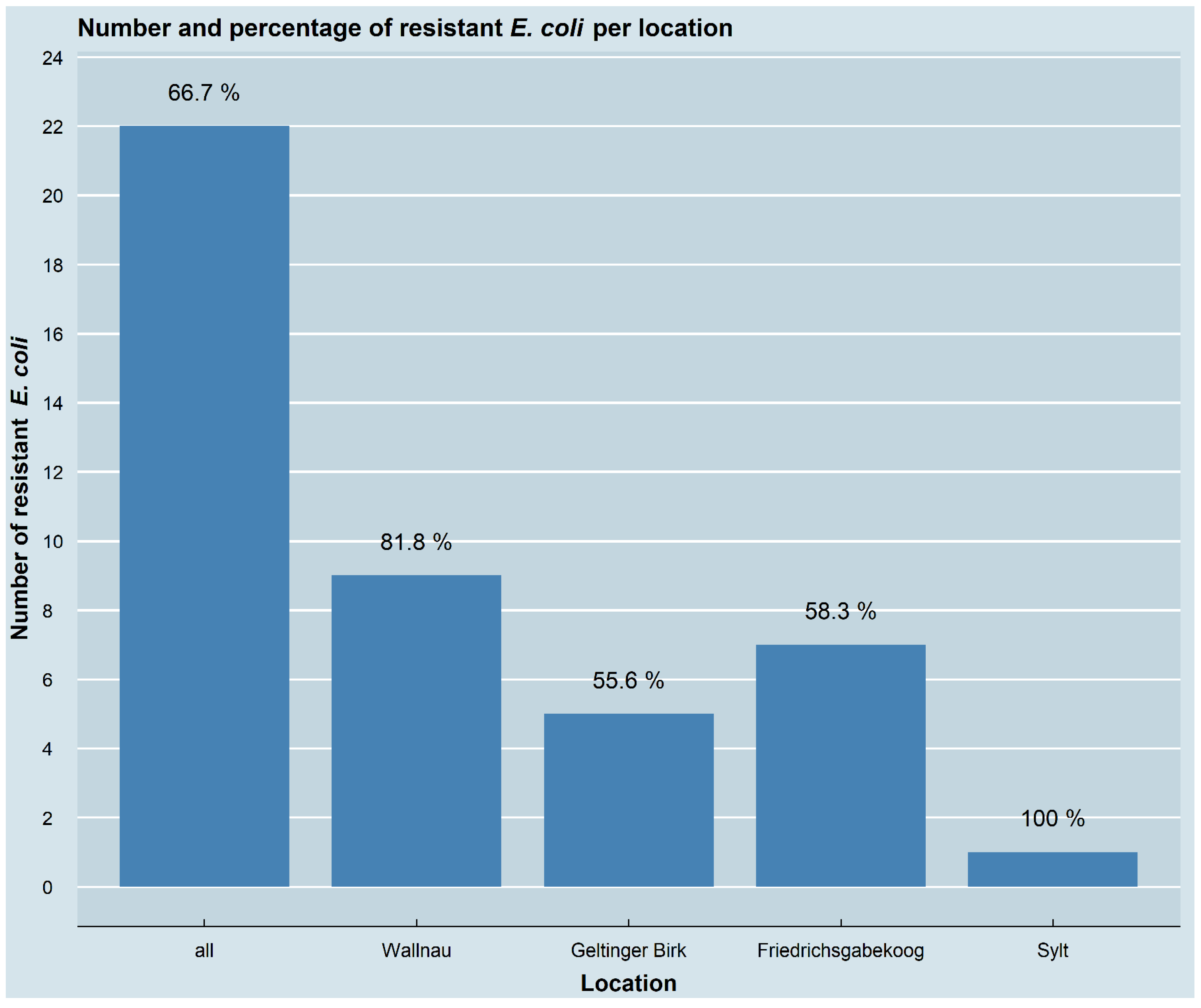
Of the 33 great cormorant samples that were collected for this study, 87.9% (29/33) had bacterial growth on at least one of the antimicrobial-containing agar plates. In 66.7% (22/33) of the samples, at least one resistant E. coli isolate was obtained, while the other isolates could not be confirmed as E. coli. Resistant E. coli were present in 14 of the 20 (70%) samples from the Baltic Sea coast, including 9 of the 11 (81.8%) samples from Wallnau and 5 of the 9 (55.6%) samples from Geltinger Birk. Regarding the samples collected at the North Sea coast, 8 of the 13 (61.5%) samples contained resistant E. coli, including 7 of the 12 (58.3%) samples collected in Friedrichsgabekoog, as well as the single sample (100%) from Sylt. No statistically significant association between the number of samples with resistant isolates and the sampling locations (p = 0.5393) was observed. Whether there were differences between nestlings and adult birds could not be calculated, because the faecal samples could also have come either from nestlings or adults. The number and the percentage of the samples containing resistant E. coli isolates, as well as their allocation for the different locations, are depicted in Figure 2. In total, 45 different resistant E. coli isolates were collected and further investigated (Figure 3).

3.2. Resistance Pheno- and Genotypes

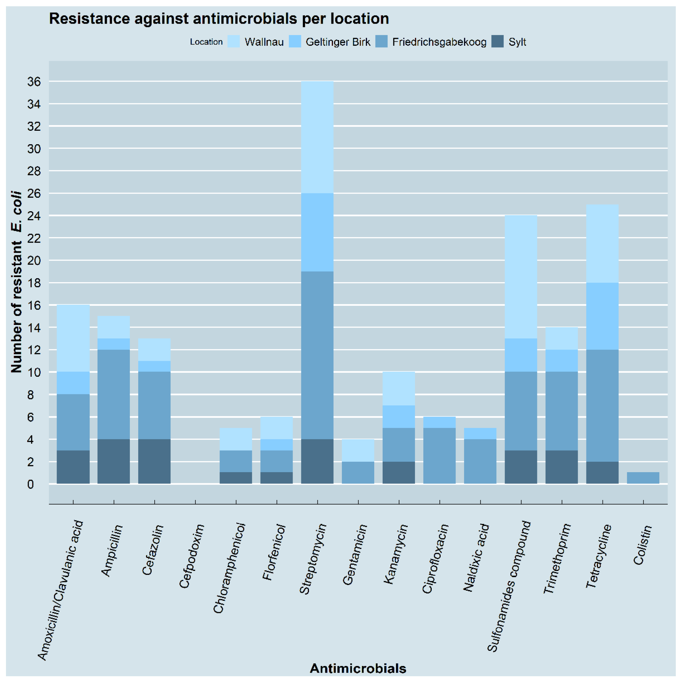
Overall, most of the 45 E. coli isolates (36/45, 80%) showed resistance to streptomycin in the diskdiffusion tests, followed by resistance to tetracycline (25/45, 55.6%) and sulfonamides (24/45, 53.3%). Resistance to amoxicillin/clavulanic acid, ampicillin, and trimethoprim ranged between 35.6 and 31.1% (14, 15 and 16/45), followed by cefazolin (13/45, 28.9%), kanamycin (10/45, 22.2%), ciprofloxacin and florfenicol (6/45, 13.3% each), and chloramphenicol and nalidixic acid (5/45, 11.1% each). The lowest rates of resistance were seen for gentamicin (4/45, 8.9%) and colistin (1/45, 2.2%). None of the isolates showed resistance against cefpodoxime.
The phenotypic resistance patterns varied in the isolates from the different sampling locations. In the isolates from the samples of Geltinger Birk (nature reserve) and Friedrichsgabekoog (agricultural landscape), the most common resistance phenotype was to streptomycin, with percentages of 77.8 (7/9) and 93.8 (15/16), respectively, while in Wallnau (waterbird reserve) most of the isolates were resistant to sulfonamides (73.3%, 11/15). Four out of the five (80%) isolates recovered from the sample taken in Sylt showed resistance to streptomycin, ampicillin, and cefazolin. Apart from the zero isolates with resistance to cefpodoxim, the isolates from the samples collected in Geltinger Birk were all susceptible to chloramphenicol; the isolates from Wallnau and Sylt were all susceptible to ciprofloxacin and nalidixic acid; and the isolates from Geltinger Birk and Sylt were all susceptible to gentamicin. Only one isolate from Friedrichsgaabekoog was resistant to colistin. The resistance patterns of the isolates from the four locations to the different antimicrobials are shown in Figure 4.
Of the 45 isolates, 10 isolates were resistant to only one antimicrobial agent (22.2%), and 7 isolates showed resistance to two antimicrobials (15.6%). Resistance to three antimicrobials was shown by 9 (20%) isolates; resistance to four and five antimicrobials was shown by 5 isolates for each (each 11.1%); resistance to six antimicrobials was shown by 4 isolates (8.9%); and resistance to seven antimicrobials was shown by 1 isolate (2.2%). Three isolates (6.7%) were resistant to 10 antimicrobials, and one isolate (2.2%) was resistant to 11 of the tested antimicrobials.
Considering the different antimicrobial classes instead of the individual antimicrobial agents revealed that the isolates showed the highest resistance rates to aminoglycosides (here represented by streptomycin, gentamicin, and kanamycin) (37/45, 82.2%), followed by resistance to tetracyclines (here represented by tetracycline) (25/45, 55.6%), folate synthesis inhibitors (here represented by sulfonamides and trimethoprim) (24/45, 53.3%), and non-extended spectrum β-lactams (here represented by amoxicillin/clavulanic acid, ampicillin, and cefazolin) (22/45, 48.9%). Lower resistance rates occurred towards quinolones (here represented by ciprofloxacin and nalidixic acid), phenicols (here represented by chloramphenicol and florfenicol) (6/45, 13.3% each), and polymyxins (here represented by colistin) (1/45, 2.2%). No extended spectrum β-lactamases (here represented by cefpodoxime) were detected. Resistance to at least one antimicrobial agent of three or more antimicrobial classes was rated as multidrug resistance, which occurred in 48.9% (22/45) of the E. coli isolates. Of these, eleven were resistant to three antimicrobial classes, six to four classes, three to five classes, and two isolates to six antimicrobial classes. Resistance to two antimicrobial classes occurred in 31.1% (14/22) of the isolates and to one antimicrobial class in 20% (9/45). Of the different locations, the highest rate of multiresistance was found in the isolates from Friedrichsgabekoog (9/16, 56.3%), followed by Wallnau (8/15, 53.3%), Sylt (2/5, 40%) and Geltinger Birk (3/9, 33.3%). The number of susceptible and resistant isolates per location, as well as their proportion of multiresistance, is depicted in Figure 5. No statistically significant association between the number of samples containing multidrug-resistant E. coli and the sampling locations (p = 0.924) was observed.
Isolates showing phenotypic resistance were tested for a selection of corresponding resistance genes. By total numbers, the resistance genes most commonly detected were sul2 (n = 20) and sul1 (n = 14) mediating resistance to sulfonamides; strA (n = 18), strB (n = 18), and aadA1 (n = 11) mediating resistance to streptomycin; and tet(A) (n = 16) and tet(B) (n = 10) mediating resistance to tetracyclines, as well as blaTEM (n = 15) mediating resistance to non-extended spectrum β-lactams. More rarely found resistance genes include dfrA5/A14 (n = 6), dfrA7/A17 (n = 3), and dfrA1/15/16 (n = 5) mediating resistance to trimethoprim; floR (n = 5) mediating resistance to chloramphenicol; aac(3)-II (n = 1) and ant(2“)-I (n = 2) mediating resistance to aminoglycosides; tet(D) (n = 1) mediating resistance to tetracyclines; sul3 (n = 1) mediating resistance to sulfonamides; and qnrS (n = 2) mediating resistance to quinolones.
Of the isolates showing phenotypic resistance to aminoglycosides (n = 37), 25 (67.6%) carried one to four corresponding resistance genes that were tested here, whereas all of the isolates resistant to tetracycline (n = 25) carried one to two corresponding resistance genes. Of the isolates with resistance to sulfonamides (n = 24), in 23 (95.8%) one to two corresponding resistance genes were detected, while all the isolates resistant to trimethoprim (n = 14) carried one corresponding resistance gene. Of the isolates showing resistance to β-lactams (n = 22), 15 (68.2%) carried one resistance gene, while of the isolates showing resistance to phenicols (n = 6), in 5 (83.3%) one corresponding resistance gene was detected. Of the isolates showing resistance to quinolones (n = 6), two carried (33.3%) one corresponding gene, and the isolate showing resistance to colistin did not carry any of the tested resistance genes. Of the 26 isolates that exhibited phenotypic resistance or intermediate resistance, but lacked a corresponding resistance gene, 21 (80.8%) exhibited intermediate resistance. Only in a single isolate with phenotypic resistance to cefazolin could no corresponding resistance gene tested be detected here. However, from the isolates without a detected plasmid-located quinolone resistance gene, three out of four showed a full phenotypic resistance. The detected resistance genes and the number of genes per location are summarized in Table 1. For each isolate, the resistance genotypes are shown in Figure 3.

3.3. Molecular Typing

The determination of major phylogenetic groups assigned most of the isolates to the phylogenetic group B1 (23/45, 51.1%), followed by the phylogenetic group D (13/45, 28.9%), the phylogenetic group B2 (5/45, 11.1%), and the phylogenetic group A (4/45, 8.9%). The phylogenetic groups of each isolate are listed in Figure 3. The results for the different sampling locations, as well as for all the isolates, are summarized in Table 2.
Eleven isolates were identified as avian pathogenic E. coli (APEC) isolates (all five indicative genes attested) while the remaining 34 isolates were presumed to be commensal E. coli isolates. Of the different locations, the one sample from Sylt had the highest rate of APEC isolates (2/5, 40%), followed by the samples from Wallnau (5/15, 33.3%) and Friedrichsgabekoog (4/16, 25%). No APEC isolates were detected in the samples from Geltinger Birk. The results of the analysis for the APEC isolates identification are shown in Figure 3.
The majority of the APEC isolates (9/11) were multidrug-resistant. With regard to the phylogenetic groups, 45.5% (5/11) of the APEC isolates belong to group B1, 36.4% (4/11) to group D, and 18.2% (2/11) to group A. None of the APEC isolates belonged to group B2. The phylogenetic group B1 isolates also had the highest proportion of multidrug resistance, with 45.5% (10/22), while 31.8% (7/22) belonged to group D, 13.6% (3/22) to group A, and 9.1% (2/22) to group B2.
Macrorestriction analyses revealed band patterns in 42 isolates, based on which the genomic relatedness could be determined. The remaining three isolates were found to be non-typeable by Xbal macrorestriction. It was possible to identify one cluster of three isolates with ≥90% similarity, involving two isolates from the same sample and one isolate from a different sample of the same sampling location and sampling date (isolates 25a, 25b and 26b), with two isolates having identical band patterns (25a and 25b). Another cluster with ≥80% similarity included three isolates of different samples of the same sampling location and sampling date (isolates 8b, 9b and 7a), with two isolates (8b and 9b) showing ≥90% similarity. Additionally, two isolates from three samples showed ≥90% similarity, with two isolates from the same sampling date and location having identical band patterns. All the other isolates showed a high degree of heterogeneity. The dendrogram of the 41 isolates is depicted in Figure 3.

4. Discussion

In the present study, antimicrobial-resistant E. coli isolates were detected in 66.7% of great cormorant samples collected at four different sites in the federal state of Schleswig-Holstein, Germany. For the three sampled great cormorant colonies, the occurrence varied between 55.6, 58.3, and 81.8%, respectively. Only a few other studies have investigated antimicrobial-resistant E. coli exclusively in cormorant species. In contrast to the present study, E. coli were isolated and then tested for their antimicrobial susceptibility, and very low resistance rates were found. In Japan, only 3.5% of E. coli isolated from great cormorants presented an antimicrobial-resistant profile [53]. A similarly low rate of 4.8% resistance was found in double-crested cormorant (Phalacrocorax auritus) chicks in Canada [72]. On the other hand, there are numerous reports of isolated antimicrobial-resistant E. coli in a multitude of wild bird species, but only a few of them found similar or higher rates of samples carrying antimicrobial resistance than were ascertained in this study. One study reported resistance rates in over 90% of the samples [73], while a few others reported around 50 [47,74,75] or 30% [76,77,78,79], respectively, of the samples carrying resistant E. coli. Lower rates varied from 1.4 to 18% [44,45,79,80]. The reasons for the wide diversity of bird species carrying resistant isolates may include bird population density [31], the feeding ecology of the birds [81], the close or absent proximity to livestock farms or wastewater run-off [82], and human population density in the sampling areas [83,84], as well as a seasonality depending on temperature [85]. It is noteworthy that differences in the study design, such as the selection of bird species (feeding ecology), the different status of the sampled animals (live/deceased, likely healthy/weak or sick), and the sampling method (throat/cloacal/faecal swab or organ samples), as well as the sample processing (only one isolate per sample/several isolates per sample), make the comparison of the results difficult. Furthermore, due to the delay in sample processing in the present study, it was of course possible that the number of E. coli was underestimated.
Multidrug resistance, defined as a resistance against three or more antimicrobial classes [86], occurred in the present study in 48.9% of the resistant E. coli isolates. Compared to other studies, this is a high rate. However, comparing studies is limited as the definition of multidrug resistance varies. Applying the same definition of multidrug resistance as in the present study, samples from different bird species collected in Poland carried multidrug-resistant E. coli in 38.6% [47] and 31.2% [73] of the cases, respectively. Furthermore, 15.3% of the samples of migratory birds collected in China [32], as well as 6.7% of the great cormorants samples collected in Switzerland [54] had multidrug-resistant E. coli isolates. Multidrug resistance, defined as resistance against two or more antimicrobial classes, was found in suburban and in wild birds in Australia in 2.1% and 1.5% of the tested samples, respectively [45]. Resistance against three or more antimicrobials was reported in 3% of adult gulls in Alaska [75] and in 3.2% of different bird species in Germany [44]. Resistance against two or more antimicrobials was detected in 19% of gulls tested in different European countries [77] and in 3.2% of the double-crested cormorant chicks in Prince Edward Island, Canada [72].
The high rate of multidrug resistance in the present study seems to indicate a high contamination of the environment by multidrug-resistant bacteria [39]. The highest amount of multidrug-resistant isolates was found in the great cormorant colony in Friedrichsgabekoog, while the location with the highest percentage of isolates with resistance to at least one antimicrobial was Wallnau. However, the highest human population densities are in Wallnau (Ost-Holstein, Germany), with 145 humans/km2, while Friedrichsgabekoog (Dithmarschen, Germany) and Geltinger Birk (Schleswig-Flensburg, Germany) are less populated, with 94 humans/km2 and 98 humans/km2, respectively [87]. Of the three colony sampling locations, Wallnau, a waterbird reserve, and Geltinger Birk, a nature reserve, may be seen as more pristine than Friedrichsgabekoog, which is an agricultural environment. Moreover, differences in the sampling method need to be noted. Only in Wallnau were cloacal swab samples from nestlings taken, while in the other two colonies the samples were obtained by swabbing fresh faeces collected from the ground, comprising a higher rate of contamination. In the latter two colonies, no information is available on the age class of the sampled birds. The faeces could have come from nestlings or adults. Although other studies found a higher occurrence of resistant bacteria in areas with a higher anthropogenic impact [45,82,83], the results of the different sampling locations in the present study did not show statistically significant differences.
Looking at the antimicrobial classes, most of the isolates in the present study showed resistance to aminoglycosides (82.2%), followed by tetracyclines (55.6%), folate synthesis inhibitors (53.3%), and non-extended spectrum β-lactams (48.9%). In most studies investigating antimicrobial-resistant E. coli in wild birds, tetracycline resistance was the most common of all the antimicrobials tested [47,72,73,76,77,78]. In other studies, ampicillin or β-lactams were named as antimicrobials with the highest rate of resistant isolates [32,47,74], while in one study this was the case for sulfadimethoxine [44]. In the present study, streptomycin resistance occurred most frequently (80%). This is of specific concern as streptomycin is listed as a watch group antibiotic in the WHO Access, Watch, Reserve (AWaRe) classification of antibiotics for evaluation and monitoring of use, indicating that it is as an essential first or second choice for empiric treatment of a limited number of specific infectious syndromes [88]. In other studies, streptomycin resistance varied from 47% [44], to 32.1% [74], and to 9.3% [78]. Again, it is difficult to compare the different studies as the tested antimicrobials were not consistent.
The findings in the present study are not in line with the antimicrobial consumption rates in Germany. Between 2011 and 2014, the amounts of antimicrobials per antimicrobial class in tons, including only those tested in this study, sold to Germany-based veterinarians had the following order: penicillins, tetracyclines, sulfonamides, aminoglycosides, folate synthesis inhibitors, fluoroquinolones, cephalosporins, and phenicols [89]. In the same period, β-lactams and fluoroquinolones were the most frequently prescribed antimicrobials in hospitals, while for outpatients β-lactams and tetracyclines had the highest prescription rates [89]. If the consumption rate alone was responsible for the amount of antimicrobial resistance and the contamination of the environment, β-lactams would have been the antimicrobials with the highest resistance rates in the present study. Some studies also found extended-spectrum beta-lactamases (ESBL) producing E. coli in wild birds [32,52,54,79,90], which was not the case in the present study.
In the present study, the resistance genes most commonly detected with regard to the total numbers were sul2 and sul1 (mediating resistance to sulfonamides); strA, strB, and aadA1 (mediating resistance to aminoglycosides); and tet(A) and tet(B) (mediating resistance to tetracyclines), as well as blaTEM (mediating resistance to non-extended spectrum β-lactams). More rarely found resistance genes include aac(3)-II and ant(2″)-I (mediating resistance to gentamicin); floR (mediating resistance to chloramphenicol); tet(D) (mediating resistance to tetracyclines); sul3 (mediating resistance to sulfonamides); dfrA5/A14, dfrA7/A17, and dfrA1/15/16 (mediating resistance to trimethoprim); and qnrS (mediating resistance to quinolones). Some of the genes detected in the present study were also reported in E. coli isolated from different wild bird species, including blaTEM, sul2, sul3, tet(A), strA, strB, and aadA [73], blaTEM, aadA, aac(3)-II, aac(3)-IV, sul1, sul2, and sul3 [74], and blaTEM-1, tet(A), tet(B), tet(M), sul1, sul2, sul3, and floR [32]. A corresponding resistance gene was not detectable for all resistant isolates. In the present study, 26 phenotypically resistant isolates (5 resistant, 21 intermediate) did not carry one of the tested resistance genes. This can be explained by the fact that only a selection of the resistance genes was tested, which is especially true for aminoglycosides [91]. Furthermore, the phenotypic resistance may be caused by a chromosomal mutation rather than by the dedicated resistance genes which we tested here. This is especially known for quinolone and colistin resistances [92,93,94,95]. In addition, other mechanisms such as chromosomally encoded efflux pumps can be involved in the formation of resistance [96,97].
To assess the pathogenetic potential of the isolated E. coli of the present study, their affiliation to phylogenetic groups and avian pathogenic E. coli (APEC) strains was determined. The assignment to phylogenetic groups is primarily performed to classify isolates with regard to their phylogenesis. In addition, this classification is used to approximately assign isolates to be either commensals or pathogens. In this regard, the isolates belonging to phylogenetic group A are considered to be mainly commensals; the isolates of group B1 may more often include pathogens, while the ones belonging to group D and especially group B2 are more likely to carry genes associated with extraintestinal pathogenicity to humans [98,99,100,101]. In the present study, the isolated E. coli belonged to 60% of the phylogenetic groups A (8.9%) and B1 (51.1%), thus being more likely to represent commensals. The remaining 40% have a higher potential to be pathogenic, belonging to the groups B2 (11.1%) and D (28.9%). Similarly to this, other studies on wild birds also found that more than 50% of the E. coli isolates belonged to the phylogenetic groups most likely harbouring commensals [40,44,46,73,74,102], leaving the proportion of potential pathogenic E. coli isolates over all listed studies between 10.4 to 44.5%. In the present study, the isolates belonging to phylogenetic groups less frequently associated with human-pathogenic isolates showed a higher proportion of multidrug resistance, with 59.1% of all the multidrug-resistant isolates belonging to group B1 (45.5%) or A (13.6%). Accordingly, only 40.9% belonged to group B2 (9.1%) or D (31.8%), with the lowest rates of multiresistance found in the isolates belonging to group B2. In contrast, another study found that 66.7% of multidrug-resistant E. coli isolates from different German bird species belonged to the phylogenetic groups B2 (46.7%) and D (20%) [44].
With regard to the APEC assignment, 24.4% of the resistant isolates of the present study were classified as APEC. Interestingly, APEC isolates belonged mainly to the phylogenetic groups harbouring potential commensals (18.2% to group A and 45.5% to group B1). Consistently with the results presented here, different studies have reported that E. coli of animal origin often noticeably belong to group B1, indicating a potential host-adaptation [103,104]. None of the APEC isolates belonged to group B2, although the isolates of this phylogenetic group are considered most likely to harbour pathogenic strains [98,100], which indicates that the classification into commensals and pathogens via phylogenetic grouping is not strict. On the other hand, most of the APEC isolates (81.8%) were multidrug-resistant. This fact is especially alarming as multidrug-resistant pathogenic E. coli transmitted, e.g., via natural waterbodies, to humans, companion animals, or livestock could cause severe disease with limited antimicrobial therapy possibilities. Thus, monitoring environmental contamination with such microorganisms is of specific importance under the one health perspective.

5. Conclusions

The present study shows that great cormorants, as has been shown for many other wild bird species, can carry antimicrobial-resistant E. coli, although wild birds are generally not subjected to antimicrobial treatment themselves. Simultaneously, they can act as vectors by shedding antimicrobial-resistant bacteria into the environment. As great cormorants are almost strictly piscivorous, they can be used as important sentinels for environmental contamination and as effective bioindicators. Of specific concern is the high number of multidrug-resistant E. coli isolates detected on the North and Baltic Sea coasts of Schleswig-Holstein. This environmental contamination poses a potential hazard to human, companion animal, and livestock health, as infections or colonization with bacteria are possible for all of those coming in close contact with the correlating environmental sources. Furthermore, regular surveillance of sentinel species is essential to determine variances in the degree of antimicrobial contamination of the environment and may help in assessing the effectiveness of mitigation measures against the global problem of antimicrobial resistance.

Supplementary Materials

The following supporting information can be downloaded at: https://www.mdpi.com/article/10.3390/pathogens11080836/s1, Table S1: Antimicrobial plates. The table shows all cormorant sample numbers and which sample grew on which antibiotic containing plate as well if E. coli was isolated from at least one of the plates. No E. coli isolation was performed in table cells with x because of missing bacterial growth on all antibiotic containing agar plates. All samples showed bacterial growth either on Blood agar, Gassner agar or in the Mossel bouillon indicating bacterial presence in the samples; Figure S1: The figure shows the dendrogram including the pulsed-field gel electrophoresis band patterns of the 42 isolates that were typeable by Xbal macrorestriction.

Author Contributions

Conceptualization, M.O., D.S. (Dieter Steinhagen), U.S. and C.K.; formal analysis, S.G., A.M., D.S. (Diana Seinige) and C.K.; investigation, S.G., A.M. and D.S. (Diana Seinige); resources, U.S. and C.K.; data curation, S.G. and A.M.; writing—original draft preparation, S.G.; writing—review and editing, A.M. and D.S. (Diana Seinige), M.O. and D.S. (Dieter Steinhagen), U.S. and C.K.; visualization, S.G. and A.M.; supervision, M.O. and D.S. (Dieter Steinhagen), U.S. and C.K.; funding acquisition, U.S. All authors have read and agreed to the published version of the manuscript.

Funding

This study was financed by BONUS BaltHealth. The BaltHealth project has received funding from BONUS (art. 185), funded jointly by the EU, Innovation Fund Denmark (grants 6180-00001B and 6180-00002B), Forschungszentrum Jülich GmbH, German Federal Ministry of Education and Research (grant number FKZ 03F0767A), Academy of Finland (decision #311966) and Swedish Foundation for Strategic Environmental Research. This Open Access publication was funded by the Deutsche Forschungsgemeinschaft (DFG, German Research Foundation)—491094227 “Open Access Publication Costs” and the University of Veterinary Medicine Hannover, Foundation.

Institutional Review Board Statement

Sampling procedures that included catching and handling of animals (cloacal swabs) were reviewed by one of the animal welfare officers of the University of Veterinary Medicine Hanover, Foundation, and approved by the responsible governmental ethical committees. These procedures were conducted under animal experiment ethical permit V241-23763/2017 (50-4/17) of the Ministry of Energy, Agriculture, the Environment, Nature and Digitalization of Schleswig-Holstein.

Informed Consent Statement

Not applicable.

Data Availability Statement

The data presented in this study are available in this published article and its supplementary information files.

Acknowledgments

The authors would like to thank Martin Altemüller, NABU Waterfowl Reserve Wallnau, Nils Kobarg and Uwe Schwippert, Förderverein Integrierte Station Geltinger Birk and Abbo van Neer, the Institute for Terrestrial and Aquatic Wildlife Research for their support in collecting faecal and rectal swabs. We also thank Bianca Unger for creating the map of the sample locations. Furthermore, we deeply thank Iris Oltrogge, Inna Pahl, and Rouwen Stucke, the Institute for Food Quality and Food Safety, University of Veterinary Medicine Hannover, Foundation, as well as Claudia Walter and Cornelia Dürrschmidt, the Institute for Veterinary Food Science, Justus-Liebig-University Giessen for their excellent technical assistance.

Conflicts of Interest

The authors declare no conflict of interest.

References

  1. World Health Organization. Antimicrobial Resistance. Available online: https://www.who.int/news-room/fact-sheets/detail/antimicrobial-resistance (accessed on 5 May 2022).
  2. Acar, J.; Davies, J.; Buckley, M. Antibiotic Resistance: An Ecological Perspective on an Old Problem; American Society for Microbiology: Washington, DC, USA, 2009. [Google Scholar]
  3. Aminov, R.I. The Role of Antibiotics and Antibiotic Resistance in Nature. Environ. Microbiol. 2009, 11, 2970–2988. [Google Scholar] [CrossRef] [PubMed]
  4. Sengupta, S.; Chattopadhyay, M.K.; Grossart, H.-P. The Multifaceted Roles of Antibiotics and Antibiotic Resistance in Nature. Front. Microbiol. 2013, 4, 47. [Google Scholar] [CrossRef] [PubMed] [Green Version]
  5. Wellington, E.M.H.; Boxall, A.B.A.; Cross, P.; Feil, E.J.; Gaze, W.H.; Hawkey, P.M.; Johnson-Rollings, A.S.; Jones, D.L.; Lee, N.M.; Otten, W.; et al. The Role of the Natural Environment in the Emergence of Antibiotic Resistance in Gram-Negative Bacteria. Lancet Infect. Dis. 2013, 13, 155–165. [Google Scholar] [CrossRef]
  6. Schijven, J.F.; Blaak, H.; Schets, F.M.; de Roda Husman, A.M. Fate of Extended-Spectrum β-Lactamase-Producing Escherichia coli from Faecal Sources in Surface Water and Probability of Human Exposure through Swimming. Environ. Sci. Technol. 2015, 49, 11825–11833. [Google Scholar] [CrossRef]
  7. Cahill, N.; O’Connor, L.; Mahon, B.; Varley, Á.; McGrath, E.; Ryan, P.; Cormican, M.; Brehony, C.; Jolley, K.A.; Maiden, M.C.; et al. Hospital Effluent: A Reservoir for Carbapenemase-Producing Enterobacterales? Sci. Total Environ. 2019, 672, 618–624. [Google Scholar] [CrossRef] [PubMed]
  8. Guenther, S.; Aschenbrenner, K.; Stamm, I.; Bethe, A.; Semmler, T.; Stubbe, A.; Stubbe, M.; Batsajkhan, N.; Glupczynski, Y.; Wieler, L.H.; et al. Comparable High Rates of Extended-Spectrum-Beta-Lactamase-Producing Escherichia coli in Birds of Prey from Germany and Mongolia. PLoS ONE 2012, 7, e53039. [Google Scholar] [CrossRef] [PubMed] [Green Version]
  9. Hirsch, R.; Ternes, T.; Haberer, K.; Kratz, K.-L. Occurrence of Antibiotics in the Aquatic Environment. Sci. Total Environ. 1999, 225, 109–118. [Google Scholar] [CrossRef]
  10. Boxall, A.B.A.; Johnson, P.; Smith, E.J.; Sinclair, C.J.; Stutt, E.; Levy, L.S. Uptake of Veterinary Medicines from Soils into Plants. J. Agric. Food Chem. 2006, 54, 2288–2297. [Google Scholar] [CrossRef]
  11. Milić, N.; Milanović, M.; Letić, N.G.; Sekulić, M.T.; Radonić, J.; Mihajlović, I.; Miloradov, M.V. Occurrence of Antibiotics as Emerging Contaminant Substances in Aquatic Environment. Int. J. Environ. Health Res. 2013, 23, 296–310. [Google Scholar] [CrossRef]
  12. Monteiro, S.C.; Boxall, A.B.A. Occurrence and fate of human pharmaceuticals in the environment. In Reviews of Environmental Contamination and Toxicology; Springer: New York, NY, USA, 2010; Volume 202, pp. 53–154. ISBN 9781441911568. [Google Scholar]
  13. Radhouani, H.; Silva, N.; Poeta, P.; Torres, C.; Correia, S.; Igrejas, G. Potential Impact of Antimicrobial Resistance in Wildlife, Environment and Human Health. Front. Microbiol. 2014, 5, 23. [Google Scholar] [CrossRef] [Green Version]
  14. Halpern, B.S.; Walbridge, S.; Selkoe, K.A.; Kappel, C.V.; Micheli, F.; D’Agrosa, C.; Bruno, J.F.; Casey, K.S.; Ebert, C.; Fox, H.E.; et al. A Global Map of Human Impact on Marine Ecosystems. Science 2008, 319, 948–952. [Google Scholar] [CrossRef] [PubMed] [Green Version]
  15. Hooban, B.; Fitzhenry, K.; Cahill, N.; Joyce, A.; O’Connor, L.; Bray, J.E.; Brisse, S.; Passet, V.; Abbas Syed, R.; Cormican, M.; et al. A Point Prevalence Survey of Antibiotic Resistance in the Irish Environment, 2018–2019. Environ. Int. 2021, 152, 106466. [Google Scholar] [CrossRef] [PubMed]
  16. Mahon, B.M.; Brehony, C.; McGrath, E.; Killeen, J.; Cormican, M.; Hickey, P.; Keane, S.; Hanahoe, B.; Dolan, A.; Morris, D. Indistinguishable NDM-Producing Escherichia coli Isolated from Recreational Waters, Sewage, and a Clinical Specimen in Ireland, 2016 to 2017. Eurosurveillance 2017, 22, 30513. [Google Scholar] [CrossRef] [PubMed] [Green Version]
  17. Nappier, S.P.; Liguori, K.; Ichida, A.M.; Stewart, J.R.; Jones, K.R. Antibiotic Resistance in Recreational Waters: State of the Science. Int. J. Environ. Res. Public Health 2020, 17, 8034. [Google Scholar] [CrossRef] [PubMed]
  18. Farrell, M.L.; Joyce, A.; Duane, S.; Fitzhenry, K.; Hooban, B.; Burke, L.P.; Morris, D. Evaluating the Potential for Exposure to Organisms of Public Health Concern in Naturally Occurring Bathing Waters in Europe: A Scoping Review. Water Res. 2021, 206, 117711. [Google Scholar] [CrossRef]
  19. Bouki, C.; Venieri, D.; Diamadopoulos, E. Detection and Fate of Antibiotic Resistant Bacteria in Wastewater Treatment Plants: A Review. Ecotoxicol. Environ. Saf. 2013, 91, 1–9. [Google Scholar] [CrossRef]
  20. Kotlarska, E.; Łuczkiewicz, A.; Pisowacka, M.; Burzyński, A. Antibiotic Resistance and Prevalence of Class 1 and 2 Integrons in Escherichia coli Isolated from Two Wastewater Treatment Plants, and Their Receiving Waters (Gulf of Gdansk, Baltic Sea, Poland). Environ. Sci. Pollut. Res. 2015, 22, 2018–2030. [Google Scholar] [CrossRef] [Green Version]
  21. Garcia, S.; Wade, B.; Bauer, C.; Craig, C.; Nakaoka, K.; Lorowitz, W. The Effect of Wastewater Treatment on Antibiotic Resistance in Escherichia coli and Enterococcus sp. Water Environ. Res. 2007, 79, 2387–2395. [Google Scholar] [CrossRef]
  22. Lachmayr, K.L.; Kerkhof, L.J.; DiRienzo, A.G.; Cavanaugh, C.M.; Ford, T.E. Quantifying Nonspecific TEM β-Lactamase (BlaTEM) Genes in a Wastewater Stream. Appl. Environ. Microbiol. 2009, 75, 203–211. [Google Scholar] [CrossRef] [Green Version]
  23. Nagulapally, S.R.; Ahmad, A.; Henry, A.; Marchin, G.L.; Zurek, L.; Bhandari, A. Occurrence of Ciprofloxacin-, Trimethoprim-Sulfamethoxazole-, and Vancomycin-Resistant Bacteria in a Municipal Wastewater Treatment Plant. Water Environ. Res. 2009, 81, 82–90. [Google Scholar] [CrossRef]
  24. Zhang, Y.; Marrs, C.F.; Simon, C.; Xi, C. Wastewater Treatment Contributes to Selective Increase of Antibiotic Resistance among Acinetobacter spp. Sci. Total Environ. 2009, 407, 3702–3706. [Google Scholar] [CrossRef] [PubMed]
  25. Quach-Cu, J.; Herrera-Lynch, B.; Marciniak, C.; Adams, S.; Simmerman, A.; Reinke, R. The Effect of Primary, Secondary, and Tertiary Wastewater Treatment Processes on Antibiotic Resistance Gene (ARG) Concentrations in Solid and Dissolved Wastewater Fractions. Water 2018, 10, 37. [Google Scholar] [CrossRef] [Green Version]
  26. Börjesson, S.; Matussek, A.; Melin, S.; Löfgren, S.; Lindgren, P.E. Methicillin-Resistant Staphylococcus aureus (MRSA) in Municipal Wastewater: An Uncharted Threat? J. Appl. Microbiol. 2010, 108, 1244–1251. [Google Scholar] [CrossRef] [PubMed]
  27. Czekalski, N.; Berthold, T.; Caucci, S.; Egli, A.; Bürgmann, H. Increased Levels of Multiresistant Bacteria and Resistance Genes after Wastewater Treatment and Their Dissemination into Lake Geneva, Switzerland. Front. Microbiol. 2012, 3, 106. [Google Scholar] [CrossRef] [Green Version]
  28. von Wintersdorff, C.J.H.; Penders, J.; van Niekerk, J.M.; Mills, N.D.; Majumder, S.; van Alphen, L.B.; Savelkoul, P.H.M.; Wolffs, P.F.G. Dissemination of Antimicrobial Resistance in Microbial Ecosystems through Horizontal Gene Transfer. Front. Microbiol. 2016, 7, 173. [Google Scholar] [CrossRef] [Green Version]
  29. Cooper, R.M.; Tsimring, L.; Hasty, J. Inter-Species Population Dynamics Enhance Microbial Horizontal Gene Transfer and Spread of Antibiotic Resistance. Elife 2017, 6, e25950. [Google Scholar] [CrossRef]
  30. Hooban, B.; Joyce, A.; Fitzhenry, K.; Chique, C.; Morris, D. The Role of the Natural Aquatic Environment in the Dissemination of Extended Spectrum Beta-Lactamase and Carbapenemase Encoding Genes: A Scoping Review. Water Res. 2020, 180, 115880. [Google Scholar] [CrossRef] [PubMed]
  31. Edge, T.A.; Hill, S. Occurrence of Antibiotic Resistance in Escherichia coli from Surface Waters and Fecal Pollution Sources near Hamilton, Ontario. Can. J. Microbiol. 2005, 51, 501–505. [Google Scholar] [CrossRef]
  32. Yuan, Y.; Liang, B.; Jiang, B.; Zhu, L.; Wang, T.; Li, Y.; Liu, J.; Guo, X.; Ji, X.; Sun, Y. Migratory Wild Birds Carrying Multidrug-Resistant Escherichia coli as Potential Transmitters of Antimicrobial Resistance in China. PLoS ONE 2021, 16, e0261444. [Google Scholar] [CrossRef]
  33. Riedel, S.; Boire, N.; Carson, K.A.; Vadlamudi, A.; Khuvis, J.; Vadlamudi, V.; Atukorale, V.; Riedel, V.A.A.; Parrish, N.M. A Survey of Antimicrobial Resistance in Enterobacteriaceae Isolated from the Chesapeake Bay and Adjacent Upper Tributaries. MicrobiologyOpen 2019, 8, e00839. [Google Scholar] [CrossRef] [Green Version]
  34. Kaper, J.B.; Nataro, J.P.; Mobley, H.L.T. Pathogenic Escherichia coli. Nat. Rev. Microbiol. 2004, 2, 123–140. [Google Scholar] [CrossRef] [PubMed]
  35. Szmolka, A.; Nagy, B. Multidrug Resistant Commensal Escherichia coli in Animals and Its Impact for Public Health. Front. Microbiol. 2013, 4, 258. [Google Scholar] [CrossRef] [PubMed] [Green Version]
  36. Poirel, L.; Madec, J.-Y.; Lupo, A.; Schink, A.-K.; Kieffer, N.; Nordmann, P.; Schwarz, S. Antimicrobial Resistance in Escherichia coli. Microbiol. Spectr. 2018, 6, 979–980. [Google Scholar] [CrossRef] [PubMed] [Green Version]
  37. van Elsas, J.D.; Semenov, A.V.; Costa, R.; Trevors, J.T. Survival of Escherichia coli in the Environment: Fundamental and Public Health Aspects. ISME J. 2011, 5, 173–183. [Google Scholar] [CrossRef] [Green Version]
  38. Marcelino, V.R.; Wille, M.; Hurt, A.C.; González-Acuña, D.; Klaassen, M.; Schlub, T.E.; Eden, J.-S.; Shi, M.; Iredell, J.R.; Sorrell, T.C.; et al. Meta-Transcriptomics Reveals a Diverse Antibiotic Resistance Gene Pool in Avian Microbiomes. BMC Biol. 2019, 17, 31. [Google Scholar] [CrossRef] [Green Version]
  39. Guenther, S.; Ewers, C.; Wieler, L.H. Extended-Spectrum Beta-Lactamases Producing E. coli in Wildlife, yet Another Form of Environmental Pollution? Front. Microbiol. 2011, 2, 246. [Google Scholar] [CrossRef] [PubMed] [Green Version]
  40. Khan, M.M.; Kolenda, R.; Schierack, P.; Weinreich, J.; Rödiger, S.; Schierack, J.; Stubbe, M.; Lkhagvasuren, D.; Guenther, S.; Schaufler, K. Investigation of Commensal Escherichia coli Populations of Cormorant Hatchlings in the Absence of Anthropogenic Impacts in Remote Areas of West Mongolia. Microorganisms 2021, 9, 372. [Google Scholar] [CrossRef]
  41. Lin, Y.; Dong, X.; Sun, R.; Wu, J.; Tian, L.; Rao, D.; Zhang, L.; Yang, K. Migratory Birds-One Major Source of Environmental Antibiotic Resistance around Qinghai Lake, China. Sci. Total Environ. 2020, 739, 139758. [Google Scholar] [CrossRef] [PubMed]
  42. Jamborova, I.; Johnston, B.D.; Papousek, I.; Kachlikova, K.; Micenkova, L.; Clabots, C.; Skalova, A.; Chudejova, K.; Dolejska, M.; Literak, I.; et al. Extensive Genetic Commonality among Wildlife, Wastewater, Community, and Nosocomial Isolates of Escherichia coli Sequence Type 131 (H 30R1 and H 30Rx Subclones) That Carry Bla CTX-M-27 or Bla CTX-M-15. Antimicrob. Agents Chemother. 2018, 62, e00519-18. [Google Scholar] [CrossRef] [Green Version]
  43. Ashbolt, N.J.; Amézquita, A.; Backhaus, T.; Borriello, P.; Brandt, K.K.; Collignon, P.; Coors, A.; Finley, R.; Gaze, W.H.; Heberer, T.; et al. Human Health Risk Assessment (HHRA) for Environmental Development and Transfer of Antibiotic Resistance. Environ. Health Perspect. 2013, 121, 993–1001. [Google Scholar] [CrossRef] [PubMed] [Green Version]
  44. Guenther, S.; Grobbel, M.; Lübke-Becker, A.; Goedecke, A.; Friedrich, N.D.; Wieler, L.H.; Ewers, C. Antimicrobial Resistance Profiles of Escherichia coli from Common European Wild Bird Species. Vet. Microbiol. 2010, 144, 219–225. [Google Scholar] [CrossRef] [PubMed]
  45. Blyton, M.D.J.; Pi, H.; Vangchhia, B.; Abraham, S.; Trott, D.J.; Johnson, J.R.; Gordon, D.M. Genetic Structure and Antimicrobial Resistance of Escherichia coli and Cryptic Clades in Birds with Diverse Human Associations. Appl. Environ. Microbiol. 2015, 81, 5123–5133. [Google Scholar] [CrossRef] [PubMed] [Green Version]
  46. Kuczkowski, M.; Krawiec, M.; Voslamber, B.; Książczyk, M.; Płoskońska-Bugla, G.; Wieliczko, A. Virulence Genes and the Antimicrobial Susceptibility of Escherichia coli, Isolated from Wild Waterbirds, in The Netherlands and Poland. Vector-Borne Zoonotic Dis. 2016, 16, 528–536. [Google Scholar] [CrossRef] [PubMed]
  47. Skarżyńska, M.; Zając, M.; Bomba, A.; Bocian, Ł.; Kozdruń, W.; Polak, M.; Wiącek, J.; Wasyl, D. Antimicrobial Resistance Glides in the Sky—Free-Living Birds as a Reservoir of Resistant Escherichia coli With Zoonotic Potential. Front. Microbiol. 2021, 12, 656223. [Google Scholar] [CrossRef]
  48. Gaye-Siessegger, J. The Great Cormorant (Phalacrocorax Carbo) at Lower Lake Constance/Germany: Dietary Composition and Impact on Commercial Fisheries. Knowl. Manag. Aquat. Ecosyst. 2014, 414, 4. [Google Scholar] [CrossRef] [Green Version]
  49. Gagliardi, A.; Preatoni, D.G.; Wauters, L.A.; Martinoli, A. Selective Predators or Choosy Fishermen? Relation between Fish Harvest, Prey Availability and Great Cormorant (Phalacrocorax carbo sinensis) Diet. Ital. J. Zool. 2015, 82, 544–555. [Google Scholar] [CrossRef] [Green Version]
  50. Lehikoinen, A.; Heikinheimo, O.; Lappalainen, A. Temporal Changes in the Diet of Great Cormorant (Phalacrocorax carbo sinensis) on the Southern Coast of Finland—Comparison with Available Fish Data. Boreal Environ. Res. 2011, 16, 61–70. [Google Scholar]
  51. Dias, E.; Morais, P.; Leopold, M.; Campos, J.; Antunes, C. Natural Born Indicators: Great Cormorant Phalacrocorax carbo (Aves: Phalacrocoracidae) as Monitors of River Discharge Influence on Estuarine Ichthyofauna. J. Sea Res. 2012, 73, 101–108. [Google Scholar] [CrossRef]
  52. Tausova, D.; Dolejska, M.; Cizek, A.; Hanusova, L.; Hrusakova, J.; Svoboda, O.; Camlik, G.; Literak, I. Escherichia coli with Extended-Spectrum -Lactamase and Plasmid-Mediated Quinolone Resistance Genes in Great Cormorants and Mallards in Central Europe. J. Antimicrob. Chemother. 2012, 67, 1103–1107. [Google Scholar] [CrossRef] [PubMed]
  53. Odoi, J.O.; Sugiyama, M.; Kitamura, Y.; Sudo, A.; Omatsu, T.; Asai, T. Prevalence of Antimicrobial Resistance in Bacteria Isolated from Great Cormorants (Phalacrocorax carbo hanedae) in Japan. J. Vet. Med. Sci. 2021, 83, 1191–1195. [Google Scholar] [CrossRef] [PubMed]
  54. Zurfluh, K.; Nüesch-Inderbinen, M.; Stephan, R.; Hächler, H. Higher-Generation Cephalosporin-Resistant Escherichia coli in Feral Birds in Switzerland. Int. J. Antimicrob. Agents 2013, 41, 296–297. [Google Scholar] [CrossRef] [PubMed] [Green Version]
  55. Bregnballe, T.; Frederiksen, M.; Gregersen, J. Seasonal Distribution and Timing of Migration of Cormorants Phalacrocorax carbo sinensis Breeding in Denmark. Bird Study 1997, 44, 257–276. [Google Scholar] [CrossRef]
  56. Bregnballe, T.; Frederiksen, M.; Gregersen, J. Effects of Distance to Wintering Area on Arrival Date and Breeding Performance in Great Cormorants Phalacrocorax carbo. Ardea 2006, 94, 619–630. [Google Scholar]
  57. Doumith, M.; Day, M.J.; Hope, R.; Wain, J.; Woodford, N. Improved Multiplex PCR Strategy for Rapid Assignment of the Four Major Escherichia coli Phylogenetic Groups. J. Clin. Microbiol. 2012, 50, 3108–3110. [Google Scholar] [CrossRef] [Green Version]
  58. CLSI Standard VET01; CLSI Performance Standards for Antimicrobial Disk and Dilution Susceptibility Tests for Bacteria Isolated from Animals. 5th ed. Clinical and Laboratory Standards Institute: Berwyn, PA, USA, 2018.
  59. CLSI Supplement VET01S; CLSI Performance Standards for Antimicrobial Disk and Dilution Susceptibility Tests for Bacteria Isolated from Animals. 5th ed. Clinical and Laboratory Standards Institute: Berwyn, PA, USA, 2020.
  60. CLSI Supplement M100; CLSI Performance Standards for Antimicrobial Susceptibility Testing. 32nd ed. Clinical and Laboratory Standards Institute: Berwyn, PA, USA, 2022.
  61. Müller, A.; Jansen, W.; Grabowski, N.T.; Kehrenberg, C. Characterization of Salmonella enterica Serovars Recovered from Meat Products Legally and Illegally Imported into the EU Reveals the Presence of Multiresistant and AmpC-Producing Isolates. Gut Pathog. 2018, 10, 40. [Google Scholar] [CrossRef] [Green Version]
  62. Randall, L.P.; Cooles, S.W.; Piddock, L.J.V.; Woodward, M.J. Effect of Triclosan or a Phenolic Farm Disinfectant on the Selection of Antibiotic-Resistant Salmonella enterica. J. Antimicrob. Chemother. 2004, 54, 621–627. [Google Scholar] [CrossRef] [PubMed]
  63. Kehrenberg, C.; Schwarz, S. Occurrence and Linkage of Genes Coding for Resistance to Sulfonamides, Streptomycin and Chloramphenicol in Bacteria of the Genera Pasteurella and Mannheimia. FEMS Microbiol. Lett. 2001, 205, 283–290. [Google Scholar] [CrossRef]
  64. Lanz, R.; Kuhnert, P.; Boerlin, P. Antimicrobial Resistance and Resistance Gene Determinants in Clinical Escherichia coli from Different Animal Species in Switzerland. Vet. Microbiol. 2003, 91, 73–84. [Google Scholar] [CrossRef]
  65. Aarestrup, F.M.; Agersø, Y.; Ahrens, P.; Jørgensen, J.C.Ø.; Madsen, M.; Jensen, L.B. Antimicrobial Susceptibility and Presence of Resistance Genes in Staphylococci from Poultry. Vet. Microbiol. 2000, 74, 353–364. [Google Scholar] [CrossRef]
  66. Liu, Y.-Y.; Wang, Y.; Walsh, T.R.; Yi, L.-X.; Zhang, R.; Spencer, J.; Doi, Y.; Tian, G.; Dong, B.; Huang, X.; et al. Emergence of Plasmid-Mediated Colistin Resistance Mechanism MCR-1 in Animals and Human Beings in China: A Microbiological and Molecular Biological Study. Lancet Infect. Dis. 2016, 16, 161–168. [Google Scholar] [CrossRef]
  67. Rebelo, A.R.; Bortolaia, V.; Kjeldgaard, J.S.; Pedersen, S.K.; Leekitcharoenphon, P.; Hansen, I.M.; Guerra, B.; Malorny, B.; Borowiak, M.; Hammerl, J.A.; et al. Multiplex PCR for Detection of Plasmid-Mediated Colistin Resistance Determinants, Mcr-1, Mcr-2, Mcr-3, Mcr-4 and Mcr-5 for Surveillance Purposes. Eurosurveillance 2018, 23, 17-00672. [Google Scholar] [CrossRef] [PubMed]
  68. Borowiak, M.; Baumann, B.; Fischer, J.; Thomas, K.; Deneke, C.; Hammerl, J.A.; Szabo, I.; Malorny, B. Development of a Novel Mcr-6 to Mcr-9 Multiplex PCR and Assessment of Mcr-1 to Mcr-9 Occurrence in Colistin-Resistant Salmonella enterica Isolates from Environment, Feed, Animals and Food (2011–2018) in Germany. Front. Microbiol. 2020, 11, 80. [Google Scholar] [CrossRef] [Green Version]
  69. Clermont, O.; Bonacorsi, S.; Bingen, E. Rapid and Simple Determination of the Escherichia coli Phylogenetic Group. Appl. Environ. Microbiol. 2000, 66, 4555–4558. [Google Scholar] [CrossRef] [PubMed] [Green Version]
  70. Johnson, T.J.; Wannemuehler, Y.; Doetkott, C.; Johnson, S.J.; Rosenberger, S.C.; Nolan, L.K. Identification of Minimal Predictors of Avian Pathogenic Escherichia coli Virulence for Use as a Rapid Diagnostic Tool. J. Clin. Microbiol. 2008, 46, 3987–3996. [Google Scholar] [CrossRef] [PubMed] [Green Version]
  71. Ribot, E.M.; Fair, M.A.; Gautom, R.; Cameron, D.N.; Hunter, S.B.; Swaminathan, B.; Barrett, T.J. Standardization of Pulsed-Field Gel Electrophoresis Protocols for the Subtyping of Escherichia coli O157:H7, Salmonella, and Shigella for PulseNet. Foodborne Pathog. Dis. 2006, 3, 59–67. [Google Scholar] [CrossRef] [PubMed] [Green Version]
  72. Dobbin, G.; Hariharan, H.; Daoust, P.-Y.; Hariharan, S.; Heaney, S.; Coles, M.; Price, L.; Anne Muckle, C. Bacterial Flora of Free-Living Double-Crested Cormorant (Phalacrocorax auritus) Chicks on Prince Edward Island, Canada, with Reference to Enteric Bacteria and Antibiotic Resistance. Comp. Immunol. Microbiol. Infect. Dis. 2005, 28, 71–82. [Google Scholar] [CrossRef]
  73. Nowaczek, A.; Dec, M.; Stępień-Pyśniak, D.; Urban-Chmiel, R.; Marek, A.; Różański, P. Antibiotic Resistance and Virulence Profiles of Escherichia coli Strains Isolated from Wild Birds in Poland. Pathogens 2021, 10, 1059. [Google Scholar] [CrossRef] [PubMed]
  74. Radhouani, H.; Poeta, P.; Igrejas, G.; Gonçalves, A.; Vinué, L.; Torres, C. Antimicrobial Resistance and Phylogenetic Groups in Isolates of Escherichia coli from Seagulls at the Berlengas Nature Reserve. Vet. Rec. 2009, 165, 138–142. [Google Scholar] [CrossRef]
  75. Bonnedahl, J.; Hernandez, J.; Stedt, J.; Waldenström, J.; Olsen, B.; Drobni, M. Extended-Spectrum β-Lactamases in Escherichia coli and Klebsiella pneumoniae in Gulls, Alaska, USA. Emerg. Infect. Dis. 2014, 20, 897–899. [Google Scholar] [CrossRef]
  76. Sacristán, C.; Esperón, F.; Herrera-León, S.; Iglesias, I.; Neves, E.; Nogal, V.; Muñoz, M.J.; de la Torre, A. Virulence Genes, Antibiotic Resistance and Integrons in Escherichia coli Strains Isolated from Synanthropic Birds from Spain. Avian Pathol. 2014, 43, 172–175. [Google Scholar] [CrossRef] [PubMed]
  77. Stedt, J.; Bonnedahl, J.; Hernandez, J.; McMahon, B.J.; Hasan, B.; Olsen, B.; Drobni, M.; Waldenström, J. Antibiotic Resistance Patterns in Escherichia coli from Gulls in Nine European Countries. Infect. Ecol. Epidemiol. 2014, 4, 21565. [Google Scholar] [CrossRef] [Green Version]
  78. Dolejska, M.; Cizek, A.; Literak, I. High Prevalence of Antimicrobial-Resistant Genes and Integrons in Escherichia coli Isolates from Black-Headed Gulls in the Czech Republic. J. Appl. Microbiol. 2007, 103, 11–19. [Google Scholar] [CrossRef] [PubMed]
  79. Literak, I.; Dolejska, M.; Janoszowska, D.; Hrusakova, J.; Meissner, W.; Rzyska, H.; Bzoma, S.; Cizek, A. Antibiotic-Resistant Escherichia coli Bacteria, Including Strains with Genes Encoding the Extended-Spectrum Beta-Lactamase and QnrS, in Waterbirds on the Baltic Sea Coast of Poland. Appl. Environ. Microbiol. 2010, 76, 8126–8134. [Google Scholar] [CrossRef] [PubMed] [Green Version]
  80. Hathcock, T.; Poudel, A.; Kang, Y.; Butaye, P.; Raiford, D.; Mobley, T.; Wang, C.; Bellah, J. Multidrug-Resistant Escherichia coli and Tetracycline-Resistant Enterococcus faecalis in Wild Raptors of Alabama and Georgia, USA. J. Wildl. Dis. 2019, 55, 482. [Google Scholar] [CrossRef] [PubMed]
  81. Ramey, A.M.; Hernandez, J.; Tyrlöv, V.; Uher-Koch, B.D.; Schmutz, J.A.; Atterby, C.; Järhult, J.D.; Bonnedahl, J. Antibiotic-Resistant Escherichia coli in Migratory Birds Inhabiting Remote Alaska. Ecohealth 2018, 15, 72–81. [Google Scholar] [CrossRef]
  82. Cole, D.; Drum, D.J.V.; Stallknecht, D.E.; Lee, M.D.; Ayers, S.; Sobsey, M.; Maurert, J.J. Free-Living Canada Geese and Antimicrobiai Resistance. Emerg. Infect. Dis. 2005, 11, 935–938. [Google Scholar] [CrossRef]
  83. Atterby, C.; Ramey, A.M.; Hall, G.G.; Järhult, J.; Börjesson, S.; Bonnedahl, J. Increased Prevalence of Antibiotic-Resistant E. coli in Gulls Sampled in Southcentral Alaska Is Associated with Urban Environments. Infect. Ecol. Epidemiol. 2016, 6, 32334. [Google Scholar] [CrossRef]
  84. Skurnik, D. Effect of Human Vicinity on Antimicrobial Resistance and Integrons in Animal Faecal Escherichia coli. J. Antimicrob. Chemother. 2006, 57, 1215–1219. [Google Scholar] [CrossRef]
  85. Kaba, H.E.J.; Kuhlmann, E.; Scheithauer, S. Thinking Outside the Box: Association of Antimicrobial Resistance with Climate Warming in Europe—A 30 Country Observational Study. Int. J. Hyg. Environ. Health 2020, 223, 151–158. [Google Scholar] [CrossRef] [PubMed]
  86. Magiorakos, A.-P.; Srinivasan, A.; Carey, R.B.; Carmeli, Y.; Falagas, M.E.; Giske, C.G.; Harbarth, S.; Hindler, J.F.; Kahlmeter, G.; Olsson-Liljequist, B.; et al. Multidrug-Resistant, Extensively Drug-Resistant and Pandrug-Resistant Bacteria: An International Expert Proposal for Interim Standard Definitions for Acquired Resistance. Clin. Microbiol. Infect. 2012, 18, 268–281. [Google Scholar] [CrossRef] [Green Version]
  87. Schleswig-Holstein. Schleswig-Holstein—Zahlen Zur Bevölkerung. Available online: https://www.schleswig-holstein.de/DE/LandLeute/ZahlenFakten/_documents/bevoelkerung.html (accessed on 27 May 2022).
  88. World Health Organization. WHO Access, Watch, Reserve (AWaRe) Classification of Antibiotics for Evaluation and Monitoring of Use. 2021. Geneva: World Health Organization; (WHO/MHP/HPS/EML/2021.04); License: CC BY-NC-SA 3.0 IGO; WHO: Geneva, Switzerland, 2021. [Google Scholar]
  89. Bundesamt für Verbraucherschutz und Lebensmittelsicherheit. Paul-Ehrlich-Gesellschaft für Chemotherapie e.V. GERMAP 2015—Bericht Über Den Antibiotikaverbrauch Und Die Verbreitung von Antibiotikaresistenzen in Der Human- Und Veterinärmedizin in Deutschland; Antiinfectives Intelligence: Rheinbach, Germany, 2016. [Google Scholar]
  90. Guenther, S.; Grobbel, M.; Beutlich, J.; Bethe, A.; Friedrich, N.D.; Goedecke, A.; Lübke-Becker, A.; Guerra, B.; Wieler, L.H.; Ewers, C. CTX-M-15-Type Extended-Spectrum Beta-Lactamases-Producing Escherichia coli from Wild Birds in Germany. Environ. Microbiol. Rep. 2010, 2, 641–645. [Google Scholar] [CrossRef] [PubMed]
  91. Ramirez, M.S.; Tolmasky, M.E. Aminoglycoside Modifying Enzymes. Drug Resist. Updat. 2010, 13, 151–171. [Google Scholar] [CrossRef] [PubMed] [Green Version]
  92. Hooper, D.C.; Jacoby, G.A. Mechanisms of Drug Resistance: Quinolone Resistance. Ann. N. Y. Acad. Sci. 2015, 1354, 12–31. [Google Scholar] [CrossRef] [Green Version]
  93. Araújo, B.F.; de Campos, P.A.; Royer, S.; Ferreira, M.L.; Gonçalves, I.R.; da Fonseca Batistão, D.W.; Resende, D.S.; de Brito, C.S.; Gontijo-Filho, P.P.; Ribas, R.M. High Frequency of the Combined Presence of QRDR Mutations and PMQR Determinants in Multidrug-Resistant Klebsiella pneumoniae and Escherichia coli Isolates from Nosocomial and Community-Acquired Infections. J. Med. Microbiol. 2017, 66, 1144–1150. [Google Scholar] [CrossRef] [PubMed]
  94. Kotb, D.N.; Mahdy, W.K.; Mahmoud, M.S.; Khairy, R.M.M. Impact of Co-Existence of PMQR Genes and QRDR Mutations on Fluoroquinolones Resistance in Enterobacteriaceae Strains Isolated from Community and Hospital Acquired UTIs. BMC Infect. Dis. 2019, 19, 979. [Google Scholar] [CrossRef]
  95. Aghapour, Z.; Gholizadeh, P.; Ganbarov, K.; Bialvaei, A.Z.; Mahmood, S.S.; Tanomand, A.; Yousefi, M.; Asgharzadeh, M.; Yousefi, B.; Samadi Kafil, H. Molecular Mechanisms Related to Colistin Resistance in Enterobacteriaceae. Infect. Drug Resist. 2019, 12, 965–975. [Google Scholar] [CrossRef] [Green Version]
  96. Blanco, P.; Hernando-Amado, S.; Reales-Calderon, J.; Corona, F.; Lira, F.; Alcalde-Rico, M.; Bernardini, A.; Sanchez, M.; Martinez, J. Bacterial Multidrug Efflux Pumps: Much More Than Antibiotic Resistance Determinants. Microorganisms 2016, 4, 14. [Google Scholar] [CrossRef] [Green Version]
  97. Webber, M.A. The Importance of Efflux Pumps in Bacterial Antibiotic Resistance. J. Antimicrob. Chemother. 2003, 51, 9–11. [Google Scholar] [CrossRef]
  98. Bingen, E.; Picard, B.; Brahimi, N.; Mathy, S.; Desjardins, P.; Elion, J.; Denamur, E. Phylogenetic Analysis of Escherichia coli Strains Causing Neonatal Meningitis Suggests Horizontal Gene Transfer from a Predominant Pool of Highly Virulent B2 Group Strains. J. Infect. Dis. 1998, 177, 642–650. [Google Scholar] [CrossRef] [Green Version]
  99. Boyd, E.F.; Hartl, D.L. Chromosomal Regions Specific to Pathogenic Isolates of Escherichia coli Have a Phylogenetically Clustered Distribution. J. Bacteriol. 1998, 180, 1159–1165. [Google Scholar] [CrossRef] [Green Version]
  100. Picard, B.; Garcia, J.S.; Gouriou, S.; Duriez, P.; Brahimi, N.; Bingen, E.; Elion, J.; Denamur, E. The Link between Phylogeny and Virulence in Escherichia coli Extraintestinal Infection. Infect. Immun. 1999, 67, 546–553. [Google Scholar] [CrossRef] [PubMed] [Green Version]
  101. Le Gall, T.; Clermont, O.; Gouriou, S.; Picard, B.; Nassif, X.; Denamur, E.; Tenaillon, O. Extraintestinal Virulence Is a Coincidental By-Product of Commensalism in B2 Phylogenetic Group Escherichia coli Strains. Mol. Biol. Evol. 2007, 24, 2373–2384. [Google Scholar] [CrossRef] [Green Version]
  102. Radhouani, H.; Poeta, P.; Gonçalves, A.; Pacheco, R.; Sargo, R.; Igrejas, G. Wild Birds as Biological Indicators of Environmental Pollution: Antimicrobial Resistance Patterns of Escherichia coli and Enterococci Isolated from Common Buzzards (Buteo buteo). J. Med. Microbiol. 2012, 61, 837–843. [Google Scholar] [CrossRef]
  103. Ishii, S.; Meyer, K.P.; Sadowsky, M.J. Relationship between Phylogenetic Groups, Genotypic Clusters, and Virulence Gene Profiles of Escherichia coli Strains from Diverse Human and Animal Sources. Appl. Environ. Microbiol. 2007, 73, 5703–5710. [Google Scholar] [CrossRef] [PubMed] [Green Version]
  104. Higgins, J.; Hohn, C.; Hornor, S.; Frana, M.; Denver, M.; Joerger, R. Genotyping of Escherichia coli from Environmental and Animal Samples. J. Microbiol. Methods 2007, 70, 227–235. [Google Scholar] [CrossRef] [PubMed]
Figure 1. Sample map. The map indicates the four sampling locations, Wallnau, Geltinger Birk, Friedrichsgabekoog, and Sylt, as well as the sample size.
Pathogens 11 00836 g001
Figure 2. Number and percentage of resistant E. coli per location. Shown are all samples (n = 22) with at least one resistant E. coli isolate as well as their distribution in the different sampling locations. Percentages indicate proportion of samples with resistant E. coli isolates to the total number of samples for all locations together, as well as for each location separately.
Pathogens 11 00836 g002
Figure 3. Overview of the pheno- and genotypes of E. coli isolates from wild cormorants. For 42 isolates the dendrogram is shown; three isolates were not typeable. For all isolates, the sampling location (Loc), the sample method, the resistance pheno- and genotypes, and the phylogenetic group (PG), as well as the detection of genes indicative for avian pathogenic E. coli (APEC), are listed. Locations: Wal = Wallnau, Gel = Geltinger Birk, Fri = Friedrichsgabekoog, Syl = Sylt. Resistance phenotype: AMC = amoxicillin/clavulanic acid, AMP = ampicillin, KZ = cefazolin, CPD = cefpodoxime, C = chloramphenicol, FFC = florfenicol, S =streptomycin, CN = gentamicin, K = kanamycin, CIP = ciprofloxacin, NA = nalidixic acid, S3 = compound sulfonamide, W = trimethoprim, TE = tetracycline, and COL = colistin.
Pathogens 11 00836 g003
Figure 4. Resistance against antimicrobials per location. The number of resistant E. coli isolates is shown for each of the tested antimicrobials. The proportion of the four locations is indicated by different colours. Isolates with more than one resistance are included multiple times. The figure was created using RStudio version 1.4.1103 (R version 4.1.2) with the package ggplot2.
Pathogens 11 00836 g004
Figure 5. Resistance to antimicrobial classes per location. For each location the number of resistant isolates plus the number of samples without resistance are indicated on the left. Straps show the proportion of the number of isolates resistant against different antimicrobial classes. Wall = Wallnau, Gelt = Geltinger Birk, Frie = Friedrichsgabekoog, 0 = sample without resistance, 1–2 = isolates with resistance against one or two antimicrobial classes, 3–4 = isolates with resistance against three or four antimicrobial classes, 5–6 = isolates with resistance against five or six antimicrobial classes. Multiresistant isolates are resistant against at least one antimicrobial from three or more antimicrobial classes. The figure was created using RStudio version 1.4.1103 (R version 4.1.2) with the packages readxl and DescTools.
Pathogens 11 00836 g005
Table 1. Resistance genes—list of detected resistance genes as well as number of resistance genes per location and over all locations. Wall = Wallnau, Gelt = Geltinger Birk, Frie = Friedrichsgabekoog.
Resistance GeneWallGeltFrieSyltTotal
blaTEM218415
strA2213118
strB2213118
aadA1802111
aac(3)-II00101
ant(2“)-I20002
floR20215
tet(A)266216
tet(B)505010
tet(D)01001
sul1833014
sul2927220
sul300011
dfrA5/A1420226
dfrA7/A1702103
dfrA1/15/1600415
qnrS00202
Table 2. Phylogenetic groups—number and percentage of phylogenetic groups of isolates from the four sampling locations, as well as for all samples.
Phylogenetic GroupAB1B2D
Wallnau08 (53.3%)2 (13.3)5 (33.3)
Geltinger Birk04 (44.4%)2 (22.2%)3 (33.3%)
Friedrichsgabekoog2 (12.5%)8 (50%)1 (6.3%)5 (31.3%)
Sylt2 (40%)3 (60%)00
total4 (8.9%)23 (51.1%)5 (11.1%)13 (28.9%)
Publisher’s Note: MDPI stays neutral with regard to jurisdictional claims in published maps and institutional affiliations.