Article

Monitoring of Antimicrobial Resistance of Salmonella Serotypes Isolated from Humans in Northwest Italy, 2012–2021

by
Monica Pitti
1,*,
Aitor Garcia-Vozmediano
2,
Clara Tramuta
1,
CeRTiS Clinical Laboratories Group
,
Cristiana Maurella
2 and
Lucia Decastelli
1
1
Centro di Riferimento per la Tipizzazione delle Salmonelle, CeRTiS, Istituto Zooprofilattico Sperimentale del Piemonte Liguria e Valle d’Aosta, Via Bologna, 148, 10154 Turin, Italy
2
S.S. Rischi Alimentari ed Epidemiologia degli Alimenti (REA), Istituto Zooprofilattico Sperimentale del Piemonte Liguria e Valle d’Aosta, Via Bologna, 148, 10154 Turin, Italy
*
Author to whom correspondence should be addressed.
Membership of CeRTiS Clinical Laboratories Group is provided in the Author Contributions.
Pathogens 2023, 12(1), 89; https://doi.org/10.3390/pathogens12010089
Submission received: 29 November 2022 / Revised: 30 December 2022 / Accepted: 3 January 2023 / Published: 5 January 2023
(This article belongs to the Special Issue Viral Diseases, Bacterial Infections, and Antimicrobial Resistance)

Abstract

:
Salmonella enterica is among the most common causes of foodborne outbreaks in humans in Europe. The global emergence of resistance to antimicrobials calls for close monitoring of the spread and prevalence of resistant Salmonella strains. In this study, we investigated the occurrence of antimicrobial resistance of Salmonella serotypes isolated from humans between 2012 and 2021 in Piedmont, northwest Italy. A total of 4814 Salmonella strains (168 serotypes) were tested against six classes of antimicrobials. Many strains (83.3%) showed resistance to at least one antibiotic: tetracycline (85.1%), ampicillin (79.2%), quinolones (47.4%), and gentamicin (28.4%). Between the first (2012–2016) and the second study period (2017–2021), a decrease in antimicrobial resistance was noted for tetracycline (from 92.4% to 75.3%), ampicillin (from 85.3% to 71.3%), quinolones (from 49.4% to 44.6%), and cefotaxime (from 34.8% to 4.0%). Many multidrug resistant Salmonella strains (43.6%) belonged to S. ser. Typhimurium, S. ser. Infantis, and S. ser. Typhimurium 1,4,[5],12:i:-. Overall, multidrug resistance decreased from 60.7% to 26.4%, indicating a reduction in the antimicrobial resistance of Salmonella strains in Piedmont and in Europe and demonstrating the effectiveness of the measures that were put in place to reduce antimicrobial resistance.

1. Introduction

Salmonella enterica is the leading cause of foodborne outbreaks in the European Union (EU). In 2020 alone, 94 foodborne outbreaks of salmonellosis were reported, causing 3686 cases of illness, 812 hospitalizations, and seven deaths [1]. In 2021, 60,494 laboratory-confirmed cases of non-typhoidal salmonellosis were reported in the EU, with an increase of 14% in cases, compared to 2020. Seventy-three cases were fatal, accounting for a case fatality rate of 0.19%. The EU notification rate for salmonellosis was 16.6 cases per 100,000 population. Italy reported 3768 cases with a notification rate of 6.4 per 100,000 population [2].
Salmonella is a ubiquitous bacterium of which six subspecies and more than 2600 serotypes are known. Serotypes of the species Salmonella enterica can be classified as typhoid and nontyphoid (NTS), based on differences in host specificity, virulence, and severity of the clinical manifestations they cause in humans [3]. Typhoid Salmonella strains include S. ser. Typhi and S. ser. Paratyphi, which are human host-restricted organisms that cause enteric fever, a severe systemic syndrome with moderate to high fatality rates when untreated [4,5]. By contrast, NTS strains usually cause self-limiting gastrointestinal infections in humans. They are often acquired through the consumption of contaminated animal food products made from domestic and wild animals, which are the natural reservoirs. Furthermore, fruits and vegetables can also serve as vehicles for pathogen transmission [6,7]. In Europe, pig and poultry food products are associated with nontyphoid human infections, most often caused by S. ser. Enteritidis, followed by S. ser. Typhimurium, S. ser. Typhimurium 1,4,[5],12:i:-, S. ser. Infantis, and S. ser. Derby [1]. Because NTS strains cause self-limiting gastrointestinal infections in humans, antimicrobial treatment is not normally required. In a small percentage of cases, however, the infection is invasive (iNTS) and antimicrobial treatment is essential and life-saving [8]. In sub-Saharan Africa iNTS infections particularly affect immunosuppressed populations, with children (<5 years) experiencing a mortality rate of 20–30%. Salmonella ser. Typhimurium (77.4%), Salmonella ser. Enteritidis (17.0%), and Salmonella ser. Dublin (0.1%) have been recorded as the main serovars involved in iNTS infections during the period from 1996 to 2016 [9]. Similarly, S. ser. Typhimurium was the most common serovar in iNTS infections from Nigeria, registering a prevalence of 39.8% during 1999–2018 and followed by S. ser. Enteritidis (29.3%) [10]. Infants, young children, the elderly, and the immunocompromised are at particular risk for bacteremia, in which multi-resistant strains are also more likely to cause invasive disease [11].
Multidrug resistant (MDR) Salmonella strains are defined as resistant to three or more antimicrobial classes [12]. The increasing resistance against first-line antimicrobials (aminopenicillins, trimethoprim-sulfamethoxazole, chloramphenicol) in the treatment of salmonellosis has led to a shift in treatment to fluoroquinolones and third-generation cephalosporins [8] and resistance has emerged with the wider use of fluoroquinolones over the last twenty years [11].
Moreover, some Salmonella serotypes have developed resistance towards broad-spectrum cephalosporins through mutations in genes encoding extended-spectrum β-lactamases [13]. The global emergence of resistance to antimicrobials calls for close monitoring of the spread and prevalence of resistant and multi-resistant strains and to detect possible qualitative and quantitative variations over time.
Our institute (Istituto Zooprofilattico Sperimentale del Piemonte, Liguria e Valle d’Aosta, IZSPLV) supports public health agencies in human/animal outbreak investigations in northwest Italy, in addition to carrying out diagnostic testing and research activities in national surveillance programs for animal health and food safety. In 2011, IZSPLV was designated a regional reference center for Salmonella typing (Centro di Riferimento per la Tipizzazione delle Salmonelle, CeRTiS) within the national surveillance networks for enteric pathogens in human medicine (Enter-NET). CeRTiS is involved in the identification and characterization of enteric pathogens. The institute performs serotyping and investigates the antimicrobial resistance (AMR) profiles of Salmonella strains. Surveillance data on enteric pathogens generated by CeRTiS activities are regularly sent to the Italian National Institute of Health (ISS) and then to the European Centre for Disease Prevention and Control within the frame of the European Food- and Waterborne Diseases and Zoonoses Network (FWD-Net).
The aim of the present study was to investigate the prevalence of Salmonella serotypes and the occurrence of antimicrobial resistance in strains isolated from humans in Piedmont between 2012 and 2021.

2. Materials and Methods

2.1. Human Sample Collection and Salmonella Serotyping

CeRTiS receives samples of enteric pathogens isolated from symptomatic human patients presenting at health care centers. A total of 22 clinical laboratories located in Piedmont (northwest Italy) sent CeRTiS Salmonella strains isolated from cases of human infections. These Salmonella strains are previously identified using the Vitek®2 System (Biomerieux, Marcy l’Étoile, France) or Wellcolex Color Salmonella Test (ThermoFisher Scientific, Waltham, MA, USA) following the manufacturers’ instructions. A total of 4814 S. enterica strains isolated from 2012 to 2021 were analyzed. The isolates were obtained from samples of feces (n = 4518), urine (n = 111), blood (n = 129), purulent exudate (n = 6), cerebrospinal fluid (n = 1), and other biological fluids (n = 49). The strains were subcultured on Columbia Blood Agar (Becton&Dickinson, Franklin Lakes, NJ, USA) at 37 °C for 24 h and then serotyped according to the Kaufmann-White and Le Minor scheme [14] using O and H antisera (Statens Serum Institut, Artillerivej, Denmark).

2.2. Antimicrobial Susceptibility Testing

Salmonella strains were tested for antimicrobial susceptibility using the agar disk diffusion method, according to the European Committee on Antimicrobial Susceptibility Testing (EUCAST) guidelines [15]. The antimicrobial panels included the following antibiotics and disk contentBiolab ZRT, Hungary): ampicillin 10 μg (AMP), cefotaxime 5 μg (FOT), chloramphenicol 30 μg (CHL), ciprofloxacin 5 μg (CIP), gentamicin 10 μg (GEN), nalidixic acid 30 μg, (NAL), and tetracycline 30 μg (TET). Breakpoints described by EUCAST [16] and the Clinical and Laboratory Standards Institute (CLSI) were used [17,18] and strains displaying intermediate susceptibility were considered resistant. Furthermore, strains showing concurrent resistance to at least three classes of antibiotics (aminoglycosides, cephalosporins, penicillins, phenicols, quinolones, tetracyclines) were defined as multidrug-resistant (MDR) strains.

2.3. Statistical Analysis

Data analysis was performed using Stata 16 [19]. The binomial exact test was applied to calculate the prevalence of the Salmonella serotypes identified and to summarize the prevalence of AMR for each class of antibiotics and their combinations. We evaluated temporal patterns in prevalence and AMR of Salmonella strains with the non-parametric Wilcoxon-type test for trend [20]. Comparisons of infection prevalence between serotypes according to the source of the samples were made using Pearson’s chi-squared test. We also applied this test to compare AMR against single drug and MDR patterns among Salmonella serotypes during the first study period (2012–2016) and the second (2017–2021). Statistical significance was set at p < 0.05.

3. Results

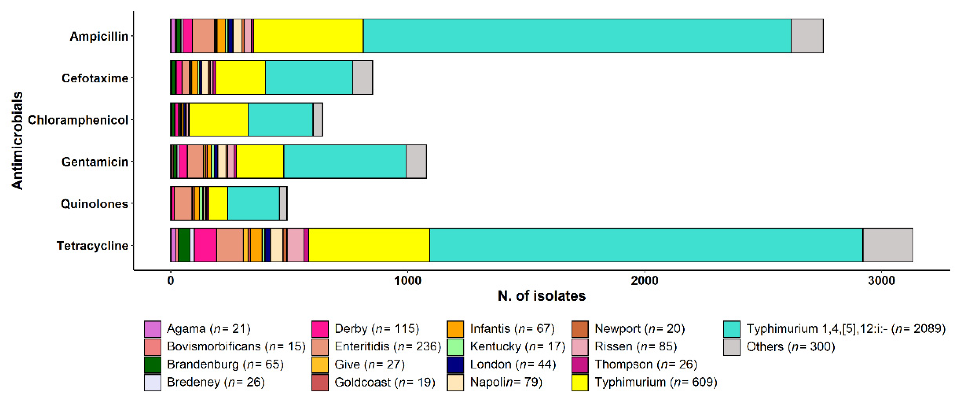
3.1. Salmonella Serotyping

A total of 168 Salmonella serotypes were identified (Table S1), albeit few were responsible for the majority of human infection cases (Table 1). S. ser. Typhimurium 1,4,[5],12:i:-, S. ser. Typhimurium, and S. ser. Enteritidis were predominant during almost the entire time period, except for the last two years when S. ser. Brandenburg (in both 2020 and 2021) and S. ser. Napoli (in 2020) were the most frequent serotypes together with S. ser. Typhimurium 1,4,[5],12:i:- (Figure 1). The distribution of Salmonella serotypes differed during the study period, with a sharp decrease in the occurrence of S. ser. Typhimurium (Wilcoxon-type test for trend, p < 0.05) and a generally stable occurrence of S. ser. Typhimurium 1,4,[5],12:i: and S. ser. Enteritidis. The prevalence of the remaining seven most frequent serotypes increased over the years, especially S. ser. Brandenburg (prevalence, 0.9% in 2012 vs. 7.8% in 2021; p < 0.05), S. ser. Infantis (0.5% vs. 2.5%; p < 0.01), and S. ser. Derby (1.9% vs. 4.6%; p < 0.05), while no substantial changes in prevalence were detected for S. ser. Napoli (prevalence, 3.5% vs. 4.3%) during the study period (Figure 1).
The ten most frequent serotypes were isolated from 3963 human patients, most of whom showing gastrointestinal infections (n = 3885). These serotypes were also recorded as causative agents of extraintestinal infections, including urinary tract infections (n = 88) and bacteriemia (n = 88). A single case of neuroinvasive infection caused by S. ser. Typhimurium was detected after isolating the bacterium from the cerebrospinal fluid. Serotypes S. ser. Derby, S. ser. Enteritidis, and S. ser. Typhimurium were also isolated from three different infections involving pus samples. S. ser. Typhimurium 1,4,[5],12:i:-, S. ser. Typhimurium, and S. ser. Enteritidis were the most frequently isolated in cases of bacteriemia, and showed comparable prevalence (p > 0.05). By contrast, S. ser. Typhimurium 1,4,[5],12:i:- was most often detected in urinary tract infections (p < 0.001), accounting for 31.8% (95% CI = 22.3–42.6) of cases, followed in frequency by S. ser. Derby (17.0%; 95% CI = 9.9–26.6), S. ser. Typhimurium (13.6%; 95% CI = 7.2–22.6), and S. ser. Enteritidis (6.8%; 95% CI = 2.5–14.3).

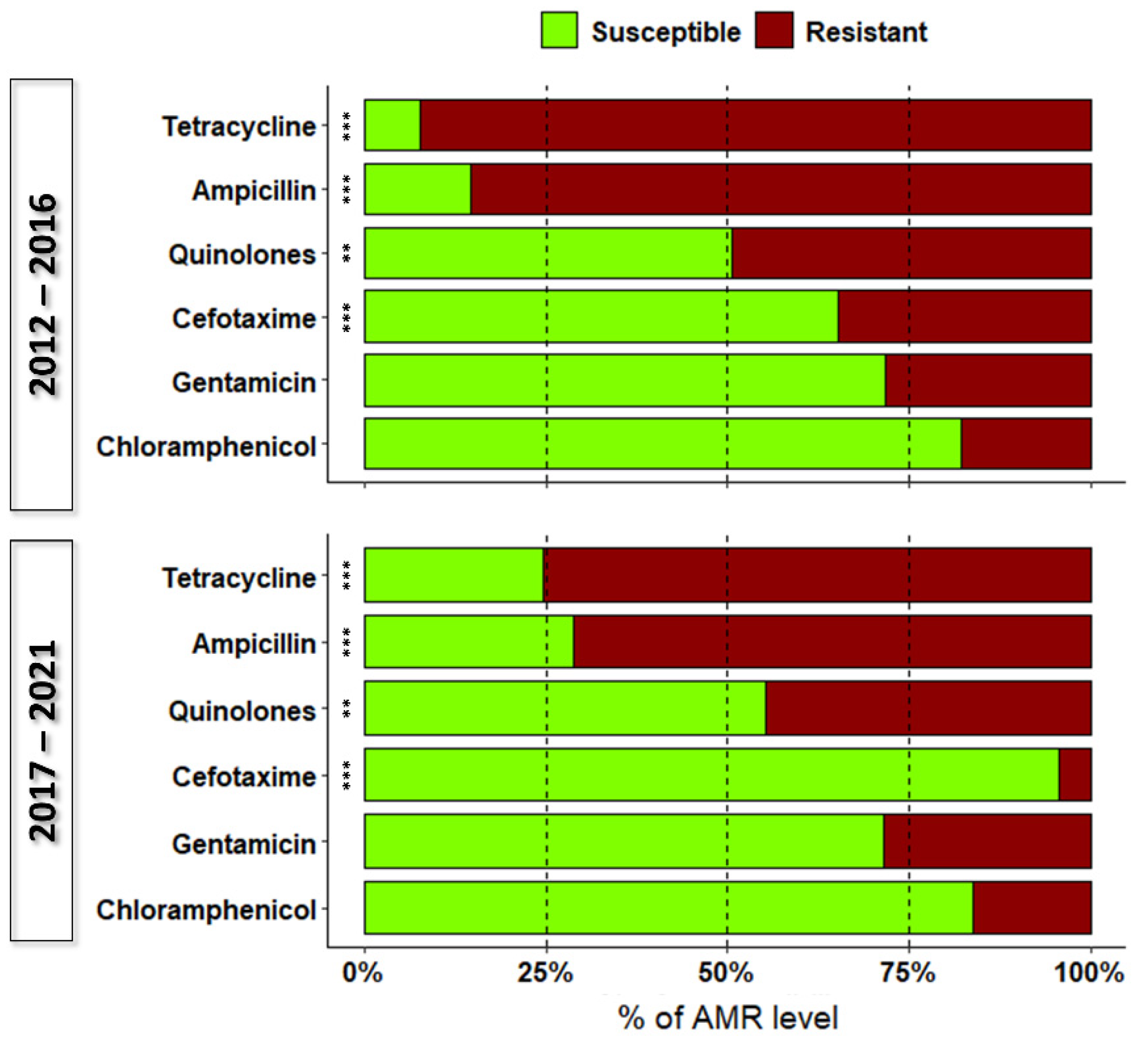
3.2. Antimicrobial Resistance

Antimicrobial resistance (AMR) was generally common in the Salmonella isolates investigated, with 83.3% (95% CI = 82.2–84.4) of the strains displaying resistance to at least one drug. However, antimicrobial susceptibility differed between the three most frequent (13.5%; 95% CI = 12.4–14.5) and minor Salmonella serotypes (34.4%; 95% CI = 31.0–38.0; Pearson’s chi-squared test, p < 0.001). The prevalence of antimicrobial resistant Salmonella strains decreased during the study period (Wilcoxon-type test, p < 0.05), with an annual prevalence of 94.4% (95% CI = 92.1–96.1) in 2012 and 64.6% (95% CI = 59.8–69.4) in 2021. This trend was evident for the ten most frequent serotypes between the first and the second half of the study period, except for S. ser. Enteritidis, S. ser. Rissen, and S. ser. London in which resistance levels remained unchanged over the same period (Figure 2). However, the pattern for S. ser. Bovismorbificans was difficult to assess because the sample size varied considerably (8/8 resistant isolates in 2012–2016 vs. 7/37 in 2017–2021).
We generally observed differences in AMR according to the type of infections (p < 0.001). Salmonella strains involved in gastrointestinal infections experienced the highest levels of AMR (80.7%; 95% CI = 79.5–81.9) compared with those involved in urinary tract infections (73.9%; 95% CI = 64.7–81.8) and bacteriemia (68.2%; 95% CI = 59.4–76.1), which displayed comparable resistance levels (p > 0.05).
Only 17.9% (95% CI = 16.7–19.2) of Salmonella strains were resistant to a single antibiotic class. The highest levels of AMR were observed against tetracycline and ampicillin, with 85.1% (95% CI = 83.9–86.3) and 79.2% (95% CI = 77.8–80.6) of Salmonella strains exhibiting resistance against these antimicrobials, respectively. These antibiotics were followed by the quinolones, nalidixic acid, and ciprofloxacin, with a joint AMR prevalence of 47.4% (95% CI = 45.7–49.1), gentamicin (28.4%; 95% CI = 26.8–29.9), and cefotaxime (21.2%; 95% CI = 19.9–22.6). The lowest resistance was displayed against chloramphenicol (17.1%; 95% CI = 15.8–18.4). Antimicrobial resistance differed for most drugs tested between the two time periods (Pearson’s chi-squared test, p < 0.001; Figure 3), except for gentamicin and chloramphenicol for which the level of AMR remained constant (p > 0.05). Resistance against the combination of two antibiotics was particularly frequent for S. ser. Typhimurium 1,4,[5],12:i:-, with 45.4% (95% CI = 43.3–47.5) of strains showing this pattern. We recorded 14 different two-drug combinations; AMR against ampicillin–tetracycline (67.8%; 95% CI = 65.3–70.3) and quinolones–gentamicin (13.5%; 95% CI = 11.7–15.4) combinations was common.
Antimicrobial resistance to tetracycline (TET) and ampicillin (AMP) was extremely high in S. ser. Agama (TET 100%; AMP 95.2%), S. ser. Typhimurium 1,4,[5],12:i:- (TET 87.6%; AMP 86.5%), S. ser. Newport (TET 85%; AMP 55%), S. ser. Typhimurium (TET 83.9%; AMP 76%), S. ser. Rissen (TET 83.5%; AMP 34.1%), and S. ser. Derby (TET 81.7%; AMP 33.9%) (Figure 4).

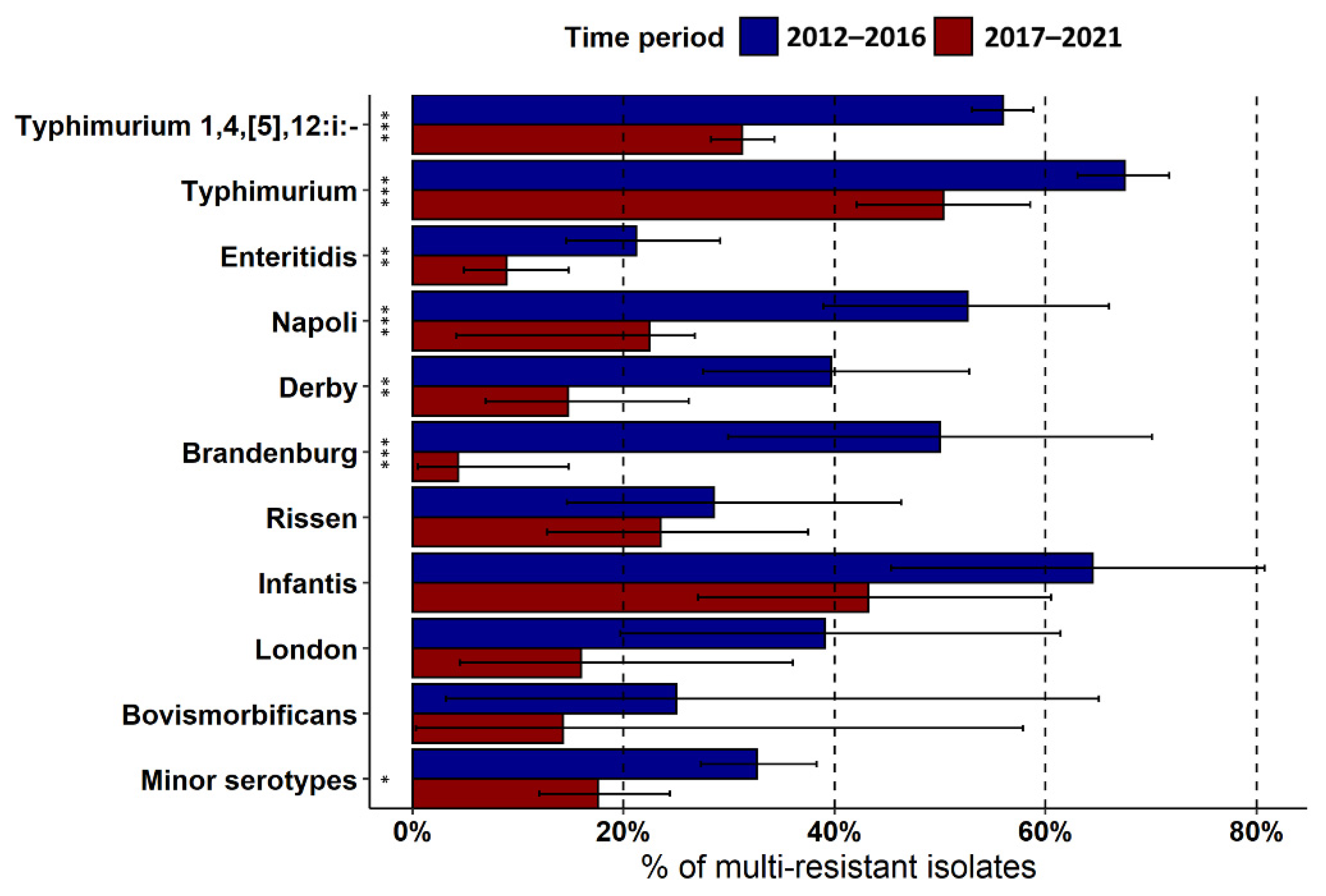
Multidrug Resistance (MDR)

Multidrug resistance patterns (≥3 classes of antibiotics) were rather frequent, accounting for 43.6% (95% CI 42.0–45.3) of the resistant strains. We observed the highest levels of MDR in S. ser. Typhimurium, S. ser. Infantis, and S. ser. Typhimurium 1,4,[5],12:i:-, which accounted for 63.3%, 52.9%, and 44.7% of resistant strains, respectively, followed in frequency by S. ser. Napoli (36.1%), S. ser. Derby (27.4%), S. ser. London (27.1%), S. ser. Rissen (25.6%), S. ser. Brandenburg (20.8%), and S. ser. Enteritidis (14.8%).
The overall prevalence of MDR in the prevailing serotypes decreased significantly over the years (Pearson’s chi-squared test, p < 0.001) and was, on average, 60.7% (95% CI = 56.2–65.1) in 2012 and 26.4% (95% CI = 20.9–32.5) in 2021. This trend was plainly evident for most of the prevailing serotypes, except for S. ser. Rissen, S. ser. Infantis, and S. ser. London (Figure 5). With regards to the type of infections, we detected comparable levels of MDR between strains involved in gastrointestinal (34.7; 95% CI = 33.2–36.3), urinary infections (34.1%; 95% CI = 24.0–45.4), and cases of bacteriemia (27.3; 95% CI = 18.3–37.8).
Multidrug resistance patterns against the six antimicrobials classes tested (AMP–FOT–CHL–GEN–TET–CIP/NAL) was observed in 87 strains. Six different combinations of antimicrobials were observed in MDR phenotypes involving five antibiotics with AMP–FOT–GEN–TET–CIP/NAL (53.5%; 95% CI 47.4–59.5), AMP–CHL–GEN–TET–CIP/NAL (22.9%; 95% CI = 18.1–28.3), and AMP–FOT–CHL–TET–CIP/NAL (14.9%; 95% CI = 10.9–19.7) being the most prevalent and observed in 275 strains. Higher numbers of combinations were ascertained in MDR phenotypes involving three (n = 18) and four (n = 13) drugs. These latter occurred in 506 strains, in which the most frequent combinations were AMP–FOT–TET–CIP/NAL (29.1%; 95% CI = 25.1–33.2), AMP–GEN–TET–CIP/NAL (28.3%; 95% CI = 24.4–32.4), and AMP–CHL–TET–CIP/NAL (16.6%; 95% CI = 13.5–20.1). The combination AMP–TET–CIP/NAL (43.7%; 95% CI = 40.0–47.6) prevailed among strains displaying resistance to three antibiotics (n = 667), followed by AMP–CHL–TET (17.2%; 95% CI = 14.4–20.3), and AMP–FOT–TET (9.4%; 95% CI = 7.3–11.9).

4. Discussion

Global monitoring of the emergence of antimicrobial resistance of Salmonella strains is essential for protecting public health. In developing countries, for example, the spread of antimicrobial resistant Salmonella strains can cause more acute and invasive infections, in addition to treatment failure and greater risk of mortality [21].
Here we analyzed the AMR of Salmonella serotypes isolated from human biological samples between 2012 and 2021 against six antimicrobial classes and 52 antimicrobial combinations. We found highly diverse resistance pattern phenotypes. Among the 168 different Salmonella serotypes involved in human infections, the three most frequent were S. ser. Typhimurium 1,4,[5],12:i:-, S. Typhimurium, and S. ser. Enteritidis, which were detected over almost the entire study period, as previously reported by global studies [1,22,23]. In addition, 3.6% and 3.4% of the isolates were S. ser. Napoli and S. ser. Derby, respectively. The most detected serotype in Piedmont was S. ser. Typhimurium 1,4,[5],12:i:-, which is consistent with previous reports published at national level [24]. By contrast, S. ser. Enteritidis is the serovar that is more frequently identified in EU, however an increase in prevalence has been observed for S. ser. Typhimurium 1,4,[5],12:i:- while S. ser. Enteritidis is following a decreasing trend [1].
Among the iNTS strains, the most frequent serotypes were S. ser. Typhimurium 1,4,[5],12:i:-, S. ser. Typhimurium, and S. ser. Enteritidis, as reported by previous studies in the Netherlands [25], Greece [26], and the United States [27]. The serotypes that were frequently identified from urinary tract infections were S. ser. Typhimurium 1,4,[5],12:i:-, S. ser. Derby, and S. ser. Typhimurium, followed by S. ser. Enteritidis. This latter serotype together with S. ser. Typhimurium were the two serotypes most often associated with urinary tract infections in Brazil [28], Spain [29], and the United States [30], whereas S. ser. Derby was sporadically detected in urinary infections in the past [30,31].
Investigation of the susceptibility of Salmonella serotypes toward drugs indicated that most strains were resistant to at least one antibiotic and that the highest levels of resistance were against tetracycline, ampicillin, quinolones, and gentamicin. The high levels of resistance we observed is shared by previous reports and is of particular concern, since these antibiotics are commonly used in the first-line treatment of human and animal infections [12]. For instance, fluoroquinolones are the gold standard in treatment against invasive salmonellosis in human medicine, and ampicillin and tetracycline are widely used in veterinary medicine as first-line treatments [32]. The resistance to third-generation cephalosporines we noted was moderate, albeit higher than that reported previously [22,33]. In the present study, chloramphenicol had the lowest level of resistance, likely due to its use in veterinary medicine having been prohibited in Europe since January 1997 [34]. Though antimicrobial resistance to chloramphenicol is still present, it appears to be decreasing [35]. To compare antibiotic resistance patterns, we divided the study into two time periods. Resistance rates significantly decreased during the latter half of the study period, especially for S. ser. Typhimurium 1,4,[5],12:i:-, S. ser. Typhimurium, S. ser. Napoli, S. ser. Derby, S. ser. Brandenburg, and S. ser. Infantis. During both periods, we observed a reduction in resistance to tetracycline, ampicillin, quinolones, and cefotaxime. Similarly, a decline in resistance to tetracyclines and ampicillin in Salmonella from humans was observed in other European countries (nine and ten countries, respectively) over the period from 2015 to 2019, which was particularly true for S. ser. Typhimurium [12]. Decreasing trends of resistance were more commonly observed for ampicillin in S. ser. Typhimurium (nine countries) and for tetracycline in Salmonella spp. (eleven countries), S. ser. Typhimurium (nine countries), S. ser. Typhimurium 1,4,[5],12:i:- (six countries), and S. ser. Infantis (two countries). Despite the decline, resistance to these antibiotics remains high in bacteria isolated from humans and animals [12]. These resistance data are of particular concern for clinicians who use antimicrobials (e.g., fluoroquinolones and cephalosporins) in the treatment of children and in the early treatment of severe gastroenteritis or invasive infections in adults.
Worldwide actions have been undertaken to prevent the emergence of drug-resistant bacteria and to promote food safety and public health through plans to ban or reduce the use of certain antimicrobials. The development of AMR can be slowed by restricting inappropriate use of antimicrobials and by improving hygiene conditions and practices in healthcare settings or in the food chain to reduce the transmission of resistant microorganisms, where more than one cause may play a role. Furthermore, the application of European regulations concerning the utilization of antibiotics in veterinary medicine has had a positive impact on slowing the spread of AMR. The JIACRA report also identifies links between antimicrobial consumption in animals and AMR in bacteria from food-producing animals, which is associated with antimicrobial resistance in bacteria from humans. Data also show that the use of antibiotics has decreased and is now lower in food-producing animals than in humans [36].
Regarding MDR strains, our data showed that almost 50% of Salmonella strains are resistant to more than three classes of antibiotics. According to European data [9], MDR is highest in S. ser. Typhimurium, S. ser. Infantis, and S. ser. Typhimurium 1,4,[5],12:i:-. In our study, the number of MDR isolates was far higher in the first half than the second half of the study period, in which the significant reduction in the occurrence of S. ser. Typhimurium 1,4,[5],12:i:-, S. ser. Typhimurium, S. ser. Enteritidis, S. ser. Napoli, S. ser. Derby, and S. ser. Brandenburg may be ascribed to the implementation of Italian and European control programs for the eradication of Salmonella in poultry [37].
We observed high levels of resistance in S. ser. Typhimurium 1,4,[5],12:i:-, S. ser. Typhimurium, S. ser. Enteritidis, S. ser. Infantis, S. ser. Derby, and S. ser. Newport against ampicillin and tetracycline. Many human Salmonella strains were resistant to ampicillin, sulfonamides, and tetracyclines, as reported in other countries [12].
This study has benefited from the well-structured and coordinated surveillance health system implemented in the region. Collaboration among different health services has made possible the collection of a significant amount of data on human salmonellosis in the long term, enabling the evaluation of temporal trends of Salmonella strains and identify the main AMR patterns occurring in human infections. Notwithstanding, our study is limited to only six antibiotic classes because of the monitoring of AMR involving other antimicrobials was sometimes interrupted, thus preventing comparisons over time. Moreover, our survey was restricted to a specific geographical context, not allowing our findings to be generalized to other Italian or European regions.

5. Conclusions

Our study focused on the occurrence and the antibiotic resistance of Salmonella spp. detected in humans in northwest Italy over a 10 year period. High AMR levels were uncovered among Salmonella strains toward tetracycline, ampicillin, and quinolones in particular. These data provide supplementary information about AMR in human strains of Salmonella. The decreasing trend of AMR experienced by Salmonella in Piedmont is consistent with data from the European Union and demonstrates the effectiveness of measures implemented in human and veterinary medicine.

Supplementary Materials

The following supporting information can be downloaded at: https://www.mdpi.com/article/10.3390/pathogens12010089/s1, Table S1: Prevalence of the Salmonella enterica serotypes involved in human infections in Piedmont, northwestern Italy, 2012–2021.

Author Contributions

Conceptualization, M.P. and C.T.; methodology, C.T. and M.P.; data curation, M.P., A.G.-V. and C.M.; writing—original draft preparation, M.P., C.T. and A.G.-V.; writing—review and editing, M.P., C.T., A.G.-V., C.M. and L.D.; supervision, M.P. and L.D.; project administration, M.P., C.M. and L.D.; CeRTiS Clinical Laboratories Group (Zaccaria T., Laboratorio di Microbiologia AOU Città della Salute e della Scienza Torino; Mussino S., Laboratorio Analisi Humanitas Gradenigo Torino; Casonato I., Laboratorio analisi chimico-cliniche e microbiologia, AO Ordine Mauriziano Torino; Vuolo A., Laboratorio analisi Presidio Martini OMV Torino; Turchi A., Laboratorio Analisi Ospedale Koelliker pro Infantia SPA Torino; Del Re S., Laboratorio di Microbiologia e Virologia PO Amedeo di Savoia Torino; Bellizia L., Laboratorio di Analisi Chimico Cliniche e Microbiologiche Ospedale Cottolengo Torino; Amarù G. and Millesimo M., Laboratorio Analisi Unificato Rivoli—Pinerolo ASL TO3 Rivoli-TO; Li Vigni N., Laboratorio Analisi e Microbiologia Ospedale Civico di Ivrea-TO; Allocco A., Laboratorio analisi chimico-cliniche e microbiologiche ASL TO5 ospedali riuniti Carmagnola, Chieri, Moncalieri-TO; Leli C., Laboratorio di Microbiologia Ospedale Civile Santi Antonio e Biagio e Cesare Arrigo Alessandria; Salerno A., Laboratorio Analisi e Microbiologia Ospedale Civile SS Antonio e Margherita Tortona-AL; Concialdi E., Laboratorio Analisi chimiche cliniche e microbiologiche Ospedale Cardinal Massaia Asti; Piana F., Laboratorio Analisi Chimico Cliniche e microbiologia AO S. Croce e Carle Cuneo; Vinai E., Laboratorio Microbiologia Ospedale Regina Montis Regalis Mondovì-CN; Comessatti A., Laboratorio Analisi—Microbiologia Ospedale Michele e Pietro Ferrero Verduno-CN; Malabaila A., Laboratorio Analisi e Microbiologia Nuovo Ospedale degli Infermi Biella; Tinivella A., Laboratorio Analisi Chimico Cliniche e Microbiologiche Ospedale SS. Trinità Borgomanero-NO; Gobatto E., Laboratorio Microbiologia e Virologia Ospedale Maggiore della Carità Novara; Lodolo L., Laboratorio Analisi Chimico Cliniche e Microbiologiche, Ospedale Castelli Verbania-VCO; Caffiero G., Laboratorio Analisi e Microbiologia Ospedale S. Andrea Vercelli; Rondinelli E., Laboratorio Analisi Istituto Auxologico Italiano Verbania-VCO) provided Salmonella strains. All authors have read and agreed to the published version of the manuscript.

Funding

This study received no external funding.

Institutional Review Board Statement

Salmonella strains, isolated from patients’ samples, examined for this study were submitted to CeRTiS for surveillance purposes, within the framework of the Directive 2003/99/EC on the monitoring of zoonoses and zoonotic agents. In addition, information and consent to medical intervention was obtained from patients visited by medical doctors, in accordance with the current Italian legislation. All data on humans were treated in anonymized modes and were used solely for the purposes of scientific research.

Informed Consent Statement

Patient consent was waived due to the anonymity of the samples used and personal data were not used.

Data Availability Statement

Not applicable.

Conflicts of Interest

The authors declare that the study was carried without personal, professional or financial relationships that could be potentially construed as a conflict of interest.

References

  1. EFSA and ECDC (European Food Safety Authority and European Centre for Disease Prevention and Control). The European Union One Health 2020 Zoonosis Report. EFSA J. 2021, 19, 6971. [Google Scholar] [CrossRef]
  2. European Centre for Disease Prevention and Control. Salmonellosis. ECDC Annual Epidemiological Report for 2021; ECDC: Stockholm, Sweden, 2022. Available online: https://www.ecdc.europa.eu/en/publications-data/salmonellosis-annual-epidemiological-report-2021 (accessed on 27 December 2022).
  3. Gal-Mor, O.; Boyle, E.C.; Grassl, G.A. Same species, different diseases: How and why typhoidal and non-typhoidal Salmonella enterica serovars differ. Front. Microbiol. 2014, 5, 391. [Google Scholar] [CrossRef] [PubMed] [Green Version]
  4. Butler, T.; Knight, J.; Nath, S.K.; Speelman, P.; Roy, S.K.; Azad, M.A. Typhoid fever complicated by intestinal perforation: A persisting fatal disease requiring surgical management. Rev. Infect. Dis. 1985, 7, 244–256. [Google Scholar] [CrossRef] [PubMed]
  5. Crump, J.A. Progress in Typhoid Fever Epidemiology. Clin. Infect. Dis. 2019, 68 (Suppl. 1), S4–S9. [Google Scholar] [CrossRef] [Green Version]
  6. Shinohara, N.K.; Barros, V.B.; Jimenez, S.M.; Machado Ede, C.; Dutra, R.A.; De Lima Filho, J.L. Salmonella spp., important pathogenic agent transmitted through foodstuffs. Ciência Saúde Coletiva 2008, 13, 1675–1683. [Google Scholar] [CrossRef] [Green Version]
  7. Berger, C.N.; Sodha, S.V.; Shaw, R.K.; Griffin, P.M.; Pink, D.; Hand, P.; Frankel, G. Fresh fruit and vegetables as vehicles for the transmission of human pathogens. Environ. Microbiol. 2010, 12, 2385–2397. [Google Scholar] [CrossRef]
  8. Grivas, G.; Lagousi, T.; Mandilara, G. Epidemiological Data, Serovar Distribution and Antimicrobial Resistance Patterns of Salmonella Species in Children, Greece 2011–2017: A Retrospective Study. Acta Med. Acad. 2020, 49, 255–264. [Google Scholar] [CrossRef] [PubMed]
  9. Marchello, C.S.; Fiorino, F.; Pettini, E.; Crump, J.A. Incidence of non-typhoidal Salmonella invasive disease: A systematic review and meta-analysis. J. Infect. 2021, 83, 523–532. [Google Scholar] [CrossRef] [PubMed]
  10. Akinyemi, K.O.; Ajoseh, S.O.; Fakorede, C.O. A systemic review of literatures on human Salmonella enterica serovars in Nigeria (1999–2018). J. Infect. Dev. Ctries. 2021, 15, 1222–1235. [Google Scholar] [CrossRef]
  11. Kariuki, S.; Gordon, M.A.; Feasey, N.; Parry, C.M. Antimicrobial resistance and management of invasive Salmonella disease. Vaccine 2015, 33 (Suppl. 3), C21–C29. [Google Scholar] [CrossRef]
  12. EFSA and ECDC (European Food Safety Authority and European Centre for Disease Prevention and Control). The European Union Summary Report on Antimicrobial Resistance in zoonotic and indicator bacteria from humans, animals and food in 2019/2020. EFSA J. 2022, 20, 7209. [Google Scholar] [CrossRef]
  13. Eng, S.K.; Pusparajah, P.; Ab Mutalib, N.S.; Ser, H.L.; Chan, K.G.; Lee, L.H. Salmonella: A review on pathogenesis, epidemiology and antibiotic resistance. Front. Life Sci. 2015, 8, 284–293. [Google Scholar] [CrossRef] [Green Version]
  14. Kauffmann-White and Le Minor. Antigenic formulae of the Salmonella serovars. Bergey’s Man. Syst. Bacteriol. 1984, 1, 429–445. Available online: https://www.pasteur.fr/sites/default/files/veng_0.pdf (accessed on 27 December 2022).
  15. Matuschek, E.; Brown, D.F.J.; Kahlmeter, G. Development of the EUCAST disk diffusion antimicrobial susceptibility testing method and its implementation in routine microbiology laboratories. Clin. Microbiol. Infect. 2014, 20, O255–O266. [Google Scholar] [CrossRef] [PubMed] [Green Version]
  16. The European Committee on Antimicrobial Susceptibility Testing (EUCAST). Breakpoint Tables for Interpretation of MICs and Zone Diameters, Version 11.0; European Society of Clinical Microbiology and Infectious Diseases: Växjö, Sweden, 2021; Available online: http://www.eucast.org (accessed on 27 December 2022).
  17. CLSI Document M100-S22; Performance Standards for Antimicrobial Susceptibility Testing: Twenty-Second Informational Supplement. Clinical and Laboratory Standards Institute: Wayne, PA, USA, 2012.
  18. CLSI Document M100-S23; Performance Standards for Antimicrobial Susceptibility Testing: Twenty-Third Informational Supplement. Clinical and Laboratory Standards Institute: Wayne, PA, USA, 2014.
  19. StataCorp LLC. Stata Statistical Software, Release 16; StataCorp LLC: Collage Station, TX, USA, 2019. [Google Scholar]
  20. Cuzick, J. A Wilcoxon-type test for trend. Stat. Med. 1985, 4, 87–90. [Google Scholar] [CrossRef]
  21. Breurec, S.; Reynaud, Y.; Thierry, F.; Farra, A.; Costilhes, G.; Weill, F.X.; Le Hello, S. Serotype distribution and antimicrobial resistance of human Salmonella enterica in Bangui, Central African Republic, from 2004 to 2013. PLoS Negl. Trop. Dis. 2019, 13, 7917. [Google Scholar] [CrossRef] [Green Version]
  22. El-Tayeb, M.; Ibrahim, A.S.S.; Al-Salamah, A.A.; Almaary, K.S.; Elbadawi, Y.B. Prevalence, serotyping and antimicrobials resistance mechanism of Salmonella enterica isolated from clinical and environmental samples in Saudi Arabia. Braz. J. Microbiol. 2017, 48, 499–508. [Google Scholar] [CrossRef]
  23. Ceyssens, P.J.; Mattheus, W.; Vanhoof, R.; Bertrand, S. Trends in Serotype distribution and Antimicrobial Susceptybility in Salmonella enterica Isolates from Humans in Belgium, 2009 to 2013. Antimicrob. Agents Chemother. 2015, 59, 544–552. [Google Scholar] [CrossRef] [Green Version]
  24. Mastrorilli, E.; Pietrucci, D.; Barco, L.; Ammendola, S.; Petrin, S.; Longo, A.; Mantovani, C.; Battistoni, A.; Ricci, A.; Desideri, A.; et al. A Comparative Genomic Analysis Provides Novel Insights Into the Ecological Success of the Monophasic Salmonella Serovar 4,[5],12:i:-. Front. Microbiol. 2018, 17, 715. [Google Scholar] [CrossRef]
  25. Mughini-Gras, L.; Pijnacker, R.; Duijster, J.; Heck, M.; Wit, B.; Veldman, K.; Franz, E. Changing epidemiology of invasive non-typhoid Salmonella infection. Clin. Microbiol. Infect. 2020, 26, 941. [Google Scholar] [CrossRef]
  26. Galanakis, E.; Bitsori, M.; Maraki, S.; Giannakopoulou, C.; Samonis, G.; Tselentis, Y. Invasive non-typhoidal salmonellosis in immunocompetent infants and children. Int. J. Infect. Dis. 2007, 11, 36–39. [Google Scholar] [CrossRef] [PubMed]
  27. Jones, T.F.; Ingram, L.A.; Cieslak, P.R.; Vugia, D.J.; Tobin-D′Angelo, M.; Hurd, S.; Medus, C.; Cronquist, A.; Angulo, F.J. Salmonellosis outcomes differ substantially by serotype. J. Infect. Dis. 2008, 198, 109–114. [Google Scholar] [CrossRef] [PubMed]
  28. Oliveira dos Reis, R.; Cecconi, M.C.; Timm, L.; Neves Souza, M.; Ikuta, N.; Wolf, J.M.; Lunge, W.R. Salmonella isolates from urine cultures: Serotypes and antimicrobial resistance in hospital settings. Braz. J. Microbiol. 2019, 50, 445–448. [Google Scholar] [CrossRef] [PubMed]
  29. Ramos, J.M.; Aguado, J.M.; García-Corbeira, P.; Alés, J.M.; Soriano, F. Clinical spectrum of urinary tract infections due on nontyphoidal Salmonella species. Clin. Infect. Dis. 1996, 23, 388–390. [Google Scholar] [CrossRef]
  30. Sivapalasingam, S.; Hoekstra, R.M.; McQuiston, J.R.; Fields, P.I.; Tauxe, R.V. Salmonella bacteriuria: An increasing entity in elderly women in the United States. Epidemiol. Infect. 2004, 132, 897–902. [Google Scholar] [CrossRef]
  31. Allerberger, F.J.; Dierich, M.P.; Ebner, A.; Keating, M.R.; Steckelberg, J.M.; Yu, P.K.; Anhalt, J.P. Urinary tract infection caused by nontyphoidal Salmonella: Report of 30 cases. Urol. Int. 1992, 48, 395–400. [Google Scholar] [CrossRef]
  32. Peruzy, M.F.; Capuano, F.; Proroga, Y.T.R.; Cristiano, D.; Carullo, M.R.; Murru, N. Antimicrobial Susceptibility Testing for Salmonella Serovars Isolated from Food Samples: Five-Year Monitoring (2015–2019). Antibiotics 2020, 9, 365. [Google Scholar] [CrossRef]
  33. Burke, L.; Hopkins, K.L.; Meunier, D.; De Pinna, E.; Fitzgerald-Hughes, D.; Humphreys, H.; Woodford, N. Resistance to third-generation cephalosporins in human non-typhoidal Salmonella enterica isolates from England and Wales, 2010–2012. J. Antimicrob. Chemother. 2014, 69, 977–981. [Google Scholar] [CrossRef] [Green Version]
  34. Regulation, H. Council Regulation (EEC) No 2377/90 of 26 June 1990 laying down a Community procedure for the establishment of maximum residue limits of veterinary medicinal products in foodstuffs of animal origin. Off. JL 1990, 224, 1–8. [Google Scholar]
  35. Callens, B.; Cargnel, M.; Sarrazin, S.; Dewulf, J.; Hoet, B.; Vermeersch, K.; Wattiau, P.; Welby, S. Associations between a decreased veterinary antimicrobial use and resistance in commensal Escherichia coli from Belgian livestock species (2011–2015). Prev. Vet. Med. 2018, 157, 50–58. [Google Scholar] [CrossRef] [Green Version]
  36. European Centre for Disease Prevention and Control (ECDC); European Food Safety Authority (EFSA); European Medicines Agency (EMA). Third joint inter-agency report on integrated analysis of consumption of antimicrobial agents and occurrence of antimicrobial resistance in bacteria from humans and food-producing animals in the EU/EEA—JIACRA III 2016–2018. EFSA J. 2021, 19, 6712. [Google Scholar] [CrossRef]
  37. Proroga, Y.T.R.; Capuano, F.; Carullo, M.R.; La Tela, I.; Capparelli, R.; Barco, L.; Pasquale, V. Occurrence and antimicrobial resistance of Salmonella strains from food of animal origin in southern Italy. Folia Microbiol. 2016, 61, 21–27. [Google Scholar] [CrossRef] [PubMed]
Figure 1. Prevalence of Salmonella serotypes involved in human infections in Piedmont during 2012–2021.
Pathogens 12 00089 g001
Figure 2. Prevalence and 95% CI of AMR in the most common serotypes recovered in the two halves of the study period. Prevalence is expressed as the number of isolates that showed resistant phenotypes against at least one drug. Asterisks denote significant differences in AMR between the two time periods (Pearson’s chi-squared test, * p < 0.05, ** p < 0.01, *** p < 0.001).
Pathogens 12 00089 g002
Figure 3. Percentage of susceptibility and resistance of Salmonella strains causing infections in humans in the two halves of the study period. Asterisks denote significant differences in AMR against each antibiotic between the two time periods (Pearson’s chi-squared test, ** p < 0.01; *** p < 0.001).
Pathogens 12 00089 g003
Figure 4. Number of strains resistant against the six classes of antimicrobials in each Salmonella serotype. Serotypes with fewer than 10 strains are summed and denoted as “Others”.
Pathogens 12 00089 g004
Figure 5. Prevalence and 95% CI of MDR in the most frequent serotypes recorded in the two halves of the study period. Asterisks denote significant differences in the prevalence of MDR between the two study periods (Pearson’s chi-squared test, * p < 0.05; ** p < 0.01; *** p < 0.001).
Pathogens 12 00089 g005
Table 1. Prevalence and 95% confidence interval (CI) of most prevailing Salmonella enterica subspp. enterica serotypes identified in human patients from Piedmont during 2012–2021.
SerotypeNo. Isolates (n = 4814)Prevalence (95% CI)
Typhimurium 1,4,[5],12:i:-218845.5 (44.0–46.9)
Typhimurium66613.8 (12.9–14.8)
Enteritidis4539.4 (8.6–10.3)
Napoli1723.6 (3.1–4.1)
Derby1643.4 (2.9–4.0)
Brandenburg1292.7 (2.2–3.2)
Rissen972.0 (1.6–2.5)
Infantis841.7 (1.4–2.2)
London681.4 (1.1–1.8)
Bovismorbificans440.9 (0.7–1.2)
Minor serotypes74915.6 (14.5–16.6)
Disclaimer/Publisher’s Note: The statements, opinions and data contained in all publications are solely those of the individual author(s) and contributor(s) and not of MDPI and/or the editor(s). MDPI and/or the editor(s) disclaim responsibility for any injury to people or property resulting from any ideas, methods, instructions or products referred to in the content.

