
Meta-Analysis of the Prevalence of Porcine Zoonotic Bacterial Pathogens in India: A 13-Year (2010–2023) Study
 , ,
, ,
Abstract
:1. Introduction
2. Materials and Methods
2.1. Literature Retrieval and Data Compilation
2.2. Methods Used for Meta-Analysis
3. Results
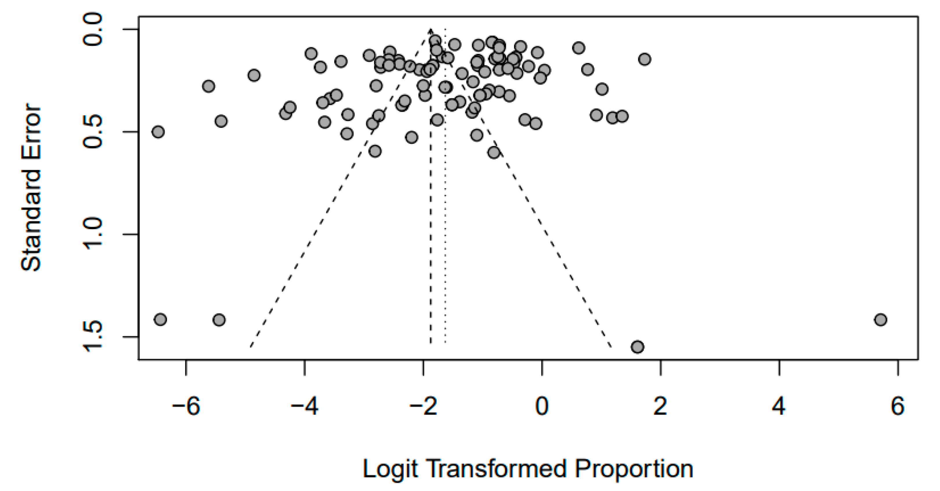
3.1. Meta-Analysis
3.2. Meta-Analysis of the Prevalence of Brucellosis in Pigs
3.3. Meta-Analysis of the Prevalence of Clostridium spp. in Pigs
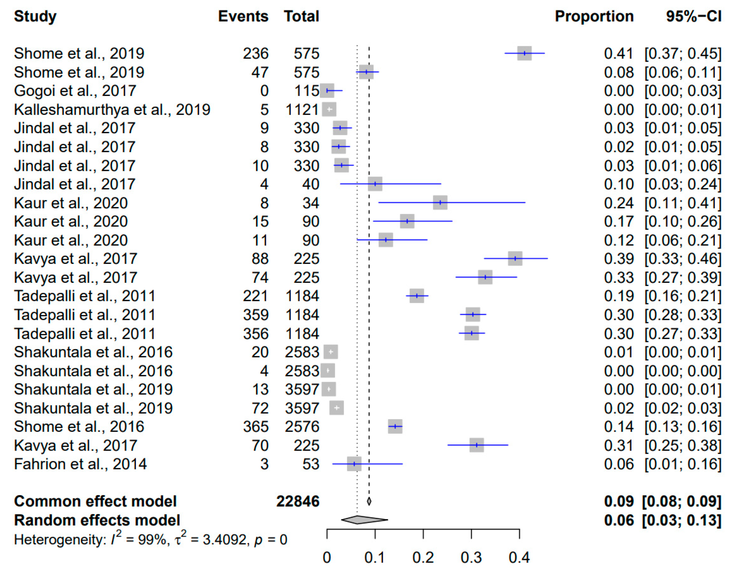
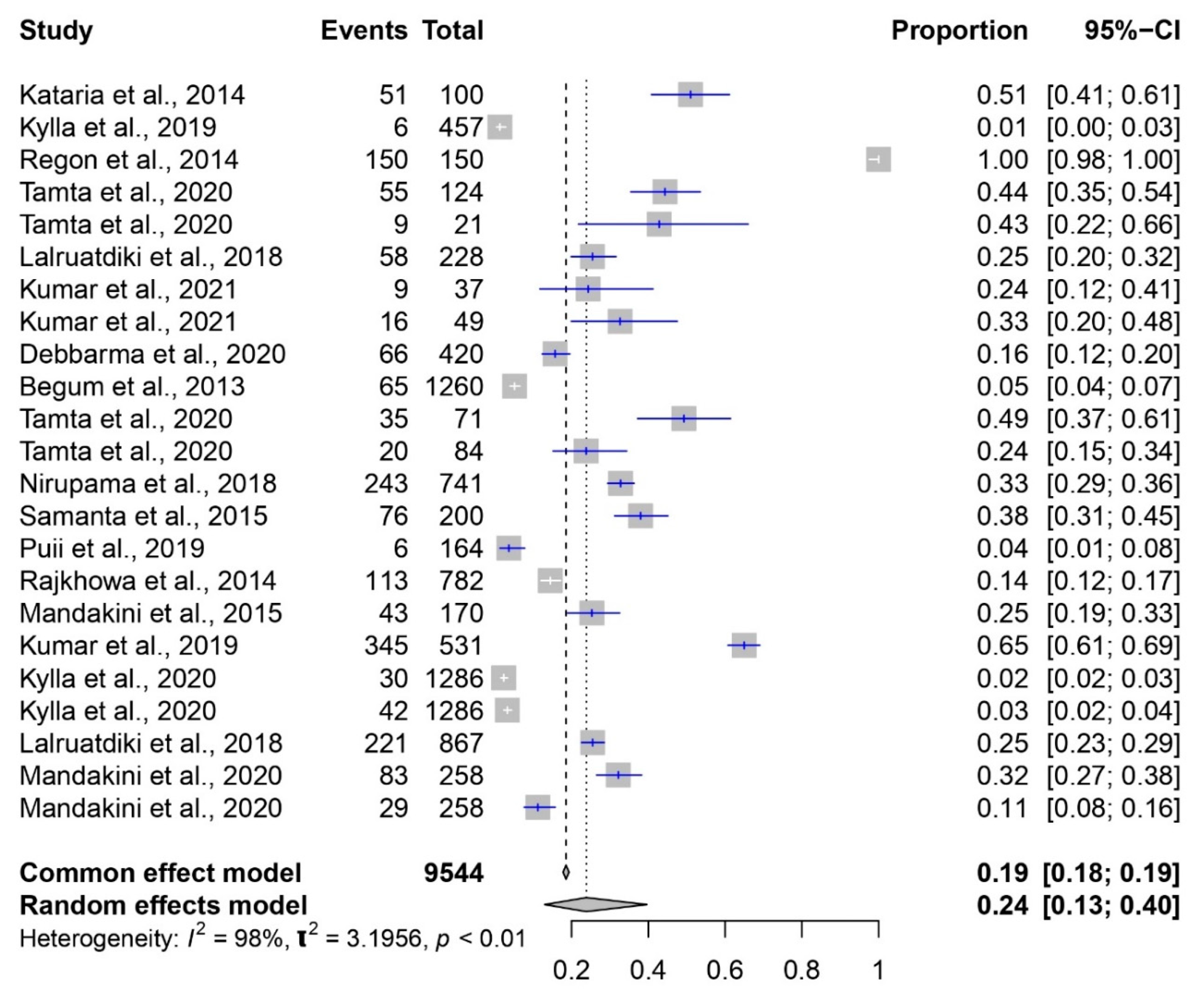
3.4. Meta-Analysis of the Prevalence of E. coli in Pigs
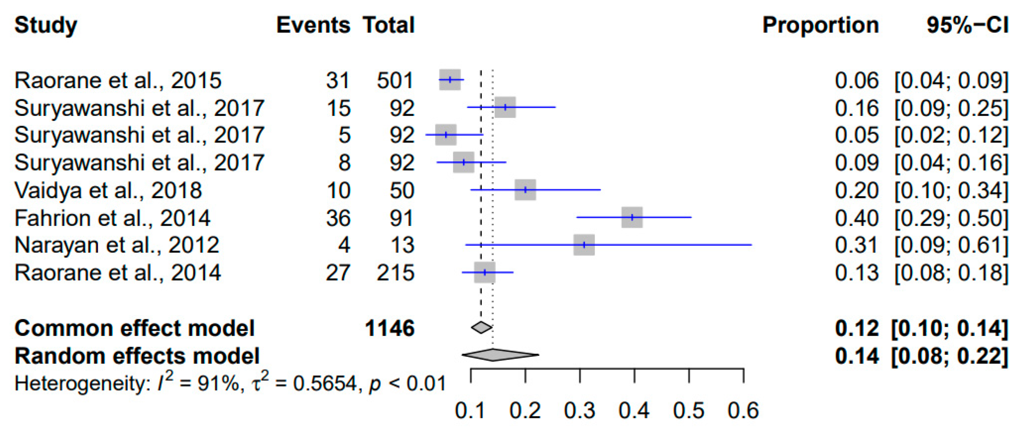
3.5. Meta-Analysis of the Prevalence of Listeria monocytogenes in Pigs
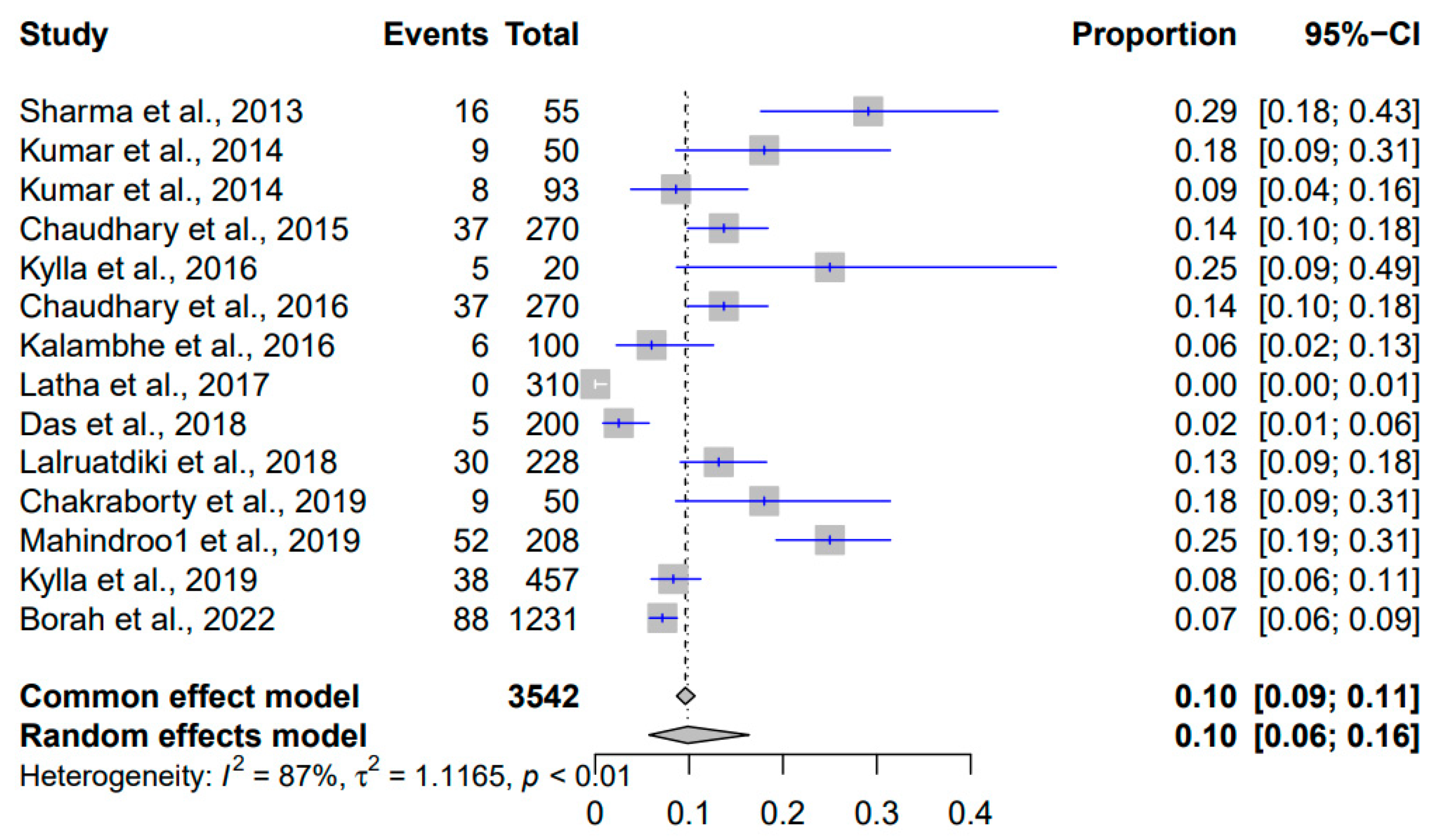
3.6. Meta-Analysis of the Prevalence of Salmonella spp. in Pigs
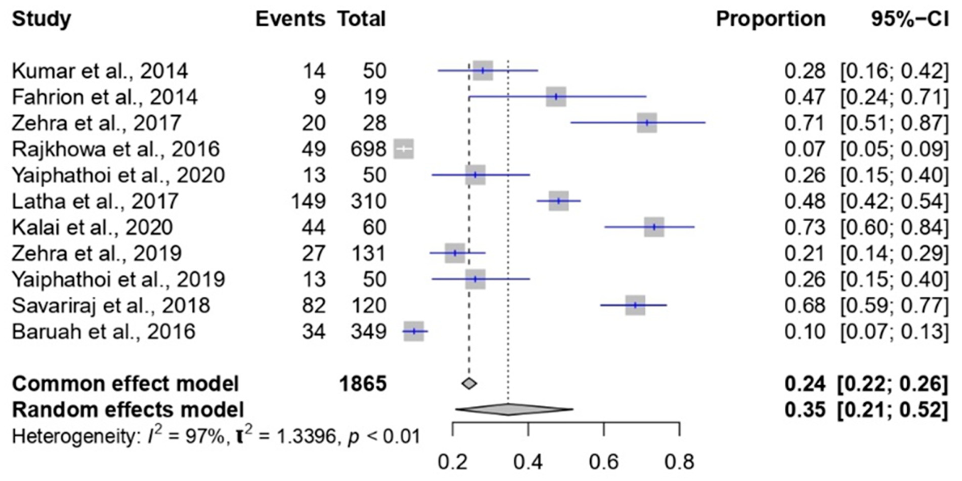
3.7. Meta-Analysis of the Prevalence of Staphylococcus spp. in Pigs
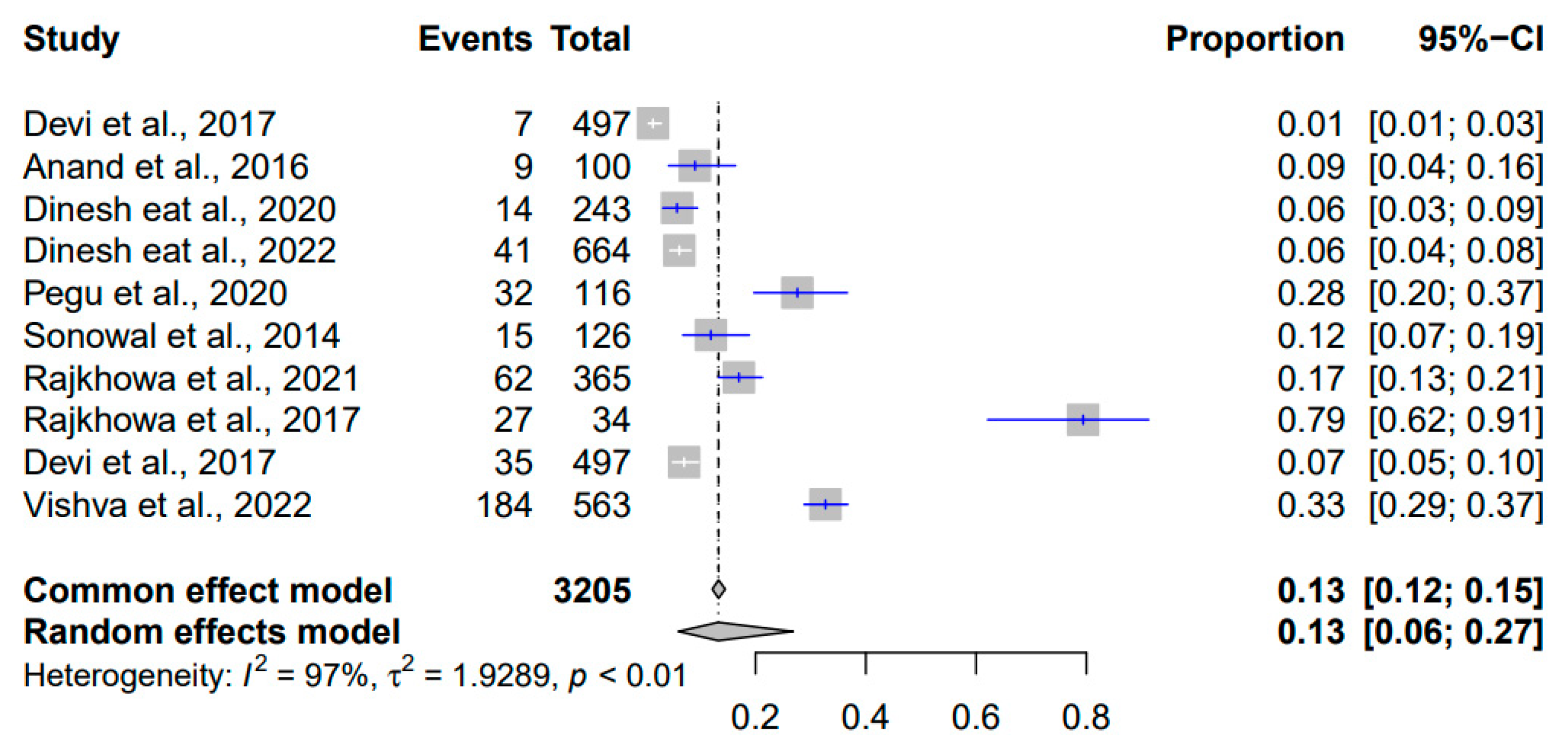
3.8. Meta-Analysis of the Prevalence of Streptococcus suis in Pigs
4. Discussion
5. Conclusions
Author Contributions
Funding
Institutional Review Board Statement
Informed Consent Statement
Data Availability Statement
Acknowledgments
Conflicts of Interest
References
- Uzal, F.A.; Navarro, M.A.; Asin, J.; Boix, O.; Ballarà-Rodriguez, I.; Gibert, X. Clostridial diarrheas in piglets: A review. Vet. Microbiol. 2023, 280, 109691. [Google Scholar] [CrossRef] [PubMed]
- Di Bella, S.; Ascenzi, P.; Siarakas, S.; Petrosillo, N.; di Masi, A. Clostridium difficile Toxins A and B: Insights into Pathogenic Properties and Extraintestinal Effects. Toxins 2016, 8, 134. [Google Scholar] [CrossRef] [PubMed]
- Pacheco, A.R.; Sperandio, V. Shiga toxin in enterohemorrhagic E.coli: Regulation and novel anti-virulence strategies. Front. Cell. Infect. Microbiol. 2012, 2, 81. [Google Scholar] [CrossRef]
- Castanheira, M.; Simner, P.J.; Bradford, P.A. Extended-spectrum β-lactamases: An update on their characteristics, epidemiology and detection. JAC-Antimicrob. Resist. 2021, 3, dlab092. [Google Scholar] [CrossRef] [PubMed]
- Bergšpica, I.; Kaprou, G.; Alexa, E.A.; Prieto, M.; Alvarez-Ordóñez, A. Extended Spectrum β-Lactamase (ESBL) Producing Escherichia coli in Pigs and Pork Meat in the European Union. Antibiotics 2020, 9, 678. [Google Scholar] [CrossRef]
- Dramsi, S.; Cossart, P. Listeriolysin O: A genuine cytolysin optimized for an intracellular parasite. J. Cell. Biol. 2002, 156, 943–946. [Google Scholar] [CrossRef] [PubMed]
- van Asten, A.J.A.M.; van Dijk, J.E. Distribution of “classic” virulence factors among Salmonella spp. FEMS Immunol. Med. Microbiol. 2005, 44, 251–259. [Google Scholar] [CrossRef] [PubMed]
- Pinchuk, I.V.; Beswick, E.J.; Reyes, V.E. Staphylococcal enterotoxins. Toxins 2010, 2, 2177–2197. [Google Scholar] [CrossRef]
- Hennekinne, J.-A.; De Buyser, M.-L.; Dragacci, S. Staphylococcus aureus and its food poisoning toxins: Characterization and outbreak investigation. FEMS Microbiol. Rev. 2012, 36, 815–836. [Google Scholar] [CrossRef]
- Tenenbaum, T.; Asmat, T.M.; Seitz, M.; Schroten, H.; Schwerk, C. Biological activities of suilysin: Role in Streptococcus suis pathogenesis. Future Microbiol. 2016, 11, 941–954. [Google Scholar] [CrossRef] [PubMed]
- Kouki, A.; Pieters, R.J.; Nilsson, U.J.; Loimaranta, V.; Finne, J.; Haataja, S. Bacterial Adhesion of Streptococcus suis to Host Cells and Its Inhibition by Carbohydrate Ligands. Biology 2013, 2, 918–935. [Google Scholar] [CrossRef]
- Barnett, T.C.; Cole, J.N.; Rivera-Hernandez, T.; Henningham, A.; Paton, J.C.; Nizet, V.; Walker, M.J. Streptococcal toxins: Role in pathogenesis and disease. Cell. Microbiol. 2015, 17, 1721–1741. [Google Scholar] [CrossRef] [PubMed]
- Hynes, W.; Sloan, M. Secreted Extracellular Virulence Factors. In Streptococcus pyogenes: Basic Biology to Clinical Manifestations; Ferretti, J.J., Stevens, D.L., Fischetti, V.A., Eds.; University of Oklahoma Health Sciences Center: Oklahoma City, OK, USA, 2016. [Google Scholar]
- Cunningham, M.W. Pathogenesis of Group A Streptococcal Infections. Clin. Microbiol. Rev. 2000, 13, 470–511. [Google Scholar] [CrossRef] [PubMed]
- Głowacka, P.; Żakowska, D.; Naylor, K.; Niemcewicz, M.; Bielawska-Drózd, A. Brucella-Virulence Factors, Pathogenesis and Treatment. Pol. J. Microbiol. 2018, 67, 151–161. [Google Scholar] [CrossRef]
- Celli, J.; de Chastellier, C.; Franchini, D.M.; Pizarro-Cerda, J.; Moreno, E.; Gorvel, J.P. Brucella evades macrophage killing via VirB-dependent sustained interactions with the endoplasmic reticulum. J. Exp. Med. 2003, 198, 545–556. [Google Scholar] [CrossRef]
- Pizarro-Cerdá, J.; Méresse, S.; Parton, R.G.; van der Goot, G.; Sola-Landa, A.; Lopez-Goñi, I.; Moreno, E.; Gorvel, J.P. Brucella abortus transits through the autophagic pathway and replicates in the endoplasmic reticulum of nonprofessional phagocytes. Infect. Immun. 1998, 66, 5711–5724. [Google Scholar] [CrossRef] [PubMed]
- World Health Organization. Brucellosis in Humans and Animals; WHO Press: Geneva, Switzerlad, 2006.
- EFSA. Scientific Opinion on the pathogenicity assessment of Shiga toxin-producing Escherichia coli (STEC) and the public health risk posed by contamination of food with STEC. EFSA J. 2020, 18, 105. [Google Scholar]
- Colello, R.; Cáceres, M.E.; Ruiz, M.J.; Sanz, M.; Etcheverría, A.I.; Padola, N.L. From Farm to Table: Follow-Up of Shiga Toxin-Producing Escherichia coli Throughout the Pork Production Chain in Argentina. Front. Microbiol. 2016, 7, 93. [Google Scholar] [CrossRef]
- Vallejo, P.; Cilla, G.; López-Olaizola, M.; Vicente, D.; Marimón, J.M. Epidemiology and Clinical Features of Listeriosis in Gipuzkoa, Spain, 2010–2020. Front. Microbiol. 2022, 13, 894334. [Google Scholar] [CrossRef]
- Schoder, D.; Guldimann, C.; Martlbauer, E. Asymptomatic Carriage of Listeria monocytogenes by Animals and Humans and Its Impact on the Food Chain. Foods 2022, 11, 3472. [Google Scholar] [CrossRef]
- Bonardi, S. Salmonella in the pork production chain and its impact on human health in the European Union. Epidemiol. Infect. 2017, 145, 1513–1526. [Google Scholar] [CrossRef]
- Werinder, A.; Aspan, A.; Backhans, A.; Sjolund, M.; Guss, B.; Jacobson, M. Streptococcus suis in Swedish grower pigs: Occurrence, serotypes, and antimicrobial susceptibility. Acta. Vet. Scand. 2020, 62, 36. [Google Scholar] [CrossRef]
- Algammal, A.M.; Hetta, H.F.; Elkelish, A.; Alkhalifah, D.H.H.; Hozzein, W.N.; Batiha, G.E.; El Nahhas, N.; Mabrok, M.A. Methicillin-Resistant Staphylococcus aureus (MRSA): One Health Perspective Approach to the Bacterium Epidemiology, Virulence Factors, Antibiotic-Resistance, and Zoonotic Impact. Infect. Drug. Resist. 2020, 13, 3255–3265. [Google Scholar] [CrossRef] [PubMed]
- Shaikh, S.; Fatima, J.; Shakil, S.; Rizvi, S.M.; Kamal, M.A. Antibiotic resistance and extended spectrum beta-lactamases: Types, epidemiology and treatment. Saudi J. Biol. Sci. 2015, 22, 90–101. [Google Scholar] [CrossRef]
- Mancuso, G.; Midiri, A.; Gerace, E.; Biondo, C. Bacterial Antibiotic Resistance: The Most Critical Pathogens. Pathogens 2021, 10, 1310. [Google Scholar] [CrossRef]
- Shome, R.; Kalleshamurthy, T.; Natesan, K.; Jayaprakash, K.R.; Byrareddy, K.; Mohandoss, N.; Sahay, S.; Shome, B.R.; Hiremath, J.; Rahman, H. Serological and molecular analysis for brucellosis in selected swine herds from Southern India. J. Infect. Public. Health. 2019, 12, 247–251. [Google Scholar] [CrossRef]
- Gogoi, S.; Hussain, P.; Sarma, P.C.; Barua, A.; Mahato, G.; Bora, D.; Konch, P.; Gogoi, P. Prevalence of bovine brucellosis in Assam, India. J. Entomol. Zool. Stud. 2017, 5, 179–185. [Google Scholar]
- Kalleshamurthy, T.; Yaranna, C.; Shekar, R.; Natesan, K.; Sahay, S.; Shome, B.R.; Rahman, H.; Barbuddhe, S.B.; Barman, N.N.; Das, S.K.; et al. Fluorescence polarization assay: Diagnostic evaluation for porcine brucellosis. J. Microbiol. Methods 2019, 156, 46–51. [Google Scholar] [CrossRef] [PubMed]
- Jindal, P.; Singh, B.B.; Kaur, P.; Gill, J.P.S. Sero-prevalence and molecular detection of Brucella species in slaughter pigs (Sus Scrofa) of Punjab, India. J. Anim. Res. 2017, 7, 495–499. [Google Scholar] [CrossRef]
- Kaur, A.; Mahajan, V.; Singh, N.; Filia, G.; Banga, H.; Leishangthem, G.; Singh, A. Patho-epidemiological and molecular diagnosis of swine brucellosis in Punjab. Indian J. Anim. Res. 2020, 54, 90–95. [Google Scholar] [CrossRef]
- Kavya, B.; Veeregowda, B.; Kamran, A.; Gomes, A.; Triveni, K.; Padmashree, B.; Shome, R. Comparative evaluation of blood based lateral flow assay for diagnosis of brucellosis in livestock species. Indian J. Anim. Sci. 2017, 87, 1068–1070. [Google Scholar]
- Tadepalli, M.; Rajendra, L.; Bhavesh, T.; Thiagarajan, D.; Srinivasan, V.A. Development and comparative evaluation of a competitive ELISA with rose bengal test and a commercial indirect ELISA for serological diagnosis of brucellosis. Indian J. Microbiol. 2011, 51, 528–530. [Google Scholar]
- Shakuntala, I.; Ghatak, S.; Sanjukta, R.; Sen, A.; Das, S.; Puro, A.; Dutta, A.; Kakoty, K. Incidence of brucellosis in livestock in North-Eastern India. Int. J. Infect. Dis. 2016, 45, 474. [Google Scholar] [CrossRef]
- Shakuntala, I.; Ghatak, S.; Das, S.; Milton, A.P.; Sanjukta, R.; Puro, K.-U.; Dutta, A.; Kakoty, K.; Karam, A.; Lalgruaipuii, L.; et al. Seroepidemiological survey of brucellosis and isolation of Brucella suis from swine herds of Meghalaya, North-East India. Indian J. Anim. Sci. 2020, 90, 12–16. [Google Scholar] [CrossRef]
- Shome, R.; Triveni, K.; Shankaranarayana, P.B.; Sahay, S.; Rao, N.; Shome, B.R.; Misri, J.; Rahman, H. Record of porcine brucellosis in India by indigenously developed indirect ELISA. Asian Pac. J. Trop. Dis. 2016, 6, 892–894. [Google Scholar] [CrossRef]
- Fahrion, A.S.; Jamir, L.; Richa, K.; Begum, S.; Rutsa, V.; Ao, S.; Padmakumar, V.P.; Deka, R.P.; Grace, D. Food-safety hazards in the pork chain in Nagaland, North East India: Implications for human health. Int. J. Environ. Res. Public Health 2014, 11, 403–417. [Google Scholar] [CrossRef]
- Yadav, J.P.; Das, S.C.; Dhaka, P.; Mukhopadhyay, A.K.; Chowdhury, G.; Naskar, S.; Malik, S.S. Pulsed-field gel electrophoresis of enterotoxic Clostridium perfringens type A isolates recovered from humans and animals in Kolkata, India. Int. J. Vet. Sci. Med. 2018, 6, 123–126. [Google Scholar] [CrossRef]
- Das, B.; Sharma, R.; Borah, P.; Das, S.; Barkalita, L.; Devi, R.M.; Baishya, B. Molecular characterization and toxin-typing of Clostridium difficile isolates of dogs and pigs from Assam and Mizoram of North East India. Curr. Sci. 2017, 113, 1099–1106. [Google Scholar] [CrossRef]
- Hazarika, R.; Sarmah, H.; Doley, M.K.; Saikia, D.P.; Hazarika, G.; Barkalita, L.M.; Deka, P.; Manoharan, S.; Sharma, R.K. Clostridioides difficile in food and food products of animal origin in Assam, India. Anaerobe 2023, 81, 102723. [Google Scholar] [CrossRef] [PubMed]
- Yadav, J.P.; Das, S.C.; Dhaka, P.; Vijay, D.; Kumar, M.; Mukhopadhyay, A.K.; Chowdhury, G.; Chauhan, P.; Singh, R.; Dhama, K. Molecular characterization and antimicrobial resistance profile of Clostridium perfringens type A isolates from humans, animals, fish and their environment. Anaerobe 2017, 47, 120–124. [Google Scholar] [CrossRef]
- Hussain, I.; Borah, P.; Sharma, R.; Rajkhowa, S.; Rupnik, M.; Saikia, D.; Hasin, D.; Hussain, I.; Deka, N.; Barkalita, L. Molecular characteristics of Clostridium difficile isolates from human and animals in the North Eastern region of India. Mol. Cell. Probes. 2016, 30, 306–311. [Google Scholar] [CrossRef]
- Hussain, M.I.; Borah, P.; Hussain, I.; Sharma, R.K.; Kalita, M.C. Densitometric analysis of rep-PCR data: Insight into genetic variability and transmission of Clostridium perfringens typed with an improved multiplex PCR. Anaerobe 2021, 70, 102383. [Google Scholar] [CrossRef] [PubMed]
- Hussain, M.I.; Borah, P.; Hussain, I.; Sharma, R.K.; Kalita, M.C. Necrotic enteritis by beta2toxin-producing Clostridium perfringens in doom pigs of assam, India. Int. J. Curr. Microbiol. Appl. Sci. 2017, 6, 1872–1876. [Google Scholar] [CrossRef]
- Kataria, J.; Dutta, T.; Roychoudhury, P.; Tiwari, J. Detection and molecular characterization of Shiga toxin producing Escherichia coli (STEC) autoagglutinating adhesion gene (saa) from piglets in Mizoram. Vet. World 2014, 7, 373–376. [Google Scholar] [CrossRef]
- Kylla, H.; Dutta, T.K.; Roychoudhury, P.; Subudhi, P.K. Coinfection of diarrheagenic bacterial and viral pathogens in piglets of Northeast region of India. Vet. World 2019, 12, 224. [Google Scholar] [CrossRef]
- Regon, M.; Pathak, D.; Tamuli, S.; Baruah, G. Serotyping of Escherichia coli isolated from piglet diarrhea. Vet. World 2014, 7, 614–616. [Google Scholar] [CrossRef]
- Tamta, S.; Kumar, O.R.V.; Singh, S.V.; Pruthvishree, B.S.; Karthikeyan, R.; Rupner, R.; Sinha, D.K.; Singh, B.R. Antimicrobial resistance pattern of extended-spectrum β-lactamase-producing Escherichia coli isolated from fecal samples of piglets and pig farm workers of selected organized farms of India. Vet. World 2020, 13, 360. [Google Scholar] [CrossRef]
- Lalruatdiki, A.; Dutta, T.; Roychoudhury, P.; Subudhi, P. Extended-spectrum β-lactamases producing multidrug resistance Escherichia coli, Salmonella and Klebsiella pneumoniae in pig population of Assam and Meghalaya, India. Vet. World 2018, 11, 868. [Google Scholar] [CrossRef]
- Kumar, S.; Gali, J.M.; Dutta, T.; Roychoudhury, P.; Subudhi, P. Development of isothermal lamp assay for detection of intimin gene (Eae) in E. coli associated with piglet diarrhea. Indian J. Anim. Res. 2021, 55, 956–959. [Google Scholar] [CrossRef]
- Debbarma, S.; Bora, D.P.; Hazarika, R.A.; Tamuly, S.; Barua, A.G.; Das, P.; Goswami, C.; Jerry, R.J.; Sinha, S.J.I.J.o.A.R. Molecular characterization of Escherichia coli isolates from food animals. Indian J. Anim. Res. 2020, 54, 985–993. [Google Scholar] [CrossRef]
- Begum, J.; Dutta, T.; Chandra, R.; Choudhary, P.R.; Varte, Z.; Bitew, M. Molecular and phenotypic characterization of shigatoxigenic Escherichia coli (STEC) and enteropathogenic E. coli (EPEC) from piglets and infants associated with diarrhoea in Mizoram, India. Afr. J. Biotechnol. 2014, 13, 1452–1461. [Google Scholar]
- Tamta, S.; OR, V.K.; Pruthvishree, B.; Karthikeyan, R.; Rupner, R.N.; Chethan, G.; Dubal, Z.; Sinha, D.; Singh, B.R. Faecal carriage of extended spectrum beta-lactamase (ESBL) and New Delhi metallo beta-lactamase (NDM) producing Escherichia coli between piglets and pig farmworkers. Comp. Immunol. Microbiol. Infect. Dis. 2020, 73, 101564. [Google Scholar] [CrossRef] [PubMed]
- Nirupama, K.; OR, V.K.; Pruthvishree, B.; Sinha, D.; Murugan, M.S.; Krishnaswamy, N.; Singh, B. Molecular characterisation of blaOXA-48 carbapenemase-, extended-spectrum β-lactamase-and Shiga toxin-producing Escherichia coli isolated from farm piglets in India. J. Glob. Antimicrob. Resist. 2018, 13, 201–205. [Google Scholar] [CrossRef]
- Samanta, I.; Joardar, S.N.; Mahanti, A.; Bandyopadhyay, S.; Sar, T.K.; Dutta, T.K. Approaches to characterize extended spectrum beta-lactamase/beta-lactamase producing Escherichia coli in healthy organized vis-a-vis backyard farmed pigs in India. Infect. Genet. Evol. 2015, 36, 224–230. [Google Scholar] [CrossRef] [PubMed]
- Puii, L.; Dutta, T.; Roychoudhury, P.; Kylla, H.; Chakraborty, S.; Mandakini, R.; Kawlni, L.; Samanta, I.; Bandopaddhay, S.; Singh, S. Extended spectrum beta-lactamase producing Shiga-toxin producing-Escherichia coli in piglets, humans and water sources in North East region of India. Lett. Appl. Microbiol. 2019, 69, 373–378. [Google Scholar] [CrossRef]
- Rajkhowa, S.; Sarma, D.K. Prevalence and antimicrobial resistance of porcine O157 and non-O157 Shiga toxin-producing Escherichia coli from India. Trop. Anim. Health. Prod. 2014, 46, 931–937. [Google Scholar] [CrossRef]
- Mandakini, R.; Dutta, T.K.; Chingtham, S.; Roychoudhury, P.; Samanta, I.; Joardar, S.N.; Pachauau, A.R.; Chandra, R. ESBL-producing Shiga-toxigenic E. coli (STEC) associated with piglet diarrhoea in India. Trop. Anim. Health Prod. 2015, 47, 377–381. [Google Scholar] [CrossRef]
- Kumar, V.O.; Singh, B.; Sinha, D.; Pruthvishree, B.; Tamta, S.; Dubal, Z.; Karthikeyan, R.; Rupner, R.N.; Malik, Y. Risk factor analysis, antimicrobial resistance and pathotyping of Escherichia coli associated with pre-and post-weaning piglet diarrhoea in organised farms, India. Epidemiol. Infect. 2019, 147, e174. [Google Scholar]
- Kylla, H.; Dutta, T.K.; Roychoudhury, P.; Subudhi, P.K.; Tolenkhomba, T.; Mandakini, R.; Kawlni, L. Antimicrobial Drug Resistance of Escherichia coli Isolated from Piglets in North East India. Curr. J. Appl. Sci. Technol. 2020, 39, 195–205. [Google Scholar] [CrossRef]
- Mandakini, R.; Roychoudhury, P.; Subudhi, P.; Kylla, H.; Samanta, I.; Bandyopadhayay, S.; Dutta, T. Higher prevalence of multidrug-resistant extended-spectrum β-lactamases producing Escherichia coli in unorganized pig farms compared to organized pig farms in Mizoram, India. Vet. World 2020, 13, 2752. [Google Scholar] [CrossRef]
- Raorane, A.V.; Doijad, S.P.; Poharkar, K.V.; Pathak, A.; Bhosle, S.; Barbuddhe, S. Isolation and genotypic characterization of Listera monocytogenes from pork and pork products. Int. J. Curr. Microbiol. App. Sci. 2015, 4, 788–798. [Google Scholar]
- Suryawanshi, R.D.; Malik, S.V.S.; Jayarao, B.; Chaudhari, S.P.; Savage, E.; Vergis, J.; Kurkure, N.V.; Barbuddhe, S.B.; Rawool, D.B. Comparative diagnostic efficacy of recombinant LLO and PI-PLC-based ELISAs for detection of listeriosis in animals. J. Microbiol. Methods 2017, 137, 40–45. [Google Scholar] [CrossRef]
- Vaidya, G.; Chaudhary, S.; Zade, N.; Khan, W.; Shinde, S.; Patil, A.; Kalambhe, D.J.I.J.o.C.M.; Sciences, A. Prevalence, virulence and antibiotic susceptibility of Listeria monocytogenes recuperated from slaughtered goats and pigs of Nagpur, Central India. Int. J. Curr. Microbiol. App. Sci. 2018, 7, 1566–1578. [Google Scholar] [CrossRef]
- Sarangi, L.N.; Panda, H. Isolation, characterization and antibiotic sensitivity test of pathogenic Listeria species in livestock, poultry and farm environment of Odisha. Indian J. Anim. Res. 2012, 46, 242–247. [Google Scholar]
- Raorane, A.; Doijad, S.; Katkar, S.; Pathak, A.; Poharkar, K.; Dubal, Z.; Barbuddhe, S. Prevalence of Listeria spp. in animals and associated environment. Adv. Anim. Vet. Sci. 2014, 2, 81–85. [Google Scholar] [CrossRef]
- Sharma, I.; Bist, B. Antibiotic Sensitivity of Salmonella Isolated from Retail Raw Meats of Goat, Pig and Poultry. Indian Vet. J. 2013, 90, 42–43. [Google Scholar]
- Kumar, P.; Rao, J.; Haribabu, Y. Microbiological quality of meat collected from municipal slaughter houses and retail meat shops from Hyderabad Karnataka region, India. APCBEE Procedia 2014, 8, 364–369. [Google Scholar] [CrossRef]
- Kumar, T.; Rajora, V.; Arora, N. Prevalence of Salmonella in pigs and broilers in the Tarai region of Uttarakhand, India. Indian J. Med. Microbiol. 2014, 32, 99. [Google Scholar] [CrossRef]
- Chaudhary, J.; Nayak, J.; Brahmbhatt, M.; Makwana, P. Virulence genes detection of Salmonella serovars isolated from pork and slaughterhouse environment in Ahmedabad, Gujarat. Vet. World 2015, 8, 121. [Google Scholar] [CrossRef] [PubMed]
- Kylla, H.; Dutta, T.K.; Roychoudhury, P.; Mandakini, R.; Subudhi, P.K. Salmonella daarle and Salmonella hiduddify associated with acute gastroenteritis in piglets in India. Microbes Health 2016, 5, 1–3. [Google Scholar] [CrossRef]
- Chaudhary, J.; Nayak, J.; Brahmbha, M.; Parmar, B. Antibiogram of Salmonella Species isolated from pork and slaughter house environment in Ahmedabad, Gujarat. Life Sci. Leafl. 2016, 71, 117–124. [Google Scholar]
- Kalambhe, D.; Zade, N.; Chaudhari, S.; Shinde, S.; Khan, W.; Patil, A. Isolation, antibiogram and pathogenicity of Salmonella spp. recovered from slaughtered food animals in Nagpur region of Central India. Vet. World 2016, 9, 176. [Google Scholar] [CrossRef] [PubMed]
- Latha, C.; Anu, C.; Ajaykumar, V.; Sunil, B. Prevalence of Listeria monocytogenes, Yersinia enterocolitica, Staphylococcus aureus and Salmonella enterica Typhimurium in meat and meat products using multiplex polymerase chain reaction. Vet. World 2017, 10, 927. [Google Scholar] [CrossRef]
- Das, M.; Motina, E.; Deka, D.; Dutta, T.; Singh, N.; Das, P.; Ghosh, S. Studies on incidence and antibiogram of Salmonella serovars isolated from raw pork in Aizawl and Imphal, India. Explor. Anim. Med. Res. 2018, 8, 40–44. [Google Scholar]
- Chakraborty, S.; Roychoudhury, P.; Samanta, I.; Subudhi, P.; Das, M.; De, A.; Bandyopadhayay, S.; Joardar, S.; Mandal, M.; Qureshi, A. Molecular detection of biofilm, virulence and antimicrobial resistance associated genes of Salmonella serovars isolated from pig and chicken of Mizoram, India. Indian J. Anim. Res. 2020, 54, 608–613. [Google Scholar] [CrossRef]
- Mahindroo, J.; Thanh, D.P.; Nguyen, T.N.T.; Mohan, B.; Thakur, S.; Baker, S.; Taneja, N. Endemic fluoroquinolone-resistant Salmonella enterica serovar Kentucky ST198 in northern India. Microb. Genom. 2019, 5, e000275. [Google Scholar] [CrossRef]
- Kylla, H.; Dutta, T.; Roychoudhury, P.; Subudhi, P.; Lalsiamthara, J. Prevalence and molecular characterization of Salmonella species associated with piglet diarrhea in North East India. Pol. J. Vet. Sci. 2019, 22, 793–797. [Google Scholar] [CrossRef]
- Borah, P.; Dutta, R.; Das, L.; Hazarika, G.; Choudhury, M.; Deka, N.K.; Malakar, D.; Hussain, M.I.; Barkalita, L.M. Prevalence, antimicrobial resistance and virulence genes of Salmonella serovars isolated from humans and animals. Vet. Res. Commun. 2022, 46, 799–810. [Google Scholar] [CrossRef]
- Zehra, A.; Singh, R.; Kaur, S.; Gill, J. Molecular characterization of antibiotic-resistant Staphylococcus aureus from livestock (bovine and swine). Vet. World 2017, 10, 598. [Google Scholar] [CrossRef]
- Rajkhowa, S.; Sarma, D.; Pegu, S. SCC mec typing and antimicrobial resistance of methicillin-resistant Staphylococcus aureus (MRSA) from pigs of Northeast India. Vet. Res. Commun. 2016, 40, 117–122. [Google Scholar] [CrossRef]
- Yaiphathoi, S.; Sharma, I. Characterization and Phylogenetic Reconstruction of MecA and Pvl Genes of Methicillin Resistant Staphylococcus aureus from Retail Meats in North East India. Indian J. Nat. Sci. 2020, 10, 27661–27670. [Google Scholar]
- Kalai, S.; Roychoudhury, P.; Dutta, T.; Subudhi, P.; Chakraborty, S.; Barman, N.; Sen, A. Multidrug resistant staphylococci isolated from pigs with exudative epidermitis in North eastern Region of India. Lett. Appl. Microbiol. 2021, 72, 535–541. [Google Scholar] [CrossRef]
- Zehra, A.; Gulzar, M.; Singh, R.; Kaur, S.; Gill, J. Prevalence, multidrug resistance and molecular typing of methicillin-resistant Staphylococcus aureus (MRSA) in retail meat from Punjab, India. J. Glob. Antimicrob. Reseist. 2019, 16, 152–158. [Google Scholar] [CrossRef] [PubMed]
- Yaiphathoi, S.; Sharma, I. Spa typing and prevalence of methicillin-resistant Staphylococcus aureus isolated in retail meats from silchar, assam, india. Adv. Anim. Vet. Sci. 2019, 7, 694–700. [Google Scholar] [CrossRef]
- Savariraj, W.R.; Ravindran, N.B.; Kannan, P.; Paramasivam, R.; Senthilkumar, T.; Kumarasamy, P.; Rao, V.A. Prevalence, antimicrobial susceptibility and virulence genes of Staphylococcus aureus isolated from pork meat in retail outlets in India. J. Food Saf. 2019, 39, e12589. [Google Scholar] [CrossRef]
- Baruah, M.S.; Phukan, A.; Sharma, R.K.; Dutta, B.; Hazarika, R.A. Clinico-Histopathological Study of Exudative Epidermitis Caused by S. hyicus in Swine. Indian J. Hill Farming 2017, 30, 35–40. [Google Scholar]
- Devi, M.; Dutta, J.B.; Rajkhowa, S.; Kalita, D.; Saikia, G.K.; Das, B.C.; Hazarika, R.A.; Mahato, G. Prevalence of multiple drug resistant Streptococcus suis in and around Guwahati, India. Vet. World 2017, 10, 556. [Google Scholar] [CrossRef] [PubMed]
- Anand, P.; Kumar, R.; Anoopraj, R.; Saikumar, G. Streptococcus suis infection in slaughtered pigs and its association with pathological lesions in the lungs, brain and tonsils. Indian J. Vet. Pathol. 2016, 40, 133–138. [Google Scholar]
- Dinesh, M.; Thakor, J.; Vishwa, K.; Pathak, M.; Patel, S.K.; Kumar, P.; Qureshi, S.; Singh, K.; Sahoo, M. Pathology and diagnosis of Streptococcus suis infections in pre-weaned piglets. Indian J. Vet. Pathol. 2020, 44, 144–153. [Google Scholar] [CrossRef]
- Dinesh, M.; Thakor, J.C.; Singh, K.P.; Singh, R.; Anbazhagan, S.; Chauhan, R.; Qureshi, S.; Sahoo, N.R.; Sahoo, M. Patho-epidemiological study of Streptococcus suis infections in slaughtered pigs from North and North-Eastern Region, India. Indian J. Vet. Pathol. 2022, 46, 26–32. [Google Scholar] [CrossRef]
- Pegu, S.; Rajkhowa, S.; Choudhury, M.; Neher, S.; Yadav, A.; Barman, K.; Das, P.; Banik, S.; Deb, R.; Kumar, S. Prevalence, pathology, molecular detection and characterization of Streptococcus suis associated with various disease conditions in pigs in Assam. Vet. Pract. 2020, 21, 294–299. [Google Scholar]
- Sonowal, S.; Barua, A.; Hazarika, R.; Rajkhowa, S.; Barua, C.; Bhattacharya, D. Detection of glutamate dehydrogenase gene (gdh) in Streptococcus suis isolated from pigs. Indian J. Anim. Sci. 2014, 84, 287–288. [Google Scholar]
- Rajkhowa, S.; Rajesh, J. Virulence associated gene profiling and antimicrobial resistance pattern of Streptococcus suis isolated from clinically healthy pigs from North East India. Lett. Appl. Microbiol. 2021, 73, 392–397. [Google Scholar] [CrossRef] [PubMed]
- Rajkhowa, S.; Sarma, D.; Pegu, S. Virulence markers and antimicrobial resistance of Streptococcus suis isolated from diseased pigs. Indian J. Anim. Sci. 2017, 87, 581–583. [Google Scholar] [CrossRef]
- Devi, M.; Dutta, J.B.; Rajkhowa, S.; Saikia, G.; Begum, S. Molecular confirmation and detection of virulence genes of Streptococcus suis from Pigs in Assam. Int. J. Chem. Stud. 2017, 5, 976–980. [Google Scholar]
- Vishva, K.; Gangwar, P.; Chaturji Thakor, J.; Dinesh, M.; Sahoo, M.; Singh, R.; Mahajan, S.; Qureshi, S.; Laddika, L.; Ranjan Sahoo, N. Carrier status of Streptococcus suis in the palatine tonsils of apparently healthy slaughtered pigs of India. J. Immunoass. Immunochem. 2022, 43, 557–578. [Google Scholar]
- Rajkhowa, S.; Neher, S.; Pegu, S.; Sarma, D. Bacterial diseases of pigs in India: A review. Indian J. Comp. Microbiol. Immunol. Infect. Dis. 2018, 39, 29–37. [Google Scholar] [CrossRef]


| Sl. No. | Criteria | Inclusion Criteria | Exclusion Criteria |
|---|---|---|---|
| 1 | Study design | Observational | Reviews, editorials, commentaries, and non-observational studies (e.g., experimental, or interventional studies |
| 2 | Geographical area | Specified to India only | Study radius outside India |
| 3 | Publication year | From 2010 to 2023 | Studies other than said period (Before 2009 and after 2023) |
| 4 | Selection of bacteria | Having zoonotic importance and at least 6 publications within the study range | Non-zoonotic bacteria and less than 6 numbers of publication within the study range |
| 5 | Specified for the organisms | Brucella spp., Clostridium spp., E. coli, Listeria monocytogenes, Salmonella spp., Staphylococcus spp., Streptococcus suis | Other than mentioned organisms |
| 6 | Sample size | More than 2 samples | Less than 2 samples |
| 7 | Target animal | Swine | Other than mentioned animal |
| 8 | Publication type | Peer-Reviewed | Non-peer-reviewed articles, conference abstracts, or unpublished data |
| 9 | Language | English | Non-English language publications |
| 10 | Sample source | Blood, tissue, body fluids, stool samples, farm waste and environmental samples etc. | Samples from human and other animals |
| Sl. No. | Author’s Name | Year of Publication | Sample Size | Organism | Number of Positives | Percent Positive | Study Area | References |
|---|---|---|---|---|---|---|---|---|
| 1 | Shome et al., 2019 | 2019 | 575 | Brucella | 236 | 41.04 | Southern India | [28] |
| 2 | Shome et al., 2019 | 2019 | 575 | Brucella | 47 | 8.17 | Southern India | [28] |
| 3 | Gogoi et al., 2017 | 2017 | 115 | Brucella | 0 | 0.00 | North Eastern India | [29] |
| 4 | Kalleshamurthya et al., 2019 | 2019 | 1121 | Brucella | 5 | 0.45 | North East India | [30] |
| 5 | Jindal et al., 2017 | 2017 | 330 | Brucella | 9 | 2.73 | Northern India | [31] |
| 6 | Jindal et al., 2017 | 2017 | 330 | Brucella | 8 | 2.42 | Northern India | [31] |
| 7 | Jindal et al., 2017 | 2017 | 330 | Brucella | 10 | 3.03 | Northern India | [31] |
| 8 | Jindal et al., 2017 | 2017 | 40 | Brucella | 4 | 10.00 | Northern India | [31] |
| 9 | Kaur et al., 2020 | 2020 | 34 | Brucella | 8 | 23.53 | Northern India | [32] |
| 10 | Kaur et al., 2020 | 2020 | 90 | Brucella | 15 | 16.67 | Northern India | [32] |
| 11 | Kaur et al., 2020 | 2020 | 90 | Brucella | 11 | 12.22 | Northern India | [32] |
| 12 | Kavya et al., 2017 | 2017 | 225 | Brucella | 88 | 39.11 | Southern India | [33] |
| 13 | Kavya et al., 2017 | 2017 | 225 | Brucella | 74 | 32.89 | Southern India | [33] |
| 14 | Tadepalli et al., 2011 | 2011 | 1184 | Brucella | 221 | 18.67 | Southern India | [34] |
| 15 | Tadepalli et al., 2011 | 2011 | 1184 | Brucella | 359 | 30.32 | Southern India | [34] |
| 16 | Tadepalli et al., 2011 | 2011 | 1184 | Brucella | 356 | 30.07 | Southern India | [34] |
| 17 | Shakuntala et al., 2016 | 2016 | 2583 | Brucella | 20 | 0.77 | North Eastern India | [35] |
| 18 | Shakuntala et al., 2016 | 2016 | 2583 | Brucella | 4 | 0.15 | North Eastern India | [35] |
| 19 | Shakuntala et al., 2020 | 2019 | 3597 | Brucella | 13 | 0.36 | North Eastern India | [36] |
| 20 | Shakuntala et al., 2020 | 2019 | 3597 | Brucella | 72 | 2.00 | North Eastern India | [36] |
| 21 | Shome et al., 2016 | 2016 | 2576 | Brucella | 365 | 14.17 | mix | [37] |
| 22 | Kavya et al., 2017 | 2017 | 225 | Brucella | 70 | 39.11 | Southern India | [33] |
| 23 | Fahrion et al., 2014 | 2014 | 53 | Brucella | 3 | 5.66 | North Eastern India | [38] |
| 24 | Yadav et al., 2018 | 2018 | 111 | Clostridium | 4 | 3.60 | Southern India | [39] |
| 25 | Das et al., 2017 | 2017 | 41 | Clostridium | 15 | 36.59 | North Eastern India | [40] |
| 26 | Hazarika et al., 2023 | 2023 | 41 | Clostridium | 6 | 14.63 | North Eastern India | [41] |
| 27 | Yadav et al., 2017 | 2017 | 154 | Clostridium | 59 | 38.31 | Eastern India | [42] |
| 28 | Hussain et al., 2016 | 2016 | 233 | Clostridium | 29 | 12.45 | North Eastern India | [43] |
| 29 | Hussain et al., 2021 | 2021 | 116 | Clostridium | 38 | 32.76 | North Eastern India | [44] |
| 30 | Hussain et al., 2017 | 2017 | 2 | Clostridium | 2 | 100.00 | North Eastern India | [45] |
| 31 | Kataria et al., 2014 | 2014 | 100 | E. coli | 51 | 51.00 | North Eastern India | [46] |
| 32 | Kylla et al., 2019 | 2019 | 457 | E. coli | 6 | 1.31 | North Eastern India | [47] |
| 33 | Regon et al., 2014 | 2014 | 150 | E. coli | 150 | 100.00 | North Eastern India | [48] |
| 34 | Tamta et al., 2020 | 2020 | 124 | E. coli | 55 | 44.35 | mix | [49] |
| 35 | Tamta et al., 2020 | 2020 | 21 | E. coli | 9 | 42.86 | Southern India | [49] |
| 36 | Lalruatdiki et al., 2018 | 2018 | 228 | E. coli | 58 | 25.44 | North Eastern India | [50] |
| 38 | Kumar et al., 2021 | 2021 | 37 | E. coli | 9 | 24.32 | North Eastern India | [51] |
| 39 | Kumar et al., 2021 | 2021 | 49 | E. coli | 16 | 32.65 | North Eastern India | [51] |
| 40 | Debbarma et al., 2020 | 2020 | 420 | E. coli | 66 | 15.71 | North Eastern India | [52] |
| 41 | Begum et al., 2013 | 2013 | 1260 | E. coli | 65 | 5.16 | North Eastern India | [53] |
| 42 | Tamta et al., 2020 | 2020 | 71 | E. coli | 35 | 49.30 | Northern India | [54] |
| 43 | Tamta et al., 2020 | 2020 | 84 | E. coli | 20 | 23.81 | Southern India | [54] |
| 44 | Nirupama et al., 2018 | 2018 | 741 | E. coli | 243 | 32.79 | mix | [55] |
| 45 | Samanta et al., 2015 | 2015 | 200 | E. coli | 76 | 38.00 | Eastern India | [56] |
| 46 | Puii et al., 2019 | 2019 | 164 | E. coli | 6 | 3.66 | North Eastern India | [57] |
| 47 | Rajkhowa et al., 2014 | 2014 | 782 | E. coli | 113 | 14.45 | North Eastern India | [58] |
| 48 | Mandakini et al., 2015 | 2015 | 170 | E. coli | 43 | 25.29 | North Eastern India | [59] |
| 49 | Kumar et al., 2019 | 2019 | 531 | E. coli | 345 | 64.97 | mix | [60] |
| 50 | Kylla et al., 2020 | 2020 | 1286 | E. coli | 30 | 2.33 | North Eastern India | [61] |
| 51 | Kylla et al., 2020 | 2020 | 1286 | E. coli | 42 | 3.27 | North Eastern India | [61] |
| 52 | Lalruatdiki et al., 2018 | 2018 | 867 | E. coli | 221 | 25.49 | North Eastern India | [50] |
| 53 | Mandakini et al., 2020 | 2020 | 258 | E. coli | 83 | 32.17 | North Eastern India | [62] |
| 54 | Mandakini et al., 2020 | 2020 | 258 | E. coli | 29 | 11.24 | North Eastern India | [62] |
| 55 | Raorane et al., 2015 | 2015 | 501 | Listeria | 31 | 6.19 | Western India | [63] |
| 56 | Suryawanshi et al., 2017 | 2017 | 92 | Listeria | 15 | 16.30 | Western India | [64] |
| 57 | Suryawanshi et al., 2017 | 2017 | 92 | Listeria | 5 | 5.43 | Western India | [64] |
| 58 | Suryawanshi et al., 2017 | 2017 | 92 | Listeria | 8 | 8.70 | Western India | [64] |
| 59 | Vaidya et al., 2018 | 2018 | 50 | Listeria | 10 | 20.00 | Central India | [65] |
| 60 | Fahrion et al., 2014 | 2014 | 91 | Listeria | 36 | 39.56 | North Eastern India | [38] |
| 61 | Sarangi et al., 2012 | 2012 | 13 | Listeria | 4 | 30.77 | Eastern India | [66] |
| 62 | Raorane et al., 2014 | 2014 | 215 | Listeria | 27 | 12.56 | Northern India | [67] |
| 63 | Sharma et al., 2013 | 2013 | 55 | Salmonella | 16 | 29.09 | Northern India | [68] |
| 64 | Kumar et al., 2014 | 2014 | 50 | Salmonella | 9 | 18.00 | Southern India | [69] |
| 65 | Kumar et al., 2014 | 2014 | 93 | Salmonella | 8 | 8.60 | Northern India | [70] |
| 66 | Chaudhary et al., 2015 | 2015 | 270 | Salmonella | 37 | 13.70 | Western India | [71] |
| 67 | Kylla et al., 2016 | 2016 | 20 | Salmonella | 5 | 25.00 | North Eastern India | [72] |
| 68 | Chaudhary et al., 2016 | 2016 | 270 | Salmonella | 37 | 13.70 | Western India | [73] |
| 69 | Kalambhe et al., 2016 | 2016 | 100 | Salmonella | 6 | 6.00 | Western India | [74] |
| 70 | Latha et al., 2017 | 2017 | 310 | Salmonella | 0 | 0.00 | Southern India | [75] |
| 71 | Das et al., 2018 | 2018 | 200 | Salmonella | 5 | 2.50 | North Eastern India | [76] |
| 72 | Lalruatdiki et al., 2018 | 2018 | 228 | Salmonella | 30 | 13.16 | North Eastern India | [50] |
| 73 | Chakraborty et al., 2019 | 2019 | 50 | Salmonella | 9 | 18.00 | North Eastern India | [77] |
| 74 | Mahindroo1 et al., 2019 | 2019 | 208 | Salmonella | 52 | 25.00 | Northern India | [78] |
| 75 | Kylla et al., 2019 | 2019 | 457 | Salmonella | 38 | 8.32 | Northern India | [79] |
| 76 | Borah et al., 2022 | 2022 | 1231 | Salmonella | 88 | 7.15 | North Eastern India | [80] |
| 77 | Kumar et al., 2014 | 2014 | 50 | Staphylococcus | 14 | 28.00 | Southern India | [69] |
| 78 | Fahrion et al., 2014 | 2014 | 19 | Staphylococcus | 9 | 47.37 | North Eastern India | [38] |
| 79 | Zehra et al., 2017 | 2017 | 28 | Staphylococcus | 20 | 71.43 | Northern India | [81] |
| 80 | Rajkhowa et al., 2016 | 2016 | 698 | Staphylococcus | 49 | 7.02 | North Eastern India | [82] |
| 82 | Yaiphathoi et al., 2020 | 2020 | 50 | Staphylococcus | 13 | 26.00 | North Eastern India | [83] |
| 83 | Latha et al., 2017 | 2017 | 310 | Staphylococcus | 149 | 48.06 | Southern India | [75] |
| 84 | Kalai et al., 2020 | 2020 | 60 | Staphylococcus | 44 | 73.33 | North Eastern India | [84] |
| 85 | Zehra et al., 2019 | 2019 | 131 | Staphylococcus | 27 | 20.61 | Northern India | [85] |
| 86 | Yaiphathoi et al., 2019 | 2019 | 50 | Staphylococcus | 13 | 26.00 | North Eastern India | [86] |
| 88 | Savariraj et al., 2018 | 2018 | 120 | Staphylococcus | 82 | 68.33 | Southern India | [87] |
| 89 | Baruah et al., 2016 | 2016 | 349 | Staphylococcus | 34 | 9.74 | North Eastern India | [88] |
| 90 | Devi et al., 2017 | 2017 | 497 | Streptococcus | 7 | 1.41 | North Eastern India | [89] |
| 91 | Anand et al., 2016 | 2016 | 100 | Streptococcus | 9 | 9.00 | Northern India | [90] |
| 92 | Dinesh et al., 2020 | 2020 | 243 | Streptococcus | 14 | 5.76 | Northern India | [91] |
| 93 | Dinesh et al., 2022 | 2022 | 664 | Streptococcus | 41 | 6.17 | Northern and North Eastern India | [92] |
| 94 | Pegu et al., 2020 | 2020 | 116 | Streptococcus | 32 | 27.59 | North Eastern India | [93] |
| 95 | Sonowal et al., 2014 | 2014 | 126 | Streptococcus | 15 | 11.90 | North Eastern India | [94] |
| 96 | Rajkhowa et al., 2021 | 2021 | 365 | Streptococcus | 62 | 16.99 | North Eastern India | [95] |
| 97 | Rajkhowa et al., 2017 | 2017 | 34 | Streptococcus | 27 | 79.41 | North Eastern India | [96] |
| 98 | Devi et al., 2017 | 2017 | 497 | Streptococcus | 35 | 7.04 | North Eastern India | [97] |
| 99 | Vishva et al., 2022 | 2022 | 563 | Streptococcus | 184 | 32.68 | Northern India | [98] |
| Organism | Total Events | Common Effect | Random Effects | Heterogeneity (I2) | Variance (τ2) | p-Value | ||
|---|---|---|---|---|---|---|---|---|
| Proportion | 95% CI (Common Effect) | Proportion | 95% CI (Random Effects) | |||||
| Brucella spp. | 23,846 | 0.09 | [0.08; 0.09] | 0.06 | [0.03; 0.13] | 99% | 3.4092 | 0 |
| Clostridium spp. | 698 | 0.22 | [0.19; 0.25] | 0.23 | [0.11; 0.41] | 90% | 1.0815 | <0.01 |
| E. coli | 9544 | 0.19 | [0.18; 0.19] | 0.24 | [0.13; 0.40] | 98% | 3.1956 | <0.01 |
| Listeria monocytogenes | 1146 | 0.12 | [0.10; 0.14] | 0.14 | [0.08; 0.22] | 91% | 0.5654 | <0.01 |
| Salmonella spp. | 3542 | 0.1 | [0.09; 0.11] | 0.1 | [0.06; 0.16] | 87% | 1.1165 | <0.01 |
| Staphylococcus spp. | 1865 | 0.24 | [0.22; 0.26] | 0.35 | [0.21; 0.52] | 98% | 1.3396 | <0.01 |
| Streptococcus suis | 3205 | 0.13 | [0.12; 0.15] | 0.13 | [0.06; 0.27] | 97% | 1.9289 | <0.01 |
Disclaimer/Publisher’s Note: The statements, opinions and data contained in all publications are solely those of the individual author(s) and contributor(s) and not of MDPI and/or the editor(s). MDPI and/or the editor(s) disclaim responsibility for any injury to people or property resulting from any ideas, methods, instructions or products referred to in the content. |
© 2023 by the authors. Licensee MDPI, Basel, Switzerland. This article is an open access article distributed under the terms and conditions of the Creative Commons Attribution (CC BY) license (https://creativecommons.org/licenses/by/4.0/).
Share and Cite
Rajkhowa, S.; Sonowal, J.; Borthakur, U.; Pegu, S.R.; Deb, R.; Das, P.J.; Sengar, G.S.; Gupta, V.K. Meta-Analysis of the Prevalence of Porcine Zoonotic Bacterial Pathogens in India: A 13-Year (2010–2023) Study. Pathogens 2023, 12, 1266. https://doi.org/10.3390/pathogens12101266
Rajkhowa S, Sonowal J, Borthakur U, Pegu SR, Deb R, Das PJ, Sengar GS, Gupta VK. Meta-Analysis of the Prevalence of Porcine Zoonotic Bacterial Pathogens in India: A 13-Year (2010–2023) Study. Pathogens. 2023; 12(10):1266. https://doi.org/10.3390/pathogens12101266
Chicago/Turabian StyleRajkhowa, Swaraj, Joyshikh Sonowal, Udipta Borthakur, Seema Rani Pegu, Rajib Deb, Pranab Jyoti Das, Gyanendra Singh Sengar, and Vivek Kumar Gupta. 2023. "Meta-Analysis of the Prevalence of Porcine Zoonotic Bacterial Pathogens in India: A 13-Year (2010–2023) Study" Pathogens 12, no. 10: 1266. https://doi.org/10.3390/pathogens12101266