Communication

Prevalence of Toxoplasma gondii in Wild American Mink (Neogale vison): The First Serological Study in Germany and Poland

1
Department of Zoology, Musée National d’Histoire Naturelle, 2160 Luxembourg, Luxembourg
2
Institute of Parasitology, University of Veterinary Medicine Vienna, Veterinärplatz 1, 1210 Vienna, Austria
3
Wildlife Biology Working Group, Justus-Liebig-University of Giessen, 35392 Giessen, Germany
4
Institute for Veterinary Sciences, Nicolaus Copernicus University, 87-100 Toruń, Poland
5
Faculty of Chemistry, Georg-August University of Göttingen, 37073 Göttingen, Germany
*
Author to whom correspondence should be addressed.
Pathogens 2024, 13(2), 153; https://doi.org/10.3390/pathogens13020153
Submission received: 28 December 2023 / Revised: 3 February 2024 / Accepted: 6 February 2024 / Published: 7 February 2024
(This article belongs to the Special Issue Advances in Parasitic Diseases—Second Edition)

Abstract

:
Toxoplasma gondii is an obligate intracellular protozoan that causes toxoplasmosis in warm-blooded animals. Although most infections in humans and animals are subclinical, an infection can nevertheless be fatal. One of the important characteristics in the epidemiology of this parasite is waterborne transmission. The American mink (Neogale vison), a mammal closely adapted to freshwater ecosystems, is a potential sentinel for T. gondii. We analysed meat juice from the heart of 194 wild minks collected between 2019 and 2022 in five study areas from Germany and Poland and tested for the presence of antibodies against T. gondii. The analysis was performed using a commercial enzyme-linked immunosorbent assay test (ELISA). Antibodies were detected in 45.36% (88/194, 95% confidence interval (CI): 38.39–52.41%) of the analysed animals. While the prevalence values ranged from 37.50% to 49.30%, there was no significant difference in seroprevalence between the study areas. Juveniles were less likely to carry T. gondii antibodies than adults (odds ratio: 0.216), whereas there was no significant difference in prevalence between the sexes (odds ratio: 0.933). The results of our study show that contact with T. gondii is widespread in minks, and the parasite is common in inland freshwater ecosystems in Germany and Poland. This indicates that watercourses play an important role in the spread of T. gondii oocysts.

1. Introduction

Toxoplasma gondii is an intracellular protozoan parasite with a global distribution. Potentially all warm-blooded vertebrates, including humans, can act as intermediate hosts [1]. The parasite has an indirect life cycle in which felids (family: Felidae) are the only definitive hosts. They excrete oocysts into the environment via faeces. Infection occurs horizontally through the ingestion of water or food contaminated with sporulated T. gondii oocysts or through the consumption of tissues from animals infected with encystised bradyzoites, as well as vertically through transplacental transmission of tachyzoites [1]. There is also the possibility of sexual transmission of toxoplasmosis [2]. Waterborne transmission of T. gondii is a growing problem. In the past, infections in humans have been linked to contaminated watercourses [3,4,5,6,7]. Sporulated oocysts are resistant to numerous environmental conditions and can remain infectious in freshwater for many months at low temperatures [1].
In addition to food, semi-aquatic mammals can also become infected via water contaminated with oocysts. The loss and degradation of wetlands due to intensive agriculture and urbanisation can facilitate the transport of oocysts from terrestrial to aquatic areas [8,9,10,11,12,13]. Furthermore, the presence and abundance of feral and domestic cats can increase the risk of T. gondii infection for wildlife species [14]. Mammals from a high tropical level in the aquatic ecosystem and can be good sentinel species for the detection of various pathogens, including T. gondii [15,16].
The American mink (Neogale vison; syns.: Neovison vison, Mustela vison), referred to henceforth as mink, is a Nearctic semi-aquatic representative of the Mustelidae family. While the species has its original distribution area in North America, it was introduced to Europe, Asia, and South America for fur farming. Following accidental escapes and deliberate releases [17], the mink has become established in Europe. It is geographically widespread in its invasive range, with the main distribution area being in northern and eastern Europe [18]. Minks were first observed in the wild in Germany in the 1950s [19] and in Poland in the 1960s [20]. The species’ main distribution area in Germany is in the eastern and northern [21] and south-western part of the country [22], whereas in Poland the mink is distributed across the entire country, with a focus on the northern lowlands [22,23,24]. The mink is one of the most invasive mammals, with major negative ecological and economic impacts [25]. As a semi-aquatic predator, the mink is an inhabitant of coasts, watercourses, lakes, ponds, and other reservoirs, including their neighboring habitats [26]. As omnivores, minks have a wide range of food that includes rodents, shrews, birds, amphibians, fish, and crustaceans. The diet composition of minks varies depending upon their habitat type. Mammals, fish, and amphibians are the most important food resources in rivers, whereas birds and fish predominate in the diet of minks living near lakes and ponds [27].
Serological studies on T. gondii infections in minks were mostly performed on animals from fur farms [28,29,30,31,32,33,34], while studies on wild minks are generally rare and often based on small sample sizes [35,36,37]. The aim of the present study was to analyse for the first time the seroprevalence of T. gondii in a larger sample of wild minks in order to assess the contamination of freshwater ecosystems in Germany and Poland.

2. Materials and Methods

2.1. Ethical Statement

The mink is an invasive species in both Germany and Poland. It is not protected by law in either country, and it can be hunted by licensed hunters [22,38]. All hunted animals were harvested legally and made available to the authors for this study. No animal was killed for the purpose of being used in this study.

2.2. Sample Collection

A total of 194 minks were collected as part of convenience sampling. All animals, which were shot during hunting activities (n = 177) in the study period between September 2019 and October 2021, as well as road-killed animals (n = 17), were included in this study. The carcasses were collected in three study areas in eastern Germany, Artern (AT: 51°20′ N/11°19′ E; size of the study area 50 km2), Havelland (HB: 52°49′ N/12°05′ E; 150 km2), and Torgau (TO: 51°35′ N/13°01′ E; 35 km2), and two study areas from western Poland, Pieńsk (PK: 51°15′ N/15°02′ E; 50 km2) and Słubice (SB: 52°22′ N/14°33′ E; 25 km2) (Figure 1).
The heart of each animal was removed and examined macroscopically. None of the hearts showed macroscopic lesions. They were then stored in a sealable polyvinyl bag at −20 °C for 48 to 72 h. The serological examination was performed on meat juice. As described by Nielsen et al. [39], each heart was placed in a plastic funnel (Mollenkopf GmbH, Stuttgart, Germany) for thawing at room temperature, and the drained meat juice was collected in tubes. The collected meat juice was clean and free of dirt, and it was refrozen at −20 °C.
As described by Heddergott et al. [40], the age of the mink was determined using the growth lines in the cementum of a fang from the upper jaw. Animals were categorised into two age classes, juvenile and adult, with adult individuals having one or more growth lines and juvenile individuals having none. The data set analysed for this study comprised 97 males and 97 females, with 83 adults and 111 juveniles.

2.3. Determination of Antibodies to T. gondii by ELISA

The meat juice was analysed for IgG antibodies against T. gondii using the ID Screen® Toxoplasmosis Indirect Multi-species ELISA kit (bioMérieux, Lyon, France) according to the manufacturer’s instructions. The determination was carried out according to the latest manufacturer’s instructions, and it was performed with a sample volume of 50 µL at a dilution of 1:2. Although the antibody concentration in meat juice is lower than in serum, serological analyses showed that both media correlate well [41]. Meat juice has been successfully used in the past to monitor for antibodies against T. gondii, including in mammalian predators [34,42,43]. The optical density (OD) was measured at 450 nm using a microplate reader (Tecan Infinite® F50 Plus, Tecan Trading AG, Männedorf, Switzerland). Using internal positive and negative controls, the ratio between the sample and the positive control (S/P ratio) was calculated using the following formula:
S P % = O D v a l u e o f t h e s a m p l e O D v a l u e o f t h e n e g a t i v e c o n t r o l O D v a l u e o f t h e p o s i t i v e c o n t r o l O D v a l u e o f t h e n e g a t i v e c o n t r o l e × 100
Samples showing S/P% ≤ 25% were classified as negative for the presence of T. gondii antibodies, those within the range of 25–30% as equivocal, and those with S/P% ≥ 30% as positive. All samples were tested twice and samples with equivocal results were tested a third time.

2.4. Statistical Analysis

The prevalence of antibodies against T. gondii was estimated from the ratio of positive samples to the total number of samples tested, with a 95% confidence interval (95% CI). Effects were considered statistically significant if p < 0.05. We used a χ2-test to test for differences in prevalence between study areas. We performed a logistic regression to test for the effect of sex and age category on the presence of T. gondii antibodies. The statistical analyses were performed in program SPSS v.22 (SPSS Inc., Chicago, IL, USA).

3. Results

We identified the presence of T. gondii antibodies in 88 of the 194 (45.36%; 95% CI 38.39–52.41%) analysed minks. None of the meat juice samples tested had a dubious result. Seropositive minks were found in all five study areas (Table 1). Although there were major differences between the seroprevalence of some study areas, including Pieńsk (37.50%) and Havelland (49.30%), there was no significant overall difference between the study areas (χ2 = 1.06; df = 4; p = 0.901). While sex had no influence on the presence of T. gondii antibodies, there was a significant difference between the age groups (p ≤ 0.001). Adults (prevalence: 66.27%; 95% CI 56.10–76.52%) were more likely to be seropositive than juveniles (prevalence: 23.73%; 95% CI 17.63–41.86%; Table 2).

4. Discussion

In the original distribution area, the seroprevalence of T. gondii in wild minks was between 40 and 100% [13,44,45,46]. In areas where the species has been introduced, including South America, Europe, and Asia, a seroprevalence of between 26 and 100% has been reported [34,47,48,49,50,51]. However, seroprevalence values were mostly based on small sample sizes and sometimes on individual animals. To our knowledge, the present study is the first serological study on wild minks in Germany. There is only one report from eastern Poland, in which a single wild mink was tested using an ELISA and the direct agglutination test (DAT), and it was shown to be seropositive in both tests [52].
Serological studies on wild mink populations with comparable sample sizes (≥200 individuals) were conducted in Denmark and Spain. The seroprevalence of 45.36% (95% CI: 38.39–52.41%) determined in the present study was comparable to the prevalence of 53.6% (126/235; 95% CI: 40.0–60.0%) reported from Denmark [34], which was also determined using ELISA based on meat juice. A significantly higher seroprevalence was reported from Spain [51], where T. gondii antibodies were found in 78.8% (534/678; 95% CI: 75.5–81.8%) of wild minks using an indirect modified agglutination test (MAT) based on serum samples. Experimental studies have shown that ELISA and MAT provide comparable results in terms of both sensitivity and reaction speed [53,54].
Studies on wild minks show significant differences in seroprevalence between study regions in South America [47,50] and in Europe [34,51]. In wild minks from Chile, the differences between study areas were associated with locally higher cat density [47,50]. Wild minks on Danish islands (Bornholm and Zealand) were significantly more seropositive than animals from the mainland, which the authors associate with regional differences in T. gondii infection of prey [34]. Different ecological and climatic characteristics as well as the higher density of the final hosts are cited as reasons for the significant differences in seroprevalence between bioregions in northern Spain [51]. In contrast, results of other studies from Argentina [49] and the USA [13] showed no differences in the seroprevalence of wild minks between different study areas, which is consistent with the results of our findings.
In general, several studies have reported a positive relationship between high T. gondii seroprevalence in wildlife and increased cat density [8,47,50,51,55]. To our knowledge, there are no current estimates of cat densities in the areas included in this study. According to information from regional hunters, all study areas harboured a fairly high density of domestic cats and feral domestic cats, which could explain the lack of differences in seroprevalence between the study areas. This assumption is supported by the fact that no regional differences in seroprevalence have been reported in omnivores, such as the raccoon (Procyon lotor), or in terrestrial predatory mammals, such as the red fox (Vulpes vulpes) in Germany [56,57].
Whether diet has an influence on the results we obtained cannot be conclusively assessed, as only a few and mainly regional studies on the feeding ecology of wild minks have been conducted in both Germany and Poland, with mostly small sample sizes [58,59].
As there was increased contact at older ages, the results suggest that the main route of infection was horizontal transmission in the minks studied, and thus it may be the consumption of prey with tissue cysts as well as the ingestion of oocysts from the environment [58,59]. However, depending on the season, these studies show a high proportion of aquatic prey, such as fish, as food. According to various reports, fish do not develop tissue cysts, and, in general, the involvement of fish in the epidemiology of T. gondii is still unclear. However, it is known that filter-feeding fish as well as mussels can ingest sporulated T. gondii oocysts from seawater and maintain their infectivity [60]. Furthermore, the ingestion of sporulated oocysts present in the water is possible. The high seroprevalence found in the predominantly piscivorous minks indicates a high fecal contamination of the freshwater ecosystem with oocysts. The few nutritional ecology studies from Germany and Poland [58,59,61] show that minks are opportunistic predators and have a wide range of prey. Seasonally, mammals and birds also play an important role in the diet of wild minks, which can then become infected with T. gondii tissue cysts by swallowing prey.
The age of the host is often identified as increasing the probability of a T. gondii infection, as the probability of contact with the parasite increases with age [62]. Our results showed a significant difference in seroprevalence between the age groups (juveniles and adults). Other studies on wild minks from Argentina [49], Chile [47], and Spain [51] came to the same conclusion, according to which older individuals were significantly more seropositive than younger individuals. In contrast, no significant difference was found between the age groups in another study on wild minks from Chile [50], although the authors point out a tendency towards higher prevalence in older animals.
Our results show no link between the presence of T. gondii antibodies and sex of the mink. This is consistent with results from studies on wild minks in Argentina [49] and Chile [47,50]. In contrast, a study on wild minks from Denmark found a significantly higher probability of the presence of T. gondii antibodies in males [34]. These authors hypothesised that the differences in seroprevalence between the sexes could have been due to different food preferences.
Surveillance of wild mink populations is appropriate because of conservation concerns for the Eurasian otter (Lutra luta) in our study areas and the highly endangered European mink (Mustela lutrealo) elsewhere. The mink potentially represents a useful sentinel species for monitoring waterborne pathogens, such as T. gondii. Toxoplasmosis has been reported in wild minks [63]. There is genetic evidence of T. gondii infection in wild minks in Poland [64], but no deaths associated with the protozoan have been confirmed in either Germany or Poland. Earlier studies on farmed minks showed that T. gondii infection in the species is mostly asymptomatic [65], whereas outbreaks, clinical cases, and spontaneous abortions have also been reported [28,29,66,67].
Demographic change and anthropogenic activity are altering the emergence and spread patterns of various pathogens [68]. As human populations, including domestic animals, increase, faecal contamination and its impact on human, domestic animal, and wildlife health will increase [69,70]. The results of our study once again support the growing evidence that freshwater ecosystems play an important role in the transmission of T. gondii. We cannot rule out the possibility that our relatively small sample does not reflect the active prevalence. Surveillance control studies, based on larger samples, are warranted and necessary to further clarify the occurrence, prevalence, and epidemiology of T. gondii in freshwater ecosystems and to assess its public health significance.

5. Conclusions

We analysed the sero-epidemiology of T. gondii infection in wild minks from different populations in Germany and Poland. Antibodies against T. gondii were present in animals from all study areas and all age groups. The high seroprevalence indicates a high degree of circulation of the parasite in the semi-aquatic habitat. Although almost half of all minks were T. gondii seropositive, the zoonotic risk was considered to be low if general hygiene guidelines were observed, and also because minks are not eaten.

Author Contributions

Conceptualisation, M.H., F.M. and P.S.; methodology, M.H. and P.S.; formal analysis, M.H.; investigation, P.S. and M.H.; writing—original draft preparation, M.H., J.P. and P.S.; writing—review and editing, P.S., J.P., N.O.-S., F.M. and M.H.; supervision, P.S.; project administration, M.H. All authors have read and agreed to the published version of the manuscript.

Funding

This research received no external funding.

Institutional Review Board Statement

Not applicable. The mink is an invasive species in both Germany and Poland. It is not protected by law in either country, and it can be hunted by licensed hunters [22,38]. All hunted animals were harvested legally and made available to the authors for this study. No animal was killed for the purpose of being used in this study.

Informed Consent Statement

Not applicable.

Data Availability Statement

Not applicable.

Acknowledgments

We thank the regional hunters and the Local Hunting Association in Germany and Poland for providing the hunted and road-killed minks and for their support of the project.

Conflicts of Interest

The authors declare that they have no conflicts of interest.

References

  1. Dubey, J.P. Toxoplasmosis of Animals and Humans; CRC Press: London, UK; New York, NY, USA, 2021. [Google Scholar]
  2. Tong, W.H.; Hlaváčová, J.; Abdulai-Saiku, S.; Kaňková, Š.; Flegr, J.; Vyas, A. Presence of Toxoplasma gondii tissue cysts in human semen: Toxoplasmosis as a potential sexually transmissible infection. J. Infect. 2023, 86, 60–65. [Google Scholar] [CrossRef] [PubMed]
  3. Bowie, W.R.; King, A.S.; Werker, D.H.; Isaac-Renton, J.L.; Bell, A.; Eng, S.B.; Marion, S.A. Outbreak of toxoplasmosis associated with municipal drinking water. Lancet 1997, 350, 173–177. [Google Scholar] [CrossRef] [PubMed]
  4. de Moura, L.; Bahia-Oliveira, L.M.G.; Wada, M.; Jones, J.; Tuboi, S.; Carmo, E.; Ramalho, W.M.; Camargo, N.; Trevisan, R.; Graça, R.M.T.; et al. Waterborne Toxoplasmosis, Brazil, from Field to Gene. Emerg. Infect. Dis. 2006, 12, 326. [Google Scholar] [CrossRef] [PubMed]
  5. Palanisamy, M.; Madhavan, B.; Balasundaram, M.B.; Andavar, R.; Venkatapathy, N. Outbreak of ocular toxoplasmosis in Coimbatore, India. Indian J. Ophthalmol. 2006, 54, 129–131. [Google Scholar] [CrossRef]
  6. Minuzzi, C.E.; Fernandes, F.D.A.; Portella, L.P.; Bräunig, P.; Sturza, D.A.F.; Giacomini, L.; Salvagni, E.; Ribeiro, J.d.S.; Silva, C.R.; Difante, C.M.; et al. Contaminated water confirmed as source of infection by bioassay in an outbreak of toxoplasmosis in South Brazil. Transb. Emerg. Dis. 2021, 68, 767–772. [Google Scholar] [CrossRef] [PubMed]
  7. Mortari, A.P.G.; Tagarra, L.G.; de Souza, M.L.; Roman, I.J.; Ratzlaff, F.R.; Braunig, P.; de Andrade, C.M.; Cargnelutti, J.F.; Sangioni, L.A.; Vogel, F.S.F. Increased seroprevalence of anti-Toxoplasma gondii antibodies in dogs in southern Brazil after an outbreak of human toxoplasmosis. Parasitol. Res. 2023, 122, 1009–1014. [Google Scholar] [CrossRef]
  8. Miller, M.A.; Gardner, I.A.; Kreuder, C.; Paradies, D.M.; Worcester, K.R.; Jessup, D.A.; Dodd, E.; Harris, M.D.; Ames, J.A.; Packham, A.E.; et al. Coastal freshwater runoff is a risk factor for Toxoplasma gondii infection of southern sea otters (Enhydra lutris nereis). Int. J. Parasitol. 2002, 32, 997–1006. [Google Scholar] [CrossRef]
  9. Conrad, P.A.; Miller, M.A.; Kreuder, C.; James, E.R.; Mazet, J.; Dabritz, H.; Jessup, D.A.; Gulland, F.; Grigg, M.E. Transmission of Toxoplasma: Clues from the study of sea otters as sentinels of Toxoplasma gondii flow into the marine environment. Int. J. Parasitol. 2005, 35, 1155–1168. [Google Scholar] [CrossRef]
  10. Jones, J.L.; Dubey, J.P. Waterborne toxoplasmosis—Recent developments. Exp. Parasitol. 2010, 124, 10–25. [Google Scholar] [CrossRef]
  11. Shapiro, K.; Largier, J.; Mazet Jonna, A.K.; Bernt, W.; Ell John, R.; Melli Ann, C.; Conrad Patricia, A. Surface Properties of Toxoplasma gondii Oocysts and Surrogate Microspheres. Appl. Environ. Microbiol. 2009, 75, 1185–1191. [Google Scholar] [CrossRef]
  12. Shapiro, K.; Silver, M.W.; Largier, J.L.; Conrad, P.A.; Mazet, J.A.K. Association of Toxoplasma gondii oocysts with fresh, estuarine, and marine macroaggregates. Limnol. Oceanogr. 2012, 57, 449–456. [Google Scholar] [CrossRef]
  13. Ahlers, A.A.; Mitchell, M.A.; Dubey, J.P.; Schooley, R.L.; Heske, E.J. Risk Factors for Toxoplasma gondii Exposure in Semiaquatic Mammals in a Freshwater Ecosystem. J. Wildl. Dis. 2015, 51, 488–492. [Google Scholar] [CrossRef]
  14. Fredebaugh, S.L.; Mateus-Pinilla, N.E.; McAllister, M.; Warner, R.E.; Weng, H.-Y. Prevalence of antibody to Toxoplasma gondii in terestrial wildlife in a nutral area. J. Wildl. Dis. 2011, 47, 381–392. [Google Scholar] [CrossRef]
  15. Bossart, G.D. Marine mammals as sentinel species for oceans and human health. Vet. Pathol. 2011, 48, 676–690. [Google Scholar] [CrossRef] [PubMed]
  16. de Moura, J.F.; Hauser-Davis, R.A.; Lemos, L.; Emin-Lima, R.; Siciliano, S. Guiana Dolphins (Sotalia guianensis) as Marine Ecosystem Sentinels: Ecotoxicology and Emerging Diseases. Rev. Environ. Contam. Toxicol. 2014, 228, 1–29. [Google Scholar] [CrossRef]
  17. Benecke, H.-G. Freilassung von Nerzen (Mink, Mustela vision) bei Burg, Sachsen-Anhalt. Säugetierkd. Inf. 2007, 35, 127–128. [Google Scholar]
  18. Bonesi, L.; Palazon, S. The American mink in Europe: Status, impacts, and control. Biol. Conserv. 2007, 134, 470–483. [Google Scholar] [CrossRef]
  19. Heidemann, G. Über das Vorkommen des Farmnerzes (Mustela vison f. dom.) in Schleswig-Holstein. Z. Jagdwiss. 1983, 29, 120–122. [Google Scholar] [CrossRef]
  20. Brzeziński, M.; Marzec, M. The origin, dispersal and distribution of the American mink Mustela vison in Poland. Acta. Theriol. 2003, 48, 505–514. [Google Scholar] [CrossRef]
  21. Zschille, J.; Heidecke, D.; Stubbe, M. Distribution and ecology of feral American mink Mustela vison Schreber, 1777 (Carnivora, Mustelidae) in Saxony-Anhalt (Germany). Hercynia 2004, 37, 103–126. [Google Scholar]
  22. Vada, R.; Illanas, S.; Acevedo, P.; Adriaens, T.; Apollonio, M.; Belova, O.; Blanco-Aguiar, J.A.; Csányi, S.; Body, G.; Fernández-De-Mera, I.G.; et al. Feral American mink Neogale vison continues to expand its European range: Time to harmonise population monitoring and coordinate control. Mam. Rev. 2023, 53, 158–176. [Google Scholar] [CrossRef]
  23. Brzeziński, M.; Zarzycka, A.; Diserens, T.A.; Zalewski, A. Does the American mink displace the European polecat? A need for more research on interspecific competition between invasive and native species. Eur. J. Wildl. Res. 2021, 67, 64. [Google Scholar] [CrossRef]
  24. Dziech, A.; Wierzbicki, H.; Moska, M.; Zatoń-Dobrowolska, M. Invasive and Alien Mammal Species in Poland—A Review. Diversity 2023, 15, 138. [Google Scholar] [CrossRef]
  25. Nentwig, W.; Kühnel, E.; Bacher, S. A Generic Impact-Scoring System Applied to Alien Mammals in Europe. Conserv. Biol. 2010, 24, 302–311. [Google Scholar] [CrossRef] [PubMed]
  26. Bevanger, K.; Henriksen, G. The distributional history and present status of the American mink (Mustela vison Schreber, 1777) in Norway. Ann. Zool. Fenn. 1995, 32, 11–14. [Google Scholar]
  27. Jędrzejewska, B.; Sidorovich, V.E.; Pikulik, M.M.; Jędrzejewski, W. Feeding habits of the otter and the American mink in Białowieża Primeval Forest (Poland) compared to other Eurasian populations. Ecography 2001, 24, 165–180. [Google Scholar] [CrossRef]
  28. Rodney, F.K. An Outbreak of Toxoplasmosis in Farmed Mink (Mustela vison S.). J. Vet. Diagn. Investig. 2001, 13, 245–249. [Google Scholar] [CrossRef]
  29. Śmielewska-Łoś, E.; Turniak, W. Toxoplasma gondii infection in Polish farmed mink. Vet. Parasitol. 2004, 122, 201–206. [Google Scholar] [CrossRef] [PubMed]
  30. Shwab, E.K.; Zhu, X.-Q.; Majumdar, D.; Pena, H.F.J.; Gennari, S.M.; Dubey, J.P.; Su, C. Geographical patterns of Toxoplasma gondii genetic diversity revealed by multilocus PCR-RFLP genotyping. Parasitology 2014, 141, 453–461. [Google Scholar] [CrossRef]
  31. Henriksen, P.; Dietz, H.H.; Uttenthal, A.; Hansen, M. Seroprevalence of Toxoplasma gondii in Danish farmed mink (Mustela vison S.). Vet. Parasitol. 1994, 53, 1–5. [Google Scholar] [CrossRef]
  32. Zheng, W.-B.; Zhang, X.-X.; Ma, J.-G.; Li, F.-C.; Zhao, Q.; Huang, S.-Y.; Zhu, X.-Q. Molecular Detection and Genetic Characterization of Toxoplasma gondii in Farmed Minks (Neovison vison) in Northern China by PCR-RFLP. PLoS ONE 2016, 11, e0165308. [Google Scholar] [CrossRef] [PubMed]
  33. Shamaev, N.D.; Shuralev, E.A.; Petrov, S.V.; Kazaryan, G.G.; Aleksandrova, N.M.; Valeeva, A.R.; Khaertynov, K.S.; Mukminov, M.N.; Kitoh, K.; Takashima, Y. Seroprevalence and B1 gene genotyping of Toxoplasma gondii in farmed European mink in the Republic of Tatarstan, Russia. Parasitol. Int. 2020, 76, 102067. [Google Scholar] [CrossRef]
  34. Sengupta, M.E.; Pagh, S.; Stensgaard, A.-S.; Chriel, M.; Petersen, H.H. Prevalence of Toxoplasma gondii and Cryptosporidium in feral and farmed American Mink (Neovison vison) in Denmark. Acta. Parasitol. 2021, 66, 1285–1291. [Google Scholar] [CrossRef] [PubMed]
  35. Dubey, J.P.; Beattie, C.P. Toxoplasmosis of Animals and Man; CRC Press: Boca Raton, FL, USA, 1988. [Google Scholar]
  36. Dubey, J.P.; Murata, F.H.A.; Cerqueira-Cézar, C.K.; Kwok, O.C.H. Recent epidemiologic and clinical Toxoplasma gondii infections in wild canids and other carnivores: 2009–2020. Vet. Parasitol. 2021, 290, 109337. [Google Scholar] [CrossRef] [PubMed]
  37. Kornacka-Stackonis, A. Toxoplasma gondii infection in wild omnivorous and carnivorous animals in Central Europe—A brief overview. Vet. Parasitol. 2022, 304, 109701. [Google Scholar] [CrossRef] [PubMed]
  38. Monaco, A.; Genovesi, P.; Middleton, A. European Code of Conduct on Hunting and IAS. 2013, Volume 20. Available online: https://www.jagdverband.de/sites/default/files/Code%20of%20Conduct%20on%20Hunting%20and%20IAS%20%2012%202013.pdf (accessed on 27 December 2023).
  39. Nielsen, B.; Ekeroth, L.; Bager, F.; Lind, P. Use of muscle fluid as a source of antibodies for serologic detection of Salmonella infection in slaughter pig herds. J. Vet. Diagn. Investig. 1998, 10, 158–163. [Google Scholar] [CrossRef]
  40. Heddergott, M.; Pohl, D.; Steinbach, P.; Salazar, L.C.; Müller, F.; Frantz, A.C. Determinants and effects of sinus worm Skrjabingylus nasicola (Nematoda: Metastrongyloidae) infestation in invasive American mink Neovison vison in Germany. Parasit. Res. 2016, 115, 3449–3457. [Google Scholar] [CrossRef]
  41. Wingstrand, A.; Lind, P.; Haugegaard, J.; Henriksen, S.A.; Bille-Hansen, V.; Sørensen, V. Clinical observations, pathology, bioassay in mice and serological response at slaughter in pigs experimentally infected with Toxoplasma gondii. Vet. Parasitol. 1997, 72, 129–140. [Google Scholar] [CrossRef]
  42. Kornacka, A.; Cybulska, A.; Popiołek, M.; Kuśmierek, N.; Moskwa, B. Survey of Toxoplasma gondii and Neospora caninum in raccoons (Procyon lotor) from the Czech Republic, Germany and Poland. Vet. Parasitol. 2018, 262, 47–50. [Google Scholar] [CrossRef]
  43. Engel, L.; Hamedy, A.; Kornacka-Stackonis, A.; Langner, T.; Birka, S.; Koethe, M. Toxoplasma gondii in raccoons (Procyon lotor) in Germany: A serosurvey based on meat juice. Parasitol. Res. 2022, 121, 3417–3425. [Google Scholar] [CrossRef]
  44. Tizard, I.R.; Billett, J.B.; Ramsden, R.O. The prevalence of antibodies against Toxoplasma gondii in some Ontario mammals. J. Wildl. Dis. 1976, 12, 322–325. [Google Scholar] [CrossRef]
  45. Smith, D.D.; Frenkel, J.K. Prevalence of antibodies to Toxoplasma gondii in wild mammals of Missouri and east central Kansas: Biologic and ecologic considerations of transmission. J. Wildl. Dis. 1995, 31, 15–21. [Google Scholar] [CrossRef]
  46. Gerhold, R.W.; Saraf, P.; Chapman, A.; Zou, X.; Hickling, G.; Stiver, W.H.; Houston, A.; Souza, M.; Su, C. Toxoplasma gondii seroprevalence and genotype diversity in select wildlife species from the southeastern United States. Parasit. Vectors 2017, 10, 508. [Google Scholar] [CrossRef]
  47. Sepúlveda, M.A.; Muñoz-Zanzi, C.; Rosenfeld, C.; Jara, R.; Pelican, K.M.; Hill, D. Toxoplasma gondii in feral American minks at the Maullín river, Chile. Vet. Parasitol. 2011, 175, 60–65. [Google Scholar] [CrossRef]
  48. Goncharuk, M.S.; Kerley, L.L.; Naidenko, S.V.; Rozhnov, V.V. Prevalence of seropositivity to pathogens in small carnivores in adjacent areas of Lazovskii Reserve. Biol. Bull. Russ. Acad. Sci. 2012, 39, 708–713. [Google Scholar] [CrossRef]
  49. Martino, P.E.; Samartino, L.E.; Stanchi, N.O.; Radman, N.E.; Parrado, E.J. Serology and protein electrophoresis for evidence of exposure to 12 mink pathogens in free-ranging American mink (Neovison vison) in Argentina. Vet. Q. 2017, 37, 207–211. [Google Scholar] [CrossRef]
  50. Barros, M.; Cabezon, O.; Dubey, J.P.; Almeria, S.; Ribas, M.P.; Escobar, L.E.; Ramos, B.; Medina-Vogel, G. Toxoplasma gondii infection in wild mustelids and cats across an urban-rural gradient. PLoS ONE 2018, 13, e0199085. [Google Scholar] [CrossRef]
  51. Ribas, M.P.; Almería, S.; Fernández-Aguilar, X.; De Pedro, G.; Lizarraga, P.; Alarcia-Alejos, O.; Molina-López, R.; Obón, E.; Gholipour, H.; Temiño, C.; et al. Tracking Toxoplasma gondii in freshwater ecosystems: Interaction with the invasive American mink (Neovison vison) in Spain. Parasitol. Res. 2018, 117, 2275–2281. [Google Scholar] [CrossRef]
  52. Kornacka, A.; Cybulska, A.; Bień, J.; Goździk, K.; Moskwa, B. The usefulness of direct agglutination test, enzyme-linked immunosorbent assay and polymerase chain reaction for the detection of Toxoplasma gondii in wild animals. Vet. Parasitol. 2016, 228, 85–89. [Google Scholar] [CrossRef]
  53. Dubey, J.P.; Hamir, A.N.; Shen, S.K.; Thulliez, P.; Rupprecht, C.E. Experimental Toxoplasma gondii infection in raccoons (Procyon lotor). J. Parasitol. 1993, 79, 548–552. [Google Scholar] [CrossRef]
  54. Gamble, H.R.; Dubey, J.P.; Lambillotte, D.N. Comparison of a commercial ELISA with the modified agglutination test for detection of Toxoplasma infection in the domestic pig. Vet. Parasitol. 2005, 128, 177–181. [Google Scholar] [CrossRef]
  55. VanWormer, E.; Conrad, P.A.; Miller, M.A.; Melli, A.C.; Carpenter, T.E.; Mazet, J.A.K. Toxoplasma gondii, source to sea: Higher contribution of domestic felids to terrestrial parasite loading despite lower infection prevalence. EcoHealth 2013, 10, 277–289. [Google Scholar] [CrossRef]
  56. Heddergott, M.; Frantz, A.C.; Stubbe, M.; Stubbe, A.; Ansorge, H.; Osten-Sacken, N. Seroprevalence and risk factors of Toxoplasma gondii infection in invasive raccoons (Procyon lotor) in Central European. Parasitol. Res. 2017, 116, 2335–2340. [Google Scholar] [CrossRef]
  57. Herrmann, D.C.; Maksimov, P.; Maksimov, A.; Sutor, A.; Schwarz, S.; Jaschke, W.; Schliephake, A.; Denzin, N.; Conraths, F.J.; Schares, G. Toxoplasma gondii in foxes and rodents from the German Federal States of Brandenburg and Saxony-Anhalt: Seroprevalence and genotypes. Vet. Parasitol. 2012, 185, 78–85. [Google Scholar] [CrossRef]
  58. Zschille, J.; Stier, N.; Roth, M.; Mayer, R. Feeding habits of invasive American mink (Neovison vison) in northern Germany—Potential implications for fishery and waterfowl. Acta Theriol. 2014, 59, 25–34. [Google Scholar] [CrossRef]
  59. Krawczyk, A.J.; Bogdziewicz, M.; Czyż, M.J. Diet of the American mink Neovison vison in an agricultural landscape in western Poland. Folia Zool. 2013, 62, 304–310. [Google Scholar] [CrossRef]
  60. Massie, G.N.; Ware, M.W.; Villegas, E.N.; Black, M.W. Uptake and transmission of Toxoplasma gondii oocysts by migratory, filter-feeding fish. Vet. Parasitol. 2010, 169, 296–303. [Google Scholar] [CrossRef]
  61. Bartoszewicz, M.; Zalewski, A. American Mink, Mustela Vison Diet and Predation on Waterfowl in the Słońsk Reserve, Western Poland; Institute of Vertebrate Biology, Academy of Sciences of the Czech Republic: Brno, Czech Republic, 2003; Volume 52, pp. 225–238. [Google Scholar]
  62. Jones, J.L.; Kruszon-Moran, D.; Wilson, M.; McQuillan, G.; Navin, T.; McAuley, J.B. Toxoplasma gondii infection in the United States: Seroprevalence and risk factors. Am. J. Epidemiol. 2001, 154, 357–365. [Google Scholar] [CrossRef]
  63. Jones, Y.L.; Fitzgerald, S.D.; Sikarske, J.G.; Murphy, A.; Grosjean, N.; Kiupel, M. Toxoplasmosis in a free-ranging mink. J. Wildl. Dis. 2006, 42, 865–869. [Google Scholar] [CrossRef]
  64. Sroka, J.; Karamon, J.; Wójcik-Fatla, A.; Dutkiewicz, J.; Bilska-Zając, E.; Zając, V.; Piotrowska, W.; Cencek, T. Toxoplasma gondii infection in selected species of free-living animals in Poland. Ann. Agric. Environ. Med. 2019, 26, 656–660. [Google Scholar] [CrossRef]
  65. Dietz, H.H.; Henriksen, P.; Lebech, M.; Henriksen, S.A. Experimental infection with Toxoplasma gondii in farmed mink (Mustela vison S.). Vet. Parasitol. 1993, 47, 1–7. [Google Scholar] [CrossRef]
  66. Mel’nikov, V.D.; Rodyukov, A.P.; Berestov, A.A. Data of toxoplasmosis in fur-animals. In Adapattsionnye Reaktsii Pushnykh Zverei; Berestov, V.A., Ed.; Karel’skiĭ Filial Akademii Nauk SSR: Petrozavodsk, Russia, 1980; pp. 124–128. [Google Scholar]
  67. Gavier-Widén, D.; Bröjer, C.; Dietz, H.H.; Englund, L.; Hammer, A.S.; Hedlund, K.-O.; Hård af Segerstad, C.; Nilsson, K.; Nowotny, N.; Puurula, V.; et al. Investigations into shaking mink syndrome: An encephalomyelitis of unknown cause in farmed mink (Mustela Vison) kits in Scandinavia. J. Vet. Diagn. Investig. 2004, 16, 305–312. [Google Scholar] [CrossRef]
  68. Jones, K.E.; Patel, N.G.; Levy, M.A.; Storeygard, A.; Balk, D.; Gittleman, J.L.; Daszak, P. Global trends in emerging infectious diseases. Nature 2008, 451, 990–993. [Google Scholar] [CrossRef] [PubMed]
  69. Fayer, R.; Dubey, J.P.; Lindsay, D.S. Zoonotic protozoa: From land to sea. Trends Parasitol. 2004, 20, 531–536. [Google Scholar] [CrossRef] [PubMed]
  70. Chadwick, E.A.; Cable, J.; Chinchen, A.; Francis, J.; Guy, E.; Kean, E.F.; Paul, S.C.; Perkins, S.E.; Sherrard-Smith, E.; Wilkinson, C.; et al. Seroprevalence of Toxoplasma gondii in the Eurasian otter (Lutra lutra) in England and Wales. Parasit Vectors 2013, 6, 75. [Google Scholar] [CrossRef]
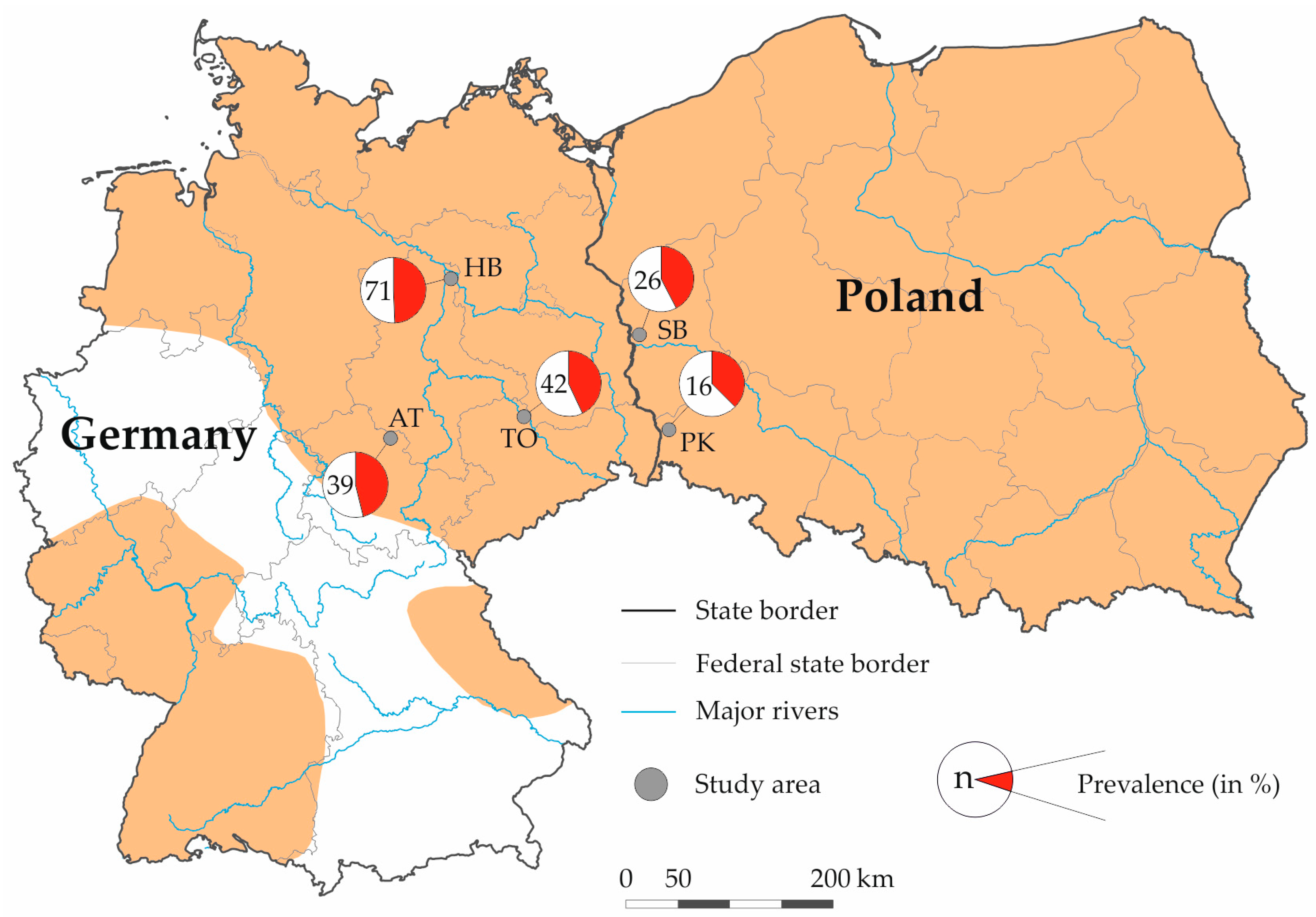
Figure 1. Sampling areas, sample size (n), and seroprevalence of Toxoplasma gondii in wild American minks (Neogale vison) from Germany and Poland. The orange area represents the geographic distribution of the American mink in Germany and Poland (after Vada et al. [22]). Germany: AT Artern (federal state: Thuringia), HB Havelland (Saxony-Anhalt), and TO Torgau (Saxony); Poland: SB Słubice (Lubusz) and PK Pieńsk (Lower Silesian).
Pathogens 13 00153 g001
Table 1. Seroprevalence of Toxoplasma gondii in wild American minks (Neogale vison) from Germany and Poland.
StateStudy AreaNo. TestedNo. PositivePrevalence in % (95% CI) 1
GermanyArtern391846.15 (30.54–61.86)
Torgau421842.86 (27.93–57.87)
Havelland713549.30 (37.67–60.93)
PolandPieńsk16637.50 (13.78–61.22)
Słubice261142.31 (23.31–61.29)
Total 1948845.36 (38.39–52.41)
1 CI–confidence interval.
Table 2. Results of logistic regression identifying predicators for the seroprevalence of Toxoplasma gondii in wild American minks (Neogale vison). The lines with no data indicate the reference category.
CoefficientsEstimates.e.Odds Ration95% CI 1 Odds Rationz-Valuep-Value
(Intercept)0.7070.2732.0281.199–3.5162.5890.010
Sex—female-- --
Sex—male−0.0690.3100.9330.508–1.718−0.2240.823
Age—adult-- --
Age—juvenile−1.5310.3120.2160.116–0.395−4.904<0.001
1 CI–confidence interval.
Disclaimer/Publisher’s Note: The statements, opinions and data contained in all publications are solely those of the individual author(s) and contributor(s) and not of MDPI and/or the editor(s). MDPI and/or the editor(s) disclaim responsibility for any injury to people or property resulting from any ideas, methods, instructions or products referred to in the content.