Zoonotic Implications of Onchocerca Species on Human Health
by
 , ,
, ,
Maria Cambra-Pellejà
1,2, 
Javier Gandasegui
3,
Rafael Balaña-Fouce
4,
José Muñoz
3 and
María Martínez-Valladares
1,2,* 1
Instituto de Ganadería de Montaña (CSIC-Universidad de León), 24346 León, Spain
2
Departamento de Sanidad Animal, Facultad de Veterinaria, Universidad de León, Campus de Vegazana, 24071 León, Spain
3
Instituto de Salud Global de Barcelona (ISGlobal), 08036 Barcelona, Spain
4
Departmento de Ciencias Biomédicas, Facultad de Veterinaria, Universidad de León, 24071 León, Spain
*
Author to whom correspondence should be addressed.
Pathogens 2020, 9(9), 761; https://doi.org/10.3390/pathogens9090761
Submission received: 31 July 2020
/
Revised: 10 September 2020
/
Accepted: 15 September 2020
/
Published: 17 September 2020
(This article belongs to the Special Issue Onchocerciasis and River Epilepsy)
Abstract
:The genus Onchocerca includes several species associated with ungulates as hosts, although some have been identified in canids, felids, and humans. Onchocerca species have a wide geographical distribution, and the disease they produce, onchocerciasis, is generally seen in adult individuals because of its large prepatency period. In recent years, Onchocerca species infecting animals have been found as subcutaneous nodules or invading the ocular tissues of humans; the species involved are O. lupi, O. dewittei japonica, O. jakutensis, O. gutturosa, and O. cervicalis. These findings generally involve immature adult female worms, with no evidence of being fertile. However, a few cases with fertile O. lupi, O. dewittei japonica, and O. jakutensis worms have been identified recently in humans. These are relevant because they indicate that the parasite’s life cycle was completed in the new host—humans. In this work, we discuss the establishment of zoonotic Onchocerca infections in humans, and the possibility of these infections to produce symptoms similar to human onchocerciasis, such as dermatitis, ocular damage, and epilepsy. Zoonotic onchocerciasis is thought to be an emerging human parasitic disease, with the need to take measures such as One Health Strategies, in order to identify and control new cases in humans.
1. Introduction
The genus Onchocerca comprises several parasitic nematode species [1], which are transmitted by arthropod vectors, namely, blackflies belonging to the genera Simulium and Culicoides [2], and it has been suggested that the biting habits of these blackflies influence the presence and distribution of microfilariae in the subcutaneous tissues of the hosts [3]. Until now, several Onchocerca species have been identified, and ungulates seem to be the main hosts, although the parasite is also found in canids, felids, and humans [4]. Each larva stays as a single male or female and grows until it becomes a mature worm, which remains separated in the skin or collected together in subcutaneous fibrous nodules because of the immunological reaction of the host; females produce microfilariae, which invade the skin, travel in it, and very often reach the eye [1,5]. As a result, onchocerciasis usually causes dermatitis and ocular damages, but epilepsy has also been reported in humans [6]. In recent years, Onchocerca species infecting animals were found in human tissues. In this work, we review the Onchocerca infections in animals and their potential zoonotic character, as well as analyzing whether the filarial worms belonging to the genus Onchocerca are able to produce clinical signs in humans, rendering onchocercasis an emerging zoonotic disease.
2. How Many Definitive Hosts Are Parasitized by Onchocerca spp.?
The genus Onchocerca comprises more than 30 species which mainly infect ungulates and have a worldwide distribution [4]. The only known host exceptions are carnivores, canids, felids, and humans [4]. In general, onchocerciasis is more prevalent in adult rather than juvenile individuals, because of the large prepatency period of the infection [5]. Onchocerca infections may have been underestimated, particularly in wildlife, where infected animals are easier to be preyed upon, especially when the Onchocerca nodules are found in the joints, making movement difficult [7]. However, Onchocerca control in wildlife is difficult because of the ubiquity of the adult insect vectors [8], the need of larviciding of vector breeding sites [6], and the migratory patterns of some hosts, leading to the expansion of Onchocerca species [9]. The most common Onchocerca species identified so far, related to their main vectors, hosts, and geographical distribution, are described in Table 1.
Some authors emphasize that knowing the evolutionary history of a parasite and its spread among its hosts is crucial to control the disease it causes [104]. The origin of the genus Onchocerca and its principal evolution may have been started in Africa. This hypothesis is based on the fact that the most primitive species were found in Africa, the continent with the highest diversity of Onchocerca species [105]. From these Onchocerca primitive African species, diversification may have occurred until reaching the current species. There are two main hypotheses about the origin of these species; the first one postulates that they evolved via co-speciation with the host, and the second one indicates that they could have been captured via horizontal transfer from other host species, naming the latter host-switching. As the host specificity in some Onchocerca species is not as strong as was thought to be [53], it seems feasible that multiple host-switching events between hosts have historically occurred [106]. Some authors support the idea that the domestication of animals may have had an important role in those horizontal transfers leading to different Onchocerca species, parasitizing both animals and humans [12] (Figure 1). Thus, onchocerciasis could be considered a zoonosis with different potential hosts [104].
Onchocerca species identification has traditionally been done through morphological examinations [39]. The number, height, and longitude between the crests in the outer cuticle, the shape of the anterior tip, the body thickness, and the presence or absence and the shape of the striae from the inner cuticle are used to achieve the morphological distinction between Onchocerca species [39]. Difficulties during worm extraction could complicate the later morphological identification; therefore, authors such as Verocai et al. [7] support the combination of classical and molecular tools to characterize the specimens collected. Regarding the molecular methods, the analysis of mitochondrial cytochrome c oxidase subunit 1 (COI) and 12S ribosomal RNA gene sequences are the most used for species identification [39].
In wild ruminants such as reed deer, moose, and sika deer, among others, Onchocerca has been described traditionally in Central and Eastern Europe, but recent evidence shows that they have a broader geographic distribution [8]. Some studies indicate that Onchocerca species located in insular regions such as Japan showed more primitive morphological characters than species from the continent, as a result of their geographical isolation [94]. In wild ruminants, adult worms have generally been identified in subcutaneous nodules located in the dorsal region and flanks [8], along the metatarsus and metacarpus [107], in the posterior dorsal region and thighs [58], and in the upper parts of the extremities [108]. However, in some cases, adult parasites were found in these hosts without being included within a nodule [47,94]. Regarding the microfilariae, they were located inside the hind limbs [8], the ventral zone [58], ears and muzzles [109] of the hosts.
Onchocerca infections affecting domestic animals such as cattle have usually been identified in slaughterhouses [13]. Onchocerca armillata has a specific location in the host, very different from other species, in the tunica intima of the aorta [10]. On the other hand, the remaining species affecting cattle such as O. gutturosa [53], O. ochengi, and O. dukei [15], among others, generally parasitized the cervical ligaments [12] and the connective tissues of the ventral and thoracic areas of the cattle [10,43,53].
In horses, the appearance of skin nodules is often asymptomatic, and it is not usually noticed by owners, but it could also manifest with dermatitis, impaired function of the ligaments, and even blindness [19]. Adult Onchocerca parasites are usually located coiled in the ligamentum nuchae of horses [30] and within nodules infecting the connective tissue of the flexor tendons or the suspensory ligament of the fetlock [110]. Several Onchocerca species also have been found in the locations described above, but infecting camels and dromedaries [12]. Until now, what is known is that O. fasciata is a species specific in camels [50], being the most prevalent species in this host [48]. Camelidae are valuable animals in some areas of the world, as these animals are used as transport and their meat is used for human consumption; therefore, onchocerciasis have a detrimental impact associated with a loss of commercial value [48] and major issues in public health [54].
Onchocerca lupi was described for the first time in the ocular tissues of wolves [73], and since then, it has mainly been reported in dogs [74,111]. In the latter, O. lupi adult worms are generally found in the ocular nodules, which could result in eye damage, such as conjunctivitis, swelling, exophthalmia, and vision loss [74]. Microfilariae seem to aggregate in specific body areas in the head, specifically ears and nose, or in the inter-scapular region [111]. However, O. lupi was also found infecting cats and again showing an ocular tropism [75,112].
In humans, onchocerciasis is caused by O. volvulus [113]. The adult worms are distributed in subcutaneous nodules under the skin, while microfilariae are generally found on the hips, shoulders, and the lower parts of the body [113]. Some people do not present any clinical signs while others exhibit itchy skin rashes and vision disorders [113]. Recently, several authors also described a specific type of epilepsy as a disorder related to onchocerciasis [114,115,116]. Human onchocerciasis is more prevalent in fertile regions of developing countries such as Venezuela and Brazil [113], Tanzania [114], Uganda [115], or South Sudan [116], causing a high morbidity burden [104].
Female blackflies of the genus Simulium are the best-known vectors that transmit Onchocerca microfilariae [39]. Simulium larvae inhabit areas with running freshwater or rivers, where they may develop to the following stages [39]. The identification of infective larvae in wild-caught blackflies is important in order to assess onchocerciasis transmission rate and putative natural Onchocerca vectors [38]. The screening of transmission vectors could be done by the morphological identification of microfilariae or via molecular methods, detecting their DNA by PCR amplification within vectors [48]. Although many species within the genus Simulium and Culicoides are likely to be putative vectors of Onchocerca spp. (Table 1), many of them may be still unidentified. The wide geographical distribution of Onchocerca vectors indicates favorable environmental conditions for spreading infection [5].
3. Does Onchocerciasis Have a Zoonotic Character?
The incidence of zoonotic diseases increases when humans live in close contact with animals, such as when both hosts overlap in a geographic region [117]. Thus, it is important to consider humans together with animals and the environment as a whole, which is emphasized in the One Health Strategy, in order to prevent and control the emergence of zoonotic diseases. Emerging zoonotic diseases impact on public health and socioeconomic aspects of the global population [117].
So far, 40 cases of Onchocerca species infecting animals have been described in humans worldwide, and all of them are within the Holarctic region [68,96,118,119,120,121,122,123,124,125,126] (Figure 2). Among these cases in humans, O. lupi was the most prevalent species identified, followed by O. dewittei japonica and, to lesser extent, O. jakutensis, O. gutturosa, and O. cervicalis. Moreover, there are several cases where Onchocerca parasites infecting animals were found in humans, but the species was not identified [127,128,129,130,131,132,133] or was only suspected without confirmation [134,135,136].
The first human case unequivocally caused by O. lupi was found infecting the subconjunctival region of a person in Turkey [76]. Since then, additional findings of this species were also reported in the ocular tissues of people from Turkey, Tunisia, and Iran [80,81,82]. The first report of O. lupi in the United States was published in 2013, and the worm was located in the individual’s upper cervical spinal cord [137]; two years later, another report involving the same tissues was identified in the same country [119]. Most findings of O. lupi in humans described between 1965 and 2014 were reviewed by Grácio et al. [120]. However, a few of them regarding other species were not included in that review, for example, O. dewittei japonica, which was isolated in individuals from Japan from different body parts such as hand [41], infraclavicular region [138], and knee [139]. Other examples not reported previously in the review were the case of a woman in Australia with multiple onchocercal nodules on the neck and face [140] or the case of another woman in the United States with a subdeltoid mass caused by O. gutturosa [66].
Since 2015, 12 reports of Onchocerca species infecting animals have been described in humans [68,96,118,119,121,122,123,124,125,126]. Five of them were caused by O. lupi, which was found in the upper cervical spinal cord, conjunctiva, head, and forearm of five individuals diagnosed in the United States [121,122]. Six more cases were reported in Japan, caused by O. dewittei japonica, which was isolated from the upper extremities in five individuals [123,124,125,126] or from the head of one person [96]. The most recent case was caused by O. jakutensis and was found in the vitreous body of the eye of a man from Poland [68]. These cases were generally caused by immature adult female worms, most of them with no evidence of fertile adult worms, and thus no microfilariae seemed to be produced [39]. However, to determine the fertile status of these worms, it was important to remove them intact from the nodules or tissues where they were located [68]. Sometimes, the worms were damaged during their extraction, and therefore, the chances of obtaining a good quality sample for morphological identification were reduced [68]. In this case, molecular tools such as PCR can be used for species identification [7].
Koehsler et al. [140] described a case caused by O. jakutensis in an Austrian woman with lupus erythematosus and with multiple nodules on the neck and face. Although the presence of fertile worms was not shown in those nodules, it is thought that the autoimmune disease could have favored the appearance of multiple nodules developed over several years, showing that worm reproduction was possible, and this was the first human case involving multiple nodules. However, in a few reports, the presence of fertile adult worms from Onchocerca species infecting animals was confirmed in human tissues. Dudley et al. [119] identified a mass causing cervical spinal cord compression in a girl from the United States, containing a gravid O. lupi female with uteri filled with microfilariae. Uni et al. [96] described an infection caused by a male O. dewittei japonica and located in the head of an individual in Japan; spermatozoids were identified in the seminal vesicle, showing the maturation of the male O. dewittei japonica in humans [96]. In addition, Bergua et al. [118] reported an eye infection caused by O. lupi; in this case, the worm was removed by surgery, and one month later a new nodule appeared again in the patient’s face, confirming the presence of O. lupi. These cases are relevant because they show that the parasite’s life cycle was able to be completed and therefore the establishment of zoonotic Onchocerca infections is possible in humans.
Considering the cases previously mentioned, O. lupi, O. dewittei japonica, and O. jakutensis are proposed as zoonotic species that could reproduce and complete the parasite’s life cycle within the human body. This phenomenon was previously suggested by Uni et al. [96], postulating that accidental zoonotic Onchocerca species could switch from one host to another through their vectors, adapting to humans and becoming a new human parasitic disease. Moreover, Grácio et al. [120] and Tahir et al. [141] proposed in their reviews that O. lupi cases in humans should be contemplated as a current emerging zoonotic disease.
4. Which Factors Are Involved in the Transmission of Animal Onchocerca Species to Humans?
There are several factors that favor the transmission of Onchocerca species infecting animals to humans. The high prevalence of the causative agents in the host animals is one of those factors [138]. A prevalence of 92% of O. dewittei japonica in wild boars in the Oita region of Japan was associated with several cases in humans in the same area [138]. The expansion of wild animal habitats due to alterations in climate, deforestation, and urbanization appears to be an important factor that favors contact between animals and humans [96]. On the other hand, climate determines the geographical distribution of potential Onchocerca vectors, and changes in the environment, such as accelerating climate warming, may modify the ranges where these vectors can live [142]. Modifications in any of these factors could lead to changes in Onchocerca species distribution [53], and as a consequence, altered patterns of exposure could lead to contact with new hosts, animals or humans [7].
Since the most frequent Onchocerca species infecting animals found in humans are O. dewittey japonica from Japanese wild boar, O. lupi from carnivores (dogs or wolfs), and O. jakutensis from red deer [68], it is important to evaluate the onchocerciasis infection rate in these hosts, especially if they share the same habitat with humans. In order to define areas of risk effectively, it is essential to plan public health strategies to prevent the transmission of Onchocerca species infecting animals to humans [126]. Although it is not clear that either an increasing number of zoonotic reports or a better diagnostic is being performed, or both [68], zoonotic onchocerciasis is likely to occur more widely than expected.
5. What Are the Lesions and Clinical Signs that Onchocerca Species Infecting Animals Cause in Humans?
Zoonotic onchocerciasis is characterized by lesions observed as subcutaneous nodules developed around adult worms in various parts of the body and, in some cases, reaching the ocular tissue [39]. The common clinical sign related with this infection is local swelling, so the pathology of the infection does not depend on Onchocerca species but on the invaded tissue [118]. As previously described, most of the Onchocerca species infecting animals but found in humans are detected by the appearance of subcutaneous nodules [124]. Nodules can grow in size, swell, and itch, and some of them are painful [124]. So far, the most effective treatment is the surgical removal of the worms [68]. Then, species identification is performed according to the cuticular morphology of the adult worms or by PCR [143].
However, three Onchocerca species infecting animals were able to reach the ocular tissues. O. lupi was found within the conjunctiva [76,80,81,82], the superior rectus muscle of the eye [122], and the anterior chamber of the eye [118,137], while O. jakutensis was located in the vitreous body of the eye [68] and O. cervicalis in the cornea [26]. Although the most characteristic lesion is the presence of an ocular mass, other cases may be accompanied by milder signs such as blurred vision, redness, irritation, or itchiness, or more severe signs such as swelling, eyelid drooping, conjunctival hyperemia, and pain [76,80,118,122,137]. Some authors have considered the possibility that the ocular invasion of O. lupi in humans could be underestimated, being more widespread than believed, as some cases could have been misdiagnosed as other species [80]. O. lupi is also able to invade the Central Nervous System (CNS) of its hosts, leading to lesions in the upper cervical spinal cord or neck pain and stiffness, but also, less frequently, sore throat, dysphagia, and fever [119,121,122,137].
5.1. Is It Possible That Onchocerca Species Infecting Animals Produce Clinical Symptoms Such as Blindness in Humans?
Onchocerciasis caused by O. volvulus is the world’s second leading cause of infectious blindness [144]. However, it is generally accepted that the true prevalence of blindness associated to onchocerciasis is underestimated [144]. Previous studies have suggested that onchocercal blindness is more common in savanna regions of Africa compared with forest areas, where dermatological symptoms are predominant [145,146], and some studies are being conducted to elucidate the parasite differences between those areas. The microfilariae produced by female O. volvulus migrate to the ocular tissues in the human host and invade every part of the eye except the lens. Living microfilariae localized in the eye do not cause inflammation [147], the ocular damage appears as a result of the host immune response to antigen release from degenerating and dead microfilariae [148]. The risk of developing blindness increases with greater exposure to a high microfilariae load [148]. The results are impaired vision from corneal opacities, cataract, chorioretinal degeneration, optic atrophy, and in severe cases permanent blindness with subsequent chronic disability and reduced life expectancy [147,149].
In animals, it is known that adult worms of O. lupi could cause vision loss in dogs [74] and O. cervicalis in horses [30]. Adult worms of O. lupi, O. cervicalis, and O. jakutensis were found invading human ocular tissues, but blindness was not diagnosed in any individual [26,68,118]. Thus, it seems that the Onchocerca species infecting animals but found in the human ocular tissues involved adult parasites present in the conjunctiva and the cornea, as it happens in their natural hosts [30,74]. This contrasts with O. volvulus infection in humans where microfilariae are found in the ocular tissues and are responsible for the ocular damage [148,149].
5.2. Is It Possible That Onchocerca Species Infecting Animals Produce Clinical Disorders Such as Epilepsy in Humans?
It has been estimated that onchocerciasis-associated epilepsy (OAE) affected between 300 and 400,000 people worldwide in 2015 [150]. The onset of OAE is between the ages of 3 and 18 years old, and OAE is an important cause of mortality among children and adolescents in onchocerciasis endemic areas with ongoing transmission [151]. Recent epidemiological studies strongly suggest that the “parasite is able to directly or indirectly trigger epilepsy” [152,153,154,155]. A post mortem study done in nine children who died of OAE including nodding syndrome showed signs of neuroinflammation and Tau deposits but no signs of parasitic infection [156].
The mechanism through which O. volvulus causes epilepsy remains unknown [6]. One of the most important challenges to study OAE etiology is the need to perform autopsies for subsequent histopathological analysis, which can be extremely difficult in most rural African regions. Different hypotheses about OAE etiology have been postulated: direct infection of the CNS by microfilariae [157], autoimmune reactions resulting in neurotoxicity [158], and the transmission of a neurotropic virus together with the Onchocerca microfilariae [159]. These hypotheses were published in the American Journal of Ophthalmology [157], Science Translational Medicine [158], and Annals of Tropical Medicine and Parasitology [159] journals, respectively. However, most of these hypotheses suggest that there is a factor related to onchocerciasis that triggers the episode of epilepsy.
The first theories published on the etiology of OAE suggested that direct infection of the CNS by O. volvulus microfilariae could be the cause of OAE onset [157,160]. However, microfilariae nor O. volvulus DNA were observed in recent studies in cerebrospinal fluid (CSF) nor brain of persons with OAE [161].
The hypothesis relating OAE and immunological factors of the host is gaining importance. Considering that these factors are known to be involved in the development of blindness, they may also be associated with the onset of epilepsy [158]. Therefore, epilepsy could be the result of an autoimmune reaction of antibodies against O. volvulus, which results in a neurotoxic reaction [162]. The entrance of O. volvulus microfilariae in the human host triggers the activation of the immune system, increasing the levels of several cytokines that migrate from the periphery into the CNS, promoting neuroinflammation [160]. As a consequence, antibodies against O. volvulus proteins could cross the blood–brain barrier and recognize antigens from the CNS of the host. This may trigger a neurotoxic reaction, which could promote the onset of OAE [160]. Several proteins found in the CNS have been proposed as cross-reacting antigen candidates, such as voltage-gated potassium channels (VGKC) [163], leiomiodin-1 (LM-1), and the human protein deglycase DJ-1 [158]. Further studies are needed to elucidate the underlying mechanism of immunological mediated toxicity.
Finally, Colebunders et al. [164] proposed a new hypothesis in which an unknown neurotropic virus or an endosymbiont of the parasite transmitted together with Onchocerca spp. and through the same vectors could cause OAE. This hypothesis was based on the findings by Mellor and Boorman [159], who observed that the vector Culicoides nubeculosus transmitted the bluetongue virus in ruminants only when O. cervicalis microfilariae where found together in the vector. In this sense, Wolbachia is known to be an endosymbiont bacterium for some Onchocerca species, including O. volvulus [148,165]. Wolbachia antigens can contribute to the appearance of some of the clinical signs related to onchocerciasis such as ocular damage [148,165], but it is unknown if they could also contribute to OAE.
Regarding Onchocerca species infecting animals, Cantey et al. [122] reported the presence of O. lupi in the cervical spinal canal of humans, evidencing that some Onchocerca species infecting animals could reach human CNS, but no recent study has found the parasite in the CSF. However, until now, epileptic seizures have not been reported in zoonotic onchocerciasis. Some zoonotic reports, with fertile female worms involved, suggested that this could be a possibility if the biological life cycle is completed and microfilariae are produced [96,118,119]. Therefore, some microfilariae could reach the CSF or trigger a neurotoxic reaction in the host, leading to epileptic seizure.
6. Concluding Remarks
Onchocerca species infecting animals have been found invading human tissues, with O. lupi, O. dewittei japonica, and O. jakutensis being the most common species. The high prevalence of causative agents in wildlife and domestic animals and the extension of their habitats due to alterations in climate and urbanization seemed to be crucial factors to bring Onchocerca parasites infecting animals closer to humans [96], increasing human-parasite contact, and thus leading to zoonotic transmission. Most of these invasions seem to produce only immature adult worms [39], but several cases with fertile worms involved have been recently reported. Those cases involved the three species mentioned above, O. lupi, O. dewittei japonica, and O. jakutensis, and we postulate them as candidate species that could reproduce and maintain the parasite’s life cycle within the human body. As this has occurred before, it is possible that Onchocerca species infecting animals could switch from their primarily hosts to humans in the future, causing symptoms similar to human onchocerciasis, such as dermatitis, eye damage, and even epilepsy. All of this makes zoonotic onchocerciasis an emerging human parasitic disease, with the need to take measures, such as One Health Strategies, in order to identify and control new cases in humans.
Author Contributions
Writing—original draft, M.C.-P.; critical review and editing, J.G., R.B.-F., J.M., M.M.-V.; conceptualization, M.M.-V. All authors have read and agreed to the published version of the manuscript.
Funding
M.C.-P. was funded by the “Stopping Transmission Of intestinal Parasites (STOP)” project (EDCTP2 program supported by the European Union; RIA2017NCT-1845—STOP), J.G. by Ramon Areces Foundation, and M.M.-V. by the Spanish “Ramon y Cajal” Program, Ministry of Economy and Competitiveness (RYC-2015-18368).
Conflicts of Interest
The authors declare no conflict of interest.
References
- Centers for Disease Control and Prevention (CDC). Parasites–Onchocerciasis (Also Known as River Blindness). Available online: https://www.cdc.gov/parasites/onchocerciasis/ (accessed on 11 May 2020).
- Muller, R. Identification of Onchocerca. Symposia of the British Society for Parasitology. In Problems in the Identification of Parasites and Their Vectors, 2nd ed.; Taylor, A.E.R., Mullers, R., Eds.; Blackwell Scientific: London, UK, 1979; Volume 17, pp. 175–206. [Google Scholar]
- Ogbogu, V.C.; Bablis, J.M.; Ajanusi, O.J. Prevalence of Microfilariae in Cattle at Slaughter in Zaria, Nigeria. Vet. Parasitol. 1990, 36, 171–175. [Google Scholar] [CrossRef]
- Lefoulon, E.; Giannelli, A.; Makepeace, B.L.; Mutafchiev, Y.; Townson, S.; Uni, S.; Verocai, G.G.; Otranto, D.; Martin, C. Whence river blindness? The domestication of mammals and host-parasite co-evolution in the nematode genus Onchocerca. Int. J. Parasitol. 2017, 47, 457–470. [Google Scholar] [CrossRef] [PubMed]
- Boijsen, B.; Uhlhorn, H.; Ågren, E.; Höglund, J. Nodular onchocercosis in red deer (Cervus elaphus) in Sweden. Int. J. Parasitol. Parasites. Wildl. 2017, 6, 340–343. [Google Scholar] [CrossRef] [PubMed]
- Gumisiriza, N.; Mubiru, F.; Siewe Fodjo, J.N.; Mbonye Kayitale, M.; Hotterbeekx, A.; Idro, R.; Makumbi, I.; Lakwo, T.; Opar, B.; Kaducu, J.; et al. Prevalence and incidence of nodding syndrome and other forms of epilepsy in onchocerciasis-endemic areas in northern Uganda after the implementation of onchocerciasis control measures. Infect. Dis. Poverty 2020, 9, 12. [Google Scholar] [CrossRef] [PubMed] [Green Version]
- Verocai, G.G.; Nelson, K.J.; Callahan, R.T.; Wekesa, J.W.; Hassan, H.K.; Hoberg, E.P. A cryptic species of Onchocerca (Nematoda: Onchocercidae) in blackflies (Simulium spp.) from southern California, USA. Parasites Vectors 2018, 11, 547. [Google Scholar] [CrossRef] [PubMed] [Green Version]
- Hidalgo, M.R.; Martínez, A.; Carreño, R.A.; González, S.; Ferreras, M.C.; Díez, N. Levels of infection, pathology and nodule size of Onchocerca flexuosa (Nematoda: Onchocercidae) in red deer (Cervus elaphus) from northern Spain. J. Helminthol. 2015, 89, 326–334. [Google Scholar] [CrossRef]
- Verocai, G.G.; Lejeune, M.; Beckmen, K.B.; Kashivakura, C.K.; Veitch, A.M.; Popko, R.A.; Fuentealba, C.; Hoberg, E.P.; Kutz, S.J. Defining parasite biodiversity at high latitudes of North America: New host and geographic records for Onchocerca cervipedis (Nematoda: Onchocercidae) in moose and caribou. Parasites Vectors 2012, 5, 242. [Google Scholar] [CrossRef] [Green Version]
- Neary, J.M.; Trees, A.J.; Ekale, D.D.; Tanya, V.N.; Hetzel, U.; Makepeace, B.L. Onchocerca armillata contains the endosymbiotic bacterium Wolbachia and elicits a limited inflammatory response. Vet. Parasitol. 2010, 174, 267–276. [Google Scholar] [CrossRef] [Green Version]
- ElBihari, S.; Hussein, H.S. The distribution and redescription of the microfilariae of Onchocerca armillata Railliet and Henry, 1909. Sudan J. Vet. Sci. Anim. Husb. 1976, 17, 77–85. [Google Scholar]
- Hussein, H.S.; Atta El Mannan, A.M.; El Sinnary, K. Onchocerca armillata Railliet and Henry, 1909 and Onchocerca gutturosa (Neumann, 1910) in camels (Camelus dromedarius L.) in the Sudan. Vet. Res. Commun. 1988, 12, 475–480. [Google Scholar] [CrossRef]
- Chodnik, K.S. Aortic onchocerciasis due to Onchocerca armillata in cattle in Ghana, with special reference to the morphology of the parasite. Ann. Trop. Med. Parasitol. 1957, 51, 216–224. [Google Scholar] [CrossRef] [PubMed]
- Cheema, A.H.; Ivoghli, B. Bovine onchocerciasis caused by Onchocerca armillata and O. gutturosa. Vet. Pathol. 1978, 15, 495–505. [Google Scholar] [CrossRef] [Green Version]
- Vassiliades, G.; Delbove, P.; Bain, O. Onchocercoses bovines au Senegal. Note preliminaire. Rev. Elev. Med. Vet. Pays. Trop. 1983, 36, 351–353. [Google Scholar]
- Trees, A.J.; McCall, P.J.; Davies, J.B. On the possibility of bovine Onchocerca species infecting Simulium damnosum s.l. in the forest zone of Sierra Leone. I. Parasitological aspects. Ann. Trop. Med. Parasitol. 1989, 83, 595–601. [Google Scholar] [CrossRef] [PubMed]
- Mtei, B.J.; Sanga, H.J. Aortic onchocercosis and elaeophorosis in traditional TSZ-cattle in Tabora (Tanzania): Prevalence and pathology. Vet. Parasitol. 1990, 36, 165–170. [Google Scholar] [CrossRef]
- Begam, R.; Islam, S.; Saikia, M.; Kalita, A.; Bulbul, K.M.; Bam, J.; Pathak, P. Prevalence of aortic onchocerciasis in cattle of Assam. Vet. Pract. 2015, 16, 225–227. [Google Scholar]
- Lia, R.P.; Mutafchiev, Y.; Veneziano, V.; Giannelli, A.; Abramo, F.; Santoro, M.; Latrofa, M.S.; Cantacessi, C.; Martin, C.; Otranto, D.; et al. Filarial infection caused by Onchocerca boehmi (Supperer, 1953) in a horse from Italy. Parasitol. Res. 2017, 116, 191–198. [Google Scholar] [CrossRef] [Green Version]
- Supperer, R. Filarosen der Pferde in Österreich. Wien. Tierärztliche Mon. 1953, 40, 193–220. [Google Scholar]
- Mirzayans, A.; Maghsoodloo, H. Filarial infection of Equidae in the Tehran area of Iran. Trop. Anim. Health Prod. 1977, 9, 19–20. [Google Scholar] [CrossRef]
- Mellor, P.S. Studies on Onchocerca cervicalis Railliet and Henry 1910: I. Onchocerca cervicalis in British horses. J. Helminthol. 1973, 47, 97–110. [Google Scholar] [CrossRef]
- Foil, L.D.; Kleir, T.R.; Miller, R.I.; Foil, C.S.; French, D.D.; Smith, J.N. Seasonal changes in density and tissue distribution of Onchocerca cervicalis microfilariae in ponies and related changes in Culicoides variipennis populations in Louisiana. J. Parasitol. 1987, 73, 320–326. [Google Scholar] [CrossRef] [PubMed]
- Riek, R.F. Studies on allergic dermatitis (Queensland itch) of the horse. I. Description, distribution, symptoms and pathology. Aust. Vet. J. 1953, 7, 177–184. [Google Scholar] [CrossRef]
- Monahan, C.M.; Chapman, M.R.; French, D.D.; Klei, T.R. Efficacy of moxidectin oral gel against Onchocerca cervicalis microfilariae. J. Parasitol. 1995, 81, 117–118. [Google Scholar] [CrossRef] [PubMed]
- Burr, W.E., Jr.; Brown, M.F.; Eberhard, M.L. Zoonotic Onchocerca (Nematoda:Filarioidea) in the cornea of a Colorado resident. Ophthalmology 1998, 105, 1494–1497. [Google Scholar] [CrossRef]
- Radwan, A.M.; Ahmed, N.E.; Elakabawy, L.M.; Ramadan, M.Y.; Elmadawy, R.S. Prevalence and pathogenesis of some filarial nematodes infecting donkeys in Egypt. Vet. World 2016, 9, 888–892. [Google Scholar] [CrossRef]
- Sato, T.; Hayashi, S.; Tanaka, K. Studies on Onchocerca gutturosa Neumann, 1910 as a causative parasite of bovine dermatitis ‘Wahi’ and Onchocerca cervicalis Railliet et Henry, 1910 as a causative parasite of equine dermatitis ‘Kasen’. Jpn. J. Parasitol. 1954, 2, 199–206. [Google Scholar]
- Lutz, J.E.; Boersema, J.H.; Németh, F. Preliminary studies on the incidence of Filaziae in equidae in the Netherlands. Tijdschr. Diergeneeskd. 1975, 100, 321–326. [Google Scholar]
- Lees, M.J.; Kleider, N.; Tuddenham, T.J. Cutaneous onchocerciasis in the horse: Five cases in southwestern british columbia. Can. Vet. J. 1983, 24, 3–5. [Google Scholar]
- French, D.D.; Klei, T.R.; Foil, C.S.; Miller, R.I.; Foil, L.D.; Chapman, M.R.; McClure, J.J. Efficacy of ivermectin in paste and injectable formulations against microfilariae of Onchocerca cervicalis and resolution of associated dermatitis in horses. Am. J. Vet. Res. 1988, 49, 1550–1554. [Google Scholar]
- Marques, S.M.T.; Scroferneker, M.L. Onchocerca cervicalis in horses from southern Brazil. Trop. Anim. Health Prod. 2004, 36, 633–636. [Google Scholar] [CrossRef]
- Franck, M.T.; Colombet, J.; Hugnet, C.; Ducos de Lahitte, J.; Desmaizières, L.M.; Delverdier, M.; Franc, M. Research of skin microfilariae on 160 horses from Poland, France and Spain. Revue. Méd. Vét. 2006, 157, 323–325. [Google Scholar]
- Pledger, D.J. Black flies (Diptera, Simuliidae) of the Swan Hills, Alberta, as possible vectors of Onchocerca cervipedis Wehr Dikmans 1935 (Nematoda; Onchocercidae) in moose (Alces alces Linnaeus). Ph.D. Thesis, University of Alberta, Edmonton, AB, Canada, 1978. [Google Scholar]
- Wehr, E.E.; Dikmans, G. New nematodes (Filariidae) from North America ruminants. Zool. Anz. 1935, 110, 202–208. [Google Scholar]
- Carreno, R.A.; Durden, L.A.; Brooks, D.R.; Abrams, A.; Hoberg, E.P. Parelaphostrongylus tenuis (Nematoda: Protostrongylidae) and other parasites of white-tailed deer (Odocoileus virginianus) in Costa Rica. Comp. Parasitol. 2001, 68, 177–184. [Google Scholar]
- Bain, O.; Ramachandran, C.P.; Petter, F.; Mak, J.W. Description d’Onchocerca dewittei n. sp. (Filarioidea) chez Sus scrofa en Malaisie. Ann. Parasitol. Hum. Comp. 1977, 52, 471–479. [Google Scholar] [CrossRef] [PubMed]
- Fukuda, M.; Otsuka, Y.; Uni, S.; Bain, O.; Takaoka, H. Molecular identification of infective larvae of three species of Onchocerca found in wild-caught females of Simulium bidentatum in Japan. Parasite 2010, 17, 39–45. [Google Scholar] [CrossRef] [Green Version]
- Takaoka, H.; Fukuda, M.; Otsuka, Y.; Aoki, C.; Uni, S.; Bain, O. Blackfly vectors of zoonotic onchocerciasis in Japan. Med. Vet. Entomol. 2012, 26, 372–378. [Google Scholar] [CrossRef]
- Uni, S.; Bain, O.; Takaoka, H.; Miyashita, M.; Suzuki, Y. Onchocerca dewittei japonica n. subsp., a common parasite from wild boar in Kyushu Island, Japan. Parasite 2001, 8, 215–222. [Google Scholar] [CrossRef] [PubMed] [Green Version]
- Takaoka, H.; Bain, O.; Uni, S.; Korenaga, M.; Shirasaka, C.; Aoki, C.; Otsuka, Y.; Fukuda, M.; Eshita, Y.; Daa, T. Zoonotic onchocerciasis caused by a parasite from wild boar in Oita, Japan. A comprehensive analysis of morphological characteristics of the worms for its diagnosis. Parasite 2004, 11, 285–292. [Google Scholar] [CrossRef] [Green Version]
- Wahl, G.; Ekale, D.; Enyong, P.; Renz, A. The development of Onchocerca dukei and O. ochengi microfilariae to infective-stage larvae in Simulium damnosum s.l. and in members of the S. medusaeforme group, following intra-thoracic injection. Ann. Trop. Med. Parasitol. 1991, 85, 329–337. [Google Scholar] [CrossRef]
- Wahl, G.; Achu-Kwi, M.D.; Mbah, D.; Dawa, O.; Renz, A. Bovine onchocercosis in north Cameroon. Vet. Parasitol. 1994, 52, 297–311. [Google Scholar] [CrossRef]
- Beaver, P.C.; Hira, P.R.; Patel, B.G. Onchocerciasis in Zambia: Report of O. volvulus in a child and its differentiation from O. dukei in cattle. Trans. R. Soc. Trop. Med. Hyg. 1983, 77, 162–166. [Google Scholar] [CrossRef]
- Denke, A.M. The prevalence of Onchocerca spp. in cattle in northern Togo in 1979. Trop. Med. Parasitol. 1986, 37, 46–48. [Google Scholar]
- Fukuda, M.; Takaoka, H.; Uni, S.; Bain, O. Infective larvae of five Onchocerca species from experimentally infected Simulium species in an area of zoonotic onchocerciasis in Japan. Parasite 2008, 15, 111–119. [Google Scholar] [CrossRef] [Green Version]
- Uni, S.; Bain, O.; Agatsuma, T.; Harada, M.; Torii, H.; Fukuda, M.; Takaoka, H. Onchocerca eberhardi n. sp. (Nematoda: Filarioidea) from sika deer in Japan; relationships between species parasitic in cervids and bovids in the Holarctic region. Parasite 2007, 14, 199–211. [Google Scholar] [CrossRef] [PubMed] [Green Version]
- Wang, R.; Yu, Z.C.; Luo, X.P.; Chen, L.J.; Shao, G.Y.; Yang, X.Y. Survey of infection and determination of the transmission vector of Onchocerca fasciata in camels (Camelus bactrianus) in Inner Mongolia, China. Med. Vet. Entomol. 2020, 34, 59–68. [Google Scholar] [CrossRef]
- Cheema, A.H.; El-Bihari, S.; Ashour, N.A.; Ali, A.S. Onchocerciasis in camels (Camelus dromedarius) in Saudi Arabia. J. Helminthol. 1984, 58, 279–285. [Google Scholar] [CrossRef]
- Chhabra, M.B.; Gupta, S.K. Parasitic diseases of camels. J. Camel. Pract. Res. 2006, 13, 81–87. [Google Scholar]
- El-Massry, A.A.; Derbala, A.A. Evidence of Onchocerca fasciata (Filaroidea: Onchocercidae) in camels (Camelus dromedarius): I- prevalence, nodular lesions appearance and parasite morphology. Vet. Parasitol. 2000, 88, 305–312. [Google Scholar] [CrossRef]
- Al-Rawashdeh, O.F.; Al-Ani, F.K.; Sharrif, L.A.; Al-Qudah, K.M.; Al-Hami, Y.; Frank, N. A survey of camel (Camelus dromedarius) diseases in Jordan. J. Zoo Wildl. Med. 2000, 31, 335–338. [Google Scholar] [PubMed]
- Krueger, A.; Fischer, P.; Morales-Hojas, R. Molecular phylogeny of the filaria genus Onchocerca with special emphasis on Afrotropical human and bovine parasites. Acta Trop. 2007, 101, 1–14. [Google Scholar] [CrossRef] [PubMed]
- Sazmand, A.; Joachim, A. Parasitic diseases of camels in Iran (1931–2017)—A literature review. Parasite 2017, 24, 21. [Google Scholar] [CrossRef] [PubMed]
- Yu, Z.C.; Zhang, W.; Li, B.; Luo, X.P.; Wang, R.; Yang, X.Y. A survey of Onchocerca fasciata infection in camels (Camelus bactrianus) with notes on morphology. PLoS ONE 2019, 14, e0214477. [Google Scholar] [CrossRef] [PubMed] [Green Version]
- Dykova, I. Results of studies on the potential vectors of microfilariae of Onchocerca flesuosa (Weld, 1856) and Wehrdikmansia cervipedis (Wehr et Dikmans, 1935). Acta Vet. 1972, 41, 203–206. [Google Scholar]
- Dikmans, G. Onchocerca flexuosa from the subcutaneous tissues of an antelope and subcutaneous abscesses of a deer. J. Parasitol. 1933, 19, 246. [Google Scholar]
- Bain, O.; Schulz-Key, H. Les onchocerques du cerf européen: Redescription d’O. flexuosa (Wedl, 1856) et description d’O. tubingensis n.sp. et O. tarsicola n.sp. Tropenmed. Parasitol. 1974, 25, 437–449. [Google Scholar]
- Demiaszkiewicz, A.W. Occurrence of tissue Filarioidea in ruminants in Poland. Wiad. Parazytol. 1989, 35, 315–321. [Google Scholar]
- Wildenburg, G.; Plenge-Bönig, A.; Renz, A.; Fischer, P.; Büttner, D.W. Distribution of mast cells and their correlation with inflammatory cells around Onchocerca gutturosa, O. tarsicola, O. ochengi, and O. flexuosa. Parasitol. Res. 1997, 83, 109–120. [Google Scholar] [CrossRef]
- McNulty, S.N.; Fischer, K.; Curtis, K.C.; Weil, G.J.; Brattig, N.W.; Fischer, P.U. Localization of Wolbachia-like gene transcripts and peptides in adult Onchocerca flexuosa worms indicates tissue specific expression. Parasites Vectors 2013, 6, 2. [Google Scholar] [CrossRef] [PubMed] [Green Version]
- Barbušinová, E.; Iglódyová, A.; Čurlík, J.; Lazar, P.; Mravcová, K.; Štrkolcová, G.; Mucha, R.; Karolová, R. First molecular detection of Onchocerca flexuosa (Wedl, 1856) in red deer in Slovakia. Parasitol. Res. 2020, 119, 985–990. [Google Scholar] [CrossRef]
- El Sinnary, K.; Hussein, H.S. Culicoides kingi, Austen: A vector of Onchocerca gutturosa (Neumann, 1910) in the Sudan. Ann. Trop. Med. Parasitol. 1980, 74, 655–656. [Google Scholar] [CrossRef]
- Holdsworth, P.A.; Moorhouse, D.E. Onchocerca gutturosa in an Australian camel. Aust. Vet. J. 1985, 62, 201–202. [Google Scholar] [CrossRef] [PubMed]
- Scott, D.W.; Miller, W.H. Equine Dermatology; Elsevier: St. Louis, MO, USA, 2003; pp. 242–245. [Google Scholar]
- Wright, R.W.; Neafie, R.C.; McLean, M.; Markman, A.W. Zoonotic onchocerciasis of the shoulder. A case report. J. Bone Joint Surg. Am. 2002, 84, 627–629. [Google Scholar] [CrossRef] [PubMed] [Green Version]
- Beytut, E.; Akca, A.; Bain, O. Teat onchocercosis in cows with reference to prevalence, species involved and pathology. Res. Vet. Sci. 2005, 78, 45–51. [Google Scholar] [CrossRef]
- Wesolowska, M.; Zając-Pytrus, H.; Masny, A.; Pytrus, W.; Knysz, B.; Golab, E.; Sałamatin, R. Onchocerca jakutensis ocular infection in Poland: A new vector-borne human health risk? Parasites Vectors 2020, 13, 61. [Google Scholar] [CrossRef] [Green Version]
- Morandi, F.; Krueger, A.; Panarese, S.; Sarli, G.; Verin, R.; Nicoloso, S.; Benazzi, C.; Galuppi, R. First description of nodular onchocercosis (Onchocerca jakutensis) in free-ranging Italian red deer (Cervus elaphus). J. Wildl. Dis. 2011, 47, 963–967. [Google Scholar] [CrossRef]
- Duscher, G.G.; Leschnik, M.; Fuehrer, H.P.; Joachim, A. Wildlife reservoirs for vector-borne canine, feline and zoonotic infections in Austria. Int. J. Parasitol. Parasites Wildl. 2014, 4, 88–96. [Google Scholar] [CrossRef] [PubMed] [Green Version]
- Bosch, F.; Manzanell, R.; Mathis, A. First description of Onchocerca jakutensis (Nematoda: Filarioidea) in red deer (Cervus elaphus) in Switzerland. Int. J. Parasitol. Parasites. Wildl. 2016, 5, 192–197. [Google Scholar] [CrossRef] [Green Version]
- Latrofa, M.S.; Annoscia, G.; Colella, V.; Cavalera, M.A.; Maia, C.; Martin, C.; Slapeta, J.; Otranto, D. A real-time PCR tool for the surveillance of zoonotic Onchocerca lupi in dogs, cats and potential vectors. PLoS Negl. Trop. Dis. 2018, 12, ce0006402. [Google Scholar] [CrossRef]
- Rodonaja, T.E. A New Species of Nematode, Onchocerca lupi n. sp., from Canis Lupus Cubanensis; Soobshchenyia Akad Nauk Gruzinoskoy SSR: Tbilisi, Georgia, 1967; Volume 45, pp. 715–719. [Google Scholar]
- Sréter, T.; Széll, Z. Onchocercosis: A newly recognized disease in dogs. Vet. Parasitol. 2008, 151, 1–13. [Google Scholar] [CrossRef]
- Maia, C.; Annoscia, G.; Latrofa, M.S.; Pereira, A.; Giannelli, A.; Pedroso, L.; Otranto, D. Onchocerca lupi Nematode in Cat, Portugal. Emerg. Infect. Dis. 2015, 21, 2252–2254. [Google Scholar] [CrossRef]
- Otranto, D.; Sakru, N.; Testini, G.; Gürlü, V.P.; Yakar, K.; Lia, R.P.; Dantas-Torres, F.; Bain, O. Case report: First evidence of human zoonotic infection by Onchocerca lupi (Spirurida, Onchocercidae). Am. J. Trop. Med. Hyg. 2011, 84, 55–58. [Google Scholar] [CrossRef] [PubMed]
- Széll, Z.; Sréter, T.; Erdélyi, I.; Varga, I. Ocular onchocercosis in dogs: Aberrant infection in an accidental host or lupi onchocercosis? Vet. Parasitol. 2001, 101, 115–125. [Google Scholar] [CrossRef]
- Komnenou, A.; Eberhard, M.L.; Kaldrymidou, E.; Tsalie, E.; Dessiris, A. Subconjunctival filariasis due to Onchocerca sp. in dogs: Report of 23 cases in Greece. Vet. Ophthalmol. 2002, 5, 119–126. [Google Scholar] [CrossRef] [PubMed]
- Sréter-Lancz, Z.; Széll, Z.; Sréter, T. Molecular genetic comparison of Onchocerca sp. infecting dogs in Europe with other spirurid nematodes including Onchocerca lienalis. Vet. Parasitol. 2007, 148, 365–370. [Google Scholar] [CrossRef]
- Otranto, D.; Dantas-Torres, F.; Cebeci, Z.; Yeniad, B.; Buyukbabani, N.; Boral, O.B.; Gustinelli, A.; Mounir, T.; Mutafchiev, Y.; Bain, O. Human ocular filariasis: Further evidence on the zoonotic role of Onchocerca lupi. Parasites Vectors 2012, 5, 84. [Google Scholar] [CrossRef] [Green Version]
- Ilhan, H.D.; Yaman, A.; Morishima, Y.; Sugiyama, H.; Muto, M.; Yamasaki, H.; Hasegawa, H.; Lebe, B.; Bajin, M.S. Onchocerca lupi infection in Turkey: A unique case of a rare human parasite. Acta Parasitol. 2013, 58, 384–388. [Google Scholar] [CrossRef] [PubMed] [Green Version]
- Mowlavi, G.; Farzbod, F.; Kheirkhah, A.; Mobedi, I.; Bowman, D.D.; Naddaf, S.R. Human ocular onchocerciasis caused by Onchocerca lupi (Spirurida, Onchocercidae) in Iran. J. Helminthol. 2014, 88, 250–255. [Google Scholar] [CrossRef] [PubMed]
- Otranto, D.; Giannelli, A.; Latrofa, M.S.; Dantas-Torres, F.; Scotty Trumble, N.; Chavkin, M.; Kennard, G.; Eberhard, M.L.; Bowman, D.D. Canine Infections with Onchocerca lupi Nematodes, United States, 2011–2014. Emerg. Infect. Dis. 2015, 21, 868–871. [Google Scholar] [CrossRef] [PubMed]
- Miró, G.; Montoya, A.; Checa, R.; Gálvez, R.; Mínguez, J.J.; Marino, V.; Otranto, D. First detection of Onchocerca lupi infection in dogs in southern Spain. Parasites Vectors 2016, 9, 290. [Google Scholar]
- Achukwi, M.D.; Harnett, W.; Renz, A. Onchocerca ochengi transmission dynamics and the correlation of O. ochengi microfilaria density in cattle with the transmission potential. Vet. Res. 2000, 31, 611–621. [Google Scholar] [CrossRef] [Green Version]
- Bussieras, J.; Amagee, E.; Barn, O. Les onchocercoses des bovins togolais a O. dukei et O. dermata. Rev. Elev. Med. Vet. Pays. Trop. 1974, 27, 189–194. (In French) [Google Scholar] [CrossRef] [PubMed]
- Séchan, Y. Développement d’onchocerques animales chez le vecteur de l’onchocercose humaine Simulium sirbanum Vajime et Dunbar, 1975 (Diptera: Simuliidae) en zone subsahélienne du Mali, Afrique de L’Ouest. Cahiers. ORSTOM 1984, 178, 1–234. (In French) [Google Scholar]
- Doyle, S.R.; Armoo, S.; Renz, A.; Taylor, M.J.; Osei-Atweneboana, M.Y.; Grant, W.N. Discrimination between Onchocerca volvulus and O. ochengi filarial larvae in Simulium damnosum (s.l.) and their distribution throughout central Ghana using a versatile high-resolution speciation assay. Parasites Vectors 2016, 9, 536. [Google Scholar] [CrossRef] [PubMed] [Green Version]
- Verocai, G.G.; Hassan, H.K.; Lakwo, T.; Habomugisha, P.; Katabarwa, M.N.; Begumisa, S.; Clouds, P.; Katamanywa, J.; Nahabwe, C.; Unnasch, T.R. Molecular Identification of Onchocerca spp. Larvae in Simulium damnosum sensu lato Collected in Northern Uganda. Am. J. Trop. Med. Hyg. 2017, 97, 1843–1845. [Google Scholar] [CrossRef] [Green Version]
- Bain, O.; Wahl, G.; Renz, A. Onchocerca ramachandrini n. sp. from the warthog in Cameroon. Ann. Parasitol. Hum. Comp. 1993, 68, 139–143. [Google Scholar] [CrossRef]
- Moignoux, J.B. Culicoides nubeculosus Meig (Diptera Ceratopogonida), possible intermediary host of the filaria Onchocerca reticulata Dies. in Camargue. C R Hebd. Seances. Acad. Sci. 1951, 233, 102–103. [Google Scholar]
- Riek, R.F. A note on the occurrence of Onchocerca Reticulata Diesing 1841 in the horse in Queensland. Vet. J. 1954, 30, 178–181. [Google Scholar] [CrossRef]
- Moignoux, J.B. Experimental study of the evolutive cycle of microfilaria Onchocerca reticulata Dies. in Camargue. C R Seances. Soc. Biol. Fil. 1951, 145, 1572–1573. [Google Scholar]
- Yagi, K.; Bain, O.; Shoho, C. Onchocerca suzukii n. sp. and O. skrjabini (= O. tarsicola) from a relict bovid, Capricornis crispus, in Japan. Parasite 1994, 1, 349–356. [Google Scholar] [CrossRef]
- Saeung, A.; Srisuka, W.; Aupalee, K.; Fukuda, M.; Otsuka, Y.; Taai, K.; Maleewong, W.; Takaoka, H. Natural infections with larvae of Onchocerca species type I in the human-biting black fly, Simulium nigrogilvum (Diptera: Simuliidae), in western Thailand. Acta Trop. 2020, 204, 105344. [Google Scholar] [CrossRef]
- Uni, S.; Fukuda, M.; Otsuka, Y.; Hiramatsu, N.; Yokobayashi, K.; Takahashi, H.; Murata, S.; Kusatake, K.; Morita, E.; Maruyama, H.; et al. New zoonotic cases of Onchocerca dewittei japonica (Nematoda: Onchocercidae) in Honshu, Japan. Parasites Vectors 2015, 8, 59. [Google Scholar] [CrossRef] [PubMed] [Green Version]
- Baranwal, V.K.; Shyamsundar, K.; Kabuyaya, V.; Biswas, J.; Vannadil, H. Study of onchocerciasis-related visual impairment in North Kivu province of the Democratic Republic of Congo in Africa. Indian J. Ophthalmol. 2020, 68, 890–894. [Google Scholar]
- Crainey, J.L.; Silva, T.R.; Encinas, F.; Marín, M.A.; Vicente, A.C.; Luz, S.L. The mitogenome of Onchocerca volvulus from the Brazilian Amazonia focus. Mem. Inst. Oswaldo Cruz. 2016, 111, 79–81. [Google Scholar] [CrossRef]
- Kazura, J.W. More Progress in Eliminating Transmission of Onchocerca volvulus and Wuchereria bancrofti in the Americas: A Portent of Global Eradication. Am. J. Trop. Med. Hyg. 2015, 93, 1128–1129. [Google Scholar] [CrossRef] [Green Version]
- Lagoro, D.K.; Arony, D.A. Nodding syndrome (NS) and Onchocerca Volvulus (OV) in Northern Uganda. Pan. Afr. Med. J. 2017, 28, 1. [Google Scholar] [CrossRef]
- Hendy, A.; Krüger, A.; Pfarr, K.; De Witte, J.; Kibweja, A.; Mwingira, U.; Dujardin, J.-C.; Post, R.; Colebunders, R.; O’Neill, S.; et al. The blackfly vectors and transmission of Onchocerca volvulus in Mahenge, south eastern Tanzania. Acta Trop. 2018, 181, 50–59. [Google Scholar] [CrossRef]
- Prince-Guerra, J.L.; Cama, V.A.; Wilson, N.; Thiele, E.A.; Likwela, J.; Ndakala, N.; Muzinga Wa Muzinga, J.; Ayebazibwe, N.; Ndjakani, Y.D.; Pitchouna, N.A.; et al. Comparison of PCR Methods for Onchocerca volvulus Detection in Skin Snip Biopsies from the Tshopo Province, Democratic Republic of the Congo. Am. J. Trop. Med. Hyg. 2018, 98, 1427–1434. [Google Scholar] [CrossRef] [PubMed] [Green Version]
- Hotterbeekx, A.; Namale Ssonko, V.; Oyet, W.; Lakwo, T.; Idro, R. Neurological manifestations in Onchocerca volvulus infection: A review. Brain Res. Bull. 2019, 145, 39–44. [Google Scholar] [CrossRef]
- Morales-Hojas, R. Molecular systematics of filarial parasites, with an emphasis on groups of medical and veterinary importance, and its relevance for epidemiology. Infect. Genet. Evol. 2009, 9, 748–759. [Google Scholar] [CrossRef]
- Chabaud, A.G.; Bain, O. The evolutionary expansion of the Spirurida. Int. J. Parasitol. 1994, 24, 1179–1201. [Google Scholar] [CrossRef]
- Bain, O. Le genre Onchocerca: Hypothèses sur son évolution et clé dichotomique des espèces. Ann. Parasitol. Hum. Comp. 1981, 56, 503–526. [Google Scholar] [CrossRef] [PubMed] [Green Version]
- Demiaszkiewicz, A.W.; Filip, K.J. Microfilariae Onchocerca alcis Bain et Rehbinder, 1986—A new parasite of moose Alces alces (L.) in Poland. Ann. Parasitol. 2018, 64, 89–91. [Google Scholar] [PubMed]
- Demiaszkiewicz, A.W. Redescription of Onchocerca jakutensis (gubanov, 11964) (nematoda, filarioidea). Acta Parasitol. 1993, 38, 124–127. [Google Scholar]
- Schulz-Key, H. Investigations on the filariidae of the cervidae in Southern Germany. 1. Development of the nodule, finding of the sexes and production of the microfilariae in Onchocerca flexuosa (Wedl, 1856), in the red deer (Cervus elaphus). Tropenmed. Parasitol. 1975, 26, 60–69. [Google Scholar]
- Pascoe, R.R.R.; Knottenbelt, D.C. Onchocercal dermatitis (onchocercosis microfilariasis). Manual Equine Dermatol. 1999, 141–143. [Google Scholar]
- Otranto, D.; Dantas-Torres, F.; Giannelli, A.; Abramo, F.; Ignjatovic Cupina, A.; Petric, D.; Cardoso, L.; Mutafchiev, Y.; Cortes, H. Cutaneous distribution and circadian rhythm of Onchocerca lupi microfilariae in dogs. PLoS Negl. Trop. Dis. 2013, 7, e2585. [Google Scholar] [CrossRef]
- Labelle, A.L.; Daniels, J.B.; Dix, M.; Labelle, P. Onchocerca lupi causing ocular disease in two cats. Vet. Ophthalmol. 2011, 14, 105–110. [Google Scholar] [CrossRef]
- Rodríguez-Pérez, M.A.; Unnasch, T.R.; Real-Najarro, O. Assessment and monitoring of onchocerciasis in Latin America. Adv. Parasitol. 2011, 77, 175–226. [Google Scholar]
- König, R.; Nassri, A.; Meindl, M.; Matuja, W.; Kidunda, A.R.; Siegmund, V.; Bretzel, G.; Löscher, T.; Jilek-Aall, L.; Schmutzhard, E.; et al. The role of Onchocerca volvulus in the development of epilepsy in a rural area of Tanzania. Parasitology 2010, 137, 1559–1568. [Google Scholar] [CrossRef]
- Foltz, J.L.; Makumbi, I.; Sejvar, J.J.; Malimbo, M.; Ndyomugyenyi, R.; Atai-Omoruto, A.D.; Alexander, L.N.; Abang, B.; Melstrom, P.; Kakooza, A.M.; et al. An Epidemiologic Investigation of Potential Risk Factors for Nodding Syndrome in Kitgum District, Uganda. PLoS ONE 2013, 8, e66419. [Google Scholar] [CrossRef] [PubMed] [Green Version]
- Dowell, S.F.; Sejvar, J.J.; Riek, L.; Vandemaele, K.A.; Lamunu, M.; Kuesel, A.C.; Schmutzhard, E.; Matuja, W.; Bunga, S.; Foltz, J.; et al. Nodding syndrome. Emerg. Infect. Dis. 2013, 19, 1374–1384. [Google Scholar] [CrossRef] [PubMed]
- McArthur, D.B. Emerging Infectious Diseases. Nurs. Clin. N. Am. 2019, 54, 297–311. [Google Scholar] [CrossRef]
- Bergua, A.; Hohberger, B.; Held, J.; Muntau, B.; Tannich, E.; Tappe, D. Human case of Onchocerca lupi infection, Germany, August 2014. Euro Surveill. 2015, 20, 21099. [Google Scholar] [CrossRef] [PubMed] [Green Version]
- Dudley, R.W.R.; Smith, C.; Dishop, M.; Mirsky, D.; Handler, M.H.; Rao, S. A cervical spine mass caused by Onchocerca lupi. Lancet 2015, 86, 1372. [Google Scholar] [CrossRef]
- Grácio, A.J.; Richter, J.; Komnenou, A.T.; Grácio, M.A. Onchocerciasis caused by Onchocerca lupi: An emerging zoonotic infection. Systematic review. Parasitol. Res. 2015, 114, 2401–2413. [Google Scholar] [CrossRef]
- Chen, T.; Moon, K.; DeMello, D.E.; Feiz-Erfan, I.; Theodore, N.; Bhardwaj, R.D. Case report of an epidural cervical Onchocerca lupi infection in a 13-year-old boy. J. Neurosurg. Pediatr. 2015, 16, 217–221. [Google Scholar] [CrossRef] [Green Version]
- Cantey, P.T.; Weeks, J.; Edwards, M.; Rao, S.; Ostovar, G.A.; Dehority, W.; Alzona, M.; Swoboda, S.; Christiaens, B.; Ballan, W.; et al. The Emergence of Zoonotic Onchocerca lupi Infection in the United States—A Case-Series. Clin. Infect. Dis. 2016, 62, 778–783. [Google Scholar] [CrossRef] [Green Version]
- Ishizawa, H.; Kato, S.; Nishimura, H.; Tanaka, R.; Fukuda, M.; Takaoka, H.; Tsutsumi, Y. Zoonotic onchocerciasis. Pathol. Int. 2015, 65, 271–273. [Google Scholar] [CrossRef]
- Fukuda, M.; Uni, S.; Otsuka, Y.; Eshita, Y.; Nakatani, J.; Ihara, K.; Yoshikawa, Y.; Goto, M.; Fujiwara, S.; Ramli, R.; et al. A new case of zoonotic onchocercosis in northern Kyushu, Japan. Parasitol. Int. 2015, 64, 519–521. [Google Scholar] [CrossRef]
- Uni, S.; Fukuda, M.; Ogawa, K.; Lim, Y.A.; Agatsuma, T.; Bunchom, N.; Saijuntha, W.; Otsuka, Y.; Bhassu, S.; Mat Udin, A.S.; et al. Zoonotic infection with Onchocerca dewittei japonica in an 11-year-old boy in Kansai Region, Western Honshu, Japan. Parasitol. Int. 2017, 66, 593–595. [Google Scholar] [CrossRef] [PubMed]
- Fukuda, M.; Uni, S.; Igari, R.; Utsumi, Y.; Otsuka, Y.; Nakatani, J.; Uga, S.; Hará, T.; Hasegawa, H.; Takaoka, H. Human case of Onchocerca dewittei japonica infection in Fukushima, Northeastern Honshu, Japan. Parasitol. Int. 2019, 72, 101943. [Google Scholar] [CrossRef] [PubMed]
- Azarova, N.S.; Miretskii, O.I.; Sonin, M.D. The first instance of detection of nematode Onchocerca Diesing, 1841 in a person in the USSR. Med. Parazitol. 1965, 34, 156–158. [Google Scholar]
- Siegenthaler, R.; Gubler, R. Paraarticuläres Nematodengranulom (einheimische Onchocerca). Schweiz. Med. Wochenschr. 1965, 95, 1102–1104. [Google Scholar]
- Beaver, P.C.; Yoshimura, H.; Takayasu, S.; Hashimoto, H.; Little, M.D. Zoonotic Onchocerca in a Japanese Child. Am. J. Trop. Med. Hyg. 1989, 40, 298–300. [Google Scholar] [CrossRef] [PubMed]
- Pampiglione, S.; Vakalis, N.; Lyssimachou, A.; Kouppari, G.; Orihel, T.C. Subconjunctival zoonotic Onchocerca in an Albanian man. Ann. Trop. Med. Parasitol. 2001, 95, 827–832. [Google Scholar] [CrossRef] [PubMed]
- Sallo, F.; Eberhard, M.L.; Fok, E.; Baska, F.; Hatvani, I. Zoonotic intravitreal Onchocerca in Hungary. Ophthalmology 2005, 112, 502–504. [Google Scholar] [CrossRef]
- Hira, P.R.; Al-Buloushi, A.; Khalid, N.; Iqbal, J.; Bain, O.; Eberhard, M.L. Zoonotic filariasis in the Arabian Peninsula: Autochthonous onchocerciasis and dirofilariasis. Am. J. Trop. Med. Hyg. 2008, 79, 739–741. [Google Scholar] [CrossRef] [PubMed] [Green Version]
- Lai, J.H.; Walsh, N.M.; Pritt, B.S.; Sloan, L.; Gibson, L.E.; Desormeau, L.; Haldane, D.J. Cutaneous manifestations of a zoonotic Onchocerca species in an adult male, acquired in Nova Scotia, Canada. J. Clin. Microbiol. 2014, 52, 1768–1770. [Google Scholar] [CrossRef] [Green Version]
- Beaver, P.C.; Horner, G.S.; Bilos, J.Z. Zoonotic onchocercosis in a resident of Illiois and observations on the identification of Onchocerca species. Am. J. Trop. Med. Hyg. 1974, 23, 595–607. [Google Scholar] [CrossRef]
- Ali-Khan, Z. Tissue pathology and comparative microanatomy of Onchocerca from a resident of Ontario and other enzootic Onchocerca species from Canada and the U.S.A. Ann. Trop. Med. Parasitol. 1977, 71, 469–482. [Google Scholar] [CrossRef]
- Takaoka, H.; Bain, O.; Tajimi, S.; Kashima, K.; Nakayama, I.; Korenaga, M.; Aoki, C.; Otsuka, Y. Second case of zoonotic Onchocerca infection in a resident of Oita in Japan. Parasite 1996, 3, 179–182. [Google Scholar] [CrossRef] [Green Version]
- Eberhard, M.L.; Ostovar, G.A.; Chundu, K.; Hobohm, D.; Feiz-Erfan, I.; Mathison, B.A.; Bishop, H.S.; Cantey, P.T. Case Report, Zoonotic Onchocerca lupi infection in a 22-month-old child in Arizona: First report in the United States and a review of the literature. Am. J. Trop. Med. Hyg. 2013, 88, 601–605. [Google Scholar] [CrossRef] [PubMed] [Green Version]
- Takaoka, H.; Yanagi, T.; Daa, T.; Anzai, S.; Aoki, C.; Fukuda, M.; Uni, S.; Bain, O. An Onchocerca species of wild boar found in the subcutaneous nodule of a resident of Oita, Japan. Parasitol. Int. 2005, 54, 91–93. [Google Scholar] [CrossRef]
- Uni, S.; Boda, T.; Daisaku, K.; Ikura, Y.; Maruyama, H.; Hasegawa, H.; Fukuda, M.; Takaoka, H.; Bain, O. Zoonotic filariasis caused by Onchocerca dewittei japonica in a resident of Hiroshima Prefecture, Honshu, Japan. Parasitol. Int. 2010, 59, 477–480. [Google Scholar] [CrossRef]
- Koehsler, M.; Soleiman, A.; Aspöck, H.; Auer, H.; Walochnik, J. Onchocerca jakutensis filariasis in humans. Emerg. Infect. Dis. 2007, 13, 1749–1752. [Google Scholar] [CrossRef]
- Tahir, D.; Davoust, B.; Parola, P. Vector-borne nematode diseases in pets and humans in the Mediterranean Basin: An update. Vet. World 2019, 12, 1630–1643. [Google Scholar] [CrossRef] [Green Version]
- Hoberg, E.P.; Brooks, D.R. Evolution in action: Climate change, biodiversity dynamics and emerging infectious disease. Philos. Trans. R. Soc. B 2015, 370, 20130553. [Google Scholar] [CrossRef]
- Sréter, T.; Széll, Z.; Egyed, Z.; Varga, I. Subconjunctival zoonotic onchocerciasis in man: Aberrant infection with Onchocerca lupi? Ann. Trop. Med. Parasitol. 2002, 96, 497–502. [Google Scholar] [CrossRef]
- Osei-Atweneboana, M.Y.; Eng, J.K.; Boakye, D.; Gyapong, J.O.; Prichard, R.K. Prevalence and intensity of Onchocerca volvulus infection and efficacy of ivermectin in endemic communities in Ghana: A two-phase epidemiological study, 2021–2029. Lancet 2007, 369, 2021–2029. [Google Scholar] [CrossRef]
- Remme, J.; Dadzie, K.Y.; Rolland, A.; Thylefors, B. Ocular onchocerciasis and intensity of infection in the community. I. West African savanna. Trop. Med. Parasitol. 1989, 40, 340–347. [Google Scholar]
- Dadzie, K.Y.; Remme, J.; Baker, R.H.; Rolland, A.; Thylefors, B. Ocular onchocerciasis and intensity of infection in the community. III. West African rainforest foci of the vector Simulium sanctipauli. Trop. Med. Parasitol. 1990, 41, 376–382. [Google Scholar] [PubMed]
- Crump, A.; Morel, C.M.; Omura, S. The onchocerciasis chronicle: From the beginning to the end? Trends Parasitol. 2012, 28, 280–288. [Google Scholar] [CrossRef]
- Pearlman, E. Immunopathogenesis of Onchocerca volvulus keratitis (river blindness): A novel role for endosymbiotic Wolbachia bacteria. Med. Microbiol. Immunol. 2003, 192, 57–60. [Google Scholar] [CrossRef]
- Hawryluk, N.A. Macrofilaricides: An Unmet Medical Need for Filarial Diseases. ACS Infect. Dis. 2020, 6, 662–671. [Google Scholar] [CrossRef]
- Vinkeles Melchers, N.V.S.; Mollenkopf, S.; Colebunders, R.; Edlinger, M.; Coffeng, L.E.; Irani, J.; Zola, T.; Siewe, J.N.; de Vlas, S.J.; Winkler, A.S.; et al. Burden of onchocerciasis-associated epilepsy: First estimates and research priorities. Infect. Dis. Poverty 2018, 7, 101. [Google Scholar] [CrossRef] [Green Version]
- Colebunders, R.; Tepage, F.; Rood, E.; Mandro, M.; Abatih, E.N.; Musinya, G.; Mambandu, G.; Kabeya, J.; Komba, M.; Levick, B.; et al. Prevalence of River Epilepsy in the Orientale Province in the Democratic Republic of the Congo. PLoS Negl. Trop. Dis. 2016, 10, 1–8. [Google Scholar] [CrossRef] [Green Version]
- Chesnais, C.; Nana-Djeunga, H.C.; Njamnshi, A.K.; Lenou-Nanga, C.G.; Boullé, C.; Zoung-Kanyi Bissek, A.C.; Kamgno, J.; Colebunders, R.; Boussinesq, M. The temporal relationship between onchocerciasis and epilepsy: A prospective population-based cohort study. Lancet Infect. Dis. 2018, 18, 1278–1286. [Google Scholar] [CrossRef]
- Colebunders, R.; Carter, J.Y.; Olore, P.C.; Puok, K.; Bhattacharyya, S.; Menon, S.; Abd-Elfarag, G.; Ojok, M.; Ensoy-Musoro, C.; Lako, R.; et al. High prevalence of onchocerciasis-associated epilepsy in villages in Maridi County, Republic of South Sudan: A community-based survey. Seizures 2018, 63, 93–101. [Google Scholar] [CrossRef] [Green Version]
- Gumisriza, N.; Kaiser, C.; Asaba, G.; Onen, H.; Mubiru, F.; Kisembo, D.; Siewe Fodjo, J.N.; Colebunders, R. Changes in epilepsy burden after onchocerciasis elimination in a hyperendemic focus of western Uganda: A comparison of two population-based, cross-sectional studies. Lancet Infect. Dis. 2020, 20, 30122–30125. [Google Scholar]
- Chesnais, C.B.; Bizet, C.; Campillo, J.T.; Njamnshi, W.Y.; Bopda, J.; Nwane, P.; Pion, S.D.; Njamnshi, A.K.; Boussinesq, M. A Second Population-Based Cohort Study in Cameroon Confirms the Temporal Relationship Between Onchocerciasis and Epilepsy. Open Forum Infect. Dis. 2020, 7, ofaa206. [Google Scholar] [CrossRef]
- Hotterbeekx, A.; Lammens, M.; Idro, R.; Akun, P.R.; Lukande, R.; Akena, G.; Nath, A.; Taylor, J.; Olwa, F.; Kumar-Singh, S.; et al. Neuroinflammation and Not Tauopathy Is a Predominant Pathological Signature of Nodding Syndrome. J. Neuropathol. Exp. Neurol. 2019, 78, 1049–1058. [Google Scholar] [CrossRef]
- Rodger, F.C. The pathogenesis and pathology of ocular onchocerciasis. Am. J. Ophthalmol. 1960, 49, 104–135. [Google Scholar] [CrossRef]
- Johnson, T.P.; Tyagi, R.; Lee, P.R.; Lee, M.H.; Johnson, K.R.; Kowalak, J.; Elkahloun, A.; Medynets, M.; Hategan, A.; Kubofcik, J.; et al. Nodding syndrome may be an autoimmune reaction to the parasitic worm Onchocerca volvulus. Sci. Transl. Med. 2017, 9, eaaf6953. [Google Scholar] [CrossRef] [Green Version]
- Mellor, P.S.; Boorman, J. Multiplication of bluetongue virus in culicoides nubeculosus (Meigen) simultaneously infected with the virus and the microfilariae of Onchocerca cervicalis (Railliet Henry). Ann. Trop. Med. Parasitol. 1980, 74, 463–469. [Google Scholar]
- Johnson, T.P.; Sejvar, J.; Nutman, T.B.; Nath, A. The Pathogenesis of Nodding Syndrome. Annu. Rev. Pathol. 2020, 15, 395–417. [Google Scholar] [CrossRef] [Green Version]
- Hotterbeekx, A.; Raimon, S.; Abd-Elfarag, G.; Carter, Y.J.; Sebit, W.; Suliman, A.; Nelson Siewe Fodjo, J.; De Witte, P.; Yibi Logora, M.; Colebunders, R.; et al. Onchocerca volvulus is not detected in the cerebrospinal fluid of persons with onchocerciasis-associated epilepsy. Int. J. Infect. Dis. 2019, 91, 119–123. [Google Scholar] [CrossRef] [Green Version]
- Siewe Fodjo, J.N.; Mandro, M.; Mukendi, D.; Tepage, F.; Menon, S.; Nakato, S.; Nyisi, F.; Abhafule, G.; Wonya’rossi, D.; Anyolito, A.; et al. Onchocerciasis-associated epilepsy in the Democratic Republic of Congo: Clinical description and relationship with microfilarial density. PLoS Negl. Trop. Dis. 2019, 13, e0007300. [Google Scholar] [CrossRef] [Green Version]
- Idro, R.; Opar, B.; Wamala, J.; Abbo, C.; Onzivua, S.; Amos Mwaka, D.; Kakooza-Mwesige, A.; Mbonye, A. Is nodding syndrome an Onchocerca volvulus-induced neuroinflammatory disorder? Uganda’s story of research in understanding the disease. Int. J. Infect. Dis. 2016, 45, 112–117. [Google Scholar] [CrossRef] [Green Version]
- Colebunders, R.; Hendy, A.; Nanyunja, M.; Wamala, J.F.; van Oijen, M. Nodding syndrome-a new hypothesis and new direction for research. Int. J. Infect. Dis. 2014, 27, 74–77. [Google Scholar] [CrossRef] [Green Version]
- Saint Andre, A.; Blackwell, N.M.; Hall, L.R.; Hoerauf, A.; Brattig, N.W.; Volkmann, L.; Taylor, M.J.; Ford, L.; Hise, A.G.; Lass, J.H.; et al. The role of endosymbiotic Wolbachia bacteria in the pathogenesis of river blindness. Science 2002, 295, 1892–1895. [Google Scholar] [CrossRef] [Green Version]
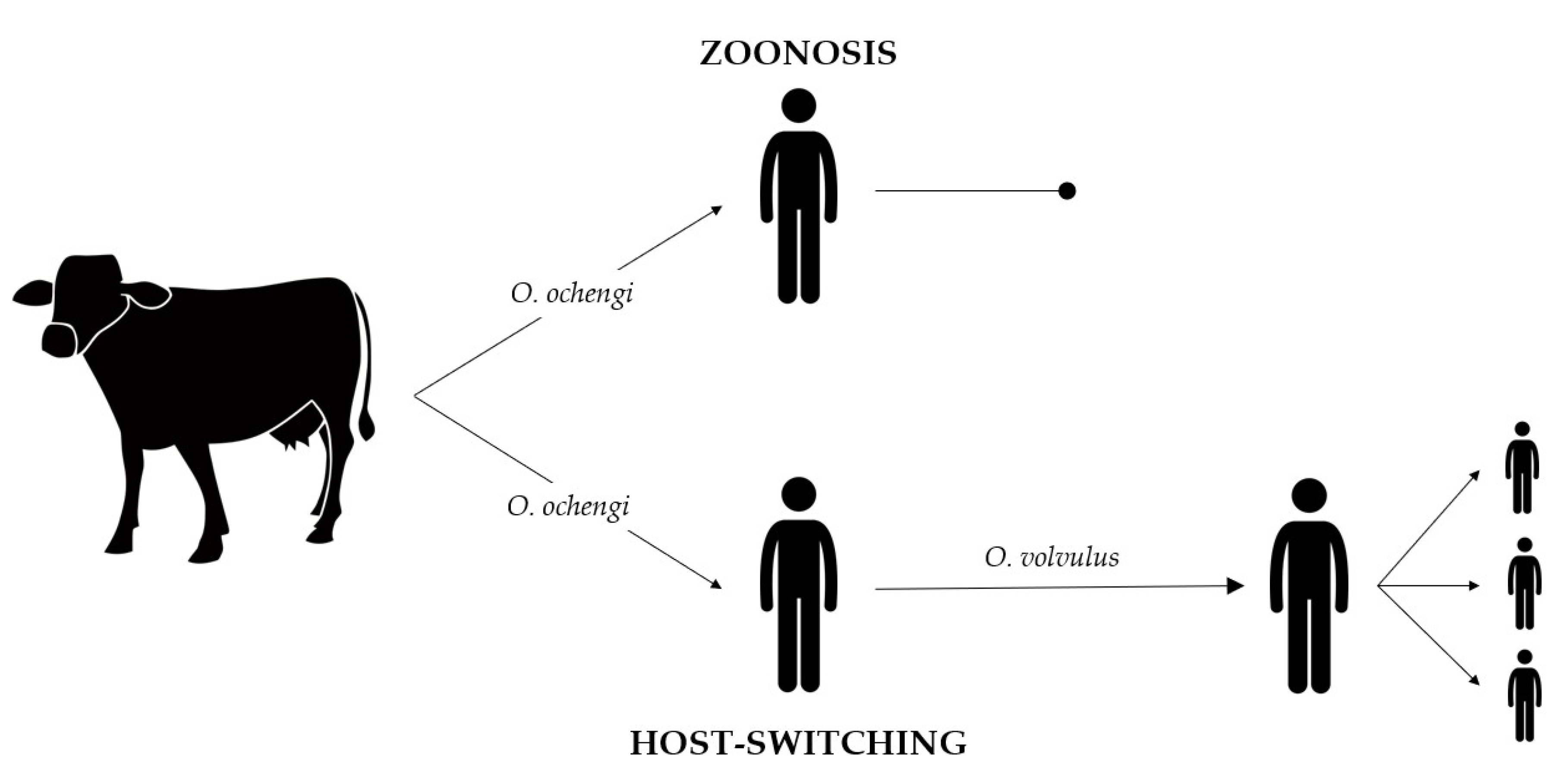
Figure 1.
Host-switching event leading to the appearance of new Onchocerca species. Onchocerca microfilariae from cattle, such as O. ochengi, are transmitted through blackflies to a new host, humans. In these new hosts, O. ochengi remain without being transmitted to other humans, leading to transmission interruption (rounded arrow), called zoonosis. Another possibility is that O. ochengi may have settled in humans and evolved to new species, such as O. volvulus. In that case, O. volvulus is transmitted through blackflies between humans (pointed arrow). This event is called host-switching.

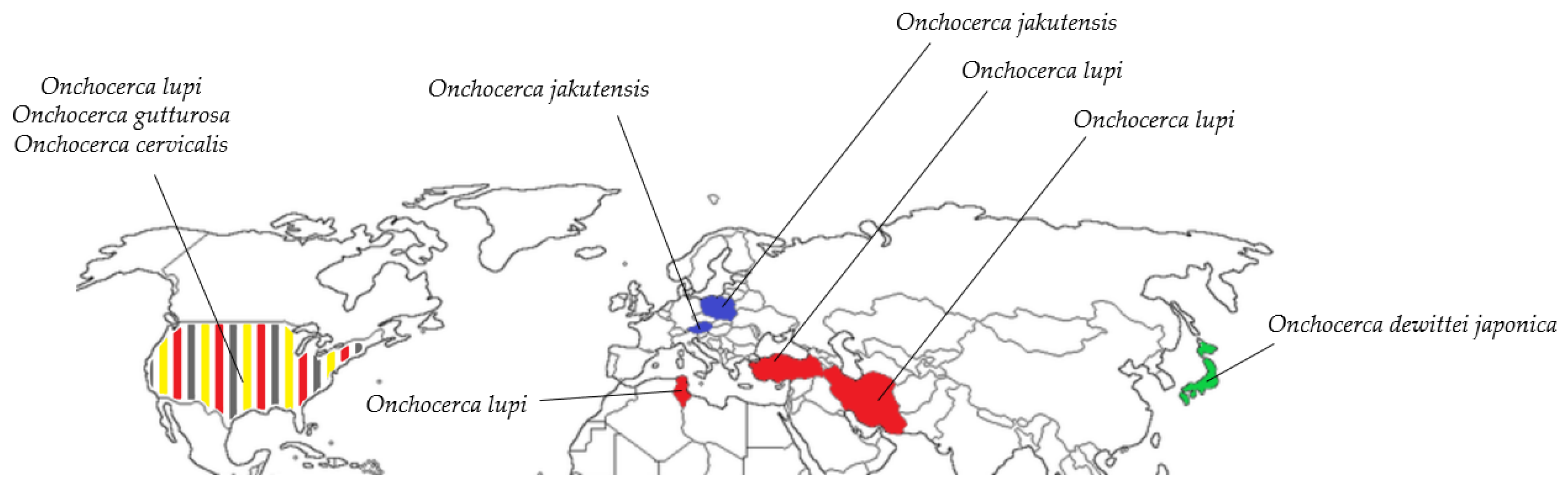
Figure 2.
Geographical distribution of Onchocerca species infecting animals identified in humans through the Holarctic region. The species found are the following: O. lupi (red), O. dewittei japonica (green), O. jakutensis (blue), O. gutturosa (grey), and O. cervicalis (yellow).
Figure 2.
Geographical distribution of Onchocerca species infecting animals identified in humans through the Holarctic region. The species found are the following: O. lupi (red), O. dewittei japonica (green), O. jakutensis (blue), O. gutturosa (grey), and O. cervicalis (yellow).


Table 1.
Description of the most common Onchocerca species identified parasitizing animals and humans, related to their main vectors, hosts, and geographical distribution.
Table 1.
Description of the most common Onchocerca species identified parasitizing animals and humans, related to their main vectors, hosts, and geographical distribution.
| Onchocerca Species | Vectors | Hosts | Geographical Distribution |
|---|---|---|---|
| O. armillata | Unknown [10] | Cattle [11], buffaloes [11], dromedaries [12], goats [11] | Ghana [13], Iran [14], Senegal [15], Sudan [12], Sierra Leona [16], Tanzania [17], Nigeria [3], Cameroon [10], India [18] |
| O. boehmi | Unknown [19] | Horses [19] | Austria [20], Iran [21], Italy [19] |
| O. cervicalis | Culicoides nubeculosus [22], C. variipennis [23] | Horses [24], ponies [25], humans [26], donkeys [27] | Australia [24], Japan [28], United Kingdom [22], Holland [29], Canada [30], United States [31], Brazil [32], Poland [33], Spain [33], Egypt [27] |
| O. cervipedis | Simulium venustum [34] | White-tailed deer [35], black-tailed deer [35], moose [9], caribou [9] | Canada [35], Costa Rica [36], United States [9], Alaska [9] |
| O. dewittei dewittei | Unknown | Wild boar [37] | Malaysia [37] |
| O. dewittei japonica | Simulium bidentatum [38], S. arakawae, S. japonicum, S. oitanum, S. quinquestriatum, S. rufibasis [39] | Wild boar [40], humans [41] | Japan [40] |
| O. dukei | Simulium hargreavesi, S. vorax, S. damnosum s.l. [42] | Cattle [43] | Zambia [44], Togo [45], Cameroon [43] |
| O. eberhardi | Simulium arakawae, S. oitanum, S. bidentatum [46] | Sika deer [47] | Japan [47] |
| O. fasciata | Culicoides puncticollis [48] | Dromedaries [49], camels [50] | Sudan [51], Somalia [51], Jordan [52], Saudi Arabia [53], Iran [54], China [55], Mongolia [48] |
| O. flexuosa | Prosimulium nigripes, Simulium ornatum [56] | Antelope [57], reed deer [58], Roe deer [59] | Uganda [60], Germany [61], Spain [8], Sweden [5], Slovakia [62] |
| O. gutturosa | Culicoides spp., C. kingi [63] | Cattle [43], camels [64], dromedaries [12], horses [65], humans [66] | Iran [14], Senegal [15], Australia [64], Togo [45], Sudan [12], Sierra Leona [16], Cameroon [43], Turkey [67], India [18] |
| O. jakutensis | Unknown | Red deer [58], humans [68] | Germany [53], Italy [69], Austria [70], Switzerland [71], Poland [68] |
| O. lupi | Simulium sp. [72], S. tribulatum [7] | Wolf [73], Dogs [74], Cats [75], Humans [76] | Georgia [73], Germany [77], Greece [78], Hungary [79], Tunis [80], Turkey [81], Iran [82], Portugal [75], United States [83], Spain [84] |
| O. ochengi | S. damnosum s.l. [85] | Cattle [43] | Burkina Faso [86], Senegal [15], Mali [87], Sierra Leona [16], Togo [45], Cameroon [43], Ghana [88] |
| O. ramachandrini | Simulium damnosum s.l. [89] | Warthogs [90] | Cameroon [90], Uganda [89] |
| O. reticulata | Culicoides nubeculosus Meig [91] | Horses [92], donkeys [27] | France [93], Australia [92], United States [65], Egypt [27] |
| O. skrjabini | Simulium arakawae, S. oitanum, S. bidentatum [46], S. japonicum, Prosimulium sp. [28] | Japanese serow [94], sika deer [47] | Japan [47,94] |
| Onchocerca spp. type I | Simulium bidentatum [38], S. sigrogilvum [95] | Wild boar [38], cattle [95] | Japan [38], Thailand [95] |
| O. suzukii | Simulium japonicum, Prosimulium sp. [28] | Japanese serow [94] | Japan [94] |
| O. takaokai | Simulium bidentatum [96] | Wild boar [96] | Japan [96] |
| O. volvulus | Simulium sp. [97] | Humans [97] | Brazil [98], Guatemala [99], Uganda [100], Tanzania [101], Democratic Republic of the Congo [102], Yemen [103] |
© 2020 by the authors. Licensee MDPI, Basel, Switzerland. This article is an open access article distributed under the terms and conditions of the Creative Commons Attribution (CC BY) license (http://creativecommons.org/licenses/by/4.0/).
Share and Cite
MDPI and ACS Style
Cambra-Pellejà, M.; Gandasegui, J.; Balaña-Fouce, R.; Muñoz, J.; Martínez-Valladares, M. Zoonotic Implications of Onchocerca Species on Human Health. Pathogens 2020, 9, 761. https://doi.org/10.3390/pathogens9090761
AMA Style
Cambra-Pellejà M, Gandasegui J, Balaña-Fouce R, Muñoz J, Martínez-Valladares M. Zoonotic Implications of Onchocerca Species on Human Health. Pathogens. 2020; 9(9):761. https://doi.org/10.3390/pathogens9090761
Chicago/Turabian StyleCambra-Pellejà, Maria, Javier Gandasegui, Rafael Balaña-Fouce, José Muñoz, and María Martínez-Valladares. 2020. "Zoonotic Implications of Onchocerca Species on Human Health" Pathogens 9, no. 9: 761. https://doi.org/10.3390/pathogens9090761
Note that from the first issue of 2016, this journal uses article numbers instead of page numbers. See further details here.