Article

Butyrate Mitigates Lipopolysaccharide-Induced Intestinal Morphological Changes in Weanling Piglets by Regulating the Microbiota and Energy Metabolism, and Alleviating Inflammation and Apoptosis

1
Key Laboratory of Feed Biotechnology of Ministry of Agriculture and Rural Affairs, Institute of Feed Research, Chinese Academy of Agricultural Science, Beijing 100081, China
2
State Key Laboratory of Animal Nutrition, Institute of Animal Sciences of Chinese Academy of Agricultural Sciences, Beijing 100193, China
3
Scientific Observing and Experiment Station of Animal Genetic Resources and Nutrition in North China of Ministry of Agriculture and Rural Affairs, Institute of Animal Science of Chinese Academy of Agricultural Sciences, Beijing 100193, China
*
Authors to whom correspondence should be addressed.
Microorganisms 2022, 10(10), 2001; https://doi.org/10.3390/microorganisms10102001
Submission received: 20 September 2022 / Revised: 4 October 2022 / Accepted: 7 October 2022 / Published: 10 October 2022
(This article belongs to the Special Issue Dietary Nutrition and Gut Microbiota)

Abstract

:
Butyrate provides energy for colonocytes and is a functional metabolite that mitigates weanling piglet stress. However, its effects and mechanisms remain largely unknown. We established a lipopolysaccharide (LPS)-induced inflammatory stress piglet model to examine how butyrate mechanisms impacted piglet intestinal histology, microbiota, and inflammation. We randomly assigned 18 crossbred male piglets to three treatment groups: CON, LPS, and BT-LPS. Coated butyrate was supplemented in the BT-LPS feed for 21 days. On days 19 and 21, piglets in LPS and BT-LPS groups were challenged with LPS at 100 μg/kg body weight. Dietary butyrate improved LPS-injured intestinal histology by significantly increasing jejunal and ileal villus height, villus height to crypt depth ratios, and decreasing histological scores. LPS challenge activated hypoxia-inducible factor 1α and nuclear factor-κB, and enhanced interleukins (IL-1β, IL-6, IL-12), tumor necrosis factor-α, and also downstream inducible nitric oxide synthase and cyclooxygenase 2, but decreased anti-inflammatory cytokines (IL-10, IL-13). Most molecule levels were significantly reversed by butyrate administration. When compared with the CON or LPS groups, the BT-LPS group had a higher relative abundance of jejunal Firmicutes, Bacteroidetes, Clostridiaceae, Lactobacillus, and Prevotella but a lower abundance of Proteobacteria, Enterobacteriaceae, and Escherichia–Shigella. Phylogenetic investigation of communities by reconstruction of unobserved states and correlation analyses suggested these bacteria contributed to butyrate-alleviating jejunal inflammation and infectious diseases. Butyrate-based diets significantly reduced apoptosis via mitochondrial pathways by downregulating apoptotic caspase 3 mRNA levels. Diets also altered enterocyte metabolism in the jejunum by upregulating peroxisome-proliferator-activated receptor α expression but downregulating carnitine palmitoyltransferase 1 level when compared with CON or LPS groups. Butyrate supplementation improved immunity homeostasis, generated beneficial shifts in microbial communities, improved enterocyte energy metabolism, and prevented apoptosis to protect intestinal histology from LPS-induced injury.

1. Introduction

Modern pig production faces many challenges, especially during weanling piglet transition periods. Weanling stress commonly arises due to shifts from liquid to solid feeding, environment changes, and mixing with new mates [1]. This stress generally induces a post-weaning growth lag, diarrhea, or even a high incidence of mortality and morbidity, leading to economic losses [2]. A crucial factor during stress processes is gastrointestinal tract underdevelopment, with poor digestive and nutrient absorption abilities. Additionally, weanling piglets are vulnerable to intestinal infections with Gram-negative enterotoxigenic Escherichia coli pathogens, as well as resultant intestinal immune system activation [3]. This activation induces diverse inflammation cell infiltration and proinflammatory cytokine production involving interleukin 1β (IL-1β), IL-6, and tumor necrosis factor-α (TNF-α), which cause mucosal dysfunction and injury [4]. Lipopolysaccharides (LPS) are composed of lipids and polysaccharides, are derived from Gram-negative bacteria outer membranes, and are strong inflammatory stimulants [5]. LPS interacts with toll-like receptor 4 (TLR4) and triggers inflammatory signaling cascades by activating nuclear factor-κB (NF-κB) and inducing its nuclear translocation, ultimately leading to proinflammatory cytokine secretion and inflammatory responses in piglets [6]. LPS challenge doses, duration, and associated mechanisms are well characterized in animals, and therefore LPS-induced intestinal inflammation models are commonly used to study in vivo inflammatory injury [7,8].
Recently, a previous study has reported close relationships between intestinal microbiota, intestinal mucosa health, and piglet health [9]. In piglet intestines, several lymphoid tissue types are present, including highly organized lymphoid follicles, mesenteric lymph nodes, isolated lymphoid follicles, and lymphocytes, which are influenced by microbiota in terms of intestinal immune function. Healthy microbial communities not only resist pathogen infection but also coordinate balanced anti- and pro-inflammatory responses [6,10]. Intestinal microbiota also regulate intestinal immune systems via flagellin, LPS, and secreted metabolites to accelerate immune system development and maturity [11]. Conversely, intestinal microbial disorders are related to intestinal inflammation [12]; however, excessive inflammatory responses exacerbate intestinal dysfunction and injury, and negatively impact piglet growth and health [13]. This evidence highlights the central role of intestinal microbiota in regulating immune functions and improving piglet health.
Butyrate is primarily derived from the microbial fermentation of dietary fiber and provides approximately 70% of energy requirements for colonocytes [14,15]. In general, butyrate exerts positive effects on livestock production, as well as multiple beneficial effects toward animal physiology. Previously, diets containing sodium butyrate significantly decreased the incidence of post-weaning diarrhea in piglets, enhanced humoral immunity levels by increasing immunoglobulin concentrations, and improved intestinal mucosa integrity [16]. Another study reported that tributyrin-supplemented diets alleviated intestinal injury and enhanced tight-junction formation in piglets [17]. The evidence also suggested that butyrate elevated enterocyte proliferation but decreased apoptosis [18]. Moreover, butyrate regulated intestinal immunity homeostasis by directly acting on immune mucosa cells, increasing Treg numbers and activity, and decreasing neutrophils, macrophages, and effector T cells [19]. A previously characterized butyrate mechanism showed that associated anti-inflammatory properties were promoted via G-protein-coupled receptor 109A signaling [11]. However, the application of sodium butyrate in livestock is generally restricted in practice because of its offensive odor, leading to an adverse effect on feed intake [1,20]. In addition, sodium butyrate is absorbed immediately at the anterior part of the gastrointestinal tract, which limits the efficiency of sodium butyrate absorption in the entire digestive tract [21,22]. Coated sodium butyrate is composed of a secondary coating of sodium butyrate with different purity via intelligent microencapsulation technology that overcomes the aforementioned limits of ordinary sodium butyrate [23]. This microencapsulated coated sodium would slowly and progressively release its active ingredients in the whole intestinal tract and display its function [21,24]. We hypothesized that coated butyrate would improve intestinal injury by shifting intestinal microbiota, regulate signal molecules related to inflammation and apoptosis in LPS-challenged piglets, and ameliorate host intestinal health.
To address these questions, we used an LPS-induced inflammatory stress piglet model to investigate underlying coated butyrate protective mechanisms toward intestinal histology when challenged by LPS. Several key physiological traits were examined: intestinal morphology, jejunal microbiota, inflammation, and enterocyte apoptosis and metabolism. Our data provided a theoretical dietary basis for butyrate in weaned piglets to improve overall host health.

2. Materials and Methods

2.1. Animals and Experimental Design

We randomly assigned 18 crossbred male weanling piglets (Duroc × Landrace × Yorkshire) with a similar initial body weight (BW = 9.10 ± 0.15 kg) to three treatments. Each treatment comprised six replicates (pens), with one piglet/pen. Treatments included a basal diet and a butyrate diet (basal diet + 3.00 g/kg coated butyrate). The test period was 21 days. The day of weaning was the first day of the experiment, and on the 19th and 21st days of the experiment, the piglets in the CON group were treated by intraperitoneal injection of sterile saline, and piglets in LPS and BT-LPS groups were treated by LPS and then labeled CON, LPS, and BT-LPS. The LPS (from E. coli serotype 055:B5; Sigma-Aldrich, St. Louis, MO, USA) was challenged at 100 μg/kg BW in sterile saline one time [7]. A non-medicated basal diet was formulated to address all weanling piglet nutrient requirements according to the National Research Council recommendations [25] (Table 1). Coated butyrate was supplemented as a partially coated sodium butyrate formulation composed of 70% sodium butyrate and 30% fat that was encapsulated using intelligent microencapsulation technology. The dose was based on previous studies [26,27].
Piglets were housed alone in one pen (1.8 × 0.8 m), and adjacent pens were separated by a closed baffle. A hard plastic and fully slotted floor was used in the pen. The room was environmentally controlled; the temperature was controlled at 28–30 °C and 26–28 °C between days 1–14 and 15–21, respectively. During the experimental period, pigs had ad libitum access to food and water.

2.2. Sample Collection

On day 21, 12-h-fasted piglets were humanely slaughtered at 4 h after LPS challenge by means of an anesthetic injection. The intestines were removed and divided into the duodenum, jejunum, and ileum sections. Middle duodenum, jejunum, and ileum in 1 cm long intestinal canal sections were obtained; washed lightly in saline; fixed in 4% formalin; and stored at 4 °C. Fresh jejunal mucosa and chyme were collected as previously described [28], immediately frozen in liquid nitrogen, and stored at −80 °C for biochemical analysis.

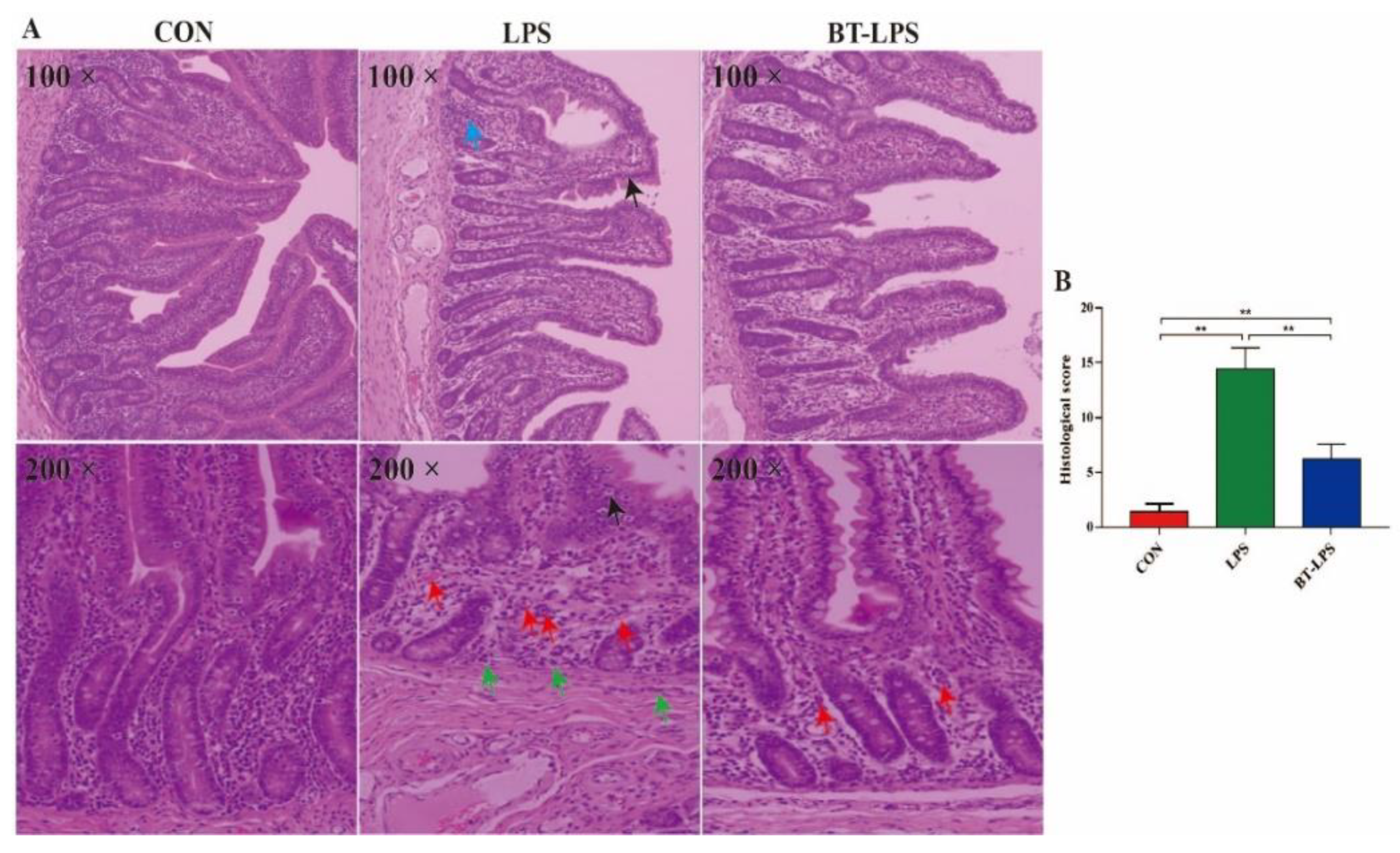
2.3. Intestinal Morphology

Duodenum, jejunum, and ileum specimens were prepared and embedded in paraffin, sectioned at 4 μm, and subjected to hematoxylin and eosin (H&E) and periodic acid–Schiff staining. Three intact crypt villi in one sample were selected, and villus height (VH) and its adjoining crypt depth (CD) were measured with an image analysis system (Version 1, Leica Imaging Systems Ltd., Cambridge, UK) [2]. VH was determined from the apex to the base of the villus crypt junction, and CD was the invagination depth between adjacent villi [29]. VH to CD (V/C) ratios were also calculated. Histological observations were conducted according to the previous study, and histological scores were blindly analyzed using a 0–30 score system for each parameter [30].

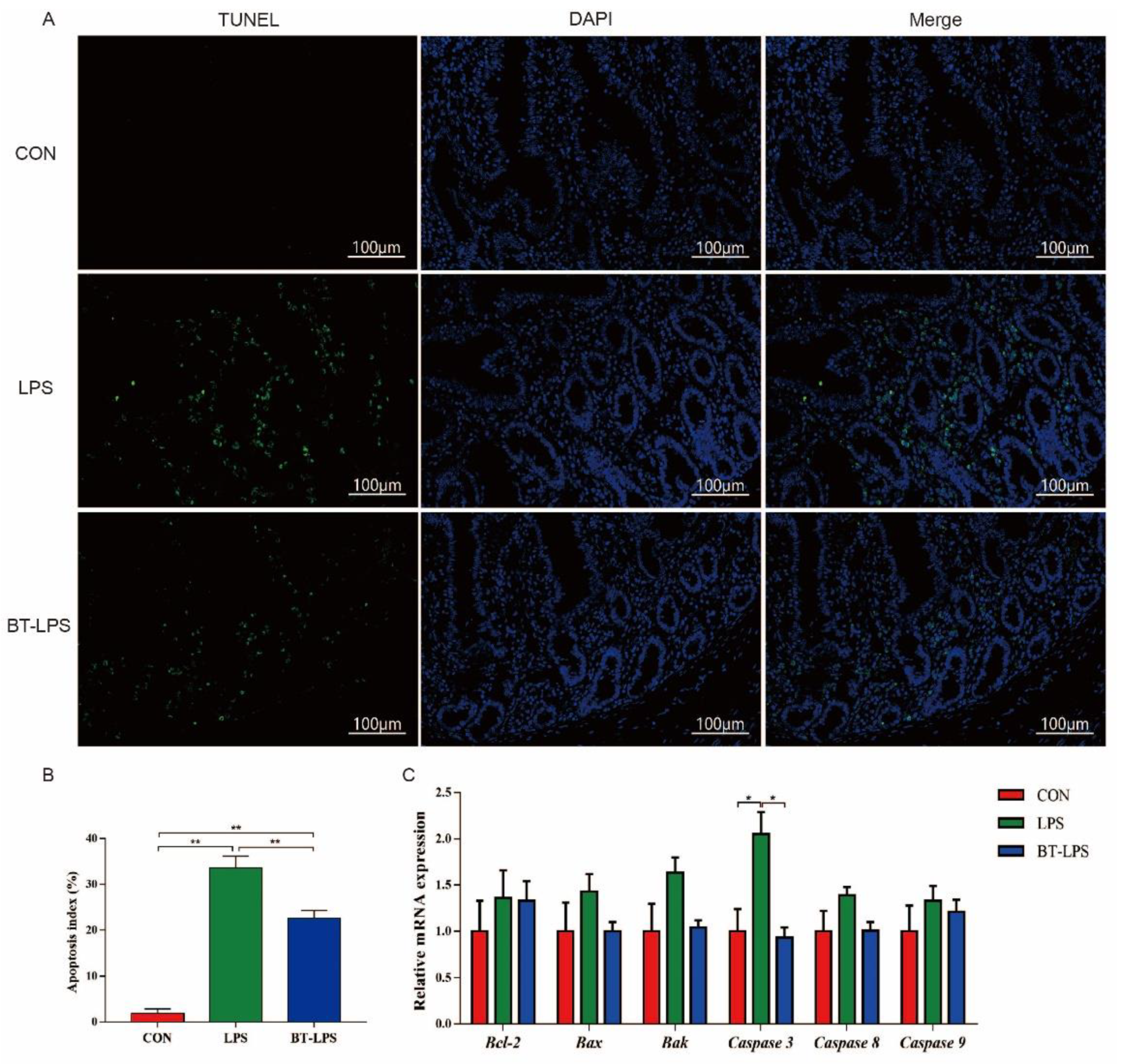
2.4. Terminal Deoxynucleotidyl Transferase Nick end Labeling (TUNEL)

Jejunum sections were prepared and stained using a commercial TUNEL kit (Fluorescein, Basel, Switzerland, cat. no. 11684817910) [31]. The TUNEL-positive nuclei fluoresced green under a fluorescence microscope, while all nuclei in the jejunum fluoresced blue. Images were taken at high magnification (200×), where five sections were selected from each group for statistical analysis. Apoptotic index = number of TUNEL-positive nuclei/total number of nuclei × 100% [31].

2.5. Jejunal Microbiota and Analysis

Bacterial DNA was extracted from jejunal contents with a Qiagen DNA stool mini kit (Qiagen, Hilden, Germany). DNA quantity and quality were determined using a NanoDrop 2000 spectrophotometer (Thermo Fisher Scientific, Waltham, MA, USA) and 1% agarose gels, respectively. The specific hypervariable region (V3–V4) 16S rRNA was amplified using the primers (forward 5′-ACTCCTACGGGAGGCAGCA-3′ and reverse 5′-GGACTACHVGGGTWTCTAAT-3′) with unique barcodes. Polymerase chain reaction (PCR) was applied in a total volume of 20 µL (including 0.2 μM each primer, 250 μM dNTP, 1 × FastPfu buffer, 1 U FastPfu polymerase (Beijing TransGen Biotech, Beijing, China), and 10 ng template DNA). PCR products were electrophoresed on 2% agarose gels and purified using a commercial kit (Qiagen, Hilden, Germany). Sequencing libraries were constructed using a TruSeq® DNA PCR-Free Sample Preparation Kit (Illumina, San Diego, CA, USA) on the basis of the manufacturer’s recommendations. Library quality was determined using a Qubit V.2.0 Fluorometer (Thermo Fisher Scientific, Waltham, MA, USA). Qualified DNA libraries were loaded into a NovaSeq platform with a capability of 2 × 250 bp paired-end sequencing reads (Novogene, Beijing, China).
Paired-end reads were generated and merged using FLASH software (V1.2.7, http://ccb.jhu.edu/software/FLASH/, accessed on 20 May 2022). Operational taxonomic units with 97% identity were gathered using Uparse (ver. 7.1, http://drive5.com/uparse/, accessed on 20 May 2022). Taxonomic annotations were performed using the Mothur algorithm (70% confidence) in the Silva database (http://www.arb-silva.de/, accessed on 20 May 2022). Alpha-diversity was analyzed using Chao 1 (http://www.mothur.org/wiki/Chao, accessed on 20 May 2022) and Shannon (http://www.mothur.org/wiki/Shannon, accessed on 20 May 2022) indices. Beta-diversity was visualized using principal coordinate analysis (PCoA) plots based on weighted and unweighted Unifrac distance. Bacterial biomarkers between groups were displayed using the linear discriminant analysis effect size (LEfSe) with linear discriminant analysis (LDA) > 3.5. Phylogenetic investigation of communities by reconstruction of unobserved states (PICRUSt) was used to assess metagenome function on the basis of different Kyoto Encyclopedia of Genes and Genomes (KEGG) levels [32]. The relative percentage of predictive pathways at KEGG level II was presented as a heatmap with normalized analysis.

2.6. Biochemical Analysis

Jejunum mucosa (appropriately 100 mg) samples were mixed and homogenized in 1 mL precooled saline using an Ultra-Turrax homogenizer (Scientz, Ningbo, China) and then vortexed for 30 s. Homogenates were centrifuged at 3000 × g for 15 min at 4 °C, and supernatants were transferred to new tubes for protein assay and other measurements. Immune indices were measured using enzyme-linked immunosorbent assay kits (Shanghai Enzyme-linked Biotechnology Co., Ltd., Shanghai, China) according to the manufacturer’s protocols. Assay molecules included hypoxia-inducible factor 1α (HIF-1α), nuclear factor-κB (NF-κB), NF-κB p65, interleukins (IL-1β, IL-6, IL-8, IL-10, IL-12, IL-13, IL-17), tumor necrosis factor α (TNF-α), transcription growth factor β (TGF-β), cyclooxygenase 2 (COX-2), and inducible nitric oxide synthase (iNOS). Inflammatory related parameters were normalized to tissue protein concentrations, which were measured using a bicinchoninic acid commercial kit (Thermo Fisher Scientific, Waltham, MA, USA).

2.7. Real-Time Quantitative PCR (RT-qPCR)

Selected mRNA abundance was determined by RT-qPCR, including apoptosis genes Bcl-2, Bak, Bax, caspase 3, caspase 8, and caspase 9 and energy metabolism genes acyl-CoA oxidase 1 (ACOX1), succinate dehydrogenase (SDH), peroxisome-proliferator-activated receptor α (PPARα), nuclear respiratory factor 1 (NRF1), uncoupling protein 2 (UCP2), mitochondrial transcription factor A (TFAM), α-ketoglutarate-dependent dioxygenase (FTO), malic enzyme 1 (ME1), and carnitine palmitoyltransferase 1 (CPT1). Total RNA was isolated from jejunum samples (approximately 0.75 mg) using an RNAprep pure tissue kit (Tiangen Biotech Co. Ltd., Beijing, China). Total RNA concentrations and quality were estimated using a NanoDrop 2000 spectrophotometer (Thermo Fisher Scientific, Waltham, MA, USA), which confirmed that high-quality absorbance ratios (260/280 nm) were between 1.8 and 2.0. RNA integrity was evaluated using agarose gel (1%) electrophoresis. Then, cDNA was synthesized from 1 μg total RNA using a PrimeScript RT reagent kit (TaKaRa Biotechnology Co., Ltd., Otsu, Japan) according to the manufacturer’s instructions. Selected mRNA reactions were detected in 10 µL (ABI Prism 7700 Sequence Detection System; Applied Biosystems, Foster City, CA, USA) using SYBR® Premix Ex TaqTM II (Tli RNaseH Plus) (TaKaRa Biotechnology Co., Otsu, Japan). The primers for energy metabolism, apoptosis-related, and housekeeping genes (glyceraldehyde 3-phosphate dehydrogenase (GAPDH)) were described previously [31,33]. The 2−ΔΔCt method was used for quantification using GAPDH as a reference gene, and relative abundance was normalized to CON group values.

2.8. Statistical Analysis

Statistical analyses were performed using one-way analysis of variance in SAS 9.4 (SAS Institute, Inc., Cary, NC, USA). Each piglet served as a statistical unit. Differences between groups were evaluated using Duncan’s multiple-range tests. Tukey’s HSD tests were used to test for differences in microbial diversity. Metastat and LEfSe analyses were used to test for significant differences between microbiota relative abundance. Results were represented as the mean with standard error of mean (SEM) in the tables and the mean with standard error (SE) in the figures, while p < 0.05 and p < 0.01 values were considered statistically and extremely significant, respectively. Bar charts were drafted in Graphpad Prism 7.0 software (GraphPad Software Inc., La Jolla, CA, USA).

3. Results

3.1. The Effects of Butyrate on LPS-Challenged Intestinal Morphology in Piglets

Piglets in BT-LPS and LPS groups had lower VH and V/C duodenum values when compared to the CON group (p < 0.05, Table 2). Compared with the CON group, the LPS challenge significantly reduced VH jejunum and ileum values and V/C ileum ratios (p < 0.05), while butyrate administration significantly increased these parameters (p < 0.05). Jejunum CD values in BT-LPS and LPS groups were lower than the CON group (p < 0.05), and V/C ratios in the BT-LPS group were higher than the LPS group (p < 0.05).

3.2. The Effects of Butyrate on LPS-Challenged Jejunum Histological Scores

Histological jejunum examinations indicated that LPS challenge increased mucosal intraepithelial lymphocytes (black arrows), as well as granulocyte infiltration and crypt damage (blue arrows) (Figure 1). Extensive inflammatory cell infiltration comprising neutrophils in the mucosa (red arrows) and monocytes in the submucosa (green arrows) were identified in LPS-challenged piglets. Dietary butyrate improved these parameters as indicated by histological scores. Piglets in the BT-LPS group had lower histological scores when compared with LPS animals but higher histological scores when compared to the CON group (p < 0.05).

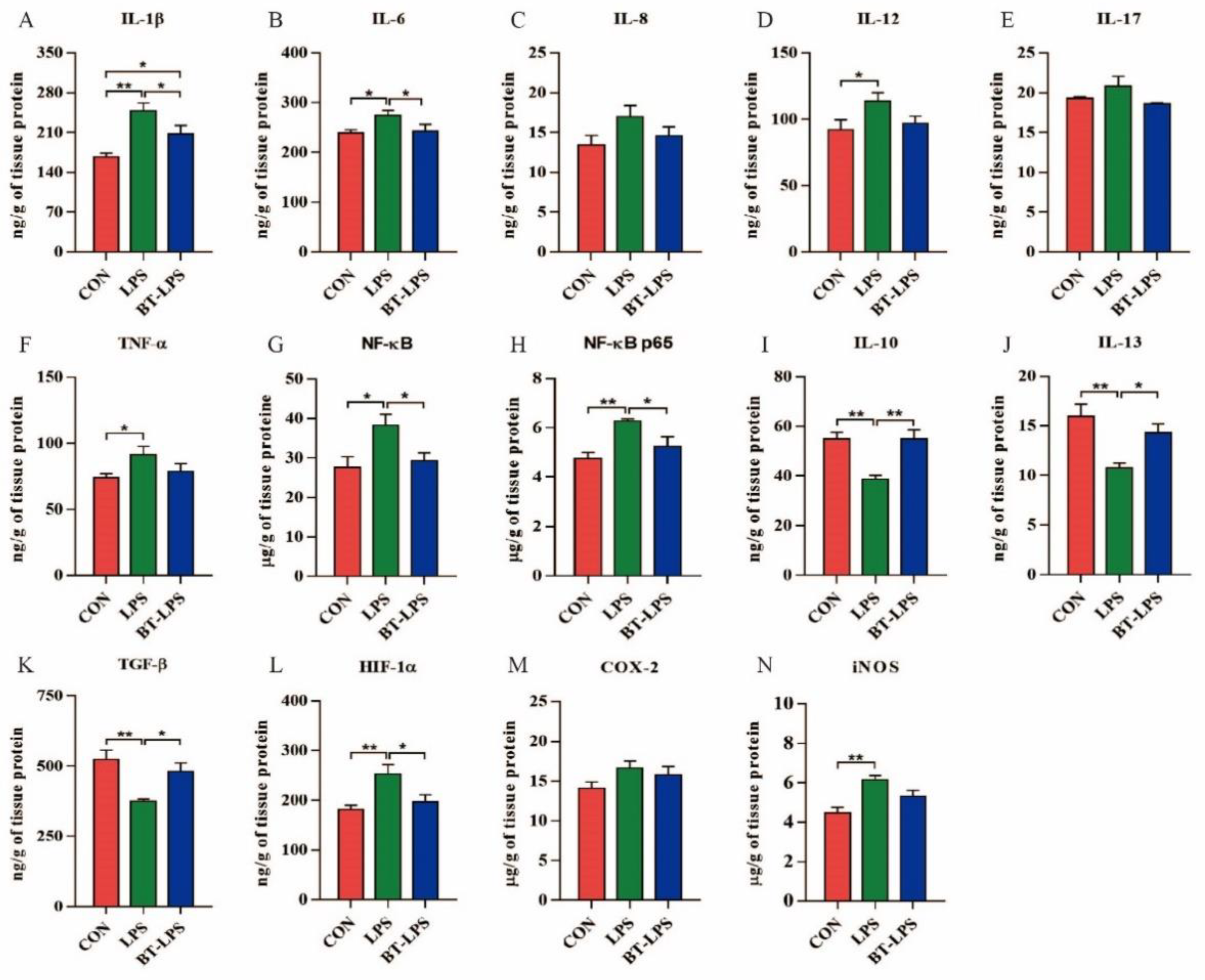
3.3. The Effects of Butyrate on LPS-Challenged Jejunum Inflammation

LPS challenge induced inflammation in jejunum tissue (Figure 2). When compared with the CON group, LPS animals displayed significantly increased pro-inflammatory-associated elements/cytokines IL-1β, IL-6, IL-12, TNF-α, NF-κB, and NF-κB p65 (p < 0.05, Figure 2A–H), while butyrate diets significantly decreased IL-1β, IL-6, NF-κB, and NF-κB p65 levels when compared with the LPS animals (p < 0.05). IL-1β level in the BT-LPS group was significantly higher than the CON group (p < 0.05). Anti-inflammatory cytokine IL-10, IL-13, and TGF-β levels in LPS animals were significantly higher than in CON and BT-LPS animals (p < 0.05, Figure 2I–K), but no differences were observed between BT-LPS and CON groups (p > 0.05). Piglets in the LPS group had higher HIF-1α and iNOS levels when compared with the CON group (p < 0.05, Figure 2L,N), while BT-LPS animals had lower HIF-1α levels when compared with the LPS group (p < 0.05).

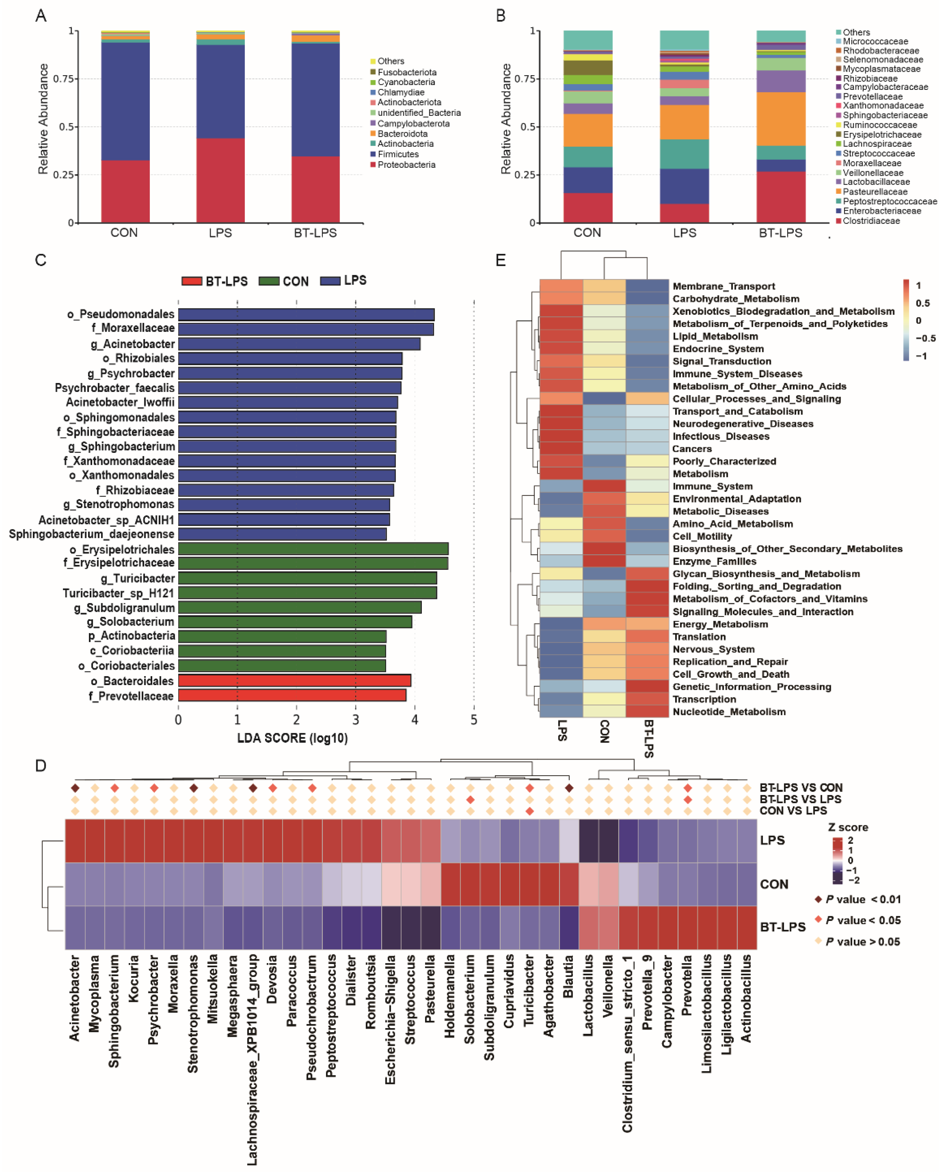
3.4. The Effects of Butyrate on Microbiota Diversity in Jejunum Exposed to LPS

In total, 1,074,885 effective sequences (1,520,294 raw reads) were generated from piglet jejunum microbiota samples, with an average of 59,716 sequences/sample. Alpha-diversity analyses showed that a butyrate diet and LPS challenge did not affect bacterial community α-diversity Observed_species, Chao 1, and Shannon indices (p > 0.05, Figure 3A–C). Beta-diversity PCoA analyses based on weighted and unweighted Unifrac distance showed that microbial communities were well separated between groups, especially between BT-LPS and the other groups (Figure 3D,E).

3.5. The Effects of Butyrate on Bacterial Community Structures, Challenged by LPS, in the Jejunum

At the phylum level, Firmicutes, Proteobacteria, Bacteroidetes, and Actinobacteria were the four major bacterial phyla in the CON group, with relative abundances of 61.38%, 32.78%, 1.64%, and 1.58%, respectively (Figure 4A). Compared with the CON group, LPS challenge increased the relative abundance of Proteobacteria, Bacteroidetes, and Actinobacteria by 35.18%, 53.05%, and 81.68%, respectively, but decreased the relative abundance of Firmicutes by 20.79% (p > 0.05). The relative abundances of Firmicutes, Proteobacteria, Bacteroidetes, and Actinobacteria were, respectively, 58.81%, 34.77%, 3.38%, and 0.86% in the BT-LPS group, which indicated that butyrate supplementation reduced the relative abundance of Firmicutes and Actinobacteria by 4.19% and 45.56%, respectively, but increased the relative abundance of Proteobacteria and Bacteroidetes by 6.07% and 106.10%, respectively, when compared to the CON group (p > 0.05).
At the family level, the Prevotellaceae relative abundance in the BT-LPS group was significantly higher than the LPS group (p < 0.05, Figure 4B), and no significant difference was observed between the CON group and other two groups (p > 0.05). The Clostridiales relative abundance in the BT-LPS group (26.95%) was higher than LPS (10.11%) and CON (15.71%) groups (p > 0.05). The Lactobacillaceae relative abundance in the BT-LPS group (11.30%) was higher than the LPS group (4.45%) and even higher than the CON group (5.47%) (p > 0.05). When compared to the CON group, LPS challenge increased the relative abundance of Enterobacteriaceae from 13.43% to 18.22%, whereas the butyrate diet decreased Enterobacteriaceae relative abundance from 18.22% to 6.19% when compared to the LPS group (p > 0.05).
LEfSe analyses indicated that g_Acinetobacter, g_Psychrobacter, g_Sphingobacterium, and g_stenotrophomonas constituted the dominant bacteria in the LPS animals (Figure 4C). In contrast, in the BT-LPS group, f_Prevotellaceae was the dominant bacteria. Of the 35 most dominant genera, the LPS challenge significantly decreased the relative abundance of Turicibacter when compared with the CON group (p < 0.05, Figure 4D), while butyrate supplementation significantly increased the relative abundance of Prevotella when compared with LPS and CON groups (p < 0.05). Compared with the CON group, the butyrate diet significantly decreased Blautia, Acinetobacter, Psychrobacter, Pseudochrobactrum, Devosia, Stenotrophomonas, and Sphingobacterium relative abundance (p < 0.05). The Escherichia–Shigella relative abundance in the BT-LPS animals (6.14%) was lower than the CON animals (13.18%) and the LPS animals (16.80%) (p > 0.05). PICRUSt analyses suggested that jejunal bacteria in butyrate-fed piglets provided a lower risk of immune system and infectious diseases when compared with LPS or CON groups (Figure 4E).

3.6. Correlation Analyses between the Jejunal Microbiota and Immune Indices

A Spearman’s correlation analysis was used to explore relationships between predominant genera and families and immune-associated parameters (Figure 5). Lactobacillus and Turicibacter were significantly positively correlated with IL-13 (r = 0.610, 0.468), while Escherichia–Shigella was significantly positively correlated with IL-1β (r = 0.332), IL-6 (r = 0.589), IL-12 (r = 0.598), and TNF-α (r = 0.485) levels (p < 0.05). Limosilactobacillus and Ligilactobacillus showed significantly positive correlations with IL-17 (r = 0.468, 0.515) and NF-κB p65 (r = 0.567, 0.578) (p < 0.05). Prevotella and Prevotella_9 were significantly negatively correlated with IL-6 (r = −0.480, −0.483), while Prevotella_9 was significantly negatively correlated with IL-12 (r = −0.471) (p < 0.05).

3.7. The Effects of Butyrate on Jejunum-LPS Challenged Cell Apoptosis

TUNEL staining showed that positive nuclei in the LPS jejunum were significantly higher than in CON and BT-LPS jejuna (p < 0.05, Figure 6A,B). Butyrate supplementation significantly decreased these numbers when compared with the LPS group, but they were still significantly higher than in the CON group (p < 0.05). Caspase 3 relative mRNA levels in the LPS group were significantly increased (p < 0.05, Figure 6C), while Bak relative mRNA levels showed an increased trend (p = 0.08) when compared with CON and BT-LPS groups. The transcript abundances of Bax, caspase 8, and caspase 9 were 43.0%, 39.0%, and 33.0% greater in the LPS animals, respectively, than in the CON animals (p > 0.05), but in the BT-LPS animals, these parameters were 30.1%, 27.3%, and 9.0% lower than LPS animals’ levels, respectively (p > 0.05).

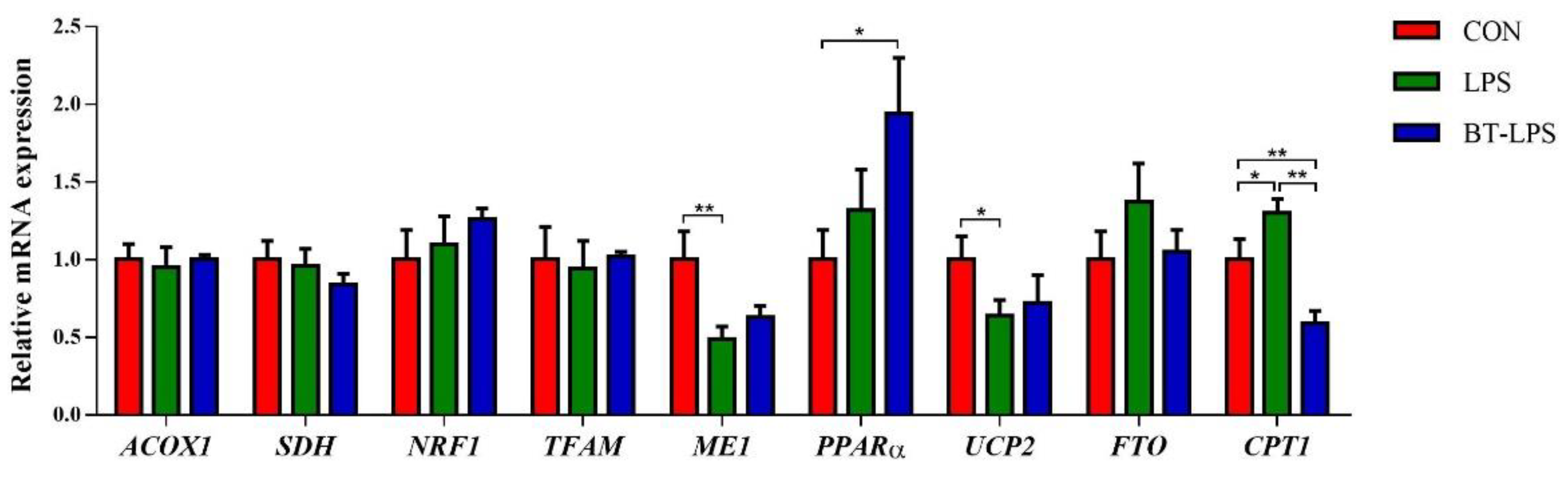
3.8. The Effects of Butyrate on Energy Metabolism Gene mRNA Levels in LPS-Induced Jejunum
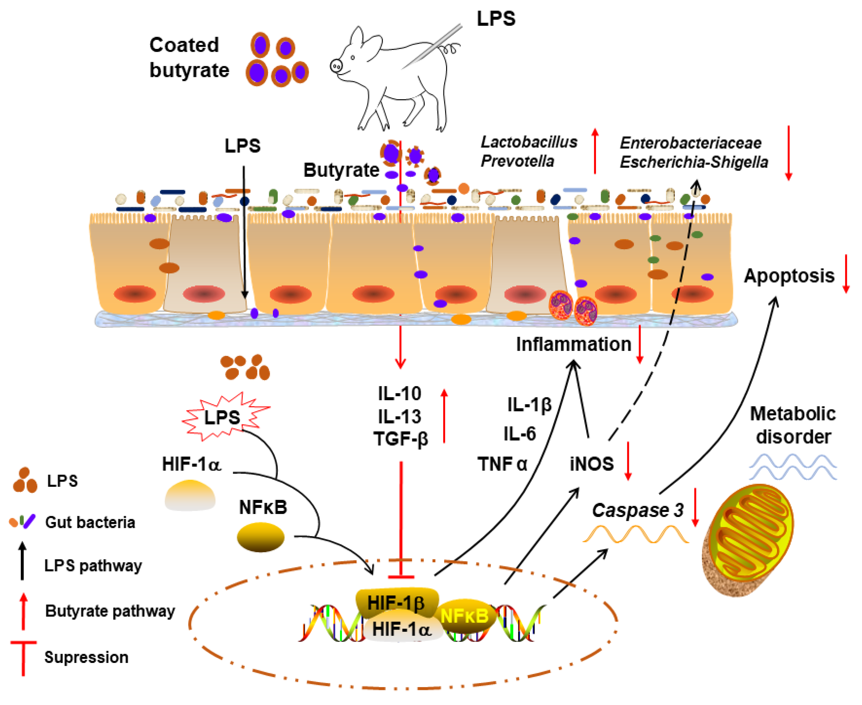
When compared with the CON group, butyrate supplementation significantly upregulated jejunal PPARα mRNA levels (p < 0.05, Figure 7), while the LPS challenge significantly decreased ME1 and UCP2 mRNA expression (p < 0.05). The LPS challenge significantly upregulated CPT1 mRNA levels when compared to the CON group (p < 0.05), but dietary butyrate significantly decreased CPT1 mRNA expression levels when compared to CON and LPS groups (p < 0.05). We observed no significance differences in ACOX1, SDH, NRF1, TFAM, and FTO transcript abundance between groups (p > 0.05).

4. Discussion

Growth retardation due to weanling stress is a common problem in weaned piglets. Previously, weanling-associated intestinal inflammation negatively affected epithelial functions in piglets [34]. LPS is a major enterotoxin produced by E. coli and damages intestinal functions or even causes piglet diarrhea, which negatively impact growth performance and generate huge economic production losses [8]. For this reason, LPS is widely used to construct acute inflammatory injury piglet models [35]. In our previous study, LPS challenge significantly decreased piglet average daily gain between days 15 and 21 and increased clinical scores between days 19 and 21 when compared with the CON group, while butyrate supplementation significantly decreased these clinical scores when compared with the LPS group [6]. Our observations were similar to a previous study showing that sodium butyrate supplementation (0.2%) to a deoxynivalenol diet improved weaned piglet performance [36]. These data were possibly attributed to improvements in gut health. To further highlight the beneficial effects of butyrate, we studied its effects on intestinal morphology, jejunal microbiota, inflammatory responses, apoptosis, and metabolism in an LPS-induced piglet model.
Intestinal morphology is an important mechanical defense barrier reflecting intestinal health and absorptive capacity. Intestinal morphological alterations, including villus atrophy and crypt hyperplasia, represent malabsorption and body growth retardation [37,38], and decreased VH indicates LPS toxicosis in the intestine [38]. In our study, LPS challenge induced acute intestinal injury, as LPS piglets had lower intestinal VH and V/C ratios when compared with the CON group. These adverse effects on intestinal morphology by LPS were significantly prevented by coated butyrate supplementation, especially for the jejunum and ileum. This was consistent with a previous study that reported that sodium butyrate administration not only increased VH and V/C values but also enhanced intestinal barrier function, as demonstrated by increased transepithelial electrical resistance and decreased fluorescein isothiocyanate dextran 4 kDa flux [39,40]. Thus, butyrate supplementation exerted positive effects on intestinal morphology and contributed to digestive capacity and barrier function. Most probably, butyrate improved homeostatic immunity, as intestinal integrity and epithelial function were negatively affected by inflammatory status [40]. Additionally, butyrate possibly alleviated intestinal mucosa damage induced by noxious bacteria via bacteriostasis, which improved intestinal morphological structures and functions [41].
The jejunum is important in preventing pathogens and toxins from the external environment into the circulation system, and its digestive enzymes and cytokines are associated with growth and immune function [42,43]. Weaned piglets with growth retardation had a higher apoptosis rate in jejunum epithelial cells and pro-inflammatory response than those with normal body weight [44]. The efficiency of glyceryl butyrate to improve enterotoxigenic Escherichia coli-induced inflammatory response by NFκB/MAPK pathway in the ileum was not as good as that in the jejunum [45]. In addition, most attention was focused on colonic microbiota, while jejunal microbiota has been shown to be essential for the host’s physiology, which includes metabolic, immune, and endocrine functions [46,47]. The jejunum responds strongly to the involved microbiota, even though there are low bacterial densities and diversity [48]. In a time-course study of metabolic responses in gastrointestinal tract mucosa to conventionalization, a strong and dynamic response was found in the jejunum but not the ileum and colon, indicating the unique role of the jejunum in response to the luminal microbiota [46,49]. Combined with the present result, dietary coated butyrate significantly improved jejunal morphology induced by LPS. Therefore, the jejunum was selected to further measure inflammation, microbiota, apoptosis, and metabolism.
During post-weaning periods, LPS derived from Gram-negative bacteria induced inflammatory responses by activating the TLR 4/NF-κB-mediated pathway, in which the inflammatory response was partly correlated with histology injury [12]. Inflammatory cell activation and mediator release are caused by intestinal barrier dysfunction [50]. In our study, LPS challenge induced severe, higher jejunal histological scores in intraepithelial lymphocyte, granulocyte, neutrophil, and monocyte infiltration. Moreover, LPS stimulation significantly increased pro-inflammatory-associated molecule and cytokine production, such as NF-κB, NF-κB p65, TNF-α, IL-1β, IL-6, IL-12, and IL-17 in the jejunum. These increases suggested that LPS successfully induced inflammatory responses, with the NF-κB pathway having key roles initiating this inflammation. HIF-1α is a transcription factor and is associated with metabolic and inflammatory disorders [31]. HIF-1α also activates the central regulator NF-κB and causes inflammation via inhibitor of NF-κB phosphorylation and the extracellular-signal-regulated kinase1/2-mediated phosphorylation of serine 276 on p65 [51]. However, short chain fatty acids (SCFAs) may decrease pro-inflammatory cytokine secretion by suppressing NF-κB [52]. This observation agreed with our data; coated butyrate significantly diminished HIF-1α increases and associated downstream pro-inflammatory molecules and cytokines, and therefore decreased histological scores induced by LPS. A similar result was observed in a Clostridium difficile-challenge mouse study, wherein butyrate significantly reduced histological scores in the colon and alleviated inflammatory responses, as reflected by decreased IL-1β, IL-6, and chemokine ligand 1 secretion [30]. Moreover, a glycerol butyrate diet was shown to modulate jejunal and ileal inflammation, as indicated by the decrease in IL-1β, IL-6, and TNF-α levels [45]. However, in our study, this diet significantly reversed decreases in anti-inflammatory cytokines induced by LPS, including IL-10, TGF-β, and IL-13. T-regulatory cell IL-10 production was recently shown to prevent exaggerated T-cell responses and protect against intestinal inflammation via a self-limiting mechanism [53]. TGF-β positively affected epithelial recovery and improved inflammation, while TGF-β deficiency accelerated immune system dysfunction [54,55]. These observations indicated that butyrate supplementation contributed to homeostatic immunity, where regulatory mechanisms were related to butyric acid and G-protein-coupled receptor interactions or histone deacetylase inhibition [15].
Intestinal microbial communities generally participate in nutrient metabolism, and associated disorders are largely associated with disrupted intestinal digestion, inflammation, and morphology [41,56]. To investigate the effects of coated butyrate on jejunal microbiota in piglets challenged by LPS, we compared microbial diversity and composition between treatment groups. However, no significant differences were observed in α-diversity between groups. As reported, the oral administration of sodium butyrate to weaned piglets decreased microbial diversity in the ileum but increased levels in the colon [57]. This discrepancy may have been due to the type and dose of butyrate and different intestinal segments analyzed. Our β-diversity results showed that microbiota in the BT-LPS group was distinct from microbiota in CON or LPS groups, which suggested coated butyrate altered jejunal microbial structures. Specifically, coated butyrate supplementation increased the relative abundance of Firmicutes and Bacteroidetes but decreased the relative abundance of Proteobacteria and Actinobacteria when compared with the LPS group. An increase in Firmicutes proportion is beneficial to intestinal health, as it contains abundant SCFA-producing bacteria [8]. Clostridiaceae, Clostridium_sensu_stricto_1, Lactobacillaceae, and Lactobacillus were involved in the Firmicutes, and their relative abundance was higher in the BT-LPS group when compared with the LPS group and even the CON group. This concurred with a previous study showing sodium butyrate infusion tended to accelerate Firmicutes and Clostridiaceae proliferation in the colon [58]. Dietary sodium butyrate also increased colonic Clostridiaceae relative abundance in weaned piglets [57]. Lactobacillus is a commonly used probiotic organism and a characterized bacteriocin and organic acid producer. Compelling previous evidence showed that Lactobacillus had excellent probiotic properties in a dextran-sulfate-sodium-induced mice model, where it maintained the intestinal epithelial barrier and alleviated inflammatory responses [59]. Other studies indicated that Lactobacillus protected intestinal health via its metabolites, bacteriostasis, and the competitive exclusion of pathogens at mucosa sites [8,60]. As demonstrated in a Staphylococcus aureus infection model, Lactobacillus supplementation effectively decreased S. aureus levels and accelerated intestinal damage recovery [61]. Another study demonstrated that the Lactobacillus stress protein GroEL enhanced IL-13 secretion and decreased apoptosis induced by LPS [62]. Additionally, in our study, Lactobacillus was significantly correlated with IL-13 levels. This compelling evidence supported the concept that butyrate administration improved intestinal histology and inflammation induced by LPS challenge.
Intestinal dysbiosis is generally associated with increased facultative anaerobic bacteria levels. Facultative anaerobes, Proteobacteria, including Enterobacteriaceae and Escherichia–Shigella, proliferate and interfere with host nutrition by metabolizing fermentation products in oxygenated conditions [63]. Escherichia–Shigella is a pathogen that induces intestinal inflammation by activating NF-κB and mitogen-activated protein kinase pathways [12,64]. Shiga toxin produced by E. coli promotes fluid and electrolyte accumulation in the bowel by disrupting balances in intestinal absorption and secretion, leading to higher diarrhea incidences and lower piglet growth [57,65]. These observations agreed with our study where an LPS challenge increased the relative abundance of Escherichia–Shigella, Enterobacteriaceae, and Proteobacteria, and caused severe inflammatory responses. However, coated butyrate supplementation decreased this bacterial abundance and decreased pro-inflammatory cytokine levels. This was partly consistent with a previous report showing that sodium butyrate administration inhibited the proliferation of facultative anaerobes [66]. We also showed that Escherichia–Shigella significantly positively correlated with TNF α, IL-1β, IL-6, and IL-12 levels, indicating that a butyrate-based diet alleviated intestinal inflammation and was possibly related to a decrease in the relative abundance of Escherichia–Shigella. As reported, host protection against intestinal infection mechanisms partly depends on the fight between facultative anaerobic Enterobacteriaceae and obligate anaerobic Clostridiaceae [67]. This observation was also reflected by our data. A lower acid intestinal environment, due to butyrate supplementation, probably suppressed the colonization of these pathogens [68]. Another reason could be iNOS levels, which were higher in the LPS animals when compared to the CON animals but were non-statistically reduced in the BT-LPS animals. Decreased iNOS levels were shown to control facultative anaerobic bacteria by mitigating nitric oxide conversion to nitrate, thus reducing levels of additional electron acceptors [69].
Bacteroidota, including Prevotellaceae, Prevotella, and Prevotella_9, are well known for modulating piglet growth due to energy derived from bacterial fermentation [57]. Prevotella promotes complex dietary polysaccharide conversion to monosaccharides for host uptake and confers performance advantages [70,71]. Additionally, Prevotella metabolizes plant cell walls and produces high SCFA levels, which are later absorbed by enterocytes to promote intestinal immunity homeostasis [72]. In our study, coated butyrate supplementation significantly increased the relative abundance of Prevotella when compared with the CON and LPS groups, while Prevotella or Prevotella_9 were significantly negatively correlated with IL-6 and IL-12 levels. This was partly explained by the fact that butyrate supplementation alleviated intestinal inflammation, and possibly explained why piglets in the BT-LPS group had better growth performances and clinical scores in our previous study [6]. These findings suggested that dietary coated butyrate improved intestinal injury and appeared to be related to a microbiota shift. Combined PICRUSt and correlation analyses indicated that coated butyrate supplementation alleviated intestinal inflammation, partly by accelerating the prevalence of Firmicutes, Bacteroidetes, Clostridiaceae, Lactobacillus, and Prevotella but partly suppressing Proteobacteria, Enterobacteriaceae, and Escherichia–Shigella proliferation.
Apoptosis is a form of physiological cell death that controls epithelial turnover in the intestinal mucosa. Pathogens and associated toxins induce cell damage and apoptosis, causing villus atrophy during piglet weaning periods [4]. This observation was consistent with our TUNEL staining data, which indicated that an LPS challenge significantly increased jejunal cell apoptosis, with upregulated caspase 3 and Bax expression. Apoptosis is an important mechanism suppressing enterocyte proliferation. Generally, mitochondrial apoptosis pathways are modulated by the Bcl-2 family, the caspase family, apoptosis-induce factor, and second mitochondria-derived activator of caspases [31,73]. A previous study reported that a deoxynivalenol challenge caused intestinal epithelial cell apoptosis by upregulating Bax and caspase 3 protein expression [9]. However, our coated-butyrate-based diet significantly mitigated LPS-induced apoptosis/as reflected by TUNEL-positive cell levels and caspase 3 and Bak expression. These observations concurred with a previous study showing that diet with 0.1% tributyrin improved apoptosis and intestinal injury [17]. The anti-apoptotic butyrate effects may be closely correlated with the butyrate-mediated alleviation of intestinal inflammation induced by LPS challenge. As reported, inflammation usually promotes small intestinal cell apoptosis [74,75]. Another probable mechanism could be that butyrate protects against apoptosis by inhibiting endoplasmic reticulum stress, which is regulated by protein-kinase-like endoplasmic reticulum kinase-eukaryotic initiation factor 2-α pathway via GPR109A [76]. Collectively, these results indicated that coated butyrate supplementation effectively ameliorated LPS-induced intestinal apoptosis and injury and was reflected by improved intestinal histology.
Our data also indicated that coated butyrate protection against LPS-induced apoptosis was mediated by a mitochondria-dependent pathway. Energy metabolism in intestinal enterocytes was measured by gene expression. ME1 is an important enzyme required for lipid synthesis as well as being a malate metabolizing enzyme that catalyzes the reversible oxidative decarboxylation of L-malate coupled to the reduction of dinucleotide cofactor nicotinamide adenine dinucleotide phosphate to yield pyruvate and carbon dioxide [33,77]. PPARα is a master regulator involved in β-oxidation and controls mitochondrial β-oxidation by modulating its downstream target gene carnitine palmitoyltransferase 1 (CPT1) [78]. In our study, coated butyrate supplementation and LPS challenge significantly increased PPARα and CPT1 mRNA levels, respectively, when compared with the CON group. This suggested that fatty acids were mobilized to provide energy for intestinal enterocyte proliferation during LPS challenge. Moreover, LPS challenge significantly decreased ME1 expression, with no differences observed between BT-LPS and CON groups. This suggested that fatty acid metabolism in the BT-LPS group was more balanced between synthesis and catabolism. As reported, CPT1 is a rate-limiting enzyme of fatty acid oxidation and controls long-chain fatty acid entry into the mitochondria [79]. This potentially explained our data, showing that coated butyrate administration significantly decreased CPT1 expression when compared with the LPS group, as CPT1 is not required for shuttling SCFA esters into mitochondrial oxidation. UCP2 has important roles in fatty acid metabolism and reduces reactive oxygen species during mitochondrial electron transport [80]. Significantly lower UCP2 expression is associated with mitochondrial dysfunction [81]. These observations were consistent with our results, showing that coated butyrate supplementation appeared to mediate significant reductions in UCP2 expression caused by LPS challenge, which may contribute to the butyrate alleviation of jejunal apoptosis induced by LPS.

5. Conclusions

We showed that coated butyrate significantly increased intestinal VH and V/C ratios, decreased jejunal histological scores, and improved jejunal homeostatic immunity by enhancing anti-inflammatory and reducing pro-inflammatory cytokines in an LPS-induced piglet model. Dietary butyrate optimized LPS-induced microbial disorders by increasing the prevalence of jejunal Firmicutes, Bacteroidetes, Clostridiaceae, Lactobacillus, and Prevotella, as well as suppressing Proteobacteria, Enterobacteriaceae, and Escherichia–Shigella. Additionally, butyrate supplementation alleviated jejunal apoptosis and shifted enterocyte energy metabolism at the mRNA level. Therefore, a butyrate-based diet protected piglet intestinal histology from LPS-induced injury by improving homeostatic immunity, microbial community, and enterocyte energy metabolism, and prevented jejunal apoptosis (Figure 8). Our data provided a theoretical basis for protecting weanling stress in piglets using coated butyrate.

Author Contributions

Conceptualization, Y.H. and J.Z.; methodology, Y.H., C.T. and Q.Z.; software, Y.H. and S.F.; validation, P.Y. and J.Z.; formal analysis, Y.H. and C.T.; investigation, Y.H. and Q.Z.; data curation, Y.H. and C.T.; writing—original draft preparation, Y.H.; writing—review and editing, J.Z. and P.Y.; supervision, J.Z.; funding acquisition, J.Z. and P.Y. All authors have read and agreed to the published version of the manuscript.

Funding

This research was funded by the National Key Research and Development Program of China (2021YFD1300301), the Special Basic Research Fund for Central Public Research Institutes (1610382022010), and the Hebei Province Key Research and Development Program of China (22326624D).

Institutional Review Board Statement

Animal studies were conducted in accordance with the recommendations of “Guidelines on Welfare and Ethical Review for Laboratory Animals” (GB/T 35892-2018) and approved by the Institutional Animal Care and Use Committee of the Institute of Animal Science of the Chinese Academy of Agricultural Sciences (IAS2019–66, Beijing, China).

Informed Consent Statement

Not applicable.

Data Availability Statement

The datasets generated for this study can be found in the NCBI sequence read archive, accession number PRJNA648691.

Acknowledgments

We gratefully acknowledge all professors and students in the Institute of Animal Sciences of Chinese Academy of Agricultural Sciences and Feed Research Institute of Chinese Academy of Agricultural Science for their assistance in conducting experiment and revising the article.

Conflicts of Interest

The authors declare this research was conducted in the absence of any commercial or financial relationships that could be construed as a potential conflict of interest.

References

  1. Bedford, A.; Gong, J. Implications of butyrate and its derivatives for gut health and animal production. Anim. Nutr. 2018, 4, 151–159. [Google Scholar] [CrossRef] [PubMed]
  2. Han, Y.; Zhan, T.; Zhao, Q.; Tang, C.; Zhang, K.; Han, Y.; Zhang, J. Effects of mixed organic acids and medium chain fatty acids as antibiotic alternatives on the performance, serum immunity, and intestinal health of weaned piglets orally challenged with Escherichia coli K88. Anim. Feed Sci. Tech. 2020, 269, 114617. [Google Scholar] [CrossRef]
  3. Lee, J.S.; Awji, E.G.; Lee, S.J.; Tassew, D.D.; Park, S.C. Effect of Lactobacillus plantarum CJLP243 on the growth performance and cytokine response of weaning pigs challenged with enterotoxigenic Escherichia coli. J. Anim. Sci. 2012, 90, 3709–3717. [Google Scholar] [CrossRef] [PubMed] [Green Version]
  4. Liu, Y. Fatty acids, inflammation and intestinal health in pigs. J. Anim. Sci. Biotechnol. 2015, 6, 41. [Google Scholar] [CrossRef] [PubMed] [Green Version]
  5. Zhang, M.; Pan, H.; Xu, Y.; Wang, X.; Qiu, Z.; Jiang, L. Allicin decreases lipopolysaccharide-induced oxidative stress and inflammation in human umbilical vein endothelial cells through suppression of mitochondrial dysfunction and activation of Nrf2. Cell Physiol. Biochem. 2017, 41, 2255–2267. [Google Scholar] [CrossRef]
  6. Han, Y.; Zhao, Q.; Tang, C.; Li, Y.; Zhang, K.; Li, F.; Zhang, J. Butyrate mitigates weanling piglets from lipopolysaccharide-induced colitis by regulating microbiota and energy metabolism of the gut-liver axis. Front. Microbiol. 2020, 11, 588666. [Google Scholar] [CrossRef]
  7. Fan, C.; Han, J.; Liu, X.; Zhang, F.; Long, Y.; Xie, Q. Modulation of hypoxia-inducible factor-1alpha/cyclo-oxygenase-2 pathway associated with attenuation of intestinal mucosa inflammatory damage by Acanthopanax senticosus polysaccharides in lipopolysaccharide-challenged piglets. Br. J. Nutr. 2019, 122, 666–675. [Google Scholar] [CrossRef]
  8. Gao, R.; Tian, S.; Wang, J.; Zhu, W. Galacto-oligosaccharides improve barrier function and relieve colonic inflammation via modulating mucosa-associated microbiota composition in lipopolysaccharides-challenged piglets. J. Anim. Sci. Biotechnol. 2021, 12, 92. [Google Scholar] [CrossRef]
  9. Qiu, Y.; Yang, J.; Wang, L.; Yang, X.; Gao, K.; Zhu, C.; Jiang, Z. Dietary resveratrol attenuation of intestinal inflammation and oxidative damage is linked to the alteration of gut microbiota and butyrate in piglets challenged with deoxynivalenol. J. Anim. Sci. Biotechnol. 2021, 12, 71. [Google Scholar] [CrossRef]
  10. Heo, J.M.; Opapeju, F.O.; Pluske, J.R.; Kim, J.C.; Hampson, D.J.; Nyachoti, C.M. Gastrointestinal health and function in weaned pigs: A review of feeding strategies to control post-weaning diarrhoea without using in-feed antimicrobial compounds. J. Anim. Physiol. Anim. Nutr. 2013, 97, 207–237. [Google Scholar] [CrossRef]
  11. Peng, J.; Tang, Y.; Huang, Y. Gut health: The results of microbial and mucosal immune interactions in pigs. Anim. Nutr. 2021, 7, 282–294. [Google Scholar] [CrossRef] [PubMed]
  12. Hu, R.; He, Z.; Liu, M.; Tan, J.; Zhang, H.; Hou, D.X.; He, J.; Wu, S. Dietary protocatechuic acid ameliorates inflammation and up-regulates intestinal tight junction proteins by modulating gut microbiota in LPS-challenged piglets. J. Anim. Sci. Biotechnol. 2020, 11, 92. [Google Scholar] [CrossRef] [PubMed]
  13. Liu, Y.; Chen, F.; Odle, J.; Lin, X.; Jacobi, S.K.; Zhu, H.; Wu, Z.; Hou, Y. Fish oil enhances intestinal integrity and inhibits TLR4 and NOD2 signaling pathways in weaned pigs after LPS challenge. J. Nutr. 2012, 142, 2017–2024. [Google Scholar] [CrossRef] [PubMed] [Green Version]
  14. Jacobi, S.K.; Odle, J. Nutritional factors influencing intestinal health of the neonate. Adv. Nutr. 2012, 3, 687–696. [Google Scholar] [CrossRef] [Green Version]
  15. Koh, A.; De Vadder, F.; Kovatcheva-Datchary, P.; Backhed, F. From dietary fiber to host physiology: Short-chain fatty acids as key bacterial metabolites. Cell 2016, 165, 1332–1345. [Google Scholar] [CrossRef] [Green Version]
  16. Fang, C.L.; Sun, H.; Wu, J.; Niu, H.H.; Feng, J. Effects of sodium butyrate on growth performance, haematological and immunological characteristics of weanling piglets. J. Anim. Physiol. Anim. Nutr. 2014, 98, 680–685. [Google Scholar] [CrossRef]
  17. Hou, Y.; Wang, L.; Yi, D.; Ding, B.; Chen, X.; Wang, Q.; Zhu, H.; Liu, Y.; Yin, Y.; Gong, J.; et al. Dietary supplementation with tributyrin alleviates intestinal injury in piglets challenged with intrarectal administration of acetic acid. Br. J. Nutr. 2014, 111, 1748–1758. [Google Scholar] [CrossRef] [Green Version]
  18. Xu, E.; Chen, C.; Fu, J.; Zhu, L.; Shu, J.; Jin, M.; Wang, Y.; Zong, X. Dietary fatty acids in gut health: Absorption, metabolism and function. Anim. Nutr. 2021, 7, 1337–1344. [Google Scholar] [CrossRef]
  19. Goncalves, P.; Araujo, J.R.; Di Santo, J.P. A cross-talk between microbiota-derived short-chain fatty acids and the host mucosal immune system regulates intestinal homeostasis and inflammatory bowel disease. Inflamm. Bowel Dis. 2018, 24, 558–572. [Google Scholar] [CrossRef]
  20. Lin, F.; Li, X.; Wen, J.; Wang, C.; Peng, Y.; Feng, J.; Hu, C. Effects of coated sodium butyrate on performance, diarrhea, intestinal microflora and barrier function of pigs during the first 2-week post-weaning. Anim. Feed Sci. Tech. 2020, 263, 114464. [Google Scholar] [CrossRef]
  21. Claus, R.; Gunthner, D.; Letzguss, H. Effects of feeding fat-coated butyrate on mucosal morphology and function in the small intestine of the pig. J. Anim. Physiol. Anim. Nutr. 2007, 91, 312–318. [Google Scholar] [CrossRef] [PubMed]
  22. Piva, A.; Pizzamiglio, V.; Morlacchini, M.; Tedeschi, M.; Piva, G. Lipid microencapsulation allows slow release of organic acids and natural identical flavors along the swine intestine. J. Anim. Sci. 2007, 85, 486–493. [Google Scholar] [CrossRef] [Green Version]
  23. Zhang, Q.; Zhang, K.; Wang, J.; Bai, S.; Zeng, Q.; Peng, H.; Zhang, B.; Xuan, Y.; Ding, X. Effects of coated sodium butyrate on performance, egg quality, nutrient digestibility, and intestinal health of laying hens. Poult. Sci. 2022, 101, 102020. [Google Scholar] [CrossRef] [PubMed]
  24. Van Immerseel, F.; Fievez, V.; de Buck, J.; Pasmans, F.; Martel, A.; Haesebrouck, F.; Ducatelle, R. Microencapsulated short-chain fatty acids in feed modify colonization and invasion early after infection with Salmonella enteritidis in young chickens. Poult. Sci. 2004, 83, 69–74. [Google Scholar] [CrossRef] [PubMed]
  25. National Research Council. Nutrient Requirements of Swine, 11th ed.; National Academy Press: Washington, DC, USA, 2012. [Google Scholar]
  26. Casanova-Higes, A.; Andrés-Barranco, S.; Mainar-Jaime, R.C. Effect of the addition of protected sodium butyrate to the feed on Salmonella spp. infection dynamics in fattening pigs. Anim. Feed Sci. Tech. 2017, 231, 12–18. [Google Scholar] [CrossRef] [Green Version]
  27. Roh, S.; Kimura, N.; Sakamoto, K.; Nishihara, K.; Suzuki, K.; Katoh, K. Effects of butyrate supplementation in antibiotic-free milk replacer and starter on growth performance in suckling calves. Anim. Sci. J. 2018, 89, 1486–1491. [Google Scholar] [CrossRef]
  28. Wang, J.; Chen, L.; Li, D.; Yin, Y.; Wang, X.; Li, P.; Dangott, L.J.; Hu, W.; Wu, G. Intrauterine growth restriction affects the proteomes of the small intestine, liver, and skeletal muscle in newborn pigs. J. Nutr. 2008, 138, 60–66. [Google Scholar] [CrossRef] [Green Version]
  29. Xu, Z.R.; Hu, C.H.; Xia, M.S.; Zhan, X.A.; Wang, M.Q. Effects of dietary fructooligosaccharide on digestive enzyme activities, intestinal microflora and morphology of male broilers. Poult. Sci. 2003, 82, 1030–1036. [Google Scholar] [CrossRef] [PubMed]
  30. Fachi, J.L.; Felipe, J.S.; Pral, L.P.; da Silva, B.K.; Correa, R.O.; de Andrade, M.C.P.; da Fonseca, D.M.; Basso, P.J.; Camara, N.O.S.; de Sales, E.S.E.L.; et al. Butyrate protects mice from Clostridium difficile-induced colitis through an HIF-1-dependent mechanism. Cell Rep. 2019, 27, 750–761. [Google Scholar] [CrossRef] [PubMed] [Green Version]
  31. Li, S.; Sun, W.; Zhang, K.; Zhu, J.; Jia, X.; Guo, X.; Zhao, Q.; Tang, C.; Yin, J.; Zhang, J. Selenium deficiency induces spleen pathological changes in pigs by decreasing selenoprotein expression, evoking oxidative stress, and activating inflammation and apoptosis. J. Anim. Sci. Biotechnol. 2021, 12, 65. [Google Scholar] [CrossRef]
  32. Langille, M.G.; Zaneveld, J.; Caporaso, J.G.; McDonald, D.; Knights, D.; Reyes, J.A.; Clemente, J.C.; Burkepile, D.E.; Vega Thurber, R.L.; Knight, R.; et al. Predictive functional profiling of microbial communities using 16S rRNA marker gene sequences. Nat. Biotechnol. 2013, 31, 814–821. [Google Scholar] [CrossRef] [PubMed]
  33. Qi, M.; Wang, J.; Tan, B.e.; Li, J.; Liao, S.; Liu, Y.; Yin, Y. Dietary glutamine, glutamate, and aspartate supplementation improves hepatic lipid metabolism in post-weaning piglets. Anim. Nutr. 2020, 6, 124–129. [Google Scholar] [CrossRef] [PubMed]
  34. Hu, C.; Xiao, K.; Luan, Z.; Song, J. Early weaning increases intestinal permeability, alters expression of cytokine and tight junction proteins, and activates mitogen-activated protein kinases in pigs. J. Anim. Sci. 2013, 91, 1094–1101. [Google Scholar] [CrossRef] [PubMed] [Green Version]
  35. Wang, X.; Wang, W.; Wang, L.; Yu, C.; Zhang, G.; Zhu, H.; Wang, C.; Zhao, S.; Hu, C.A.; Liu, Y. Lentinan modulates intestinal microbiota and enhances barrier integrity in a piglet model challenged with lipopolysaccharide. Food Funct. 2019, 10, 479–489. [Google Scholar] [CrossRef] [PubMed]
  36. Wang, S.; Zhang, C.; Yang, J.; Wang, X.; Wu, K.; Zhang, B.; Zhang, J.; Yang, A.; Rajput, S.A.; Qi, D. Sodium butyrate protects the intestinal barrier by modulating intestinal host defense peptide expression and gut microbiota after a challenge with deoxynivalenol in weaned piglets. J. Agric. Food Chem. 2020, 68, 4515–4527. [Google Scholar] [CrossRef]
  37. Zhu, H.; Wang, H.; Wang, S.; Tu, Z.; Zhang, L.; Wang, X.; Hou, Y.; Wang, C.; Chen, J.; Liu, Y. Flaxseed oil attenuates intestinal damage and inflammation by regulating necroptosis and TLR4/NOD signaling pathways following lipopolysaccharide challenge in a piglet model. Mol. Nutr. Food Res. 2018, 62, e1700814. [Google Scholar] [CrossRef] [PubMed]
  38. Wang, X.; Xiao, K.; Yu, C.; Wang, L.; Liang, T.; Zhu, H.; Xu, X.; Liu, Y. Xylooligosaccharide attenuates lipopolysaccharide-induced intestinal injury in piglets via suppressing inflammation and modulating cecal microbial communities. Anim. Nutr. 2021, 7, 609–620. [Google Scholar] [CrossRef] [PubMed]
  39. Lu, J.; Zou, X.; Wang, Y. Effects of sodium butyrate on the growth performance, intestinal microflora and morphology of weanling pigs. Anim. Feed Sci. Tech. 2008, 17, 568–578. [Google Scholar] [CrossRef] [Green Version]
  40. Wang, C.C.; Wu, H.; Lin, F.H.; Gong, R.; Xie, F.; Peng, Y.; Feng, J.; Hu, C.H. Sodium butyrate enhances intestinal integrity, inhibits mast cell activation, inflammatory mediator production and JNK signaling pathway in weaned pigs. J. Innate Immun. 2018, 24, 40–46. [Google Scholar] [CrossRef]
  41. Chen, J.; Xu, Q.; Li, Y.; Tang, Z.; Sun, W.; Zhang, X.; Sun, J.; Sun, Z. Comparative effects of dietary supplementations with sodium butyrate, medium-chain fatty acids, and n-3 polyunsaturated fatty acids in late pregnancy and lactation on the reproductive performance of sows and growth performance of suckling piglets. J. Anim. Sci. 2019, 97, 4256–4267. [Google Scholar] [CrossRef]
  42. Bai, K.; Jiang, L.; Li, Q.; Zhang, J.; Zhang, L.; Wang, T. Dietary dimethylglycine sodium salt supplementation alleviates redox status imbalance and intestinal dysfunction in weaned piglets with intrauterine growth restriction. Anim. Nutr. 2022, 10, 188–197. [Google Scholar] [CrossRef]
  43. Tang, X.; Xiong, K. Intrauterine growth retardation affects intestinal health of suckling piglets via altering intestinal antioxidant capacity, glucose uptake, tight junction, and immune responses. Oxid. Med. Cell Longev. 2022, 2022, 2644205. [Google Scholar] [CrossRef]
  44. Niu, Y.; Zhao, Y.; He, J.; Yun, Y.; Shen, M.; Gan, Z.; Zhang, L.; Wang, T. Dietary dihydroartemisinin supplementation alleviates intestinal inflammatory injury through TLR4/NOD/NF-kappaB signaling pathway in weaned piglets with intrauterine growth retardation. Anim. Nutr. 2021, 7, 667–678. [Google Scholar] [CrossRef] [PubMed]
  45. Tian, M.; Li, L.; Tian, Z.; Zhao, H.; Chen, F.; Guan, W.; Zhang, S. Glyceryl butyrate attenuates enterotoxigenic Escherichia coli-induced intestinal inflammation in piglets by inhibiting the NF-kappaB/MAPK pathways and modulating the gut microbiota. Food Funct. 2022, 13, 6282–6292. [Google Scholar] [CrossRef] [PubMed]
  46. Zhong, X.; Zhang, Z.; Wang, S.; Cao, L.; Zhou, L.; Sun, A.; Zhong, Z.; Nabben, M. Microbial-driven butyrate regulates jejunal homeostasis in piglets during the weaning stage. Front. Microbiol. 2018, 9, 3335. [Google Scholar] [CrossRef] [PubMed] [Green Version]
  47. Matamoros, S.; Gras-Leguen, C.; Le Vacon, F.; Potel, G.; de La Cochetiere, M.F. Development of intestinal microbiota in infants and its impact on health. Trends Microbiol. 2013, 21, 167–173. [Google Scholar] [CrossRef]
  48. El Aidy, S.; van den Bogert, B.; Kleerebezem, M. The small intestine microbiota, nutritional modulation and relevance for health. Curr. Opin. Biotechnol. 2015, 32, 14–20. [Google Scholar] [CrossRef]
  49. El Aidy, S.; Merrifield, C.A.; Derrien, M.; van Baarlen, P.; Hooiveld, G.; Levenez, F.; Dore, J.; Dekker, J.; Holmes, E.; Claus, S.P.; et al. The gut microbiota elicits a profound metabolic reorientation in the mouse jejunal mucosa during conventionalisation. Gut 2013, 62, 1306–1314. [Google Scholar] [CrossRef] [PubMed]
  50. Moeser, A.J.; Ryan, K.A.; Nighot, P.K.; Blikslager, A.T. Gastrointestinal dysfunction induced by early weaning is attenuated by delayed weaning and mast cell blockade in pigs. Am. J. Physiol-Gastrointest. Liver Physiol. 2007, 293, G413–G421. [Google Scholar] [CrossRef] [PubMed] [Green Version]
  51. Scortegagna, M.; Cataisson, C.; Martin, R.J.; Hicklin, D.J.; Schreiber, R.D.; Yuspa, S.H.; Arbeit, J.M. HIF-1alpha regulates epithelial inflammation by cell autonomous NFkappaB activation and paracrine stromal remodeling. Blood 2008, 111, 3343–3354. [Google Scholar] [CrossRef] [PubMed]
  52. Meijer, K.; de Vos, P.; Priebe, M.G. Butyrate and other short-chain fatty acids as modulators of immunity: What relevance for health? Curr. Opin. Clin. Nutr. Metab. Care 2010, 13, 715–721. [Google Scholar] [CrossRef] [PubMed]
  53. Furusawa, Y.; Obata, Y.; Fukuda, S.; Endo, T.A.; Nakato, G.; Takahashi, D.; Nakanishi, Y.; Uetake, C.; Kato, K.; Kato, T.; et al. Commensal microbe-derived butyrate induces the differentiation of colonic regulatory T cells. Nature 2013, 504, 446–450. [Google Scholar] [CrossRef] [PubMed]
  54. Geng, S.; Cheng, S.; Li, Y.; Wen, Z.; Ma, X.; Jiang, X.; Wang, Y.; Han, X. Faecal microbiota transplantation reduces susceptibility to epithelial injury and modulates tryptophan metabolism of the microbial community in a piglet model. J. Crohns Colitis. 2018, 12, 1359–1374. [Google Scholar] [CrossRef]
  55. Kotlarz, D.; Marquardt, B.; Baroy, T.; Lee, W.S.; Konnikova, L.; Hollizeck, S.; Magg, T.; Lehle, A.S.; Walz, C.; Borggraefe, I.; et al. Human TGF-beta1 deficiency causes severe inflammatory bowel disease and encephalopathy. Nat. Genet. 2018, 50, 344–348. [Google Scholar] [CrossRef] [PubMed]
  56. Libertucci, J.; Dutta, U.; Kaur, S.; Jury, J.; Rossi, L.; Fontes, M.E.; Shajib, M.S.; Khan, W.I.; Surette, M.G.; Verdu, E.F.; et al. Inflammation-related differences in mucosa-associated microbiota and intestinal barrier function in colonic Crohn’s disease. Am. J. Physiol-Gastrointest. Liver Physiol. 2018, 315, G420–G431. [Google Scholar] [CrossRef]
  57. Huang, C.; Song, P.; Fan, P.; Hou, C.; Thacker, P.; Ma, X. Dietary sodium butyrate decreases postweaning diarrhea by modulating intestinal permeability and changing the bacterial communities in weaned piglets. J. Nutr. 2015, 145, 2774–2780. [Google Scholar] [CrossRef] [PubMed] [Green Version]
  58. Chen, X.; Xu, J.; Su, Y.; Zhu, W. Effects of intravenous infusion with sodium butyrate on colonic microbiota, intestinal development- and mucosal immune-related gene expression in normal growing pigs. Front. Microbiol. 2018, 9, 1652. [Google Scholar] [CrossRef] [PubMed]
  59. Miyauchi, E.; Morita, H.; Tanabe, S. Lactobacillus rhamnosus alleviates intestinal barrier dysfunction in part by increasing expression of zonula occludens-1 and myosin light-chain kinase in vivo. J. Dairy Sci. 2009, 92, 2400–2408. [Google Scholar] [CrossRef] [PubMed] [Green Version]
  60. Ouattara, A.S.; Bassolé, I.; Traore, A. Bacteriocins and lactic acid bacteria-a minireview. Afr. J. Biotechnol. 2006, 5, 678. [Google Scholar]
  61. Liu, G.; Pang, B.; Li, N.; Jin, H.; Li, J.; Wu, W.; Ai, C.; Jiang, C.; Shi, J. Therapeutic effect of Lactobacillus rhamnosus SHA113 on intestinal infection by multi-drug-resistant Staphylococcus aureus and its underlying mechanisms. Food Funct. 2020, 11, 6226–6239. [Google Scholar] [CrossRef]
  62. Dias, A.M.M.; Douhard, R.; Hermetet, F.; Regimbeau, M.; Lopez, T.E.; Gonzalez, D.; Masson, S.; Marcion, G.; Chaumonnot, K.; Uyanik, B.; et al. Lactobacillus stress protein GroEL prevents colonic inflammation. J. Gastroenterol. 2021, 56, 442–455. [Google Scholar] [CrossRef] [PubMed]
  63. Litvak, Y.; Byndloss, M.X.; Baumler, A.J. Colonocyte metabolism shapes the gut microbiota. Science 2018, 362, eaat9076. [Google Scholar] [CrossRef] [PubMed] [Green Version]
  64. Wang, X.; Gao, X.; Hardwidge, P.R. Heat-labile enterotoxin-induced activation of NF-kappaB and MAPK pathways in intestinal epithelial cells impacts enterotoxigenic Escherichia coli (ETEC) adherence. Cell Microbiol. 2012, 14, 1231–1241. [Google Scholar] [CrossRef] [PubMed] [Green Version]
  65. Laohachai, K.N.; Bahadi, R.; Hardo, M.B.; Hardo, P.G.; Kourie, J.I. The role of bacterial and non-bacterial toxins in the induction of changes in membrane transport: Implications for diarrhea. Toxicon 2003, 42, 687–707. [Google Scholar] [CrossRef] [PubMed]
  66. Fang, W.; Xue, H.; Chen, X.; Chen, K.; Ling, W. Supplementation with sodium butyrate modulates the composition of the gut microbiota and ameliorates high-fat diet-induced obesity in mice. J. Nutr. 2019, 149, 747–754. [Google Scholar] [CrossRef] [PubMed]
  67. Spees, A.M.; Lopez, C.A.; Kingsbury, D.D.; Winter, S.E.; Baumler, A.J. Colonization resistance: Battle of the bugs or ménage à trois with the host? PLoS Pathog. 2013, 9, e1003730. [Google Scholar] [CrossRef] [Green Version]
  68. Shibata, N.; Kunisawa, J.; Kiyono, H. Dietary and microbial metabolites in the regulation of host immunity. Front. Microbiol. 2017, 8, 2171. [Google Scholar] [CrossRef]
  69. Winter, S.E.; Winter, M.G.; Xavier, M.N.; Thiennimitr, P.; Poon, V.; Keestra, A.M.; Laughlin, R.C.; Gomez, G.; Wu, J.; Lawhon, S.D.; et al. Host-derived nitrate boosts growth of E. coli in the inflamed gut. Science. 2013, 339, 708–711. [Google Scholar] [CrossRef] [Green Version]
  70. Ellekilde, M.; Selfjord, E.; Larsen, C.S.; Jakesevic, M.; Rune, I.; Tranberg, B.; Vogensen, F.K.; Nielsen, D.S.; Bahl, M.I.; Licht, T.R.; et al. Transfer of gut microbiota from lean and obese mice to antibiotic-treated mice. Sci. Rep. 2014, 4, 5922. [Google Scholar] [CrossRef] [Green Version]
  71. Anguita, M.; Canibe, N.; Perez, J.F.; Jensen, B.B. Influence of the amount of dietary fiber on the available energy from hindgut fermentation in growing pigs: Use of cannulated pigs and in vitro fermentation. J. Anim. Sci. 2006, 84, 2766–2778. [Google Scholar] [CrossRef] [Green Version]
  72. Ramayo-Caldas, Y.; Mach, N.; Lepage, P.; Levenez, F.; Denis, C.; Lemonnier, G.; Leplat, J.J.; Billon, Y.; Berri, M.; Dore, J.; et al. Phylogenetic network analysis applied to pig gut microbiota identifies an ecosystem structure linked with growth traits. ISME J. 2016, 10, 2973–2977. [Google Scholar] [CrossRef] [PubMed]
  73. Abate, M.; Festa, A.; Falco, M.; Lombardi, A.; Luce, A.; Grimaldi, A.; Zappavigna, S.; Sperlongano, P.; Irace, C.; Caraglia, M.; et al. Mitochondria as playmakers of apoptosis, autophagy and senescence. Semin. Cell Dev. Biol. 2020, 98, 139–153. [Google Scholar] [CrossRef] [PubMed]
  74. Williams, J.M.; Duckworth, C.A.; Watson, A.J.; Frey, M.R.; Miguel, J.C.; Burkitt, M.D.; Sutton, R.; Hughes, K.R.; Hall, L.J.; Caamano, J.H.; et al. A mouse model of pathological small intestinal epithelial cell apoptosis and shedding induced by systemic administration of lipopolysaccharide. Dis. Model Mech. 2013, 6, 1388–1399. [Google Scholar] [CrossRef] [PubMed] [Green Version]
  75. Chou, D.H.; Holson, E.B.; Wagner, F.F.; Tang, A.J.; Maglathlin, R.L.; Lewis, T.A.; Schreiber, S.L.; Wagner, B.K. Inhibition of histone deacetylase 3 protects beta cells from cytokine-induced apoptosis. Chem. Biol. 2012, 19, 669–673. [Google Scholar] [CrossRef] [Green Version]
  76. Zhao, Y.; Hu, N.; Jiang, Q.; Zhu, L.; Zhang, M.; Jiang, J.; Xiong, M.; Yang, M.; Yang, J.; Shen, L.; et al. Protective effects of sodium butyrate on rotavirus inducing endoplasmic reticulum stress-mediated apoptosis via PERK-eIF2alpha signaling pathway in IPEC-J2 cells. J. Anim. Sci. Biotechnol. 2021, 12, 69. [Google Scholar] [CrossRef] [PubMed]
  77. Yu, Y.H.; Ginsberg, H.N. The role of acyl-CoA: Diacylglycerol acyltransferase (DGAT) in energy metabolism. Ann. Med. 2004, 36, 252–261. [Google Scholar] [CrossRef]
  78. Evans, R.M.; Barish, G.D.; Wang, Y.X. PPARs and the complex journey to obesity. Nat. Med. 2004, 10, 355–361. [Google Scholar] [CrossRef]
  79. Qu, Q.; Zeng, F.; Liu, X.; Wang, Q.J.; Deng, F. Fatty acid oxidation and carnitine palmitoyltransferase I: Emerging therapeutic targets in cancer. Cell Death Dis. 2016, 7, e2226. [Google Scholar] [CrossRef] [Green Version]
  80. Giardina, T.M.; Steer, J.H.; Lo, S.Z.; Joyce, D.A. Uncoupling protein-2 accumulates rapidly in the inner mitochondrial membrane during mitochondrial reactive oxygen stress in macrophages. BBA Bioenerg. 2008, 1777, 118–129. [Google Scholar] [CrossRef] [Green Version]
  81. Chatterjee, A.; Maity, S.; Banerjee, S.; Dutta, S.; Adhikari, M.; Guchhait, R.; Biswas, C.; De, S.; Pramanick, K. Toxicological impacts of nanopolystyrene on zebrafish oocyte with insight into the mechanism of action: An expression-based analysis. Sci. Total Environ. 2022, 830, 154796. [Google Scholar] [CrossRef]
Figure 1. The effects of butyrate on LPS-induced jejunal histology in weaned piglets. (A) Representative jejunum histological sections of LPS-induced and butyrate-treated piglets. (B) Jejunal histological scores on day 21 based on a 0–30 score evaluation system. Arrows in images indicate major histological differences between treatments: intraepithelial lymphocytes (black arrows), granulocyte inflammation (blue arrow), neutrophil inflammation (red arrows), monocyte inflammation (green arrows). Values are the mean ± standard error (n = 5). ** p < 0.01.
Microorganisms 10 02001 g001
Figure 2. The effects of butyrate on LPS-induced jejunal inflammation. (AH) The pro-inflammatory associated cytokine IL-1β, IL-6, IL-8, IL-12, IL-17, and TNF-α levels; (IK) The anti-inflammatory cytokine IL-10, IL-13, and TGF-β levels; (G,H) The transcription factor NF-κB and NF-κB p65 levels; and (LN) HIF-1α, COX-2, and iNOS levels in LPS-induced jejunum. Values are the mean ± standard error (n = 6). * p < 0.05 and ** p < 0.01.
Microorganisms 10 02001 g002
Figure 3. The effects of butyrate on microbiota diversity in LPS-induced jejunum. (AC) Bacterial richness and diversity were estimated using Observed_species, Chao 1 value, and Shannon indices; “.” represents outlier points. (D,E) Principal coordinate analysis based on weighted and unweighted Unifrac distance showed separations among groups. Values are the mean ± standard error (n = 6).
Microorganisms 10 02001 g003
Figure 4. The effects of butyrate on bacterial compositions in LPS-induced jejunum. (A,B) Distribution of jejunal bacteria at phylum and family levels. (C) Taxa enrichment based on LEfSe analysis identified significant microbial community differences between groups. A logarithmic LDA score > 3.5 was selected. (D) Metastat was used to test for significant differences between the top 35 genera relative abundance; dark pink diamonds indicate p < 0.01, and deep pink diamonds indicate p < 0.05 differences between two groups. (E) The top 35 predicted metabolism pathways based on KEGG II levels from PICRUSt analysis. Values are the mean ± standard error (n = 6).
Microorganisms 10 02001 g004
Figure 5. Correlations between jejunal microbiota and immune parameters. Red indicates a positive correlation and blue indicates a negative correlation. * p < 0.05 and ** p < 0.01 indicate significant and extremely significant correlations, while # 0.05 < p < 0.01 represents a correlation trend.
Microorganisms 10 02001 g005
Figure 6. The effects of butyrate on LPS-induced jejunal apoptosis. (A,B) TUNEL staining of jejunal tissue and the apoptotic index. Values are the mean ± standard error (n = 5). (C) Relative apoptosis gene mRNA levels in the jejunum (n = 6). * p < 0.05 and ** p < 0.01.
Microorganisms 10 02001 g006
Figure 7. The effects of butyrate on energy metabolism relative gene mRNA levels in LPS-induced jejunum. Values are the mean ± standard error (n = 6). * p < 0.05 and ** p < 0.01.
Microorganisms 10 02001 g007
Figure 8. Schematic showing how butyrate putatively protects weanling piglets from LPS-induced intestinal histology changes by regulating microbiota and energy metabolism levels, as well as alleviating inflammation and apoptosis.
Microorganisms 10 02001 g008
Table 1. Ingredients and chemical composition of experimental diets (as-fed basis).
Ingredient (%)Content
Extruded corn55.25
Soybean meal11.30
Extruded soybean10.00
Fish meal5.00
Soybean protein concentrate4.00
Whey powder8.00
Sucrose2.00
Soy oil1.50
Dicalcium phosphate1.00
Limestone0.50
Sodium chloride0.20
L-Lysine-HCl0.30
DL-Methionine0.20
L-Threonine0.15
L-Tryptophan0.10
Premix 0.50
Nutrient composition
Digestible energy (MJ/kg)14.50
Crude protein (%)19.10
Calcium (%)0.82
Total phosphorus (%)0.72
Available phosphorus (%)0.49
SID * lysine (%)1.23
SID * methionine (%)0.36
SID * threonine (%)0.74
SID * tryptophan (%)0.20
Provided per kg of diet: vitamin A, 2200 IU; vitamin D3, 220 IU; vitamin E, 11 IU; vitamin K3, 0.5 mg; vitamin B12, 0.015 mg; riboflavin, 4 mg; niacin, 30 mg; pantothenic acid, 10 mg; choline chloride, 400 mg; folic acid, 0.3 mg; thiamine, 1.5 mg; vitamin B6, 3 mg; biotin, 0.1 mg; zinc, 100 mg; manganese, 4 mg; iron, 84 mg; copper, 6 mg; iodine, 0.14 mg; and selenium, 0.35 mg. Nutrient levels are calculated. * SID = standardized ileal digestibility. The feed formulation was cited from Han et al. (2020) [6].CC.BY.
Table 2. The effects of butyrate on intestinal morphology in piglets challenged with LPS.
ItemDietary Treatment 1SEM 2p-Value
CONLPSBT-LPS
Duodenum
Villus height (μm)391.43 a274.29 b284.76 b8.9600.002
Crypt depth (μm)333.21291.43306.676.6330.464
Villus height/crypt depth1.16 a0.93 b0.90 b0.029<0.001
Jejunum
Villus height (μm)348.19 a281.24 b334.38 a6.408<0.001
Crypt depth (μm)281.52 a242.66 b252.38 b4.292<0.001
Villus height/crypt depth1.25 ab1.16 b1.34 a0.0250.018
Ileum
Villus height (μm)344.29 a265.90 b325.05 a7.472<0.001
Crypt depth (μm)269.33250.47253.903.4840.059
Villus height/crypt depth1.28 a1.06 b1.28 a0.024<0.001
a,b Mean values within a row with different superscript letters indicate significant differences between groups (p < 0.05). 1 CON, control group, basal diet; LPS, lipopolysaccharide group, basal diet, and intraperitoneally challenged with LPS; BT-LPS, butyrate-LPS group, basal diet supplemented with coated butyrate at 3.0 g/kg, and intraperitoneally challenged with LPS. 2 SEM, standard error of the mean, n = 6.
Publisher’s Note: MDPI stays neutral with regard to jurisdictional claims in published maps and institutional affiliations.

