Article

Dual Antimicrobial Effect of Medium-Chain Fatty Acids against an Italian Multidrug Resistant Brachyspira hyodysenteriae Strain

1
DIMEVET, Dipartimento di Scienze Mediche Veterinarie, Università di Bologna, Via Tolara di Sopra 50, Ozzano dell’Emilia, 40064 Bologna, Italy
2
Vetagro S.p.A., Via Porro 2, 42124 Reggio Emilia, Italy
3
Vetagro Inc., 17 East Monroe Street, Suite #179, Chicago, IL 60603, USA
*
Author to whom correspondence should be addressed.
Microorganisms 2022, 10(2), 301; https://doi.org/10.3390/microorganisms10020301
Submission received: 3 December 2021 / Revised: 5 January 2022 / Accepted: 24 January 2022 / Published: 27 January 2022

Abstract

:
The fastidious nature of Brachyspira hyodysenteriae limits an accurate in vitro pre-screening of conventionally used antibiotics and other candidate alternative antimicrobials. This results in a non-judicious use of antibiotics, leading to an exponential increase of the antibiotic resistance issue and a slowdown in the research for new molecules that might stop this serious phenomenon. In this study we tested four antibiotics (tylosin, lincomycin, doxycycline, and tiamulin) and medium-chain fatty acids (MCFA; hexanoic, octanoic, decanoic, and dodecanoic acid) against an Italian field strain of B. hyodysenteriae and the ATCC 27164 strain as reference. We determined the minimal inhibitory concentrations of these substances, underlining the multidrug resistance pattern of the field strain and, on the contrary, a consistent and stable inhibitory effect of the tested MCFA against both strains. Then, sub-inhibitory concentrations of antibiotics and MCFA were examined in modulating a panel of B. hyodysenteriae virulence genes (tlyA, tlyB, bhlp16, bhlp29.7, and bhmp39f). Results of gene expression analysis were variable, with up- and downregulations not properly correlated with particular substances or target genes. Decanoic and dodecanoic acid with their direct and indirect antimicrobial property were the most effective among MCFA, suggesting them as good candidates for subsequent in vivo trials.

1. Introduction

Swine dysentery is an economically important disease that affects fattening pigs, causing low mortality rate but a decline in growth performance and feed conversion. Animals can either show a mucohemorragic diarrhea with colitis, or no clinical signs. This condition complicates the diagnosis of swine dysentery, which already difficult due to the nature of the etiological agent: Brachyspira hyodysenteriae is a Gram-negative fastidious anaerobic spirochaete, problematic to in vitro isolate and cultivated from feces or infected colonic tissues of ill pigs [1]. Severe disease is caused by strongly beta-hemolytic strains, whereas weakly-hemolytic ones are usually avirulent and thus associated to less acute illness [2]. Since cultivation of B. hyodysenteriae is difficult, many research areas are still lacking, although hemolysis has been recognized as one of the main virulence factors. The expression of tly genes has been associated to the hemolytic phenotype of the strain [3]. In particular, tlyA, tlyB, and tlyC encode for hemolysin A, a caseinolytic protease (Clp), and hemolysin C respectively [4,5,6]. Other Brachyspira virulence factors are identified as the group of the outer membrane proteins that regulate the interactions with the host’s epithelial intestinal cells as well as the immune evasion although in a not yet defined mechanism [7].
Other than growth performance decline, the main economic loss due to the onset of swine dysentery is represented by the cost of prevention and treatment of the disease, since no valid vaccines are currently available [1]. The most used antibiotics in treating swine dysentery are macrolides, lincosamides, pleuromutilins, and tetracyclines [8]. In Italy, recommended antibiotics are lincomycin, tiamulin, and valnemulin, avoiding the use of macrolides for which resistance is raising exponentially [9]. Nowadays, multidrug resistance strains are spreading all over the world, making antibiotics frequently ineffective [8]. Therefore, the research of alternative antimicrobial molecules is of great importance for the control of B. hyodysenteriae. The main struggle is the lack of in vitro pre-screening of commonly used antibiotics and alternative molecules due to the fastidious nature of this microorganism. Medium-chain fatty acids (MCFA), particularly saturated fatty acids with a chain length of 6–12 carbon atoms, are known to be good antimicrobials, as well as gut health and growth promoters when used as feed additives in pig production [10,11,12]. For this reason, their role in control Brachyspira hyodysenteriae could be promising and their in vitro pre-screening is fundamental for further studies.
The aim of this study was to evaluate, in vitro, the antimicrobial power of conventional antibiotics for B. hyodysenteriae and MCFA against a field strain isolated in northern Italy, including the ATCC 27164 strain as reference. Moreover, a possible modulation of a panel of Brachyspira virulence genes has been then investigated using sub-inhibitory concentrations of these substances.

2. Materials and Methods

2.1. Bacterial Strains and Culture Conditions

The ATCC strain Brachyspira hyodysenteriae 27164 and a strongly-hemolytic field strain isolated in northern Italy in January 2020 from a symptomatic pig were used in this study. The strains were stored at −80 °C and, when thawed, were maintained on Tryptone Soya Agar with 5% Sheep Blood (Oxoid, Basingstoke, UK) for up to 4 days in anaerobic jars (Oxoid, Basingstoke, UK) with AnaeroGen™ sachets (Oxoid, Basingstoke, UK) at 37 °C.

2.2. Chemicals and Test Solutions

Antibiotics and MCFA were purchased from Alfa Aesar (Thermo Fisher GmbH, Kandel, Germany). Stock solutions of hexanoic acid, octanoic acid, decanoic acid, dodecanoic acid, and doxycycline were prepared in 70% (v/v) ethanol at 800 mM (MCFA) and 2560 μg/mL (doxycycline), so that the final and maximum concentration of ethanol tested in the agar and broth dilution method was 2.2% and 0.55% for MCFA, respectively, and 1.75% for doxycycline in both the tests. The remaining antibiotics (tylosin, lincomycin, and tiamulin) were prepared at 256 μg/mL in Tryptone Soya Broth (TSB; Oxoid, Basingstoke, UK) at pH 7 or in ATCC Medium 1827 (Brain Heart Infusion broth supplemented with heat inactivated fetal bovine serum and glucose) at pH 6.5 for agar dilution and broth dilution method, respectively. All solutions were filter-sterilized, then stored at +4 °C and brought back to room temperature before each use.

2.3. Antimicrobial Susceptibility Testing

The antimicrobial activity of the tested compounds was determined using agar dilution method and broth dilution method in 48-well microtiter plates. Testing conditions were performed following the guidelines of Clinical and Laboratory Standards Institute (CLSI) [13].

2.3.1. Agar Dilution Method

Tryptone Soya Agar plates were prepared in triplicate by adding 5% (v/v) defibrinated horse blood (Oxoid, Basingstoke, UK) and 14% (v/v) of serial two-fold dilutions of antibiotics or MCFA. Antibiotics were tested at final concentrations ranging from 64 to 0.008 μg/mL, whereas MCFA from 25 to 0.39 mM. Control plates with TSB or ethanol at 2.2% (depending on the stock solution used) were made to control the growth of the strains. All the plates were inoculated with one of the two B. hyodysenteriae strains as follows: 4-day cultures of either the ATCC or the field strain on agar were harvested and resuspended in TSB up to the turbidity corresponding to McFarland #1. The bacterial suspension was then diluted in order to reach 106 CFU/mL. With this suspension, every plate was inoculated with seven drops of 20 μL each.
After 4 days of anaerobic incubation at 37 °C, Minimal Inhibitory Concentrations (MIC) was established. MIC was defined as the lowest concentration of antimicrobial substance able to prevent the hemolysis of blood agar plates.

2.3.2. Broth Dilution Method

For the broth dilution test in 48-well microtiter plates, two-fold dilutions of the substances were prepared in ATCC Medium 1827. Antibiotics were tested at final concentrations ranging from 64 to 0.008 μg/mL, whereas in a separate plate MCFA from 6.25 to 0.20 mM. Every concentration was tested in triplicate. Control wells with TSB or ethanol at 1.75% (depending on the stock solution used) were made to control the growth of the strains.
Again, the inoculum was prepared for the agar dilution method to reach a final inoculum of 106 CFU/well in a final volume of 500 μL/well.
After 4 days of anaerobic incubation at 37 °C on a shaker (150 rpm), the MIC value was defined as the lowest concentration that resulted in null absorbance (630 nm) registered with Varioskan™ LUX Multimode Microplate Reader (Thermo fisher Scientific Inc., Waltham, MA, USA). Sub-inhibitory doses were chosen for the following virulence gene expression study. More precisely, the doses below the detected MIC (half MIC doses) or the highest concentration tested (if no MIC was found) were selected.

2.4. Virulence Gene Expression Study

Gene expression analysis was performed on samples of B. hyodysenteriae strains cultured with sub-inhibitory doses of antibiotics or MCFA from the broth dilution method. In brief, the content of each selected well (500 μL) was harvested, centrifuged for 5 min at 5000× g, and the supernatants were discarded. The pellets were resuspended in 100 µL of TE buffer supplemented with 1 mg/mL of lysozyme and incubated for 10 min at 37 °C. The RNA extraction and its conversion in cDNA were performed according to Giovagnoni et al. [14]. B. hyodysenteriae virulence gene expression was determined with a real-time PCR according to Giovagnoni et al. [14], testing the genes listed in Table 1.
mRNA expression was normalized using gyrB and rpoD as housekeeping genes. After determining the threshold cycle (Ct) for each gene, the relative changes in mRNA expression of B. hyodysenteriae strains grown with antibiotics or MCFA compared to controls were calculated using the 2−ΔΔCt method [15].

2.5. Statistical Analysis

The data were analyzed with GraphPad Prism v. 9.2.0 (GraphPad Software, Inc., San Diego, CA, USA), and differences were considered significant at p ≤ 0.05. For gene expression data, t test was performed in order to compare every substance with the control.

3. Results

3.1. Antimicrobial Susceptibility Testing

MIC values of antibiotics and MCFA obtained by agar and broth dilution methods are summarized in Table 2 for both the ATCC and the field strain.

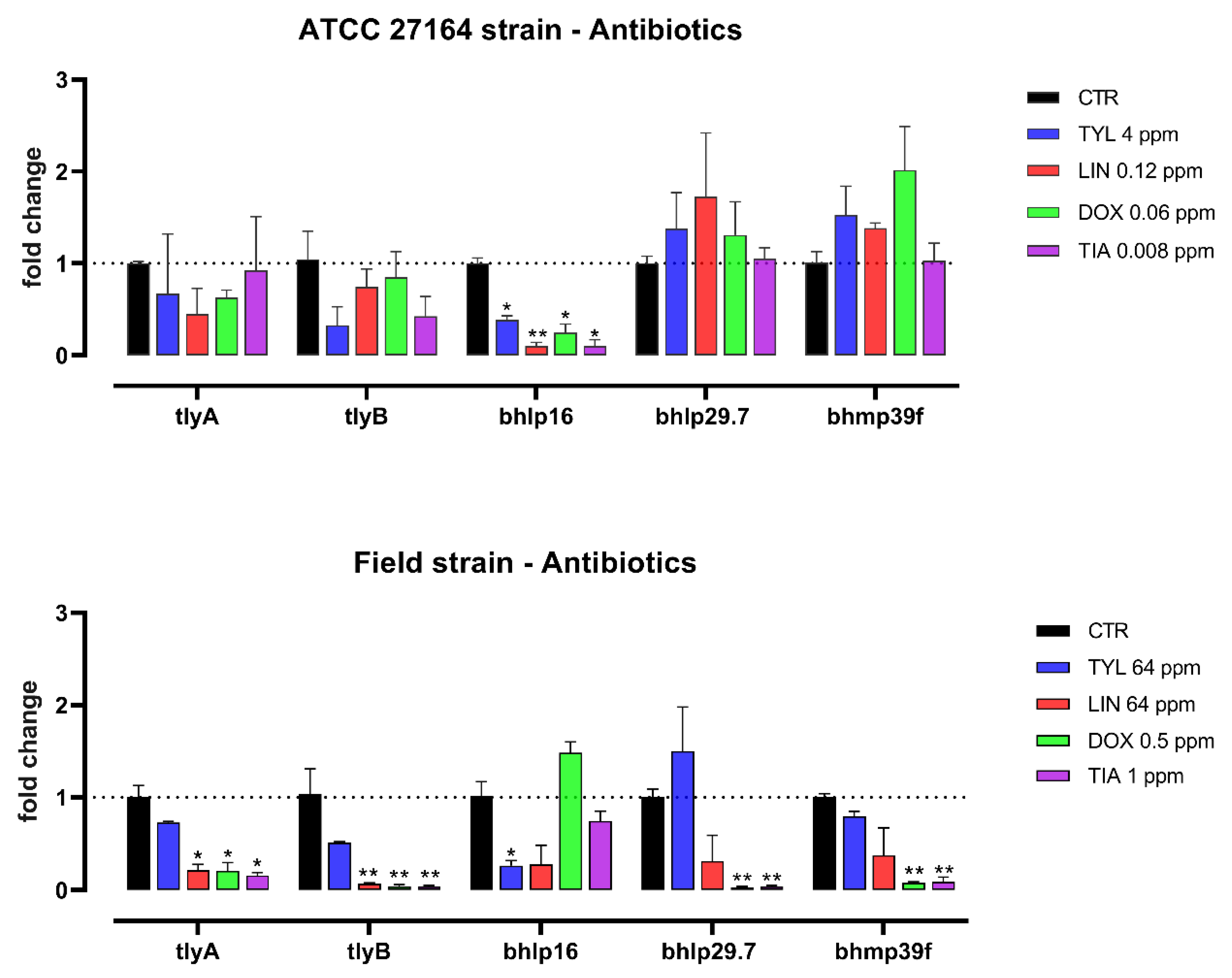
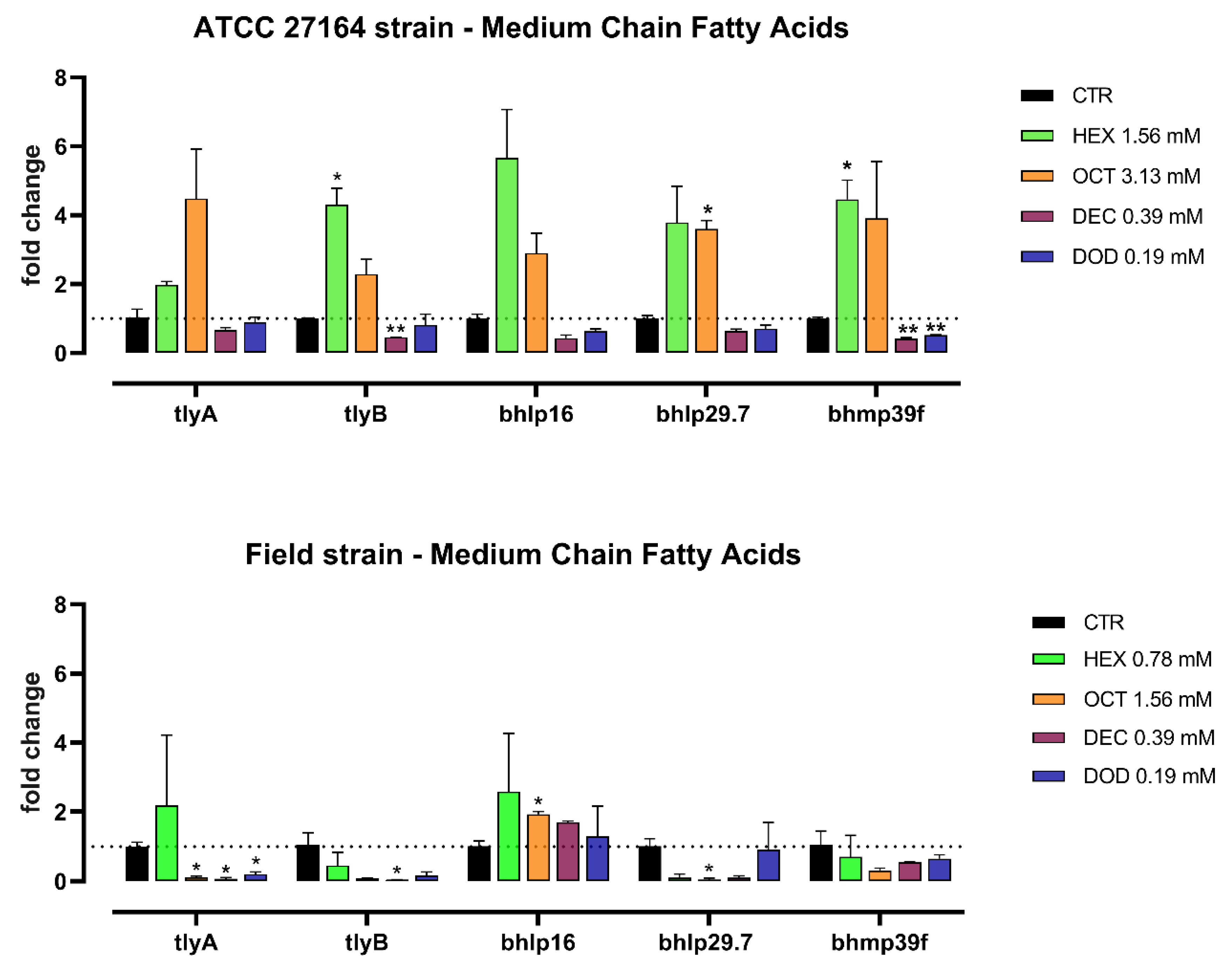
3.2. Gene Expression Study

The expression of Brachyspira virulence genes is represented in Figure 1 and Figure 2 for antibiotics and MCFA, respectively.
The sub-inhibitory doses of the tested substances have variously regulated the expression of hemolysin-associated genes and outer membrane proteins genes. Tylosin significantly decreased the expression of bhlp16 for both the strains. Lincomycin downregulated bhlp16 in ATCC strain and both the hemolysin-associated genes (tlyA, tlyB) in the field strain (p < 0.05), showing a trend for lower bhlp16 level (p = 0.10). For the field strain, tlyA, tlyB, bhlp29.7, and bhmp39f were downregulated by doxycycline, which also reduced bhlp16 mRNA level in the ATCC strain. Tiamulin significantly decreased the expression of bhlp16 in ATCC strain and tlyA, tlyB, bhlp29.7, and bhmp39f in the field strain.
Hexanoic acid significantly upregulated the level of tlyB and bhmp39f in the ATCC strain. In the field strain, octanoic acid downregulated tlyA and upregulated bhlp16, whereas the same acid increased the expression of bhlp29.7 in the ATCC strain. Decanoic acid was able to decrease the level of tlyB and bhmp39f in the ATCC strain (p < 0.01), and the level of tlyA and tlyB in the field strain (p < 0.05). Finally, bhmp39f was downregulated in the ATCC strain by dodecanoic acid (p < 0.01), which also decreased the expression of tlyA in the field strain (p < 0.05).

4. Discussion

B. hyodysenteriae is a fastidious bacterium, able to grow only in particular and enriched conditions. For this reason, antimicrobial susceptibility tests are difficult to carry out and, without a proper in vitro preliminary screening, a possible misuse of antibiotics and a consequent spread of antibiotic resistant bacteria are likely to occur. Moreover, information about in vitro antimicrobial activity of bioactive compounds as alternative to antibiotics is still scarce. This can be considered a major problem dealing with non-antibiotic treatment of swine dysentery, since the possibility of in vitro pre-screening these molecules could considerably limit the large number of in vivo trials.
During the previous years, numerous studies worldwide assessed the antimicrobial susceptibility of various B. hyodysenteriae strains, as recently reviewed by Hampson et al. [8]. These studies were carried out even using commercially available panels trying to establish a standardized and unique protocol [16,17]. This effort stems from the need to obtain antimicrobial susceptibility testing data before treating animals, since numerous cases of antibiotic resistance have been recorded. In fact, only in 2017 the Clinical and Laboratory Standards Institute (CLSI) provided guidance for antimicrobial agent susceptibility testing and breakpoints for fastidious bacteria for veterinary use, including B. hyodysenteriae [13].
In this study, for both the ATCC and the field strain, agar and broth dilution methods were found to be quite comparable, with MIC values being found with broth generally lower than those of agar dilution method, as already documented [18,19,20]. A panel of antibiotics including tylosin, lincomycin, doxycycline, and tiamulin was tested because of their wide use in treatment of swine dysentery [21,22]. All of these antibiotics are protein synthesis inhibitors by targeting the 50S (tylosin, lincomycin, and tiamulin) or 30S (doxycycline) ribosomal unit. Universal breakpoints are suggested by the CLSI only for tylosin and tiamulin [13]. Resistance breakpoints of tylosin and tiamulin for the broth microdilution method are identified as ≥128 μg/mL and ≥1 μg/mL, respectively, indicating that in our study the ATCC strain showed susceptibility (MICTYL = 8 μg/mL; MICTIA = 0.016 μg/mL) and the field strain showed resistance (MICTYL ≥ 64 μg/mL; MICTIA = 2 μg/mL) to both antibiotics. For tiamulin, the result was confirmed by the agar dilution method with an outcome MIC of 0.125 μg/mL for the ATCC strain and 4 μg/mL for field strain, with the CLSI resistance cutoff being at ≥2 μg/mL. No official breakpoints for lincomycin and doxycycline are available. To fill the gap of lacking breakpoints and standardize the antimicrobial susceptibility tests of B. hyodysenteriae, cutoff values for six antibiotics were proposed by Pringle et al. [23]: even based on these assumptions, in this study the MIC of the ATCC strain always fell below these agreed values, while the MIC of the field strain fell above. The B. hyodysenteriae ATCC 27164 was proposed by Pringle et al. [19] as a suitable quality control strain for antimicrobial susceptibility tests, although its lower MIC for pleuromutilins pushed the research of new reference strains for tiamulin and valnemulin [17,18]. Accordingly with previous studies, our data showed reproducible results about the ATCC 27164 strain for both the broth and the agar dilution method [16,17,19,20,24,25,26]. Considering the MIC values of the field strain instead, we can assume that it can be considered a multidrug-resistant strain of B. hyodysenteriae isolated in the North of Italy. Unfortunately, there is limited data about antibiotic resistance in B. hyodysenteriae from Italy. Rugna et al. [27] reported a reduction of susceptibility to tiamulin from 2003 to 2012, whereas De Luca et al. [28] described the multidrug resistance of 10 Italian B. hyodysenteriae strains to tylosin, lincomycin, tiamulin, and for some also to doxycycline. In particular, decreased susceptibility of Italian strains to tylosin and lincomycin was confirmed by the presence of A2058T mutation and the acquisition of the transposon associated-lnu(C) gene [28], already correlated with resistance patterns to macrolides and lincosamides [29,30,31,32]. According to the Italian situation, in the majority of European studies that investigated the resistance to tylosin, lincomycin, doxycycline, and tiamulin in the last decade, it is evident that the reduction in sensitivity to both tylosin and lincomycin is common and frequently concomitant. On the other hand, resistance to tiamulin and/or doxycycline is not always observed among European strains, although, when present, it covers a high percentage of the tested strains [23,31,33,34,35,36]. This can be explained by a selective environmental pressure that results in an increasing multidrug resistance, as happens in our field strain. Clearly, antibiotic resistance is less evident for the ATCC 27164 strain because of its sampling, dating back to 1972. New effective antimicrobial alternatives can be sourced among molecules not influenced by environmental pressure.
Organic acids are widely used as feed additives in swine farming due to their dual use as antimicrobials and growth promoters in the dose range of 0.2–3% [10,11,12]. Depending on their acid dissociation constant, undissociated organic acids can pass through the bacterial membrane and, in response to the release of H+ and RCOO ions, are able to exert their antimicrobial action: on one hand, bacteria waste energy to restore intracellular pH and, furthermore, the anion has a toxic effect by targeting replication and metabolic functions [37]. In particular, antibacterial properties of MCFA are recognized as the most effective ones against Gram-positive bacteria and, to a lesser extent, against Gram-negative bacteria [38]. MCFA antibacterial activity against B. hyodysenteriae was investigated by Vande Maele et al. using broth dilution method [39]. From their findings, the bacterial response to these acids varied depending on the pH conditions and the length of the carbon chain [39]. The longer is the carbon chain and the lower is the pH, the stronger is the antimicrobial activity, with dodecanoic acid being the most powerful antimicrobial against three strains of B. hyodysenteriae, including the ATCC 27164 [39]. Consistently, we did observe the same differences comparing the agar dilution method (pH 7) and the broth dilution method (pH 6.5). This behavior for MCFA can be explained by the anion model theory, according to which the inhibitory effect of organic acids is highly related to their acid dissociation constant and undissociated form that in turn depends on the environmental pH [37,40]. Decanoic and dodecanoic acid showed the lowest MIC, also confirming the longer the chain, the more effective the antibacterial properties of MCFA are, the only exception being hexanoic acid with a lower MIC compared to octanoic acid, as already shown for other Gram-negative bacterial species, like Escherichia coli [41]. Despite the difference in relation to pH and the carbon chain length, in our study, MIC values of MCFA were more consistent between the two strains compared to the tested antibiotics. In fact, resistance development to organic acids is much less frequent and problematic than for antibiotics [10], because the bacterial susceptibility to MCFA is an intrinsic property, meaning that the bioactive power of these molecules meets a broad spectrum of microorganisms with lower probability of failure.
To our knowledge, this is the first study investigating the role of antibiotics and MCFA in modulating B. hyodysenteriae virulence gene expression. Targeting virulence represents a novel and alternative approach to find new antimicrobials and diminish the selective pressure of resistance issue: the target of interest is not the killing or the inhibition of pathogens, but the reduction of virulence factors required to cause disease thanks to sub-lethal or sub-inhibitory doses of antimicrobials [42]. We investigated the regulation of two categories of virulence genes correlated with an increase of severity of swine dysentery [43]: hemolysis-associated (tlyA and tlyB) and outer membrane proteins genes (bhlp16, bhlp29.7, bhmp39f). TlyA and TlyB are considered a pore-forming hemolysin and a caseinolytic protease (Clp), respectively [4,5]: their role in modulating the hemolytic phenotype and the virulence has been investigated, although the genetic background is not yet fully understood. TlyA negative B. hyodysenteriae mutants showed reduced hemolysis in vitro and less severe lesions in pigs and mice [44,45]. On the other side, the specific functions of bhlp16, bhlp29.7, and bhmp39f are unknown, but they are presumably involved in the interaction with epithelial cells. Indeed Gömmel et al. postulated that the in vitro attachment of B. hyodysenteriae strain B204 to IPEC-J2 cells can be driven by the action of these proteins [46].
In our multidrug resistant strain, the regulation of virulence genes appeared to be different for tylosin and lincomycin. The strain was strongly resistant to both antibiotics, consistently with the common genetic basis of the resistance to macrolides and lincosamides already reported in literature [29]. However, when treated with high but not inhibitory doses of the two antibiotics, the hemolysis-associated markers were significantly reduced by lincomycin but not by tylosin, which instead reduced only one outer membrane protein. The exact mode of action behind this dual effect is not clear and needs to be further investigated, but is likely not merely related to the resistance traits. Our field strain has been considered resistant also to doxycycline and tiamulin, although both antibiotics showed an inhibitory action in the MIC tests. In this case, low doses of both drugs below the MIC values were able to downregulate the expression of four out of five markers of virulence analyzed. Therefore, it can be suggested that doxycycline and tiamulin are more likely to positively modulate the bacterial virulence, even at sub-inhibitory doses compared to drugs towards which a strong bacterial resistance is established (tylosin and lincomycin). Similarly, it has been shown that the expression of E. coli K88 toxins and other pathogenic factors can be reduced by the treatment with sub-inhibitory doses of colistin and doxycycline, but not affected by high concentrations of amoxicillin against which the strain is highly resistant [41].
The ATCC strain used as reference resulted to be more sensitive to antibiotics, as already discussed, and also more stable in terms of virulence modulation. Indeed, the antibiotics used at sub-MIC doses did not affect the expression of the analyzed markers, except for one outer membrane protein that was consistently reduced irrespective of the antibiotic type. This different behavior for the ATCC strain compared to the field strain could be related to the lower doses, but higher efficacy, of antibiotics against the reference strain. However, the detailed mechanism still needs to be clarified.
The effect of MCFA on virulence gene expression was quite variable for both the field strain and the ATCC strain. We did observe a general pattern of upregulation of most virulence markers in the ATCC strain with hexanoic and octanoic acid, whereas longer MCFA, such as decanoic and dodecanoic acid, did reduce hemolysin genes in the field strain. We did not expect such difference between the two strains based on the equivalent susceptibility to the antimicrobial action of MCFA found in our MIC tests. However, this can be explained by the specific toxic effect of the anionic form that each different MCFA acquires inside bacterial cells [47].
In a recent review by Jackman et al. [12] it has been reported how MCFA can be included in feed additives as mixtures with other compounds, ranging from 0.2% to 3%, with the aim to curb Salmonella and Escherichia coli infections. Comparing these inclusions with our tested concentrations, the latter are clearly lower, but could have an interesting potential to be investigated with in vivo studies, maybe with new technologies like microencapsulation that can help the delivering of small amounts of protected ingredients to target sites of the intestine [48].

5. Conclusions

Although with some limitations, these data suggest that, even in a multidrug-resistant strain, virulence genes can be modulated thanks to both antibiotics and MCFA in a variable manner. Decanoic and dodecanoic acid had the lowest and most stable MIC against B. hyodysenteriae, and their sub-inhibitory concentrations also decreased the expression of some important virulence genes correlated to a worsening of swine dysentery. It will be interesting to elucidate the mechanism of decanoic and dodecanoic acid in the in vitro modulation of hemolytic phenotype and attachment to epithelial intestinal cells, and eventually verify their potential to control swine dysentery in in vivo.

Author Contributions

Conceptualization, E.G.; methodology, B.T. and G.G.; investigation, G.G.; writing—original draft preparation, G.G.; writing—review and editing, E.G. and B.T.; supervision, E.G. and A.P. All authors have read and agreed to the published version of the manuscript.

Funding

This research received no external funding.

Data Availability Statement

Data available on request due to restrictions, e.g., for privacy or ethical reasons.

Acknowledgments

We thank Giovanni Alborali (IZSLER) for the isolation of the field strain.

Conflicts of Interest

Andrea Piva serves as a professor at the University of Bologna and is a member of the board of directors of Vetagro S.p.A. (Reggio Emilia, Italy). Ester Grilli serves as an advisor of Vetagro S.p.A.

References

  1. Hampson, D.J.; Burrough, E.R. Swine Dysentery and Brachyspiral Colitis. In Diseases of Swine; John Wiley & Sons, Ltd.: Hoboken, NJ, USA, 2019; pp. 951–970. ISBN 978-1-119-35092-7. [Google Scholar]
  2. Mahu, M.; De Pauw, N.; Vande Maele, L.; Verlinden, M.; Boyen, F.; Ducatelle, R.; Haesebrouck, F.; Martel, A.; Pasmans, F. Variation in Hemolytic Activity of Brachyspira Hyodysenteriae Strains from Pigs. Vet. Res. 2016, 47, 66. [Google Scholar] [CrossRef] [Green Version]
  3. Joerling, J.; Willems, H.; Ewers, C.; Herbst, W. Differential Expression of Hemolysin Genes in Weakly and Strongly Hemolytic Brachyspira Hyodysenteriae Strains. BMC Vet. Res. 2020, 16, 169. [Google Scholar] [CrossRef]
  4. Muir, S.; Koopman, M.B.; Libby, S.J.; Joens, L.A.; Heffron, F.; Kusters, J.G. Cloning and Expression of a Serpula (Treponema) Hyodysenteriae Hemolysin Gene. Infect. Immun. 1992, 60, 529–535. [Google Scholar] [CrossRef] [Green Version]
  5. ter Huurne, A.A.; Muir, S.; van Houten, M.; van der Zeijst, B.A.; Gaastra, W.; Kusters, J.G. Characterization of Three Putative Serpulina Hyodysenteriae Hemolysins. Microb. Pathog. 1994, 16, 269–282. [Google Scholar] [CrossRef]
  6. Hsu, T.; Hutto, D.L.; Minion, F.C.; Zuerner, R.L.; Wannemuehler, M.J. Cloning of a Beta-Hemolysin Gene of Brachyspira (Serpulina) Hyodysenteriae and Its Expression in Escherichia Coli. Infect. Immun. 2001, 69, 706–711. [Google Scholar] [CrossRef] [Green Version]
  7. Trott, D.J.; Alt, D.P.; Zuerner, R.L.; Wannemuehler, M.J.; Stanton, T.B. The Search for Brachyspira Outer Membrane Proteins That Interact with the Host. Anim. Health Res. Rev. 2001, 2, 19–30. [Google Scholar] [CrossRef]
  8. Hampson, D.J.; Lugsomya, K.; La, T.; Phillips, N.D.; Trott, D.J.; Abraham, S. Antimicrobial Resistance in Brachyspira—An Increasing Problem for Disease Control. Vet. Microbiol. 2019, 229, 59–71. [Google Scholar] [CrossRef]
  9. Regione Emilia Romagna Linee Guida: Uso Prudente Degli Antibiotici Nell’allevamento Suino 2018. Available online: https://www.alimenti-salute.it/sites/default/files/Linee%20Guida%20SUINI%202018.pdf (accessed on 28 October 2021).
  10. Desbois, A.P.; Smith, V.J. Antibacterial Free Fatty Acids: Activities, Mechanisms of Action and Biotechnological Potential. Appl. Microbiol. Biotechnol. 2010, 85, 1629–1642. [Google Scholar] [CrossRef] [Green Version]
  11. Tugnoli, B.; Giovagnoni, G.; Piva, A.; Grilli, E. From Acidifiers to Intestinal Health Enhancers: How Organic Acids Can Improve Growth Efficiency of Pigs. Animals 2020, 10, 134. [Google Scholar] [CrossRef] [Green Version]
  12. Jackman, J.A.; Boyd, R.D.; Elrod, C.C. Medium-Chain Fatty Acids and Monoglycerides as Feed Additives for Pig Production: Towards Gut Health Improvement and Feed Pathogen Mitigation. J. Anim. Sci. Biotechnol. 2020, 11, 44. [Google Scholar] [CrossRef] [Green Version]
  13. CLSI VET06: Methods for Antimicrobial Susceptibility Testing of Infrequently Isolated or Fastidious Bacteria Isolated from Animals 2017. Available online: https://clsi.org/standards/products/veterinary-medicine/documents/vet06/ (accessed on 14 October 2021).
  14. Giovagnoni, G.; Rossi, B.; Tugnoli, B.; Ghiselli, F.; Bonetti, A.; Piva, A.; Grilli, E. Thymol and Carvacrol Downregulate the Expression of Salmonella Typhimurium Virulence Genes during an In Vitro Infection on Caco-2 Cells. Microorganisms 2020, 8, 862. [Google Scholar] [CrossRef]
  15. Livak, K.J.; Schmittgen, T.D. Analysis of Relative Gene Expression Data Using Real-Time Quantitative PCR and the 2(-Delta Delta C(T)) Method. Methods San Diego Calif. 2001, 25, 402–408. [Google Scholar] [CrossRef]
  16. Karlsson, M.; Fellström, C.; Gunnarsson, A.; Landén, A.; Franklin, A. Antimicrobial Susceptibility Testing of Porcine Brachyspira (Serpulina) Species Isolates. J. Clin. Microbiol. 2003, 41, 2596–2604. [Google Scholar] [CrossRef] [Green Version]
  17. Stubberfield, E.; Pringle, M.; Landén, A.; Veldman, K.T.; Geurts, Y.; Jouy, E.; Le Devendec, L.; Rubin, J.E.; Kulathunga, D.G.R.S.; Kristensen, K.A.; et al. Validation of an Antimicrobial Susceptibility Testing Protocol for Brachyspira Hyodysenteriae and Brachyspira Pilosicoli in an International Ring Trial. Vet. Microbiol. 2020, 244, 108645. [Google Scholar] [CrossRef]
  18. Rohde, J.; Kessler, M.; Baums, C.G.; Amtsberg, G. Comparison of Methods for Antimicrobial Susceptibility Testing and MIC Values for Pleuromutilin Drugs for Brachyspira Hyodysenteriae Isolated in Germany. Vet. Microbiol. 2004, 102, 25–32. [Google Scholar] [CrossRef]
  19. Pringle, M.; Aarestrup, F.M.; Bergsjø, B.; Fossi, M.; Jouy, E.; Landén, A.; Mevius, D.; Perry, K.; Teale, C.; Thomson, J.; et al. Quality-Control Ranges for Antimicrobial Susceptibility Testing by Broth Dilution of the Brachyspira Hyodysenteriae Type Strain (ATCC 27164T). Microb. Drug Resist. Larchmt. N 2006, 12, 219–221. [Google Scholar] [CrossRef]
  20. Mirajkar, N.S.; Gebhart, C.J. Comparison of Agar Dilution and Antibiotic Gradient Strip Test with Broth Microdilution for Susceptibility Testing of Swine Brachyspira Species. J. Vet. Diagn. Investig. 2016, 28, 133–143. [Google Scholar] [CrossRef] [Green Version]
  21. Alvarez-Ordóez, A.; Martínez-Lobo, F.J.; Arguello, H.; Carvajal, A.; Rubio, P. Swine Dysentery: Aetiology, Pathogenicity, Determinants of Transmission and the Fight against the Disease. Int. J. Environ. Res. Public. Health 2013, 10, 1927–1947. [Google Scholar] [CrossRef] [Green Version]
  22. Burrough, E.R. Swine Dysentery: Etiopathogenesis and Diagnosis of a Reemerging Disease. Vet. Pathol. 2017, 54, 22–31. [Google Scholar] [CrossRef]
  23. Pringle, M.; Landén, A.; Unnerstad, H.E.; Molander, B.; Bengtsson, B. Antimicrobial Susceptibility of Porcine Brachyspira Hyodysenteriae and Brachyspira Pilosicoli Isolated in Sweden between 1990 and 2010. Acta Vet. Scand. 2012, 54, 54. [Google Scholar] [CrossRef] [Green Version]
  24. Karlsson, M.; Oxberry, S.L.; Hampson, D.J. Antimicrobial Susceptibility Testing of Australian Isolates of Brachyspira Hyodysenteriae Using a New Broth Dilution Method. Vet. Microbiol. 2002, 84, 123–133. [Google Scholar] [CrossRef]
  25. Lugsomya, K.; Zeeh, F.; La, T.; Phillips, N.; Hampson, D.J. First Identification and Characterisation of Brachyspira Hyodysenteriae in Pigs in Hong Kong. Porc. Health Manag. 2019, 5, 27. [Google Scholar] [CrossRef]
  26. Lobová, D.; Smola, J.; Cizek, A. Decreased Susceptibility to Tiamulin and Valnemulin among Czech Isolates of Brachyspira Hyodysenteriae. J. Med. Microbiol. 2004, 53, 287–291. [Google Scholar] [CrossRef] [Green Version]
  27. Rugna, G.; Bonilauri, P.; Carra, E.; Bergamini, F.; Luppi, A.; Gherpelli, Y.; Magistrali, C.F.; Nigrelli, A.; Alborali, G.L.; Martelli, P.; et al. Sequence Types and Pleuromutilin Susceptibility of Brachyspira Hyodysenteriae Isolates from Italian Pigs with Swine Dysentery: 2003-2012. Vet. J. Lond. Engl. 1997 2015, 203, 115–119. [Google Scholar] [CrossRef] [Green Version]
  28. De Luca, S.; Nicholson, P.; Magistrali, C.F.; García-Martín, A.B.; Rychener, L.; Zeeh, F.; Frey, J.; Perreten, V. Transposon-Associated Lincosamide Resistance Lnu(C) Gene Identified in Brachyspira Hyodysenteriae ST83. Vet. Microbiol. 2018, 214, 51–55. [Google Scholar] [CrossRef]
  29. Karlsson, M.; Fellström, C.; Heldtander, M.U.K.; Johansson, K.-E.; Franklin, A. Genetic Basis of Macrolide and Lincosamide Resistance in Brachyspira (Serpulina) Hyodysenteriae. FEMS Microbiol. Lett. 1999, 172, 255–260. [Google Scholar] [CrossRef] [Green Version]
  30. Hidalgo, Á.; Carvajal, A.; Vester, B.; Pringle, M.; Naharro, G.; Rubio, P. Trends towards Lower Antimicrobial Susceptibility and Characterization of Acquired Resistance among Clinical Isolates of Brachyspira Hyodysenteriae in Spain. Antimicrob. Agents Chemother. 2011, 55, 3330–3337. [Google Scholar] [CrossRef] [Green Version]
  31. Mahu, M.; Pasmans, F.; Vranckx, K.; De Pauw, N.; Vande Maele, L.; Vyt, P.; Vandersmissen, T.; Martel, A.; Haesebrouck, F.; Boyen, F. Presence and Mechanisms of Acquired Antimicrobial Resistance in Belgian Brachyspira Hyodysenteriae Isolates Belonging to Different Clonal Complexes. Vet. Microbiol. 2017, 207, 125–132. [Google Scholar] [CrossRef]
  32. Achard, A.; Villers, C.; Pichereau, V.; Leclercq, R. New Lnu(C) Gene Conferring Resistance to Lincomycin by Nucleotidylation in Streptococcus Agalactiae UCN36. Antimicrob. Agents Chemother. 2005, 49, 2716–2719. [Google Scholar] [CrossRef] [Green Version]
  33. Šperling, D.; Smola, J.; Čížek, A. Characterisation of Multiresistant Brachyspira Hyodysenteriae Isolates from Czech Pig Farms. Vet. Rec. 2011, 168, 215. [Google Scholar] [CrossRef]
  34. Zmudzki, J.; Szczotka, A.; Nowak, A.; Strzelecka, H.; Grzesiak, A.; Pejsak, Z. Antimicrobial Susceptibility of Brachyspira Hyodysenteriae Isolated from 21 Polish Farms. Pol. J. Vet. Sci. 2012, 15, 259–265. [Google Scholar] [CrossRef] [PubMed] [Green Version]
  35. Kirchgässner, C.; Schmitt, S.; Borgström, A.; Wittenbrink, M.M. Antimicrobial Susceptibility of Brachyspira Hyodysenteriae in Switzerland. Schweiz. Arch. Tierheilkd. 2016, 158, 405–410. [Google Scholar] [CrossRef] [PubMed]
  36. García-Martín, A.B.; Perreten, V.; Rossano, A.; Schmitt, S.; Nathues, H.; Zeeh, F. Predominance of a Macrolide-Lincosamide-Resistant Brachyspira Hyodysenteriae of Sequence Type 196 in Swiss Pig Herds. Vet. Microbiol. 2018, 226, 97–102. [Google Scholar] [CrossRef] [PubMed]
  37. Hirshfield, I.N.; Terzulli, S.; O’Byrne, C. Weak Organic Acids: A Panoply of Effects on Bacteria. Sci. Prog. 2003, 86, 245–270. [Google Scholar] [CrossRef] [PubMed]
  38. Yoon, B.K.; Jackman, J.A.; Valle-González, E.R.; Cho, N.-J. Antibacterial Free Fatty Acids and Monoglycerides: Biological Activities, Experimental Testing, and Therapeutic Applications. Int. J. Mol. Sci. 2018, 19, 1114. [Google Scholar] [CrossRef] [PubMed] [Green Version]
  39. Vande Maele, L.; Heyndrickx, M.; Maes, D.; De Pauw, N.; Mahu, M.; Verlinden, M.; Haesebrouck, F.; Martel, A.; Pasmans, F.; Boyen, F. In Vitro Susceptibility of Brachyspira Hyodysenteriae to Organic Acids and Essential Oil Components. J. Vet. Med. Sci. 2016, 78, 325–328. [Google Scholar] [CrossRef] [Green Version]
  40. Russell, J.B.; Diez-Gonzalez, F. The Effects of Fermentation Acids on Bacterial Growth. Adv. Microb. Physiol. 1998, 39, 205–234. [Google Scholar] [CrossRef]
  41. Bonetti, A.; Tugnoli, B.; Rossi, B.; Giovagnoni, G.; Piva, A.; Grilli, E. Nature-Identical Compounds and Organic Acids Reduce E. Coli K88 Growth and Virulence Gene Expression In Vitro. Toxins 2020, 12, 468. [Google Scholar] [CrossRef]
  42. Heras, B.; Scanlon, M.J.; Martin, J.L. Targeting Virulence Not Viability in the Search for Future Antibacterials. Br. J. Clin. Pharmacol. 2015, 79, 208–215. [Google Scholar] [CrossRef]
  43. Burrough, E.R.; Strait, E.L.; Kinyon, J.M.; Bower, L.P.; Madson, D.M.; Wilberts, B.L.; Schwartz, K.J.; Frana, T.S.; Songer, J.G. Comparative Virulence of Clinical Brachyspira Spp. Isolates in Inoculated Pigs. J. Vet. Diagn. Investig. Off. Publ. Am. Assoc. Vet. Lab. Diagn. Inc. 2012, 24, 1025–1034. [Google Scholar] [CrossRef] [Green Version]
  44. ter Huurne, A.A.H.M.; van Houten, M.; Muir, S.; Kusters, J.G.; van der Zeijst, B.A.M.; Gaastra, W. Inactivation of a Serpula (Treponema) Hyodysenteriae Hemolysin Gene by Homologous Recombination: Importance of This Hemolysin in Pathogenesis of S. Hyodysenteriae in Mice. FEMS Microbiol. Lett. 1992, 92, 109–113. [Google Scholar] [CrossRef]
  45. Hyatt, D.R.; ter Huurne, A.A.; van der Zeijst, B.A.; Joens, L.A. Reduced Virulence of Serpulina Hyodysenteriae Hemolysin-Negative Mutants in Pigs and Their Potential to Protect Pigs against Challenge with a Virulent Strain. Infect. Immun. 1994, 62, 2244–2248. [Google Scholar] [CrossRef] [PubMed] [Green Version]
  46. Gömmel, M.; Barth, S.; Heydel, C.; Baljer, G.; Herbst, W. Adherence of Brachyspira Hyodysenteriae to Porcine Intestinal Epithelial Cells Is Inhibited by Antibodies against Outer Membrane Proteins. Curr. Microbiol. 2013, 66, 286–292. [Google Scholar] [CrossRef] [PubMed]
  47. Warnecke, T.; Gill, R.T. Organic Acid Toxicity, Tolerance, and Production in Escherichia Coli Biorefining Applications. Microb. Cell Fact. 2005, 4, 25. [Google Scholar] [CrossRef] [Green Version]
  48. Piva, A.; Pizzamiglio, V.; Morlacchini, M.; Tedeschi, M.; Piva, G. Lipid Microencapsulation Allows Slow Release of Organic Acids and Natural Identical Flavors along the Swine Intestine. J. Anim. Sci. 2007, 85, 486–493. [Google Scholar] [CrossRef] [Green Version]
Figure 1. mRNA expression of tlyA, tlyB, bhlp16, bhlp29.7, and bhmp39f for B. hyodysenteriae ATCC 27164 and the field strain cultured alone (CTR), or with sub-inhibitory doses of antibiotics. Data are presented as mean (n = 3) ± SEM. For each gene, significant differences between each substance and its control are marked by asterisks (* for p < 0.05 and ** for p < 0.01). TYL = tylosin, LIN = lincomycin, DOX = doxycycline, TIA = tiamulin.
Microorganisms 10 00301 g001
Figure 2. mRNA expression of tlyA, tlyB, bhlp16, bhlp29.7, and bhmp39f for B. hyodysenteriae ATCC 27164 and the field strain cultured alone (CTR), or with sub-inhibitory doses of medium-chain fatty acids. Data are presented as mean (n = 3) ± SEM. For each gene, significant differences between each substance and its control are marked by asterisks (* for p < 0.05 and ** for p < 0.01). HEX = hexanoic acid, OCT = octanoic acid, DEC = decanoic acid, DOD = dodecanoic acid.
Microorganisms 10 00301 g002
Table 1. List of primers used for real-time PCR.
GeneFunctionSequence (5′ → 3′)Product Length (bp)Accession NumberReference
tlyAHemolysin AF: AAAGGCGTTTGTAGAATTTGGAAT
R: TGTCCTACATCAAGAGCATAAACTTTTT
131MT304819.1[3]
tlyBClp proteaseF: AAGGATTCGATAAGAAGTATGGTGCTA
R: TTCGGTACTCACATAATCCTCTATCTCT
79MT304820.1[3]
bhlp16 (smpA)Outer membrane proteinF: GCAGGTGTAGAAAAGGGATTTGG
R: TCTGAAGAACTTGCTCCACCTT
107CP015910.2This study
bhlp29.7 (bmpB)Outer membrane proteinF: TGGTTTTGCTGGAGAGTCTGA
R: TCTCCGTCATTCAAAGCCTGAT
132AY706761.1This study
bhmp39fOuter membrane proteinF: AGCCTTTCGGTATTGGCGTA
R: ACAGCTATTTGAACAGGAACTGC
130AY027775.1This study
gyrBHousekeepingF: TGCAGGCGGTACTGCTAAAG
R: GCACCTACACCGCATCCTAA
159CP015910.2This study
rpoDHousekeepingF: AGCTTTTGCCTCTATCTGACGA
R: ACAGTTTGCCGGACAGAGAA
137CP015910.2This study
F = forward; R = reverse.
Table 2. Minimal inhibitory concentrations (MIC) of antibiotics and medium chain fatty acids (MCFA) tested against the ATCC 27164 strain and the field strain using the agar and broth dilution method.
ATCC 27164 StrainField Strain
Agar DilutionBroth DilutionAgar DilutionBroth Dilution
AntibioticsTylosin16 μg/mL8 μg/mL>64 μg/mL>64 μg/mL
Lincomycin2 μg/mL0.25 μg/mL>64 μg/mL>64 μg/mL
Doxycycline2 μg/mL0.12 μg/mL4 μg/mL1 μg/mL
Tiamulin0.125 μg/mL0.016 μg/mL4 μg/mL2 μg/mL
MCFAHexanoic acid25 mM3.13 mM25 mM1.56 mM
Octanoic acid>25 mM6.25 mM25 mM3.13 mM
Decanoic acid3.13 mM0.78 mM3.13 mM0.78 mM
Dodecanoic acid0.39 mM0.39 mM0.39 mM0.39 mM
Publisher’s Note: MDPI stays neutral with regard to jurisdictional claims in published maps and institutional affiliations.

