Article

Comparative De Novo and Pan-Genome Analysis of MDR Nosocomial Bacteria Isolated from Hospitals in Jeddah, Saudi Arabia

by
Molook Alghamdi
1,
Effat Al-Judaibi
1,
Mohammed Al-Rashede
2 and
Awatif Al-Judaibi
1,*
1
Department of Biological Sciences, Microbiology Section, Faculty of Science, Jeddah University, Jeddah 21959, Saudi Arabia
2
Maternity and Children Hospital Almosadya, Jeddah 21425, Saudi Arabia
*
Author to whom correspondence should be addressed.
Microorganisms 2023, 11(10), 2432; https://doi.org/10.3390/microorganisms11102432
Submission received: 3 July 2023 / Revised: 14 September 2023 / Accepted: 26 September 2023 / Published: 28 September 2023
(This article belongs to the Special Issue Virulence Factors and Antibiotic Resistance of Enterobacterales 2.0)

Abstract

:
Multidrug-resistant (MDR) bacteria are one of the most serious threats to public health, and one of the most important types of MDR bacteria are those that are acquired in a hospital, known as nosocomial. This study aimed to isolate and identify MDR bacteria from selected hospitals in Jeddah and analyze their antibiotic-resistant genes. Bacteria were collected from different sources and wards of hospitals in Jeddah City. Phoenix BD was used to identify the strains and perform susceptibility testing. Identification of selected isolates showing MDR to more than three classes on antibiotics was based on 16S rRNA gene and whole genome sequencing. Genes conferring resistance were characterized using de novo and pan-genome analyses. In total, we isolated 108 bacterial strains, of which 75 (69.44%) were found to be MDR. Taxonomic identification revealed that 24 (32%) isolates were identified as Escherichia coli, 19 (25.3%) corresponded to Klebsiella pneumoniae, and 17 (22.67%) were methicillin-resistant Staphylococcus aureus (MRSA). Among the Gram-negative bacteria, K. pneumoniae isolates showed the highest resistance levels to most antibiotics. Of the Gram-positive bacteria, S. aureus (MRSA) strains were noticed to exhibit the uppermost degree of resistance to the tested antibiotics, which is higher than that observed for K. pneumoniae isolates. Taken together, our results illustrated that MDR Gram-negative bacteria are the most common cause of nosocomial infections, while MDR Gram-positive bacteria are characterized by a wider antibiotic resistance spectrum. Whole genome sequencing found the appearance of antibiotic resistance genes, including SHV, OXA, CTX-M, TEM-1, NDM-1, VIM-1, ere(A), ermA, ermB, ermC, msrA, qacA, qacB, and qacC.

1. Introduction

Antibiotic resistance develops from the abuse and overuse of antibiotics. Specifically, a selection of bacteria resistant to most forms of antibiotics emerges from the widespread and unjustified use of broad-spectrum antibiotics [1]. In a clinical environment, such as in hospitals, the overuse of antibiotics, even under controlled administration, breeds antibiotic resistance. More alarming is the presence of multidrug-resistant (MDR) bacteria, which refers to bacteria that exhibit resistance to at least three different classes of antimicrobials [2]. MDR bacteria represent a major global health problem. Infections of MDR bacteria are associated with unfavorable prognoses, even in clinically managed cases, resulting in financial stress to both the patients and the healthcare system [2]. There has been an increase in antibiotic resistance despite the increase in commercially available antibiotics over the past few decades. In response, the World Health Organization (WHO) [3] has issued an urgent demand for new treatments against the world’s leading antibiotic-resistant bacteria [4]. Specifically, research into innovative drugs (i.e., drugs with an active ingredient that does not demonstrate cross-resistance to existing antibiotics) is warranted [2,4]. However, developing an active substance requires understanding the mechanisms of antibiotic resistance. The antibiotic resistance of MDR bacteria results from antibiotic inactivation through one or a combination of molecular mechanisms. One of the most documented mechanisms of resistance to antibiotics is the production of enzymes that inactivate their biological activity. For example, β-lactamase inactivates the β-lactam ring, the primary active component of several antibiotics that are classified as β-lactams, such as penicillins, cephalosporins, carbapenems, and monobactams [5]. These β-lactam antibiotics target penicillin-binding proteins (PBPs) by acylating the transpeptidase, resulting in inhibition of the final processes of peptidoglycan synthesis [6]. K. pneumoniae carbapenemase also hydrolyzes and inactivates a specific class of β-lactams, namely, the carbapenems. Antibiotic-resistant S. aureus and E. coli also produce carbapenemase. In addition to β-lactamase, enzymes that inactivate antibiotic activity include acetyltransferase, phosphotransferase, and adenyltransferase [2]. Altering the antibiotic target without interrupting cellular processes represents another mechanism of antibiotic resistance. For instance, methicillin-resistant S. aureus (MRSA) carries mecA, which encodes the novel penicillin-binding protein PBP2a, which differs from the PBP targeted by older β-lactam antibiotics, rendering them ineffective against MRSA [7]. Notably, staphylococcal cassette chromosome (SCCmec) of classes I, II, III, or IV was acquired by horizontal gene transfer across hospital-acquired MRSA (HA-MRSA) strains of different lineages [7]. The horizontal transfer of genes that confer antibiotic resistance to pathogens represents an emerging cause of concern for public health [8]. This threat already exists for HA-MRSA, one of the most common hospital-acquired infections caused by MDR bacteria [9].
MRSA produces aggressive biofilms, reducing the permeability of the bacteria [10]. Reducing cellular permeability is a mechanism of antibiotic resistance characterized by alterations in the structure of the cell surface casing [11]. In the case of MRSA and P. aeruginosa, aggressive biofilm production increases their resistance to the host immune system as well as antibiotics [9]. According to the Ministry of Health (MOH), a work plan is in place because of the threat of antimicrobial resistance to public health, necessitating various activities, and coordination between government agencies, the private sector, and the public [11]. Compared to the rates in the 1990s in Saudi Arabia, during the subsequent decade, a considerable increase in the prevalence of carbapenem-resistant Gram-negative bacteria was observed, specifically of strains that produce extended-spectrum beta-lactamase (ESBL). At the same time, some institutes have reported ESBL rates of 29% for E. coli and 65% for K. pneumoniae of all isolated MDR bacteria. These rising rates have been linked to several outbreaks and fatalities ranging from 11% to 40%. According to Saudi Arabia’s national surveillance on Gram-positive cocci, 32% of S. aureus strains are methicillin-resistant (MRSA), and 33% and 26% of S. pneumoniae strains are resistant to penicillin G and erythromycin, respectively [12].
Jeddah is the second largest city in Saudi Arabia, with a population of 3.2 million people. Jeddah is commercially important and serves as the entry point to the holy cities of Makkah and Medina [13]. According to studies conducted in three private hospitals and hospitals of MOH in Jeddah, among the Gram-negative MDR bacteria, species of the Enterobacteriaceae family were the most frequently isolated, with E. coli being the most common organism, representing 40.0% [14,15]. Regarding Gram-positive MDR bacteria, a study conducted on the incidence of Gram-positive MDR bacteria at King Abdulaziz University Hospital, Jeddah, found that the most prevalent isolates (20%) were S. aureus (MRSA) [16].
This study aims to identify MDR isolates based on the definition of MDR bacteria as those that are resistant to at least three or more antibiotic classes.

2. Materials and Methods

2.1. Ethical Approvement

This study did not require ethical approval, as no human subjects were involved.

2.2. Clinical Specimens

A total of 108 isolates were isolated from different types of medical specimens (stool; sputum; blood; urine; high vaginal swabs; and swabs from wounds, ears, eyes, noses, etc.) and from several hospitals wards, e.g., the neonatal intensive care unit, intensive care unit (ICU), emergency room, and outpatient department (OPD). The isolates were collected from three hospitals (MOH) between December 2018 and November 2019. All of the isolates from each hospital laboratory were grown on blood agar and then transferred to the microbiology laboratory at Jeddah University for further studies.

2.3. Isolation and Identification of MDR Bacteria

The combination panels of the BD Phoenix M50 identification system (NMIC/ID-26; catalog no. 448026) were used for phenotype identification and antibiotic susceptibility testing of the bacterial isolates, in accordance with the manufacturer’s recommendations. This system uses a redox indicator to detect the growth of organisms in the presence of an antimicrobial agent [17]. The bacteria were identified at the Maternity and Children’s Hospital in Jeddah.

2.4. Amplification of 16S rRNA Gene, Sequencing, and Phylogenetic Analysis

Seven MDR bacterial DNA samples were extracted using the Qiagen DNA extraction kit (Qiagen, Hilden, Germany), according to the manufacturer’s instructions [18]. The extracted DNA was stored at −20 °C.
The 16S rRNA gene was amplified by PCR using the following couples of forward and reverse primers: 518F 5′-CCAGCAGCCGCGGTAATACG-3′ and 800R 5′-TACCAGGGTATCTAATCC-3′; 27F 5′-AGAGTTTGATCMTGGCTCAG-3′ and 1492R 5′-GGTTACCTTGTTACGACTT-3′.
The primers used for the amplification of the 16S rRNA gene were designed based on the conserved regions in the 16S rDNA sequences [19]. DNA sequencing was conducted by MiSeq (Illumina) using the v3 chemistry (600 cycles); sequencing of amplification products was carried out by the Infection Disease Unit, King Fahad Medical Center for Research, King Abdulaziz University 8/2/2021, and the sequenced data were analyzed using BLAST-National Center for Biotechnology Information (NCBI) (available online: http://blast.ncbi.nlm.nih.gov/Blast.cgi (accessed on 29 August 2021)). The phylogenetic tree diagram of the seven isolated MDR bacteria samples based on 16S rDNA sequencing was constructed using the maximum likelihood (ML) algorithm in MEGA7 software (version 10.2, megasoftware.net (accessed on 2 September 2021)). Node stability was assessed by bootstrapping 1000 replicates. The accession number of the 16S rDNA gene of each strain was obtained from the National Center for Biotechnology Information (NCBI) (http://blast.ncbi.nlm.nih.gov/Blast.cgi (accessed on 29 August 2021)).

2.5. Whole Genome Sequencing and Analysis Workflow

Genomic DNA was extracted from the seven isolated bacteria samples according to the protocol provided by the Qiagen genome DNA extraction kit. After completion of quality control, the selected isolates were used for library TruSeq Nano DNA (350), the run type Throughput Run, in 36 Scale. For cluster generation, the library was loaded onto a flow cell, where fragments were captured on a lawn of surface-bound oligos complementary to the library adapter sequences and proceeded using an average 350 bp insert size with “covaries”. Each fragment was amplified into distinct clonal clusters through bridge amplification. When cluster generation was complete, the templates were ready for sequencing. As all four reversible, terminator-bound dNTPs are present during each sequencing cycle, natural competition minimizes incorporation bias and greatly reduces the raw error rates compared with other technologies. This results in highly accurate base-by-base sequences where sequence-context-specific errors are virtually eliminated, even within repetitive sequence regions and homopolymers. After sequencing, the identified bases and the predicted quality of each base were converted into raw data for analysis. In this step, BCL/cBCL files were converted into FASTQ (http://www.bioinformatics.babraham.ac.uk/projects/fastqc (accessed on 15 May 2022)) files using the Illumina package bcl2fastq [20].

2.6. De Novo Assembly of MDR Bacterial Sequences

For the de novo assembly, the overlap–layout–consensus (OLC) algorithm was adopted, which identifies overlaps between reads, with the layout of the reads and building of consensus sequences of the read sequences generated by the Illumina sequencing platform (Novaseq-6000) with the De Bruijn graph (DBG) algorithm. The total read bases were Pseudomonase_1 “3.526 GB”, E. coli_7 “2.774 GB”, K. pneumoniae_3 “3.047 GB”, K. pneumoniae_6 “2.918 GB”, S. aureus_9 “3.508 GB”, S. aureus_10 “2.313 GB”, and S. aureus_12 “2.254 GB”. After de novo assembly was achieved, the contigs (Pseudomonas_1 “100”, E. coli_7 “47”, K. pneumoniae_3 “45”, K. pneumoniae_6 “50”, S. aureus_9 “48”, S. aureus_10 “20”, and S. aureus_12 “28”) were generated with N50, and the generated reads of the size range of bp were as follows: Pseudomonase_1, 669, E_coli_7, 632, K_pneumoniae_3, 652, K_pneumoniae_6, 641, S_aureus_9, 653, S_aureus_10, 664, and S_aureus_12, 652. These were assigned a taxonomic classification based on alignment using the nucleotide Basic Local Alignment Search Tool (BLASTN) against the nucleotide collection database (NCBI database). Following quality trimming, trimmed paired-end reads were de novo assembled into contigs. Contigs with a total read count of >1000 reads were chosen for alignment with software that used the DBG algorithm, including SPAdes version 3.15.4 (http://cab.spbu.ru/software/spades/ (accessed on 15 May 2022)), Platanus, and SOAPdenovo, for bacterial identification. The DBG algorithm divides each read into pieces with a length K, referred to as K-mers, and progressively extends them by 1 base to select the most expected sequence [21,22].

2.7. Pan-Genome Analysis

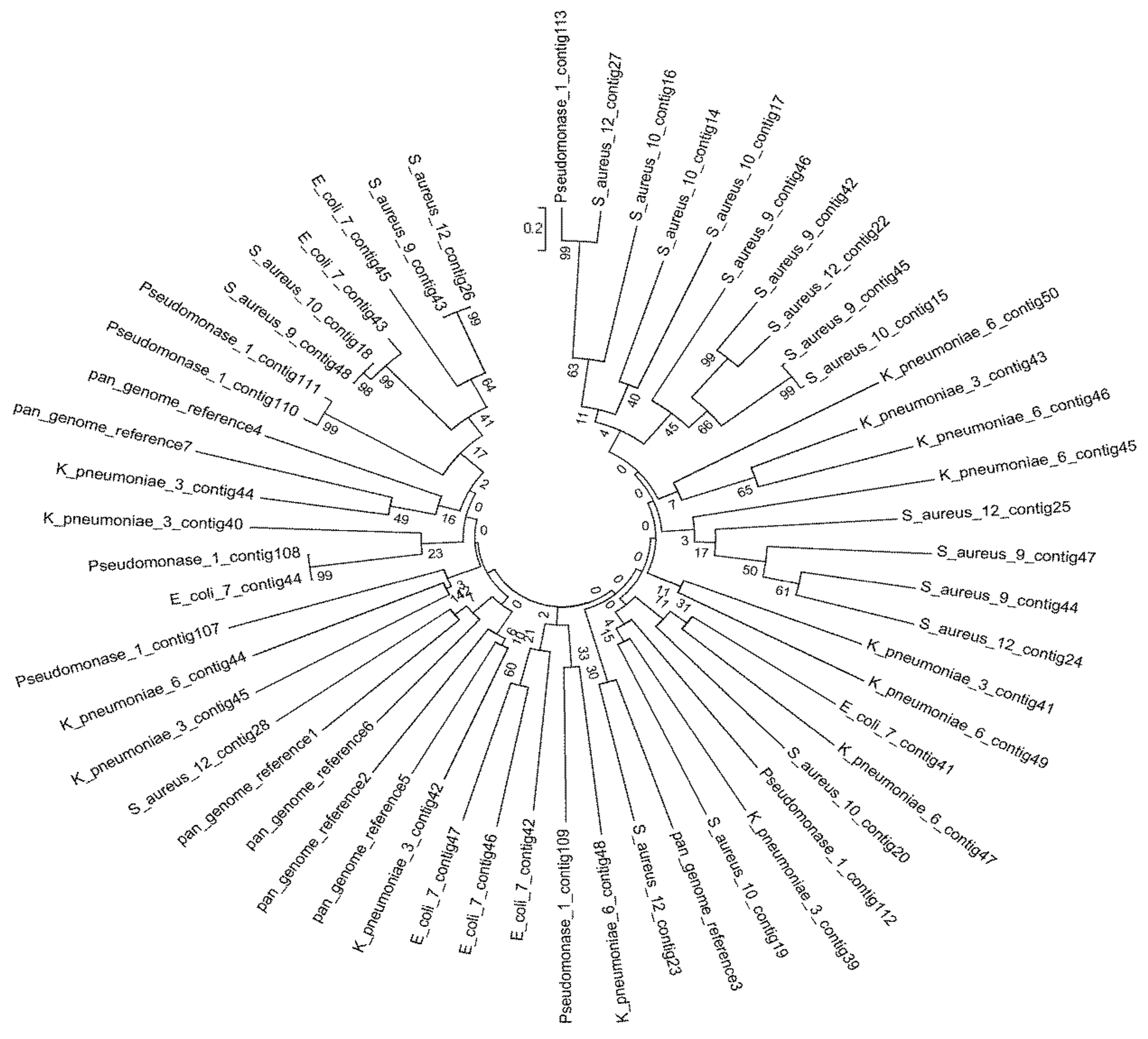
2.7.1. Calculation of Mash Distances and ANI

To identify the taxonomic boundary of the genomes, average nucleotide identity (ANI) was calculated using the Mash tool [21]. The ANI calculator (https://www.ezbiocloud.net/tools/ani (accessed on 15 May 2022)) was used to compare two prokaryotic genomes based on ANI values calculated for both genomes, followed by the classification and identification of different bacteria. The pan-genome was used as a reference genome in all cases.
For quality control assessment and to ensure error-free sequences, the genomic sequences were subjected to Panaroo [23]. This helped to identify the Mash distances between the assemblies by using Mash. Mash calculates the dissimilarities among the genomes as well as genome-scale similarity, referred to as ANI [21]. To interpret the results, Pheatmap in R was used [24]. Pan-genome analysis was performed by Macrogen, Seoul, Republic of Korea (https://www.macrogenusa.com/ (accessed on 22 July 2022)).

2.7.2. Profiling of Virulence and Antibiotic Resistance Genes

ABRicate v0.8 (https://github.com/tseemann/abricate (accessed on 22 July 2022)) was used to identify virulence and antibiotic resistance genes and characterize their presence and absence.

3. Statistical Analysis

Statistical analyses were performed using the Statistical Package for the Social Sciences, version 20 (IBM, Armonk, NY, USA). The results are expressed as the mean ± standard deviation. Differences between samples and homogeneity between groups were determined using ANOVA. The differences were considered significant at p ≤ 0.05.
The total number of base reads, GC (%), Q20 (%), and Q30 (%) were calculated for each isolate. The phylogenetic indices that serve as a quantitative index for the number of species are found in a dataset (a community), as are the phylogenetic relationships (co-distribution, species affinity, and species richness) between distributed individuals (Table 1). After sequencing, the FastQC program was used for quality control. According to quality control results, the number of data read qualities, GC distributions, k-mer distributions, and possible adapter contamination were determined and evaluated for each sample [25]. After quality control, reads with poor quality (Phred score < Q20, 30 bp window range) were removed from the data. Furthermore, low-quality base reads at terminal regions and chimeric sequences with possible adapter contaminants were removed using the Trimmomatic tool and Genomes OnLine Database (GOLD) [26]. Taxonomic profiling was performed using Kraken2 [27], and the Silva (2020) database was used as a reference dataset [28]. The OTU groups in each sample were determined after alignment. R scripts were used for data reporting, statistical analyses, and data visualization (https://www.R-project.org/ (accessed on 22 July 2022)).

4. Results

A total of 108 isolates were analyzed in this study, of which 75 were MDR bacteria. In terms of patient age, the number of bacterial isolates was 41 (37.97%) from 0–10-year-olds, 10 (9.26%) from 11–20-year-olds, 20 (18.52%) from 21–30-year-olds, 19 (17.59%) from 31–40-year-olds, and 18 (16.67%) from >41-year-olds. Gender distribution analysis showed that 32 (29.63%) strains were isolated from male patients and 76 (70.37%) from female patients.

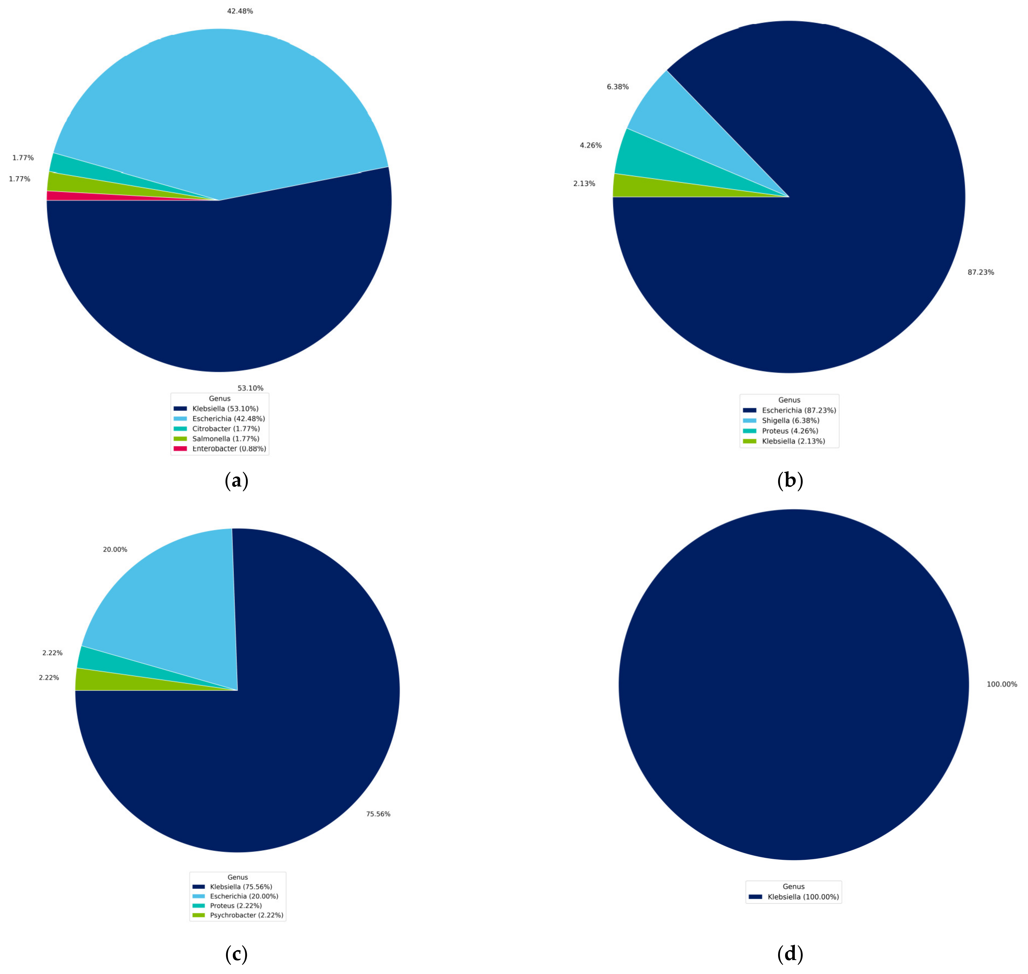
4.1. Identification of Isolated Bacteria

Phenotype analysis of the 108 bacterial isolates identified 33 (30.56%) as E. coli, 27 (25%) as K. pneumoniae, 2 (1.85%) as K. aerogenes, 9 (8.33%) as P. aeruginosa, 3 (2.78%) as E. cloacae, 2 (1.85%) as Salmonella enterica, 20 (18.52%) as S. aureus, 1 (0.93%) as S. epidermidis, 2 (1.85%) as Streptococcus pneumoniae, 8 (7.41%) as S. agalactiae (B), and 1 (0.93%) as Acinetobacter baumannii. The AST results identified one isolate of P. aeruginosa, three isolates of E. coli, four of K. Pneumoniae, and three of S. aureus as being resistant to more than three classes of antibiotics.
Of the 108 isolates, 75 (69.44%) were identified as MDR bacteria. These isolates were predominately represented by the following strains: S. aureus (MRSA), 17 (22.67%); MDR S. epidermidis, 1 (1.33%); MDR S. pneumoniae, 2 (2.67%); ESBL-producing Gram-negative Enterobacteriaceae, such as ESBL E. coli, 24 (32%); ESBL K. pneumoniae, 19 (25.33%); K. aerogenes 1(1.33%); E. cloacae, 2 (2.67%); and MDR P. aeruginosa, 9 (12%). Following this initial screening step, we selected two E. coli isolates in addition to two K. pneumoniae and three S. aureus isolates to perform the remaining investigations.

4.2. Identification Based on 16S rRNA Gene and Whole Genome Sequencing

The genus-level identification based on the top five hits of the contigs ordered by length and use of the NCBI NT database of the seven selected isolates confirmed the preliminary molecular identification results based on 16S rRNA gene sequencing, except for isolates of P. aeruginosa_1, whose identification differed from that indicated by the biochemical and preliminary molecular tests. The isolated MDR bacteria were identified as members of the genera Klebsiella (53.10%), Escherichia (42.48%), Citrobacter (1.77%), Salmonella (1.77%), and Enterobacter (0.88%) (Figure 1a). Furthermore, the identity of E. coli_7 and K. pneumoniae_3 was confirmed on the basis of 16S rDNA sequence identities of 87.23% and 75.56%, respectively. The isolates K. pneumoniae_6, S. aureus_9, S. aureus_10, and S. aureus_12 showed 100% identity at the genus level (Figure 1b–g).

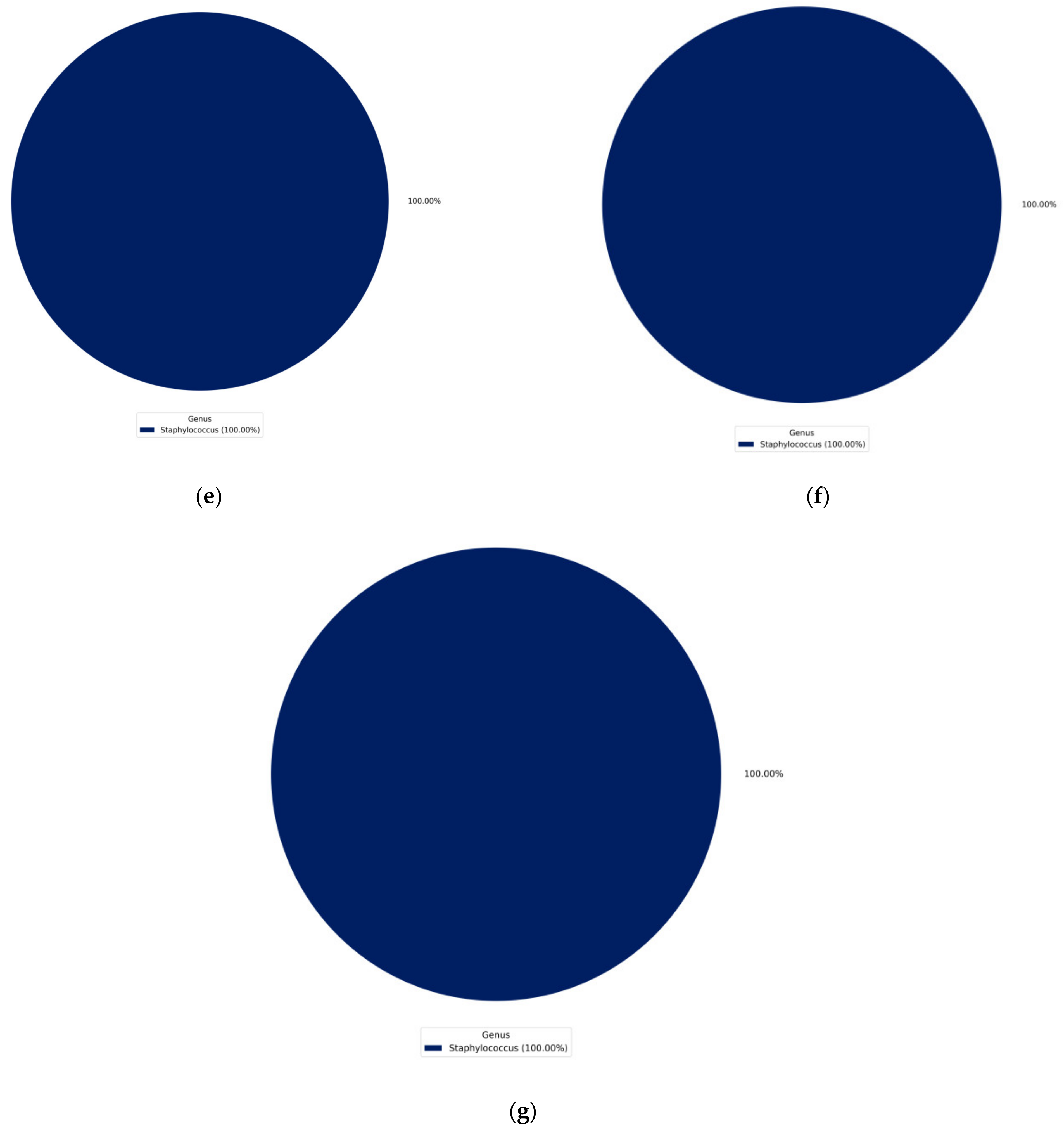
4.3. Phylogenetic Tree of the Seven Selected MDR Bacteria

The phylogenetic tree shows the multiple alignments of MDR genome contigs sequences of the seven isolated species (Figure 2). The results in Figure 3 each isolate were used to assess the completeness of genome assembly using Benchmarking Universal Single-Copy Orthologs (BUSCO) analysis and ANI. Nucleotide-level genomic similarity between the species from the same taxon available in the NCBI database was used as the measurement unit to determine the accession number for each isolate. For Pseudomonas_1, the rate of complete single-copy BUSCOs (S) was 6.45%, and 93.55% had complete and duplicated BUSCOs (D) (Figure 3a1), whereas the top five genomes identified by ANI analysis were GCA_023366235.1_E. coli_PNUSAE100989, with an ANI of 98.87% and 39.89% coverage by the assembly sequence alignments against the compared genome; GCA_023366335.1_E. coli_1193, with an ANI of 98.82% and 41.29% coverage; GCA_023367035.1_E. coli_131, with an ANI of 98.55% and 42.62% coverage; GCA_023367635.1_E. coli_131, with an ANI of 98.55% and 42.39% coverage; and GCA_023368475.1_E. coli_131, with an ANI of 98.54% and 42.41% (Figure 3b1,c1). E. coli_7 had 100% complete single-copy BUSCOs (S) (Figure 3a2), whereas the top five genomes identified by ANI analysis were GCA_023368155.1_E.coli_4532, with an ANI of 99.26% and 89.32% coverage; GCA_023367075.1_E. coli_4532, with an ANI of 99.26% and 89.29% coverage; GCA_023367595.1_E. coli_, with an ANI of 99.19% and 92.15% coverage; GCA_023369735.1_E. coli_DC5_C10, with an ANI of 99.13% and 88.78% coverage; and GCA_023367495.1_E. coli_191, with an ANI of 99.12% and 88.78% coverage (Figure 3b2,c2). K. pneumoniae_3 had 98.39% complete single-copy BUSCOs (S) and 1.61% missing BUSCOs (M) (Figure 3a3), whereas the top five genomes identified by ANI analysis were GCA_023368715.1_K. pneumoniae_11, with an ANI of 99.30% and 86.83% coverage; GCA_023368275.1_K. pneumoniae_11, with an ANI of 99.29% and 88.69% coverage; GCA_023368575.1_K. pneumoniae_1161, with an ANI of 99.29% and 87.91% coverage; GCA_023367775.1_K. pneumoniae_1, with an ANI of 99.29% and 88.16% coverage; and GCA_023367655.1_K. pneumoniae_133, with an ANI of 99.29% and 87.19% coverage (Figure 3b3,c3). In addition, K. pneumoniae_6 had 98.39% complete single-copy BUSCOs (S) and 1.61% missing BUSCOs (M) (Figure 3a4), whereas the top five genomes identified by ANI analysis were GCA_023369875.1_K. pneumoniae_13, with an ANI of 99.94% and 94.96% coverage; GCA_023369885.1_K. pneumoniae_14, with an ANI of 99.94% and 94.44% coverage; GCA_023370005.1_K. pneumoniae_6, with an ANI of 99.93% and 95.29% coverage; GCA_023370035.1_K. pneumoniae_2, with an ANI of 99.93% and 98.04% coverage; and GCA_023369995.1_K. pneumoniae_3, with an ANI of 99.93% and 98.0% coverage (Figure 3b4,c4).
Gram-positive S. aureus_9 had 100% complete single-copy BUSCOs (S) (Figure 3a5), whereas the top five genomes identified by ANI analysis were GCA_023361315.1_S. aureus_, with an ANI of 99.13% and 94.31% coverage by assembly sequence alignments against the compared genome; GCA_023361575.1_S. aureus_, with an ANI of 99.11% and 94.17% coverage; GCA_023363605.1_S. aureus_, with an ANI of 99.11% and 93.82% coverage; GCA_023361695.1_S. aureus_, with an ANI of 99.10% and 93.77% coverage; and GCA_023361615.1_S. aureus_, with an ANI of 99.10% and 93.71% coverage (Figure 3b5,c5). S. aureus_10 had 98.39% complete single-copy BUSCOs (S) (Figure 3a6), whereas the top five genomes identified by ANI analysis were GCA_023362615.1_S. aureus_, with an ANI of 99.93% and 98.42% coverage; GCA_023362315.1_S.aureus_, with an ANI of 99.13% and 93.37% coverage; GCA_023362475.1_S. aureus_, with an ANI of 99.10% and 93.98% coverage; GCA_023361575.1_S. aureus_, with an ANI of 99.09% and 92.55% coverage; and GCA_023361495.1_S. aureus_, with an ANI of 99.08% and 93.22% coverage (Figure 3b6,c6). S. aureus_12 had 100.00% complete single-copy BUSCOs (S) (Figure 3a7), whereas the top five genomes identified by ANI analysis were GCA_023360955.1_S.aureus_, with an ANI of 99.87% and 94.39% coverage; GCA_023362575.1_S.aureus_, with an ANI of 99.87% and 93.26% coverage; GCA_023361655.1_S. aureus_, with an ANI of 99.86% and 94.67% coverage; GCA_023362455.1_S. aureus_, with an ANI of 99.86% and 96.15% coverage; and GCA_023362395.1_S. aureus_, with an ANI of 99.83% and 96.75% coverage (Figure 3b7,c7).

4.4. Detection of MDR Genes

The MDR genes were detected in Pesudomonas_1, E. coli_7, K. pneumonia_3, K. pneumonia_6, S. aureus_9, S. aureus_10, and S. aureus_12. The results are illustrated in Figure 4. SHV, OXA, CTX-M, TEM-1, NDM-1, VIM-1, and ere(A) genes were detected in Pesudomonas_1, E. coli_7, K. pneumonia_3, and K. pneumonia_6, whereas ermA, ermB, ermC, msrA, qacA, qacB, and qacC genes were detected in S. aureus_9, S. aureus_10, and S. aureus_12. All genomic information is available in the Supplementary Materials (Tables S1–S7). Venn diagram showing the susceptibility of multidrug-resistant bacteria isolated from selected hospitals in Jeddah to 108 universal antibiotics, the result shows that Pseudomonas_1 and E. coli_7 are resistant to 34 antibiotics, while S. aureus_9, S. aureus_10, and S. aureus_12, are resistant to 15 antibiotics (Figure 5, data on antibiotics are available in Table S8).

5. Discussion

MDR is defined as resistance to antimicrobial drugs from at least one to three or more categories. Hospitals are an ideal environment for bacterial colonization and a potential source of bacterial infection, thereby increasing the possibility of infection, particularly by MDR nosocomial bacteria. Thus, conducting studies on MDR nosocomial bacteria is necessary to control and prevent infection in hospitals.
With the emergence of MDR bacteria as a global public health concern, a novel approach for the management of MDR bacteria is warranted. Identifying these MDR bacteria and comparing them with the global isolates determines the extent of the spread of different species and provides a statement of new species, thereby allowing the identification and inventory of species in each country and of the common species among different countries. By comparing the isolated species with species published in other research, a similarity is observed between the species in our study, including E. coli, K. pneumoniae, and S. aureus, and the species isolated from different countries in several publications [29,30,31,32].
Antibiotic resistance may be a result of an energy-driven efflux system within the cell that removes foreign antibiotic agents from the cell. For instance, the resistance of pathogens, including K. pneumoniae and E. coli, to tetracyclines is caused by the acquisition of the tet(A) and tet(B) genes by Gram-negative bacteria and tet(K) and tet(L) genes by Gram-positive bacteria. In this study, Gram-negative bacteria were resistant to multiple antibiotics, in addition to tetracyclines, due to the presence of SHV, OXA, CTX-M, TEM-1, NDM-1, and VIM-1 genes. These results are consistent with those reported in other studies [33,34,35,36]. Gram-positive bacteria carried resistance owing to the presence of ermA, ermB, ermC, msrA, qacA, qacB, and qacC genes. Our results are in accordance with other reports detecting the presence of the genes ermB [37]; ermA, msrA, and qacA [8,38,39]; and ermA, ermB, and ermC [40] in S. aureus. These genes encode efflux pumps [2]. However, antibiotic resistance can also emerge from the activation of an alternative metabolic pathway in the presence of an antibiotic that inhibits a cellular process. For instance, sulfamidics that inhibit folic acid synthesis are ineffective against bacteria that have the ability to synthesize folic acid through alternative metabolic pathways [41].
The increase in the spread of infections by isolated MDR bacteria in the winter season may be a result of frequent visits to hospitals because of diseases associated with winter; in this respect, the results of our study agree with those of other reports [42,43,44,45]. Interestingly, the bacteria occurring most often in winter are respiratory pathogens, such as S. aureus MRSA. This could be due to resistance, stability in dehydrating conditions, and presence in the ventilation streams of air conditioners [46,47,48].
To examine contamination, inanimate objects and patient-care equipment were sampled for MDR bacteria because pathogenic bacteria tend to accumulate in service locations, such as restrooms and canteens. S. aureus has the characteristic ability to survive in dry conditions and both high and low temperatures. The difference in the prevalence of MDR bacteria in different samples was found to be caused by variable exposure to antibiotics [49].
Antibiotics are becoming ineffective because of MDR characteristics. In this study, we report the presence of MDR in all groups of bacteria. The genera that were identified by de novo analysis were Klebsiella, Escherichia, and Staphylococcus. The species that were identified were Pseudomonas_1, E. coli_7, K. pneumoniae_10, S. aureus_19, and S. aureus_12. There were no significant differences in antibiotic resistance in the isolated strains of E. coli, K. pneumoniae, K. aerogenes, E. cloacae, and P. aeruginosa for all tested antibiotics, except gentamicin in E. cloacae. Several studies have produced results in accordance with our findings [50,51,52,53]. The increase in the resistance of E. coli, Pseudomonas, and S. aureus to antibiotics may have been caused by the indiscriminate use of antibiotics without prescriptions. Many studies have reported the resistance of E. coli, Pseudomonas, and S. aureus to one or more antibiotics and have identified some of the genes responsible for this resistance [54,55,56,57,58].
Gram-positive bacteria are more sensitive than Gram-negative bacteria, which are more stable to change [59,60,61], with K. pneumoniae demonstrating stability in biochemical reactions [62]. A higher rate of resistance to most commonly used antibiotics has been observed [63]. E. coli and K. pneumoniae were the most frequently isolated among Gram-negative MDR bacteria, with high resistance rates. These findings are in agreement with those of other studies [64,65,66]. The results of E. coli, Pseudomonas_1, and K. aerogenes in our study are in accordance with the results of other investigations [9,67,68]. Regarding Gram-positive isolates, our findings illustrated that S. aureus MRSA and S. epidermidis are resistant to most antibiotics, while S. pneumoniae is less resistant to antibiotics as reported by previous other studies [69,70,71].
The result in this study indicates that MDR bacteria represent a healthcare problem in hospitals for patients of all ages and awards. This result may be due to the adaptation of pathogenic bacteria, which leads to the development of mechanisms by which they can survive and acquire antibiotic resistance [72,73,74]. The result of this study is consistent with that of previous studies [12,74,75].
Gram-negative bacteria were more resistant to antibiotics than Gram-positive bacteria because of the differences in the mechanism of gene transfer between Gram-negative and Gram-positive bacteria. In addition, Gram-negative bacteria have an active uptake mechanism under iron-depleted conditions, which improves stability to different types of β-lactamases [76,77]. Bacterial resistance to antibiotics is also due to bacterial mutation, and the appearance of antibiotic-resistant genes in Gram-negative and Gram-positive bacteria can occur through the production of new generations, with a timescale of just a few hours, or new mutations in a parent cell can become a widespread mutation, resulting in the microevolution of resistant colonies [78,79]. Our finding is consistent with that of previous studies [80,81,82,83,84].

6. Conclusions

There is an urgent need to identify bacteria that are resistant to more than one antibiotic, to discover antibiotics that inhibit their growth. This study provides valuable insights into the presence of MDR bacteria in hospitals and antibiotic-resistance genes in Gram-positive and Gram-negative bacteria. The results presented here identified strains of S. aureus, E. coli, K. pneumoniae, and P. aeruginosa as MDR bacteria isolated from hospitals in Jeddah, Saudi Arabia, and Gram-negative isolates were resistant to antibiotics, in contrast with Gram-positive isolates. Detecting some genes that cause bacterial resistance promotes the development of a mechanism to inhibit the infection caused by these resistant bacteria. Our findings are largely consistent with those of previous studies, but further investigation at other hospitals is needed to assess their generalizability.

Supplementary Materials

The following supporting information can be downloaded at: https://www.mdpi.com/article/10.3390/microorganisms11102432/s1, Table S1: Gene presence/absence of Pseudomonas_1. Table S2: Gene presence/absence of E. coli_7. Table S3: Gene presence/absence of K. pneumoniae_3. Table S4: Gene presence/absence of K. pneumoniae_6. Table S5: Gene presence/absence of S. aureus_9. Table S6: Gene presence/absence of S. aureus_10. Table S7: Gene presence/absence of S. aureus_12. Table S8: Antibiotics-resistant bacteria.

Author Contributions

Data curation, E.A.-J.; Formal analysis, A.A.-J. and E.A.-J.; Investigation, M.A. and M.A.-R.; Resources, E.A.-J. and M.A.; Software, M.A.; Supervision, A.A.-J.; Validation, M.A.-R.; Visualization, E.A.-J. and A.A.-J.; Writing—original draft, M.A.; Writing—review and editing, A.A.-J. All authors have read and agreed to the published version of the manuscript.

Funding

This research did not receive any specific grant from funding agencies in the public, commercial, or not-for-profit sectors.

Institutional Review Board Statement

Not applicable.

Informed Consent Statement

Not applicable.

Data Availability Statement

Data of biochemical identification of MDR bacteria are available in the Ministry of Health Jeddah branch. Data of molecular identification are available on Blast; BLAST: Basic Local Alignment Search Tool (nih.gov) (accessed on 22 May 2022).

Acknowledgments

The authors would like to express special thanks to Essam Ibrahim Azhar for his contribution to DNA extraction and sequencing of isolated bacteria, and the Ministry of Health for their support and the facilitation of antibiotic delivery and testing. In addition, we would like to thank the Saudi Digital Library for the free access to the selected publications.

Conflicts of Interest

The authors declare no conflict of interest.

References

  1. Bentivegna, E.; Luciani, M.; Arcari, L.; Santino, I.; Simmaco, M.; Martelletti, P. Reduction of multidrug-resistant (MDR) bacterial infections during the COVID-19 pandemic: A retrospective study. Int. J. Environ. Res. Public Health 2021, 18, 1003. [Google Scholar] [CrossRef]
  2. Terreni, M.; Taccani, M.; Pregnolato, M. New antibiotics for multidrug-resistant bacterial strains: Latest research developments and future perspectives. Molecules 2021, 26, 2671. [Google Scholar] [CrossRef]
  3. World Health Organization (WHO). Antimicrobial Resistance. 2021. Available online: https://www.who.int/news-room/fact-sheets/detail/antimicrobial-resistance (accessed on 17 November 2021).
  4. Falagas, M.E.; Tansarli, G.S.; Karageorgopoulos, D.E.; Vardakas, K.Z. Deaths attributable to carbapenem-resistant Enterobacteriaceae infections. Emerg. Infect. Dis. 2014, 20, 1170–1175. [Google Scholar] [CrossRef]
  5. Sato, T.; Yamawaki, K. Cefiderocol: Discovery, chemistry, and in vivo profiles of a novel siderophore cephalosporin. Clin. Infect. Dis. 2019, 69 (Suppl. 7), S538–S543. [Google Scholar] [CrossRef]
  6. Pandey, N.; Cascella, M. Beta Lactam Antibiotics. StatPearls. 2022. Available online: https://www.ncbi.nlm.nih.gov/books/NBK545311/ (accessed on 26 September 2022).
  7. Formosa-Dague, C.; Speziale, P.; Foster, T.J.; Geoghegan, J.A.; Dufrêne, Y.F. Zinc-dependent mechanical properties of Staphylococcus aureus biofilm-forming surface protein SasG. Proc. Natl. Acad. Sci. USA 2016, 113, 410–415. [Google Scholar] [CrossRef]
  8. Karlowsky, J.A.; Hackel, M.A.; Tsuji, M.; Yamano, Y.; Echols, R.; Sahm, D.F. In vitro activity of cefiderocol, a siderophore cephalosporin, against Gram-negative bacilli isolated by clinical laboratories in North America and Europe in 2015–2016: SIDERO-WT-2015. Int. J. Antimicrob. Agents 2019, 53, 456–466. [Google Scholar] [CrossRef]
  9. Bukhari, S.A.R.; Irfan, M.; Ahmad, I.; Chen, L. Comparative genomics and pan-genome driven prediction of a reduced genome of Akkermansia muciniphila. Microorganisms 2022, 10, 1350. [Google Scholar] [CrossRef] [PubMed]
  10. Hassoun-Kheir, N.; Stabholz, Y.; Kreft, J.U.; de la Cruz, R.; Romalde, J.L.; Nesme, J.; Sørensen, S.J.; Smets, B.F.; Graham, D.; Paul, M. Comparison of antibiotic-resistant bacteria and antibiotic resistance genes abundance in hospital and community wastewater: A systematic review. Sci. Total Environ. 2020, 743, 140804. [Google Scholar] [CrossRef] [PubMed]
  11. Szekeres, E.; Baricz, A.; Chiriac, C.M.; Farkas, A.; Opris, O.; Soran, M.L.; Andrei, A.S.; Rudi, K.; Balcázar, J.L.; Dragos, N.; et al. Abundance of antibiotics, antibiotic resistance genes and bacterial community composition in wastewater effluents from different Romanian hospitals. Environ. Pollut. 2017, 225, 304–315. [Google Scholar] [PubMed]
  12. The Pew Charitable Trust Tracking the Global Pipeline of Antibiotics in Development. March 2021. Available online: https://www.pewtrusts.org/en/research-and-analysis/issue-briefs/2021/03/tracking-the-global-pipeline-of-antibiotics-in[1]development (accessed on 1 May 2021).
  13. World Health Organization. Meeting Report of the World Health Organization Expert Consultation on the Definition of Extensively Drug-Resistant Tuberculosis, 27–29 October 2020; World Health Organization: Geneva, Switzerland, 2021. [Google Scholar]
  14. Zowawi, H.M. Antimicrobial resistance in Saudi Arabia: An urgent call for an immediate action. Saudi Med. J. 2016, 37, 935–940. [Google Scholar] [CrossRef]
  15. Enani, M.A.; Alzahrani, R.M.; Alzubaidy, K.A.; Ajjaj, R.G.; Saeed, A.M. The prevalence and characters of hospital acquired infections in three private hospital, Jeddah, Saudi Arabia. Int. J. Adv. Res. 2019, 7, 1262–1269. [Google Scholar] [CrossRef] [PubMed]
  16. Halwani, M.A.; Tashkandy, N.A.J.; Aly, M.M.; Al Masoudi, S.B.; Dhafar, O.O. Incidence of antibiotic resistance bacteria in Jeddah’s Ministry of Health Hospitals, Saudi Arabia. Adv. Microbiol. 2015, 5, 780–786. [Google Scholar] [CrossRef]
  17. Helmi, N.R.; Zaman, R.M.; Aly, M.M. Prevalence of Gram-positive bacteria in Jeddah, Kingdom of Saudi Arabia: Study of antimicrobial resistance patterns and molecular typing. Int. J. Pharm. Biol. Sci. 2013, 4, 1231. [Google Scholar]
  18. Qiagen. Dneasy Tissue Handbook; Qiagen: Hilden, Germany, 2006. [Google Scholar]
  19. Sakdapetsiri, C.; Ngaemthao, W.; Suriyachadkun, C.; Pinyakong, O. Paeniglutamicibacter quisquiliarum sp. nov., isolated from midden soil waste. Int. J. Sys. Evo. Micr. 2022, 72, 005651. [Google Scholar] [CrossRef]
  20. Guillen, I.A.; Camacho, H.; Tuero, A.D.; Bacardí, D.; Palenzuela, D.O.; Aguilera, A.; Silva, J.A.; Estrada, R.; Gell, O.; Suárez, J.; et al. PCR conditions for 16S primers for analysis of microbes in the colon of rats. J. Biomol. Technol. 2016, 27, 105–112. [Google Scholar] [CrossRef]
  21. Ekblom, R.; Wolf, J.B. A field guide to whole-genome sequencing, assembly and annotation. Evol. Appl. 2014, 7, 1026–1042. [Google Scholar] [CrossRef]
  22. Peker, N.; Garcia-Croes, S.; Dijkhuizen, B.; Wiersma, H.H.; Van Zanten, E.; Wisselink, G.; Friedrich, A.W.; Kooistra-Smid, M.; Sinha, B.; Rossen, J.W.A.; et al. A comparison of three different bioinformatics analyses of the 16S–23S rRNA encoding region for bacterial identification. Front. Microbiol. 2019, 10, 620. [Google Scholar] [CrossRef]
  23. Ondov, B.D.; Treangen, T.J.; Melsted, P.; Mallonee, A.B.; Bergman, N.H.; Koren, S.; Phillippy, A.M. Mash: Fast genome and metagenome distance estimation using MinHash. Genome Biol. 2016, 17, 132. [Google Scholar] [CrossRef]
  24. Tonkin-Hill, G.; MacAlasdair, N.; Ruis, C.; Weimann, A.; Horesh, G.; Lees, J.A.; Gladstone, R.A.; Lo, S.; Beaudoin, C.; Floto, R.A.; et al. Producing polished prokaryotic pangenomes with the Panaroo pipeline. Genome Biol. 2020, 21, 180. [Google Scholar] [CrossRef]
  25. Kolde, R.; Kolde, M.R. Package ‘pheatmap’. In pheatmap: Pretty Heatmaps version 1.0.12 from CRAN; For. Rev. Version 1.0.10; 2018; Available online: Rdrr.io (accessed on 12 May 2022).
  26. Andrews, S. FastQC: A Quality Control Tool for High Throughput Sequence Data; Babraham Bioinformatics: Cambridge, UK, 2010; Available online: https://www.bioinformatics.babraham.ac.uk/projects/fastqc/ (accessed on 22 May 2022).
  27. Bolger, A.M.; Lohse, M.; Usadel, B. Trimmomatic: A flexible trimmer for Illumina sequence data. Bioinformatics 2014, 30, 2114–2120. [Google Scholar] [CrossRef]
  28. Wood, D.E.; Lu, J.; Langmead, B. Improved metagenomic analysis with Kraken 2. Genome Biol. 2019, 20, 257. [Google Scholar] [CrossRef] [PubMed]
  29. Jaén-Luchoro, D.; Kahnamouei, A.; Yazdanshenas, S.; Lindblom, A.; Samuelsson, E.; Åhrén, C.; Karami, N. Comparative Genomic Analysis of ST131 Subclade C2 of ESBL-Producing, E. coli Isolates from Patients with Recurrent and Sporadic Urinary Tract Infections. Microorganisms 2023, 11, 1622. [Google Scholar] [CrossRef] [PubMed]
  30. Li, D.; Elankumaran, P.; Kudinha, T.; Kidsley, A.K.; Trott, D.J.; Jarocki, V.M.; Djordjevic, S.P. Dominance of Escherichia coli sequence types ST73, ST95, ST127 and ST131 in Australian urine isolates: A genomic analysis of antimicrobial resistance and virulence linked to F plasmids. Microb. Genom. 2023, 9, 001068. [Google Scholar] [CrossRef] [PubMed]
  31. Castillo-Polo, J.A.; Hernández-García, M.; Morosini, M.I.; Pérez-Viso, B.; Soriano, C.; De Pablo, R.; Cantón, R.; Ruiz-Garbajosa, P. Outbreak by KPC-62-producing ST307 Klebsiella pneumoniae isolates resistant to ceftazidime/avibactam and cefiderocol in a university hospital in Madrid, Spain. J. Antimicrob. Chemother. 2023, 78, 1259–1264. [Google Scholar] [CrossRef] [PubMed]
  32. Chew, C.H.; Yeo, C.C.; Che Hamzah, A.M.; Al-Trad, E.A.; Jones, S.U.; Chua, K.H.; Puah, S.M. Multidrug-Resistant Methicillin-Resistant Staphylococcus aureus Associated with Hospitalized Newborn Infants. Diagnostics 2023, 13, 1050. [Google Scholar] [CrossRef]
  33. Krüger, H.; Ji, X.; Hanke, D.; Schink, A.K.; Fiedler, S.; Kaspar, H.; Wang, Y.; Schwarz, S.; Wu, C.; Feßler, A.T. Novel macrolide-lincosamide-streptogramin B resistance gene erm (54). Int. J. Antimicrob. Chemother. 2022, 77, 2296–2298. [Google Scholar] [CrossRef]
  34. Szymanek-Majchrzak, K.; Młynarczyk, G. Genomic Insights of first erm B-positive ST338-SCC mec VT/CC59 Taiwan clone of community-associated methicillin-resistant Staphylococcus aureus in Poland. Int. J. Mol. Sci. 2022, 23, 8755. [Google Scholar] [CrossRef]
  35. Mahesh, S.; Kalyan, R.; Gupta, P.; Veram, S.; Venkatesh, V.; Trİpathİ, P. MecA and ermA gene discrepancy from their phenotypic profile in Staphylococcus aureus isolates. J. Microbiol. Infect. Dis. 2022, 12, 6–11. [Google Scholar] [CrossRef]
  36. Suma, T.A.; Alam, N.; Raihan, S.Z.; Zahid, M.A.; Mandal, S.C.; Suchana, F.J.; Kundu, R.; Hossain, A.; Muhit, M.A. Association of antibacterial susceptibility profile with the prevalence of genes encoding efflux proteins in the Bangladeshi clinical isolates of Staphylococcus aureus. Antibiotics 2023, 12, 305. [Google Scholar] [CrossRef]
  37. Medis, S.; Dissanayake, T.; Weerasekera, M.; Namali, D.; Gunasekara, S.; Kottahachchi, J. Distribution of mecA and qacA/B genes among coagulase negative staphylococci isolated from central venous catheters of intensive care unit patients. Indian J. Med. Microbiol. 2022, 40, 505–509. [Google Scholar] [CrossRef]
  38. Mohamad Farook, N.A.; Argimón, S.; Abdul Samat, M.N.; Salleh, S.A.; Sulaiman, S.; Tan, T.L.; Periyasamy, P.; Lau, C.L.; Ismail, Z.; Muhammad Azami, N.A.; et al. Diversity and dissemination of methicillin-resistant Staphylococcus aureus (MRSA) genotypes in Southeast Asia. Trop. Med. Infect. Dis. 2022, 7, 438. [Google Scholar] [CrossRef] [PubMed]
  39. Zhang, Y.; Kashikar, A.; Bush, K. In vitro activity of plazomicin against β-lactamase-producing carbapenem-resistant Enterobacteriaceae (CRE). J. Antimicrob. Chemother. 2017, 72, 2792–2795. [Google Scholar] [CrossRef] [PubMed]
  40. Ito, A.; Sato, T.; Ota, M.; Takemura, M.; Nishikawa, T.; Toba, S.; Kohira, N.; Miyagawa, S.; Ishibashi, N.; Matsumoto, S.; et al. In vitro antibacterial properties of cefiderocol, a novel siderophore cephalosporin, against Gram-negative bacteria. Antimicrob. Agents Chemother. 2018, 62, 10–1128. [Google Scholar] [CrossRef] [PubMed]
  41. Behroozynia, M. Knowledge, attitude and practice of intensive care unit nurses about prevention and control of nosocomial infections. Sci. J. Iranshahr Univ. Med. Sci. 2022, 1, 11–15. [Google Scholar]
  42. Burnham, J.P.; Feldman, M.F.; Calix, J.J. Seasonal changes in the prevalence of antibiotic-susceptible Acinetobacter calcoaceticus-baumannii complex isolates result in increased multidrug resistance rates during winter months. Open Forum Infect. Dis. 2019, 6, ofz245. [Google Scholar] [CrossRef]
  43. Schwab, F.; Gastmeier, P.; Hoffmann, P.; Meyer, E. Summer, sun and sepsis—The influence of outside temperature on nosocomial bloodstream infections: A cohort study and review of the literature. PLoS ONE 2020, 15, e0234656. [Google Scholar] [CrossRef]
  44. Yamaguto, G.E.; Zhen, F.; Moreira, M.M.; Montesanti, B.M.; Raboni, S.M. Community respiratory viruses and healthcare-associated infections: Epidemiological and clinical aspects. J. Hosp. Infect. 2022, 122, 187–193. [Google Scholar] [CrossRef]
  45. Zhou, Z.C.; Liu, Y.; Lin, Z.J.; Shuai, X.Y.; Zhu, L.; Xu, L.; Meng, L.X.; Sun, Y.J.; Chen, H. Spread of antibiotic resistance genes and microbiota in airborne particulate matter, dust, and human airways in the urban hospital. Environ. Int. 2021, 153, 106501. [Google Scholar] [CrossRef]
  46. Adalbert, J.R.; Varshney, K.; Tobin, R.; Pajaro, R. Clinical outcomes in patients co-infected with COVID-19 and Staphylococcus aureus: A scoping review. BMC Infect. Dis. 2021, 21, 985. [Google Scholar] [CrossRef]
  47. Hsu, B.M.; Chen, J.S.; Lin, I.C.; Hsu, G.J.; Koner, S.; Hussain, B.; Huang, S.W.; Tsai, H.C. Molecular and antimicrobial Re-sistance (AMR) profiling of methicillin-Resistant Staphylococcus aureus (MRSA) from hospital and long-term care facilities (LTCF) environment. Antibiotics 2021, 10, 748. [Google Scholar] [CrossRef]
  48. Yu, C.H.; Shen, S.; Huang, K.-Y.A.; Huang, Y.C. The trend of environmental and clinical methicillin-resistant Staphylococcus aureus in a hospital in Taiwan: Impact of USA300. J. Microbiol. Immunol. Infect. 2022, 55, 241. [Google Scholar] [CrossRef] [PubMed]
  49. Adhikari, S.; Khadka, S.; Sapkota, S.; Rana, J.C.; Khanal, S.; Neupane, A.; Sharma, B. Prevalence and antibiograms of uropathogens from the suspected cases of urinary tract infections in Bharatpur Hospital, Nepal. J. Coll. Med. Sci. 2019, 15, 260–266. [Google Scholar] [CrossRef]
  50. Vivas, R.; Barbosa, A.A.T.; Dolabela, S.S.; Jain, S. Multidrug-resistant bacteria and alternative methods to control them: An overview. Microb. Drug Resist. 2019, 25, 890–908. [Google Scholar] [CrossRef] [PubMed]
  51. Liu, S.; Huang, N.; Zhou, C.; Lin, Y.; Zhang, Y.; Wang, L.; Zheng, X.; Zhou, T.; Wang, Z. Molecular mechanisms and epidemiology of carbapenem-resistant Enterobacter cloacae complex isolated from Chinese patients during 2004–2018. Infect. Drug Resist. 2021, 14, 3647–3658. [Google Scholar] [CrossRef]
  52. Pot, M.; Reynaud, Y.; Couvin, D.; Ducat, C.; Ferdinand, S.; Gravey, F.; Gruel, G.; Guérin, F.; Malpote, E.; Breurec, S.; et al. Wide distribution and specific resistance pattern to third-generation cephalosporins of Enterobacter cloacae complex members in humans and in the environment in Guadeloupe (French West Indies). Front. Microbiol. 2021, 12, 628058. [Google Scholar] [CrossRef]
  53. Manandhar, S.; Nguyen, Q.; Nguyen Thi Nguyen, T.; Pham, D.T.; Rabaa, M.A.; Dongol, S.; Basnyat, B.; Dixit, S.M.; Baker, S.; Karkey, A. Genomic epidemiology, antimicrobial resistance and virulence factors of Enterobacter cloacae complex causing potential community-onset bloodstream infections in a tertiary care hospital of Nepal. JAC Antimicrob Resist. 2022, 4, dlac050. [Google Scholar] [CrossRef]
  54. Baiou, A.; Elbuzidi, A.A.; Bakdach, D.; Zaqout, A.; Alarbi, K.M.; Bintaher, A.A.; Ali, M.M.B.; Elarabi, A.M.; Ali, G.A.M.; Daghfal, J.; et al. Clinical characteristics and risk factors for the isolation of multi-drug-resistant Gram-negative bacteria from critically ill patients with COVID-19. J. Hosp. Infect. 2021, 110, 165–171. [Google Scholar] [CrossRef]
  55. Derakhshan, S.; Navidinia, M.; Haghi, F. Antibiotic susceptibility of human-associated Staphylococcus aureus and its relation to agr typing, virulence genes, and biofilm formation. BMC Infect. Dis. 2021, 21, 627. [Google Scholar] [CrossRef]
  56. Miklasińska-Majdanik, M. Mechanisms of resistance to macrolide antibiotics among Staphylococcus aureus. Antibiotics 2021, 10, 1406. [Google Scholar] [CrossRef]
  57. Nji, E.; Kazibwe, J.; Hambridge, T.; Joko, C.A.; Larbi, A.A.; Damptey, L.A.O.; Nkansa-Gyamfi, N.A.; Stålsby Lundborg, C.S.; Lien, T.Q. High prevalence of antibiotic resistance in commensal Escherichia coli from healthy human sources in community settings. Sci. Rep. 2021, 11, 3372. [Google Scholar] [CrossRef]
  58. Ma, Y.; Guo, Z.; Xia, B.; Zhang, Y.; Liu, X.; Yu, Y.; Tang, N.; Tong, X.; Wang, M.; Ye, X.; et al. Identification of antimicrobial peptides from the human gut microbiome using deep learning. Nat. Biotechnol. 2022, 40, 921. [Google Scholar] [CrossRef] [PubMed]
  59. Nasrin, S.; Hegerle, N.; Sen, S.; Nkeze, J.; Sen, S.; Permala-Booth, J.; Choi, M.; Sinclair, J.; Tapia, M.D.; Johnson, J.K.; et al. Distribution of serotypes and antibiotic resistance of invasive Pseudomonas aeruginosa in a multi-country collection. BMC Microbiol. 2022, 22, 13. [Google Scholar] [CrossRef]
  60. Hwang, W.; Yoon, S.S. Virulence characteristics and an action mode of antibiotic resistance in multidrug-resistant Pseudomonas aeruginosa. Sci. Rep. 2019, 9, 487. [Google Scholar] [CrossRef] [PubMed]
  61. Nogueira, J.O.E.; Campolina, G.A.; Batista, L.R.; Alves, E.; Caetano, A.R.S.; Brandão, R.M.; Nelson, D.L.; Cardoso, M.D.G. Mechanism of action of various terpenes and phenylpropanoids against Escherichia coli and Staphylococcus aureus. FEMS Microbiol. Lett. 2021, 368, fnab052. [Google Scholar] [CrossRef]
  62. Rahman, M.A.; Amirkhani, A.; Chowdhury, D.; Mempin, M.; Molloy, M.P.; Deva, A.K.; Vickery, K.; Hu, H. Proteome of Staphylococcus aureus biofilm changes significantly with aging. Int. J. Mol. Sci. 2022, 23, 6415. [Google Scholar] [CrossRef]
  63. Tu, I.F.; Lin, T.L.; Yang, F.L.; Lee, I.M.; Tu, W.L.; Liao, J.H.; Ko, T.P.; Wu, W.J.; Jan, J.T.; Ho, M.R.; et al. Structural and biological insights into Klebsiella pneumoniae surface polysaccharide degradation by a bacteriophage K1 lyase: Implications for clinical use. J. Biomed. Sci. 2022, 29, 9. [Google Scholar] [CrossRef]
  64. Fesseha, H.; Mathewos, M.; Aliye, S.; Mekonnen, E. Isolation and antibiogram of Escherichia coli O157: H7 from diarrhoeic calves in urban and peri-urban dairy farms of Hawassa town. Vet. Med. Sci. 2022, 8, 864–876. [Google Scholar] [CrossRef]
  65. Gebremedhin, E.Z.; Merga, D.; Sarba, E.J.; Marami, L.M.; Tola, G.K.; Endale, S.S. Prevalence, risk factors and antibiogram of Escherichia coli isolated from dogs in Ambo, Gojo and Bako towns of Oromia region, Ethiopia. Ethiop. Vet. J. 2021, 25, 1–22. [Google Scholar] [CrossRef]
  66. Mapipa, Q.; Digban, T.O.; Nnolim, N.E.; Nwodo, U.U. Antibiogram profile and virulence signatures of pseudomonas aeruginosa isolates recovered from selected agrestic hospital effluents. Sci. Rep. 2021, 11, 11800. [Google Scholar] [CrossRef]
  67. KuKanich, K.S.; Bagladi-Swanson, M.; KuKanich, B. Pseudomonas aeruginosa susceptibility, antibiogram and clinical interpretation, and antimicrobial prescribing behaviors for dogs with otitis in the Midwestern United States. J. Vet. Pharmacol. Ther. 2022, 45, 440–449. [Google Scholar] [CrossRef]
  68. Founou, L.L.; Amoako, D.G.; Founou, R.C.; Essack, S.Y. Antibiotic resistance in food animals in Africa: A systematic review and meta-analysis. Microb. Drug Resist. 2018, 24, 648–665. [Google Scholar] [CrossRef]
  69. Cazer, C.L.; Westblade, L.F.; Simon, M.S.; Magleby, R.; Castanheira, M.; Booth, J.G.; Jenkins, S.G.; Gröhn, Y.T. Analysis of multidrug resistance in Staphylococcus aureus with a machine learning-generated antibiogram. Antimicrob. Agents Chemother. 2021, 65, e02132-20. [Google Scholar] [CrossRef]
  70. Roth, B.M.; Laps, A.; Yamba, K.; Heil, E.L.; Johnson, J.K.; Stafford, K.; Hachaambwa, L.M.; Kalumbi, M.; Mulenga, L.; Patel, D.M.; et al. Antibiogram development in the setting of a high frequency of multi-drug resistant organisms at University Teaching Hospital, Lusaka, Zambia. Antibiotics 2021, 10, 782. [Google Scholar] [CrossRef]
  71. Baron, S.A.; Diene, S.M.; Rolain, J.-M. Human microbiomes and antibiotic resistance. Hum. Microbiome J. 2018, 10, 43–52. [Google Scholar] [CrossRef]
  72. Alemayehu, T. Prevalence of multidrug-resistant bacteria in Ethiopia: A systematic review and meta-analysis. J. Glob. Antimicrob. Resist. 2021, 26, 133–139. [Google Scholar] [CrossRef] [PubMed]
  73. Uddin, T.M.; Chakraborty, A.J.; Khusro, A.; Zidan, B.R.M.; Mitra, S.; Emran, T.B.; Dhama, K.; Ripon, M.K.H.; Gajdács, M.; Sahibzada, M.U.K.; et al. Antibiotic resistance in microbes: History, mechanisms, therapeutic strategies and future prospects. J. Infect. Public Health 2021, 14, 1750–1766. [Google Scholar] [CrossRef] [PubMed]
  74. Ito-Horiyama, T.; Ishii, Y.; Ito, A.; Sato, T.; Nakamura, R.; Fukuhara, N.; Tsuji, M.; Yamano, Y.; Yamaguchi, K.; Tateda, K. Stability of novel siderophore cephalosporin S-649266 against clinically relevant carbapenemases. Antimicrob. Agen. chemoth. 2016, 60, 4384–4386. [Google Scholar] [CrossRef] [PubMed]
  75. Bhowmik, D.; Chetri, S.; Das, B.J.; Dhar Chanda, D.D.; Bhattacharjee, A. Distribution of virulence genes and SCCmec types among methicillin-resistant Staphylococcus aureus of clinical and environmental origin: A study from community of Assam, India. BMC Res. Notes 2021, 14, 58. [Google Scholar] [CrossRef]
  76. de Souza, G.H.A.; Dos Santos Radai, J.A.; Mattos Vaz, M.S.; Esther da Silva, K.; Fraga, T.L.; Barbosa, L.S.; Simionatto, S. In vitro and in vivo antibacterial activity assays of carvacrol: A candidate for development of innovative treatments against KPC-producing Klebsiella pneumoniae. PLoS ONE 2021, 16, e0246003. [Google Scholar] [CrossRef]
  77. Meirelles, L.A.; Perry, E.K.; Bergkessel, M.; Newman, D.K. Bacterial defenses against a natural antibiotic promote collateral resilience to clinical antibiotics. PLOS Biol. 2021, 19, e3001093. [Google Scholar] [CrossRef]
  78. Roemhild, R.; Bollenbach, T.; Andersson, D.I. The physiology and genetics of bacterial responses to antibiotic combinations. Nat. Rev. Microbiol. 2022, 20, 478. [Google Scholar] [CrossRef] [PubMed]
  79. Bojar, B.; Sheridan, J.; Beattie, R.; Cahak, C.; Liedhegner, E.; Munoz-Price, L.S.; Hristova, K.R.; Skwor, T. Antibiotic resistance patterns of Escherichia coli isolates from the clinic through the wastewater pathway. Int. J. Hyg. Environ. Health 2021, 238, 113863. [Google Scholar] [CrossRef] [PubMed]
  80. Langendonk, R.F.; Neill, D.R.; Fothergill, J.L. The building blocks of antimicrobial resistance in Pseudomonas aeruginosa: Implications for current resistance-breaking therapies. Front. Cell. Infect. Microbiol. 2021, 11, 665759. [Google Scholar] [CrossRef] [PubMed]
  81. Sharahi, J.Y.; Hashemi, A.; Ardebili, A.; Davoudabadi, S. Molecular characteristics of antibiotic-resistant Escherichia coli and Klebsiella pneumoniae strains isolated from hospitalized patients in Tehran, Iran. Ann. Clin. Microbiol. Antimicrob. 2021, 20, 32. [Google Scholar] [CrossRef]
  82. Haeili, M.; Shoghi, Y.; Moghimi, M.; Ghodousi, A.; Omrani, M.; Cirillo, D.M. Genomic features of in vitro selected mutants of Escherichia coli with decreased susceptibility to tigecycline. J. Glob. Antimicrob. Resist. 2022, 31, 32–37. [Google Scholar] [CrossRef] [PubMed]
  83. Van An, N.; Hoang, L.H.; Le, H.H.; Thai Son, N.; Hong, L.T.; Viet, T.T.; Le, T.D.; Thang, T.B.; Vu, L.H.; Nguyen, V.T.; et al. Distribution and antibiotic resistance characteristics of bacteria isolated from blood culture in a teaching hospital in Vietnam during 2014–2021. Infec. Drug Resist. 2023, 16, 1677–1692. [Google Scholar] [CrossRef] [PubMed]
  84. Fröhlich, C.; Sørum, V.; Tokuriki, N.; Johnsen, P.J.; Samuelsen, Ø. Evolution of β-lactamase-mediated cefiderocol resistance. J. Antimicrob. Chemoth. 2022, 77, 2429–2436. [Google Scholar] [CrossRef]
Figure 1. Genus-level summary of de novo assembly and analysis of the RNA-seq data obtained by BLASTN for the isolated MDR bacteria: (a) P. aeruginosa_1; (b) E. coli_7; (c) K. pneumoniae_3; (d) K. pneumoniae_6; (e) S. aureus_9; (f) S. aureus_10; and (g) S. aureus_12.
Microorganisms 11 02432 g001aMicroorganisms 11 02432 g001b
Figure 2. Phylogenetic tree of the MDR bacterial isolates based on contigs sequencing.
Microorganisms 11 02432 g002
Figure 3. De novo assembly of Pseudomonas_1, E. coli_7, K. pneumoniae_3, K. pneumoniae_6, S. aerueus_9, S. aerueus_10, and S. aureus_12 isolated from selected hospitals in Jeddah. (a) Plot of BUSCO assessment results. (b) Identity heatmaps (%) identified by ANI analysis. (c) Heatmaps of alignment coverage from ANI analysis.
Microorganisms 11 02432 g003aMicroorganisms 11 02432 g003bMicroorganisms 11 02432 g003cMicroorganisms 11 02432 g003d
Figure 4. Pan-genome analysis of (a) gene clusters (total = 11,156), shell genes (10,826), and core genes (330) for Pseudomonas_1. Gene presence/absence of 4061 gene clusters commonly present in four strains (a1). (b) Gene clusters (total = 6418), shell genes (2746), and core genes (3672) for E. coli_7. Gene presence/absence of 3671 gene clusters commonly present in six strains (b1). (c) Gene clusters (total = 11,848), shell genes (11,523), and core genes (325) for K. pneumoniae_3. Gene presence/absence of 324 gene clusters commonly present in the six strains (c1). (d) Gene clusters (total = 6003), shell genes (1076), and core genes (4927) for K. pneumoniae_3. Gene presence/absence of 4926 gene clusters commonly present in the six strains (d1). (e) Gene clusters (total = 3249), shell genes (1097), and core genes (2152) for S. aureus_9. Gene presence/absence of 2151 gene clusters commonly present in the four strains (e1). (f) Gene clusters (total = 2763), shell genes (370), and core genes (2393) for S. aureus_10. Gene presence/absence of 2392 gene clusters commonly present in the four strains (f1). (g) Gene clusters (total = 3022), shell genes (740), and core genes (2282) for S. aureus_12. Gene presence/absence of 2281 gene clusters commonly present in the four strains (g1).
Microorganisms 11 02432 g004aMicroorganisms 11 02432 g004bMicroorganisms 11 02432 g004c
Figure 5. Venn diagram showing the susceptibility of multidrug-resistant bacteria isolated from selected hospitals in Jeddah to 108 universal antibiotics (data on antibiotics are available in Table S8).
Microorganisms 11 02432 g005
Table 1. The index information of the isolated bacteria.
Sample IDIndex 5Index 7
Pseudomonas_1CTAGGCAACTCACCAA
K_pneumoniae_3CTGTATTAATATGGAT
K_pneumoniae_6TCACGCCGGCGCAAGC
E_coli_7ACTTACATAAGATACT
S_aureus_9GTCCGTGCGGAGCGTC
S_aureus_10AAGGTACCATGGCATG
S_aureus_12GGAACGTTGCAATGCA
Disclaimer/Publisher’s Note: The statements, opinions and data contained in all publications are solely those of the individual author(s) and contributor(s) and not of MDPI and/or the editor(s). MDPI and/or the editor(s) disclaim responsibility for any injury to people or property resulting from any ideas, methods, instructions or products referred to in the content.

