Article

Dysbiosis by Eradication of Helicobacter pylori Infection Associated with Follicular Gastropathy and Pangastropathy

by
Uriel Gomez-Ramirez
1,2,
Carolina G. Nolasco-Romero
1,2,
Araceli Contreras-Rodríguez
3,
Gerardo Zuñiga
4,
Sandra Mendoza-Elizalde
1,
Francisco-Javier Prado-Galbarro
5,
Fernando Pérez Aguilar
6,
Jonatan Elihu Pedraza Tinoco
7,
Pedro Valencia-Mayoral
8,* and
Norma Velázquez-Guadarrama
1,*
1
Laboratorio de Investigación en Enfermedades Infecciosas, Hospital Infantil de México Federico Gómez, Mexico City 06720, Mexico
2
Posgrado en Ciencias Quimicobiológicas, Escuela Nacional de Ciencias Biológicas, Instituto Politécnico Nacional, Mexico City 11340, Mexico
3
Departamento de Microbiología, Escuela Nacional de Ciencias Biológicas, Instituto Politécnico Nacional, Mexico City 11340, Mexico
4
Laboratorio de Variación Biológica y Evolución, Departamento de Zoología, Escuela Nacional de Ciencias Biológicas, Instituto Politécnico Nacional, Mexico City 11340, Mexico
5
Dirección de Investigación, Hospital Infantil de México Federico Gómez, Mexico City 06720, Mexico
6
Servicio de Endoscopía Gastrointestinal, Hospital General Dr. Fernando Quiroz, Instituto de Seguridad y Servicios Sociales de los Trabajadores del Estado, Mexico City 01140, Mexico
7
Clinica Medizama Tulum, Tulum 77760, Mexico
8
Departamento de Patología Clínica y Experimental, Hospital Infantil de México Federico Gómez, Mexico City 06720, Mexico
*
Authors to whom correspondence should be addressed.
Microorganisms 2023, 11(11), 2748; https://doi.org/10.3390/microorganisms11112748
Submission received: 28 September 2023 / Revised: 3 November 2023 / Accepted: 7 November 2023 / Published: 10 November 2023

Abstract

:
Dysbiosis plays an important role in the development of bacterial infections in the gastric mucosa, particularly Helicobacter pylori. The international guidelines for the treatment of H. pylori infections suggest standard triple therapy (STT). Nevertheless, because of the increasing resistance rates to clarithromycin, metronidazole has been widely considered in several countries. Unfortunately, the non-justified administration of antibiotics induces dysbiosis in the target organ. We characterized the gastric microbiota of patients diagnosed with follicular gastropathy and pangastropathy attributed to H. pylori infection, before and after the administration of STT with metronidazole. Dominant relative abundances of Cutibacterium were observed in pre-treatment patients, whereas H. pylori was observed at <11%, suggesting the multifactor property of the disease. The correlation of Cutibacterium acnes and H. pylori with gastric infectious diseases was also evaluated using quantitative real-time polymerase chain reaction. The dominance of C. acnes over H. pylori was observed in gastritis, gastropathies, and non-significant histological alterations. None of the microorganisms were detected in the intestinal metaplasia. Post-treatment alterations revealed an increase in the relative abundances of Staphylococcus, Pseudomonas, and Klebsiella. Non-H. pylori gastrointestinal bacteria can be associated with the initiation and development of gastric diseases, such as pathobiont C. acnes.

1. Introduction

Gastropathy is a condition that affects the gastric mucosa [1]. It is characterized by the prevalence of mononuclear infiltrates, the formation of lymphoid follicles with germinal centres in the lamina propria, and subsequent tissue inflammation and regeneration. Histological injury is frequently located in the gastric antrum; however, it sometimes extends to the gastric corpus [2]. Gastropathies are classified according to their aetiologies. Follicular gastropathy (also known as reactive or chemical gastropathy) is mainly attributed to irritants, and their recurrent abuse, including alcohol and nonsteroidal anti-inflammatory drugs (NSAIDs) [1], which play an important role in the development of bacterial infections in the mucosa, particularly those induced by Helicobacter pylori [3]. Approximately 50% of the worldwide population is colonized with H. pylori, representing up to 90% of the gastric microbiota of healthy subjects [4]. However, only 1–2% of this population develops several degrees of chronic mucosal inflammation with clinical manifestations and severe complications [5]. The Mexican consensus on the diagnosis, prevention, and treatment of NSAID-induced gastropathy and enteropathy [6] has determined that the consumption of NSAIDs in subjects with H. pylori infection is a risk factor (relative risk = 20.8) for the development of gastroduodenopathies [7].
H. pylori is classified as a high-priority pathogen because of its resistance to multiple antibiotics [8]. Its association with several infectious diseases has led us to seriously consider the most accurate diagnoses for the optimal selection of treatment therapies. Several non-invasive diagnostic techniques, such as the urea breath test, blood and stool tests, and upper gastrointestinal series [9], have been widely developed for the detection of this bacterium. However, endoscopic studies, which are considered invasive methods for clinical diagnosis, remain helpful because they facilitate the early identification of the aetiological agent [10].
H. pylori eradication has been suggested in patients with gastropathy, who are undergoing NSAIDs therapy, to avoid subsequent complications (i.e., development of ulcerations) [6]. International guidelines for the treatment of H. pylori infections suggest the use of the standard triple therapy (STT), which is considered the first therapeutic alternative for effective eradication [11]. This therapy consists of two antibiotics and a proton pump inhibitor (PPI), usually composed of clarithromycin (CLR), amoxicillin (AMX), and omeprazole [12]. However, clinical guides for the eradication of H. pylori in Mexico and several European countries have replaced the use of CLR by considering metronidazole (MTZ) as a first-line drug for treating this infection [13,14].
Antibiotics have improved the outcomes of treatment therapies for bacterial infectious diseases. They are considered one of the major contributors to the rise in life expectancy by exponentially diminishing morbidity and mortality rates. Unfortunately, the sometimes-unjustified employment and abuse of drugs for the relief of gastric symptomatology or self-treatment of several infectious diseases have contributed to the global crisis of resistance to multiple antibiotics. Although antibiotics are widely employed for the treatment of specific bacterial infections [15], they can cause several states of dysbiosis.
Dysbiosis, defined as the induction of several alterations in the native microbiota of the host, can be observed as a significant reduction or permanent loss of commensal species in the affected microbial communities and/or the establishment, colonization, or overgrowth of gastrointestinal pathobionts (native commensal microbiota that presents pathogen behaviours under dysbiotic conditions) [16]. Although H. pylori induces dysbiosis in infected patients by colonizing up to 90% of the total gastric microenvironment for its establishment, recent studies have highlighted the possible association of non-H. pylori bacteria with the initiation and development of not only follicular gastropathy and pangastropathy but also several gastric infectious diseases [17] by correlating bacterial overgrowth under non-favourable microenvironmental conditions [18], such as the pathobiont Cutibacterium acnes.
The species of the genus Cutibacterium reside as dominant commensal bacteria in the human skin [19]. Although most Cutibacterium species are adapted to inhabit the human skin [20,21], C. acnes is mainly associated with the maintenance of skin homeostasis due to its multiple benefits in this organ. This species was first reported in healthy gastric mucosa [22,23,24]; however, its functions in the gastric microenvironment have not been fully elucidated. C. acnes has recently been classified as a possible trigger of gastric clinical outcomes, such as corpus-dominant lymphocytic gastritis [25], and has been considered a high-risk factor for the initiation and development of gastric cancer [26].
This study aimed to characterize the gastric microbiota of patients with follicular gastropathy and pangastropathy, evaluate the effects of STT on the gastric environment, and correlate the presence of pathobiont C. acnes with the development of gastric infectious diseases.

2. Materials and Methods

2.1. Characterization of the Gastric Microenvironment

2.1.1. Study Subjects and Sample Collection

From June to August 2015, 17 patients presented to the Gastroenterology Service at the Hospital General Dr. Fernando Quiroz Gutiérrez, Instituto de Seguridad y Servicios Sociales de los Trabajadores del Estado (ISSSTE), Mexico City, Mexico, and provided informed consent for gastric sampling. This study was conducted according to the Declaration of Helsinki and was approved by the Institutional Ethics Committee of the Hospital Infantil de México Federico Gómez (protocol code HIM/2017/010 SSA 1305). Patients were selected according to the following inclusion criteria: patients aged 18–65 years, and those who were endoscopically diagnosed with follicular gastropathy and pangastropathy. The exclusion criteria were as follows: patients who did not wish to participate in this study; those who did not follow the protocol indications; and those with medical conditions that could not allow them to stand without assistance. According to the inclusion and exclusion criteria, only six patients were finally included in the study for a second gastric biopsy sampling (Figure 1).
Eighteen biopsies (three pre-treatment samples per patient) were first obtained from the endoscopic study. Six samples were processed for histopathological evaluation. Six additional samples were stored in Brucella broth for microbiological and molecular analyses. The remaining biopsy samples were stored in 1 mL GeneAll® RiboEx for next-generation sequencing (NGS).
According to the symptomatology and the endoscopic study, all patients were diagnosed with follicular gastropathy and follicular pangastropathy associated with H. pylori infection. The study subjects were treated with an STT (1 g AMX twice a day, 1 g MTZ per day, and 40 mg omeprazole twice a day) administered for 14 days. The remaining 18 gastric biopsy samples were collected 30 days after the administration of STT (post-treatment samples) and stored as described above.

2.1.2. Histopathological Evaluation

Twelve gastric biopsy samples were paraffined, cut into 10 μm-thick sections, and stained with Haematoxylin-Eosin (H&E), Giemsa, and immunohistochemical staining techniques. Sections were observed and analysed as a double-blind trial. The categorization of histological damage was established using the Updated Sydney grading system [27]. The classification of histological injury was determined by scores (0: absent; 1: minimum; 2: moderate; 3: severe). Additional histological alterations associated with H. pylori infection were evaluated as well.

2.1.3. Isolation and Characterization of H. pylori Cultures

Twelve gastric biopsies were processed for the isolation of H. pylori as follows: samples were placed in frosted glass mortars with 100 μL Brucella broth for maceration. Approximately 100 μL from each macerated tissue were inoculated in Casman solid medium with 7.5% horse defibrinated blood and incubated at 37 °C for up to 5 days under microaerophilic conditions. H. pylori primary cultures were subcultured in Casman solid medium using the cross-streak method and incubated at 37 °C for up to 5 days under microaerophilic conditions. Subcultures were identified by Gram staining, catalase, and oxidase to determine their susceptibility profile and genotype.

Antibiotic Susceptibility Profiling and Molecular Genotyping

Both primary and secondary cultures were evaluated to determine their susceptibility profiles to AMX, CLR, MTZ, and levofloxacin (LVX) using the minimum inhibitory concentration (MIC) method via the agar dilution test, according to the Clinical Laboratory Standard Institute (CLSI) guidelines. The Helicobacter pylori ATCC (American Type Culture Collection, Manassas, VA, USA) 43504 reference strain was used to validate the technique. The breakpoint values (μg/mL) used for the determination of the MIC were taken from CLSI, 2015 [28]. Strains were classified as sensitive (S), intermediate (I), or resistant I.
The subcultures were then processed for DNA isolation using the Wizard Genomic DNA Purification Kit (Promega, Madison, WI, USA), according to the manufacturer’s instructions. The DNA was used to determine the genotypes of each subculture, which was performed via end-point PCR, according to the conditions described by Mendoza-Elizalde et al. [29]. The PCR products were then loaded onto 1.5% agarose gel with SYBR Safe DNA Gel Stain (Thermo-Fisher Scientific, Waltham, MA, USA). The running conditions for electrophoresis were as follows: 90 V, 350 mA per 50 min. Amplicons were visualized in the iBright CL1000 imaging system (Thermo-Fisher Scientific, Waltham, MA, USA).

2.1.4. NGS Sequencing

Twelve gastric biopsy samples were processed for the isolation and purification of total RNA using the GeneAll® RiboEx reagent, and complementary DNA (cDNA) was synthesized using the Jena Biosciences SCRIPT cDNA Synthesis Kit. The samples were shipped to Zymo Research (Irvine, CA, USA) for Illumina MiSeq NGS service by sequencing the 16S rRNA V3V4 hypervariable regions.

Metagenomic Analysis: Sequencing Data Quality Control and Processing

The sequences were analysed using Quantitative Insights Into Microbial Ecology (QIIME2) v.2022.4 pipeline [30]. Demultiplexed sequences were processed for denoising, dereplication, clustering, and remotion of chimeras using the DADA2 algorithm, which is included in the QIIME2 pipeline. The retained reads were processed for alignment; taxonomic assignation, performed at 97% sequence identity with the SILVA v132 database [31]; and clustering into phylum and genus operational taxonomy units (OTUs). Representative sequences of dominant OTUs were extracted for multiple alignments using the online Basic Local Alignment Search Tool (BLAST) [32] for species identification. Estimation of Alpha- (Shannon’s (H); Faith’s PD; Simpson’s diversity index; Chao1) and Beta-diversity indexes for pre-treatment and post-treatment patients were also estimated using QIIME2. The sampling depth for the estimation of alpha- and beta-diversity indexes was determined using rarefaction tests. Additionally, Good’s coverage index was also estimated using the QIIME2 pipeline to determine the sampling quality in each sample. Beta-diversity analyses were performed via Principal Coordinate Analysis (PCoA) using a Bray–Curtis dissimilarity distance matrix. Student’s t-test, Mann–Whitney U test, and analysis of similarity (ANOSIM) analyses were performed as well. The R console v 4.2.1 was used for data visualization [33].

2.2. Correlation of the Presence of C. acnes and H. pylori with Gastric Diseases

2.2.1. Study Subjects and Sample Collection

Forty formalin-fixed paraffin-embedded (FFPE) gastric biopsy samples from patients who attended the Pathology Service and who previously signed an informed consent for gastric sampling were obtained and stored at room temperature. The study was also conducted according to the Declaration of Helsinki and was approved by the Institutional Ethics Committee of the Hospital Infantil de México Federico Gómez (protocol code HIM/2017/010 SSA 1305). Patients were selected according to the following inclusion criteria: patients possibly referred for H. pylori infection, with non-significant histological alterations, gastritis, gastropathies, or intestinal metaplasia. Two sections (10 μm each) from each FFPE sample were deparaffinized for RNA isolation.

2.2.2. FFPE Deparaffinization for RNA Isolation

FFPE tissues from patients with non-significant alterations (n = 5), gastritis (n = 17), gastropathies (n = 7), and intestinal metaplasia (n = 11) (Supplementary Table S1) were processed as follows: two sections from each sample were added to 1 mL Xylene and incubated for 3 min at 50 °C. Then, tubes were centrifuged at 13,000 rpm for 2 min to discard the supernatant. This process was repeated until the gastric tissues were paraffin-free. One millilitre of absolute ethanol was then added to each sample. The mixture was incubated at room temperature for 3 min. Tubes were then centrifuged as mentioned above to discard the supernatant. Then, each sample was added to 1 mL of absolute ethanol. Tubes were incubated and centrifuged as described above to discard the supernatant. The tubes were incubated at 50 °C for complete drying of the tissue. After total drying, each sample was rehydrated with 85 μL H2O DEPC, and treated with 15 μL of proteinase K (20 mg/mL) and 600 μL of a mixture containing 600 μL Nuclei Lysis Solution (Promega), and 120 μL of ethylenediaminetetraacetic acid (EDTA) 0.5 M at pH 8. Samples were homogenised and incubated at 55 °C overnight. After total digestion of the tissue, each sample was collected in 800 μL TRIzol ® reagent for RNA isolation, according to the manufacturer’s instructions.
Total RNA from each sample was subjected to double-stranded DNA (dsDNA) remotion and cDNA synthesis using DNAse I (Thermo-Fisher Scientific, Waltham, MA, USA) and the SCRIPT cDNA Synthesis (Jena Bioscience, Jena, Germany) kits, respectively, according to the manufacturer’s instructions.

2.2.3. Quantitative Real-Time PCR (qRT-PCR)

Identification and quantification of C. acnes and H. pylori in the FFPE samples were performed using qRT-PCR, with the primers for identification of C. acnes [34] and H. pylori [35], which are enlisted as follows: C. acnes 16S rRNA-F: 5′ GCG TGA GTG ACG GTA ATG GGT A 3′; 16S rRNA-R: 5′ TTC CGA CGC GAT CAA CCA 3′; H. pylori 16S rRNA-F: 5′ TCG GAA TCA CTG GGC GTA A 3′; and 16S rRNA-R: 5′ TTC TAT GGT TAA GCC ATA GGA TTT CAC 3′.
Two internal standard curves were built for the validation of the qRT-PCR reactions: for C. acnes, reactions were performed in 14 μL containing 7 μL qPCR SybrMaster HighROX (Jena Bioscience), 2 pmol forward and reverse primers, and 1:2 serial dilutions of cDNA from a clinical strain of C. acnes. For H. pylori, reactions were also performed in 14 μL containing 7 μL qPCR SybrMaster HighROX, 4 pmol forward and reverse primers, and 1:10 serial dilutions of cDNA from the H. pylori ATCC 26695 reference strain. All reactions were tested in triplicate in the AriaMx Real-Time PCR System (Agilent Technologies, Santa Clara, CA, USA) under the following conditions: initial denaturation at 95 °C for 2 min, followed by 35 cycles of denaturation at 95 °C for 15 s, annealing at 60 °C for 20 s, and fluorescence detection at 72 °C for 10 s. Melting curves were built under the following conditions: denaturation at 95 °C for 30 s, followed by annealing at 60 °C for 20 s, and a final extension at 72 °C for 10 s.

2.2.4. Validation of qRT-PCR, Data Treatment and Statistical Analysis

The Cq values were plotted against the logarithm of the concentrations used for the internal standard curves to obtain the slope of each gene. The efficiency (E) of each reaction was calculated using the following formula: E = 10(−1/slope). For the determination of the number of molecules (N) in each gastric sample, the formula N = ECq was used. All statistical analyses were performed using the Statistical Package for the Social Sciences (SPSS) program (IBM Corp., Armonk, NY, USA).

3. Results

3.1. Characterization of the Gastric Microenvironment

3.1.1. Histopathological Evaluation

Six patients were diagnosed with follicular gastropathy and pangastropathy, associated with H. pylori infection. Table 1 shows the histopathological evaluation performed before and after the administration of STT.
Mucosal atrophy was minimal in patients 1B and 6B, whereas it was moderate in patient 4B. Patients 2B, 3B, and 5B did not present mucosal atrophy. Cellular infiltrates were also identified. Mononuclear infiltrates were observed, as shown in Figure 2. Patients 3B and 6B presented with minimal mononuclear infiltrates, while 1B and 5B presented moderate activity, and 4B presented with severe activity. No patient presented neutrophilic infiltrates, except for 4B (severe).
Mononuclear infiltrates were minimal in all post-treatment patients, except for patient 4A (moderate) and 5A (severe). Metaplastic tissue was not observed in pre-treatment patients. Moreover, moderate and severe nodulations were identified in patients 3B and 4B, respectively. Foveolar hyperplasic tissue was identified in patients 1B and 6B, whereas patient 4B presented with hyperplasic tissue in up to 50% of the tissue (Figure 3).
Almost all pre-treatment patients (except for patients 1B and 4B) lacked regenerative tissue. The bacterial structures were observed in patients 3B, 4B, and 2A. However, in patient 2, no histological injury was observed.

3.1.2. Molecular Identification of H. pylori and Urease Detection

Patients 1B, 5B, and 6B tested positive for H. pylori by PCR and culture (Table 2). However, no bacillary structures were found during the histopathological evaluation. Patients 1B and 5B presented a moderate level of mononuclear infiltrates, whereas patient 6B had a minimal level of mononuclear infiltrates. The three remaining patients were considered negative. Moreover, patients 3B and 4B presented with histological injuries and bacillary structures. Urease activity was observed in patients 1B, 5B, and 4B. However, in patient 6B, no such activity was detected.

3.1.3. Antibiotic Susceptibility Profiling and Molecular Genotyping

The susceptibility profiles of the primary cultures revealed resistance to CLR. Additionally, two isolates were resistant to AMX and LVX, and highly resistant to MTZ (Supplementary Table S2). However, the subcultures presented multiple susceptibility profiles (Supplementary Table S3).
Molecular genotyping of the subcultures revealed relevant genotypic diversity. The cagA+ genotype was dominant. The vacA s1 region was the most frequent. High polymorphisms were observed in the m-region. The most common genotypic profiles were cagA+ vacA s1+m1+ and cagA+ vacA s1+m1+m2+ (Supplementary Table S4).

3.1.4. Metagenomic Analysis

From the 362,231 raw reads, 78,053 sequences were finally retained for metagenomic analysis. After quality control, a minimum and maximum frequency of sequences per sample ranging from 4167 to 9811, respectively, was observed in the filtered libraries, with an average mean frequency of 6504.42 sequences per patient. All the samples presented a coverage index value of 1 (Table 3; Supplementary Figures S1 and S2). In total, 440 OTUs were observed. Patient 1 presented with the lowest number of OTUs. Patient 5 presented with the highest pre-treatment and post-treatment OTU values.
The most abundant phyla in pre-treatment patients were Actinobacteria (up to 75.99%), Proteobacteria (up to 46.17%), Chloroflexi (up to 15.83%), and Firmicutes (up to 14.41%). In contrast, Acidobacteriota was absent in all pre-treatment samples. The phylum Bacteroidota almost presented the same behaviour, with relative abundances < 6% in one pre-treatment patient. However, the post-treatment analysis revealed the absence of the phylum Campylobacterota in patient 6B; a decrease in Actinobacteria and Firmicutes, and an increase in Proteobacteria (up to 59.19%), Cyanobacteria (up to 42.86%), and Bacteroidota (up to 10.86%) (Supplementary Figure S3; Supplementary File S1).
At the genus level (Figure 4), patient 6B presented with relative abundances of Helicobacter < 9%, whereas relative abundances < 1% of this bacterium were determined in patients 3B and 5B; however, it was absent in patient 1B (H. pylori-positive patient previously confirmed through microbiological and molecular analyses). Cutibacterium was dominant in almost all pre-treatment patients, with relative abundances of up to 36.44%, except for in patient 6B. Non-dominant bacteria were determined in low relative abundances before the administration of STT, e.g., Nocardioides, Kocuria, Bifidobacterium, Brevundimonas, Tetragenococcus, and Delftia. Specific genera involved in the maintenance of homeostasis were observed to increase their relative abundances after the administration of STT, e.g., Staphylococcus, Neisseria, and Methylobacterium. Cutibacterium and Helicobacter representative sequences were all extracted from the QIIME2 representative sequence files and imported into the BLAST program for taxonomic identification at the species level. All sequences were identified as the species Cutibacterium acnes and Helicobacter pylori (Supplementary File S1).
Pathobionts of clinical interest increased their relative abundances after the administration of STT, including Pseudomonas, Klebsiella, Streptococcus, Pantoea, Escherichia, and Acinetobacter. Helicobacter was present in patient 2A with relative abundances of <1%.
Dominance, diversity, and richness indexes presented a similar trend before the administration of STT (Figure 5A, Table 4; Supplementary Figures S4–S7). All pre-treatment patients had low species diversity and richness indexes. In contrast, post-treatment patients presented with higher richness and diversity values, attributed to dysbiosis in the microbial structure and composition of the pre-treatment bacterial communities, which led to bacterial recolonization that resisted the antimicrobial treatment. Simpson’s dominance index revealed statistically non-significant values in both groups.
The parallel PCoA plot revealed the highest variation (29.65%) among the study samples (Figure 5B). Moreover, when determining clusters in all variation axes, no correlation between pretreatment and posttreatment patients was observed, suggesting an almost unique community structure and composition in each sample. This fact was confirmed using the ANOSIM test (Table 5), where no statistical significance was observed regarding the similarities between the two study groups. These results confirmed the multiple dysbiotic events that occurred due to STT.
The paired Student’s t test and the Wilcoxon signed rank test were also performed to determine statistically significant differences in the bacterial diversity and richness in both pretreatment and post-treatment groups (Table 6). However, statistically non-significant results were determined between the study groups.
A heatmap of the behaviour of specific bacteria of clinical interest before and after the administration of STT (Figure 6) revealed full eradication of Brevundimonas, Tetragenococcus, and Shewanella. Moreover, Pantoea, Klebsiella, Pseudomonas, Prevotella, and Acinetobacter colonized during the resilience period. Although STT induced severe alterations, Corynebacterium, Staphylococcus, and Cutibacterium only diminished their relative abundances. The heatmap also revealed the clustering of the patients before and after the administration of STT, indicating the similarity of the samples, regardless of the treatment status. Four clusters were determined according to the similarity in the microbial community structure, as observed in patients 1A, 2A, and 4A; 1B, 2B, and 5B; patients 3A and 6B; and patients 4B, 5A, and 6A. However, patient 3B could not be clustered into any of these groups.

3.2. Correlation of C. acnes in Gastric Diseases

Forty FFPE gastric biopsy samples (Supplementary Table S1) from patients with non-significant histologic alterations, gastritis, gastropathies, and intestinal metaplasia were evaluated using qRT-PCR. Figure 7 shows the correlation between the presence of C. acnes and H. pylori in the FFPE samples obtained from patients with gastric infectious diseases. Seventeen patients revealed C. acnes transcripts, whereas H. pylori was determined as absent. Five patients with non-significant alterations were exclusively colonized with C. acnes. However, when evaluating gastropathies, six samples were presented with both bacteria. The number of C. acnes transcripts was significantly higher than that of H. pylori transcripts. Finally, when evaluating the samples from patients diagnosed with intestinal metaplasia, only one patient presented with C. acnes transcripts. The remaining patients did not present C. acnes or H. pylori transcripts (Supplementary File S2).
As observed in Table 7, the number of C. acnes transcripts was determined as statistically significant compared with the number of H. pylori transcripts, with the non-parametric Mann–Whitney U-Test.
The number of C. acnes transcripts was determined significantly higher than the number of H. pylori transcripts (Table 8).
Additionally, the significance of the number of C. acnes transcripts by pathology was determined using the Kruskal–Wallis test (Table 9). As observed, the number of C. acnes transcripts was higher in gastritis (maximum = 573,263,008.44), whereas it was lower in gastropathies (maximum = 48,151,777.41).

4. Discussion

The health status of the host can be significantly altered by the induction of dysbiosis in the human gastrointestinal microbiome, which increases the susceptibility to gastric disorders [16], and difficulting both clinical diagnosis and treatment of infections.
The diagnosis for the identification of H. pylori infection is not easy [36]. Therefore, different methodologies are necessary for clinical diagnosis. Nevertheless, each technique presents variable sensitivity and specificity. Histopathological evaluation revealed that patients 3B and 4B tested positive for H. pylori. However, microbiological and molecular analyses only confirmed patients 1B, 5B, and 6B as H. pylori positive. Metagenomics revealed H. pylori representative sequences in patients 3B (interpreted as an occult infection event), 5B, and 6B, but not in patient 1B. Although this may be interpreted as a false-negative result, we attribute this result to the sampling site. Conflicting reports regarding the best sampling site for the detection of H. pylori continue because of the migration mechanisms of this bacterium in the stomach [37,38]. Advanced stages of histological injury have been also implied in the diminishment of H. pylori populations, reducing its detection rates [39] and hindering its identification using molecular methods, including metagenomics.
Coccoid structures were detected in patient 5A via histopathological evaluation. Although H. pylori can modify its native morphology under dysbiotic microenvironmental conditions (i.e., antibiotic and antisecretory therapies, and accumulation of toxigenic metabolic products (reactive O2 species, pyrimidine nucleotides)) as an adaptative mechanism [40,41], it was impossible to conclude its presence. Therefore, we attribute this finding to the muco-microbiotic layer, which is described from a morphofunctional and histological point of view, as the first line of defense under hostile conditions. This structure is the product of the union of the mucus layer and microorganisms. Although in most routine histologic evaluations the muco-microbiotic layer is unfortunately not visible due to the processing of the sample, the detection of microbial morphologies in H. pylori-negative subjects, as observed in our study, can elucidate a possible key role of these bacteria in the gastrointestinal physiology and pathophysiology [42].
Patients 1B, 6B, and 4B presented with urealytic activity. Although the urea breath test is one of the fastest and most employed tests for clinical diagnosis [43], studies have reported false-positive rates of up to 16.9% [44,45], supporting other reports that have also observed native commensal bacteria with urealytic activity in the stomach, e.g., Staphylococcus epidermidis, Streptococcus salivarius, and Staphylococcus capitis urealiticum [46].
Although these results strongly suggest a lack of trustworthy in the different tests for clinical diagnosis, this study attempts to highlight the importance of the integration of all diagnostic tools for the identification of H. pylori. We suggest not treating all results as mutually excluding but as complementary data.
Although the international guidelines for the treatment of H. pylori infection usually suggest the use of the STT composed of CLR, AMX, and omeprazole [8,9], this therapy has lost its effectiveness due to the increasing resistance rates to antibiotics [47], lengthened periods of treatment up to 2 weeks, and the intention to reduce treatment periods to less than 1 week [48]. To date, STT with CLR is considered one of the least effective treatments since its eradication rates stand < 73% [49], forcing the clinical practice to reconsider other broad-spectrum antibiotics, such as MTZ, which has shown an improvement in the symptomatology and eradication rates > 94.3% when combined with AMX and omeprazole [48].
The lack of objectivity in selecting an optimal therapy according to the phenotypic and genotypic characteristics of H. pylori remains a major challenge, particularly with regard to the increasing rates of antimicrobial resistance, which in consequence hamper the treatment of the bacterial infection, and rapidly increases the gastroduodenal morbidity rates [50]. Three primary cultures isolated from the pretreatment patients presented with specific susceptibility profiles to the four antibiotics. However, when testing the subcultures, discrepancies were observed in their susceptibility profiles. Although the emergence of multidrug-resistant strains has been recognized as a growing problem for the treatment of these infections, heteroresistance, a non-widely discussed issue, must be examined [50].
Heteroresistance, which is defined as the different susceptibility profiles to specific antibiotics in H. pylori subpopulations [51], has been poorly detected mainly due to the lack of standardized methods for its characterization. However, our findings justify the need for new methods for the optimal detection and genotyping of these subpopulations prior to a multiple-antibiotic therapy prescription [50].
Clinical outcomes are attributed to strain-specific virulence factors [52]. Although cagA+ increases the risk of mucosal inflammation [53,54], highly diverse vacA s1m1/s2m2 variants were correlated with more severe histological lesions and clinical outcomes [55,56]. Demirturk et al. [57] reported severe atrophy in diverse cagA and vacA genotypes, revealing a higher risk of progression of precancerous lesions than each virulence factor considered separately. Urtiz-Estrada et al. [58] identified that the cagA+vacAs1+m1 genotype is the most prevalent genotype in Mexican patients and is correlated with various gastric diseases. In our study, it was the most observed genotype; however, some studies widely suggest the possible association of non-H. pylori microbiota with the induction of histological injuries in H. pylori-negative patients [25].
Patient 2A revealed H. pylori representative sequences after the administration of STT. Buffie et al. [59] reported that dysbiotic events can facilitate the establishment of infections as recent acquisition events during the recolonization period, which was determined after the administration of STT. Although some studies reported de novo infections via the inadequate sterilization of surgical materials [60,61], other factors must be considered for these types of events, such as the habits of the host during the infection treatment process, including age, sex, diet, fomites, lipid metabolism, smoking, alcohol consumption, and physical activity [62].
Although dysbiosis is mainly attributed to STT administration, the broad-spectrum activity of the antibiotics prevented the development of infections by not allowing exogenous bacteria to infiltrate the mucosal tissue during therapy, in addition to the competition mechanisms for the inhibition of colonization resistance, pressure selection, and regulation of overgrowth by the survivor gastric microbiota (production of bacteriocins, alterations in gastric pH, consumption of limited resources for competition, and promotion of the epithelial barrier by antimicrobial peptides) [63,64].
Bacterial communities that were altered during dysbiosis (Figure 7) have been reported to be involved in the maintenance of gastrointestinal homeostasis, e.g., signalling for the release of gastric acids [65]; regulation of pathobiont overgrowth [66]; synthesis of precursors in the synthesis of short-chain fatty acids [67]; metabolism of processed foods and production of histamine under halophilic conditions [68]; immunomodulation [69,70,71,72]; and lipid digestion processes [73]. Some of these bacteria have also been shown to be associated with secondary infections in immunocompromised patients [74,75,76,77,78,79,80].
Diversity and richness index values after the recolonization period varied between individuals. The overgrowth and dominance of pathobiont bacteria of clinical interest were observed; however, both metrics after the administration of STT slightly increased, suggesting recolonization which we attribute to the lifestyle [16]. Recolonization by pathobionts was observed. Palleja et al. [81] observed recolonization of the intestinal microbiota after the administration of a multiple-antibiotic therapy. Most species recovered their almost native relative abundances 42 days after treatment, suggesting the modulation of recovery patterns by antibiotics resistance genes (ARGs). In our study, recolonization was observed to be predominantly performed by non-dominant facultative bacteria on day 30. Birg, Ritz, and Lin. [64] reported that eradication treatments generate organ-specific dysbiosis by inducing oxygenation of the gastric tissue via severe inflammation processes, favouring the growth of facultative anaerobic pathogens with antibiotic resistance genes (ARGs).
Recolonization by pathobiont bacteria is an actual concern due to their ability to infect patients with an increased risk of infection. Our study revealed the dominance of Pseudomonas after dysbiosis over time [82]. Although this genus colonizes healthy subjects, it can overgrow on almost any surface due to its non-restrictive metabolic requirements. Pseudomonas aeruginosa and Pseudomonas fluorescens are considered to be of clinical interest because of their role as opportunist pathogens in healthcare-associated infections (HAIs) [83,84], in addition to the risk of establishment of carbapenem-resistant P. aeruginosa [82].
Microbial communities with specific functions (the inhibition of H. pylori growth and its conversion to coccoid structures via the modulation of uremic toxins [18]; acquisition and competition of nutrients to prevent the establishment of Escherichia coli pathotypes [85,86,87,88]; bioeradication and recovery from infectious diseases [89,90]; and generation of energy [91,92]) increased their relative abundances in the gastric microenvironment of the study subjects. However, these bacteria have been also associated with immunocompromised patients and those with gastric diseases [85,86,87,88,91,92,93,94,95].
According to the results of our study, we strongly suggest that dysbiotic events, such as bacterial eradication by the administration of a multiple-antibiotic treatment, and its consequences, e.g., survival and overgrowth of adapted microbial communities, indicate a true ecological opportunity for these pathobionts to persist in the microenvironment by benefitting of the alterations induced in the native microbiota, e.g., eradication of non-resistant native microbiota, whose principal functions might include the regulation of the establishment or overgrowth of specific pathobionts or exogenous microbiota, both of clinical interest.
All pre-treatment patients were initially diagnosed with H. pylori infection. Histological injury was observed as well. However, only three patients showed relative abundances < 9% and <1% of this bacterium. Both presence and bacterial overgrowth are widely correlated with the pathogenesis of infectious diseases, which were not observed for H. pylori in this study. Through metagenomics, C. acnes was observed to be dominant in almost all pre-treatment patients, regardless of the presence–absence of histological injury (i.e., patient 2), strongly suggesting the role of non-H. pylori microbiota in not only development but also the initiation of gastric infectious diseases [96].
To support our findings, FFPE gastric biopsy samples were evaluated. H. pylori was exclusively present in samples from patients with gastropathies. This bacterium is the most frequent cause of gastroduodenal diseases due to its evident dominant relative abundances in subjects with these conditions [97]. However, H. pylori has coevolved within humans through time as a pathobiont by inducing multiple benefits within the host [98]. Additionally, studies have reported the incidence of gastric alterations in the absence of this bacterium in worldwide populations, in which the aetiology of the cases could not be determined [99,100], suggesting a possible role of specific gastric bacteria under dysbiosis.
Research on the microbiome and its significant findings regarding the dysbiosis of several clinical outcomes opens the opportunity to study other bacterial agents possibly involved in the initiation and development of infectious diseases [101]. Studies have reported an overgrowth of Paludibacter sp. and Dialister sp. in patients with gastritis [102], whereas other authors have observed the role of an overabundance of Streptococcus spp., Haemophilus parainfluenzae, and Treponema spp. in the development and progression of dysbiosis in patients with non-H. pylori gastritis [103], suggesting new potential associations between the absence of this pathobiont and the development of infectious diseases [101]. Native commensal bacteria (i.e., Lactobacillus spp., Prevotella melaninogenica, Streptococcus anginosus) have also been shown to be associated with the development of peptic ulcer and gastric cancer [17,18]. The skin pathobiont Cutibacterium acnes was present in almost all FFPE samples.
Neither H. pylori nor C. acnes transcripts were identified in intestinal metaplasia gastric biopsy samples, except in one patient. Studies have determined significant differences in gastric microbial diversity, which is gradually reduced while progressing from non-atrophic gastritis to gastric cancer [104] due to an increase in the production of proinflammatory cytokines and subsequent progressive inflammation [105]. Although these conditions result in an inhospitable microenvironment for most native bacteria, studies have reported dysregulation of the bacterial overgrowth of lactic acid bacteria, which can also promote the development of neoplasia [101,105]. Patient 30 was colonized with C. acnes, which has shown overabundance in gastric tumoral tissues because of its ability to induce inflammation via the production of interleukin-15 [105].

5. Conclusions

NGS tools enable us to determine alterations in the microbiota under specific health-disease conditions. The dysbiotic events of specific pathobionts were highlighted by the dominance of C. acnes in the gastric microenvironment, suggesting the possible role of this bacterium in the initiation or development of diseases. However, some limitations could not allow us to conclude its possible role in the stomach. To support our findings, the presence of C. acnes was evaluated, allowing us to highlight its dominance in different gastric alterations. Therefore, gastroduodenal disorders should no longer be considered as self-limiting diseases. Studies regarding the characterization of C. acnes must be conducted to evaluate the functions of this bacterium in the gastric microenvironment and determine its role in the pathogenesis of infectious diseases. This study takes part in the list of studies that have characterized dysbiotic events, in addition to the recent emergence of pathobiont bacterial species, which under a disequilibrium state can induce severe injury to the host, instead of generating multiple benefits to a specific organ.

Supplementary Materials

The following supporting information can be downloaded at: https://www.mdpi.com/article/10.3390/microorganisms11112748/s1, Supplementary Figure S1: Alpha rarefaction for Good’s Coverage. Supplementary Figure S2: Alpha rarefaction for observed features. Supplementary Figure S3: Metagenomic profile at Phylum level of patients diagnosed with follicular gastropathy and pangastropathy initially associated to infection with H. pylori. Supplementary Figure S4: Alpha rarefaction for Shannon’s H index. Supplementary Figure S5: Alpha rarefaction for Faith’s PD. Supplementary Figure S6: Alpha rarefaction for Chao1 index. Supplementary Figure S7: Alpha rarefaction for Simpson’s index. Supplementary Table S1: Histological findings of the FFPE gastric biopsies for evaluation of the presence of C. acnes and H. pylori. Supplementary Table S2: Evaluation of the susceptibility to four different antibiotics in three primary cultures of H. pylori isolated from three H. pylori patients with follicular gastropathy and pangastropathy. Supplementary Table S3: Frequency of resistance in the H. pylori subcultures. Supplementary Table S4: Frequence of genotypes in H. pylori strains isolated from the study subjects before and after the administration of STT. Supplementary File S1: Metagenomic relative abundances and representative sequences. Supplementary File S2: qRT-PCR data.

Author Contributions

Conceptualization, U.G.-R. and N.V.-G.; methodology, U.G.-R., S.M.-E., J.E.P.T. and P.V.-M.; software, U.G.-R.; formal analysis, U.G.-R., F.-J.P.-G., G.Z. and N.V.-G.; investigation, C.G.N.-R.; writing—original draft preparation, U.G.-R.; review and editing, A.C.-R., P.V.-M., U.G.-R. and N.V.-G.; visualization, C.G.N.-R.; supervision, F.P.A.; project administration, N.V.-G.; funding acquisition, N.V.-G. All authors have read and agreed to the published version of the manuscript.

Funding

This research was funded by Federal Resources of the Hospital Infantil de México Federico Gómez, grant number HIM2017/010 SSA 1305, Mexico.

Data Availability Statement

The original datasets generated and analysed for this study can be found in the EMBL’s EBI European Nucleotide Archive repository, under the accession number PRJEB60885. Submitted on 8 April 2023. LINK: [https://www.ebi.ac.uk/ena/browser/view/PRJEB60885].

Acknowledgments

The authors thank the Posgrado en Ciencias Quimicobiológicas, Escuela Nacional de Ciencias Biológicas, Instituto Politécnico Nacional, Mexico City, Mexico. This work was part of the Ph.D. dissertation of UGR, who is a CONACyT (814136). The authors also thank the participation of the Chemists Bacteriologists and Parasitologists Elizabeth Chávez López, Itzel Alejandra Aguirre Carvajal, and Diana María Hernández Valadez; and the Chemist Pharmaceutical Biologist Anayeli Maribel Arguello Bustos, for their technical contributions.

Conflicts of Interest

The authors declare no conflict of interest.

References

  1. Zhang, S.L.; Lollie, T.K.; Chen, Z.; Narasimhalu, T.; Wang, H.L. Histopathologic diagnosis of gastritis and gastropathy: A narrative review. Dig. Med. Res. 2022, 6, 3. [Google Scholar] [CrossRef]
  2. Carlosama-Rosero, Y.; Bolaños-Bravo, H.; Sierra-Tórres, C.; Rosero, E. Asociación de los genotipos cagA, vacA e IceA de H. pylori con la gastritis crónica y folicular en una población colombiana con alto riesgo de cáncer gástrico. Rev. Gastroenterol. Mex. 2019, 84, 158–164. [Google Scholar] [CrossRef] [PubMed]
  3. Kayacetin, S.; Guresci, S. What is gastritis? What is gastropathy? How is it classified? Turk. J. Gastroenterol. Off. J. Turk. Soc. Gastroenterol. 2014, 25, 233–247. [Google Scholar] [CrossRef] [PubMed]
  4. Atherton, J.C.; Blaser, M.J. Coadaptation of Helicobacter pylori and humans: Ancient history, modern implications. J. Clin. Investig. 2009, 119, 2475–2487. [Google Scholar] [CrossRef] [PubMed]
  5. Yang, J.-C. Treatment of Helicobacter pylori infection: Current status and future concepts. World J. Gastroenterol. 2014, 20, 5283–5293. [Google Scholar] [CrossRef]
  6. Bielsa-Fernández, M.; la Cuesta, J.T.-D.; Lizárraga-López, J.; Remes-Troche, J.; Carmona-Sánchez, R.; Aldana-Ledesma, J.; Avendaño-Reyes, J.; Ballesteros-Amozorrutia, M.; De Ariño, M.; de Giau-Triulzi, L.; et al. Consenso mexicano sobre diagnóstico, prevención y tratamiento de la gastropatía y enteropatía por antiinflamatorios no esteroideos. Rev. Gastroenterol. Mex. 2020, 85, 190–206. [Google Scholar] [CrossRef]
  7. Lanza, F.L.; Chan, F.K.; Quigley, E.M.; Practice Parameters Committee of the American College of Gastroenterology. Guidelines for Prevention of NSAID-Related Ulcer Complications. Am. J. Gastroenterol. 2009, 104, 728–738. [Google Scholar]
  8. Tacconelli, E.; Carrara, E.; Savoldi, A.; Harbarth, S.; Mendelson, M.; Monnet, D.L.; Pulcini, C.; Kahlmeter, G.; Kluytmans, J.; Carmeli, Y.; et al. Discovery, research, and development of new antibiotics: The WHO priority list of antibiotic-resistant bacteria and tuberculosis. Lancet Infect. Dis. 2018, 18, 318–327. [Google Scholar] [CrossRef]
  9. National Institute of Diabetes and Digestive and Kidney Diseases. Gastritis & Gastropathy. Available online: https://www.niddk.nih.gov/health-information/digestive-diseases/gastritis-gastropathy (accessed on 12 October 2023).
  10. Kato, T.; Yagi, N.; Kamada, T.; Shimbo, T.; Watanabe, H.; Ida, K.; Study Group for Establishing Endoscopic Diagnosis of Chronic Gastritis. Diagnosis of Helicobacter Pylori Infection in Gastric Mucosa by Endoscopic Features: A Multicenter Prospective Study. Dig. Endosc. Off. J. Jpn. Gastroenterol. Endosc. Soc. 2013, 25, 508–518. [Google Scholar] [CrossRef]
  11. Malfertheiner, P.; Megraud, F.; Rokkas, T.; Gisbert, J.P.; Liou, J.-M.; Schulz, C.; Gasbarrini, A.; Hunt, R.H.; Leja, M.; O’Morain, C.; et al. Management of Helicobacter pylori infection: The Maastricht VI/Florence consensus report. Gut 2022, 71, 1724–1762. [Google Scholar] [CrossRef]
  12. Ladrón-De-Guevara, L.; Bornstein-Quevedo, L.; González-Huezo, S.; Castañeda-Romero, B.; Costa, F.; di Silvio-López, M. Erradicación de Helicobacter pylori en México con un esquema basado en levofloxacina versus la triple terapia estándar: Resultados de un estudio clínico de fase iiib, abierto, aleatorizado, de no inferioridad. Rev. Gastroenterol. Mex. 2018, 84, 274–283. [Google Scholar] [CrossRef] [PubMed]
  13. Guía de Práctica Clínica Diagnóstico y Tratamiento de la Dispepsia Funcional. 2009. México. Instituto Mexicano del Seguro Social. Available online: http://www.imss.gob.mx/sites/all/statics/guiasclinicas/071GER.pdf (accessed on 15 October 2022).
  14. Wasielica-Berger, J.; Gugnacki, P.; Mlynarczyk, M.; Rogalski, P.; Swidnicka-Siergiejko, A.; Antonowicz, S.; Krzyzak, M.; Maslach, D.; Dabrowski, A.; Daniluk, J. Comparative Effectiveness of Various Eradication Regimens for Helicobacter Pylori Infection in the Northeastern Region of Poland. Int. J. Environ. Res. Public Health 2022, 19, 6921. [Google Scholar] [CrossRef]
  15. Patangia, D.V.; Ryan, C.A.; Dempsey, E.; Ross, R.P.; Stanton, C. Impact of antibiotics on the human microbiome and consequences for host health. Microbiologyopen 2022, 11, e1260. [Google Scholar] [CrossRef] [PubMed]
  16. Gomez-Ramirez, U.; Valencia-Mayoral, P.; Mendoza-Elizalde, S.; Murillo-Eliosa, J.R.; Santos, F.S.; Contreras-Rodríguez, A.; Zúñiga, G.; Aguilar-Rodea, P.; Jiménez-Rojas, V.L.; Galindo, J.C.V.; et al. Role of Helicobacter pylori and Other Environmental Factors in the Development of Gastric Dysbiosis. Pathogens 2021, 10, 1203. [Google Scholar] [CrossRef] [PubMed]
  17. Li, Q.; Honggang, Y. The Role of Non-H. Pylori Bacteria in the Development of Gastric Cancer. Am. J. Cancer Res. 2020, 10, 2271–2281. [Google Scholar] [PubMed]
  18. Petra, C.V.; Rus, A.; Dumitrașcu, D.L. Gastric Microbiota: Tracing the Culprit. Med. Pharm. Rep. 2017, 90, 369–376. [Google Scholar] [CrossRef]
  19. Boisrenoult, P. Cutibacterium acnes prosthetic joint infection: Diagnosis and treatment. Orthop. Traumatol. Surg. Res. 2018, 104, S19–S24. [Google Scholar] [CrossRef]
  20. Claesen, J.; Spagnolo, J.B.; Ramos, S.F.; Kurita, K.L.; Byrd, A.L.; Aksenov, A.A.; Melnik, A.V.; Wong, W.R.; Wang, S.; Hernandez, R.D.; et al. A Cutibacterium acnes antibiotic modulates human skin microbiota composition in hair follicles. Sci. Transl. Med. 2020, 12, eaay5445. [Google Scholar] [CrossRef]
  21. Fournière, M.; Latire, T.; Souak, D.; Feuilloley, M.G.J.; Bedoux, G. Staphylococcus epidermidis and Cutibacterium acnes: Two Major Sentinels of Skin Microbiota and the Influence of Cosmetics. Microorganisms 2020, 8, 1752. [Google Scholar] [CrossRef]
  22. Delgado, S.; Suárez, A.; Mayo, B. Identification, typing and characterisation of Propionibacterium strains from healthy mucosa of the human stomach. Int. J. Food Microbiol. 2011, 149, 65–72. [Google Scholar] [CrossRef]
  23. Grice, E.A.; Segre, J.A. The skin microbiome. Nat. Rev. Microbiol. 2011, 9, 244–253. [Google Scholar] [CrossRef] [PubMed]
  24. Brüggemann, H.; Salar-Vidal, L.; Gollnick, H.P.M.; Lood, R. A Janus-Faced Bacterium: Host-Beneficial and -Detrimental Roles of Cutibacterium acnes. Front. Microbiol. 2021, 12, 673845. [Google Scholar] [CrossRef] [PubMed]
  25. Montalban-Arques, A.; Wurm, P.; Trajanoski, S.; Schauer, S.; Kienesberger, S.; Halwachs, B.; Högenauer, C.; Langner, C.; Gorkiewicz, G. Propionibacterium acnes overabundance and natural killer group 2 member D system activation in corpus-dominant lymphocytic gastritis. J. Pathol. 2016, 240, 425–436. [Google Scholar] [CrossRef] [PubMed]
  26. Liu, X.; Shao, L.; Liu, X.; Ji, F.; Mei, Y.; Cheng, Y.; Liu, F.; Yan, C.; Li, L.; Ling, Z. Alterations of gastric mucosal microbiota across different stomach microhabitats in a cohort of 276 patients with gastric cancer. EBioMedicine 2018, 40, 336–348. [Google Scholar] [CrossRef] [PubMed]
  27. Dixon, M.F.; Genta, R.M.; Yardley, J.H.; Correa, P. Classification and Grading of Gastritis. The updated Sydney System. International Workshop on the Histopathology of Gastritis, Houston 1994. Am. J. Surg. Pathol. 1996, 20, 1161–1181. [Google Scholar] [CrossRef]
  28. CLSI. Performance Standards for Antimicrobial Susceptibility Testing, 31st ed.; Wayne, P.A., Ed.; CLSI Supplement M100; Clinical and Laboratory Standards Institute: Malvern, PA, USA, 2015. [Google Scholar]
  29. Mendoza-Elizalde, S.; Arteaga-Resendiz, N.K.; Valencia-Mayoral, P.; Luna, R.C.; Moreno-Espinosa, S.; Arenas-Huertero, F.; Zúñiga, G.; Velázquez-Guadarrama, N. Diversification of the vacAs1m1 and vacAs2m2 Strains of Helicobacter pylori in Meriones unguiculatus. Front. Microbiol. 2016, 7, 1758. [Google Scholar] [CrossRef]
  30. Bolyen, E.; Rideout, J.R.; Dillon, M.R.; Bokulich, N.A.; Abnet, C.C.; Al-Ghalith, G.A.; Alexander, H.; Alm, E.J.; Arumugam, M.; Asnicar, F.; et al. Reproducible, Interactive, Scalable and Extensible Microbiome Data Science using QIIME 2. Nat. Biotechnol. 2019, 37, 852–857. [Google Scholar] [CrossRef]
  31. Quast, C.; Pruesse, E.; Yilmaz, P.; Gerken, J.; Schweer, T.; Yarza, P.; Peplies, J.; Glöckner, F.O. The SILVA Ribosomal RNA Gene Database Project: Improved Data Processing and Web-Based Tools. Nucleic Acids Res. 2013, 41, D590–D596. [Google Scholar] [CrossRef]
  32. Altschul, S.F.; Gish, W.; Miller, W.; Myers, E.W.; Lipman, D.J. Basic local alignment search tool. J. Mol. Biol. 1990, 215, 403–410. [Google Scholar] [CrossRef]
  33. R Core Team. R: A Language and Environment for Statistical Computing; R Foundation for Statistical Computing: Vienna, Austria, 2022; Available online: https://www.r-project.org/ (accessed on 15 October 2022).
  34. Miura, Y.; Ishige, I.; Soejima, N.; Suzuki, Y.; Uchida, K.; Kawana, S.; Eishi, Y. Quantitative PCR of Propionibacterium acnes DNA in samples aspirated from sebaceous follicles on the normal skin of subjects with or without acne. J. Med. Dent. Sci. 2010, 57, 65–74. [Google Scholar]
  35. Roshdy, T.M.; Saleh, S.A.; Abass, N.H.; Bassiouny, K.; Khalil, H. Disturbing intracellular replication of Helicobacter pylori by sorafenib treatment in-vitro. Afr. J. Biol. Sci. 2021, 17, 285–295. [Google Scholar] [CrossRef]
  36. Abadi, A.T.B. Diagnosis of Helicobacter pylori Using Invasive and Noninvasive Approaches. J. Pathog. 2018, 2018, 1–13. [Google Scholar] [CrossRef] [PubMed]
  37. Ailloud, F.; Didelot, X.; Woltemate, S.; Pfaffinger, G.; Overmann, J.; Bader, R.C.; Schulz, C.; Malfertheiner, P.; Suerbaum, S. Within-host evolution of Helicobacter pylori shaped by niche-specific adaptation, intragastric migrations and selective sweeps. Nat. Commun. 2019, 10, 2273. [Google Scholar] [CrossRef] [PubMed]
  38. Kim, C.G.; Choi, I.J.; Lee, J.Y.; Cho, S.-J.; Nam, B.-H.; Kook, M.-C.; Hong, E.K.; Kim, Y.-W. Biopsy site for detecting Helicobacter pylori infection in patients with gastric cancer. J. Gastroenterol. Hepatol. 2009, 24, 469–474. [Google Scholar] [CrossRef]
  39. Noh, C.-K.; Lee, G.H.; Park, J.W.; Roh, J.; Han, J.H.; Lee, E.; Park, B.; Lim, S.G.; Shin, S.J.; Cheong, J.Y.; et al. Diagnostic accuracy of “sweeping” method compared to conventional sampling in rapid urease test for Helicobacter pylori detection in atrophic mucosa. Sci. Rep. 2020, 10, 18483. [Google Scholar] [CrossRef]
  40. Ierardi, E.; Losurdo, G.; Mileti, A.; Paolillo, R.; Giorgio, F.; Principi, M.; Di Leo, A. The Puzzle of Coccoid Forms of Helicobacter pylori: Beyond Basic Science. Antibiotics 2020, 9, 293. [Google Scholar] [CrossRef]
  41. Pennelli, G.; Grillo, F.; Galuppini, F.; Ingravallo, G.; Pilozzi, E.; Rugge, M.; Fiocca, R.; Fassan, M.; Mastracci, L. Gastritis: Update on etiological features and histological practical approach. Pathologica 2020, 112, 153–165. [Google Scholar] [CrossRef]
  42. Fucarino, A.; Burgio, S.; Paladino, L.; Bavisotto, C.C.; Pitruzzella, A.; Bucchieri, F.; Cappello, F. The Microbiota Is Not an Organ: Introducing the Muco-Microbiotic Layer as a Novel Morphofunctional Structure. Anatomia 2022, 1, 186–203. [Google Scholar] [CrossRef]
  43. Nakamura, H.; Yoshiyama, H.; Takeuchi, H.; Mizote, T.; Okita, K.; Nakazawa, T. Urease Plays an Important Role in the Chemotactic Motility of Helicobacter pylori in a Viscous Environment. Infect. Immun. 1998, 66, 4832–4837. [Google Scholar] [CrossRef]
  44. Brandi, G.; Biavati, B.; Calabrese, C.; Granata, M.; Nannetti, A.; Mattarelli, P.; Di Febo, G.; Saccoccio, G.; Biasco, G. Urease-Positive Bacteria Other than Helicobacter pylori in Human Gastric Juice and Mucosa. Am. J. Gastroenterol. 2006, 101, 1756–1761. [Google Scholar] [CrossRef]
  45. Ramírez-Lázaro, M.J.; Lario, S.; Calvet, X.; Sánchez-Delgado, J.; Montserrat, A.; Quílez, E.M.; Casalots, A.; Suarez, D.; Campo, R.; Brullet, E.; et al. Occult H. pylori infection partially explains ‘false-positive’ results of 13 C-urea breath test. United Eur. Gastroenterol. J. 2015, 3, 437–442. [Google Scholar] [CrossRef] [PubMed]
  46. Non, H. Pylori Bacteria with Urease Activity Identified in the Human Stomach. Nat. Clin. Pract. Gastroenterol. Hepatol. 2006, 3, 605. [Google Scholar] [CrossRef]
  47. Arama, S.S.; Tiliscan, C.; Negoita, C.; Croitoru, A.; Arama, V.; Mihai, C.M.; Pop, F.; Garg, A. Efficacy of 7-Day and 14-Day Triple Therapy Regimens for the Eradication of Helicobacter pylori: A Comparative Study in a Cohort of Romanian Patients. Gastroenterol. Res. Pract. 2016, 2016, 5061640. [Google Scholar] [CrossRef] [PubMed]
  48. Langdon, A.; Crook, N.; Dantas, G. The effects of antibiotics on the microbiome throughout development and alternative approaches for therapeutic modulation. Genome Med. 2016, 8, 39. [Google Scholar] [CrossRef] [PubMed]
  49. Dingsdag, S.A.; Hunter, N. Metronidazole: An update on metabolism, structure–cytotoxicity and resistance mechanisms. J. Antimicrob. Chemother. 2017, 73, 265–279. [Google Scholar] [CrossRef]
  50. Rizvanov, A.A.; Haertlé, T.; Bogomolnaya, L.; Abadi, A.T.B. Helicobacter pylori and Its Antibiotic Heteroresistance: A Neglected Issue in Published Guidelines. Front. Microbiol. 2019, 10, 1796. [Google Scholar] [CrossRef]
  51. Boyanova, L.; Hadzhiyski, P.; Gergova, R.; Markovska, R. Evolution of Helicobacter pylori Resistance to Antibiotics: A Topic of Increasing Concern. Antibiotics 2023, 12, 332. [Google Scholar] [CrossRef]
  52. Kaklikkaya, N.; Cubukcu, K.; Aydin, F.; Bakir, T.; Erkul, S.; Tosun, I.; Topbas, M.; Yazici, Y.; Buruk, C.K.; Erturk, M. Significance of cagA status and vacA subtypes of Helicobacter pylori in determining gastric histopathology: Virulence markers of H. pylori and histopathology. J. Gastroenterol. Hepatol. 2006, 21, 1042–1047. [Google Scholar] [CrossRef]
  53. Liu, X.; Nie, W.; Liang, J.; Li, Y. Interaction of Helicobacter Pylori with Other Microbiota Species in the Development of Gastric Cancer. Arch. Clin. Microbiol. 2017, 8, 67. [Google Scholar] [CrossRef]
  54. Vital, J.S.; Tanoeiro, L.; Lopes-Oliveira, R.; Vale, F.F. Biomarker Characterization and Prediction of Virulence and Antibiotic Resistance from Helicobacter pylori Next Generation Sequencing Data. Biomolecules 2022, 12, 691. [Google Scholar] [CrossRef]
  55. Nogueira, C.; Figueiredo, C.; Carneiro, F.; Gomes, A.T.; Barreira, R.; Figueira, P.; Salgado, C.; Belo, L.; Peixoto, A.; Bravo, J.C.; et al. Helicobacter pylori Genotypes May Determine Gastric Histopathology. Am. J. Pathol. 2001, 158, 647–654. [Google Scholar] [CrossRef] [PubMed]
  56. Roesler, B.M.; Rabelo-Gonçalves, E.M.; Zeitune, J.M. Virulence Factors of Helicobacter pylori: A Review. Clin. Med. Insights Gastroenterol. 2014, 7, 9–17. [Google Scholar] [CrossRef] [PubMed]
  57. Demırtürk, L.; Özel, A.M.; Yazgan, Y.; Solmazgül, E.; Yildirim; Gültepe, M.; Gürbüz, A.K. CagA Status in Dyspeptic Patients with and without Peptic Ulcer Disease in Turkey: Association with Histopathologic Findings. Helicobacter 2001, 6, 163–168. [Google Scholar] [CrossRef]
  58. Norma, U.E.; Angelina, C.J.N.; Lilia, M.V.L.V.L.; Mayra, C.L.C.; Aurora, M.N.R.; Joseacute, A.H.H.N.; Juan, A.R.C.; Estela, R.B. Prevalence of Helicobacter pylori cagA and vacA genotypes in a population from Northeastern Mexico with chronic gastritis and intestinal metaplasia. Afr. J. Microbiol. Res. 2013, 7, 1409–1414. [Google Scholar] [CrossRef]
  59. Buffie, C.G.; Jarchum, I.; Equinda, M.; Lipuma, L.; Gobourne, A.; Viale, A.; Ubeda, C.; Xavier, J.; Pamer, E.G. Profound Alterations of Intestinal Microbiota following a Single Dose of Clindamycin Results in Sustained Susceptibility to Clostridium difficile-Induced Colitis. Infect. Immun. 2012, 80, 62–73. [Google Scholar] [CrossRef]
  60. Duynhoven, Y.T.V.; Jonge, R.D. Transmission of Helicobacter Pylori: A Role for Food? Bull. World Health Organ. 2001, 79, 455–460. [Google Scholar]
  61. Akamatsu, T.; Tabata, K.; Hironga, M.; Kawakami, H.; Yyeda, M. Transmission of Helicobacter pylori infection via flexible fiberoptic endoscopy. Am. J. Infect. Control. 1996, 24, 396–401. [Google Scholar] [CrossRef]
  62. Kim, D.-H.; Son, B.K.; Min, K.-W.; Han, S.K.; Na, J.U.; Choi, P.C.; Kim, H.-L.; Kwon, M.J.; Oh, Y.H.; Jung, W.Y.; et al. Chronic Gastritis Is Associated with a Decreased High-Density Lipid Level: Histological Features of Gastritis Based on the Updated Sydney System. J. Clin. Med. 2020, 9, 1856. [Google Scholar] [CrossRef]
  63. Kamada, N.; Chen, G.Y.; Inohara, N.; Núñez, G. Control of pathogens and pathobionts by the gut microbiota. Nat. Immunol. 2013, 14, 685–690. [Google Scholar] [CrossRef]
  64. Birg, A.; Ritz, N.L.; Lin, H.C. The Unknown Effect of Antibiotic-Induced Dysbiosis on the Gut Microbiota. Microbiome Metabolome Diagn. Ther. Other Strateg. Appl. 2019, 195–200. [Google Scholar] [CrossRef]
  65. Reed, T.; Lushington, G.H.; Xia, Y.; Hirakawa, H.; Travis, D.M.; Mure, M.; Scott, E.E.; Limburg, J. Crystal Structure of Histamine Dehydrogenase from Nocardioides simplex. J. Biol. Chem. 2010, 285, 25782–25791. [Google Scholar] [CrossRef]
  66. Hagiya, H.; Sugiyama, J.; Kuroe, Y.; Nojima, H.; Naito, H.; Hagioka, S.; Morimoto, N.; Murase, T. Delftia acidovorans bacteremia caused by bacterial translocation after organophosphorus poisoning in an immunocompetent adult patient. J. Infect. Chemother. 2013, 19, 338–341. [Google Scholar] [CrossRef]
  67. Ghattargi, V.C.; Nimonkar, Y.S.; Sape, K.; Prakash, O.; Suryavanshi, M.V.; Shouche, Y.S.; Meti, B.S.; Pawar, S.P. Functional and Comparative Genomics of Niche-Specific Adapted Actinomycetes Kocuria rhizophila Strain D2 Isolated from Healthy Human Gut. bioRxiv 2018, 400242. [Google Scholar] [CrossRef]
  68. Kuda, T. Quality Improvement and Fermentation Control in Fish Products. In Advances in Fermented Foods and Beverages: Improving Quality, Technologies and Health Benefits; Woodhead Publishing: Cambridge, UK, 2015; pp. 377–390. [Google Scholar] [CrossRef]
  69. Gomi, A.; Harima-Mizusawa, N.; Shibahara-Sone, H.; Kano, M.; Miyazaki, K.; Ishikawa, F. Effect of Bifidobacterium bifidum BF-1 on gastric protection and mucin production in an acute gastric injury rat model. J. Dairy Sci. 2013, 96, 832–837. [Google Scholar] [CrossRef] [PubMed]
  70. O’Callaghan, A.; van Sinderen, D. Bifidobacteria and Their Role as Members of the Human Gut Microbiota. Front. Microbiol. 2016, 7, 925. [Google Scholar] [CrossRef] [PubMed]
  71. Butta, H.; Sardana, R.; Vaishya, R.; Singh, K.N.; Mendiratta, L. Bifidobacterium: An Emerging Clinically Significant Metronidazole-resistant Anaerobe of Mixed Pyogenic Infections. Cureus 2017, 9, e1134. [Google Scholar] [CrossRef] [PubMed]
  72. Esaiassen, E.; Hjerde, E.; Cavanagh, J.P.; Simonsen, G.S.; Klingenberg, C. Bifidobacterium Bacteremia: Clinical Characteristics and a Genomic Approach To Assess Pathogenicity. J. Clin. Microbiol. 2017, 55, 2234–2248. [Google Scholar] [CrossRef]
  73. Segers, P.; Vancanneyt, M.; Pot, B.; Torck, U.; Hoste, B.; Dewettinck, D.; Falsen, E.; Kersters, K.; DE Vos, P. Classification of Pseudomonas diminuta Leifson and Hugh 1954 and Pseudomonas vesicularis Busing, Doll, and Freytag 1953 in Brevundimonas gen. nov. as Brevundimonas diminuta comb. nov. and Brevundimonas vesicularis comb. nov., Respectively. Int. J. Syst. Evol. Microbiol. 1994, 44, 499–510. [Google Scholar] [CrossRef]
  74. Schwartz, M.A.; Tabet, S.R.; Collier, A.C.; Wallis, C.K.; Carlson, L.C.; Nguyen, T.T.; Kattar, M.M.; Coyle, M.B. Central Venous Catheter–Related Bacteremia Due to Tsukamurella Species in the Immunocompromised Host: A Case Series and Review of the Literature. Clin. Infect. Dis. 2002, 35, e72–e77. [Google Scholar] [CrossRef]
  75. Lee, M.; Srinivasan, S.; Kim, M.K. New taxa in Alphaproteobacteria: Brevundimonas olei sp. nov., an esterase-producing bacterium. J. Microbiol. 2010, 48, 616–622. [Google Scholar] [CrossRef]
  76. Kandi, V.; Palange, P.; Vaish, R.; Bhatti, A.B.; Kale, V.; Kandi, M.R.; Bhoomagiri, M.R. Emerging Bacterial Infection: Identification and Clinical Significance of Kocuria Species. Cureus 2016, 8, e731. [Google Scholar] [CrossRef] [PubMed]
  77. Alkanany, F.N.; Gmais, S.A.; Maki, A.A.; Altaee, A.M. Estimation of Bacterial Biodegradability of PAH in Khor Al-Zubair Channel, Southern Iraq. Int. J. Mar. Sci. 2017, 7, 42. [Google Scholar] [CrossRef]
  78. Wisplinghoff, H. Pseudomonas spp., Acinetobacter spp. and Miscellaneous Gram-Negative Bacilli. In Infectious Diseases; Elsevier: Amsterdam, The Netherlands, 2017; pp. 1579–1599.e2. [Google Scholar] [CrossRef]
  79. Peduto, L.; Philippe, J.S.; Giulia, N. Chapter Three-Intestinal Stem Cells and Their Niche at Homeo-stasis and Under Stress. In Advances in Stem Cells and Their Niches; Bonnet, D., Ed.; Elsevier: Amsterdam, The Netherlands, 2018; Volume 2, pp. 77–97. [Google Scholar] [CrossRef]
  80. Ryan, M.P.; Pembroke, J.T. Brevundimonas spp.: Emerging global opportunistic pathogens. Virulence 2018, 9, 480–493. [Google Scholar] [CrossRef] [PubMed]
  81. Palleja, A.; Mikkelsen, K.H.; Forslund, S.K.; Kashani, A.; Allin, K.H.; Nielsen, T.; Hansen, T.H.; Liang, S.; Feng, Q.; Zhang, C.; et al. Recovery of gut microbiota of healthy adults following antibiotic exposure. Nat. Microbiol. 2018, 3, 1255–1265. [Google Scholar] [CrossRef] [PubMed]
  82. Pettigrew, M.M.; Gent, J.F.; Kong, Y.; Halpin, A.L.; Pineles, L.; Harris, A.D.; Johnson, J.K. Gastrointestinal Microbiota Disruption and Risk of Colonization with Carbapenem-resistant Pseudomonas aeruginosa in Intensive Care Unit Patients. Clin. Infect. Dis. Off. Publ. Infect. Dis. Soc. Am. 2018, 69, 604–613. [Google Scholar] [CrossRef]
  83. Cogen, A.L.; Nizet, V.; Gallo, R.L. Skin microbiota: A source of disease or defence? Br. J. Dermatol. 2008, 158, 442–455. [Google Scholar] [CrossRef]
  84. Wagner, J.; Short, K.; Catto-Smith, A.G.; Cameron, D.J.S.; Bishop, R.F.; Kirkwood, C.D. Identification and Characterisation of Pseudomonas 16S Ribosomal DNA from Ileal Biopsies of Children with Crohn’s Disease. PLoS ONE 2008, 3, e3578. [Google Scholar] [CrossRef]
  85. Gao, Y.-D.; Zhao, Y.; Huang, J. Metabolic Modeling of Common Escherichia coli Strains in Human Gut Microbiome. BioMed Res. Int. 2014, 2014, 694967. [Google Scholar] [CrossRef]
  86. Conway, T.; Cohen, P.S. Commensal and Pathogenic Escherichia coli Metabolism in the Gut. Microbiol. Spectr. 2015, 3, 343–362. [Google Scholar] [CrossRef]
  87. Sonnenborn, U. Escherichia coli strain Nissle 1917—From bench to bedside and back: History of a special Escherichia coli strain with probiotic properties. FEMS Microbiol. Lett. 2016, 363, fnw212. [Google Scholar] [CrossRef]
  88. Fang, K.; Jin, X.; Hong, S.H. Probiotic Escherichia coli inhibits biofilm formation of pathogenic E. coli via extracellular activity of DegP. Sci. Rep. 2018, 8, 4939. [Google Scholar] [CrossRef] [PubMed]
  89. Dutkiewicz, J.; Mackiewicz, B.; Lemieszek, M.K.; Golec, M.; Milanowski, J. Pantoea agglomerans: A mysterious bacterium of evil and good. Part IV. Beneficial effects. Ann. Agric. Environ. Med. AAEM 2016, 23, 206–222. [Google Scholar] [CrossRef] [PubMed]
  90. Büyükcam, A.; Tuncer, Ö.; Gür, D.; Sancak, B.; Ceyhan, M.; Cengiz, A.B.; Kara, A. Clinical and microbiological characteristics of Pantoea agglomerans infection in children. J. Infect. Public Health 2018, 11, 304–309. [Google Scholar] [CrossRef] [PubMed]
  91. Delgado, S.; Cabrera-Rubio, R.; Mira, A.; Suárez, A.; Mayo, B. Microbiological Survey of the Human Gastric Ecosystem Using Culturing and Pyrosequencing Methods. Microb. Ecol. 2013, 65, 763–772. [Google Scholar] [CrossRef]
  92. Kovaleva, J.; Degener, J.E.; van der Mei, H.C. Methylobacterium and Its Role in Health Care-Associated Infection. J. Clin. Microbiol. 2014, 52, 1317–1321. [Google Scholar] [CrossRef]
  93. Mukherjee, S.; Ramesh, A. Bacteriocin-producing strains of Lactobacillus plantarum inhibit adhesion of Staphylococcus aureus to extracellular matrix: Quantitative insight and implications in antibacterial therapy. J. Med. Microbiol. 2015, 64, 1514–1526. [Google Scholar] [CrossRef]
  94. Mohajeri, M.H.; Brummer, R.J.M.; Rastall, R.A.; Weersma, R.K.; Harmsen, H.J.M.; Faas, M.; Eggersdorfer, M. The role of the microbiome for human health: From basic science to clinical applications. Eur. J. Nutr. 2018, 57 (Suppl. 1), 1–14. [Google Scholar] [CrossRef]
  95. Liu, J.; Xue, Y.; Zhou, L. Detection of gastritis-associated pathogens by culturing of gastric juice and mucosa. Int. J. Clin. Exp. Pathol. 2018, 11, 2214–2220. [Google Scholar]
  96. Barbosa, A.J.A.; Boldt, M.S.; Rodrigues, C.B.; Silva, C.S.C.; Ferreira, H.M.; Pereira, R.D. Histopathological features of mucosa atrophy in atrophic body gastritis. J. Bras. De Patol. E Med. Lab. 2016, 52, 50–54. [Google Scholar] [CrossRef]
  97. Jonaitis, P.; Kupcinskas, L.; Kupcinskas, J. Molecular Alterations in Gastric Intestinal Metaplasia. Int. J. Mol. Sci. 2021, 22, 5758. [Google Scholar] [CrossRef]
  98. Schubert, J.P.; Rayner, C.K.; Costello, S.P.; Roberts-Thomson, I.C.; Forster, S.C.; Bryant, R.V. Helicobacter pylori: Have potential benefits been overlooked? JGH Open 2022, 6, 735–737. [Google Scholar] [CrossRef] [PubMed]
  99. Nordenstedt, H.; Graham, D.Y.; Kramer, J.R.; Rugge, M.; Verstovsek, G.; Fitzgerald, S.; Alsarraj, A.; Shaib, Y.; Velez, M.E.; Abraham, N.; et al. Helicobacter pylori -Negative Gastritis: Prevalence and Risk Factors. Am. J. Gastroenterol. 2013, 108, 65–71. [Google Scholar] [CrossRef] [PubMed]
  100. Miftahussurur, M.; Waskito, L.A.; Syam, A.F.; Nusi, I.A.; Wibawa, I.D.N.; Rezkitha, Y.A.A.; Siregar, G.; Yulizal, O.; Akil, F.; Uwan, W.B.; et al. Analysis of risks of gastric cancer by gastric mucosa among Indonesian ethnic groups. PLoS ONE 2019, 14, e0216670. [Google Scholar] [CrossRef] [PubMed]
  101. Waskito, L.A.; Rezkitha, Y.A.A.; Vilaichone, R.-K.; Sugihartono, T.; Mustika, S.; Wibawa, I.D.N.; Yamaoka, Y.; Miftahussurur, M. The role of non-Helicobacter pylori bacteria in the pathogenesis of gastroduodenal diseases. Gut Pathog. 2022, 14, 19. [Google Scholar] [CrossRef]
  102. Miftahussurur, M.; Waskito, L.A.; El-Serag, H.B.; Ajami, N.J.; Nusi, I.A.; Syam, A.F.; Matsumoto, T.; Rezkitha, Y.A.A.; Doohan, D.; Fauzia, K.A.; et al. Gastric microbiota and Helicobacter pylori in Indonesian population. Helicobacter 2020, 25, e12695. [Google Scholar] [CrossRef]
  103. Gantuya, B.; El-Serag, H.B.; Matsumoto, T.; Ajami, N.J.; Oyuntsetseg, K.; Azzaya, D.; Uchida, T.; Yamaoka, Y. Gastric Microbiota in Helicobacter pylori-Negative and -Positive Gastritis Among High Incidence of Gastric Cancer Area. Cancers 2019, 11, 504. [Google Scholar] [CrossRef]
  104. Zhang, X.; Li, C.; Cao, W.; Zhang, Z. Alterations of Gastric Microbiota in Gastric Cancer and Precancerous Stages. Front. Cell. Infect. Microbiol. 2021, 11, 559148. [Google Scholar] [CrossRef]
  105. Kim, H.-N.; Kim, M.-J.; Jacobs, J.P.; Yang, H.-J. Altered Gastric Microbiota and Inflammatory Cytokine Responses in Patients with Helicobacter pylori-Negative Gastric Cancer. Nutrients 2022, 14, 4981. [Google Scholar] [CrossRef]
Figure 1. Patient enrolment flowchart.
Microorganisms 11 02748 g001
Figure 2. Histological findings in the gastric mucosal tissue of patients with follicular gastropathy and pangastropathy. (A) Lymphoid follicles are highlighted with ↑. Diminishment of glands (H&E; 10×). (B) Mononuclear cells and glandular regenerative changes (H&E; 10×). (C) Active destruction of glands by cellular infiltrates (neutrophils, eosinophils, and mononuclear cells; highlighted with ↑) (H&E; 40×). (D) Glandular destruction, diffuse combined inflammatory infiltrates, and intraluminal microabscessation (H&E; 40×).
Microorganisms 11 02748 g002
Figure 3. Histological injury and detection of H. pylori in the gastric mucosal tissue of patients with follicular gastropathy and pangastropathy. (A) Prominent foveolar hyperplasia determined in > 50% of total length mucosa (H&E; 10×). (B) Atrophy, inflammatory alterations, and goblet cells (H&E; 40×). (C) Typical bacillary structures of H. pylori (highlighted with ↑) observed using the Giemsa staining technique (H&E; 10×). (D) Detection of H. pylori using immunohistochemical staining (H&E; 10×).
Microorganisms 11 02748 g003
Figure 4. Metagenomic profile at the genus level of patients diagnosed with follicular gastropathy and pangastropathy, initially associated with H. pylori infection. The presence of C. acnes is observed in all pre-treatment patients (highlighted with *). H. pylori was identified in relative abundances <9% (highlighted with ▲) and <1% (not shown). After the administration of STT, overgrowth of the pathobiont bacteria of clinical interest was observed, including Pseudomonas (highlighted with ○). A reduction in the relative abundances of Cutibacterium due to STT was identified as well (highlighted with Microorganisms 11 02748 i001
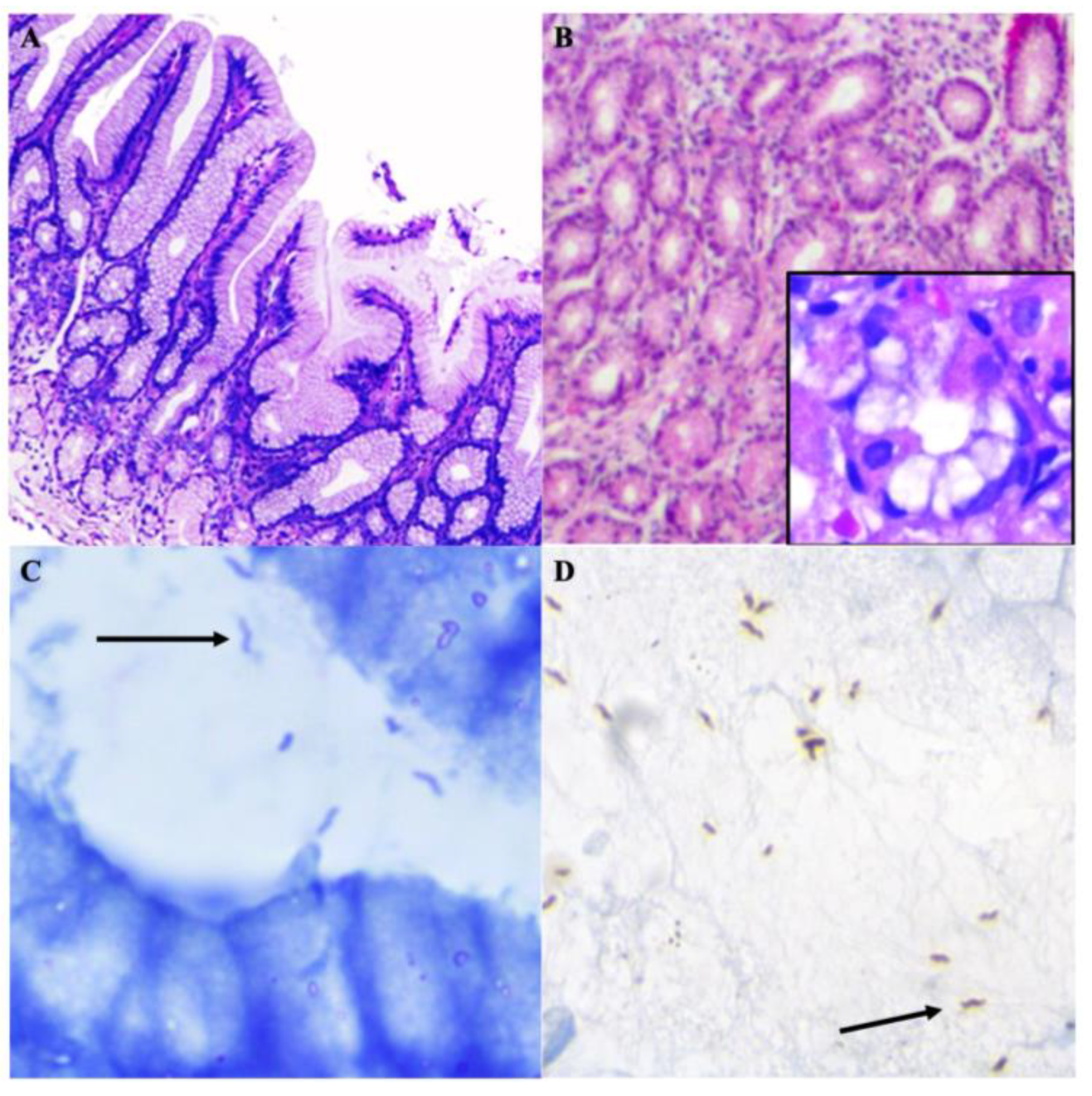
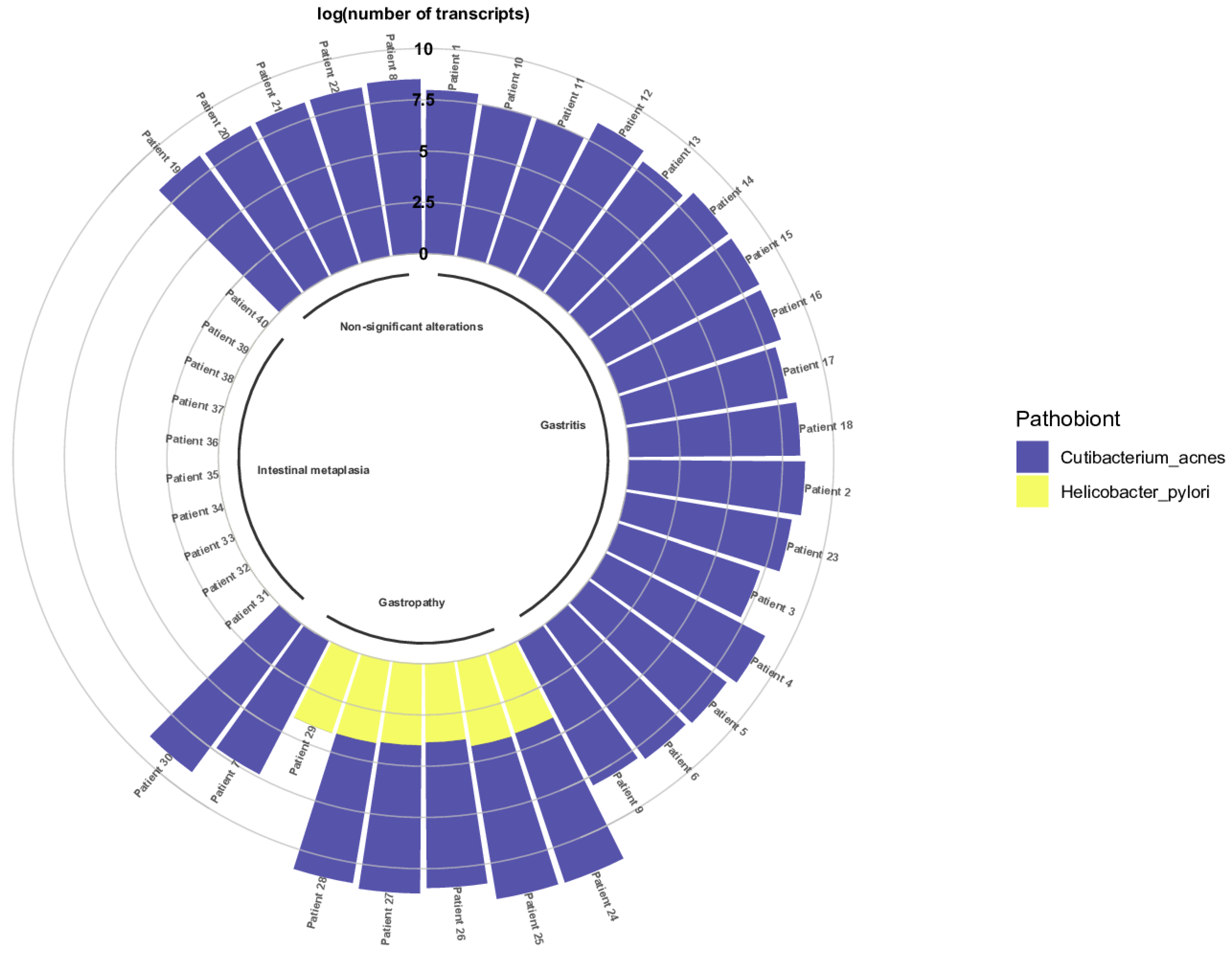
). Helicobacter representative sequences were observed in a posttreatment patient (highlighted with Microorganisms 11 02748 i002
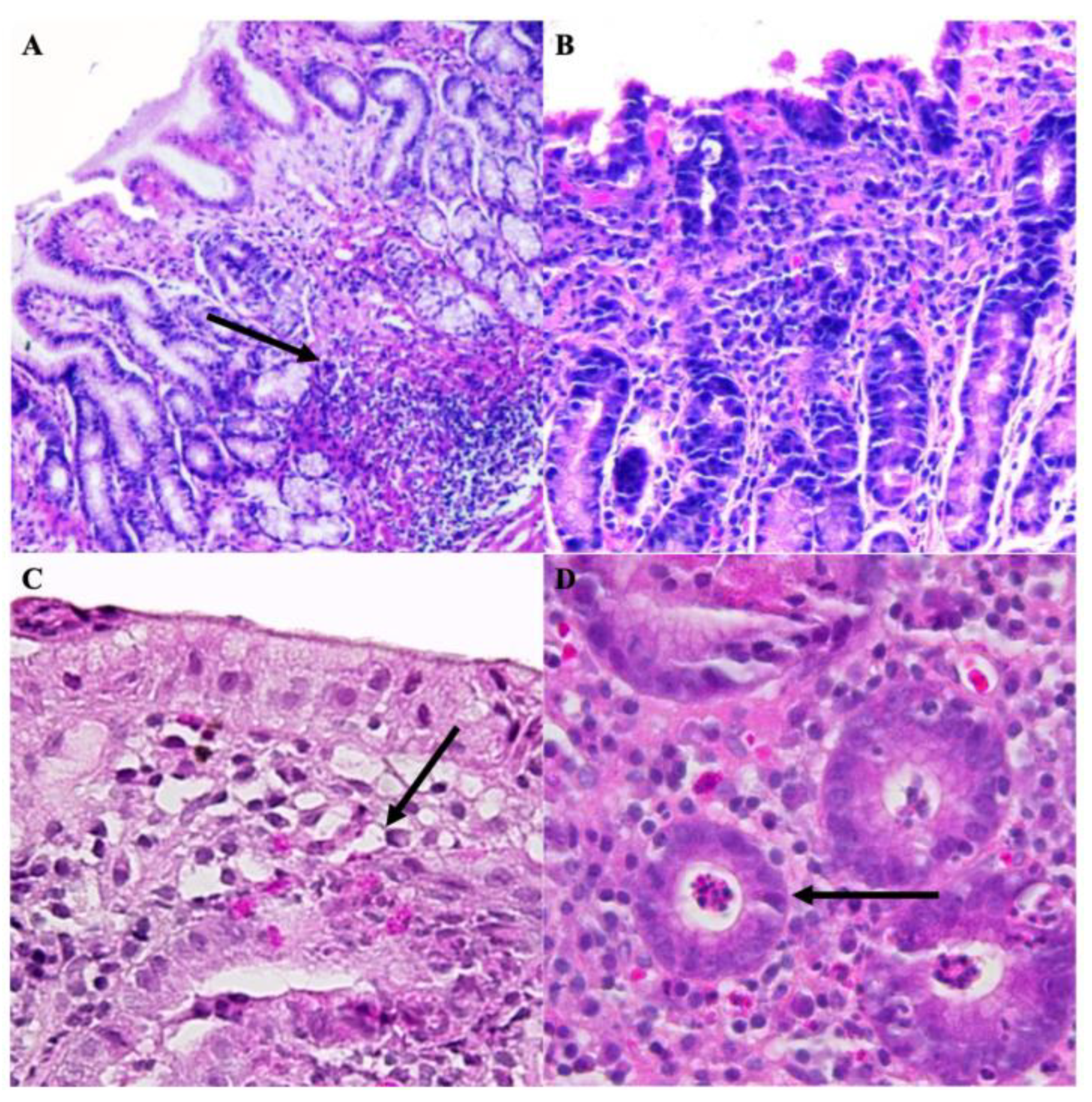
), B: Pre-treatment patient. A: Post-treatment patient.
Microorganisms 11 02748 g004
Figure 5. Alpha- and Beta-diversity indexes. (A) Alpha diversity estimations in pre-treatment and post-treatment patients. (B) Parallel Principal Coordinate Analysis (PCoA) indicates the variation between the pre-treatment and post-treatment microbiota. Axis 1 variation value = 29.65%; Axis 2 variation value = 18.39%; Axis 3 variation value = 14.77%; Axis 4 variation value = 9.933%; Axis 5 variation value = 7.860%.
Microorganisms 11 02748 g005
Figure 6. Effect of the STT on the gastric microbial communities of clinical interest. The heatmap highlights specific bacteria of clinical interest (rows) and the alterations of their relative abundances after the administration of the STT. Metagenomic profiles were clustered according to their own microbial community structure. B: Pre-treatment patient. A: Post-treatment patient.
Microorganisms 11 02748 g006
Figure 7. Presence of C. acnes and H. pylori in patients with gastric diseases. Evaluation of the presence of C. acnes and H. pylori in FFPE gastric biopsies via the determination of the log of the number of transcripts of both bacteria in the gastric biopsy samples.
Microorganisms 11 02748 g007
Table 1. Histopathological evaluation.
Patients
Category1B1A2B2A3B3A4B4A5B5A6B6A
Atrophy100000200110
Mononuclear infiltrates210011322311
Neutrophilic
infiltrates
000000310101
Metaplasia000000000100
Lymphoid follicles000020310100
Glandular atrophy000001110010
Foveolar hyperplasia2
60%
000002
50%
2
60%
01
30%
1
30%
1
30%
Regenerative changes110001210100
Bacillary structures0000101001 *00
B: Pre-treatment patient; A: Post-treatment patient. 0: Absent injure; 1: Minimum injury; 2: Moderate injury; 3: Severe injury. *: Coccoid forms.
Table 2. Identification of H. pylori by microbiological and molecular methods.
PatientPCRUrease
glmMcagAvacA
Before STTAfter STTBefore STTAfter STT
1+ *,c+ c+ c+ *,c
2
3
4+ *,c
5+ *,c+ c+ c+ *,c
6+ *,c+ c+ c
+: Positive test. −: Negative test. C: Identification from culture. *: Identification from gastric biopsy.
Table 3. Estimation of Good’s coverage and Observed OTUs indexes.
PatientGood’s CoverageObserved OTUs
BABA
1112727
2113440
3114229
4113537
5115144
6113143
B: Pre-treatment patient. A: Post-treatment patient.
Table 4. Alpha indexes.
PatientShannon’s (H)Faith’s PDChao1Simpson’s Index
BABABABA
14.2074.3736.86114.00927270.9310.944
24.6334.9798.80220.56634400.9510.965
35.0684.08224.4886.42342290.9670.920
44.8534.67914.98714.85635370.9590.953
55.1045.04614.09724.58951440.9630.959
64.3814.96813.39415.40931430.9390.962
B: Pre-treatment patient. A: Post-treatment patient.
Table 5. Analysis of similarity (ANOSIM) test.
Group 1Group 2nPermutationsRpq
BA129990.0730.2730.273
B: Pre-treatment patients. A: Post-treatment patients.
Table 6. Paired Student’s t test and Wilcoxon signed rank test for beta-diversity analyses.
Good’s
Coverage
Observed OTUsShannon’s (H)Faith’s PDChao1Simpson’s Index
Paired t test
p-value
110.9140.64310.933
Wilcoxon (W)
p-value
10.810.7490.4230.810.936
Table 7. Significance of the number of C. acnes and H. pylori transcripts.
MetricPathobiont
C. acnesH. pylori
Mean192,315,183.0011,538.67
Standard deviation203,649,007.303168.79
n296
Median157,942,606.4011,973.67
P2536,511,873.519478.76
P75230,486,322.7012,922.57
n: sample size; p-value < 0.001.
Table 8. Determination of the significance between the number of C. acnes and H. pylori transcripts in the gastropathy group using the Mann–Whitney U-Test.
Number of Transcripts
(n = 7)
Median25th Percentile75th PercentileMinimumMaximump-Value
C. acnes16,915,313.118,383,738.7436,511,872.510.9048,151,777.410.017
H. pylori11,383.786807.3212,922.570.9016,076.05
n: sample size.
Table 9. Determination of the significance of the presence of C. acnes in the gastric pathologies.
PathologyMedian25th Percentile75th PercentileMinimumMaximump-Value
Gastritis157,942,606.4094,564,890.45272,850,376.4819,229,694.53573,263,008.44<0.001
Gastropathy19,388,023.3012,826,312.7336,511,872.518,383,738.7448,151,777.41
Non-significant
alterations
221,339,080.30202,746,241.91230,486,321.70194,699,912.73318,670,312.02
Intestinal metaplasia<1<1<1<1990,328,266.98
p-value < 0.001.
Disclaimer/Publisher’s Note: The statements, opinions and data contained in all publications are solely those of the individual author(s) and contributor(s) and not of MDPI and/or the editor(s). MDPI and/or the editor(s) disclaim responsibility for any injury to people or property resulting from any ideas, methods, instructions or products referred to in the content.

