Article

Comparative Genomics Reveals Genetic Diversity and Variation in Metabolic Traits in Fructilactobacillus sanfranciscensis Strains

1
School of Life Science, Shanxi University, Taiyuan 030006, China
2
Lesaffre Insituut of Science and Technology, 101 Rue de Menin, 59700 Marc-en-Baroeul, France
3
Advanced Therapies Group, School of Dentistry, Cardiff University, Cardiff CF14 4XY, UK
4
Flanders Research Institute for Agriculture, Fisheries and Food (ILVO), Technology and Food Science Unit, Burgemeester Van Gansberghelaan 92/1, 9820 Merelbeke, Belgium
*
Author to whom correspondence should be addressed.
These authors contributed equally to this work.
Microorganisms 2024, 12(5), 845; https://doi.org/10.3390/microorganisms12050845
Submission received: 8 April 2024 / Revised: 16 April 2024 / Accepted: 18 April 2024 / Published: 23 April 2024
(This article belongs to the Special Issue Food Microorganisms and Genomics)

Abstract

:
Fructilactobacillus sanfranciscensis is a significant and dominant bacterial species of sourdough microbiota from ecological and functional perspectives. Despite the remarkable prevalence of different strains of this species in sourdoughs worldwide, the drivers behind the genetic diversity of this species needed to be clarified. In this research, 14 F. sanfranciscensis strains were isolated from sourdough samples to evaluate the genetic diversity and variation in metabolic traits. These 14 and 31 other strains (obtained from the NCBI database) genomes were compared. The values for genome size and GC content, on average, turned out to 1.31 Mbp and 34.25%, respectively. In 45 F. sanfranciscensis strains, there were 162 core genes and 0 to 51 unique genes present in each strain. The primary functions of core genes were related to nucleotide, lipid transport, and amino acid, as well as carbohydrate metabolism. The size of core genes accounted for 41.18% of the pan-genome size in 14 F. sanfranciscensis strains, i.e., 0.70 Mbp of 1.70 Mbp. There were genetic variations among the 14 strains involved in carbohydrate utilization and antibiotic resistance. Moreover, exopolysaccharides biosynthesis-related genes were annotated, including epsABD, wxz, wzy. The Type IIA & IE CRISPR-Cas systems, pediocin PA-1 and Lacticin_3147_A1 bacteriocins operons were also discovered in F. sanfranciscensis. These findings can help to select desirable F. sanfranciscensis strains to develop standardized starter culture for sourdough fermentation, and expect to provide traditional fermented pasta with a higher quality and nutritional value for the consumers.

1. Introduction

Wheat- and barley-based breads have unique significance in the human diet globally as they are important dietary sources of energy, carbohydrates, and plant proteins [1]. The last decade has witnessed a rapid expansion in China’s bakery sector. The total annual sale of bread from 2015 to 2020, especially packaged and unpackaged leavened bread, reached more than $6 billion, which accounted for 18% of the total sales of China’s bakery industry [2]. Steamed bread is the conventional staple food in China [3], which undergoes fermentation by a traditional starter culture termed sourdough. Archaeological evidence suggests that baking leavened bread using sourdough originated in ancient Egypt 4000–5000 years ago [4,5]. Nowadays, sourdough is widely used in industrial and artisan bakeries in various products ranging from a loaf of bread to crackers, baguettes, pancakes, and pizzas. Sourdoughs are the product of natural or starter culture initiation, which is mainly composed of a mixture of cereal flour and water [6,7], consisting mainly of a microbial ecosystem of lactic acid bacteria (LAB), such as Fructilactobacillus sanfranciscensis (formerly Lactobacillus sanfranciscensis), Lactiplantibacillus plantarum, and Pediococcus pentosaceus, and yeast, such as Saccharomyces cerevisiae and Candida humilis [3,8,9]. Based on the texture, flavor, and nutritional final characteristics of inoculum and baked goods, four distinct types of sourdough can be distinguished: I, II, III, and IV [10].
Fructilactobacillus sanfranciscensis is the most prevalent and functionally important Lactobacillus species in the Type I sourdough. As the native key bacterium of the sourdough ecology [3,11,12], F. sanfranciscensis has always been of great interest to ecologists and microbiologists. F. sanfranciscensis can metabolize carbohydrates (e.g., sucrose or maltose) to produce exopolysaccharides (EPS) and hydrolyze proteins to peptides. These metabolites influence the flavor, nutritional quality, and shelf life of sourdough [3,13]. Interestingly, multiple studies have shown that a large number of F. sanfranciscensis strains are prevalent in different types of sourdoughs from diverse origins [3,14,15,16]. These strains have been shown to interact differentially with prevalent yeast species and other LAB in sourdough [8,17,18].
Researchers have demonstrated that F. sanfranciscensis can be isolated from sourdough and also from the gastrointestinal tract of Drosophila melanogaster, and tea floral pollen [3,19]. Different strains of F. sanfranciscensis have shown various biological and chemical functions of interest to the bakery industry. Corsetti et al. (1996) [20] reported that bacteriocins produced by F. sanfranciscensis C57 displayed effective antibacterial properties. Zhang et al. (2020) [21] studied the physiological and biochemical characteristics of F. sanfranciscensis Ls-1001, which had a strong inhibitory effect against Staphylococcus aureus, Escherichia coli, and other foodborne pathogens. Furthermore, it was discovered that the EPS produced by strain Ls-1001 had vigorous antioxidant activity in vitro [22]. Glutathione reductase activity (thiol-exchange reactions) of F. sanfranciscensis has been shown to reduce wheat germ agglutinin, which is linked to nonCeliac wheat sensitivity [23]. Due to its gastrointestinal resistance at lower pH, GRAS (generally recognized as safe), has proven health benefits, so there is an increasing interest in using F. sanfranciscensis as a probiotic in various foods [24]. To exploit the biological and physiological functions of F. sanfranciscensis, a deeper understanding of genetic variations in this species and the factors governing these variations is mandatory.
A genotype-phenotype study of F. sanfranciscensis has shown several differences in carbohydrate and external electron receptor utilization among different strains of this species [11,17,25]. Rogalski et al. (2020c) [16] also provided powerful evidence for this with the help of comparative genomics supported by physiological data. Baek et al. (2021) [26] proposed that desirable strains of this species should be selected for a standard starter culture based on their phenotypic and physiological characteristics and their ability to interact with other sourdough microbiota. However, selecting desirable strains without the knowledge of genetic drivers remains elusive. Thus, it is important to compare whole genomes of various strains of this species to determine different factors that distinguish strains and regulate the physiology, metabolism, and diversity among F. sanfranciscensis strains. This research provides a solid theoretical basis for selecting desirable strains of F. sanfranciscensis with more significant functional characteristics to enhance fermented baked goods at domestic and commercial levels.

2. Materials and Methods

2.1. Strains, Genome Sequencing and Assembly

Genomes of 45 strains of F. sanfranciscensis were analyzed and compared. Among the 45 F. sanfranciscensis strains, 14 strains were stored at our lab (Laboratory for Exploration and Utilization of Food Microbial Resources, Shanxi University, Taiyuan, China), while 31 strains genomes were obtained from NCBI. All 14 F. sanfranciscensis were stored at −80 °C and preserved in 30% glycerol. Strains were activated in a modified MRS (mMRS) medium [22] 2 times and then cultured at 30 °C under anaerobic conditions. All 14 F. sanfranciscensis strains were sequenced using the Illumina Hiseq × 10 platform (Majorbio BioTech Co., Shanghai, China) using the pair-end library of 400 bp fragments, generating 2 × 150 bp. SOAPdenovo 2.04 software was used to handle the raw sequencing reads for quality assessment [27] and assembly. The assembled fragments were constructed after filling the assembly in undetermined regions using the software of GapCloser 1.12 [27], generating the draft genomes used in subsequent analyses.

2.2. Genomic Characteristics Prediction and Pan-Genome and Core Genes Analysis

The GC content of 45 F. sanfranciscensis strains were determined. Glimmer 3.02 and Prodigal 2.6.3 software were used for coding sequence (CDS) prediction. The sequences were annotated using the Swiss-prot database (accessed on 1 November 2023) and the RefSeq nonredundant protein (NR) database (accessed on 10 November 2023) [28] with the E value set at 1 × 10⁻⁵.
PGAP 1.2.1 software was utilized to analyze the pan-genome size and the clustering of the functional genes [29] using all 45 strains of F. sanfranciscensis. Protein sequences of 45 strains of F. sanfranciscensis were compared using Orthomcl v2.0.9 software to create Venn diagrams based on the obtained data [30]. The clusters of orthologous groups of proteins (COGs) database (accessed on 10 November 2023) was used to classify the proteins encoded genomes.

2.3. Phylogenetic Analysis and Determination of the Average Nucleotide Identity (ANI) Value

The ANI values were calculated using pyani software (accessed on 15 November 2023) based on an average of the comparison of the coding sequences of all lineal homologous proteins between the two genomes, which reflected the evolutionary distance between the genomes. Then these captured data were visualized using heat map tools (https://cloud.majorbio.com/page/tools/, accessed on 18 November 2023). OrthoMCL 2.0.9 software was used to compare and summarize all the homologous genes of 45 strains of F. sanfranciscensis [31,32]. To further investigate the potential evolutionary relationship between these strains, MEGA 7.0 software was used to create phylogenetic trees [33].

2.4. Genotypic and Phenotypic Analysis of Carbohydrate Metabolism

The carbohydrate-active enzymes of all the 14 F. sanfranciscensis strains were categorized and analyzed using the carbohydrate active enzymes database (CAZymes, http://www.cazy.org/, accessed on 22 November 2023), using Hummer and Dianond software. The heat map in the Origin Pro software was used to draw the visual clustering relationship map.
D-glucose, D-fructose, D-trehalose, D-sucrose, D-galactose, L-arabinose provided by Shanghai McLean Biochemical Technology Co., Ltd. (Shanghai, China), and D-lactose, soluble starch, D-maltose, and mannitol provided by Beijing Aobo Star Biotechnology Co., Ltd. (Beijing, China) were used as sole carbon sources to verify the expression of carbohydrate utilization genes in 14 F. sanfranciscensis strains. Ten different carbohydrates were added individually into the mMRS medium at the concentration of 2 g/100 mL followed by the addition of 0.5% (w/v) bromocresol purple solution (provided by Beijing Aobo Star Biotechnology Co., Ltd.) at the ratio of 1.5% (v/v). Culture media were all sterilized at 115 °C for 20 min in an autoclave. The 1% (v/v) activated bacterial cultures were added into the prepared medium containing different carbohydrates respectively and incubated anaerobically at 30 °C for 24 to 48 h. The control medium (without bacterial inoculum) was prepared using the same method. Color changes were observed at 48 h to evaluate the carbohydrate utilization. The growth characteristics strain F. sanfranciscensis Fs_1010 with high EPS yield (202.341 mg/L) using different carbohydrate as carbon source were further studied. For this purpose, the autoclaved mMRS medium was prepared without mono and disaccharides, supplemented with sterile filtered 2 g/100 mL of either glucose, fructose, maltose, and glucose + fructose, respectively. After inoculation at 1% (v/v), the strain was incubated at 30 °C for 48 h while growth was followed by measuring the absorbance values at 600 nm (A600 nm). Concomitantly, to determine the rate of acid production, pH and TTA were measured according to Zhang et al. (2020) [21].

2.5. Genotypic Analysis of EPS-Producing Strains

EPS-producing ability of the 14 F. sanfranciscensis strains was assayed via the phenol–sulfuric acid method [22]. Functional annotation information and blast alignment were used to predict and analyze the genes contributed to EPS synthesis in 14 F. sanfranciscensis strains (e-value was <1 × 10⁻⁵). The Origin Pro 2024 software was used to create a heat map.

2.6. Genotypic and Phenotypic Analysis of Antibiotic Resistance

The antibiotic resistance genes in 14 F. sanfranciscensis strains were analyzed using the CARD database (Comprehensive Antibiotic Resistance Database, http://arpcard.Mcmaster.ca, accessed on 25 December 2023). If the sequence matching degree of the resistance gene reaches 20% (e-value < 1 × 10⁻⁵), the antibiotic resistance gene is considered to exist. The Origin 2023 software was used to build a thermal map of the predicted data.
Based on the American Association for Clinical and Laboratory Standards Institute (CLSI) guidelines [34], the antibiotic resistance of the 14 F. sanfranciscensis strains was tested using the disk diffusion method. The antibiotics per disk were as follows: gentamicin, kanamycin, streptomycin, erythromycin, clindamycin, benzathine, ampicillin, tetracycline, chloramphenicol, and mitomycin-sulfamethoxazole, purchased from Liofilchem. The strains that were classified as susceptible (S, zone diameter > 20), intermediate (IR, 15 < zone diameter < 19), or resistant (R, zone diameter ≤ 14) hinged on the diameter of the zone of inhibition around the disk [30].

2.7. CRISPR Identification and Bacteriocin-Derived Genes Analysis

CRISPRCas Finder [35] (https://crisprcas.i2bc.paris-saclay.fr/CrisprCasFinder/Index, accessed on 1 January 2024) was used to predict the genome of CRISPR/Cas system, including the sequence of DRs (repeat sequence), Spacers (spacer region), and Cas proteins via WebLogo (http://weblogo.berkeley.edu/) for visual conservative repetitive sequencing directly. DRs RNA secondary structure via RNAfold (http://rna.tbi.univie.ac.at/cgi-bin/RNAWebSuite/, accessed on 5 January 2024) was used to forecast the default parameters. Using online tools BAGEL4 (http://bagel5.molgenrug.nl/, accessed on 6 January 2024), the bacterium strain gene prediction was compared to find assume operon gene cluster information.

3. Results

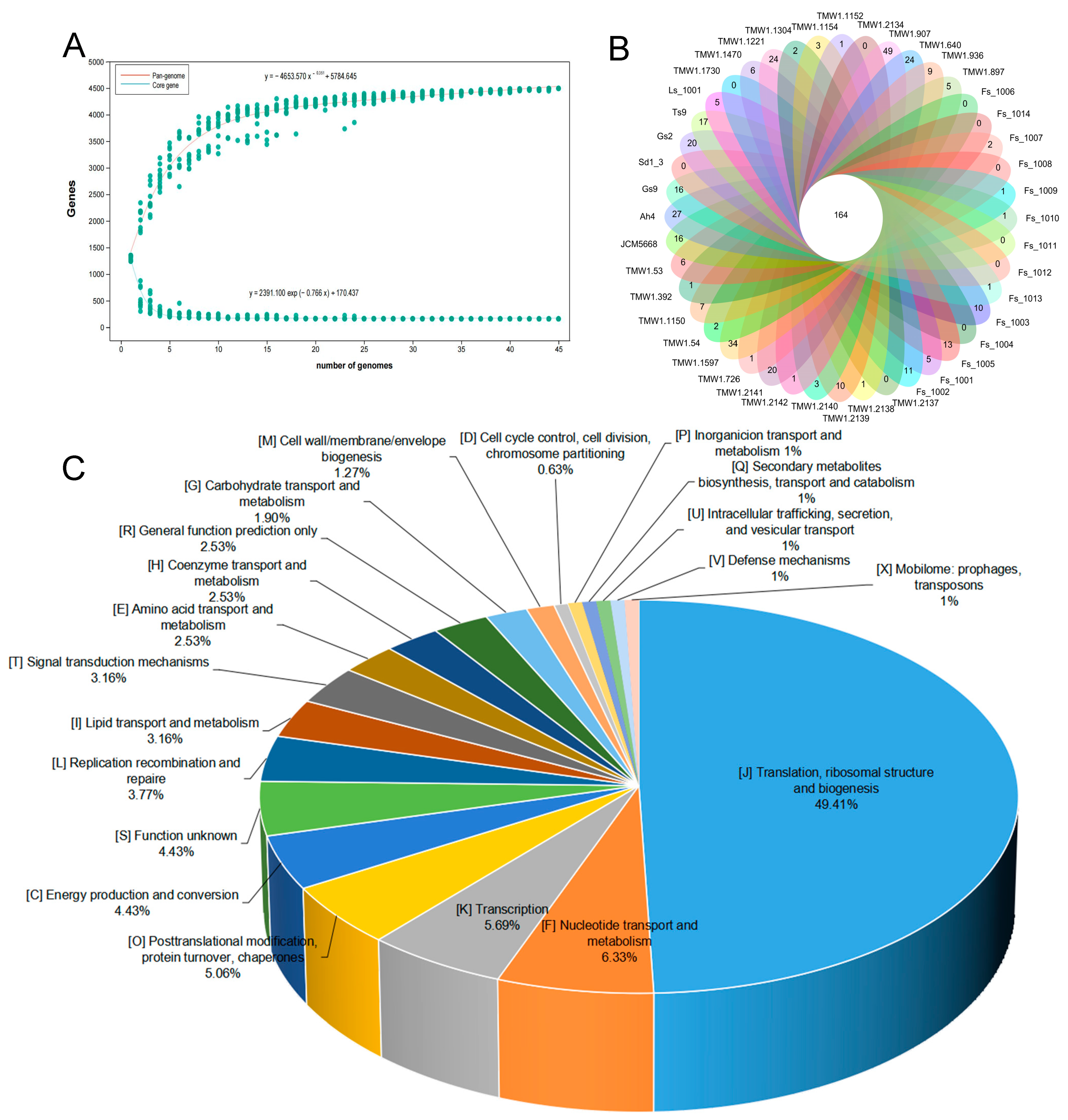
3.1. Genome Characteristics of F. sanfranciscensis

Genomic information of all the above-mentioned strains is listed in Table 1. In this study, the genome size of 45 F. sanfranciscensis strains ranged from 1.26 Mbp to 1.37 Mbp, with an average of 1.31 Mbp. Similarly, the average value of GC content was 34.25%, ranging from 33.07% to 35.20%. The genome was predicted to have an average number of 1274 protein-coding sequences (CDS) that ranged from 1184 to 1386.

3.2. Pan-Genome and Core Genes of F. sanfranciscensis

The basic genomic features of F. sanfranciscensis, pan-genome and core genome analyses were further investigated. The genomic dynamic characteristics between the number of core genes and pan-genes and the sequenced strains are depicted (Figure 1A). When the number of F. sanfranciscensis strains increased, the quantity of pan-genomes expanded further, whereas the core-genome remained at a steady level. After adding the 45th genome, the pan-genome appeared stable with 4494 genes, whereas there were only 159 genes in the core genome. However, the exponent γ value was less than zero, meaning the pan-genome of F. sanfranciscensis was closed. A Venn diagram (Figure 1B) examines the specific core genes and accessory genes of the 45 different F. sanfranciscensis strains. The 45 F. sanfranciscensis strains used in this research had an average total number of homologous gene clusters of 1307 with 12.40% core genes with 0 to 51 unique genes per strain. In 14 F. sanfranciscensis strains, the size of core genes, or 0.70 Mbp of 1.70 Mbp, accounted for 41.18% of the pan-genome size. The functions of the core genes of 45 F. sanfranciscensis are mainly related to nucleotide metabolism, energy production and conversion, lipid transport and metabolism, amino acid metabolism, and carbohydrate metabolism (Figure 1C). Among them, 49.41% (3514/7112) functional genes were related to translation, ribosomal structure and biogenesis, 2.53% (180/7112) were related to amino acid transport and metabolism, and 1.90% (135/7112) were related to carbohydrate transport and metabolism. Furthermore, 4.43% (315/7112) unknown function proteins were discovered in 45 F. sanfranciscensis strains.

3.3. ANI and Phylogenetic Analysis of F. sanfranciscensis

Strains should be considered as the same species when the ANI value exceeds 95% [30,44]. To further explore the relatedness of F. sanfranciscensis strains, the ANI values of 45 F. sanfranciscensis strains were analyzed. Figure 2A shows that the calculation of the ANI values of all 45 strains resulted in 99.75% to 100% similarity. The results showed that all the 45 strains were F. sanfranciscensis and there were no subspecies. Phylogenetic analysis divided the 45 strains into three major genetic clusters based on homologous genes (Figure 2B). In contrast to the other 43 F. sanfranciscensis strains, the strain of F. sanfranciscensis Gs2 and Gs9 was distributed into two different branches, respectively.

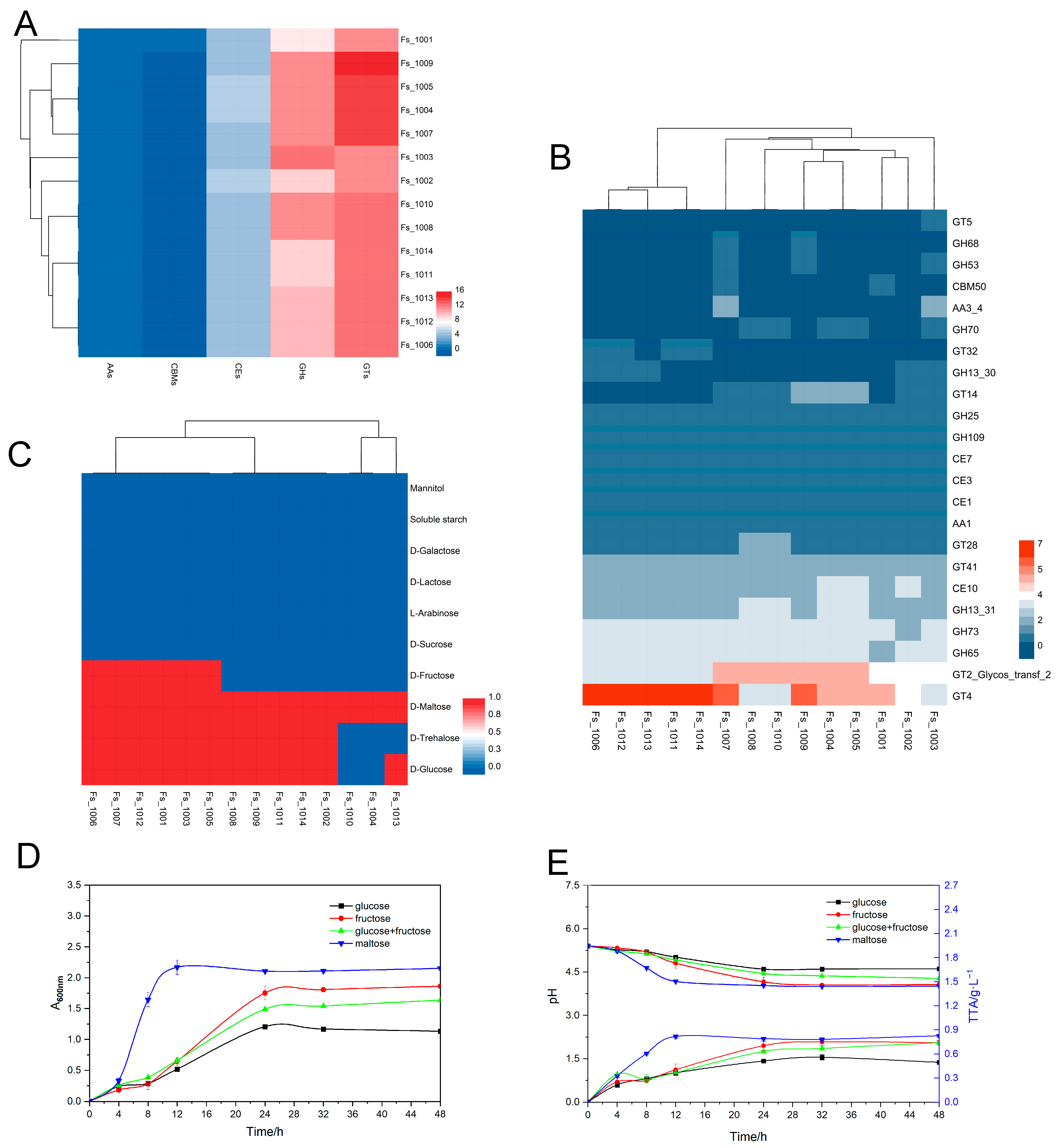
3.4. Genotypic and Phenotypic Analysis of Carbohydrate Metabolism

According to gene functional characterization, numerous genes were involved in carbohydrate transport and metabolism in all strains. Figure 3A,B show that 23 genes encoding carbohydrate-active enzymes were present in the 14 F. sanfranciscensis strains, respectively. The CAZyme-coding genes mainly belonged to glycosyltransferase families (GTs), glycoside hydrolases families (GHs), and carbohydrate esterases (CEs) families, which are responsible for the formation of glycosidic bonds, hydrolysis of rearranged glycosidic bonds, and hydrolysis of carbohydrate esters, respectively. These genes reflect the potential of LAB to synthesize and hydrolyze different carbohydrates. Among them, GT4 (sucrose synthase; EC 2.4.1.13), GH25 (lysozyme; EC 3.2.1.17), GH65 (α, α-alglucan; EC 3.2.1.28), GH109 (α-N-acetylgalactosaminidase; EC 3.2.1.49), and CE10 (carboxyl esterase; EC 3.1.1.3) were more abundant in all the 14 F. sanfranciscensis strains.
By analyzing the 14 F. sanfranciscensis strains’ abilities to utilize carbohydrates, we aimed to determine whether the genotypic classification corresponded to phenotypic characteristics. The results showed that all of these strains were able to utilize D-maltose by expressing genes belonging to family GH65 (maltose phosphorylase; EC 2.4.1.8), but did not utilize soluble starch. In addition, there were differences in the ability of F. sanfranciscensis strains to utilize D-glucose (85.71%), D-trehalose (78.57%), and D-fructose (42.86%). It is worth mentioning that all 14 F. sanfranciscensis strains were unable to utilize common carbon sources in sourdough, such as D-sucrose, although they had the genes of malZ (α-glucosidase; EC:3.2.1.20) which hydrolyzes sucrose to release D-fructose and D-glucose as an endocellular enzyme. Although GH70 (alternate sucrase; EC 2.4.1.140) were present in F. sanfranciscensis (Fs_1003, Fs_1004, Fs_1005, Fs_1008, Fs_10010) indicating their potential to metabolize sucrose efficiently, they failed to metabolize sucrose due to the lack of corresponding sucrase genes (such as sucrose PTS permease, EC 2.7.1.211; sucrase–isomaltase, EC 3.2.1.48 3.2.1.10; etc.). In addition, none of the 14 F. sanfranciscensis strains could utilize these carbohydrates, including D-lactose, D-galactose, L-arabinose, and mannitol.
The above results showed that all 14 F. sanfranciscensis strains could metabolize maltose. However, there was strain variability in the utilization of D-glucose, D-fructose, and D-trehalose (Figure 3C). Notably, the D-glucose utilization capacity of F. sanfranciscensis Fs_1010 were different between the carbohydrate utilization phenotype test (which did not utilize glucose) (Figure 3C) and the growth curve test (which utilized glucose but grew more slowly than other carbon sources) (Figure 3D). The growth density of Fs_1010 during 4 to 12 h of fermentation was the frontrunner when grown in a medium containing maltose (as a single carbon source) than in a medium containing only three other carbon sources. After 12 h of fermentation, the growth density of strain Fs_1010 was relatively higher on fructose as a single carbon source compared to its growth on glucose or a mixture of glucose and fructose. These results were consistent with the growth features of F. sanfranciscensis, preferring fructose-rich substrates.
Acidification is important for improving the characteristics of baked products [45]. The ability of strain Fs_1010 to produce acid during fermentation with different carbon sources was further assessed, as shown in Figure 3E. The results of pH and TTA showed that the acid levels produced by strain Fs_1010 varied with carbon sources. When maltose and fructose were used as the only carbon sources for the fermentation solution for 48 h, the final pH of the solution reached 4.0, but the pH of the medium containing glucose decreased only to 4.6.

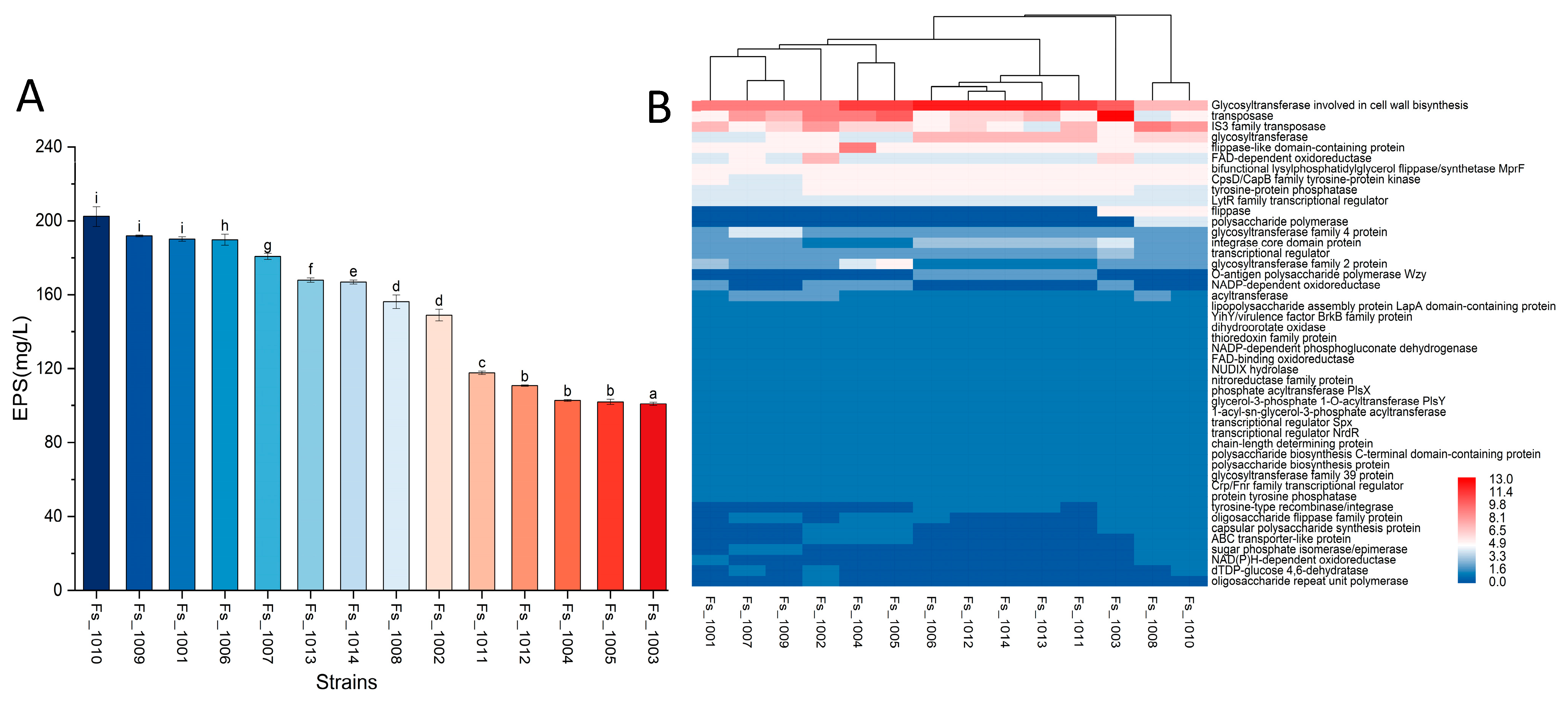
3.5. Genotypic Analysis of EPS

The EPS made by F. sanfranciscensis are recognized to improve the texture, rheology, and shelf-life of bread [3]. In this research, the biosynthesis genes and production of EPS were analyzed. Genes related to EPS biosynthesis were annotated using the NCBI database (https://www.ncbi.nlm.nih.gov/, accessed on 5 March 2024). The genes of epsA, epsB and epsD have been identified, which are responsible for encoding LytR-transcription regulatory factor, tyrosine protein kinase, and tyrosine protein phosphatase respectively (Table 2). These eps genes play an important role in gene expression, signaling pathway regulation and biological activity functions during EPS synthesis. In addition, wzy and wzx genes were identified. The wzy gene encodes a polysaccharide polymerase, which catalyzes the polymerization of sugar residues and extends and forms EPS. The wzx gene promotes the assembly and extracellular transport of EPS. The genomic analysis also revealed many genes encoding glycosyltransferase (GTF) for biosynthesis of EPS repeat units. Other genes related to EPS synthesis in 14 F. sanfranciscensis strains are displayed in Figure 4B. The results revealed that the distribution of the gene of glycosyltransferase was significantly different, whereas the composition of chain-length determining protein and transcriptional regulator was conservative.
Then, we also analyzed the carbohydrate-active enzymes genes related to EPS production across the 14 F. sanfranciscensis strains. The result showed that the production of EPS and the distribution of carbohydrate enzyme genes were different (Figure 4A, Table S1). The genes of GH68 (levansucrase; EC 2.4.1.10) and GH53 (endo-β-1,4-galactanase; EC 3.2.1.89) family were only present in F. sanfranciscensis strains Fs_1007 and Fs_1009, which might suggest that Fs_1009 and Fs 1007 strains were more adept than other strains in sucrose utilization. The high EPS production, 191.9 mg/L, by strain Fs_1009 could be related to its ability to produce levansucrase (Figure 3B), thanks to the gene that encodes inulosucrase (EC:2.4.1.9), which can metabolize sucrose to inulin. The strain Fs_1010, reaching up to 202.3 mg/L of EPS contains glycoside hydrolase family 70 (GH70) genes (dextransucrase; EC 2.4.1.5) that regulate glucan production. The genes from GT5 (α-1,3-glucan synthase; EC 2.4.1.183) only exist in Fs_1003 out of all the 14 F. sanfranciscensis strains.

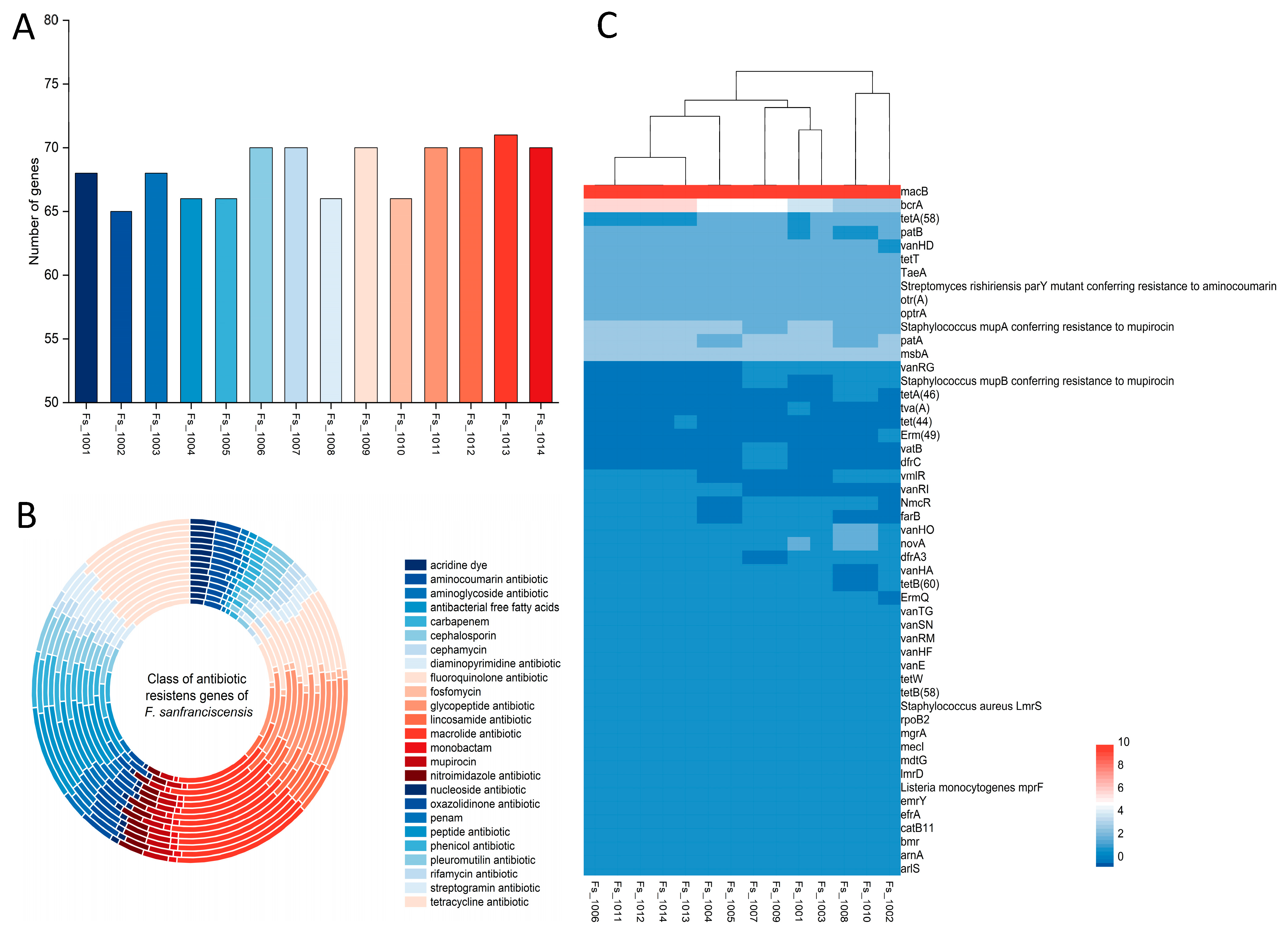
3.6. Phenotypic and Genotypic Analysis of Antibiotic Resistance

Figure 5A shows that 14 F. sanfranciscensis strains had antibiotic resistance genes ranging from 65 to 71. Strain Fs_1002 appeared to contain the fewest antibiotic resistance genes, while Fs_1013 turned out to have the most. The putative antibiotic resistance genes were annotated to 51 antibiotic resistance genes in 14 F. sanfranciscensis strains. All the 14 F. sanfranciscensis strains in our trial contained the types of antibiotic resistance genes of peptide, such as bcrA; macrolide, such as macB; lincosamide, such as lmrD; rifamycin, such as efrA; tetracycline, such as tetT; and glycopeptide, such as vanHD and vanHO (Figure 5B,C). F. sanfranciscensis Fs_1001, Fs_1002, and Fs_1013 strains had the unique distribution of resistance genes, each containing one unique gene, tva (A), Erm (49), and tet (44), respectively, while Fs_1002 was the only one of the 14 strains without ErmQ resistance genes. The dfrC and vatB genes were only present in Fs_1007 and Fs_1009, and tetA (46) was only present in Fs_1008 and FS_1010. However, compared with other F. sanfranciscensis strains, Fs_1001, Fs_1002, and Fs_1013 did not contain the three resistance genes dfrC, vatB, and tetA (46). The antibiotic resistance of the 14 F. sanfranciscensis strains was further determined using the disk diffusion method. As shown in Table 3, the 14 F. sanfranciscensis strains were sensitive to erythromycin (E), clindamycin (CD), ampicillin (AMP), and chloramphenicol (C). However, it showed resistance towards aminoglycosides gentamicin (CN), kanamycin (K), streptomycin (S), and to Trimethoprim-sulfamethoxazole (SXT).
In addition, 14 F. sanfranciscensis strains showed different levels of resistance towards penicillin; oxacilin OXO (low susceptibility) and ampicillin AMP (high susceptibility). Even though tetracycline resistance genes tetA (58), tetT, and tetB (58) were present in all strains, still these three species showed different levels of resistance towards tetracycline TE. Through the CRAD database analysis, the susceptibility of these strains to various antibiotics exhibited substantial variations; the following scenario will explain. Few strains exhibited resistance and had genes corresponding to antibiotic resistance genes that showed resistance (for instance, all of these strains contained Staphylococcus aureus LmrSdu resistance gene that showed resistance to aminoglycosides antibiotics, CK, K, and S). Few strains had the antibiotic resistance phenotype but no associated resistance genes, for instance, all the 14 F. sanfranciscensis strains were resistant to SXT, but no sulfonamide resistance gene could be found. There is another possibility that strains with resistance genes may not exhibit the resistance phenotype. For instance, 14 F. sanfranciscensis strains contained catB11 gene that conferred chloramphenicol resistance but these strains were not resistant to chloramphenicol, suggesting that the gene catB11 was probably not the main gene for chloramphenicol resistance. Moreover, all the 14 F. sanfranciscensis strains contained gene macB that encodes an ABC transporter protein, but the phenotypic test results of all strains did not show resistance to erythromycin (E).

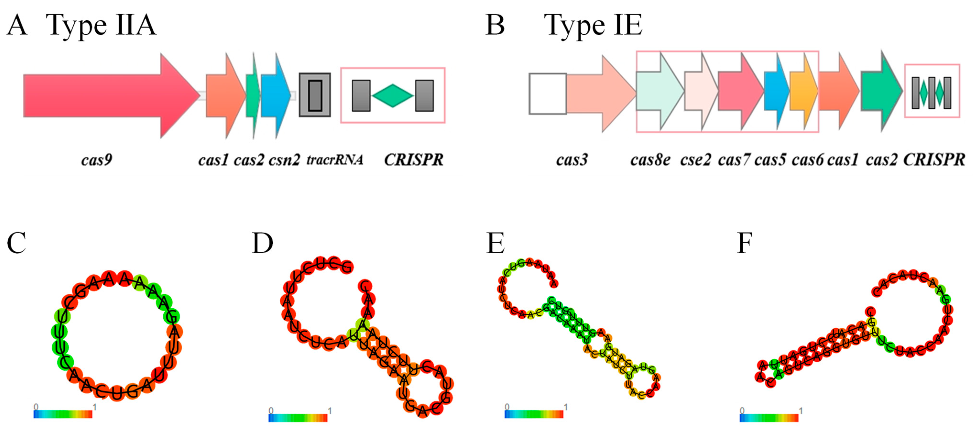
3.7. CRISPR Identification of F. sanfranciscensis

The CRISPR-Cas systems of genome of 45 F. sanfranciscensis strains were analyzed. All 45 strains contained CRISPR sequences, CRISPR sequences without Cas proteins were ignored due to its inability to silence foreign DNA. Therefore, of the 45 F. sanfranciscensis strains, 42 strains contained complete CRISPR-Cas systems (Table S2). The most common Cas gene cluster was Type ⅡA (92.86% of strains), which mainly contained cas9, cas1, cas2, and csn2 genes (Figure 6A). The results showed that Type ⅡA was prevalent in F. sanfranciscensis. In addition, Type IE was also found in 6 F. sanfranciscensis strains (Table S2). Remarkably, F. sanfranciscensis TMW 1.640, Gs2 and Fs_10001 contained both Type ⅡA and Type IE CRISPR-Cas systems (Figure 6A,B). In addition, the RNA secondary structure and the minimum free energy (MFE) of DR sequences were predicted using the RNAfold tool. The results revealed that the secondary structure could be divided into three types of typical structures (Figure 6). The first type was a circular structure (0.00 kcal/mol), which may not have the active ability to silence foreign protein expression; this needs further confirmation. The second structure consisted of a circular structure at the head and tail and a stem-like structure in the middle (−2.70 kcal/mol). Compared with the second structure type, the middle stem structure of the third structure also had the characteristics of conserved RNA secondary structure, i.e., containing one or two small rings and G:U base pairs, corresponding to the minimum free energy of −10.10 kcal/mol and −5.70 kcal/mol, respectively. Such structures are important for CRISPR function.
Furthermore, the genomes of 45 F. sanfranciscensis strains were analyzed to determine the ability of the CRISPR-Cas system to protect against foreign invasion. Among the 45 genomes, 45 phage regions were found, whereas 10 strains had no phages annotated. There were two complete phage regions, one suspicious region, and 42 incomplete regions (Table S3). Only two strains (TMW 1.1597 and TMW 1.907) had complete phage regions, meaning that only 4% of the strains had functional phages. Nonetheless, these results still require further research to better understand the role of the CRISPR-Cas system in F. sanfranciscensis.

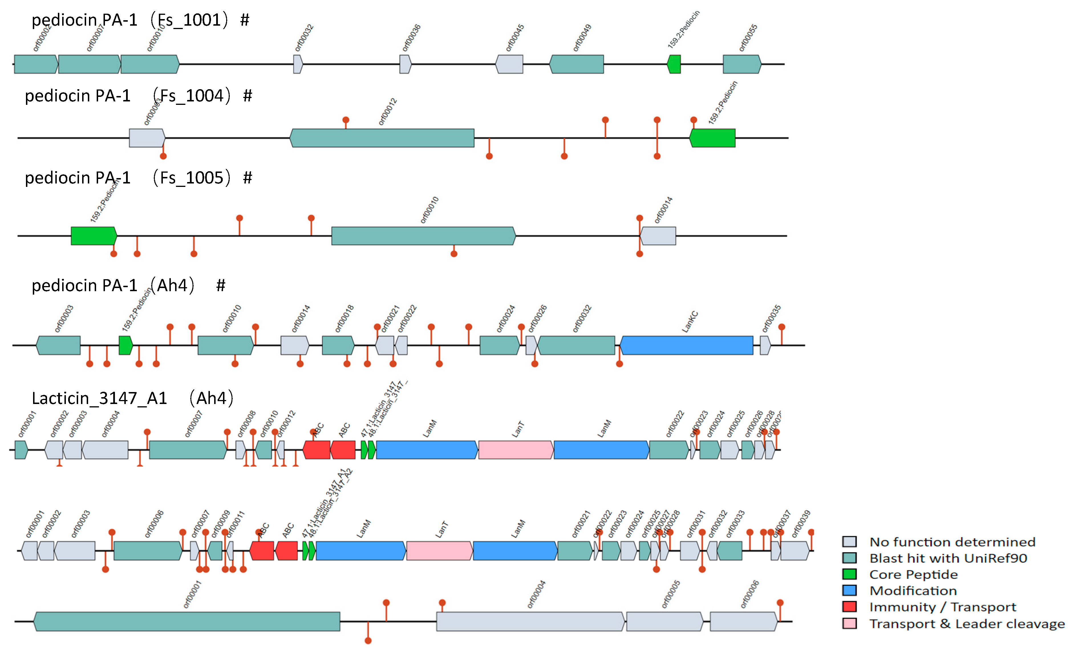
3.8. Bacteriocin Operons Analysis in F. sanfranciscensis

Using the BAGEL4 tool, the bacteriocin operons of 45 F. sanfranciscensis strains were predicted. There were seven bacteriocin operons, five of which have no transporter and one core peptide that has not yet been described (Figure 7). Therefore, these five bacteriocin operons were considered incomplete.
The pediocin PA-1 operon was found in the genomic plasmid sequences of F. sanfranciscensis Fs_1001, Fs_1004, and Fs_1005. The monopeptidycin pediocin PA-1 is one of class IIA bacteriocins showing strong activity against Listeria monocytogenes. In addition to pediocin PA-1, Lacticin_3147_A1 was also found in F. sanfranciscensis Ah4 strain. These findings suggest that Ah4 strains have antimicrobial activity potentiality owing to the bacteriocin operons. However, because some operons were incomplete or core peptides have not yet been described, further research is needed to understand their detailed properties and functions.

4. Discussion

Fructilactobacillus sanfranciscensis is a key bacterial species of the sourdough microbiota which plays an essential role in improving the aroma, texture, and nutritional characteristics of traditional Type I sourdough. Several strains of F. sanfranciscensis with different phenotypic and genotypic characteristics have been reported from various sourdough samples from diverse geographical origins [11]. Moreover, the beneficial effects of F. sanfranciscensis on sourdough is a strain-dependent characteristic [3]. Many phenotypic characteristics of F. sanfranciscensis strains are of industrial interest, acid production capacity, antibacterial capacity, metabolites, etc. Primarily its carbohydrate metabolism has attracted much attention. It is imperative to discriminate strains of F. sanfranciscensis using comparative genomics to formulate effective starter cultures for sourdough-based bread. F. sanfranciscensis strains with a relatively higher ability to ferment maltose are competitive against other strains, showing inverse behavior. F. sanfranciscensis TMW 1.392 was the most common strain due to its ability to utilize different carbohydrates and exploit electron acceptors like oxygen and fructose compared with other F. sanfranciscensis strains in the research of Rogalski et al. (2021) [17].
Whole genome sequences of only a few strains of F. sanfranciscensis are available, limiting the possibilities of genome-based comparative studies. Complete genome sequences are important for the precise description of core genomes, for the identification of strain-specific genes, and for the effective selection of important functional traits among a large number of strains. Therefore, this study analyzed the genomes of 14 F. sanfranciscensis strains isolated from sourdough samples and 31 genomes acquired from the NCBI database. Genotypic and phenotypic characteristics in terms of carbohydrate metabolism and antibiotic resistance were compared and combined with comparative genomics to analyze similarities and differences among the strains.
The small genome size of F. sanfranciscensis indicates that it participates in a limited number of chemical reactions, which can be linked to its successful adaptation towards the sourdough niche. According to streamlining theory in microbial ecology, bacteria with small genomes effectively use nutrients in populations when the adequate population size is large and nutrients limit growth [46]. Microbial niches where adaptation success is determined by resource competition, the phrase of Leonardo da Vinci fits in: ‘simplicity is the ultimate sophistication.’ F. sanfranciscensis has the smallest genome among all lactobacilli, and it has the highest density of ribosomal RNA operons per Mbp, when compared with all known genomes of free-living bacteria in nature [3,11,42]. The aforementioned features are associated with the rapid growth characteristics of the organism [42]. The findings of genomic functional annotation analysis revealed that the large multifunctional genes of F. sanfranciscensis were primarily involved in nucleotide metabolism, energy production and conversion, amino acid metabolism, and carbohydrate metabolism. However, the functional landscape of an entire set of genes has not yet been unveiled because 4.43% of gene functions still need to be determined.
Fructilactobacillus sanfranciscensis strains are known to have variability in genes related to carbohydrate metabolism [11]. This study revealed that the strains had a significant variability in phenotypic characteristics related to carbohydrate metabolism, rather than in genes related to carbohydrate utilization. Based on the carbohydrate utilization phenotypes of 14 F. sanfranciscensis strains, they can be divided into two categories (Figure 3C). There are two groups strains, one can use both maltose and glucose, while another group can only use maltose. The results of this study agree with the result of Rogalski et al. (2020a) [16] and Foschino et al. (2001) [25]. Moreover, 14 F. sanfranciscensis strains were divided into two major clusters based on the family genes number of GT4 and GT2_Glycos_transf_2 (Figure 3B). In this study, 14 F. sanfranciscensis strains were found to utilize maltose, and contained genes of maltose phosphorylase map and phosphoglucomutase pgm. In this study, all 14 F. sanfranciscensis strains contained genes for phosphoketolase pathway from maltose, one of the key carbohydrates in the bread, to produce energy and glucose. And one of the main reasons that F. sanfranciscensis can thrive in sourdough is its ability to utilize maltose more efficiently. This characteristic is also linked to the ability of F. sanfranciscensis to establish a symbiotic relationship with S. cerevisiae, resulting in better adaptation to the sourdough environment [11,17]. There have also been reports of strain-specific interactions between F. sanfranciscensis and the yeasts [17]. S. cerevisiae metabolizes sugars during fermentation to produce significant levels of CO2, which improves the bread volume and crumb texture. Moreover, yeast creates a favorable environment for the growth of anaerobic LAB, such as F. sanfranciscensis. At the same time, F. sanfranciscensis can hydrolyze maltose and proteins through its metabolism to provide glucose and amino acids to baker’s yeast for its enhanced growth [47].
The result may be due to the fact that F. sanfranciscensis takes longer to adapt to growing on glucose, as the genetic setting should allow for all strains that exploit glucose [11,48], such as the genes of glk. In addition, fructokinase exists in all 14 F. sanfranciscensis strains, but there are still eight strains cannot utilize fructose (Figure 3C). The reason for this incapability is the gene of fructokinase in these eight strains which may be not functional. In the literature, it is described that fructose is favored by F. sanfranciscensis for use as an external electron acceptor to increase energy and speed up growth [11,49]. The variations in the sourdough fermentation behavior can be largely attributed to strain-species differences in carbohydrate metabolism of F. sanfranciscensis.
The EPS of LAB have high industrial value, can significantly improve the texture and rheological properties of fermented food, including antioxidant, antibacterial, immune regulation, and other biological activities [50]. Via the analysis of carbohydrate enzyme genes and genes related to EPS synthesis, this article provides valuable insights for the analysis of EPS biosynthesis of F. sanfranciscensis. We identified multiple EPS biosynthesis genes and carbohydrate enzyme genes, including flippase, polymerase, and GTs in 14 F. sanfranciscensis strains. The identification of these genes contributes to further understanding of the molecular mechanisms and regulatory networks involved in the EPS production of F. sanfranciscensis, which has far-reaching implications for optimizing EPS production, modifying EPS structure, and exploring the broad potential of EPS applications.
Globally, food safety is becoming an increasingly important topic while exploring the LAB’s probiotic or food fermentation potential [51]. Therefore, the presence of antibiotic resistance genes and their dissemination potential should be thoroughly evaluated before declaring a bacterial strain safe. Many studies have determined antibiotic resistance of bacteria from fermented foods and probiotic formulations [52,53,54]. The antibiotic susceptibility assessment in this study revealed that all the 14 F. sanfranciscensis strains included multidrug antibiotic resistance patterns. A total of 14 F. sanfranciscensis strains were sensitive to erythromycin (E) and clindamycin (CD), but resistant to aminoglycosides (streptomycin (S), kanamycin (K), and gentamicin (CN), as also presented in the description of current research [34,55]. Membrane impermeability is considered the primary mechanism of aminoglycoside resistance in LAB due to the lack of a cytochrome-related electron transport system that promotes drug uptake [34]. All 14 F. sanfranciscensis strains contained at least one aminoglycoside resistance gene (such as Staphylococcus aureus LmrS, baeS, baeR, aadA23, kdpE), and aminoglycoside antibiotic genes mainly through the efflux of antibiotics to antibiotic resistance. In addition, all 14 F. sanfranciscensis strains contained the macB gene. However, they did not produce resistance to macrolide antibiotics (erythromycin), revealing that macB was not a crucial gene for erythromycin resistance [30]. It was speculated that this resistance gene could be silent in these strains. The macB gene did not have high sequence similarity with the CRAD database (<44.9%). Antibiotic resistance in LAB may be impacted by the expression of antibiotic resistance genes. The 14 strains of F. sanfranciscensis showed various degrees of resistance to the tetracycline analog tetracycline TE. Despite corresponding resistance genes, no risk of horizontal transfer was found in mobile elements such as prophages. Therefore, we assumed that these strain did not pose a safety risk concerning the function or dissemination of gene macB. So, all F. sanfranciscensis strains used in this research were safe regarding antibiotic resistance and can be further applied to make sourdough-based products.
The CRISPR-Cas (clustered regularly interspaced short palindromic repeats, CRISPR-associated proteins) system offers acquired resistance against invasive elements such as phages and plasmids in most bacteria [56,57]. CRISPR consists of a number of short, conserved repeat regions and spacers. Cas, found near the CRISPR site, is a double-stranded DNA nuclease that cuts the target site. Cas functions as a cluster of three to more than 10 genes, which can be divided into six types (I to VI) and more than 30 subtypes [58]. There is species-specific in the distribution of Cas gene clusters. For instance, Type ⅡA mainly exists in the CRISPR system of Pediococcus pentosus, Latilactobacills curvatus, and Lactobacillus reuteri. The TypeIE Cas gene cluster mainly exists in Lactobacillus acidophilus and Lactobacillus ferment. The genome sequence of Lactobacillus generally contains Type II CRISPR-CAS system [59], which is naturally active and effectively targets invasive and genomic DNA. There were 31 strains of F. sanfranciscensis with Type IIA CRISPR-Cas system containing the cas gene (Table S2); Cas9 has the function of targeting and cutting the viral genome and can mediate genome editing. Therefore, F. sanfranciscensis can be used as a candidate for gene editing and provides the basis for cracking lysed phages in the food industry.
Bacteriocins in LAB have great biosafety and wide industrial application [60]. For example, bacteriocins can be used to develop new types of food biological preservatives due to its natural antibacterial activity [61]. In addition, bacteriocins have the potential to tailor host flora and control foodborne pathogens as the relatively narrow and specific killing spectrum [62]. Bacteriocins can also be used as an alternative to antibiotics in animal feed additives [63]. In this research, F. sanfranciscensis Ah4 could be considered the potential candidate as a source of bacteriocins. However, due to the regulatory complexity of bacteriocins in the production process, and that their biosynthesis and transport mechanisms are not fully understood, further research should provide enough experimental evidence to clarify the mechanism and promote upscale industrial application.

5. Conclusions

A comparative genomic analysis of 45 strains of F. sanfranciscensis revealed that the pan-genome of this species was closed. Moreover, the strain-specific differences were primarily reflected in carbohydrate utilization, EPS biosynthesis, antibiotic resistance, and immune/competition related factors (CRISPR-Cas and bacteriocins operon). Additionally, the CRISPR/Cas system was predicted to be of Type IIA and Type IE. There were two bacteriocins operon identified, including pediocin PA-1 and Lacticin_3147_A1. As a result of genotypic and phenotypic characterization, the biotechnological potential of F. sanfranciscensis strains can be further explored and the derived knowledge can be used to develop standardized sourdough starter culture.

Supplementary Materials

The following supporting information can be downloaded at: https://www.mdpi.com/article/10.3390/microorganisms12050845/s1, Table S1: The carbohydrate-active enzymes genes related of exopolysaccharides (EPS) of 14 strains of Fructilactobacillus sanfranciscensis. Table S2: CRISPR-Cas system in Fructilactobacillus sanfranciscensis. Table S3: Prediction of intact prophage regions of Fructilactobacillus sanfranciscensis.

Author Contributions

X.H.: Methodology, Investigation, Conceptualization, Formal analysis, Visualization, Writing—reviewing & editing. Y.Y.: Writing—original draft, Conceptualization, Writing—reviewing & editing. R.K.: Conceptualization, Writing—reviewing & editing. L.J.: Conceptualization, Writing—reviewing & editing. F.A.S.: Investigation, Conceptualization, Writing—reviewing & editing. G.Z.: Conceptualization, Resources, Funding acquisition, Writing—reviewing & editing, Supervision. All authors have read and agreed to the published version of the manuscript.

Funding

This work was supported by the National Natural Science Foundation of China [grant number 32172179].

Data Availability Statement

The genome sequences are available in the NCBI, and Table 1 has listed all strains’ accession numbers.

Conflicts of Interest

The authors declare no conflicts of interest.

References

  1. Poutanen, K.S.; Kårlund, A.O.; Gómez-Gallego, C.; Johansson, D.P.; Scheers, N.M.; Marklinder, I.M.; Eriksen, A.K.; Silventoinen, P.C.; Nordlund, E.; Sozer, N.; et al. Grains—A major source of sustainable protein for health. Nutr. Res. Rev. 2022, 80, 1648–1663. [Google Scholar] [CrossRef]
  2. China’s Rising Bakery Sector. Available online: https://www.fas.usda.gov/data/china-s-rising-bakery-sector (accessed on 3 April 2023).
  3. Zhang, G.H.; Tu, J.; Sadiq, F.A.; Zhang, W.Z.; Wang, W. Prevalence, genetic diversity, and technological functions of the Lactobacillus sanfranciscensis in sourdough: A review. Compr. Rev. Food Sci. Food Saf. 2019, 18, 1209–1226. [Google Scholar] [CrossRef]
  4. Samuel, D. Bread in archaeology. Civilisations 2002, 49, 27–36. [Google Scholar] [CrossRef]
  5. Tamang, J.P.; Cotter, P.D.; Endo, A.; Han, N.S.; Kort, R.; Liu, S.Q.; Mayo, B.; Westerik, N.; Hutkins, R. Fermented foods in a global age: East meets West. Compr. Rev. Food Sci. Food Saf. 2020, 19, 184–217. [Google Scholar] [CrossRef]
  6. Van Kerrebroeck, S.; Comasio, A.; Harth, H.; De Vuyst, L. Impact of starter culture, ingredients, and flour type on sourdough bread volatiles as monitored by selected ion flow tube-mass spectrometry. Food Res. Int. 2018, 106, 254–262. [Google Scholar] [CrossRef]
  7. De Vuyst, L.; Harth, H.; Van Kerrebroeck, S.; Leroy, F. Yeast diversity of sourdoughs and associated metabolic properties and functionalities. Int. J. Food Microbiol. 2016, 239, 26–34. [Google Scholar] [CrossRef] [PubMed]
  8. Landis, E.A.; Oliverio, A.M.; McKenney, E.A.; Nichols, L.M.; Kfoury, N.; Biango-Daniels, M.; Shell, L.K.; Madden, A.A.; Shapiro, L.; Sakunala, S.; et al. The diversity and function of sourdough starter microbiomes. Elife 2021, 10, e61644. [Google Scholar] [CrossRef] [PubMed]
  9. Wang, X.Y.; Huangfu, X.Y.; Zhao, M.Y.; Zhao, R.Y. Chinese traditional sourdough steamed bread made by retarded sponge-dough method: Microbial dynamics, metabolites changes and bread quality during continuous propagation. Food Res. Int. 2023, 163, 112145. [Google Scholar] [CrossRef] [PubMed]
  10. Pérez-Alvarado, O.; Zepeda-Hernández, A.; Garcia-Amezquita, L.E.; Requena, T.; Vinderola, G.; García-Cayuela, T. Role of lactic acid bacteria and yeasts in sourdough fermentation during breadmaking: Evaluation of postbiotic-like components and health benefits. Front. Microbiol. 2022, 13, 969460. [Google Scholar] [CrossRef]
  11. Rogalski, E.; Ehrmann, M.A.; Vogel, R.F. Intraspecies diversity and genome-phenotype-associations in Fructilactobacillus sanfranciscensis. Microbiol. Res. 2020, 243, 126625. [Google Scholar] [CrossRef]
  12. Yang, H.Y.; Liu, T.J.; Zhang, G.H.; Chen, J.C.; Gu, J.S.; Yuan, L.; He, G.Q. Genotyping of Lactobacillus sanfranciscensis isolates from Chinese traditional sourdoughs by multilocus sequence typing and multiplex RAPD-PCR. Int. J. Food Microbiol. 2017, 258, 50–57. [Google Scholar] [CrossRef] [PubMed]
  13. Korakli, M.; Rossmann, A.; Gänzle, M.G.; Vogel, R.F. Sucrose metabolism and exopolysaccharide production in wheat and rye sourdoughs by Lactobacillus sanfranciscensis. J. Agric. Food Chem. 2001, 49, 5194–5200. [Google Scholar] [CrossRef]
  14. Zapparoli, G.; Torriani, S.; Dellaglio, F. Differentiation of Lactobacillus sanfranciscensis strains by randomly amplified polymorphic DNA and pulsed-field gel electrophoresis. FEMS Microbiol. Lett. 1998, 166, 325–332. [Google Scholar] [CrossRef]
  15. Kitahara, M.; Sakata, S.; Benno, Y. Biodiversity of Lactobacillus sanfranciscensis strains isolated from five sourdoughs. Lett. Appl. Microbiol. 2005, 40, 353–357. [Google Scholar] [CrossRef] [PubMed]
  16. Rogalski, E.; Vogel, R.F.; Ehrmann, M.A. Monitoring of Lactobacillus sanfranciscensis strains during wheat and rye sourdough fermentations by CRISPR locus length polymorphism PCR. Int. J. Food Microbiol. 2020, 316, 108475. [Google Scholar] [CrossRef] [PubMed]
  17. Rogalski, E.; Ehrmann, M.A.; Vogel, R.F. Strain-specific interaction of Fructilactobacillus sanfranciscensis with yeasts in the sourdough fermentation. Eur. Food Res. Technol. 2021, 247, 1437–1447. [Google Scholar] [CrossRef]
  18. Boudaoud, S.; Aouf, C.; Devillers, H.; Sicard, D.; Segond, D. Sourdough yeast-bacteria interactions can change ferulic acid metabolism during fermentation. Food Microbiol. 2021, 98, 103790. [Google Scholar] [CrossRef] [PubMed]
  19. Sun, S.K.; Chen, S.L.; Xu, X.P.; Lin, X.Z.; Hu, F.L. Isolation and identification of bacteria from pollen and bee bread. J. Zhejiang Univ. Sci. B 2001, 27, 627–630. Available online: https://www.zjujournals.com/agr/CN/Y2001/V27/I6/627 (accessed on 6 April 2024).
  20. Corsetti, A.; Gobbetti, M.; Smacchi, E. Antibacterial activity of sourdough lactic acid bacteria isolation of a bacteriocin like inhibitory substance from Lactobacillus sanfrancisco. Food Microbiol. 1996, 13, 447–456. [Google Scholar] [CrossRef]
  21. Zhang, G.H.; Zhang, W.Z.; Liang, W.L.; Wang, W.; Tu, J. Studies on identification and characterization of a high-yield exopolysaccharides lactic acid bacteria. J. Chin. Inst. Food Sci. Technol. 2020, 20, 229–238. (In Chinese) [Google Scholar] [CrossRef]
  22. Zhang, G.H.; Zhang, W.Z.; Sun, L.J.; Sadiq, F.A.; Yang, Y.K.; Gao, J.; Sang, Y.X. Preparation screening, production optimization and characterization of exopolysaccharides produced by Lactobacillus sanfranciscensis Ls-1001 isolated from Chinese traditional sourdough. Int. J. Biol. Macromol. 2019, 139, 1295–1303. [Google Scholar] [CrossRef] [PubMed]
  23. Tovar, L.E.R.; Gänzle, M.G. Degradation of wheat germ agglutinin during sourdough fermentation. Foods 2021, 10, 340. [Google Scholar] [CrossRef] [PubMed]
  24. Zhu, W.J.; Lyu, F.Z.; Naumovski, N.; Ajlouni, S.; Ranadheera, C.S. Functional efficacy of probiotic Lactobacillus sanfranciscensis in apple, orange and tomato juices with special reference to storage stability and in vitro gastrointestinal survival. Beverages 2020, 6, 13. [Google Scholar] [CrossRef]
  25. Foschino, R.; Arrigoni, C.; Picozzi, C.; Mora, D.; Galli, A. Phenotypic and genotypic aspects of Lactobacillus sanfranciscensis strains isolated from sourdoughs in Italy. Food Microbiol. 2001, 18, 277–285. [Google Scholar] [CrossRef]
  26. Baek, H.W.; Bae, J.H.; Lee, Y.G.; Kim, S.A.; Min, W.; Shim, S.; Han, N.S.; Seo, J.H. Dynamic interactions of lactic acid bacteria in Korean sourdough during back-slopping process. J. Appl. Microbiol. 2021, 131, 2325–2335. [Google Scholar] [CrossRef] [PubMed]
  27. Luo, R.B.; Liu, B.H.; Xie, Y.L.; Li, Z.Y.; Huang, W.H.; Yuan, J.Y.; He, G.Z.; Chen, Y.X.; Pan, Q.; Liu, Y.J.; et al. SOAPdenovo2 an empirically improved memory-efficient short-read de novo assembler. GigaScience 2012, 1, 2047-2217X-1-2018, Erratum in GigaScience 2015, 4, s13742-015-0069-2. [Google Scholar] [CrossRef] [PubMed]
  28. Pruitt, K.D.; Tatusova, T.; Maglott, D.R. NCBI reference sequences (RefSeq): A curated non-redundant sequence database of genomes, transcripts and proteins. Nucleic Acids Res. 2007, 35, D61–D65. [Google Scholar] [CrossRef]
  29. Zhao, Y.B.; Wu, J.Y.; Yang, J.H.; Sun, S.X.; Xiao, J.F.; Yu, J. PGAP: Pan-genomes analysis pipeline. Bioinformatics 2012, 28, 416–418. [Google Scholar] [CrossRef]
  30. Yu, L.L.; Zang, X.J.; Chen, Y.; Gao, Y.H.; Pei, Z.M.; Yang, B.; Zhang, H.; Narbad, A.; Tian, F.W.; Zhai, Q.X.; et al. Phenotype-genotype analysis of Latilactobacills curvatus from different niches: Carbohydrate metabolism, antibiotic resistance, bacteriocin, phage fragments and linkages with CRISPR-Cas systems. Food Res. Int. 2022, 160, 111640. [Google Scholar] [CrossRef]
  31. Kelleher, P.; Bottacini, F.; Mahony, J.; Kilcawley, K.N.; Van Sinderen, D. Comparative and functional genomics of the Lactococcus lactis taxon; insights into evolution and niche adaptation. BMC Genom. 2017, 18, 267. [Google Scholar] [CrossRef]
  32. Lin, G.P.; Liu, Q.; Wang, L.Y.; Li, H.T.; Zhao, J.X.; Zhang, H.; Wang, G.; Chen, W. The comparative analysis of genomic diversity and genes involved in carbohydrate metabolism of eighty-eight Bifidobacterium pseudocatenulatum isolates from different niches of China. Nutrients 2022, 14, 2347. [Google Scholar] [CrossRef]
  33. Mailund, T.; Brodal, G.S.; Fagerberg, R.; Pedersen, C.N.; Phillips, D. Recrafting the neighbor-joining method. BMC Bioinf. 2006, 7, 29. [Google Scholar] [CrossRef]
  34. Andriani, D.; Hasan, P.N.; Utami, T.; Suroto, D.A.; Rachma, W.; Rahayu, E.S. Genotypic and phenotypic analyses of antibiotic resistance in indonesian indigenous Lactobacillus probiotics. Appl. Food Biotechnol. 2021, 8, 267–274. [Google Scholar] [CrossRef]
  35. Grissa, I.; Vergnaud, G.; Pourcel, C. The CRISPRdb database and tools to display CRISPRs and to generate dictionaries of spacers and repeats. BMC Bioinf. 2007, 8, 172. [Google Scholar] [CrossRef]
  36. Kline, L.; Sugihara, T.F. Microorganisms of the San Francisco sourdough bread process: II. Isolation and characterization of undescribed bacterial species responsible for the souring activity. Appl. Microbiol. 1971, 21, 459–465. [Google Scholar] [CrossRef] [PubMed]
  37. Stolz, P.; Vogel, R.F.; Hammes, W.P. Utilization of electron acceptors by lactobacilli isolated from sourdough. Eur. Food Res. Technol. 1995, 201, 91–96. [Google Scholar] [CrossRef]
  38. Böcker, G.; Stolz, P.; Hammes, W.P. Neue Erkenntnisse zum Ökosystem Sauerteig und zur Physiologie der sauerteigtypischen Stämme Lactobacillus sanfrancisco aund Lactobacillus pontis. Getreide Mehl Brot 1995, 49, 370–374. Available online: https://pascal-francis.inist.fr/vibad/index.php?action=getRecordDetail&idt=2933309 (accessed on 3 April 2024).
  39. Ehrmann, M.A.; Vogel, R.F. Characterisation of IS153, an IS3-family insertion sequence isolated from Lactobacillus sanfranciscensis and its use for strain differentiation. Syst. Appl. Microbiol. 2001, 24, 443–450. [Google Scholar] [CrossRef]
  40. Liske, R.B.; Niessen, L.; Vogel, R.F. Potential of lactic acid bacteria to reduce the growth of Fusarium culmorum in the malting process. Mycotoxin Res. 2000, 16 (Suppl. S1), 62–65. [Google Scholar] [CrossRef]
  41. Rogalski, E.; Ehrmann, M.A.; Vogel, R.F. Role of Kazachstania humilis and Saccharomyces cerevisiae in the strain-specific assertiveness of Fructilactobacillus sanfranciscensis strains in rye sourdough. Eur. Food Res. Technol. 2020, 246, 1817–1827. [Google Scholar] [CrossRef]
  42. Vogel, R.F.; Pavlovic, M.; Ehrmann, M.A.; Wiezer, A.; Liesegang, H.; Offschanka, S.; Voget, S.; Angelov, A.; Böcker, G.; Liebl, W. Genomic analysis reveals Lactobacillus sanfranciscensis as stable element in traditional sourdoughs. Microb. Cell Fact. 2011, 10 (Suppl. S1), S6. [Google Scholar] [CrossRef]
  43. De Angelis, M.; Di Cagno, R.; Gallo, G.; Curci, M.; Siragusa, S.; Crecchio, C.; Parente, E.; Gobbetti, M. Molecular and functional characterization of Lactobacillus sanfranciscensis strains isolated from sourdoughs. Int. J. Food Microbiol. 2007, 114, 69–82. [Google Scholar] [CrossRef]
  44. Chan, J.Z.; Halachev, M.R.; Loman, N.J.; Constantinidou, C.; Pallen, M.J. Defining bacterial species in the genomic era: Insights from the genus Acinetobacter. BMC Microbiol. 2012, 12, 302. [Google Scholar] [CrossRef]
  45. Gänzle, M.G.; Zheng, J. Lifestyles of sourdough lactobacilli-Do they matter for microbial ecology and bread quality? Int. J. Food Microbiol. 2019, 302, 15–23. [Google Scholar] [CrossRef]
  46. Giovannoni, S.J.; Cameron Thrash, J.; Temperton, B. Implications of streamlining theory for microbial ecology. ISME J. 2014, 8, 1553–1565. [Google Scholar] [CrossRef]
  47. Yang, H.Y.; Sadiq, F.A.; Liu, T.J.; Zhang, G.H.; He, G.Q. Use of physiological and transcriptome analysis to infer the interactions between Saccharomyces cerevisiae and Lactobacillus sanfranciscensis isolated from Chinese traditional sourdoughs. LWT–Food Sci. Technol. 2020, 126, 109268. [Google Scholar] [CrossRef]
  48. Stolz, P.; Böcker, G.; Vogel, R.F.; Hammes, W.P. Utilisation of maltose and glucose by lactobacilli isolated from sourdough. FEMS Microbiol. Lett. 1993, 109, 237–242. [Google Scholar] [CrossRef]
  49. Zheng, J.S.; Wittouck, S.; Salvetti, E.; Franz, C.; Harris HM, B.; Mattarelli, P.; O’Toole, P.W.; Pot, B.; Vandamme, P.; Walter, J.; et al. A taxonomic note on the genus Lactobacillus: Description of 23 novel genera, emended description of the genus Lactobacillus Beijerinck 1901, and union of Lactobacillaceae and Leuconostocaceae. Int. J. Syst. Evol. Microbiol. 2020, 70, 2782–2858. [Google Scholar] [CrossRef]
  50. Wu, J.; Han, X.; Ye, M.; Li, Y.; Wang, X.; Zhong, Q. Exopolysaccharides synthesized by lactic acid bacteria: Biosynthesis pathway, structure-function relationship, structural modification and applicability. Crit. Rev. Food Sci. Nutr. 2022, 63, 7043–7064. [Google Scholar] [CrossRef]
  51. Ferri, M.; Ranucci, E.; Romagnoli, P.; Giaccone, V. Antimicrobial resistance: A global emerging threat to public health systems. Crit. Rev. Food Sci. Nutr. 2017, 57, 2857–2876. [Google Scholar] [CrossRef]
  52. Charteris, W.P.; Kelly, P.M.; Morelli, L.; Collins, J.K. Antibiotic susceptibility of potentially probiotic Lactobacillus species. J. Food Prot. 1998, 61, 1636–1643. [Google Scholar] [CrossRef]
  53. Gueimonde, M.; Sánchez, B.; de los Reyes-Gavilán, C.G.; Margolles, A. Antibiotic resistance in probiotic bacteria. Front. Microbiol. 2013, 4, 202. [Google Scholar] [CrossRef]
  54. Zheng, M.; Zhang, R.; Tian, X.; Zhou, X.; Pan, X.; Wong, A. Assessing the risk of probiotic dietary supplements in the context of antibiotic resistance. Front. Microbiol. 2017, 8, 908. [Google Scholar] [CrossRef]
  55. Anisimova, E.A.; Yarullina, D.R. Antibiotic resistance of Lactobacillus strains. Curr. Microbiol. 2019, 76, 1407–1416. [Google Scholar] [CrossRef]
  56. Scaltriti, E.; Carminati, D.; Cortimiglia, C.; Ramoni, R.; Sørensen, K.I.; Giraffa, G.; Zago, M. Survey on the CRISPR arrays in Lactobacillus helveticus genomes. Lett. Appl. Microbiol. 2019, 68, 394–402. [Google Scholar] [CrossRef]
  57. Barrangou, R.; Fremaux, C.; Deveau, H.; Richards, M.; Boyaval, P.; Moineau, S.; Romero, D.A.; Horvath, P. CRISPR provides acquired resistance against viruses in prokaryotes. Science 2007, 315, 1709–1712. [Google Scholar] [CrossRef]
  58. Makarova, K.S.; Wolf, Y.I.; Iranzo, J.; Shmakov, S.A.; Koonin, E.V. Evolutionary classification of CRISPR–Cas systems: A burst of class 2 and derived variants. Nat. Rev. Microbiol. 2019, 18, 67–83. [Google Scholar] [CrossRef]
  59. Sun, Z.; Harris, H.M.B.; Mccann, A.; Guo, C.; Argimón, S.; Zhang, W.; Yang, X.; Jeffery, I.B.; Cooney, J.C.; Kagawa, T.F. Expanding the biotechnology potential of lactobacilli through comparative genomics of 213 strains and associated genera. Nat. Commun. 2015, 6, 8322. [Google Scholar] [CrossRef]
  60. Juturu, V.; Wu, J.C. Microbial production of bacteriocins: Latest research development and applications. Biotechnol. Adv. 2018, 36, 2187–2200. [Google Scholar] [CrossRef] [PubMed]
  61. Ibrahim, S.A.; Ayivi, R.D.; Zimmerman, T.; Siddiqui, S.A.; Altemimi, A.B.; Fidan, H.; Esatbeyoglu, T.; Bakhshayesh, R.V. Lactic acid bacteria as antimicrobial agents: Food safety and microbial food spoilage prevention. Foods 2021, 10, 3131. [Google Scholar] [CrossRef] [PubMed]
  62. Yap, P.G.; Lai, Z.W.; Tan, J.S. Bacteriocins from lactic acid bacteria: Purification strategies and applications in food and medical industries: A review. Beni-Suef Univ. J. Basic Appl. Sci. 2022, 11, 51. [Google Scholar] [CrossRef]
  63. Yang, E.; Fan, L.; Jiang, Y.; Doucette, C.; Fillmore, S. Antimicrobial activity of bacteriocin-producing lactic acid bacteria isolated from cheeses and yogurts. AMB Express 2012, 2, 48. [Google Scholar] [CrossRef]
Figure 1. Pangenome and core genes of Fructilactobacillus sanfranciscensis. (A) Pangenome and core genes of 45 F. sanfranciscensis strains. (B) Venn diagram showing the unique and core genes. (C) The function annotation of core genomes based on COG database.
Microorganisms 12 00845 g001
Figure 2. ANI and phylogenetic analysis of Fructilactobacillus sanfranciscensis. (A) Heat map of 45 strains of F. sanfranciscensis via ANI-TETRA. (B) Phylogenetic tree of 45 F. sanfranciscensis genomes based on homologous genes.
Microorganisms 12 00845 g002
Figure 3. Comparative carbohydrate utilization genotype-phenotype analysis of 14 strains of Fructilactobacillus sanfranciscensis. (A): Heat map of gene number distribution of five carbohydrate-active enzymes in different strains. (B): Heat map of genes associated with carbohydrate-active enzymes in strains. (C): Carbohydrate utilization capacity of different strains. (D): Growth curve of F. sanfranciscensis Fs_1010 on different carbon sources. (E): Acid production performance of F. sanfranciscensis Fs_1010 on different carbon sources, The declining trend line graph represents the changes in pH at different fermentation times. The upward trend line graph represents the changes in total acidity (TTA) at different fermentation times.
Microorganisms 12 00845 g003
Figure 4. The exopolysaccharides (EPS) production and genes associated with regulating EPS synthesis of 14 Fructilactobacillus sanfranciscensis strains. (A) The EPS production of 14 F. sanfranciscensis strains. Different lowercase letters (a–i) indicated a significant difference (p < 0.05). (B) Heat map of genes associated with synthetic EPS genes in 14 F. sanfranciscensis strains.
Microorganisms 12 00845 g004
Figure 5. Comparative genotypic and phenotypic analysis of antibiotic resistance in 14 strains of Fructilactobacillus sanfranciscensis. (A) Number of resistance genes present in different strains. (B) Classification of antibiotic resistance genes of 14 strains of F. sanfranciscensis strains, from inside to out, in order of strain number from smallest to largest. (C): Heat map analysis of antibiotic resistance genes of different strains.
Microorganisms 12 00845 g005
Figure 6. Prediction of Cas protein and RNA secondary structure of CRISPR DR in Fructilactobacillus sanfranciscensis. (A,B) are the type of the CRISPR/Cas locus of F. sanfranciscensis, respectly. The prediction of RNA secondary structure of CRISPR DR in F. sanfranciscensis (CF).
Microorganisms 12 00845 g006
Figure 7. The potential bacteriocin operons identified in Fructilactobacillus sanfranciscensis. “#” indicate lack of core peptide and transporter, respectively.
Microorganisms 12 00845 g007
Table 1. Information of 45 strains of Fructilactobacillus sanfranciscensis.
OrganismStrainAccession No. Isolation
Source
Genome Size (bp)GC Content (%)CDS
Coding (Total)
References
Fructilactobacillus sanfranciscensisDSM20541; TMW 1.53MIYJ00000000 Sourdough, USA; San Francisco sourdough1,295,53934.761221[36]
Fructilactobacillus sanfranciscensisTMW 1.54NZ_MIYE01000000 Rye sourdough, Germany 1,308,58434.621225[37]
Fructilactobacillus sanfranciscensisTMW 1.392NZ_MIYH01000000 Sourdough, Freising Germany1,262,09334.491185[38]
Fructilactobacillus sanfranciscensisTMW 1.640SCEZ00000000Wheat sourdough, Switzerland 1,297,10834.851243[39]
Fructilactobacillus sanfranciscensisTMW 1.726 NZ_MIYD01000000 Sourdough, Italy1,253,14934.671184[40]
Fructilactobacillus sanfranciscensisTMW 1.897 SCEP00000000 Sourdough, Greece; Athens1,249,68734.601209[41]
Fructilactobacillus sanfranciscensisTMW 1.907SCEY00000000Sourdough, Greece; Athens1,281,50034.691257[16]
Fructilactobacillus sanfranciscensisTMW 1.936SCEX00000000 Sourdough, Greece; Athens1,242,51234.741190[16]
Fructilactobacillus sanfranciscensisTMW 1.1150 NZ_MIYG01000000 Sourdough, Germany1,281,79134.691195[16]
Fructilactobacillus sanfranciscensisTMW 1.1152SCEV00000000Sourdough, USA 1,253,07634.581186[16]
Fructilactobacillus sanfranciscensisTMW 1.1154SCEU00000000 Sourdough, USA 1,242,51634.521191[16]
Fructilactobacillus sanfranciscensisTMW 1.1221 SCET00000000Sourdough, France 1,256,83034.481203[16]
Fructilactobacillus sanfranciscensisTMW 1.1304SCES00000000 Rye sourdough, Germany 1,309,93634.591280[42]
Fructilactobacillus sanfranciscensisTMW 1.1470 SCER00000000 Sourdough, Russia 1,264,12034.551211[16]
Fructilactobacillus sanfranciscensisTMW 1.1597 NZ_MIYF01000000Rye sourdough, Germany 1,318,23034.811232[16]
Fructilactobacillus sanfranciscensisTMW 1.1730 SCEQ00000000 Sourdough, Germany1,307,87434.601279[16]
Fructilactobacillus sanfranciscensisTMW 1.2137 NZ_MIXX01000000 Sourdough, Italy 1,272,75334.731210[43]
Fructilactobacillus sanfranciscensisTMW 1.2138NZ_MIXY01000000Sourdough, Italy 1,252,72034.681190[43]
Fructilactobacillus sanfranciscensisTMW 1.2139NZ_MIXZ01000000 Sourdough, Italy 1,329,22834.771256[43]
Fructilactobacillus sanfranciscensisTMW 1.2140NZ_MIYA01000000Sourdough, Italy 1,293,14434.711201[43]
Fructilactobacillus sanfranciscensisTMW 1.2141NZ_MIYB01000000 Sourdough, Italy 1,309,59434.711241[43]
Fructilactobacillus sanfranciscensisTMW 1.2142 NZ_MIYC01000000 Sourdough, Italy 1,278,40934.711212[43]
Fructilactobacillus sanfranciscensisTMW 1.2134SCEW00000000.1Rye sourdough, Germany 1,254,13834.461230[16]
Fructilactobacillus sanfranciscensisLS451NZ_CP045563.1San Francisco sourdough; South Korea1,310,99135.151318(Korea University; 3 November 2019)
Fructilactobacillus sanfranciscensisJCM 5668NZ_QRFO00000000San Francisco sourdough1,267,44834.821318(ChunLab; 2 August 2018)
Fructilactobacillus sanfranciscensisLs-1001NZ_RPFX00000000Sourdough, Shanxi China1,349,33134.491386(our lab; 20 May 2018)
Fructilactobacillus sanfranciscensisAh4NZ_QFCR00000000.1 Sourdough, Anhui, China 1,368,47634.681408(our lab; 20 May 2018)
Fructilactobacillus sanfranciscensisGs2NZ_QGEE00000000.1Sourdough, Gansu China1,373,33234.531401(our lab; 20 May 2018)
Fructilactobacillus sanfranciscensisGs9NZ_QGEF00000000.1 Sourdough, Gansu China1,365,82234.431400(our lab; 20 May 2018)
Fructilactobacillus sanfranciscensisTs9GCA_006334515.1Sourdough, Shanxi China1,325,80734.781386(our lab; 20 May 2018)
Fructilactobacillus sanfranciscensisSd1_3NZ_QGHM00000000.1 Sourdough, Shandong China1,304,53534.551335(our lab; 20 May 2018)
Fructilactobacillus sanfranciscensisFs_1001SAMN32652484Sourdough, China1,341,61834.721367(our lab; this study; 25 January 2023)
Fructilactobacillus sanfranciscensisFs_1002SAMN32652485Sourdough, China1,318,49534.611342(our lab; this study; 25 January 2023)
Fructilactobacillus sanfranciscensisFs_1003SAMN32652486Sourdough, China1,328,17434.691368(our lab; this study; 25 January 2023)
Fructilactobacillus sanfranciscensisFs_1004SAMN32652487Sourdough, China1,333,88334.751353(our lab; this study; 25 January 2023)
Fructilactobacillus sanfranciscensisFs_1005SAMN32652488Sourdough, China1,351,17934.771374(our lab; this study; 25 January 2023)
Fructilactobacillus sanfranciscensisFs_1006SAMN32652489Sourdough, China1,299,07434.571336(our lab; this study; 25 January 2023)
Fructilactobacillus sanfranciscensisFs_1007SAMN32652490Sourdough, China1,293,76634.601315(our lab; this study; 25 January 2023)
Fructilactobacillus sanfranciscensisFs_1008SAMN32652491Sourdough, China1,305,97434.701329(our lab; this study; 25 January 2023)
Fructilactobacillus sanfranciscensisFs_1009SAMN32652492Sourdough, China1,292,33034.591315(our lab; this study; 25 January 2023)
Fructilactobacillus sanfranciscensisFs_1010SAMN32652493Sourdough, China1,302,25434.701324(our lab; this study; 25 January 2023)
Fructilactobacillus sanfranciscensisFs_1011SAMN32652494Sourdough, China1,300,22634.581336(our lab; this study; 25 January 2023)
Fructilactobacillus sanfranciscensisFs_1012SAMN32652495Sourdough, China1,302,32834.591339(our lab; this study; 25 January 2023)
Fructilactobacillus sanfranciscensisFs_1013SAMN32652496Sourdough, China1,302,21634.621338(our lab; this study; 25 January 2023)
Fructilactobacillus sanfranciscensisFs_1014SAMN32652497Sourdough, China1,300,97234.581332(our lab; this study; 25 January 2023)
Table 2. Identified EPS biosynthesis genes in the Fructilactobacillus sanfranciscensis genome.
Predicted GeneNR HitNR Description
epsAEKK20066.1Cell envelope-associated transcriptional attenuator LytR-CpsA-Psr
POH10471.1LytR family transcriptional regulator
epsBWP_041817972.1CpsD/CapB family tyrosine-protein kinase
AEN99639.1Putative tyrosine-protein kinase capB
epsDMVF15937.1tyrosine-protein phosphatase
wzxWP_103429181.1bifunctional lysylphosphatidylglycerol flippase/synthetase MprF
WP_046041031.1flippase
WP_014082292.1flippase-like domain-containing protein
WP_014081504.1oligosaccharide flippase family protein
wzyWP_139571209.1polysaccharide polymerase
WP_198988114.1O-antigen polysaccharide polymerase Wzy
rgpI, waaBWP_139571129.1Glycosyltransferase
Table 3. Antibiotic resistance of 14 strains of Fructilactobacillus sanfranciscensis.
AntibioticsAminoglycolsidesSulfa-MidoTetra-
Cycline
PenicillinsChloram-PhenicolLinco-
Samides
Macro
Lide
Strains CNKSSXTTEOXOAMPCCDE
Fs_1001RRRRIRIRSSSS
Fs_1002RRRRIRSSSSS
Fs_1003RRRRRSSSSS
Fs_1004RRRRIRIRSSSS
Fs_1005RRRRRSSSSS
Fs_1006RRRRRSSSSS
Fs_1007RRRRRRSSSS
Fs_1008RRRRRIRSSSS
Fs_1009RRRRRSSSSS
Fs_1010RRRRIRSSSSS
Fs_1011RRRRRSSSSS
Fs_1012RRRRRSSSSS
Fs_1013RRRRIRIRSSSS
Fs_1014RRRRRIRSSSS
Gentamicin, 10 μg (CN); Kanamycin, 30 μg (K); Streptomycin, 10 μg (S); Erythromycin, 15 μg (E); Clindamycin, 2 μg (CD); Oxacilin, 1 μg (OXO); Ampicillin, 10 μg (AMP); Tetracycline, 30 μg (TE); Chloramphenicol, 30 μg (C); Trimethoprim-sulfamethoxazole, 25 μg (SXT). The antibiotic resistance of the 14 F. sanfranciscensis strains were classified as susceptible (S, zone diameter > 20), intermediate (IR, 15 < zone diameter < 19), or resistant (R, zone diameter ≤ 14).
Disclaimer/Publisher’s Note: The statements, opinions and data contained in all publications are solely those of the individual author(s) and contributor(s) and not of MDPI and/or the editor(s). MDPI and/or the editor(s) disclaim responsibility for any injury to people or property resulting from any ideas, methods, instructions or products referred to in the content.

