Re-Evaluating the Age of Deep Biosphere Fossils in the Lockne Impact Structure
by
 , and
, and
Mikael Tillberg
1,2,*, 
Magnus Ivarsson
3,4,
Henrik Drake
1,
Martin J. Whitehouse
5,
Ellen Kooijman
5 and
Melanie Schmitt
5 1
Department of Biology and Environmental Science, Linnaeus University, 392 31 Kalmar, Sweden
2
Department of Earth Sciences, University of Gothenburg, Box 460, 40530 Göteborg, Sweden
3
Department of Biology, University of Southern Denmark, Campusvej 55, 5230 Odense, Denmark
4
Department of Paleobiology, Swedish Museum of Natural History, Box 50 007, 104 05 Stockholm, Sweden
5
Department of Geosciences, Swedish Museum of Natural History, Box 50 007, 104 05 Stockholm, Sweden
*
Author to whom correspondence should be addressed.
Geosciences 2019, 9(5), 202; https://doi.org/10.3390/geosciences9050202
Submission received: 15 March 2019
/
Revised: 26 April 2019
/
Accepted: 4 May 2019
/
Published: 7 May 2019
(This article belongs to the Special Issue Tracking the Deep Biosphere through Time)
Abstract
:Impact-generated hydrothermal systems have been suggested as favourable environments for deep microbial ecosystems on Earth, and possibly beyond. Fossil evidence from a handful of impact craters worldwide have been used to support this notion. However, as always with mineralized remains of microorganisms in crystalline rock, certain time constraints with respect to the ecosystems and their subsequent fossilization are difficult to obtain. Here we re-evaluate previously described fungal fossils from the Lockne crater (458 Ma), Sweden. Based on in-situ Rb/Sr dating of secondary calcite-albite-feldspar (356.6 ± 6.7 Ma) we conclude that the fungal colonization took place at least 100 Myr after the impact event, thus long after the impact-induced hydrothermal activity ceased. We also present microscale stable isotope data of 13C-enriched calcite suggesting the presence of methanogens contemporary with the fungi. Thus, the Lockne fungi fossils are not, as previously thought, related to the impact event, but nevertheless have colonized fractures that may have been formed or were reactivated by the impact. Instead, the Lockne fossils show similar features as recent findings of ancient microbial remains elsewhere in the fractured Swedish Precambrian basement and may thus represent a more general feature in this scarcely explored habitat than previously known.
1. Introduction
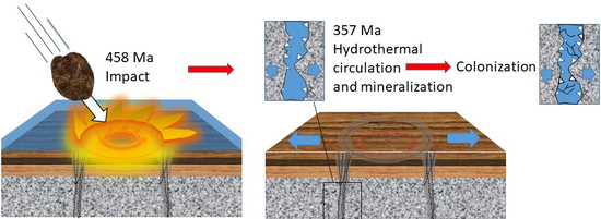
Impact craters and associated impact-generated hydrothermal systems have been suggested as favourable environments for microbial life [1,2,3,4]. Extensive fracturing at depth caused by impacts provides pore space for endolithic communities, and heat generated by the impact drives hydrothermal convection favourable for deep ecosystems [5,6]. Depending on the size and extension of the impact melt, heat can sustain for hundreds of thousands, up to millions of years [7,8,9,10,11]. A handful of reports of fossil- and geochemical signatures support post-impact colonization of impact paleo-hydrothermal systems. Sulfur isotope signatures of sulfates from the Haughton crater, Canada, suggest rapid colonization of the impact-generated hydrothermal system by sulfate-reducing bacteria [12]. Tubular ichnofossils in impact glass from the Ries crater, Germany, indicate microbial activity in a post-impact hydrothermal system [13]. The presence of putative fossilized microorganisms in samples from the Dellen crater in Sweden [14] as well as fossilized biofilm in the Siljan crater [15], suggests colonization of the hydrothermal systems, respectively. Exceptionally well preserved fungal fossils were described in drill cores at a depth of ~170 to 220 meters below the surface from the Lockne crater (458 Ma), Sweden [16]. The fungal fossils were preserved as carbonaceous matter, and displayed characteristic fungal morphologies such as repetitive septa, frequent branching and anastomosis between branches resulting in mycelium-like communities. The close association with oils and minerals interpreted as products of the hydrothermal activity was used to relate the fossils to the post-impact hydrothermal system, presumed to have prevailed about 10,000 years after the impact, and thus indirectly providing an age constraint for the fossils.
With the exception of the subseafloor igneous crust, the fossil archive of the deep biosphere is generally less explored as its living and modern counterpart [17] and age determination of those fossils is often associated with large uncertainties. Commonly, ages of fossils are associated with the radiometric age of the host rock and/or to major tectonic events affecting the rock, which, however, cannot directly be assigned to microbial colonization and subsequent fossilization [18]. In recent years, application of in situ radiometric dating of authigenic minerals produced by the ancient microbial community have been demonstrated to be successful in gaining time constraints on the colonization, both by utilization of U-Pb dating of secondary carbonate [19] and Rb/Sr dating of secondary calcite/fluorite-feldspar and calcite-clay minerals [19,20,21,22]. In situ stable carbon isotope analysis of secondary calcite (δ13C), sulfide and sulfate (δ34S, mainly for pyrite, but also chalcopyrite and barite) have been used to understand the microbial processes (e.g., methane oxidation, methanogenesis and microbial sulfate reduction) in deep fracture systems within the continental crystalline crust [23,24,25,26].
Here, we present in situ dating of secondary vein minerals associated with the Lockne fungal fossils [10], which challenges the fossil association with the impact-generated system, and suggests that fracture-reactivation due to hydrothermal activity occurred in the fracture system long after the impact (100 Ma) and thus infers a previously unknown, younger maximum age, for the fossils. In addition, we present microscale stable isotope data of 13C-enriched calcite suggesting the presence of methanogens contemporary with the fungi. Collectively, the Lockne community bears close similarities to other fossil fungal-prokaryotic consortia described in Swedish crystalline basement [23,27].
2. Geological Setting
The Lockne impact crater, Sweden, is a concentric structure with an inner crater diameter of 7.5 km developed in the crystalline basement. The oldest post-impact sediments have been dated at 458 Ma [28]. At the time of impact, the area was covered by a more than 500 m deep sea [29]. The targets were the 1.86–1.85 Ga Revsund granitoids [30] and the 1250–1200 Ma alkali- and olivine-rich Åsby dolerite [31] overlain by a sedimentary cover of Cambrian bituminous black mud (today alum shale) and Ordovician consolidated limestone [32].
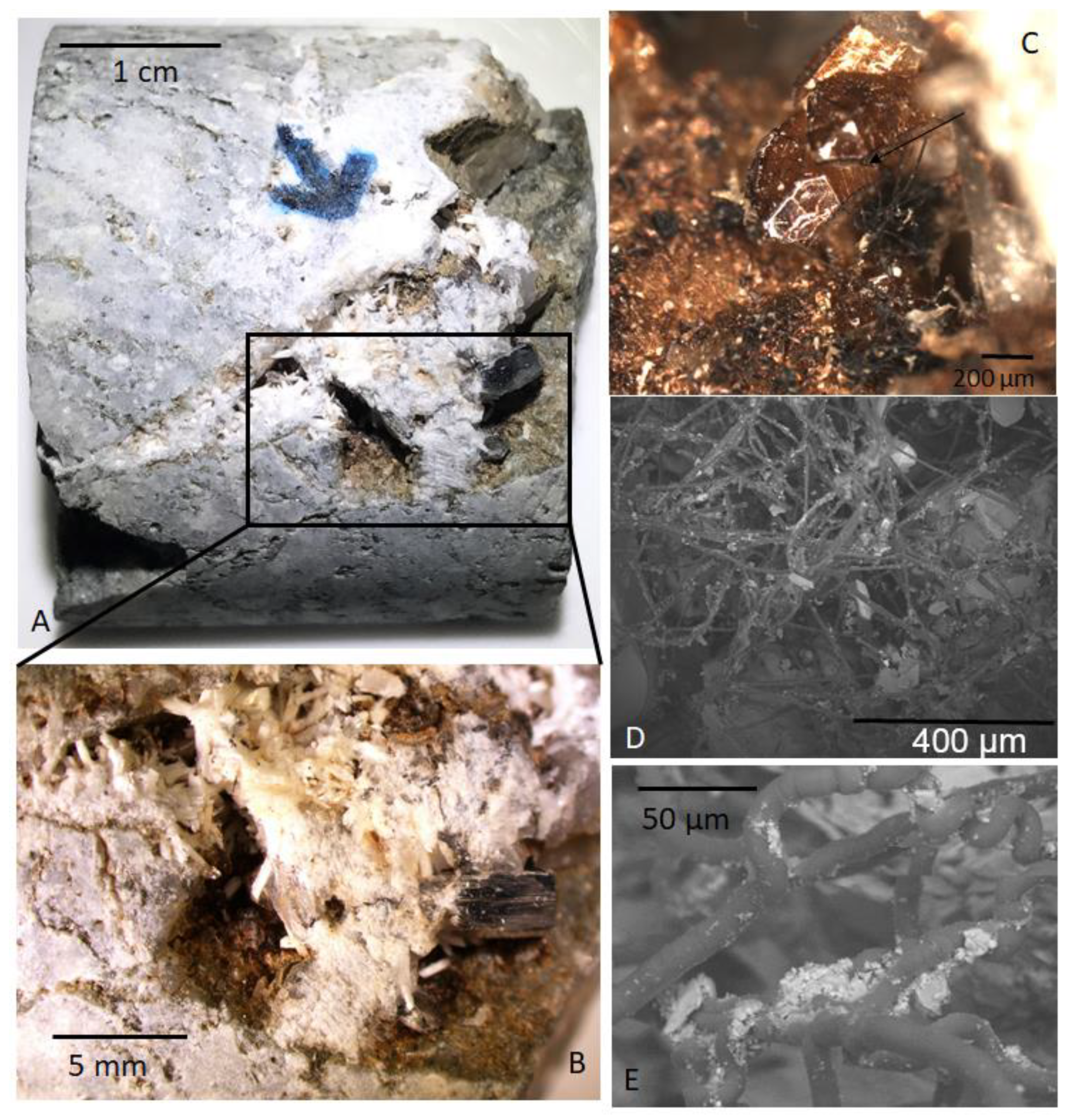
The drill core used in this study intersects the north-western part of the Lockne impact structure [33]. Structurally, this represents the marginal, shallower rim of the final crater, and not the deeper central parts. The cored sequence comprises crystalline impact breccia and fractured basement rock, slump- and resurge deposits, and secular sediments. Veins and vugs in the brecciated basement rock are partly filled with hydrothermally formed calcite and quartz and to minor extent with pyrite, chalcopyrite, galena and sphalerite [33]. Bitumen is abundant and associated with kerogenous matter that covers the hydrothermal minerals as a thin film. This film is a couple of micrometers thick and has a yellow-brownish appearance in optical microscopy. The C-rich film is associated with long, undulating and curvilinear filaments that are preserved as complex networks forming entangled, almost chaotic assemblages. Based on morphological traits identical to fungi the filaments were interpreted as fossilized hyphae forming mycelia in the open pore space of the granitoids [16]. The fungi were introduced into the system after the impact, and colonized the secondary mineralizations by the formation of an initial biofilm from which hyphae grew and formed the mycelium (Figure 1). The fungi were described to have colonized the rock during the hydrothermal activity and upon death become impregnated by oils migrating through the hydrothermal system. This process eventually preserved the fungal mycelia as carbonaceous fossils [16].
3. Sample Material
Mineral samples with fungal mycelia (Figure 1) were taken from drill core LOC1 (total length 225.15 m), and are the samples as presented in [16]. Bitumen occurs in a section between 171.30 and 219.90 m depth in the core in veins and vugs in the fractured and brecciated basement rock. A carbonaceous film from which fungal hyphae propagate and form complex mycelium-like networks covers many euhedral blocky (short c-axis) calcite crystals that occur together with quartz, K-feldspar (adularia), albite and chalcopyrite.
4. Methods
Crystals were physically removed from the fracture voids of the impact breccia, and individually embedded in epoxy. The epoxy mounts were polished and examined by a scanning electron microscope (SEM, Hitachi) equipped with an energy-dispersive spectrometer (EDS), at the University of Gothenburg, Sweden. Sample characterization of the polished crystals was carried out before isotope analysis described below.
4.1. Secondary Ion Mass Spectrometry (SIMS) Stable Isotope Analysis
Calcite and chalcopyrite crystals were mounted in epoxy, polished to expose cross-sections and examined with SEM to trace zonations and impurities prior to SIMS analysis. Intra-crystal SIMS-analysis (10 μm lateral beam dimension, 1-2 μm depth dimension) of sulfur isotopes in chalcopyrite and carbon isotopes in calcite was performed on a CAMECA IMS1280 ion microprobe (CAMECA, SAS, Gennevilliers, France) following the analytical settings and tuning reported previously [19,20]. Sulfur was sputtered using a 133Cs+ primary beam with 20 kV incident energy (10 kV primary, -10 kV secondary) and a primary beam current of ~1.5 nA. A normal incidence electron gun was used for charge compensation. Analyses were performed in automated sequences, with each analysis comprising a 70 second pre-sputter to remove the gold coating over a rastered 15 × 15 µm area, centering of the secondary beam in the field aperture to correct for small variations in surface relief and data acquisition in sixteen four second integration cycles. The magnetic field was locked at the beginning of the session using a nuclear magnetic resonance (NMR) field sensor. Secondary ion signals for 32S and 34S were detected simultaneously using two Faraday detectors with a common mass resolution of 4860 (M/ΔM). Data were normalised for instrumental mass fractionation using matrix-matched reference materials which were mounted together with the sample mounts and analysed after every sixth sample analysis. Results are reported as per mil (‰) δ34S based on the Canon Diablo Troilite (V-CDT)-reference value. Up to 17 crystals were analysed from each fracture sample. In total, 37 analyses were made for δ34S (34S/32S) of chalcopyrite from 15 crystals from fracture-coating sample 185 m. The Trout Lake chalcopyrite reference material with a conventionally determined value of +0.3‰ [34] was used. Typical precision on a single δ34S value, after propagating the within run and external uncertainties from the reference material measurements was ±0.08‰.
For calcite, 13 δ13C SIMS-analyses were performed on the same CAMECA IMS1280 described above. Settings follow those described for S isotopes, with some differences: a Faraday cage/electron multiplier (FC/EM) combination with mass resolution 2500 on the 12C peak and 4000 on the 13C peak was used to resolve it from 12C1H. Influence of organic carbon was avoided by careful spot placement to areas in the crystals without micro-fractures or inclusions and at a sufficient distance from grain-boundaries where fine-grained clusters of other minerals and remnants of organic material may appear. The uncertainty associated with potential organic inclusions and matrix composition is therefore considered to be insignificant compared to the isotopic variations. Calcite results are reported as per mil (‰) δ13C based on the Pee Dee Belemnite (V-PDB) reference value. Analyses were carried out running blocks of six unknowns bracketed by two reference material analyses. Analytical transects of up to six spots were made from core to rim in the crystals. Analyses were made in three crystals from two fracture samples (181 and 185 m). Isotope data from calcite were normalised using calcite reference material S0161 from a granulite facies marble in the Adirondack Mountains, kindly provided by R.A. Stern (University of Alberta). The values used for IMF correction were determined by conventional stable isotope mass spectrometry at Stockholm University on ten separate pieces, yielding δ13C = 0.22 ± 0.11‰V-PDB (1 std. dev.). Precision was δ13C: ±0.4–0.5‰. Values of the reference material measurements are listed together with the samples in Table 1 and Table 2.
4.2. Laser Ablation Multi-Collector Inductively Coupled Plasma Mass Spectrometry (LA-MC-ICP-MS) 87Sr/86Sr
The 87Sr/86Sr values of calcite in sample 185 m were determined by laser ablation multi-collector inductively coupled plasma mass spectrometry (LA-MC-ICP-MS) analysis at the Vegacenter, Swedish Museum of Natural History, Stockholm, Sweden, using a Nu plasma II MC-ICP-MS (Nu Instruments Ltd, Wrexham, UK), and an electrospray ionization (ESI) NWR193 ArF excimer laser ablation system (Elemental Scientific Lasers, Bozeman, MT, USA). Four of the six spots analysed for δ13C using SIMS were also analysed for Sr isotopes by LA-ICP-MS (using larger spots). Ablation frequency was 15 Hz, spot size was 80 μm and fluence was 2.8 J/cm2. Samples were ablated for 45 seconds, followed by 45 seconds wash-out time. The 87Sr/86Sr analyses were normalised to an in-house brachiopod reference material ’Ecnomiosa gerda’ (linear drift and accuracy correction) using a value established by thermal ionisation mass spectrometry (TIMS) of 0.709181 (2sd 0.000004, [35]). A modern oyster shell from Western Australia was used as a secondary reference material and analysed at regular intervals together with the primary reference. The accuracy of these analyses was quantified by comparison to the modern seawater value for 87Sr/86Sr of 0.7091792 ± 0.0000021 [36]. Values of the reference material measurements are listed together with the sample data in Table 2.
4.3. LA-ICP-MS Analyses for Rb-Sr Dating
The Rb-Sr dating system builds on the beta-decay of 87Rb to 87Sr in minerals. One or several Rb-rich minerals (showing increased 87Sr/86Sr and decreased 87Rb/86Sr with time) along with a co-genetic Sr-rich mineral (constant 87Sr/86Sr with time), in our case secondary adularia in paragenesis with calcite or albite, were analysed by Rb-Sr geochronology via high spatial resolution LA-ICP-MS [37] at the Earth Sciences Centre, University of Gothenburg, Sweden. The Rb/Sr spot analyses in fine-grained adularia (n = 10), calcite (n = 4) and albite (n = 6 (of which 3 = rejected)) from sample 185 m and adularia from sample 216 m (n = 13) were performed using an ESI 213NWR laser ablation system (Elemental Scientific Lasers, Bozeman, MT, USA) connected to Agilent 8800QQQ ICP-MS (Agilent Technologies Inc, Santa Clara, CA, USA) with an ORS3 octopole reaction system reaction cell sandwiched between two quadrupoles. Following laser warm-up, ablation occurred with static spot mode in a constant He flow (800 mL/min). The ablated material was mixed with N2 and Ar before entering the ICP-MS torch region and reacted with N2O gas in the reaction cell to chemically separate 87Rb from 87Sr by producing oxide of 87Sr, and thereby enable calculation of 87Rb/86Sr and 87Sr/86Sr ratios [38]. While the octopole bias was set to negative voltage, N2O flow rates in the reaction cell were varied to obtain optimal SrO+ production rates. In tandem mass spectrometry (MS/MS) mode both quadrupoles were controlled while reactive gas was in the reaction cell with the quadrupoles set at different masses to measure reaction products in mass-shifted mode. On mass 85Rb/mass-shifted 86Sr and mass-shifted 87Sr/mass-shifted 86Sr raw ratios were used to calculate 87Rb/86Sr and 87Sr/86Sr, respectively. 85Rb is used as a proxy for 87Rb as it is constant on Earth and within 0.02–0.05% [39]. The raw ratios were converted by correction factors derived from repeated analysis of reference materials NIST SRM 610 and BCR-2G, which are documented to be feasible for calibration of in situ LA-ICP-MS/MS Rb-Sr isotopic data [37,38]. The reference materials were selected to ensure that the pulse/analog setting of each measured isotope was identical in samples and reference materials. The NIST SRM 610 certified reference material with 87Sr/86Sr of 0.7097 [40] was used for 87Sr/86Sr ratio calibration of the sample data. 87Rb/86Sr calibration was performed by using glass reference material BCR-2G [41], shown in Table 3. For 87Rb/86Sr and 87Sr/86Sr respectively, BCR-2G yielded precisions at 1.12% and 0.21%, while NIST SRM 610 yielded 1.16% and 0.35%. The secondary reference material was LP01, a sample constituting mm-sized euhedral biotite from granodiorite of the La Posta intrusion, California. As suggested by [37], we use a weighted mean age of 91.6 ± 1.2 Ma derived from U-Pb TIMS, Ar-Ar and Rb-Sr studies. The isochron age of LP01 in the analytical session was 94.2 ± 2.9 Ma (n = 13, Table 3). The resulting ages are isochron model fits constructed using the Rb decay constant of Villa et al. [42]. Rho (ρ) values for each spot were calculated using 85Rb/86Sr, 87Sr/86Sr and 85Rb/87Sr ratios (Table 4). Average count rate calculation of reference material data is conducted by Glitter©, whereas sample data reduction and within-run error calculation of important element and isotopic ratios is performed using an in-house spreadsheet. Isotopic homogeneity is ensured through evaluation of analytically reliable laser ablation signals on the scales of single spots, grains and assemblages (justification of rejected spots given in Table 4). No error propagation from uncertainties in literature data or within-run errors of reference materials is applied to sample errors as internally calculated errors are significantly larger as the established 1.5% external errors of this method [38].
5. Results
Polished cross sections reveal growth zonation in the calcite crystals (Figure 2c). Ivarsson et al. [16] interpreted the fossils propagating from calcite crystals as fungi based on the size and appearance of the filaments and mycelia, as well as the presence of fungal characteristic morphologies like frequent branching, anastomoses between branches and repeated septa. All fungal fossils including the film are preserved as disordered carbonaceous matter according to Raman spectroscopy and Time-of-Flight-SIMS analyses [16].
5.1. Isotope Compositions
The overall variability of the δ13C in calcite is large, (Table 2, Figure 2), ranging from 13C-depleted (−19.4‰, V-PDB) to 13C-enriched values (+12.3‰, V-PDB). The δ13C composition is, however, highly sample specific, and even crystal specific. Sample 181 m has 13C-depleted values: Crystal 1 has values of −8.8‰ to −6.7‰, and crystal 2 −19.4‰ to −16.9‰. In contrast, the crystal analysed in sample 185 m has 13C-enriched composition throughout all spots (n = 6): +9.9‰ to +12.7‰, as shown in the spot transects (Figure 2d). The correlation of the isotopic variance to the growth zonation is related to minor temporal chemical and isotopic fluctuation in the precipitating fluids. The 87Sr/86Sr values of the calcite crystal in sample 185 m also shows small differences of 0.7444 ± 0.0015 to 0.7471 ± 0.0012 (Figure 2d). The δ34Schalcopyrite values in sample 185 m, show a span from +0.4‰ to +3.6‰ V-CDT (n = 37, Table 1), and there is no significant variation within the crystals targeted by several microanalyses (examples in Figure 3).
5.2. Rb/Sr Dating
The isochron constructed from 10 LA-ICP-MS analyses of adularia and three LA-ICP-MS analyses in addition to four LA-MC-ICP-MS analyses of calcite for sample 185 m yield an age of 356.6 ± 6.7 Ma (Figure 4, Table 4). Adularia crystals from sample 216 m paired with albite acquired in the 185m fracture yield an age of 1457 ± 63 Ma (Appendix A Figure A1), but as this age represents fracture formation at more than 1 Gyr before the Lockne impact structure formed, we do not discuss it in more detail. It is, nevertheless, an important and expected observation that shows that pre-existing fractures were reactivated during the impact.
6. Discussion
The isotope record of the carbonate and sulfide crystals serves as an archive for processes in the fracture system over long time periods [19,20,43,44]. In the following sections the various (mostly microbial) processes that can be responsible for the observed isotope signatures are discussed. It is important to emphasize that the isotope composition of the minerals may represent the result of one or several (cryptic) processes in the fracture system. These processes (when biological) are usually distinguished by the kinetic isotope effect that occurs when microbial populations utilize a substrate (i.e., they alter the isotope composition due to preferential utilization of a specific isotope). In addition, the source of the C and S compounds and processes occurring during transport can also influence the isotope composition before the element is incorporated into a mineral. Several of the potential source compounds may also overlap in isotope composition making certain diagnostic determinations about processes and sources difficult when using isotope composition of minerals only. Abiotic fractionation can also overlap in magnitude with microbial fractionation, which may inhibit certain conclusions about microbial processes in the fracture system. However, some magnitudes of isotope fractionation and systematics have been used extensively as diagnostic tracers for specific microbial processes. These include C isotope markers for methanogenesis and methane oxidation and S isotope markers for microbial sulfate reduction [45,46,47,48].
6.1. Ancient Methanogenesis in the Fracture Voids
Methane is usually 13C-depleted compared to other carbon compounds [49], especially when the methane is microbial in origin. As a consequence of the fractionation occurring during methanogenesis, which discriminates against 13C, the residual CO2 becomes 13C-rich [50]. Subsequent involvement of the residual C into precipitating carbonate minerals is therefore a useful diagnostic C-isotope tracer for methanogenesis, for instance by 13C-rich secondary carbonates in sedimentary basins [51] or in fractured crystalline rocks in the Fennoscandian shield [24,43,52,53]. The significantly 13C-enriched calcite in the Lockne sample 185 m (δ13Ccalcite values as heavy as +12.7‰, Figure 2d) is thus a strong line of evidence for microbial methanogenesis in situ in the deep fracture system especially since potential abiotic methane-forming chemical processes such as serpentinization, graphite metamorphism or Fischer-Tropsch type reactions [54,55] are unlikely under the local physico-chemical and geological conditions. Strong 13C-enrichment in carbonate is typically associated with microbial methane formed by carbonate reduction, as opposed to acetate (methyle-type) fermentation that involves smaller kinetic carbon isotope effects [56]. Heavy δ13CCO2 values are particularly typical for secondary microbial methane formed from thermogenic precursors [56], which may involve a previous methane oxidation step that can occur anaerobically, if associated with microbial reduction of e.g., sulfate [56]. Sulfide minerals can form as a result of microbial sulfate reduction (MSR) and the chalcopyrite observed in sample 185 m may, therefore, have formed due to MSR in a step preceding methanogenesis. The MSR metabolism involves a kinetic isotope effect that discriminates against 34S resulting in production of a typically strongly 34S-depleted hydrogen sulfide [57]. As there is no significant S isotope fractionation during sulfide precipitation the δ34Ssulfide composition serves as a diagnostic marker for MSR, because abiotic sulfate reduction at higher temperatures involves smaller fractionation [58]. However, the δ34Schalcopyrite values determined by SIMS (+0.4 to 3.6‰) are not very depleted. The initial δ34S composition of the sulfate in the system is unknown, which makes estimates of the fractionation difficult. If we anticipate that the source sulfate had a similar composition as Paleozoic seawater (+15 to +35‰ [59]) an isotope enrichment of 10–35‰ would be needed to produce the detected δ34Schalcopyrite values. This degree of isotope enrichment and uncertainty of initial sulfate composition, inhibit any certain interpretation of the δ34Schalcopyrite values as microbial in origin, because they cannot be fully distinguished from thermochemical reduction, and furthermore are overlapping with hydrothermal sulfide of magmatic origin [58,60,61]. Rayleigh isotope reservoir effects may also have occurred in the fracture system, which can modify the S isotope signature significantly [20].
The overall low δ13Ccalcite values of the 181 m sample are more in line with an organic origin, such as plants or oil/petroleum/kerogen/bitumen [62,63], and may reflect microbial utilization (oxidation) of such carbon sources, for instance coupled with sulfate reduction. The crystal-specific δ13C variance in this sample can be due to temporal variation of the microbial processes and substrates in the fracture and/or due to spatial micro-scale variability within the fracture voids, as reported in other deep fractured bedrock systems in the Fennoscandian Shield [19,24]. Organic compounds may have descended from overlying, but presently completely eroded Paleozoic sedimentary successions that once covered the Fennoscandian shield [64]. Thermochronological studies show that the sedimentary successions had considerable thickness in the mid-Paleozoic era [65]. Infiltration of bitumen and other organic compounds of surficial origin into the crystalline bedrock occurred presumably from thermally heated organic-rich shales in the lower parts of the sedimentary pile [19,66] of the Fennoscandian shield, and elsewhere (e.g., UK and Australia [67,68]). Putative bitumen occurrences have also been reported from the Lockne fracture system [33], and may thus have been influential for the deep biosphere community in the deep fracture system.
6.2. New Age Constraints for the Fracture Assemblage in the Impact Structure and Microbial Communities
The age of the feldspar-calcite assemblage in sample 185 m of 356.6 ± 6.7 Ma (Figure 4) is about 100 Myr younger than the estimated age of the impact [28]. The fracture system and mineral paragenesis of the impact structure have previously been considered to be of the same age as the impact [33], and consequently the fossilized microbial communities (dominantly fungi) observed in the fracture system have been assigned the same age [16]. Our new radiometric age determination requires an updated model for the timing of the fracture system activation in the impact structure, and consequently also for the preserved microbial remains within this system. The obtained age, at the Devonian–Carboniferous transition, is in line with secondary mineral assemblages in fractures elsewhere in the Fennoscandian shield, i.e., feldspar-calcite veins, with fossilized microorganisms at 300 m depth at Forsmark, Sweden, dated to between 355 ± 14 and 402 ± 10 Ma, and at Oskarshamn dated to 358 ± 12 and 393 ± 15 Ma by in situ Rb/Sr geochronology [19,20,21]. At Forsmark, a few calcite crystals showed methanogenesis-related 13C-rich composition, and/or 13C-depleted values typical for anaerobic oxidation of methane [19,21]. This suggests that a regional fracture-activation event at these times enabled fluid circulation, which induced microbial activity and secondary mineral formation in the bedrock. Fluids from overlying organic-rich shales were at this event allowed to descend into the deeper crystalline bedrock fracture network, such as the deeply fragmented bedrock at Lockne and thereby provided substrates to the otherwise energy-poor deep biosphere.
Fluid inclusions of secondary calcite from the current drill core have indicated fluids with a salinity around 20 eq. wt% CaCl2 corresponding to brine, and homogenization temperatures in the range of 77–218 °C [33], which at least for the salinities correspond to fluid inclusions in similar fracture coating calcite in Oskarshamn and Forsmark in Sweden, from southern Finland, and to Caledonian mineralizations [19,53,69,70,71]. The δ18O values of calcite and quartz [33,72] at Lockne are also in line with compositions of the fracture-filling calcite reported from other parts of the Fennoscandian shield [19,53,69,70,71,73]. Most of the fluid inclusion homogenization temperatures in the previous study [33] were in the 100–180 °C range, which is generally too high for microbial activity. The large temperature span of 77–218 °C, however, does suggest that fluid temperatures dropped at later stages of the fluid circulation event in the fractures. During the early part of the fluid circulation when temperatures were >100 °C, K-feldspar-albite-calcite-quartz formed and thermally mature bitumen was probably transported from overlying sediments, because asphaltite can become viscous and mobilized at elevated temperatures [66]. The occurrence of methane in the fluid inclusions in calcite of this stage also marks mobility of thermogenic hydrocarbons from the shale source [33] into the deeper fracture system. When temperatures decreased after the initial phase of hydrothermal circulation, deep subsurface microbial communities could use the bitumen as an energy source by oxidizing the organic matter and subsequently producing secondary methane as discussed above, as well as sustaining heterotrophic eukaryotic activity in the form of fungi. This late stage calcite formation is in line with brine type fluid inclusions with lower homogenization temperatures, down to 77 °C, which is more suitable for microbial colonization, or alternatively a group of calcite crystals in the 50–70 °C span [33].
The revised radiometric ages of the fracture re-activation set the fungal colonization in a different environmental context. At the time of the impact, the area was covered by a shallow sea [33], and it was believed that marine fungi were introduced into the impact-generated hydrothermal system by ingress of seawater through the fractured oceanic crust [16]. Similarly, a seawater origin is suggested for biological material responsible for sulfur reduction in the Rochechouart impact crater in France [74]. However, 356 Myr ago the current area of the Fennoscandian shield hosting the Lockne crater was not marine but continental [31], which indicates that the fungi are not marine but represent fungi belonging to the continental deep biosphere at that time. Although the brine-type fluid inclusions of the calcites may, at first glance, point to a marine origin, it is relevant to consider that deep continental brines are result of several different processes. In addition to marine infiltration, these processes include prolonged water-rock interaction, mixing of fluids with different sources, concentration during freezing and descent of sedimentary brines [75,76,77,78]. The latter has importance in Canada where this process has been studied extensively [79]. There, it has been suggested that deep continental brines result from infiltration of saline water to the crystalline basement aquifer from basinal sedimentary brines and evaporites when the shield was covered by sedimentary successions in the Paleozoic [79]. This means that brines may develop under continental conditions if marine sedimentary successions overlay the shield.
Contemporaneous methanogenesis is in agreement with previous fossilized fungal-prokaryotic communities in granitic deep settings [23], and suggest a potential synergy. Methanogenic activity and formation of sulfides indicates anoxic conditions in the system and explain the pristine carbonaceous nature of the fungal fossils and lack of mineralization by clays and Fe-oxides, which is common among fungal fossilization in oxygenated deep environments and dominates the findings in the oceanic crust [80].
Although impact craters and associated impact-generated hydrothermal systems likely are favourable environments for microbial life [1,2] owing to their increased pore space for endolithic communities, and heat convection favourable for ecosystems [5], our revised model for the ancient Lockne microbial community shows that colonization took place long after the impact, in contrast to previous models. The role of the impact-induced hydrothermal system is, therefore, negligible for the fossilized community observed in the fractures. Instead, reactivation of the fractures occurred 100 Myr after the impact event, in a post-Caledonian extension event documented previously throughout the Fennoscandian shield. Fractures and pore space opened as a result of the impact thus likely provided pathways for the extension-related fluids. However, the extent of reutilization is likely to be local given that the relatively small amounts of melt rocks are scattered along the crater rims, implying that small-scale hydrothermal cells did not produce extensive wall-rock dissolution. This extension correlates with a heating episode in the crystalline basement of southern Sweden that relates to burial by Caledonian foreland sediments [65], whereas Caledonian nappe structures overthrusted the Lockne area during the Silurian and early Devonian [81]. Tectonothermal or thermometamorphic overprinting has caused resetting of 40Ar/39Ar melt rock ages in several impact structures, including Gardnos in Norway [82], Acraman in Australia [83] and Charlevoix in Canada [84]. Following rapid uplift after Caledonian orogenic collapse, thermochronology records temperatures of 250–300 °C at 350 Ma [85] towards western Norway where the metamorphic temperatures were the highest during the orogeny as well as in northwestern Norway [86]. In the nappes immediately west to the Lockne area, cooling rates of 15 °C/Myr brought the rocks through 475 °C at 425 Ma [87]. Similar rapid cooling episodes at 350 Ma in western Norway have been attributed to extension concurrent with rapid uplift [88]. Despite the fact that the Rb-Sr isotopic system is more resistant to heating than Ar systems, the lack of thermochronological constraints in the Lockne area at 350 Ma means that temperatures high enough to disturb the radiogenic chronometer cannot be ruled out. However, the hydrothermal minerals of Lockne neither feature any traces of such an overprint in terms of diffusion or dissolution textures nor radiogenic isotopic inhomogeneity or disturbances on spatial and depth scales. Furthermore, no signs of severe degradation of the fossils are observed, which would certainly be expected if experiencing a regional thermal event. Instead, the pristine fungi structure indicates precipitation through extension-facilitated hydrothermal activity where bituminous material from overlying shales was introduced into the fracture system and provided energy for microbial activity. In that sense, the majority of the studied hydrothermal mineralizations are not impact induced and thus not of astrobiological importance in a Martian context as was previously suggested [16].
7. Conclusions
The age of previously described fungal fossils from the Lockne crater (458 Ma), Sweden, is here revised. In situ Rb/Sr dating of secondary calcite-feldspar (356.6 ± 6.7 Ma) shows that the fungal colonization is not associated with the impact-induced hydrothermal system but took place at least 100 Myr after the impact event. This revised age excludes the previous notion that marine fungi were introduced by seawater recharge, and instead suggests that the fungi may have been established in the deep continental crust underneath remnant marine successions already at 356 Ma or shortly after. Microscale stable isotope data of 13C-enriched calcite further suggest methanogenesis occurred in the fracture system, potentially in synergy with the fungi.
Author Contributions
M.I. M.T. H.D. performed the SEM-analyses, M.T. performed the LA-ICP-MS Rb–Sr dating, data reduction and interpretation, H.D. performed the SIMS and LA-MC-ICP analyses together with M.J.W. and E.K.+M.S., respectively. M.T. wrote the paper together with H.D. and M.I.
Funding
This research was funded by Swedish research council (contract 2017-05186 to H.D., 2017-04129 to M.I.), Formas (contract 2017-00766 to H.D. and M.W.), and a Villum Investigator Grant to Don Canfield (No. 16518).
Acknowledgments
Thanks to Erik Sturkell for access to drill cores. We acknowledge NordSIM for provision of facilities and experimental support under Swedish Research Council grant no 2014-06375. K. Lindén, and Johan Hogmalm are thanked for analytical or sample preparation assistance. This is NordSIM publication #603 and Vegacenter publication #017.
Conflicts of Interest
The authors declare no conflict of interest and the founding sponsors had no role in the design of the study; in the collection, analyses, or interpretation of data; in the writing of the manuscript, and in the decision to publish the results.
Appendix A
Figure A1.
In situ Rb-Sr dating results of the 216 m sample with (a) albite spots as white circles, rejected albite spots as red circles, (b) adularia spots in one of three grains as black circles on BSE images and (c) the isochron diagram and the resulting data.
Figure A1.
In situ Rb-Sr dating results of the 216 m sample with (a) albite spots as white circles, rejected albite spots as red circles, (b) adularia spots in one of three grains as black circles on BSE images and (c) the isochron diagram and the resulting data.

References
- Versh, E.; Kirsimäe, K.; Jõeleht, A. Development of potential ecological niches in impact-induced hydrothermal systems: The small-to-medium size impacts. Planet. Space Sci. 2006, 54, 1567–1574. [Google Scholar] [CrossRef]
- Osinski, G.R.; Tornabene, L.L.; Banerjee, N.R.; Cockell, C.S.; Flemming, R.; Izawa, M.R.; McCutcheon, J.; Parnell, J.; Preston, L.J.; Pickersgill, A.E.; et al. Impact-generated hydrothermal systems on Earth and Mars. Icarus 2013, 224, 347–363. [Google Scholar] [CrossRef]
- Kring, D.A. Impact events and their effect on the origin, evolution, and distribution of life. GSA today 2000, 10, 1–7. [Google Scholar]
- Cockell, C.S. The origin and emergence of life under impact bombardment. Philos. Trans. Soc. B: Boil. Sci. 2006, 361, 1845–1856. [Google Scholar] [CrossRef] [PubMed]
- Cockell, C.S.; Lee, P. The biology of impact craters – a review. Boil. Rev. 2002, 77, 279–310. [Google Scholar]
- Naumov, M.V. Principal features of impact-generated hydrothermal circulation systems: Mineralogical and geochemical evidence. Geofluids 2002, 5, 165–184. [Google Scholar] [CrossRef]
- Ames, D.E.; Watkinson, D.H.; Parrish, R.R. Dating of a regional hydrothermal system induced by the 1850 Ma Sudbury impact event. Geology 1998, 26, 447. [Google Scholar] [CrossRef]
- Jõeleht, A.; Kirsimäe, K.; Plado, J.; Versh, E.; Ivanov, B. Cooling of the Kärdla impact crater: II. Impact and geothermal modeling. Meteor. Planet. Sci. 2005, 40, 21–33. [Google Scholar] [CrossRef]
- Arp, G.; Kolepka, C.; Simon, K.; Karius, V.; Nolte, N.; Hansen, B.T. New evidence for persistent impact-generated hydrothermal activity in the Miocene Ries impact structure, Germany. Meteorit. Planet. Sci. 2013, 48, 2491–2516. [Google Scholar] [CrossRef]
- Schmieder, M.; Jourdan, F. The Lappajärvi impact structure (Finland): Age, duration of crater cooling, and implications for early life. Geochim. Cosmochim. Acta 2013, 112, 321–339. [Google Scholar] [CrossRef]
- Kenny, G.G.; Schmieder, M.; Whitehouse, M.J.; Nemchin, A.A.; Morales, L.F.; Buchner, E.; Bellucci, J.J.; Snape, J.F. A new U-Pb age for shock-recrystallised zircon from the Lappajärvi impact crater, Finland, and implications for the accurate dating of impact events. Geochim. Cosmochim. Acta 2019, 245, 479–494. [Google Scholar] [CrossRef]
- Parnell, J.; Boyce, A.; Thackrey, S.; Muirhead, D.; Lindgren, P.; Mason, C.; Taylor, C.; Still, J.; Bowden, S.; Osinski, G.R.; et al. Sulfur isotope signatures for rapid colonization of an impact crater by thermophilic microbes. Geology 2010, 38, 271–274. [Google Scholar] [CrossRef]
- Sapers, H.M.; Osinski, G.R.; Banerjee, N.R.; Preston, L.J. Enigmatic tubular features in impact glass. Geology 2014, 42, 471–474. [Google Scholar] [CrossRef]
- Lindgren, P.; Ivarsson, M.; Neubeck, A.; Broman, C.; Henkel, H.; Holm, N.G. Putative fossil life in a hydrothermal system of the Dellen impact structure, Sweden. Int. J. Astrobiol. 2010, 9, 137–146. [Google Scholar] [CrossRef]
- Hode, T.; Cady, S.L.; von Dalwigk, I.; Kristiansson, P. Evidence of Ancient Microbial Life in an Impact Structure and Its Implications for Astrobiology. In From Fossils to Astrobiology: Records of Life on Earth and Search for Extraterrestrial Biosignatures; Seckbach, J., Walsh, M., Eds.; Springer Netherlands: Dordrecht, The Netherlands, 2008; pp. 249–273. [Google Scholar]
- Ivarsson, M.; Broman, C.; Sturkell, E.; Ormö, J.; Siljeström, S.; Van Zuilen, M.; Bengtson, S. Fungal colonization of an Ordovician impact-induced hydrothermal system. Sci. Rep. 2013, 3, 3487. [Google Scholar] [CrossRef]
- Ivarsson, M.; Holm, N.G.; Neubeck, A. The Deep Biosphere of the Subseafloor Igneous Crust. In Trace Metal Biogeochemistry and Ecology of Deep-Sea Hydrothermal Vent Systems; Demina, L.L., Galkin, V.S., Eds.; Springer International Publishing: Cham, Switzerland, 2016; pp. 143–166. [Google Scholar]
- Ivarsson, M.; Bengtson, S.; Skogby, H.; Lazor, P.; Broman, C.; Belivanova, V.; Marone, F. A Fungal-Prokaryotic Consortium at the Basalt-Zeolite Interface in Subseafloor Igneous Crust. PLOS ONE 2015, 10, e0140106. [Google Scholar] [CrossRef] [PubMed]
- Drake, H.; Heim, C.; Roberts, N.M.; Zack, T.; Tillberg, M.; Broman, C.; Ivarsson, M.; Whitehouse, M.J.; Åström, M.E. Isotopic evidence for microbial production and consumption of methane in the upper continental crust throughout the Phanerozoic eon. Earth Planet. Sci. Lett. 2017, 470, 108–118. [Google Scholar] [CrossRef]
- Drake, H.; Whitehouse, M.J.; Heim, C.; Reiners, P.W.; Tillberg, M.; Hogmalm, K.J.; Dopson, M.; Broman, C.; Åström, M.E. Unprecedented 34 S-enrichment of pyrite formed following microbial sulfate reduction in fractured crystalline rocks. Geobiology 2018, 16, 556–574. [Google Scholar] [CrossRef] [PubMed]
- Drake, H.; Ivarsson, M.; Tillberg, M.; Whitehouse, M.J.; Kooijman, E. Ancient Microbial Activity in Deep Hydraulically Conductive Fracture Zones within the Forsmark Target Area for Geological Nuclear Waste Disposal, Sweden. Geosciences 2018, 8, 211. [Google Scholar] [CrossRef]
- Tillberg, M.; Drake, H.; Zack, T.; Hogmalm, J.; Åström, M. In Situ Rb-Sr Dating of Fine-grained Vein Mineralizations Using LA-ICP-MS. Procedia Earth Planet. Sci. 2017, 17, 464–467. [Google Scholar] [CrossRef]
- Drake, H.; Ivarsson, M.; Bengtson, S.; Heim, C.; Siljeström, S.; Whitehouse, M.J.; Broman, C.; Belivanova, V.; Åström, M.E. Anaerobic consortia of fungi and sulfate reducing bacteria in deep granite fractures. Nat. Commun. 2017, 8, 55. [Google Scholar] [CrossRef]
- Drake, H.; Åström, M.E.; Heim, C.; Broman, C.; Åström, J.; Whitehouse, M.; Ivarsson, M.; Siljestrom, S.; Sjövall, P. Extreme 13C depletion of carbonates formed during oxidation of biogenic methane in fractured granite. Nat. Commun. 2015, 6, 7020. [Google Scholar] [CrossRef]
- Drake, H.; Tullborg, E.-L.; Whitehouse, M.; Sandberg, B.; Blomfeldt, T.; Åström, M.E. Extreme fractionation and micro-scale variation of sulphur isotopes during bacterial sulphate reduction in deep groundwater systems. Geochim. Cosmochim. Acta 2015, 161, 1–18. [Google Scholar] [CrossRef]
- Drake, H.; Åström, M.E.; Tullborg, E.-L.; Whitehouse, M.; Fallick, A.E. Variability of sulphur isotope ratios in pyrite and dissolved sulphate in granitoid fractures down to 1km depth – Evidence for widespread activity of sulphur reducing bacteria. Geochim. Cosmochim. Acta 2013, 102, 143–161. [Google Scholar] [CrossRef]
- Drake, H.; Ivarsson, M. The role of anaerobic fungi in fundamental biogeochemical cycles in the deep biosphere. Fungal Boil. Rev. 2018, 32, 20–25. [Google Scholar] [CrossRef]
- Alwmark, C.; Schmitz, B. Extraterrestrial chromite in the resurge deposits of the early Late Ordovician Lockne crater, central Sweden. Earth Planet. Sci. Lett. 2007, 253, 291–303. [Google Scholar] [CrossRef]
- Shuvalov, V.; Ormö, J.; Lindström, M. Hydrocode Simulation of the Lockne Marine Target Impact Event. In Impact Tectonics; Koeberl, C., Henkel, H., Eds.; Springer: Berlin/Heidelberg, Germany, 2005; pp. 405–422. [Google Scholar]
- Högdahl, K. 1.86–1.85 Ga intrusive ages of K-feldspar megacryst-bearing granites in the type area of the Revsund granites in Jämtland County, central Sweden. GFF 2000, 122, 359–366. [Google Scholar] [CrossRef]
- Lindström, M.; Lundqvist, J.; Lundqvist, T. Sveriges Geologi Från Urtid till Nutid; Studentlitteratur: Lund, Sweden, 2000. [Google Scholar]
- Lindström, M.; Sturkell, E.F. Geology of the Early Palaeozoic Lockne impact structure, Central Sweden. Tectonophysics 1992, 216, 169–185. [Google Scholar] [CrossRef]
- Sturkell, E.F.; Broman, C.; Forsberg, P.; Torssander, P. Impact-related hydrothermal activity in the Lockne impact structure, Jämtland, Sweden. Eur. J. Miner. 1998, 10, 589–606. [Google Scholar] [CrossRef]
- Crowe, D.E.; Vaughan, R.G. Characterization and use of isotopically homogeneous standards for in situ laser microprobe analysis of 34 S/ 32 S ratios. Am. Miner. 1996, 81, 187–193. [Google Scholar] [CrossRef]
- Kiel, S.; Glodny, J.; Birgel, D.; Bulot, L.G.; Campbell, K.A.; Gaillard, C.; Graziano, R.; Kaim, A.; Lazăr, I.; Sandy, M.R.; et al. The Paleoecology, Habitats, and Stratigraphic Range of the Enigmatic Cretaceous Brachiopod Peregrinella. PLOS ONE 2014, 9, e109260. [Google Scholar] [CrossRef] [PubMed]
- Mokadem, F.; Parkinson, I.J.; Hathorne, E.C.; Anand, P.; Allen, J.T.; Burton, K.W. High-precision radiogenic strontium isotope measurements of the modern and glacial ocean: Limits on glacial–interglacial variations in continental weathering. Earth Planet. Sci. Lett. 2015, 415, 111–120. [Google Scholar] [CrossRef]
- Zack, T.; Hogmalm, K.J. Laser ablation Rb/Sr dating by online chemical separation of Rb and Sr in an oxygen-filled reaction cell. Chem. Geol. 2016, 437, 120–133. [Google Scholar] [CrossRef]
- Hogmalm, K.J.; Zack, T.; Karlsson, A.K.-O.; Sjöqvist, A.S.L.; Garbe-Schönberg, D. In situ Rb–Sr and K–Ca dating by LA-ICP-MS/MS: an evaluation of N 2 O and SF 6 as reaction gases. J. Anal. At. Spectrom. 2017, 32, 305–313. [Google Scholar] [CrossRef]
- Nebel, O.; Mezger, K.; Scherer, E.; Münker, C. High precision determinations of 87Rb/85Rb in geologic materials by MC-ICP-MS. Int. J. Mass Spectrom. 2005, 246, 10–18. [Google Scholar] [CrossRef]
- Jochum, K.P.; Weis, U.; Stoll, B.; Kuzmin, D.; Yang, Q.; Raczek, I.; Jacob, D.E.; Stracke, A.; Birbaum, K.; Frick, D.A.; et al. Determination of Reference Values for NIST SRM 610-617 Glasses Following ISO Guidelines. Geostand. Geoanalytical 2011, 35, 397–429. [Google Scholar] [CrossRef]
- Elburg, M.; Vroon, P.; Van Der Wagt, B.; Tchalikian, A. Sr and Pb isotopic composition of five USGS glasses (BHVO-2G, BIR-1G, BCR-2G, TB-1G, NKT-1G). Chem. Geol. 2005, 223, 196–207. [Google Scholar] [CrossRef]
- Villa, I.; De Bièvre, P.; Holden, N.; Renne, P. IUPAC-IUGS recommendation on the half life of 87 Rb. Geochim. Cosmochim. Acta 2015, 164, 382–385. [Google Scholar] [CrossRef]
- Sahlstedt, E.; Karhu, J.A.; Pitkänen, P.; Whitehouse, M. Biogenic processes in crystalline bedrock fractures indicated by carbon isotope signatures of secondary calcite. Appl. Geochem. 2016, 67, 30–41. [Google Scholar] [CrossRef]
- Sahlstedt, E.; Karhu, J.; Pitkanen, P.; Whitehouse, M. Implications of sulfur isotope fractionation in fracture-filling sulfides in crystalline bedrock, Olkiluoto, Finland. Appl. Geochem. 2013, 32, 52–69. [Google Scholar] [CrossRef]
- Knittel, K.; Boetius, A. Anaerobic Oxidation of Methane: Progress with an Unknown Process. Annu. Microbiol. 2009, 63, 311–334. [Google Scholar] [CrossRef] [PubMed]
- Peckmann, J.; Thiel, V. Carbon cycling at ancient methane–seeps. Chem. Geol. 2004, 205, 443–467. [Google Scholar] [CrossRef]
- Canfield, D.E.; Olesen, C.A.; Cox, R.P. Temperature and its control of isotope fractionation by a sulphate-reducing bacterium. Geochim. Cosmochim. Acta 2006, 70, 548–561. [Google Scholar] [CrossRef]
- Johnston, D.T.; Wing, B.A.; Farquhar, J.; Kaufman, A.J.; Strauss, H.; Lyons, T.W.; Kah, L.C.; Canfield, D.E. Active Microbial Sulfur Disproportionation in the Mesoproterozoic. Science 2005, 310, 1477–1479. [Google Scholar] [CrossRef] [PubMed]
- Whiticar, M.J. Carbon and hydrogen isotope systematics of bacterial formation and oxidation of methane. Chem. Geol. 1999, 161, 291–314. [Google Scholar] [CrossRef]
- Boehme, S.E.; Blair, N.E.; Chanton, J.P.; Martens, C.S. A mass balance of 13C and 12C in an organic-rich methane-producing marine sediment. Geochim. Cosmochim. Acta 1996, 60, 3835–3848. [Google Scholar] [CrossRef]
- Budai, J.M.; Walter, L.M.; Ku, T.C.W.; Martini, A.M. Fracture-fill calcite as a record of microbial methanogenesis and fluid migration: a case study from the Devonian Antrim Shale, Michigan Basin. Geofluids 2002, 2, 163–183. [Google Scholar] [CrossRef]
- Drake, H.; Tullborg, E.-L.; Hogmalm, K.J.; Åström, M.E. Trace metal distribution and isotope variations in low-temperature calcite and groundwater in granitoid fractures down to 1km depth. Geochim. Cosmochim. Acta 2012, 84, 217–238. [Google Scholar] [CrossRef]
- Sandström, B.; Tullborg, E.-L. Episodic fluid migration in the Fennoscandian Shield recorded by stable isotopes, rare earth elements and fluid inclusions in fracture minerals at Forsmark, Sweden. Chem. Geol. 2009, 266, 126–142. [Google Scholar] [CrossRef]
- Kietäväinen, R.; Purkamo, L. The origin, source, and cycling of methane in deep crystalline rock biosphere. Front. Microbiol. 2015, 6, 725. [Google Scholar] [CrossRef]
- Etiope, G.; Lollar, B.S. ABIOTIC METHANE ON EARTH. Rev. Geophys. 2013, 51, 276–299. [Google Scholar] [CrossRef]
- Pallasser, R. Recognising biodegradation in gas/oil accumulations through the δ 13 C compositions of gas components. Org. Geochem. 2000, 31, 1363–1373. [Google Scholar] [CrossRef]
- Sim, M.S.; Bosak, T.; Ono, S. Large sulfur isotope fractionation does not require disproportionation. Science 2011, 333, 74–77. [Google Scholar] [CrossRef] [PubMed]
- Machel, H.G.; Krouse, H.R.; Sassen, R. Products and distinguishing criteria of bacterial and thermochemical sulfate reduction. Appl. Geochem. 1995, 10, 373–389. [Google Scholar] [CrossRef]
- Strauss, H. The isotopic composition of sedimentary sulfur through time. Palaeogeogr. Palaeoclim. Palaeoecol. 1997, 132, 97–118. [Google Scholar] [CrossRef]
- Field, C.W.; Fifarek, R.H. Light stable-isotopes systematics in the epithermal environment. Rev. Econ. Geology 1985, 2, 99–128. [Google Scholar]
- Kiyosu, Y.; Krouse, H.R. The role of organic acid in the abiogenic reduction of sulfate and the sulfur isotope effect. Geochem. J. 1990, 24, 21–27. [Google Scholar] [CrossRef]
- Vlierboom, F.; Collini, B.; Zumberge, J. The occurrence of petroleum in sedimentary rocks of the meteor impact crater at Lake Siljan, Sweden. Org. Geochem. 1986, 10, 153–161. [Google Scholar] [CrossRef]
- Roberts, H.H.; Aharon, P. Hydrocarbon-derived carbonate buildups of the northern Gulf of Mexico continental slope: A review of submersible investigations. Geo-Marine Lett. 1994, 14, 135–148. [Google Scholar] [CrossRef]
- Koistinen, T.; Stephens, M.B.; Bogatchev, V.; Nordgulen, Ø.; Wennerström, M.; Korkonen, J. Geological Map of the Fennoscandian Shield Scale 1:2,000,000; Geological Surveys of Finland, Norway and Sweden and the Northwest Department of Natural Resources of Russia: Espoo, Finland, 2001. [Google Scholar]
- Guenthner, W.R.; Reiners, P.W.; Drake, H.; Tillberg, M. Zircon, titanite, and apatite (U-Th)/He ages and age-eU correlations from the Fennoscandian Shield, southern Sweden. Tectonics 2017, 36, 1254–1274. [Google Scholar] [CrossRef]
- Sandström, B.; Tullborg, E.-L.; De Torres, T.; Ortiz, J.E. The occurrence and potential origin of asphaltite in bedrock fractures, Forsmark, central Sweden. GFF 2006, 128, 233–242. [Google Scholar] [CrossRef]
- Parnell, J.; Baba, M.; Bowden, S.; Muirhead, D. Subsurface biodegradation of crude oil in a fractured basement reservoir, Shropshire, UK. J. Geol. Soc. 2017, 174, 655–666. [Google Scholar] [CrossRef]
- McCready, A.J.; Stumpfl, E.F.; Melcher, F. U/Th-rich bitumen in Archean granites and Palaeoproterozoic metasediments, Rum Jungle Mineral Field, Australia: implications for mineralizing fluids. Geofluids 2003, 3, 147–159. [Google Scholar] [CrossRef]
- Drake, H.; Tullborg, E.-L. Paleohydrogeological events recorded by stable isotopes, fluid inclusions and trace elements in fracture minerals in crystalline rock, Simpevarp area, SE Sweden. Appl. Geochem. 2009, 24, 715–732. [Google Scholar] [CrossRef]
- Sahlstedt, E.; Karhu, J.A.; Pitkänen, P. Indications for the past redox environments in deep groundwaters from the isotopic composition of carbon and oxygen in fracture calcite, Olkiluoto, SW Finland. Isot. Environ. Heal. Stud. 2010, 46, 370–391. [Google Scholar] [CrossRef]
- Alm, E.; Sundblad, K.; Huhma, H. Sm-Nd Isotope Determinations of Low-Temperature Fluorite-Calcite-Galena Mineralization in the Margins of the Fennoscandian Shield; Report R-05-66; Svensk Kärnbränslehantering AB: Stockholm, Sweden, 2005; p. 58. [Google Scholar]
- Broman, C.; Sturkell, E.; Fallick, A.E. Oxygen isotopes and implications for the cavity-grown quartz crystals in the Lockne impact structure, Sweden. GFF 2011, 133, 101–107. [Google Scholar] [CrossRef]
- Maskenskaya, O.M.; Drake, H.; Broman, C.; Hogmalm, J.K.; Czuppon, G.; Astrom, M.E. Source and character of syntaxial hydrothermal calcite veins in Paleoproterozoic crystalline rocks revealed by fine-scale investigations. Geofluids 2014, 14, 495–511. [Google Scholar] [CrossRef]
- Simpson, S.; Boyce, A.; Lambert, P.; Lindgren, P.; Lee, M.; Simpson, S.; Boyce, A. Evidence for an impact-induced biosphere from the δ 34 S signature of sulphides in the Rochechouart impact structure, France. Earth Planet. Sci. Lett. 2017, 460, 192–200. [Google Scholar] [CrossRef]
- Bottomley, D.; Katz, A.; Chan, L.; Starinsky, A.; Douglas, M.; Clark, I.; Raven, K. The origin and evolution of Canadian Shield brines: evaporation or freezing of seawater? New lithium isotope and geochemical evidence from the Slave craton. Chem. Geol. 1999, 155, 295–320. [Google Scholar] [CrossRef]
- Lüders, V.; Rickers, K. Fluid inclusion evidence for impact-related hydrothermal fluid and hydrocarbon migration in Creataceous sediments of the ICDP-Chicxulub drill core Yax-1. Meteorit. Planet. Sci. 2004, 39, 1187–1197. [Google Scholar] [CrossRef]
- Zurcher, L.; Kring, D.A.; Barton, M.D.; Dettman, D.; Rollog, M. Stable isotope record of post-impact fluid activity in the Yaxcopoil-1 borehole, Chicxulub impact structure, Mexico T. In Large Meteorite Impacts III; Kenkmann, T., Hörz, F., Deutsch, A., Eds.; Geological Society of America: Boulder, CO, USA, 2005; pp. 223–238. [Google Scholar]
- Katz, A.; Starinsky, A.; Marion, G.M. Saline waters in basement rocks of the Kaapvaal Craton, South Africa. Chem. Geol. 2011, 289, 163–170. [Google Scholar] [CrossRef]
- Gascoyne, M. Hydrogeochemistry, groundwater ages and sources of salts in a granitic batholith on the Canadian Shield, southeastern Manitoba. Appl. Geochem. 2004, 19, 519–560. [Google Scholar] [CrossRef]
- Ivarsson, M.; Bengtson, S.; Drake, H.; Francis, W. Fungi in Deep Subsurface Environments. In Advances in Applied Microbiology; Academic Press: Cambridge, MA, USA, 2017. [Google Scholar]
- Ladenberger, A.; Be’eri-Shlevin, Y.; Claesson, S.; Gee, D.G.; Majka, J.; Romanova, I.V. Tectonometamorphic evolution of the Åreskutan Nappe–Caledonian history revealed by SIMS U–Pb zircon geochronology. Geol. Soc. London Publ. 2014, 390, 337–368. [Google Scholar] [CrossRef]
- Grier, J.A.; Swindle, T.D.; Kring, D.A.; Melosh, H.J. Argon-40/argon-39 analyses of samples from the Gardnos impact structure, Norway. Meteorit. Planet. Sci. 1999, 34, 803–807. [Google Scholar] [CrossRef]
- Schmieder, M.; Tohver, E.; Jourdan, F.; Denyszyn, S.; Haines, P. Zircons from the Acraman impact melt rock (South Australia): Shock metamorphism, U–Pb and 40 Ar/ 39 Ar systematics, and implications for the isotopic dating of impact events. Geochim. Cosmochim. Acta 2015, 161, 71–100. [Google Scholar] [CrossRef]
- Buchner, E.; Schmieder, M.; Schwarz, W.H.; Trieloff, M.; Hopp, J.; Spray, J.G. Dating the Charlevoix Impact Structure (Québec, Canada) – A Tough Nut to Crack in 40Ar/39Ar Geochronology. In Proceedings of the 41st Lunar and Planetary Science Conference, The Woodlands, TX, USA, 1–5 March 2010; p. 41. [Google Scholar]
- Ksienzyk, A.K.; Wemmer, K.; Jacobs, J.; Fossen, H.; Schomberg, A.C.; Süssenberger, A.; Lünsdorf, N.K.; Bastesen, E. Post-Caledonian brittle deformation in the Bergen area, West Norway: Results from K–Ar illite fault gouge dating. Nor. J. Geol. 2016, 96, 275–299. [Google Scholar] [CrossRef]
- Steltenpohl, M.G.; Carter, B.T.; Andresen, A.; Zeltner, D.L. 40Ar/39Ar Thermochronology of Late- and Postorogenic Extension in the Caledonides of North-Central Norway. J. Geol. 2009, 117, 399–414. [Google Scholar] [CrossRef]
- Hacker, B.R.; Gans, P.B. Continental collisions and the creation of ultrahigh-pressure terranes: Petrology and thermochronology of nappes in the central Scandinavian Caledonides. GSA Bull. 2005, 117, 117. [Google Scholar] [CrossRef]
- Eide, E.A.; Torsvik, T.H.; Andersen, T.B.; Arnaud, N.O. Early Carboniferous Unroofing in Western Norway: A Tale of Alkali Feldspar Thermochronology. J. Geol. 1999, 107, 353–374. [Google Scholar] [CrossRef]
Figure 1.
(A) Photograph of a piece of drill core from 181.7 meters depth. (B) Close up from (A) of a vug containing fossilized mycelia. (C) Microphotograph of a calcite covered by a carbonaceous biofilm from which black hyphae is protruding (black arrow). (D) Environmental scanning electron microscope (ESEM) image of fungal mycelia. (E) ESEM image of branching and coiled hyphae.

Figure 2.
Transects of δ13C and 87Sr/86Sr values from microanalyses within polished calcite crystals mounted in epoxy. (a) Back-scattered electron (BSE) image of two crystals from sample 181 m with δ13C for each crystal transect shown in (b). (c) BSE image of a crystal from sample 185 m with δ13C and 87Sr/86Sr values shown in (d). The crystal in (c) is more heterogeneous in δ13C and BSE intensity than crystals in (a), due to higher degree of growth zonation in the former. Note the larger spot size of the Sr isotope analysis (80 µm, spot craters shown in Figure 4) compared to C isotope analysis (10 µm). Error bars represent 2σ.

Figure 3.
Transects of δ34S values in polished chalcopyrite crystals. (a) BSE image with analytical spots marked. (b) δ34S values of three crystals.
Figure 3.
Transects of δ34S values in polished chalcopyrite crystals. (a) BSE image with analytical spots marked. (b) δ34S values of three crystals.

Figure 4.
In situ Rb-Sr dating results of the 185m sample with (a) the isochron diagram and data, and (b,c) individual spots marked as black circles on BSE images of the analysed adularia and calcite mineral grains.
Figure 4.
In situ Rb-Sr dating results of the 185m sample with (a) the isochron diagram and data, and (b,c) individual spots marked as black circles on BSE images of the analysed adularia and calcite mineral grains.


Table 1.
SIMS-analyses of δ34S in chalcopyrite in sample 185 m. Sample and Trout Lake chalcopyrite reference material, in analytical sequence.
Table 1.
SIMS-analyses of δ34S in chalcopyrite in sample 185 m. Sample and Trout Lake chalcopyrite reference material, in analytical sequence.
| Crystal/Reference | 32S cps (× 109) | 34S/32S | ± abs | δ34SCDT (°) | ±σ (°) |
|---|---|---|---|---|---|
| Drift Corrected | |||||
| Reference | 0.66 | 0.0442764 | 0.0000017 | 0.36 | 0.06 |
| Reference | 0.68 | 0.0442713 | 0.0000023 | 0.24 | 0.07 |
| 1 | 0.66 | 0.0443742 | 0.0000021 | 2.57 | 0.07 |
| 1 | 0.66 | 0.0443723 | 0.0000018 | 2.52 | 0.07 |
| 1 | 0.65 | 0.0443817 | 0.0000030 | 2.73 | 0.09 |
| 2 | 0.66 | 0.0443697 | 0.0000026 | 2.46 | 0.08 |
| 2 | 0.66 | 0.0443756 | 0.0000018 | 2.60 | 0.07 |
| 3 | 0.65 | 0.0443733 | 0.0000019 | 2.55 | 0.07 |
| Reference | 0.66 | 0.0442731 | 0.0000013 | 0.28 | 0.06 |
| 3 | 0.66 | 0.0443553 | 0.0000031 | 2.14 | 0.09 |
| 3 | 0.66 | 0.0443692 | 0.0000023 | 2.45 | 0.07 |
| 3 | 0.66 | 0.0443696 | 0.0000020 | 2.46 | 0.07 |
| 3 | 0.66 | 0.0443794 | 0.0000029 | 2.68 | 0.08 |
| 4 | 0.66 | 0.0443826 | 0.0000045 | 2.76 | 0.11 |
| 4 | 0.66 | 0.0443832 | 0.0000020 | 2.77 | 0.07 |
| Reference | 0.66 | 0.0442755 | 0.0000018 | 0.34 | 0.07 |
| 4 | 0.66 | 0.0443779 | 0.0000025 | 2.65 | 0.08 |
| 5 | 0.65 | 0.0443602 | 0.0000021 | 2.25 | 0.07 |
| 6 | 0.65 | 0.0443216 | 0.0000023 | 1.38 | 0.07 |
| 7 | 0.66 | 0.0443781 | 0.0000026 | 2.65 | 0.08 |
| 8 | 0.66 | 0.0443895 | 0.0000026 | 2.91 | 0.08 |
| 8 | 0.66 | 0.0443752 | 0.0000024 | 2.59 | 0.07 |
| Reference | 0.66 | 0.0442745 | 0.0000027 | 0.31 | 0.08 |
| 9 | 0.65 | 0.0443958 | 0.0000019 | 3.05 | 0.07 |
| 9 | 0.65 | 0.0444002 | 0.0000023 | 3.15 | 0.07 |
| 9 | 0.66 | 0.0443685 | 0.0000030 | 2.44 | 0.08 |
| 9 | 0.66 | 0.0443857 | 0.0000017 | 2.83 | 0.06 |
| 10 | 0.66 | 0.0444065 | 0.0000030 | 3.30 | 0.09 |
| 10 | 0.65 | 0.0444176 | 0.0000021 | 3.55 | 0.07 |
| Reference | 0.64 | 0.0442718 | 0.0000022 | 0.25 | 0.07 |
| 10 | 0.64 | 0.0444125 | 0.0000028 | 3.43 | 0.08 |
| 10 | 0.63 | 0.0444023 | 0.0000035 | 3.20 | 0.09 |
| 11 | 0.63 | 0.0443028 | 0.0000034 | 0.95 | 0.09 |
| 11 | 0.64 | 0.0442781 | 0.0000025 | 0.39 | 0.08 |
| 11 | 0.64 | 0.0444033 | 0.0000022 | 3.22 | 0.07 |
| 12 | 0.64 | 0.0443509 | 0.0000027 | 2.04 | 0.08 |
| Reference | 0.64 | 0.0442744 | 0.0000020 | 0.31 | 0.07 |
| 13 | 0.63 | 0.0443912 | 0.0000024 | 2.95 | 0.07 |
| 13 | 0.64 | 0.0444193 | 0.0000022 | 3.59 | 0.07 |
| 14 | 0.63 | 0.0443902 | 0.0000027 | 2.93 | 0.08 |
| 14 | 0.64 | 0.0443839 | 0.0000021 | 2.79 | 0.07 |
| 14 | 0.63 | 0.0443679 | 0.0000025 | 2.42 | 0.08 |
| 14 | 0.66 | 0.0443547 | 0.0000032 | 2.12 | 0.09 |
| 15 | 0.64 | 0.0443929 | 0.0000042 | 2.99 | 0.11 |
| Reference | 0.65 | 0.0442740 | 0.0000026 | 0.30 | 0.08 |
| Reference | 0.65 | 0.0442776 | 0.0000023 | 0.38 | 0.07 |
| Reference | 0.65 | 0.0442705 | 0.0000024 | 0.22 | 0.07 |
Table 2.
SIMS and laser ablation multi-collector inductively coupled plasma mass spectrometry (LA-MC-ICP-MS) data for calcite. SIMS-analyses of δ13C in calcite in samples 181 and 185 m and LA-MC-ICP-MS-analyses of 87Sr/86Sr in sample 185 m. Sample and reference material, in analytical sequence (SIMS). Reference materials for 87Sr/86Sr are listed in table below. SD=Standard deviation, SE=Standard error.
| Crystal/Reference | 12C cps [× 109] | 13C/12C (Drift Corrected) | ±abs (°) | δ13CPDB (°) | ±σ (°) | Sr isotopes (Sampling Time) | 87Sr/86Sr 1 | 2SD 2 | 84Sr/86Sr | 2SE | 87Rb/86Sr3 | 2SE | Total Sr-Beam |
| Reference | 0.0179 | 0.0108741 | 0.0000025 | 0.20 | 0.45 | ||||||||
| Reference | 0.0181 | 0.0108747 | 0.0000030 | 0.26 | 0.48 | ||||||||
| Reference | 0.0180 | 0.0108753 | 0.0000025 | 0.31 | 0.45 | ||||||||
| Reference | 0.0180 | 0.0108720 | 0.0000025 | 0.01 | 0.45 | ||||||||
| 181 m, crystal 1 | 0.0184 | 0.0107828 | 0.0000030 | −8.20 | 0.47 | ||||||||
| 181 m, crystal 1 | 0.0187 | 0.0107763 | 0.0000027 | −8.79 | 0.46 | ||||||||
| 181 m, crystal 1 | 0.0184 | 0.0107798 | 0.0000032 | −8.47 | 0.48 | ||||||||
| Reference | 0.0180 | 0.0108795 | 0.0000025 | 0.70 | 0.45 | ||||||||
| 181 m, crystal 1 | 0.0184 | 0.0107996 | 0.0000025 | −6.65 | 0.45 | ||||||||
| 181 m, crystal 2 | 0.0184 | 0.0106878 | 0.0000030 | −16.93 | 0.47 | ||||||||
| 181 m, crystal 2 | 0.0186 | 0.0106803 | 0.0000025 | −17.62 | 0.44 | ||||||||
| 181 m, crystal 2 | 0.0176 | 0.0106614 | 0.0000032 | −19.37 | 0.48 | ||||||||
| 185 m, crystal 1 | 0.0185 | 0.0110004 | 0.0000025 | 11.82 | 0.45 | ||||||||
| 185 m, crystal 1 | 0.0184 | 0.0109904 | 0.0000041 | 10.90 | 0.54 | 31.0 | 0.7464 | 0.0013 | 0.0580 | 0.0038 | 0.00015 | 0.00047 | 0.167 |
| Reference | 0.0179 | 0.0108689 | 0.0000037 | −0.27 | 0.51 | ||||||||
| 185 m, crystal 1 | 0.0186 | 0.0110087 | 0.0000028 | 12.58 | 0.47 | 30.6 | 0.7466 | 0.0012 | 0.0602 | 0.0047 | <DL | 0.140 | |
| 185 m, crystal 1 | 0.0184 | 0.0109799 | 0.0000042 | 9.93 | 0.55 | 31.0 | 0.7471 | 0.0012 | 0.0548 | 0.0035 | 0.00014 | 0.00028 | 0.238 |
| 185 m, crystal 1 | 0.0183 | 0.0110098 | 0.0000034 | 12.68 | 0.50 | 31.0 | 0.7444 | 0.0015 | 0.0578 | 0.0047 | 0.00010 | 0.00051 | 0.144 |
| 185 m, crystal 1 | 0.0187 | 0.0109870 | 0.0000027 | 10.58 | 0.46 | ||||||||
| Reference | 0.0179 | 0.0108738 | 0.0000028 | 0.17 | 0.47 | ||||||||
| Reference | 0.0179 | 0.0108801 | 0.0000029 | 0.75 | 0.47 | ||||||||
| Reference | 0.0179 | 0.0108717 | 0.0000025 | −0.02 | 0.45 | ||||||||
| Reference | 0.0176 | 0.0108796 | 0.0000031 | 0.71 | 0.48 | ||||||||
| Reference | 0.0177 | 0.0108739 | 0.0000028 | 0.19 | 0.46 | ||||||||
| Reference | 0.0174 | 0.0108713 | 0.0000026 | −0.05 | 0.45 | ||||||||
| Measurements of Sr isotope reference materials Primary reference material: ’Ecnomiosa gerda’ | |||||||||||||
| Spot Number | Sampling Time | 87Sr/86Sr1 | 2SD2 | 84Sr/86Sr | 2SE | 87Rb/86Sr3 | 2SE | Total Sr-Beam | |||||
| 1 | 30.8 | 0.70914 | 0.00019 | 0.05629 | 0.00017 | 0.000055 | 0.000025 | 3.26 | |||||
| 2 | 30.4 | 0.70923 | 0.00015 | 0.05657 | 0.00018 | 0.000047 | 0.000017 | 3.77 | |||||
| 3 | 30.4 | 0.70906 | 0.00015 | 0.05646 | 0.00017 | 0.000007 | 0.000024 | 4.02 | |||||
| 4 | 30.8 | 0.70926 | 0.00016 | 0.05656 | 0.00014 | 0.000043 | 0.000016 | 4.36 | |||||
| 5 | 30.4 | 0.70914 | 0.00016 | 0.05646 | 0.00014 | 0.000055 | 0.000016 | 4.04 | |||||
| 6 | 30.8 | 0.70921 | 0.00015 | 0.05658 | 0.00017 | 0.000035 | 0.000016 | 4.10 | |||||
| 7 | 31.2 | 0.70913 | 0.00017 | 0.05656 | 0.00016 | 0.000031 | 0.000021 | 3.61 | |||||
| 8 | 30.4 | 0.70913 | 0.00019 | 0.05646 | 0.00013 | 0.000009 | 0.000015 | 4.22 | |||||
| 9 | 30.4 | 0.70915 | 0.00024 | 0.05642 | 0.00021 | 0.000015 | 0.000025 | 2.81 | |||||
| 10 | 30.4 | 0.70908 | 0.00021 | 0.05639 | 0.00020 | 0.000036 | 0.000021 | 3.29 | |||||
| 11 | 30.8 | 0.70923 | 0.00017 | 0.05650 | 0.00018 | 0.000043 | 0.000018 | 3.55 | |||||
| 12 | 30.4 | 0.70918 | 0.00018 | 0.05640 | 0.00019 | 0.000047 | 0.000019 | 3.51 | |||||
| 13 | 30.8 | 0.70923 | 0.00016 | 0.05643 | 0.00013 | 0.000065 | 0.000017 | 3.96 | |||||
| Average | 0.70917 | 0.05647 | |||||||||||
| 2 SD | 0.00012 | 0.00017 | |||||||||||
| Secondary reference material: modern oyster shell from Western Australia | |||||||||||||
| Spot Number | Sampling Time | 87Sr/86Sr1 | 2SD2 | 84Sr/86Sr | 2SE | 87Rb/86Sr3 | 2SE | Total Sr-Beam | |||||
| 1 | 29.2 | 0.70915 | 0.00019 | 0.05666 | 0.00032 | 0.000050 | 0.000036 | 2.23 | |||||
| 2 | 29.2 | 0.70932 | 0.00019 | 0.05664 | 0.00024 | 0.000079 | 0.000025 | 2.50 | |||||
| 3 | 29.6 | 0.70927 | 0.00020 | 0.05616 | 0.00026 | 0.000109 | 0.000030 | 2.50 | |||||
| Average | 0.70925 | 0.05649 | |||||||||||
| 2 SD | 0.00017 | 0.00057 | |||||||||||
Modern seawater: 87Sr/86Sr = 0.7091792 ± 0.0000021 (Mokadem et al. (2015)) 1 normalised to reference value for primary reference material ‘Ecnomiosa gerda’ (TIMS: 87Sr/86Sr = 0.709181 ± 0.000004, Kiel et al., (2014)). 2 propagated 2SD from repeated reference material measurements. 3 87Rb was calculated from 85Rb (Rb-Factor = 0.3861), <DL = below detection limit.
Table 3.
LA-ICP-MS in situ Rb-Sr data of reference materials.
| Primary 87Rb/86Sr Reference Material: BCR-2G | ||||||||||||||
| Spot No. | Spot Size (μm) | 87Rb/86Sr | 1s error (%) | 87Sr/86Sr | 1s Error (%) | ρ | Age Error | Ratio RSE | Signal Length (sec) | |||||
| 6 | 50 | 0.3917 | 0.75% | 0.70217 | 0.27% | 0.18 | - | 0.67% | 65 | |||||
| 25 | 50 | 0.3994 | 0.64% | 0.70428 | 0.32% | 0.23 | - | 0.81% | 65 | |||||
| 26 | 50 | 0.3966 | 0.62% | 0.70494 | 0.22% | 0.14 | - | 0.54% | 65 | |||||
| 47 | 50 | 0.3941 | 0.58% | 0.70713 | 0.30% | 0.20 | - | 0.74% | 65 | |||||
| 48 | 50 | 0.3865 | 0.67% | 0.70684 | 0.28% | 0.44 | - | 0.73% | 65 | |||||
| 72 | 50 | 0.3920 | 0.53% | 0.70481 | 0.28% | 0.28 | - | 0.68% | 65 | |||||
| 73 | 50 | 0.3841 | 0.61% | 0.70376 | 0.29% | 0.23 | - | 0.72% | 65 | |||||
| 95 | 50 | 0.3935 | 0.62% | 0.70451 | 0.27% | 0.17 | - | 0.64% | 65 | |||||
| 96 | 50 | 0.3895 | 0.53% | 0.70536 | 0.25% | 0.35 | - | 0.63% | 65 | |||||
| 119 | 50 | 0.3858 | 0.64% | 0.70474 | 0.22% | 0.32 | - | 0.59% | 65 | |||||
| 120 | 50 | 0.3914 | 0.70% | 0.70619 | 0.31% | 0.29 | - | 0.78% | 65 | |||||
| 146 | 50 | 0.3884 | 0.52% | 0.70788 | 0.25% | 0.16 | - | 0.61% | 65 | |||||
| 147 | 50 | 0.3933 | 0.59% | 0.70478 | 0.31% | 0.19 | - | 0.74% | 65 | |||||
| 170 | 50 | 0.3886 | 0.81% | 0.70568 | 0.38% | 0.39 | - | 1.00% | 50 | |||||
| 171 | 50 | 0.3870 | 0.57% | 0.70518 | 0.28% | 0.17 | - | 0.66% | 65 | |||||
| 191 | 50 | 0.3906 | 0.60% | 0.70494 | 0.27% | 0.28 | - | 0.70% | 65 | |||||
| 192 | 50 | 0.3896 | 0.60% | 0.70518 | 0.26% | 0.23 | - | 0.66% | 65 | |||||
| 213 | 50 | 0.3860 | 0.67% | 0.70455 | 0.28% | 0.34 | - | 0.77% | 65 | |||||
| 214 | 50 | 0.3819 | 0.65% | 0.70204 | 0.33% | 0.44 | - | 0.85% | 65 | |||||
| Average | 0.39 | 0.70500 | ||||||||||||
| SD | 1.12% | 0.21% | ||||||||||||
| Primary 87Sr/86Sr Reference Material: NIST-SRM-610 | ||||||||||||||
| Spot No. | Spot Size (μm) | 87Rb/86Sr | 1s Error (%) | 87Sr/86Sr | 1s Error (%) | ρ | Age Error | Ratio RSE | Signal Length (sec) | |||||
| 1 | 50 | 2.3244 | 0.36% | 0.70436 | 0.28% | 0.41 | - | 0.81% | 65 | |||||
| 2 | 50 | 2.3246 | 0.44% | 0.70910 | 0.23% | 0.37 | - | 0.69% | 65 | |||||
| 3 | 50 | 2.3651 | 0.38% | 0.71060 | 0.28% | 0.32 | - | 0.82% | 65 | |||||
| 4 | 50 | 2.3469 | 0.42% | 0.71208 | 0.25% | 0.25 | - | 0.73% | 65 | |||||
| 23 | 50 | 2.3611 | 0.41% | 0.71341 | 0.27% | 0.24 | - | 0.75% | 65 | |||||
| 24 | 50 | 2.3586 | 0.41% | 0.71120 | 0.34% | 0.36 | - | 1.00% | 65 | |||||
| 45 | 50 | 2.3320 | 0.35% | 0.70624 | 0.26% | 0.20 | - | 0.75% | 65 | |||||
| 46 | 50 | 2.3416 | 0.42% | 0.70980 | 0.32% | 0.33 | - | 0.90% | 65 | |||||
| 70 | 50 | 2.3375 | 0.68% | 0.70592 | 0.33% | 0.45 | - | 0.95% | 65 | |||||
| 71 | 50 | 2.3407 | 0.57% | 0.71322 | 0.32% | 0.39 | - | 0.92% | 65 | |||||
| 93 | 50 | 2.3685 | 0.51% | 0.71297 | 0.37% | 0.31 | - | 1.10% | 65 | |||||
| 94 | 50 | 2.3635 | 0.63% | 0.70978 | 0.27% | 0.26 | - | 0.77% | 55 | |||||
| 117 | 50 | 2.3586 | 0.41% | 0.70905 | 0.27% | 0.32 | - | 0.81% | 65 | |||||
| 118 | 50 | 2.3488 | 0.45% | 0.71169 | 0.24% | 0.28 | - | 0.69% | 65 | |||||
| 144 | 50 | 2.3456 | 0.51% | 0.71069 | 0.32% | 0.29 | - | 0.92% | 65 | |||||
| 145 | 50 | 2.3474 | 0.48% | 0.71028 | 0.23% | 0.33 | - | 0.70% | 65 | |||||
| 168 | 50 | 2.3556 | 0.53% | 0.70719 | 0.27% | 0.28 | - | 0.79% | 65 | |||||
| 169 | 50 | 2.3328 | 0.38% | 0.71201 | 0.29% | 0.33 | - | 0.83% | 65 | |||||
| 189 | 50 | 2.3042 | 0.43% | 0.70903 | 0.25% | 0.43 | - | 0.74% | 65 | |||||
| 190 | 50 | 2.2924 | 0.40% | 0.70751 | 0.25% | 0.33 | - | 0.74% | 65 | |||||
| 211 | 50 | 2.2880 | 0.42% | 0.71029 | 0.23% | 0.19 | - | 0.64% | 65 | |||||
| 212 | 50 | 2.2683 | 0.55% | 0.70698 | 0.30% | 0.36 | - | 0.87% | 40 | |||||
| Average | 2.3366 | 0.7097 | ||||||||||||
| SD | 1.16% | 0.35% | ||||||||||||
| Secondary Reference Material: La Posta | ||||||||||||||
| Spot No. | Spot Size (μm) | Mineral | 87Rb/86Sr | 1s error (%) | 87Sr/86Sr | 1s error (%) | ρ | Age Error | Ratio RSE | Signal Length (sec) | Status | Note | ||
| 15 | 50 | Biotite | 849 | 4.32% | 1.83 | 4.26% | 0.94 | 3.63% | 13.15% | 55 | ||||
| 16 | 50 | Biotite | 1025 | 5.23% | 2.03 | 5.03% | 0.94 | 4.29% | 29.62% | 55 | ||||
| 51 | 50 | Biotite | 891 | 21.45% | 1.97 | 21.22% | 0.95 | 9.81% | 22.48% | 10 | rejected | short signal | ||
| 52 | 50 | Biotite | 250 | 5.01% | 1.03 | 4.06% | 0.67 | 12.02% | 12.49% | 30 | ||||
| 53 | 50 | Biotite | 261 | 4.93% | 1.06 | 4.12% | 0.76 | 12.45% | 15.29% | 30 | ||||
| 99 | 50 | Biotite | 35.2 | 10.92% | 0.764 | 1.37% | 0.78 | 50.36% | 3.86% | 65 | rejected | unstable ablation, high 87Rb/86Sr error | ||
| 100 | 50 | Biotite | 261 | 3.54% | 1.06 | 2.73% | 0.76 | 6.07% | 6.75% | 58 | ||||
| 123 | 50 | Biotite | 512 | 4.34% | 1.39 | 3.56% | 0.87 | 4.91% | 11.94% | 60 | ||||
| 124 | 50 | Biotite | 858 | 4.61% | 1.83 | 4.31% | 0.92 | 3.60% | 11.24% | 67 | ||||
| 150 | 50 | Biotite | 140 | 5.14% | 0.896 | 2.49% | 0.65 | 12.29% | 8.81% | 37 | ||||
| 151 | 50 | Biotite | 185 | 4.11% | 0.947 | 3.19% | 0.75 | 17.51% | 15.68% | 60 | ||||
| 203 | 50 | Biotite | 53.4 | 14.51% | 0.904 | 4.84% | 0.25 | 39.23% | 11.82% | 8 | rejected | short signal | ||
| 204 | 50 | Biotite | 19.1 | 24.36% | 0.792 | 3.31% | 0.13 | 59.06% | 8.89% | 15 | rejected | unstable ablation, high 87Rb/86Sr error | ||
Initial 87Sr/86Sr: 0.70483 ± 0.00014 [37]; Isochron age: 94.2 ± 2.9 Ma, MSWD=0.101.
Table 4.
LA-ICP-MS in situ Rb-Sr data of samples 216m and 185m. nd = not detected.
| Sample: 216m | ||||||||||||
| Spot No. | Spot Size (μm) | Mineral | 87Rb/86Sr | 1s error (%) | 87Sr/86Sr | 1s error (%) | ρ | Age Error | Ratio RSE | Ablation Length (sec) | Status | Note |
| 1 | 50 | K-feldspar | 17.0 | 0.70% | 1.06 | 0.49% | 0.54 | 1.25% | 1.41% | 65 | ||
| 2 | 50 | K-feldspar | 17.9 | 1.68% | 1.06 | 0.72% | 0.75 | 1.40% | 2.02% | 65 | ||
| 3 | 50 | K-feldspar | 20.4 | 1.63% | 1.12 | 0.79% | 0.51 | 1.87% | 2.42% | 25 | ||
| 4 | 50 | K-feldspar | 15.6 | 1.31% | 1.03 | 0.53% | 0.69 | 1.22% | 1.62% | 65 | ||
| 5 | 50 | K-feldspar | 16.8 | 0.94% | 1.05 | 0.51% | 0.45 | 1.33% | 1.46% | 50 | ||
| 6 | 50 | K-feldspar | 13.8 | 0.73% | 1.00 | 0.72% | 0.67 | 2.03% | 2.07% | 35 | ||
| 7 | 50 | K-feldspar | 17.3 | 0.86% | 1.07 | 0.63% | 0.64 | 1.43% | 1.91% | 40 | ||
| 8 | 50 | K-feldspar | 15.0 | 0.64% | 1.02 | 0.67% | 0.52 | 1.91% | 1.96% | 30 | ||
| 9 | 50 | K-feldspar | 13.0 | 0.91% | 0.981 | 0.53% | 0.31 | 1.88% | 1.57% | 40 | ||
| 10 | 50 | K-feldspar | 14.3 | 0.71% | 1.00 | 0.47% | 0.63 | 1.24% | 1.32% | 60 | ||
| 11 | 50 | K-feldspar | 11.7 | 0.77% | 0.962 | 0.54% | 0.57 | 1.74% | 1.58% | 40 | ||
| 12 | 50 | K-feldspar | 14.7 | 0.90% | 1.00 | 0.53% | 0.55 | 1.52% | 1.63% | 55 | ||
| 13 | 50 | K-feldspar | 15.8 | 1.52% | 1.03 | 0.64% | 0.67 | 1.49% | 1.86% | 65 | ||
| 14 | 50 | K-feldspar | 32.9 | 2.43% | 1.29 | 1.06% | 0.79 | 1.50% | 3.13% | 30 | rejected | unstable ablation, age mix? |
| 15 | 50 | K-feldspar | 37.0 | 62.90% | n.d. | n.d. | 3.99 | 88.04% | 66.99% | 5 | rejected | short ablation |
| 16 | 50 | K-feldspar | 30.1 | 25.84% | 1.32 | 36.36% | −1.06 | 78.49% | 63.24% | 5 | rejected | short ablation |
| 17 | 50 | Albite | 3.01 | 0.87% | 0.771 | 0.39% | 0.27 | 12.49% | 1.11% | 30 | mineral sample from 185m | |
| 18 | 50 | Albite | 3.59 | 0.81% | 0.781 | 0.30% | 0.19 | 6.87% | 0.87% | 55 | mineral sample from 185m | |
| 19 | 50 | Albite | 4.19 | 0.93% | 0.783 | 0.39% | 0.25 | 8.07% | 1.13% | 40 | mineral sample from 185m | |
| 20 | 50 | Albite | 1.16 | 15.86% | 0.773 | 1.61% | −0.11 | 47.10% | 16.29% | 65 | rejected | high 87Rb/86Sr errors |
| 21 | 50 | Albite | 3.72 | 7.93% | 0.805 | 2.32% | 0.47 | 35.30% | 6.16% | 30 | rejected | high 87Rb/86Sr errors |
| 22 | 50 | Albite | 3.10 | 8.69% | 0.796 | 1.77% | 0.28 | 47.17% | 4.36% | 30 | rejected | high 87Rb/86Sr errors |
| Sample: 185m | ||||||||||||
| Spot No. | Spot Size (μm) | Mineral | 87Rb/86Sr | 1s Error (%) | 87Sr/86Sr | 1s Error (%) | ρ | Age Error | Ratio RSE | Ablation Length (sec) | Status | Note |
| 23 | 50 | K-feldspar | 241 | 1.24% | 1.93 | 1.21% | 0.87 | 1.01% | 3.62% | 65 | ||
| 24 | 50 | K-feldspar | 263 | 1.92% | 2.06 | 1.69% | 0.86 | 1.36% | 5.33% | 50 | ||
| 25 | 50 | K-feldspar | 158 | 1.43% | 1.57 | 0.99% | 0.82 | 1.03% | 2.90% | 60 | ||
| 26 | 50 | K-feldspar | 203 | 2.22% | 1.73 | 1.74% | 0.93 | 1.35% | 4.94% | 60 | ||
| 27 | 50 | K-feldspar | 212 | 1.51% | 1.83 | 1.34% | 0.87 | 1.19% | 3.93% | 60 | ||
| 28 | 50 | K-feldspar | 222 | 1.41% | 1.84 | 1.08% | 0.81 | 1.06% | 3.15% | 65 | ||
| 29 | 50 | K-feldspar | 169 | 1.34% | 1.61 | 1.20% | 0.85 | 1.24% | 3.42% | 60 | ||
| 30 | 50 | K-feldspar | 166 | 1.37% | 1.57 | 1.19% | 0.80 | 1.37% | 3.60% | 60 | ||
| 31 | 50 | K-feldspar | 169 | 1.52% | 1.63 | 1.59% | 0.75 | 1.85% | 4.31% | 60 | ||
| 32 | 50 | K-feldspar | 546 | 2.60% | 3.17 | 2.46% | 0.95 | 1.07% | 7.34% | 60 | rejected | unstable ablation, high errors |
| 33 | 50 | K-feldspar | 182 | 1.78% | 1.66 | 1.36% | 0.90 | 1.15% | 4.11% | 60 | ||
| 34 | 50 | Calcite | - | - | 0.752 | 0.68% | 0.17 | - | - | 55 | ||
| 35 | 50 | Calcite | - | - | 0.745 | 1.26% | −0.39 | - | - | 15 | ||
| 36 | 50 | Calcite | - | - | 0.759 | 2.24% | −0.11 | - | - | 25 | ||
© 2019 by the authors. Licensee MDPI, Basel, Switzerland. This article is an open access article distributed under the terms and conditions of the Creative Commons Attribution (CC BY) license (http://creativecommons.org/licenses/by/4.0/).
Share and Cite
MDPI and ACS Style
Tillberg, M.; Ivarsson, M.; Drake, H.; Whitehouse, M.J.; Kooijman, E.; Schmitt, M. Re-Evaluating the Age of Deep Biosphere Fossils in the Lockne Impact Structure. Geosciences 2019, 9, 202. https://doi.org/10.3390/geosciences9050202
AMA Style
Tillberg M, Ivarsson M, Drake H, Whitehouse MJ, Kooijman E, Schmitt M. Re-Evaluating the Age of Deep Biosphere Fossils in the Lockne Impact Structure. Geosciences. 2019; 9(5):202. https://doi.org/10.3390/geosciences9050202
Chicago/Turabian StyleTillberg, Mikael, Magnus Ivarsson, Henrik Drake, Martin J. Whitehouse, Ellen Kooijman, and Melanie Schmitt. 2019. "Re-Evaluating the Age of Deep Biosphere Fossils in the Lockne Impact Structure" Geosciences 9, no. 5: 202. https://doi.org/10.3390/geosciences9050202
Note that from the first issue of 2016, this journal uses article numbers instead of page numbers. See further details here.