Article

Integrated Multifunctional Laryngoscope for Medical Diagnosis and Treatment

1
School of Electronics and Information Technology, Sun Yat-sen University, Guangzhou 510006, China
2
Department of Otorhinolaryngology-Head&Neck Surgery, Sun Yat-Sen Memorial Hospital, Sun Yat-Sen University, Guangzhou 510006, China
3
School of Artificial Intelligence, Guilin University of Electronic Technology, Guilin 541004, China
*
Authors to whom correspondence should be addressed.
Appl. Sci. 2020, 10(21), 7491; https://doi.org/10.3390/app10217491
Submission received: 15 September 2020 / Revised: 16 October 2020 / Accepted: 23 October 2020 / Published: 25 October 2020

Abstract

:
Laryngeal lesions can cause great inconvenience to patients. Early diagnosis and corresponding treatments are critical to the survival of patients. However, the diagnosis and precise removal of tumors remain a challenge under the use of a white light laryngoscope. In this work, an integrated, multifunctional laryngoscope was designed and tested for the imaging evaluation and precision laser surgery for laryngeal tissue. This integrated diagnostic and therapeutic endoscopic system included two imaging modes (i.e., optical coherence tomography and white light endoscopy) and a laser ablation treatment mode. The endoscope had a common-path design to ensure that the same position could be imaged and treated simultaneously. The ex vivo porcine larynx experimental results showed that the system imaging modes could simultaneously acquire both superficial and cross-sectional images of the sample tissue, and the ablation treatment could be performed under imaging guidance. This multifunctional laryngoscope has great potential for the early diagnosis of and accurate laser ablation surgery for laryngeal tumors.

1. Introduction

The larynx plays a vital role in airway protection, respiration, and phonation. Therefore, laryngeal lesions, such as laryngeal cancer, can cause great inconvenience and even fatality to patients [1,2]. Improvements in disease management, including earlier diagnosis and better use of comprehensive treatment options, can improve the survival rate of laryngeal cancer to some extent [3,4,5,6]. In the diagnosis of laryngeal lesions, the white light laryngoscope [7], which can provide superficial morphology information, can be used during biopsy [8]. However, some early-stage laryngeal cancers are very similar to benign diseases in terms of morphology and may occur in the epithelial layer [9], where they cannot be distinguished by a white light endoscope. False-negative results or voice damage may occur due to medical errors [10,11]. Therefore, a more reliable and accurate cross-sectional imaging technology is needed.
Optical coherence tomography (OCT) is an emerging noninvasive cross-sectional imaging technique [12,13] that has been widely used in a variety of clinical studies [14,15,16]. Combining OCT with laryngoscopy can simultaneously acquire superficial morphology information and cross-sectional structure information from the examined tissue. Sergeev and Gelikonov et al. integrated the fiber-based sample arm of OCT into the lumen of a standard endoscopic device; they obtained images of mucous membranes in the larynx, revealing that the technique could be used for precise guidance in the early diagnosis and excisional biopsy of tumors [17,18]. Guo and Yu et al. developed a rigid OCT probe that could be used with a laryngoscope for office-based patient examination without anesthesia [19,20]. However, this system was a “double-barreled” configuration, and the device was large and difficult to operate. A laryngoscope with an integrated OCT beam path was presented by Wisweh et al. [21,22]; this single-channel system enabled simultaneous OCT and video laryngoscopy imaging. However, this probe diameter was 10 mm and contained excessive lenses, which would lead to a great loss of the OCT signal. Therefore, a more compact and easy-to-handle dual imaging modality laryngoscope is needed.
OCT can also be used to evaluate laser surgery for laryngeal lesions [23,24,25,26]. Boppart et al. used a spectroscopic element to couple the OCT imaging path with the ablation path; they performed real-time OCT imaging to guide the surgical laser ablation of various tissue types [27]. Shakhov et al. showed that the integration of OCT and laser ablation enhanced the efficacy of laryngeal organ-preserving surgery [28]. Guided by OCT imaging, the accuracy of the treatment position could be further ensured, and surgical errors could be avoided. However, the diagnostic system is often independent of the treatment system.
In this work, an integrated multifunctional laryngoscope for medical diagnosis and treatment is presented. The two imaging modes of OCT, i.e., a white light endoscope and a laser ablation treatment mode, are integrated into a common-path system by sharing one probe. The superficial and cross-sectional images can be acquired simultaneously and used for preoperative diagnosis and postoperative evaluation. In addition, under the guidance of dual-mode imaging, the position and depth of the laser ablation treatment system could be accurately controlled. The preliminary results show the potential of this multifunctional integrated endoscopic system for the diagnosis and treatment of laryngeal lesions.

2. Materials and Methods
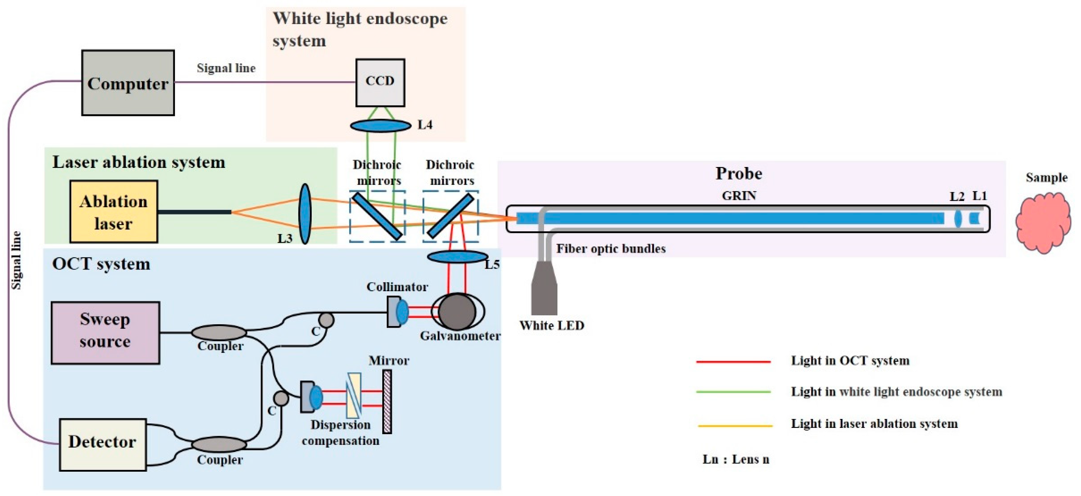
2.1. System Design

Figure 1 shows a schematic of the integrated multifunctional laryngoscope. The white light endoscope and the OCT system were coupled to the probe by two 45° dichroic mirrors, while the laser ablation treatment system was in a straight line. The endoscope was a common-path structure.
The OCT unit was a swept-source OCT system with an A-scan rate of 100 kHz (HSL-20-100, Santec Corporation, Japan). The full spectral sweep range was 87 nm centered at 1310 nm, which had lower scattering than at a shorter wavelength [29,30]. The incident light from the source was coupled into an 80:20 ratio coupler; 80% entered the sample endoscope and 20% entered the reference arm. In the reference arm, the light was reflected by the mirror and propagated back to the interferometer. The collimated beam in the sample arm was scanned by a two-dimensional galvanometer (GVS002, Thorlabs, Newton, NJ, USA) and focused with an achromatic doublet L5. The beam was guided to the probe by a customized dichroic mirror and then focused on the sample with a working distance of 15 mm. The OCT interference spectrum was acquired by using a balanced detector (PBD430c, Thorlabs, Newton, NJ, USA) and a high-speed digitizer (ATS9360, Alazar Technologies, Pointe-Claire, QC, Canada ).
The white light imaging unit consisted of the probe, the proximal objective, and the camera. Illumination light from a white LED was transmitted to the sample through fiber optic bundles. The reflected light from the sample surface was deflected using a customized dichroic mirror that passed the proximal objective onto the CMOS (Complementary Metal Oxide Semiconductor) sensor (CatchBEST Optoelectronic Equipment, Beijing, China) with 1024 × 1248 pixels.
For the laser ablation unit, the medical ablation laser (Leonardo Dual 45, Biolitec biomedical technology, Jena, Germany ) output beam was guided to the system via a multimode fiber and then coupled to the probe by a doublet L3, as shown in Figure 1. The ablation laser had dual wavelengths at 980 and 1470 nm, and the maximal output power was 45 W. The ratio of power between the two wavelengths could be adjusted for different kinds of applications.

2.2. Probe Design

The probe had a common path design, including the distal objective and the relay system. As shown in Figure 2, there were two lenses in the distal objective: a plano-concave lens L1 with a focal length of −9.8 mm and an achromatic doublet L2 with a focal length of 10 mm. The interval between the two lenses was 15 mm. A GRIN (Gradient-index) lens (Gradient Lens Corporation, Rochester, NY, USA) with a length of one pitch (218 mm) was used as a relay. The homemade optical fiber bundle was placed between the inner metal tube and the outer metal tube and was connected to a LED white light source, which provided additional lighting for the white light endoscope; the fiber used for illumination is a multimode polymers fiber bundle with a 600 μm core. The diameter and length of the probe were 8 and 250 mm, respectively. The working distance of the probe was set to 15 mm.

3. Experiments and Results

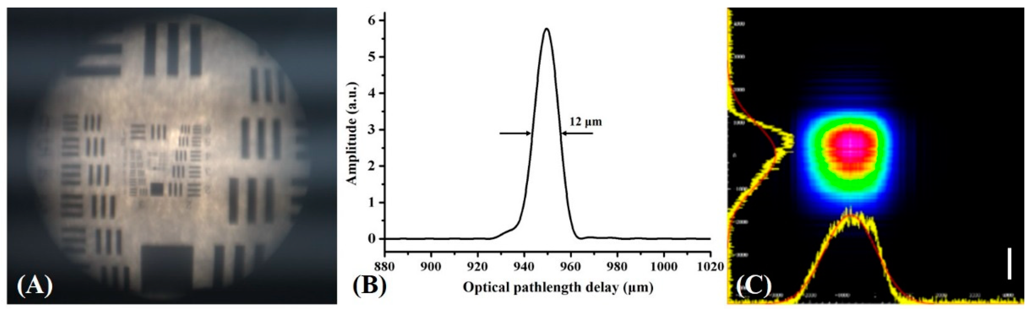
3.1. Evaluation of System Parameters

The parameters of the imaging and ablation unit were tested at the designed working distance of 15 mm. As shown in Figure 3A, for the white light endoscope, the imaging range was 14 mm, and the resolution was 32 lp/mm at the central area by using a test target (1951 USAF test target). The OCT system transverse resolution was 121.7 µm at the central field of view of the OCT system. The axial resolution was 12 μm, which was obtained by analyzing the point spread function, as shown in Figure 3B. Since the light intensity decayed dramatically at the edge of the scanning area, the scanning range of the OCT B-scan was set to 10 mm, which was slightly less than that of the white light endoscope. The B-scan rate was 100 frames/second. This imaging range coincided with that of the white light endoscope. The beam diameter of the ablation laser was 2200 μm at a working distance of 15 mm, as shown in Figure 3C. In conventional laser surgery, the light beam diverges from the multimode fiber. At a distance of 5 mm, the beam diameter is usually larger than 2000 μm. Therefore, this spot diameter at the object distance of 15 mm is acceptable for laser ablation.

3.2. Ex Vivo Porcine Larynx Experiments:

The ex vivo porcine larynx was imaged before and after laser ablation. In total, over 25 tests on three ex vivo porcine larynx samples were carried out. The imaging area is indicated in Figure 4A; the corresponding white light image of the laryngeal surface is shown in Figure 4B; and the cross-sectional OCT image of the black line area is shown in Figure 4C. In the OCT image, three layers can be distinguished: the epithelial layer (E), the mucosa lamina propria (LP), and the smooth muscle layer (SM). The boundary was obvious. Moreover, the basement membrane (BM) in the OCT image was clear, which was the boundary between the epithelial layer and the intrinsic mucosa layer. Some studies have shown that malignant lesions lead to the disappearance of the BM [17,31,32]. Therefore, the coaxial dual-modality images could provide more useful information for diagnosis within one test process.
An ablation power of 15 W was utilized to test the ablation function. The ablation zone (AZ) in Figure 5A indicates the ablation area. After ablation, a round ablation spot can be seen in the white light image. In the OCT image, the ablation effect and boundary can be presented more accurately, as shown in Figure 5B. The preliminary results showed that the multimodal endoscope could be a useful tool for an accurate guiding of laser ablation surgery and could evaluate the ablation effect after surgery.

4. Discussion

A coaxial multifunctional endoscopic system for the diagnosis and treatment of laryngeal lesions was developed. The cross-sectional OCT imaging, superficial white light laryngoscope, and laser ablation function were integrated into one signal probe with a diameter of 8 mm and length of 250 mm. With this coaxial design, the imaging modes and laser ablation position could register more accurately and reduce the examination time compared to the single-modality probe [7,17].
However, some limits still need to be improved. The 8 mm diameter is suitable for the ex vivo study, but for the in vivo study, a smaller diameter is needed since the commonly used laryngoscope diameter is around 5 mm [33]. We plan to use smaller illumination fiber and thinner wall tubing to solve this problem. The lateral resolution of OCT was about 120 μm. It was not as good as the single-channel OCT endoscope [34], which is usually around tens of microns. This was because we wanted to ensure a large field of view of the white light imaging. In the future, we hope to find a balance between the field of view and the lateral resolution. For the ex vivo experiments, the A-scan rate was 100 kHz. It was fast enough to acquire images without large motion artifacts. However, for in vivo studies, the A-scan rate could be further increased to 200 kHz, and a motion feedback function could further increase system stability [35].

5. Conclusions

In this work, a multifunctional laryngoscope was designed and fabricated. OCT and white light imaging and laser ablation surgery function were integrated into one common-path probe. The preliminary ex vivo porcine larynx experimental results showed that this multifunctional imaging and laser ablation endoscope could be a useful tool for diagnosing laryngeal lesions and accurate positioning during laser ablation surgery procedure. We hope to keep improving the performance of the system and try to utilize the system in clinical studies.

Author Contributions

Conceptualization, S.L. and J.Z. (Jun Zhang); methodology, S.L., X.L., and J.K.; software, X.L.; supervision, S.L. and J.Z. (Jun Zhang); validation, X.L., J.K., and J.Z. (Jiebin Zou); resources, F.L.; writing—original draft, S.L. and X.L.; review and editing, S.L. All authors have read and agreed to the published version of the manuscript.

Funding

This research was funded by the National Natural Science Foundation of China (No. 61975246, 61505267) and the Science and Technology Program of Guangzhou (No. 201903010065) and Research Fund for Guangxi Distinguished Experts.

Conflicts of Interest

The authors declare no conflict of interest.

References

  1. Lewis, C.M.; Laccourreye, O.; Weber, R.S.; Holsinger, F.C. Cancers of the Larynx: Tis, T1, T2 Evaluation and Management. Head and Neck Cancer; Springer: New York, NY, USA, 2011. [Google Scholar]
  2. Rudolph, E.; Dyckhoff, G.; Becher, H.; Dietz, A.; Ramroth, H. Effects of tumour stage, comorbidity and therapy on survival of laryngeal cancer patients: A systematic review and a meta-analysis. Eur. Arch. Oto-Rhino-L 2011, 268, 165–179. [Google Scholar] [CrossRef] [PubMed]
  3. Liu, Y.; Zhao, Q.; Ding, G.; Zhu, Y.; Li, W.; Chen, W. Incidence and mortality of laryngeal cancer in China, 2008–2012. Chin. J. Cancer Res. 2018, 30, 299–306. [Google Scholar] [CrossRef]
  4. Hoffman, H.T.; Porter, K.; Karnell, L.H.; Cooper, J.S.; Weber, R.S.; Langer, C.J.; Ang, K.; Gay, G.; Stewart, A.; Robinson, R.A. Laryngeal cancer in the United States: Changes in demographics, patterns of care, and survival. Laryngoscope 2006, 116, 1–13. [Google Scholar] [CrossRef] [PubMed] [Green Version]
  5. Lee, L.A.; Cheng, A.J.; Fang, T.J.; Huang, C.G.; Liao, C.T.; Chang, J.T.; Li, H.Y. High incidence of malignant transformation of laryngeal papilloma in Taiwan. Laryngoscope 2008, 118, 50–55. [Google Scholar] [CrossRef] [PubMed]
  6. Chu, E.A.; Kim, Y.J. Laryngeal cancer: Diagnosis and preoperative work-up. Otolaryng Clin. N. Am. 2008, 41, 673–695. [Google Scholar] [CrossRef] [PubMed]
  7. Davaris, N.; Kropf, S.; Arens, C. Influence of prior laryngeal surgery on the diagnostic value of laryngeal endoscopy. Laryngo-Rhino-Otologie 2018, 97, 10263. [Google Scholar]
  8. Lin, K.; Zheng, W.; Lim, C.M.; Huang, Z. Real-time in vivo diagnosis of laryngeal carcinoma with rapid fiber-optic Raman spectroscopy. Biomed. Opt. Express 2016, 7, 3705–3715. [Google Scholar] [CrossRef] [Green Version]
  9. Song, P. Assessment of vocal cord function and voice disorders. In Principles and Practice of Interventional Pulmonology; Ernst, A., Herth, F., Eds.; Springer: New York, NY, USA, 2013. [Google Scholar]
  10. Bergström, L.; Ward, E.C.; Finizia, C. The impact of laryngeal biopsy on voice outcomes: A pilot study. Otorhinolaryngol. Head Neck Surg. 2016, 1, 33–37. [Google Scholar] [CrossRef] [Green Version]
  11. Wohl, D.L. Nonsurgical management of pediatric vocal fold nodules. Arch. Otolaryngol. Head Neck Surg. 2005, 131, 68–70. [Google Scholar] [CrossRef]
  12. Huang, D.; Swanson, E.A.; Lin, C.P.; Schuman, J.S.; Stinson, W.G.; Chang, W.; Hee, M.R.; Flotte, T.; Gregory, K.; Puliafito, C.A. Optical coherence tomography. Science 1991, 254, 1178–1181. [Google Scholar] [CrossRef] [Green Version]
  13. Fercher, A.F.; Drexler, W.; Hitzenberger, C.K.; Lasser, T. Optical coherence tomography—Principles and applications. Rep. Prog. Phys. 2003, 66, 239–303. [Google Scholar] [CrossRef]
  14. Costa, R.A.; Skaf, M.; Melo, L.A.S.; Calucci, D.; Cardillo, J.A.; Castro, J.C.; Huang, D.; Wojtkowski, M. Retinal assessment using optical coherence tomography. Prog. Retin. Eye Res. 2006, 25, 325–353. [Google Scholar] [CrossRef] [PubMed]
  15. Tsai, T.H.; Lee, H.C.; Ahsen, O.O.; Liang, K.; Giacomelli, M.G.; Potsaid, B.M.; Tao, Y.K.; Jayaraman, V.; Figueiredo, M.; Huang, Q.; et al. Ultrahigh speed endoscopic optical coherence tomography for gastroenterology. Biomed. Opt. Express 2014, 5, 4387–4404. [Google Scholar] [CrossRef] [Green Version]
  16. Sinclair, H.; Bourantas, C.; Bagnall, A.; Mintz, G.S.; Kunadian, V. OCT for the identification of vulnerable plaque in acute coronary syndrome. JACC Cardiovasc. Imaging 2015, 8, 198–209. [Google Scholar] [CrossRef] [Green Version]
  17. Sergeev, A.; Gelikonov, V.; Gelikonov, G.; Feldchtein, F.; Kuranov, R.; Gladkova, N.; Shakhova, N.; Snopova, L.; Shakhov, A.; Kuznetzova, I.; et al. In vivo endoscopic OCT imaging of precancer and cancer states of human mucosa. Opt. Express 1997, 1, 432–440. [Google Scholar] [CrossRef] [PubMed]
  18. Gelikonov, V.M.; Gelikonov, G.V.; Gladkova, N.D.; Shahova, N.M.; Feldchtein, F.I.; Sergeev, A.M.; Imalux, C. Optical Coherent Tomography Apparatus, Fiberoptic Lateral Scanner and Method for Studying Biological Tissues In Vivo. U.S. Patent No. 6,608,684, 19 August 2003. [Google Scholar]
  19. Guo, S.; Hutchison, R.; Jackson, R.P.; Kohli, A.; Sharp, T.; Orwin, E.; Haskell, R.; Chen, Z.; Wong, B.J.F. Office-based optical coherence tomographic imaging of human vocal cords. J. Biomed. Opt. 2006, 11, 30501–30503. [Google Scholar] [CrossRef] [Green Version]
  20. Yu, L.; Liu, G.; Rubinstein, M.; Saidi, A.; Wong, B.J.; Chen, Z. Office-based dynamic imaging of vocal cords in awake patients with swept-source optical coherence tomography. J. Biomed. Opt. 2009, 14, 64020. [Google Scholar] [CrossRef] [Green Version]
  21. Wisweh, H.; Rohrbeck, N.; Krüger, A.; Kraft, M.; Aleksandrov, K.; Lubatschowski, H. In a laryngoscope for office-based imaging of human vocal folds using OCT. In Proceedings of the European Conference on Biomedical Optics, Munich, Germany, 14–18 June 2009; p. 73720I. [Google Scholar]
  22. Donner, S.; Bleeker, S.; Ripken, T.; Ptok, M.; Jungheim, M.; Krueger, A. Automated working distance adjustment enables optical coherence tomography of the human larynx in awake patients. J. Med. Imaging 2015, 2, 26003. [Google Scholar] [CrossRef] [Green Version]
  23. Torkian, B.A.; Guo, S.; Jahng, A.W.; Liaw, L.H.; Chen, Z.; Wong, B.J. Noninvasive measurement of ablation crater size and thermal injury after CO2 laser in the vocal cord with optical coherence tomography. Otolaryngol. Head Neck Surg. 2006, 134, 86–91. [Google Scholar] [CrossRef] [Green Version]
  24. Fleming, C.P.; Quan, K.J.; Rollins, A.M. Toward guidance of epicardial cardiac radiofrequency ablation therapy using optical coherence tomography. J. Biomed. Opt. 2010, 15, 41510. [Google Scholar] [CrossRef]
  25. Murgu, S.D.; Colt, H.G.; Mukai, D.; Brenner, M. Multimodal imaging guidance for laser ablation in tracheal stenosis. Laryngoscope 2010, 120, 1840–1846. [Google Scholar] [CrossRef] [PubMed] [Green Version]
  26. Tsai, T.H.; Zhou, C.; Tao, Y.K.; Lee, H.C.; Ahsen, O.O.; Figueiredo, M.; Kirtane, T.; Adler, D.C.; Schmitt, J.M.; Huang, Q.; et al. Structural markers observed with endoscopic 3-dimensional optical coherence tomography correlating with Barrett’s esophagus radiofrequency ablation treatment response (with videos). Gastrointest. Endosc. 2012, 76, 1104–1112. [Google Scholar] [CrossRef] [PubMed] [Green Version]
  27. Boppart, S.A.; Herrmann, J.; Pitris, C.; Stamper, D.L.; Brezinski, M.E.; Fujimoto, J.G. High-Resolution optical coherence tomography-guided laser ablation of surgical tissue. J. Surg. Res. 1999, 82, 275–284. [Google Scholar] [CrossRef] [PubMed] [Green Version]
  28. Shakhov, A.V.; Terentjeva, A.B.; Kamensky, V.A.; Snopova, L.B.; Gelikonov, V.M.; Feldchtein, F.I.; Sergeev, A.M. Optical coherence tomography monitoring for laser surgery of laryngeal carcinoma. J. Surg. Oncol. 2001, 77, 253–258. [Google Scholar] [CrossRef] [PubMed]
  29. Brezinski, M.E.; Tearney, G.J.; Bouma, B.E.; Izatt, J.A.; Hee, M.R.; Swanson, E.A.; Southern, J.F.; Fujimoto, J.G. Optical coherence tomography for optical biopsy. Properties and demonstration of vascular pathology. Circulation 1996, 93, 1206–1213. [Google Scholar] [CrossRef] [PubMed]
  30. Parsa, P.; Jacques, S.L.; Nishioka, N.S. Optical properties of rat liver between 350 and 2200 nm. Appl. Opt. 1989, 28, 2325. [Google Scholar] [CrossRef] [PubMed]
  31. Kraft, M.; Glanz, H.; von Gerlach, S.; Wisweh, H.; Lubatschowski, H.; Arens, C. Clinical value of optical coherence tomography in laryngology. Head Neck 2008, 30, 1628–1635. [Google Scholar] [CrossRef]
  32. Wong, B.J.; Jackson, R.P.; Guo, S.; Ridgway, J.M.; Mahmood, U.; Su, J.; Shibuya, T.Y.; Crumley, R.L.; Gu, M.; Armstrong, W.B.; et al. In vivo optical coherence tomography of the human larynx: Normative and benign pathology in 82 patients. Laryngoscope 2005, 115, 1904–1911. [Google Scholar] [CrossRef] [Green Version]
  33. Liao, C.C.; Liu, F.C.; Li, A.H.; Yu, H.P. Video laryngoscopy-assisted tracheal intubation in airway management. Expert Rev. Med. Devices 2018, 15, 265–275. [Google Scholar] [CrossRef]
  34. Gora, M.J.; Suter, M.J.; Tearney, G.J.; Li, X. Endoscopic optical coherence tomography: Technologies and clinical applications [Invited]. Biomed. Opt. Express 2017, 8, 2405. [Google Scholar] [CrossRef] [Green Version]
  35. Kraus, M.F.; Potsaid, B.; Mayer, M.A.; Bock, R.; Baumann, B.; Liu, J.J.; Hornegger, J.; Fujimoto, J.G. Motion correction in optical coherence tomography volumes on a per A-scan basis using orthogonal scan patterns. Biomed. Opt. Express 2012, 3, 1182. [Google Scholar] [CrossRef] [PubMed] [Green Version]
Figure 1. Schematic of the integrated multifunctional laryngoscope. L#: lens; GRIN: Gradient-index lens; CCD: charge-coupled device; C: circulator.
Applsci 10 07491 g001
Figure 2. Front (left) and cross-sectional (right) views of the probe. L1: plano-concave lens −9.8 mm; L2: achromatic doublet 10 mm; LMT: inner metal tube; OMT: outer metal tube.
Applsci 10 07491 g002
Figure 3. (A) The test target white light image at 15 mm; (B) Optical coherence tomography (OCT) point spread function (PSF); (C) laser ablation beam profile at 15 mm (scale bar: 1000 μm).
Applsci 10 07491 g003
Figure 4. Ex vivo porcine larynx experiment. (A) Picture of the ex vivo porcine larynx; (B) white light laryngoscope image; (C) OCT image of the black line region. E: epithelial layer; LP: mucosa lamina propria; SM: smooth muscle layer; BM: basement membrane.
Applsci 10 07491 g004
Figure 5. (A) White light laryngoscope image after laser ablation and (B) OCT image after laser ablation.
Applsci 10 07491 g005
Publisher’s Note: MDPI stays neutral with regard to jurisdictional claims in published maps and institutional affiliations.

