Article

Immunochromatographic System for Serodiagnostics of Cattle Brucellosis Using Gold Nanoparticles and Signal Amplification with Quantum Dots

by
Dmitriy V. Sotnikov
1,†,
Lyubov V. Barshevskaya
1,†,
Anatoly V. Zherdev
1,
Saule Z. Eskendirova
2,
Kassym K. Mukanov
2,
Kanatbek K. Mukantayev
2,
Yerlan M. Ramankulov
2 and
Boris B. Dzantiev
1,*
1
A.N. Bach Institute of Biochemistry, Russian Academy of Sciences, Leninsky Prospect 33, Moscow 119071, Russia
2
Republican State Enterprise “National Center for Biotechnology” under the Science Committee of Ministry of Education and Science of the Republic of Kazakhstan, Nursultan 010000, Kazakhstan
*
Author to whom correspondence should be addressed.
Equal contribution.
Appl. Sci. 2020, 10(3), 738; https://doi.org/10.3390/app10030738
Submission received: 27 November 2019 / Revised: 5 January 2020 / Accepted: 15 January 2020 / Published: 21 January 2020
(This article belongs to the Special Issue Chemical and Biological Sensors Applied to Environment and Health)

Abstract

:
In this article, we describe an immunochromatographic test system developed for rapid serodiagnostics of cattle brucellosis using two markers: Gold nanoparticles (GNPs) and quantum dots (QDs). The test system was compared with immunochromatographic serodiagnostics systems that use only one marker. The approbation of the test system was conducted on samples of cattle sera with low, but diagnostically significant titers of specific antibodies. We show that when two conjugates are used, the intensity of the detectable signal increases by 2–3 times compared with the test system using the QD conjugate and by more than nine times compared with the system using the GNP conjugate.

1. Introduction

In recent decades, immunochromatographic assay (ICA) has become widespread in medical and veterinary diagnostics [1]. Immunochromatographic test strips are used for diagnostics of infectious diseases [2,3]; detection of pathogens and toxic substances in food [4,5,6], and other tasks [7]. The main advantage of this technique is the possibility of rapid assaying without expensive equipment, which makes ICA convenient for off-laboratory use. Upon contact of the test strip with a liquid sample, the front of the liquid moves under the action of capillary forces and reactants applied to different zones of the strip interact with each other. These interactions result in the formation of immune complexes that are detected as staining in zones with immobilized reagents.
The most commonly used labels in the ICA are gold nanoparticles (GNPs), due to stability, optical properties and well-developed methodology for modification with biomolecules [8]. The manifestation of coloration in the analytical and control zones of the immunochromatographic test strip is caused by the surface plasmon resonance and influenced by such characteristics of GNPs as their shape, size, dielectric capacity and surroundings [9]. However, even though they are preferred in commercial and research assaying, more sensitive test systems are often needed. Approaches to signal amplification in ICA can be divided into three classes: Alternative markers (i), using markers’ catalytic properties (ii), and an increase in the number of markers in the analytical zone (iii) [10,11]. For instance, to increase the sensitivity, a mixture of conjugates with small and large GNPs was used [12,13]. Another method of signal amplification is the deposition of silver on GNPs by reduction of the silver salt [14]. Parolo et al. proposed an ICA with GNPs coated with a peroxidase. When adding a substrate, such as 3,3′,5,5′-tetramethylbenzidine, 3-amino-9-ethylcarbazole, or 3,3′-diaminobenzidine tetrahydrochloride, insoluble colored products are formed; they are concentrated in the test and control zones of the strip, contributing to their more intense staining [15].
One of the applications of ICA is serodiagnostics—the detection of antibodies against antigens of various pathogens in the blood. The use of this approach is complicated by the fact that the antibodies present in the sample usually account for just a few percent of the total amount of immunoglobulins, even in hyperimmune sera [16,17,18,19]. Blocking immunoglobulin-binding sites on the surface of the label by nonspecific immunoglobulins leads to a reduction in the signal, which in turn makes it difficult to interpret the results of the assay accurately [20]. Therefore, an increase in sensitivity is most in demand for immunochromatographic serodiagnosis [21,22]. In a number of works, the use of quantum dots (QDs) was suggested as a means to increase the sensitivity of ICA [23,24,25,26,27]. QDs are semiconductor nanocrystals that have a narrow and symmetric emission spectrum, which depends on their size and composition [28,29]. QDs allow the reduction of the detection limit in ICA, due to their intense fluorescence and a low background signal.
A combined approach in serodiagnosis in which the use of ICA with GNPs is followed by the stage of signal amplification with QDs is suggested here for the first time. At the first stage, a standard ICA with the conjugate of GNP-(protein G) is carried out. All immunoglobulins in the sample are associated with this conjugate. When the resulting complex enters the analytical zone, it binds to the immobilized antigen by means of its interaction with specific immunoglobulins, which comprise a small fraction of immunoglobulins in the blood. Thus, the binding of the bulk of immunoglobulins to GNP-(protein G) conjugate interferes with the interaction in the analytical zone. In the suggested ICA scheme, the GNP-(protein G) conjugate plays the role of seed. The second conjugate (QD-(protein G)) binds to the GNP-(protein G)-IgG complex in the analytical zone. In this case, the interaction occurs with all immunoglobulins associated with the GNP-(protein G) conjugate. Therefore, in the suggested method, nonspecific immunoglobulins do not interfere with the assay but, on the contrary, become additional marker binding sites. Such an assay scheme and the use of a highly active fluorescent label provide the possibility of highly sensitive detection of antibodies.
The efficacy of this approach was shown in the immunochromatographic serodiagnostics of cattle brucellosis caused by Brucella abortus bacteria. Brucellosis is one of the most widespread zoonotic infections. It causes significant economic damage to agriculture and leads to large livestock losses in the animal husbandries [30]. Brucellosis also poses a threat to human health, due to the possibility of transmission through the consumption of animal products, in particular, raw milk or direct contact with infected animals [31,32]. Transmission of infection in person-to-person contact is rare, therefore, to prevent the spread of the disease, it is necessary to eliminate its source in reservoir animals [33]. The diagnosis of brucellosis is quite difficult because of the long incubation period (from two weeks to approximately nine months), different clinical stages of the disease and various symptoms and signs [34]. The gold standard for the brucellosis diagnosis is still bacteriological testing, but these methods have such limitations as lack of sensitivity, time-consuming assays (1–4 weeks) and the possibility of their implementation only under laboratory conditions [35,36,37,38]. Various serological methods are used in the diagnosis of brucellosis, such as Rose Bengal test, agglutination test, complement fixation, enzyme-linked immunosorbent assay (ELISA) and others, but most of them require special equipment and trained personnel to conduct analysis for a long time, which is critical for diagnostics. Thus, the development of quick, easy to use and highly sensitive tools for the diagnosis of brucellosis is still relevant.

2. Materials and Methods

2.1. Synthesis of GNPs

GNPs were synthesized according to the Frens method [39]—1 mL of 1% aqueous solution of HAuCl4 (Sigma-Aldrich, St. Louis, MO, USA) was added to 100 mL of water. The resulting mixture was brought to boiling, and 1.5 mL of 1% aqueous solution of sodium citrate was added with vigorous stirring. The solution was boiled for 15 min, then cooled and stored at +4 °C.

2.2. Synthesis of Conjugates of GNPs with Streptococcal Immunoglobulin-Binding Protein G

The GNP solution (optical density at 520 nm: D520 = 1) was adjusted with potassium carbonate to pH 8.5–9.0, after which protein G (Imtek, Moscow, Russia) was added to this solution to a concentration of 10 μg/mL. The mixture was incubated for 1 h at room temperature, then mixed with 10% BSA solution (Boval Biosolutions, Fort Worth, TX, USA) and kept for another 10 min with stirring. The resulting solution was centrifuged for 15 min at 10,000× g. The precipitate was collected, and 10% BSA solution was added to 1 mL.

2.3. Preparation of Conjugates of QDs with Protein G

Protein G and QDs with excitation wavelength of 585 nm (Invitrogen, Carlsbad, CA, USA) were conjugated according to [28] with slight modifications as follows—300 μL of protein G (144 μg/mL concentration) in 10 mM borate buffer solution, pH 8.6, was mixed with 25 μL of QDs (8 μM concentration) at a molar ratio of 10:1. Then, 50 μL of N-(4-dimethylaminopropyl)-N′-ethyl-carbodiimide hydrochloride (Fluka, St. Gallen, Switzerland) and 50 μL of sodium N-hydroxysulfosuccinimide (Sigma, St. Louis, MO, USA) at concentrations of 308 μg/mL and 348 μg/mL, respectively, were added to the mixture of protein G and QDs. The resulting mixture was stirred by an Intelli-Mixer RM-2 shaker (Elmi, Rīga, Latvia) for two hours in the dark. The resulting conjugate was concentrated and dialyzed against 10 mM borate buffer solution, pH 8.6, through Amicon Ultracel 30 K filters (Millipore, Burlington, MA, USA) at 10,000 × g for 15 min to remove excess activators and unbound protein. After centrifugation, the resulting conjugate was concentrated by 10 times compared to the reaction volume and kept at +4 °C before application.

2.4. Cattle Serum Panel and B. Abortus Antigen Preparation

A characterized panel of blood serum of cows infected with B. abortus, as well as lipopolysaccharide (LPS) of B. abortus, was provided by the Republican State Enterprise “National Center for Biotechnology” (Nursultan, Kazakhstan). The diagnosis of “brucellosis” was made on the basis of ELISA of the sera. For diagnosing, a standard cutoff serum (Biok, Russia) was used.

2.5. ELISA of Antibodies Against LPS of B. Abortus

The adsorption of LPS in the wells of a 96-well microplate was performed overnight at 4 °C from 100 μL of LPS solution (1 μg/mL concentration) in 50 mM carbonate buffer, pH 9.6. The microplate was washed four times with 50 mM K-phosphate buffer, pH 7.4, comprising 0.1 M NaCl and 0.05% Triton X-100 (PBST), after which 100 μL of sera diluted with PBST from 1:102 to 1:105 in increments of 2 was added to the wells, and the microplate was incubated for 1 h at 37 °C. Then the microplate was washed again, 100 μL of a solution of monoclonal anti-cattle IgG antibodies labeled with horseradish peroxidase (Imtek, Moscow, Russia) at a concentration of 160 ng/mL in PBST were added to each well, and the microplate was incubated for 1 h at 37 °C. After washing the microplate (3 times with PBST and once with distilled water), the peroxidase activity of the enzyme label bound to the carrier was determined. For this, 100 μL of 0.4 mM solution of the substrate 3,3′,5,5′–tetramethylbenzidine (TMB) (Sigma, USA) comprising 0.01% H2O2 was added to the wells, and the microplate was incubated for 15 min at room temperature; then, D450 was measured on a Zenyth 3100 microplate spectrophotometer (Anthos Labtec Instruments, Wals, Austria).

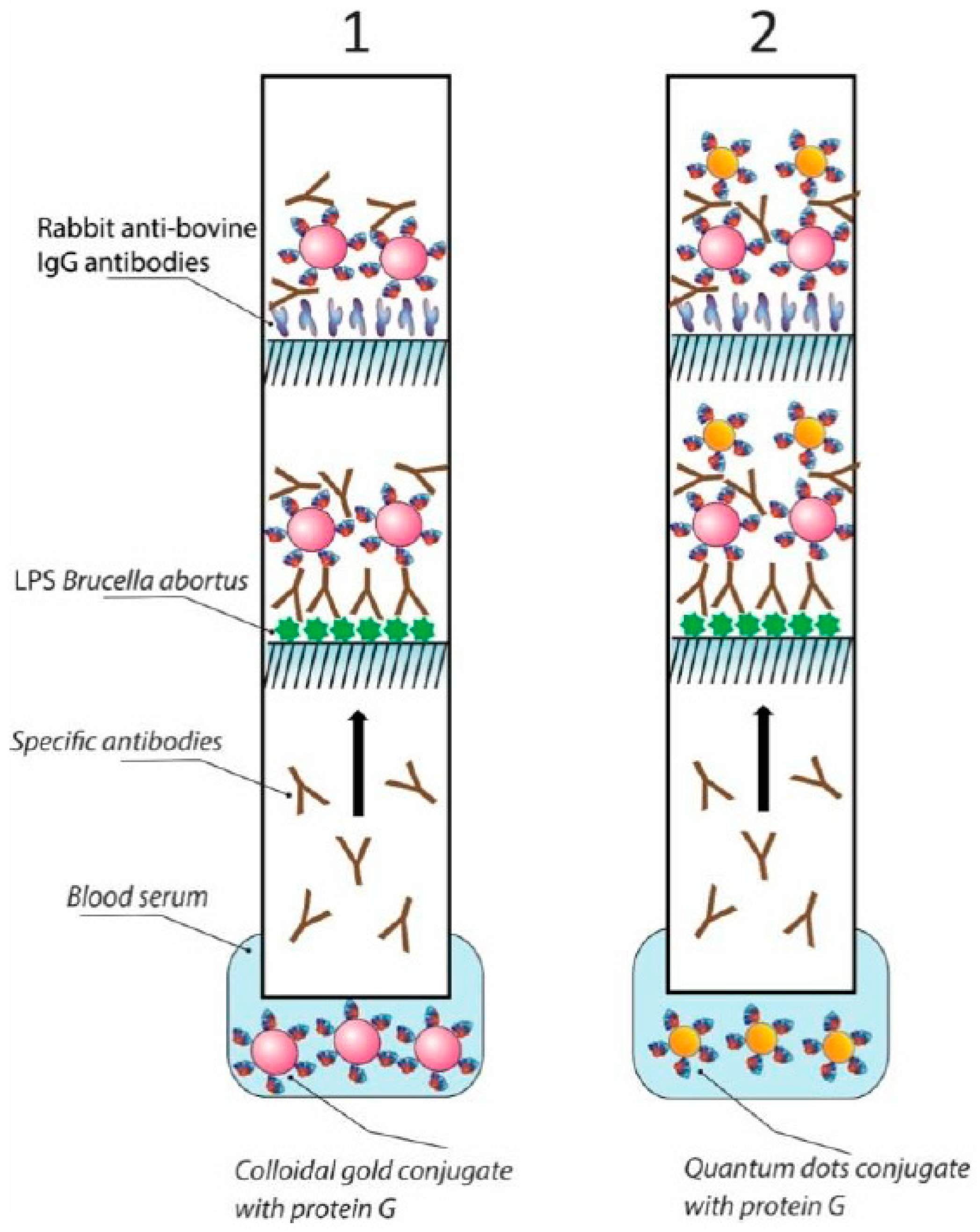
2.6. Assembly of Immunochromatographic Test Systems

The complete set of test strips included a working nitrocellulose membrane, membranes for sample and conjugate application, and a final absorbent membrane. The mdi Easypack CNPH90 membrane (Advanced Microdevices, Ambala Cantt, India) was used as a working membrane. The absorbent membrane CFSP 223,000 (Millipore, USA) was used as a membrane for the sample application. The untreated PT-R5 mdi membrane (Advanced Microdevices, Ambala Cantt, India) was used as a membrane for conjugate application. An IsoFlow automatic dispenser (Imagene Technology, Lebanon, NH, USA) was used to create, on the working nitrocellulose membrane, an analytical zone with immobilized LPS (0.32 mg/mL) in Na-carbonate buffer, pH 9.2, as well as a control zone with immobilized rabbit antibodies against cattle immunoglobulins at a concentration of 0.5 mg/mL (Imtek, Moscow, Russia). Conjugates of GNPs with protein G (D520 = 10, application volume—13 μL/mm) were applied to the conjugate pad. For a test system with QDs, the QD-(protein G) conjugate was applied to the conjugate pad at a concentration of 34 μM with a deposition volume of 1 μL per 1 mm (0.12 nmol per test strip). Atomizing nozzle of the IsoFlow dispenser was used to apply GNPs and QD conjugates with protein G.
After application of the reagents, the membranes were air-dried at 20–22 °C for at least 20 h. A multimembrane composite was assembled, from which 3.5 mm wide strips were made using an Index Cutter-1 automatic guillotine cutter (A-Point Technologies, Gibbstown, NJ, USA). Cutting and packaging were carried out at 20–22 °C in a special room with a relative humidity of not more than 30%. Packed test strips were stored at 20–22 °C.

2.7. Immunochromatographic Detection of Antibodies against LPS of B. abortus

ICA was performed at room temperature. First, 1 μL of blood serum and three drops of PBS (~100 μL) containing 1% Tween-20 were added to an Eppendorf tube. Then, a test strip was vertically placed in the tube. After 10 min, the result of ICA was evaluated. When using a test system with two labels, the conjugate of GNPs with protein G was applied to the conjugate pad, and the conjugate of QDs with protein G was applied to the sample membrane in an amount of 35 μL at a concentration of 3.4 μM (0.12 nmol per test strip) after registration of staining in the analytical zone. Finally, 10 min after the addition of the QD-(protein G) conjugate, the ICA signal was registered in a fluorescent chamber upon exposure to UV light with a wavelength of 365 nm. After analysis procedure, the obtained test strips with GNPs were scanned using a 9000F Mark II scanner (Canon, Tokyo, Japan); the test strips with QDs were photographed on a mobile phone camera under UV light. For quantitative assessment of the color intensity of analytical and control zone of the test strip, the resulting images were digitized and converted from color to monochrome using TotalLab TL120 software (Nonlinear Dynamics, Newcastle upon Tyne, UK).

3. Results and Discussion

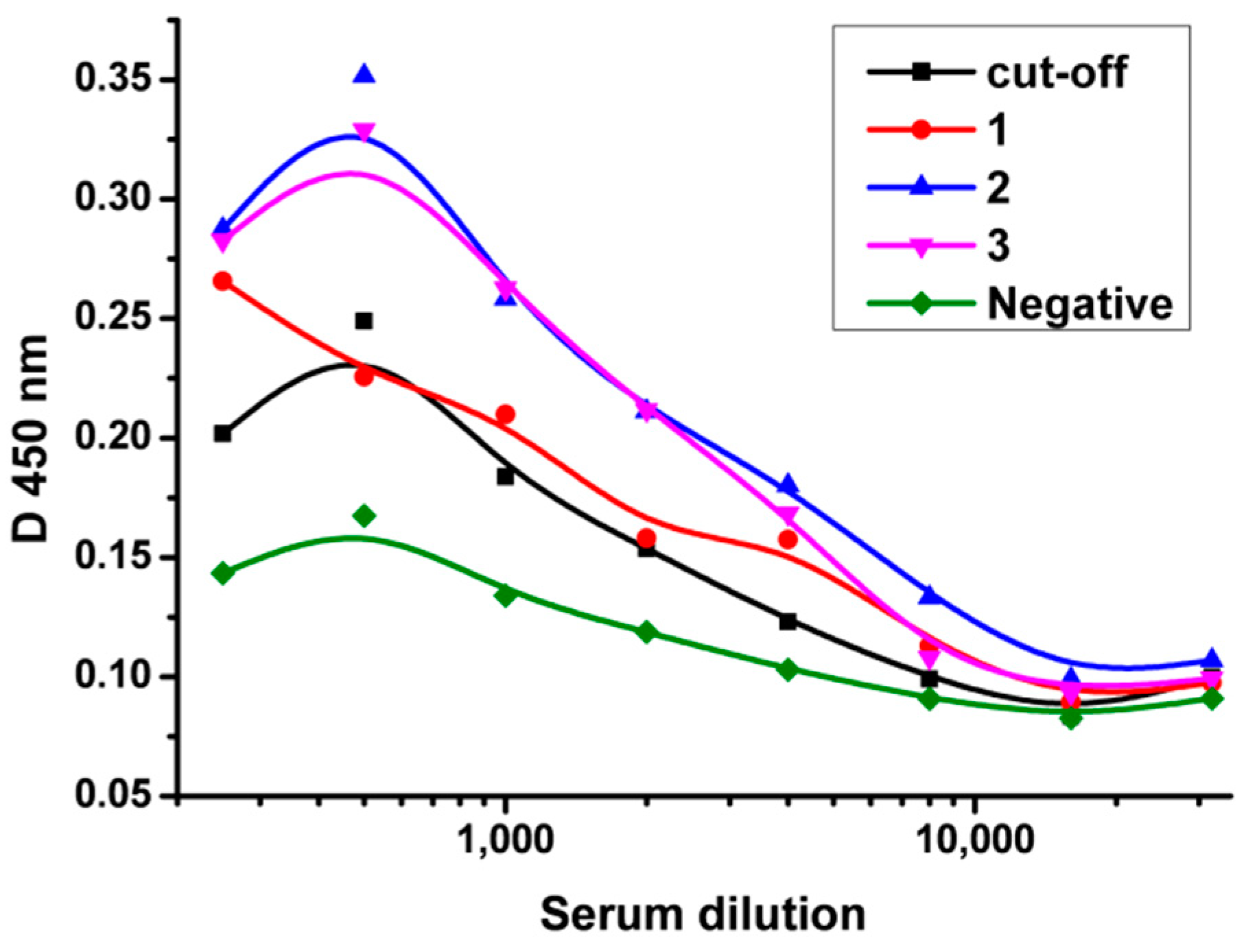
3.1. Characterization of Cattle Sera by ELISA Method

A cattle serum panel consisting of 83 positive samples and 50 samples from healthy animals was characterized by the ELISA method to determine the titers of specific antibodies and confirm the preliminary diagnosis. All sera with an antibody titer higher than the antibody titer in a standard weakly positive cutoff serum were considered positive. Quantitative values of ELISA and ICA for 83 tested positive cattle sera are presented in Table A1 (Appendix A). Although these levels are not strictly proportional (since in ICA reagents interact in the kinetic mode, and the overall level of immunoglobulins affects the result), the groups of samples with low signals intersect well.
Because the main problem of serodiagnostics by the ICA method is poor staining for weakly positive sera, three sera with the lowest antibody titers exceeding the threshold level were selected from the panel. The results of ELISA testing of these sera, together with the cutoff serum and negative control, are presented in Figure 1. These weakly positive sera were used as control bioassays to compare the sensitivities of the three ICA systems.

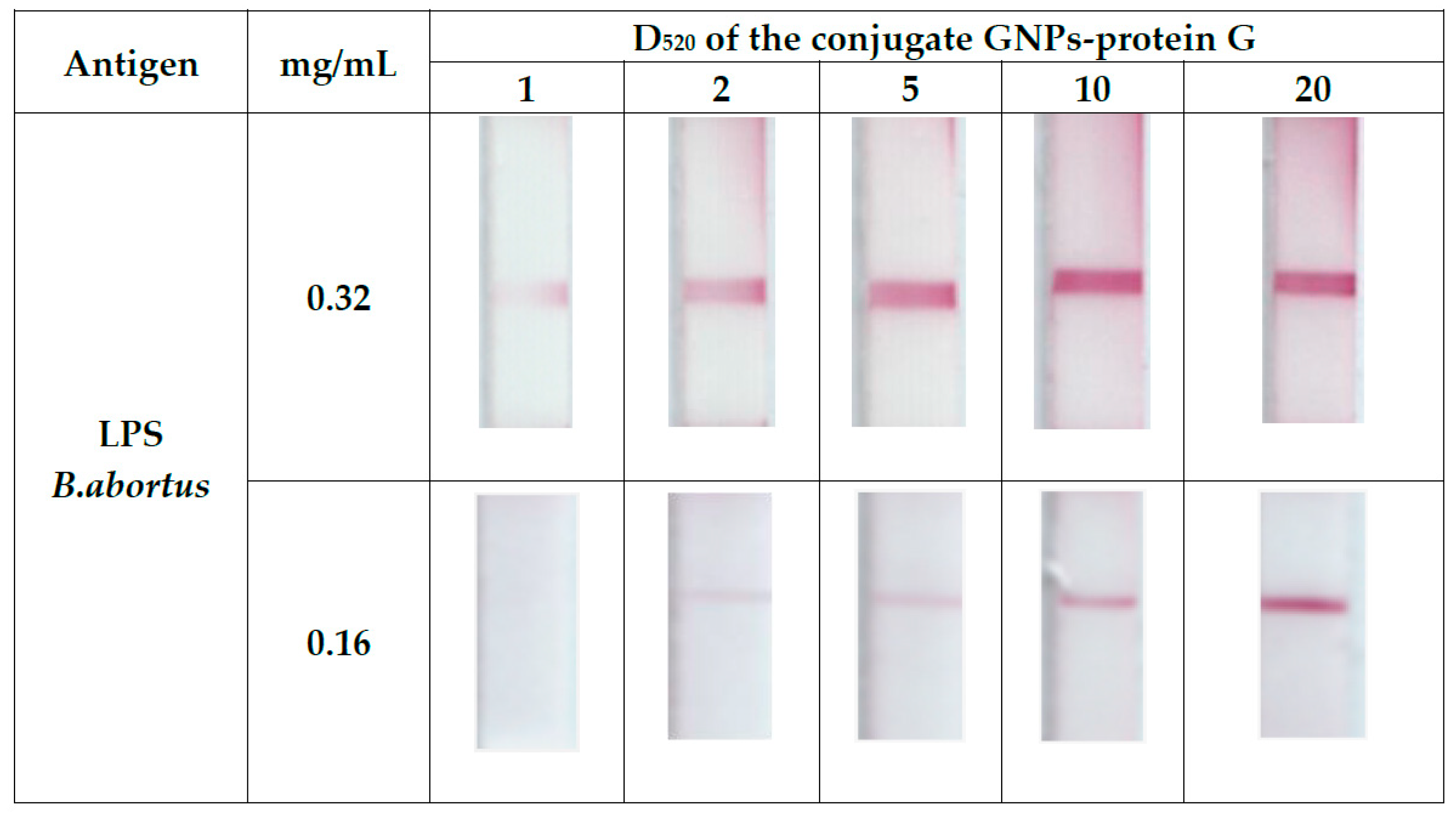
3.2. Optimization of the Concentration of GNP-(Protein G) Conjugate and LPS B. Abortus in the Immunochromatographic Serodiagnostics of Cattle Brucellosis

The dependence of the staining of the analytical zone of the test strip on the concentration of the GNP-(protein G) conjugate was studied to choose the optimal concentrations of reagents used in the final configuration of the test system. For this purpose, the GNP-(protein G) conjugate was tested at five dilutions having D520 values 1, 2, 5, 10, and 20 and at two antigen concentrations, namely, 0.16 and 0.32 mg/mL. The obtained data (Figure 2) demonstrate that with the growth of the GNP-(protein G) conjugate concentration the color intensity of the analytical zone increases, but nonspecific staining of the working membrane also increases. When the conjugate is concentrated to D520 = 20, incomplete removal from the conjugate pad is also observed, which may be associated with partial aggregation of the conjugate. As a result of the above, a working dilution of the GNP-(protein G) conjugate corresponding to D520 =10 was chosen for the final configuration of the test system.

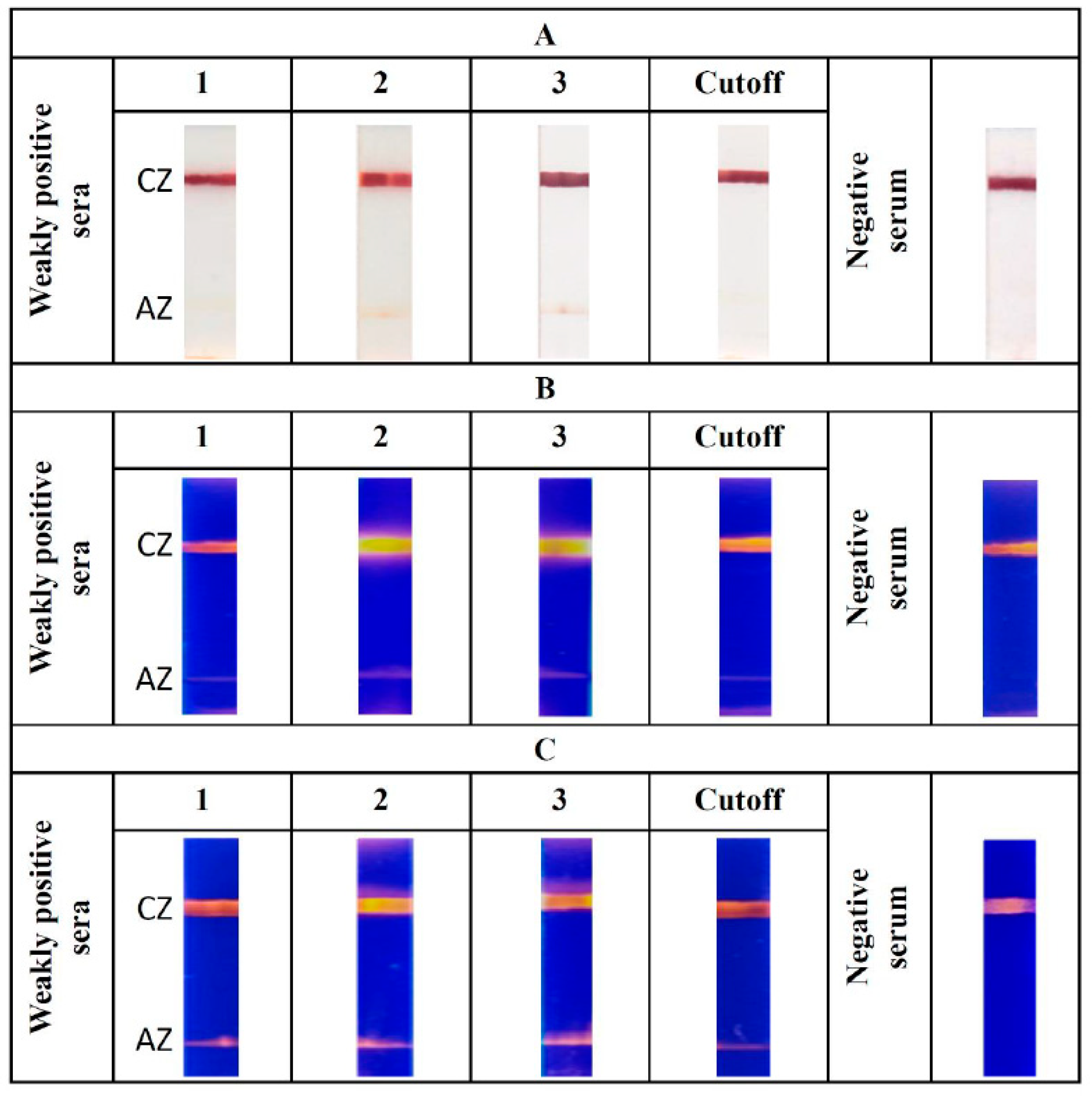
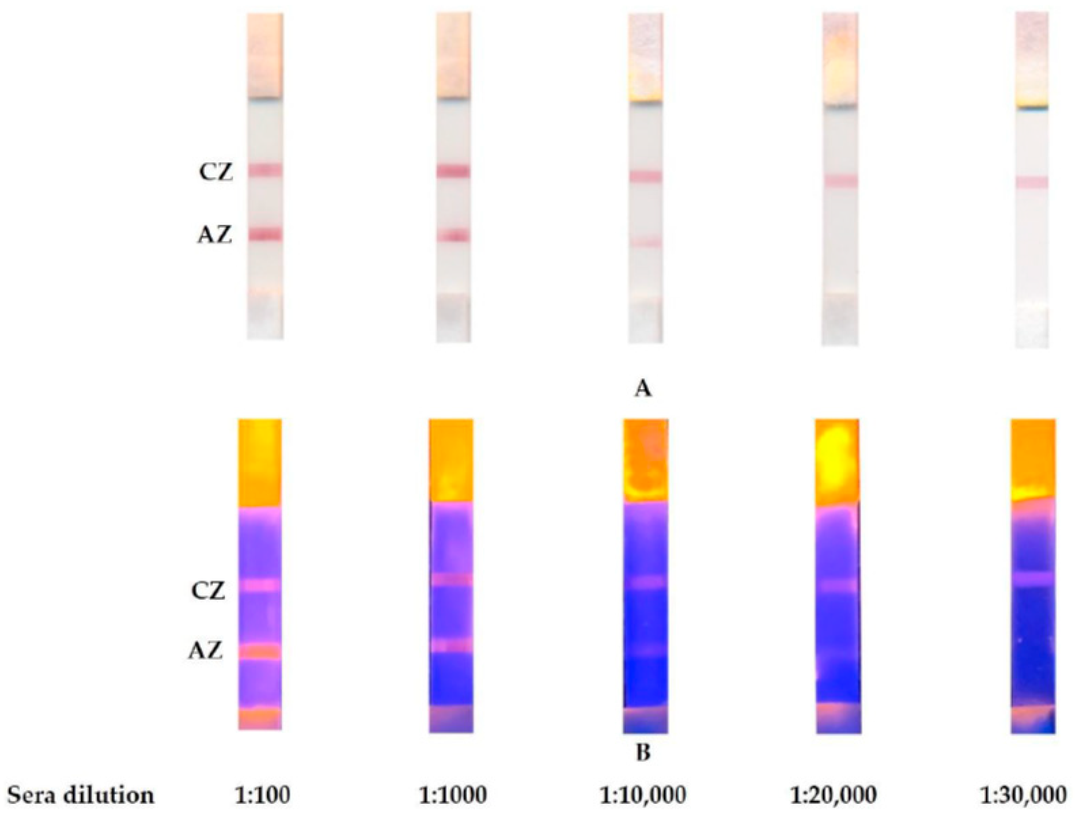
3.3. ICA on Cattle Sera with QD Amplification

ICA was implemented according to the common scheme of ICA serodiagnostics [40]. According to this scheme, when a liquid sample migrates along a test strip, specific immunoglobulins interact with the marker-protein G conjugate. Subsequent detection of the formed complex is carried out by binding of the obtained complex with immobilized LPS in the analytical zone.
All immunoglobulins in the sample can bind to the marker-protein G conjugate, and the number of specific antibodies capable of forming complexes with both the conjugate of GNPs and LPS in the analytical zone is tens of times lower than the total amount of immunoglobulins. For this reason, the ICA sensitivity is often insufficient for reliable serodiagnostics.
In this work, we included in the ICA scheme an additional stage of adding the QD-(protein G) conjugate. Two processes take place in this case: (1) Interaction of the QD-(protein G) conjugate with specific immunoglobulins that did not bind to the GNP-(protein G) conjugate, but bound to LPS in the analytical zone and (2) interaction of the nonspecific immunoglobulins bound to the GNP-(protein G) conjugate in the analytical zone with the QD-(protein G) conjugate. That is, in this case, nonspecific immunoglobulins do not interfere with the assay as they do in the common ICA scheme but, on the contrary, play the role of additional binding sites in the analytical zone. This process results in an increase in signal intensity, which helps obtain more reliable results. The proposed scheme for immunochromatographic serodiagnostics is presented in Figure 3.
Approbation of immunochromatographic test systems for detection of specific antibodies against LPS of B. abortus was performed on a panel of 83 positive sera with a confirmed diagnosis of brucellosis and 50 negative sera. Using the standard ICA technique with the GNP-(protein G) conjugate, 79 out of 83 sera were found to be strictly positive (signal > 0.5) (Table A1). Thus, the diagnostic sensitivity of this test system was 95% of that of ELISA. Using the TotalLab software, the images of the test systems were digitized, which allowed the obtaining of quantitative data on the color intensity in the control and analytical zones. As can be seen from the photographs of the test strips after testing four samples with the lowest antibody titers (Figure 4), the GNP-based ICA system showed negative results for two of the four weakly positive sera. A further two sera showed weak staining and were characterized as conditionally positive. All sera that produced staining of the analytical zone of fewer than 0.2 units were considered negative. A signal from 0.2–1 unit was considered conditionally positive.
Using the test system with QD, 81 out of 83 sera were identified as positive, and two gave questionable results. That is, the sensitivity of the system that uses QDs as a marker and fluorescence registration of the signal increased to 98% of the sensitivity of ELISA. Finally, for a system with two labels–GNPs and QDs–the sensitivity was not inferior to ELISA, and all 83 samples were identified as positive.
To establish the limit of detection for developed test-system 1 μL of standard serum sample was diluted with 1% PBS containing 1% Triton X-100 from 1:100 to 1:30,000 and then tested using GNP-based and GNP+QD-based immunochromatographic test-systems. As shown in Figure 5, the detection limit (the disappearance of color in the analytical zone) of GNP-based tests accords to dilution of standard serum sample 1:10,000, whereas, for GNP+QD-based tests it is equal to 1:20,000.
The obtained data confirm that the sensitivity of the assay is increased if a stage of binding with QDs is included. The results presented in Table 1 show that when two stages of signal generation, with the GNP conjugate and the QD conjugate, are used, the signal intensity in the analytical zone increases by 2–3 times compared with the test system based only on the QD conjugate and by more than nine times compared with the system based on the GNP conjugate. When we tested 50 sera obtained from animals without signs of brucellosis, false-positive results were not observed in any of the assay systems.
To characterize cross-reactions with other pathogens, washed preparations of alternative microbial cells were immobilized on the working membrane. Br. Melitensis, Br. Suis, Yersinia enterocolitica O:9, Y. enterocolitica 287, Francisella tularensis, Vibrio cholerae Inaba, Salmonella enteritidis yena, S. typhimurium TA 100, Escherichia coli 565, E. coli 113-3, Shigella sonne, Sh. Flexneri, Staphylococcus aureus and Citrobacter freundii were used for this purpose at concentrations of 109 cells/ml. The only causative agent of yersiniosis Y. enterocolitica O:9 gave nonspecific staining for cattle sera testing, whereas, all other compounds did not demonstrate cross-reactivity. The reason for the observed exception is the similarity of the O-specific polysaccharide, which is part of the LPS structure of Br. abortus 19 and Y. enterocolitica O:9 [41,42].

4. Conclusions

The obtained data demonstrate that the use of ICA with two conjugates allows the increase in the signal intensity in the analytical zone by 2–3 times compared to ICA based only on the conjugate with QDs and more than nine times compared to ICA based on the conjugate with GNP.
The additional step of adding the conjugate of QDs with protein G helps increase the sensitivity of the assay and allows the obtaining of more accurate and reliable results that are not inferior to ELISA. The revealing of subclinical stages of the infection could be another application of the tests after their additional focused approbation. The proposed format of the immunochromatographic test system is an easy to use, accurate and effective tool for serodiagnosis of cattle brucellosis. The use of the test system does not imply its use with expensive equipment, and signal detection can be performed using a portable UV lamp. The format of the immunochromatographic test system demonstrated through the detection of specific immunoglobulins against Brucella abortus can be used to increase the reliability of serodiagnostics of other diseases.

Ethics Commitment

This research obtained the local ethics commitment at the NCB and approved in accordance with protocols No 3, 23 September 2019 of the local ethics commitment at the NCB.

Author Contributions

Conceptualization, D.V.S., A.V.Z. and B.B.D.; Investigation, L.V.B., D.V.S., S.Z.E. and K.K.M. (Kanatbek K. Mukantayev); Methodology, D.V.S. and B.B.D.; Resources, K.K.M. (Kassym K. Mukanov), Y.M.R. and B.B.D.; Supervision, B.B.D.; Validation, L.V.B. and D.V.S.; Visualization, D.V.S. and A.V.Z.; Writing—original draft, D.V.S. and L.V.B.; Writing—review and editing, D.V.S., L.V.B., A.V.Z., Y.M.R., K.K.M. (Kassym K. Mukanov), K.K.M. (Kanatbek K. Mukantayev) and B.B.D. All authors have read and agreed to the published version of the manuscript.

Funding

This study was financially supported by the Ministry of Science and Higher Education of the Russian Federation (grant agreement No. 14.613.21.0080 on 22.11.2017, unique identifier RFMEFI61317X0080) and by the Science Committe of the Ministry of Education and Science of the Republic of Kazakhstan (Science Technical Program № BR05236334).

Conflicts of Interest

The authors declare no conflict of interest.

Appendix A

Table A1. Comparison between quantitative values of enzyme-linked immunosorbent assay (ELISA) and immunochromatographic assay (ICA) for 83 tested positive cattle sera.
ELISAICA with GNPs
D450 for 1000 Times Dilution of Serum Coloration of Analytical Zone. rel. Units
10.6579.6
20.6538.3
30.471.1
40.45816.4
50.5479.3
60.9825.4
70.70515.4
80.5593.5
90.5315.5
100.5483.4
110.5118.0
120.72216.8
131.09624.2
140.3922.6
150.3033.0
160.85914.2
170.89323.2
180.3951.7
190.3752.6
200.6198.7
210.4958.6
220.3801.5
230.3462.0
240.3091.9
250.3441.2
260.4043.3
270.3916.9
280.2918.1
290.3311.5
300.311.5
310.3121.2
320.3300.7
330.3350.7
340.3350.9
350.3401.0
360.3651.5
370.3503.5
380.4214.0
390.4555.0
400.91215.0
410.2630
420.2822.0
430.3111.5
440.4505.0
450.2772.0
460.6137.0
470.2580.5
480.3200.9
490.3001.0
500.4505.0
510.4174.0
520.2090.2
530.5350.7
540.8845.9
551.18512.3
560.3991.1
570.7872.9
580.7382.0
590.2861.5
600.5971.3
610.621.5
620.5883.8
630.6773.0
640.6045.2
650.3856.5
660.6988.3
670.7011.5
680.4895.1
690.7313.2
700.8391.7
710.5021.1
720.6526.0
731.0072.0
740.9645.3
751.22614
760.9893.1
770.6542.2
781.02310.3
790.8323.6
800.8282.6
810.8753.5
820.4997.9
83 *0.1840
* Cutoff serum. Three sera with the lowest antibody titers exceeding the threshold level and cutoff serum are marked with gray color.

References

  1. Banerjee, R.; Jaiswal, A. Recent advances in nanoparticle-based lateral flow immunoassay as a point-of-care diagnostic tool for infectious agents and diseases. Analyst 2018, 143, 1970–1996. [Google Scholar] [CrossRef] [PubMed]
  2. Lee, J.H.; Seo, H.S.; Kwon, J.H.; Kim, H.T.; Kwon, K.C.; Sim, S.J.; Cha, Y.J.; Lee, J. Multiplex diagnosis of viral infectious diseases (AIDS, hepatitis C, and hepatitis A) based on point of care lateral flow assay using engineered proteinticles. Biosens. Bioelectron. 2015, 69, 213–225. [Google Scholar] [CrossRef] [PubMed]
  3. Anfossi, L.; Di Nardo, F.; Profiti, M.; Nogarol, C.; Cavalera, S.; Baggiani, C.; Giovannoli, C.; Spano, G.; Ferroglio, E.; Mignone, W.; et al. A versatile and sensitive lateral flow immunoassay for the rapid diagnosis of visceral leishmaniasis. Anal. Bioanal. Chem. 2018, 410, 4123–4134. [Google Scholar] [CrossRef] [PubMed]
  4. Majdinasab, M.; Sheikh-Zeinoddin, M.; Soleimanian-Zad, S.; Li, P.; Zhang, Q.; Li, X.; Tang, X. Ultrasensitive and quantitative gold nanoparticle-based immunochromatographic assay for detection of ochratoxin A in agro-products. J. Chromatogr. B 2015, 974, 147–154. [Google Scholar] [CrossRef]
  5. Urusov, A.E.; Petrakova, A.V.; Zherdev, A.V.; Dzantiev, B.B. “Multistage in one touch” design with a universal labelling conjugate for high-sensitive lateral flow immunoassays. Biosens. Bioelectron. 2016, 86, 575–579. [Google Scholar] [CrossRef]
  6. Tominaga, T. Enhanced sensitivity of lateral-flow test strip immunoassays using colloidal palladium nanoparticles and horseradish peroxidase. LWT 2017, 86, 566–570. [Google Scholar] [CrossRef]
  7. Wang, K.; Qin, W.; Hou, Y.; Xiao, K.; Yan, W. The application of lateral flow immunoassay in point of care testing: A review. Nano Biomed. Eng. 2016, 8, 172–183. [Google Scholar] [CrossRef] [Green Version]
  8. Goryacheva, I.; Lenain, P.; Saeger, S. Nanosized labels for rapid immunotests. Trends Analyt. Chem. 2013, 46, 30–43. [Google Scholar] [CrossRef]
  9. Cordeiro, M.; Ferreira Carlos, F.; Pedrosa, P.; Lopez, A.; Baptista, P.V. Gold nanoparticles for diagnostics: Advances towards points of care. Diagnostics 2016, 6, 43. [Google Scholar] [CrossRef]
  10. Ye, H.; Xia, X. Enhancing the sensitivity of colorimetric lateral flow assay (CLFA) through signal amplification techniques. J. Mater. Chem. B 2018, 6, 7102–7111. [Google Scholar] [CrossRef]
  11. Zhou, Y.; Ding, L.; Wu, Y.; Huang, X.; Xiong, Y. Emerging Strategies to develop sensitive AuNP-based ICTS nanosensors. Trends Anal. Chem. 2019, 112, 147–160. [Google Scholar] [CrossRef]
  12. Chen, M.; Yu, Z.; Liu, D.; Peng, T.; Liu, K.; Wang, S.; Xiong, Y.; Wei, H.; Xu, H. Dual gold nanoparticle lateflow immunoassay for sensitive detection of Escherichia coli O157:H7. Anal. Chim. Acta 2015, 87, 71–76. [Google Scholar] [CrossRef] [PubMed]
  13. Shen, G.; Zhang, S.; Hu, X. Signal enhancement in a lateral flow immunoassay based on dual gold nanoparticle conjugates. Clin. Biochem. 2013, 46, 1734–1738. [Google Scholar] [CrossRef] [PubMed]
  14. Yang, W.; Li, X.; Liu, G.; Zhang, B.; Zhang, Y.; Kong, T.; Tang, J.; Li, D.; Wang, Z. A colloidal gold probe-based silver enhancement immunochromatographic assay for the rapid detection of abrin-a. Biosens. Bioelectron. 2011, 26, 3710–3713. [Google Scholar] [CrossRef] [PubMed]
  15. Parolo, C.; de la Escosura-Muñiz, A.; Merkoçi, A. Enhanced lateral flow immunoassay using gold nanoparticles loaded with enzymes. Biosen. Bioelectron. 2013, 40, 412–416. [Google Scholar] [CrossRef] [PubMed] [Green Version]
  16. Kemper, M.J.; Altrogge, H.; Ganschow, R.; Müller-Wiefel, D.E. Serum levels of immunoglobulins and IgG subclasses in steroid sensitive nephrotic syndrome. Pediatr. Nephrol. 2002, 17, 413–417. [Google Scholar] [CrossRef] [PubMed]
  17. Tuerlinckx, D.; Florkin, B.; Ferster, A.; De Schutter, I.; Chantrain, C.; Haerynck, F.; Philippet, P.; Strengers, P.; Laub, R. Pneumococcal antibody levels in children with PID receiving immunoglobulin. Pediatrics 2014, 133, e154–e162. [Google Scholar] [CrossRef] [PubMed] [Green Version]
  18. Turner, P.; Turner, C.; Green, N.; Ashton, L.; Lwe, E.; Jankhot, A.; Day, N.P.; White, N.J.; Nosten, F.; Goldblatt, D. Serum antibody responses to pneumococcal colonization in the first 2 years of life: Results from an SE Asian longitudinal cohort study. Clin. Microbiol. Infect. 2013, 19, E551–E558. [Google Scholar] [CrossRef] [Green Version]
  19. Goldammer, A.; Derfler, K.; Herkner, K.; Bradwell, A.R.; Hörl, W.H.; Haas, M. Influence of plasma immunoglobulin level on antibody synthesis. Blood 2002, 100, 353–355. [Google Scholar] [CrossRef]
  20. Sotnikov, D.V.; Zherdev, A.V.; Avdienko, V.G.; Dzantiev, B.B. Immunochromatographic assay for serodiagnosis of tuberculosis using an antigen-colloidal gold conjugate. Appl. Biochem. Microbiol. 2015, 51, 834–839. [Google Scholar] [CrossRef]
  21. Sotnikov, D.V.; Zherdev, A.V.; Dzantiev, B.B. Theoretical and experimental comparison of different formats of immunochromatographic serodiagnostics. Sensors 2017, 18, 36. [Google Scholar] [CrossRef] [PubMed] [Green Version]
  22. Bishop, J.D.; Hsieh, H.V.; Gasperino, D.J.; Weigl, B.H. Sensitivity enhancement in lateral flow assays: A systems perspective. Lab Chip 2019, 19, 2486–2499. [Google Scholar] [CrossRef] [PubMed] [Green Version]
  23. Sotnikov, D.V.; Berlina, A.N.; Zherdev, A.V.; Eskendirova, S.Z.; Mukanov, K.K.; Ramanculov, Y.M.; Mukantayev, K.N.; Dzantiev, B.B. Comparison of three schemes of quantum dots-based immunochromatography for serodiagnosis of brucellosis in cattle. J. Eng. Appl. Sci 2019, 14, 3711–3718. [Google Scholar] [CrossRef] [Green Version]
  24. Sotnikov, D.V.; Berlina, A.N.; Zherdev, A.V.; Eskendirova, S.Z.; Mukanov, K.K.; Ramankulov, Y.M.; Mukantayev, K.N.; Dzantiev, B.B. Immunochromatographic serodiagnosis of brucellosis in cattle using gold nanoparticles and quantum dots. Int. J. Vet. Sci. 2019, 8, 28–34. [Google Scholar]
  25. Mak, W.C.; Beni, V.; Turner, A.P.F. Lateral-flow technology: From visual to instrumental. Trends Anal. Chem. 2016, 79, 297–305. [Google Scholar] [CrossRef]
  26. Mahmoudi, T.; de la Guardia, M.; Shirdel, B.; Mokhtarzadeh, A.; Baradaran, B. Recent advancements in structural improvements of lateral flow assays towards point-of-care testing. Trends Anal. Chem. 2019, 116, 13–30. [Google Scholar] [CrossRef]
  27. Bahadır, E.B.; Sezgintürk, M.K. Lateral flow assays: Principles, designs and labels. Trends Anal. Chem. 2016, 82, 286–306. [Google Scholar] [CrossRef]
  28. Berlina, A.N.; Taranova, N.A.; Zherdev, A.V.; Sankov, M.N.; Andreev, I.V.; Martynov, A.I.; Dzantiev, B.B. Quantum-Dot-based immunochromatographic assay for total IgE in human serum. PLoS ONE 2013, 8. [Google Scholar] [CrossRef]
  29. Jaiswal, J.K.; Simon, S.M. Potentials and pitfalls of fluorescent quantum dots for biological imaging. Trends Cell Biol. 2004, 14, 497–504. [Google Scholar] [CrossRef]
  30. McDermott, J.J.; Grace, D.; Zinsstag, J. Economics of brucellosis impact and control in low-income countries. Rev. Sci. Tech. 2013, 32, 249–261. [Google Scholar] [CrossRef] [Green Version]
  31. Corbel, M.J. Brucellosis: An overview. Emerg. Infect. Dis. 1997, 3, 213–221. [Google Scholar] [CrossRef] [PubMed]
  32. Dadar, M.; Shahali, Y.; Whatmore, A.M. Human brucellosis caused by raw dairy products: A review on the occurrence, major risk factors and prevention. Int. J. Food Microbiol. 2019, 292, 39–47. [Google Scholar] [CrossRef] [PubMed]
  33. Seleem, M.N.; Boyle, S.M.; Sriranganathan, N. Brucellosis: A re-emerging zoonosis. Vet. Microbiol. 2010, 140, 392–398. [Google Scholar] [CrossRef] [PubMed]
  34. Leal-Hernandez, M.; Díaz-Aparicio, E.; Pérez, R.; Andrade, L.H.; Arellano-Reynoso, B.; Alfonseca, E.; Suárez-Güemes, F. Protection of Brucella abortus RB51 revaccinated cows, introduced in a herd with active Brucellosis, with presence of atypical humoral response. Comp. Immunol. Microbiol. Infect. Dis. 2005, 28, 63–70. [Google Scholar] [CrossRef] [PubMed]
  35. Nielsen, K.; Yu, W. Serological diagnosis of brucellosis. Prilozi 2010, 31, 65–89. [Google Scholar] [PubMed]
  36. Godfroid, J.; Nielsen, K.; Saegerman, C. Diagnosis of brucellosis in livestock and wildlife. Croat. Med. J. 2010, 51, 296–305. [Google Scholar] [CrossRef] [Green Version]
  37. Yagupsky, P. Detection of Brucellae in blood cultures. J. Clin. Microbiol. 1999, 37, 3437–3442. [Google Scholar] [CrossRef] [Green Version]
  38. Corbel, M.; Alton, G.G.; Ariza, J.; Banai, M.; Cosivi, O.; Diaz, R.; Dranovskaya, E.A.; Elber, S.S.; Garin-Bastuji, B.; Kolar, J.; et al. Brucellosis in Humans and Animals; WHO Press: Geneva, Switzerland, 2006; p. 28. [Google Scholar]
  39. Frens, G. Controlled nucleation for the regulation of the particle size in monodisperse gold suspensions. Nat. Phys. Sci. 1973, 241, 20–22. [Google Scholar] [CrossRef]
  40. Ducrotoy, M.J.; Conde-Álvarez, R.; Blasco, J.M.; Moriyón, I. A review of the basis of the immunological diagnosis of ruminant brucellosis. Vet. Immunol. Immunopathol. 2016, 171, 81–102. [Google Scholar] [CrossRef]
  41. Muñoz, P.M.; Marín, C.M.; Monreal, D.; González, D.; Garin-Bastuji, B.; Díaz, R.; Mainar-Jaime, R.C.; Moriyón, I.; Blasco, J.M. Efficacy of several serological tests and antigens for diagnosis of bovine brucellosis in the presence of false-positive serological results due to Yersinia enterocolitica O:9. Clin. Diagn. Lab. Immunol. 2005, 12, 141–151. [Google Scholar] [CrossRef] [Green Version]
  42. Mainar-Jaime, R.C.; Muñoz, P.M.; de Miguel, M.J.; Grilló, M.J.; Marín, C.M.; Moriyón, I.; Blasco, J.M. Specificity dependence between serological tests for diagnosing bovine brucellosis in Brucella-free farms showing false positive serological reactions due to Yersinia enterocolitica O:9. Can. Vet. J. 2005, 46, 913–916. [Google Scholar] [PubMed]
Figure 1. Enzyme-linked immunosorbent assay (ELISA) detection of antibodies against LPS of Brucella abortus in cattle serum.
Applsci 10 00738 g001
Figure 2. Testing a serum sample by gold nanoparticles (GNP)-based immunochromatographic system with varied D520 of the GNP-(protein G) conjugate and varied antigen concentrations.
Applsci 10 00738 g002
Figure 3. Scheme of immunochromatographic assay (ICA) with antibodies against LPS of B. abortus using GNPs and signal amplification with quantum dots (QDs). 1. Conducting ICA using the GNP-(protein G) conjugate. 2. Addition of the QD-(protein G) conjugate.
Applsci 10 00738 g003
Figure 4. Results of immunochromatographic serodiagnostics of cattle brucellosis using the GNP-(protein G) conjugate (A); using the QD-(protein G) conjugate (B); and using the signal amplification with the QD-(protein G) conjugate (C). AZ—analytical zone; CZ—control zone.
Applsci 10 00738 g004
Figure 5. Testing of diluted standard serum sample by immunochromatographic systems for serodiagnostics of cattle brucellosis with GNPs (A) and GNPs+QDs (B). AZ—analytical zone; CZ—control zone.
Applsci 10 00738 g005
Table 1. Results of digitization of images of test systems for immunochromatographic serodiagnostics of cattle brucellosis.
Cattle SeraLabel
GNPsQDsGNPs + QDs
Detection
Instrumental *VisualInstrumental *VisualInstrumental *Visual
10.00.6+/−1.8+
20.2+/−1.8+3.7+
30.5+/−1.2+4.8+
Cutoff0.00.7+/−1.3+
Negative serum0.00.00.0
* Analytical zone, rel. units.

