Article

The Antimicrobial Effect of Radiant Catalytic Ionization on the Bacterial Attachment and Biofilm Formation by Selected Foodborne Pathogens under Refrigeration Conditions

by
Krzysztof Skowron
1,*,
Karolina Jadwiga Skowron
2,
Justyna Bauza-Kaszewska
3,
Ewa Wałecka-Zacharska
4,
Joanna Kwiecińska-Piróg
1,
Katarzyna Grudlewska-Buda
1,
Natalia Wiktorczyk
1 and
Eugenia Gospodarek-Komkowska
1
1
Department of Microbiology, Nicolaus Copernicus University in Toruń, Ludwik Rydygier Collegium Medicum, 9 M. Skłodowskiej-Curie Street, 85-094 Bydgoszcz, Poland
2
Institute of Telecommunications and Computer Science, UTP University of Science and Technology, Al. S. Kaliskiego 7, 85-796 Bydgoszcz, Poland
3
Department of Microbiology and Food Technology, UTP University of Science and Technology, 6 Bernardyńska St., 85-029 Bydgoszcz, Poland
4
Department of Food Hygiene and Consumer Health, Wrocław University of Environmental and Life Sciences, 31 C.K. Norwida St., 50-375 Wrocław, Poland
*
Author to whom correspondence should be addressed.
Appl. Sci. 2020, 10(4), 1364; https://doi.org/10.3390/app10041364
Submission received: 4 January 2020 / Revised: 2 February 2020 / Accepted: 6 February 2020 / Published: 17 February 2020
(This article belongs to the Special Issue Biofilms in Focus: A Threat to Foods)

Abstract

:
The decontamination of food contact surfaces is a major problem for the food industry. The radiant catalytic ionization (RCI) method, based on the ionization process, may be an alternative for conventional decontamination procedures. The advantage of this technique is the possibility of its application to household refrigerating appliances and industrial cold rooms. This study aimed to assess the effect of RCI on the reduction of Campylobacter jejuni, Listeria monocytogenes, and Salmonella Enteritidis from the biofilms formed on a glass surface under refrigeration conditions. Bacterial biofilms were exposed to RCI for 24 h and after 12 (variant I) and 72 h (variant II) of the glass surface contamination. In the last variant (III), the contaminated meat was placed on the glass surface in the refrigerator and subjected to RCI treatment for 72 h. The significantly highest values of absolute reduction efficiency coefficient E were found for the bacterial attachment stage of biofilm formation (variant I). The research proves the efficiency of the RCI method in the reduction of bacteria number from a glass surface.

1. Introduction

Epidemiological risk related to the consumption of food contaminated with pathogenic bacteria is a worldwide problem. According to EFSA (European Food Safety Authority) reports [1], campylobacteriosis and salmonellosis have been the most prevalent zoonoses in the European Union for over a decade. In 2018, 246,571 and 91,857 confirmed cases of human campylobacteriosis and salmonellosis, respectively, were reported. Although the incidence of L. monocytogenes infections (2549 cases) is rather low, an extremely high fatality rate (15.6%) allows to include this bacterium as one of the most important foodborne human pathogens. Due to its ability to survive and grow in the low-temperature range, which is a crucial factor of the food cold chain efficacy, Listeria monocytogenes is considered a microorganism of special concern for the food industry. The largest foodborne Listeria outbreak, with more than 1000 confirmed listeriosis cases and more than 200 fatalities, was reported in 2017–2018 in South Africa [2,3]. In 2018, as a result of the multi-country (Denmark, Finland, UK) outbreak of L. monocytogenes ST6, linked to frozen vegetables, 46 individuals were hospitalized and 5 died [1]. In the same year, in Australia, the consumption of rockmelon contaminated with L. monocytogenes contributed to the death of 7 people [4]. In 2019, outbreaks in Spain [5] and Germany [6] including 3 and 2 deaths, respectively, were noted.
Since Campylobacter, Salmonella, and Listeria spp. are foodborne pathogens of major importance, a number of research studies on their prevalence and survival in food products have been carried out [7,8,9,10,11,12,13,14,15]. It has been proven that these bacteria are commonly present on poultry carcasses and in poultry processing plants, making poultry meat one of the main risk factors for the infections. Poultry meat, due to its high content of proteins and water activity, provides favorable conditions for the growth of both saprotrophic and pathogenic microorganisms. The contamination of poultry may occur in the slaughterhouse or in the processing, packaging, and handling environments. Due to the direct contact with food, working surfaces and equipment are also a serious source of the pathogens in the food. Bacteria may form a biofilm both on the biotic surface (fruits, vegetables, meat, bones) and the abiotic surface (steel, glass, polypropylene, glaze) [16]. Biofilm is a community of surface-attached microorganisms encased in a self-produced extracellular matrix. The ability of some pathogenic bacteria to form biofilm on different materials, used in food processing plants, increases the risk of food recontamination during production. Specific properties of bacterial biofilm make the cells highly resistant to the action of many stress factors, namely, desiccation, temperature, chemical disinfectants, or antibiotics [17,18]. The major part of biofilm structure is the hydrated extracellular polymeric substance (EPS) matrix. The EPS matrix consists mainly of polysaccharides, proteins, lipids, and extracellular DNA (eDNA) that protect bacterial cells. The biofilm formation depends on the availability of the nutrients, synthesis and secretion of the extracellular material, environmental stress, and the competition with other microorganisms. The EPS molecules mediate in the formation of the biofilm architecture, which is a dynamically changing spatial structure. The biofilm structure resembles a sponge that easily absorbs all the molecules secreted in the environment [19,20].
To eliminate pathogens from food contact surfaces, various disinfection procedures are applied in food processing plants. Chemical methods are the most common and effective means of controlling and reducing the number of microorganisms in the food industry. However, considering the increased persistence of the biofilm to the chemicals, the efficiency of this disinfection type may be doubtful or, at least, unpredictable. Moreover, an increased consumer awareness about the negative effects of chemicals on human health results in the search for new methods allowing the elimination of bacteria in the production environment [9,21]. A reasonable alternative for a chemical disinfection might be the application of different types of radiation [22]. One of them is a technology defined as radiant catalytic ionization (RCI) [8,23,24,25,26,27,28]. The RCI technique was initially applied in hospitals and offices to purify air, which in consequence reduced also the level of indoor surface contamination [8]. The effect of RCI on microorganism cells results from the generation of various reactive oxygen species (ROS), among which hydroxyl radical (OH) and singlet oxygen (1O2) are extremely toxic [29]. ROS effectively inhibit growth of Gram-positive and Gram-negative bacteria, fungi, and viruses. Their preventive action against biofilm formation has also been proven [22,30]. The main mechanisms by which ROS affect pathogens activity are as follows: damage of various cellular sites, destruction of proteins and genetic material, inactivation of cellular enzymes, or disturbance of metabolic pathways [28,29].
Since the contamination of food contact surfaces contributes to the recontamination of food products, increasing the risk of human infections, it is extremely important to find methods allowing the elimination of both planktonic forms of bacteria and biofilms from the food production environment. RCI is a cutting-edge technology that has no direct contact with the food and does not leave any chemicals on the treated surface. According to the manufacturer, this method has no negative impact on the food and humans. This is the first study to determine its antibacterial efficacy under refrigeration conditions. As glass is the most popular material used in refrigerators, we decided to use this type of surface in our experiments. In addition, in previous studies, the effectiveness of RCI against bacteria on a glass surface was not determined.
The aim of this study was to assess the effect of the RCI method on the reduction of Campylobacter jejuni, Salmonella Enteritidis, and Listeria monocytogenes from the biofilm formed on a glass surface under refrigeration conditions.

2. Materials and Methods

2.1. Materials

The study materials consisted of 3 Campylobacter jejuni strains (one reference strain ATCC 33560 and two isolated from poultry meat), 3 Listeria monocytogenes strains (one reference strain ATCC 19111 and two isolated from poultry meat), and 3 Salmonella Enteritidis strains (one reference strain ATCC 13076 and two isolated from poultry meat).
The microbial carriers were 3 × 3 cm pieces of chicken breast. The meat samples were thoroughly washed, dried, and packed in paper and foil bags for sterilization before examination. The packages were transported at 4 °C to the Institute of Nuclear Chemistry and Technology in Warsaw and sterilized with a high-energy electron beam (EB). The transport time of the samples took 4 h in each direction. The sterility of the meat was determined after delivery to the laboratory. For this purpose, the samples were homogenized and the obtained suspensions were plated on appropriate media and incubated. The procedure was as described in point 4.
The glass shelf from the refrigerator was cut into 5 × 5 cm fragments. The prepared coupons were thoroughly washed using a detergent, rinsed with distilled water, dried and disinfected with Virusolve + EDS (Amity International, Barnsley, United Kingdom), and finally rinsed with sterile distilled water and wiped with a sterile towel. The effectiveness of the procedure was checked by placing randomly selected disinfected glass fragments (5 coupons) on the surface of Columbia Agar plates with 5% sheep blood and incubation in aerobic and microaerophilic conditions at 37 °C for 72 h.

2.2. Preparation of Bacterial Suspension

For all the strains tested, standardized microbial suspensions in sterile PBS (BTL, Warsaw Poland) with an optical density of 0.5 McFarland standard were prepared. For each suspension, serial 10-fold dilutions in PBS were prepared and a 100 µL portion of each dilution was plated on Columbia Agar with 5% sheep blood (bioMerieux, Craponne, France). Media inoculated with C. jejuni were incubated at 42 °C under microaerophilic conditions (microaerophilic generator CampyGen, Oxoid, Hampshire, United Kingdom) for 48 h, and Listeria and Salmonella cultures were kept at 37 °C for 24 h. After the incubation, the number of bacteria in 1 mL was determined. The bacterial suspensions of 107 CFU × mL−1 were used to contaminate the meat.

2.3. Assessment of the Biofilm Formation by the Tested Strains

Bacterial suspensions of the tested strains were prepared in sterile brain heart infusion (BHI, Becton-Dickinson, Franklin Lakes, New Jersey, USA) according to the procedure described in Section 2. The sterile fragments of the glass shelves from the refrigerator were immersed in bacterial suspensions and placed for 72 h at 4 °C. Then, glass coupons were washed 3 times with sterile PBS, placed in a fresh sterile PBS and sonicated for 5 min. After15 min of shaking (400 rpm), serial ten-fold dilutions in sterile PBS were prepared and a 100 µL portion of each dilution was inoculated onto Columbia Agar medium with 5% sheep blood (bioMerieux, bioMerieux, Craponne, France). After the incubation (conditions described in Section 2.2), the grown colonies were counted and expressed as log CFU × cm−2.

2.4. Contamination of Meat and Glass Coupons

Prepared pieces of chicken breast were injected with the standardized bacterial suspensions and immersed for 1 min in the appropriate suspension (each strain to a separate meat sample). Then, the meat samples were placed separately in sterile Petri dishes in a sterile laminar chamber and allowed to dry for 30 min. The meat contamination procedure was carried out in triplicate for each strain tested. After drying, the number of bacteria in the samples was determined. For this purpose, the meat pieces were placed in sterile bags containing 100 mL of sterile PBS and were homogenized for 10 min in a laboratory stomacher (Bag Mixer 400 VW, Bag Mixer Interscience, Saint-Nom-la-Breteche, France). Next, serial ten-fold dilutions in sterile PBS were prepared and a 100 µL portion of each dilution was plated onto Columbia Agar medium with 5% sheep blood (bioMerieux, bioMerieux, Craponne, France). After the incubation (conditions described in Section 2.2), the grown colonies were counted. The ratio (C) of the number of bacteria recovered from the meat (M, [log CFU × mL−1]) to the number of bacteria in the suspension (S, [log CFU × g−1]) was determined according to the following formula:
C =   M S
The contaminated pieces of chicken breast were then placed on the glass coupons and left under refrigeration conditions for 12 (variant I) or 72 h (variants II and III) (temperature 4 °C). The number of Listeria isolated from the contaminated glass surface directly after the meat was removed and before carrying out further experimental procedures was determined (I).

2.5. Experimental Design

The tests were carried out in three variants. In variant I, the contaminated meat fragments were placed on a sterile glass piece and left for 12 h in the refrigerator. Then, the meat sample was removed and radiant catalytic ionization (RCI) was applied for 24 h (Induct 750, ActivTek, Kielce, Poland). In variant II, the contaminated meat samples were left on the glass surface for 72 h and then removed, and the device generating radiant catalytic ionization was turned on in the refrigerator for 24 h (Induct 750, ActivTek, Kielce, Poland). In variant III, the contaminated meat was placed on sterile glass fragments in the refrigerator and exposed to RCI (Induct 750, ActivTek, Kielce, Poland) for 72 h. The applied variants of the experiment allowed to check the effect of RCI under refrigeration conditions on the cell attachment (variant I), mature biofilm (variant II), and the process of biofilm formation (variant III).
Next, the glass fragments were washed 3 times with sterile PBS, placed in sterile PBS and subjected to 5 min sonication (Ultrasonic DU-4 sonicator, Nickel-Electro, Weston-super-mare, United Kingdom). After 15 min of shaking (400 rpm), serial ten-fold dilutions in sterile PBS were prepared and a 100 µL portion of each dilution was plated onto Columbia Agar medium with 5% sheep blood (bioMerieux). After the incubation (conditions described in Section 2.2), the grown colonies were counted and expressed as log CFU × cm−2. The positive controls were glass fragments contaminated with the poultry meat according to the procedure appropriate for the individual experimental variant (I, II, or III), but not treated with RCI. The negative controls were sterile pieces of poultry meat and fragments of glass shelves.
The level of reduction (R) of the number of bacteria recovered from RCI-treated (+RCI) and non-treated (K+; -RCI) glass coupons relative to the initial number of bacteria on glass coupons after contact with the contaminated meat was calculated according to the following formula:
R =   I B I
where:
  • R—bacterial count reduction
  • I—initial number of bacteria on glass coupons after contact with the contaminated meat [log CFU × cm−2]
  • B—the number of bacteria recovered in a given experimental variant for the control (K+; -RCI) and tested samples (+RCI) [log CFU × cm−2]
In the case of the experimental variant III, the formula was as follows:
R =   ( R C I ) ( + R C I ) ( R C I )
where:
  • R—bacterial count reduction
  • (-RCI)—number of bacteria determined on glass plates in the control variant without the RCI technology [log CFU × cm−2]
  • (+RCI)—number of bacteria recovered after RCI technology [log CFU × cm−2]
The coefficient of absolute RCI efficiency was also calculated, according to the following formula:
E =   R + R C I R R C I
  • ERCI absolute effectiveness coefficient
  • R+RCI—level of bacterial reduction as a result of RCI application
  • R-RCI—level of bacterial reduction without RCI application

2.6. Statistical Analysis

The obtained results were analyzed with Statistica 13.0 PL software (StatSoft). Multivariate analysis of variance was performed based on general linear models (GLMs). To check whether significant differences existed between the tested experimental groups, the post-hoc Tukey’s test at the significance level α = 0.05 was applied.

3. Results

3.1. Biofilm Formation

The research confirmed the biofilm formation ability by all tested strains. The strongest biofilm was formed by S. Enteritidis. The average bacteria number for Salmonella spp. was 8.80 log CFU × cm−2 (Figure 1). The intensity of biofilm formation between Salmonella spp. strains was not significant and varied from 8.74 log CFU × cm−2 (S. Enteritidis ATCC) to 8.86 log CFU × cm−2 (S. Enteritidis strain 2). The number of cells recovered from the biofilms of each Salmonella strain was significantly higher compared to C. jejuni, which had the weakest biofilm-formation capabilities. The average bacteria number of C. jejuni was 7.37 log CFU × cm−2 (from 7.14 log CFU × cm−2 for C. jejuni strain ATCC to 7.53 log CFU × cm−2 for C. jejuni strain 2) (Figure 1). The number of bacteria reisolated from the biofilm formed by L. monocytogenes varied from 7.85 log CFU × cm−2 for L. monocytogenes ATCC strain to 7.95 log CFU × cm−2 for L. monocytogenes strain 1 (the average of 7.91 log CFU × cm−2 (Figure 1). No significant differences were observed between individual strains within the species investigated.

3.2. Recovery Ratio of Bacteria from Contaminated Samples

The average number of bacteria reisolated from the contaminated poultry meat samples varied from 5.966 log CFU × g−2 for S. Enteritidis strains to 6.395 log CFU × g−2 for C. L. monocytogenes strains (Figure 2). No significant differences between individual strains of the examined species were noted.
The C ratio, describing the effectiveness of the bacteria recovery from the contaminated food samples, ranged from 0.709 (S. Enteritidis strain 2) to 0.822 (L. monocytogenes ATCC 19111), but these differences were not statistically significant.

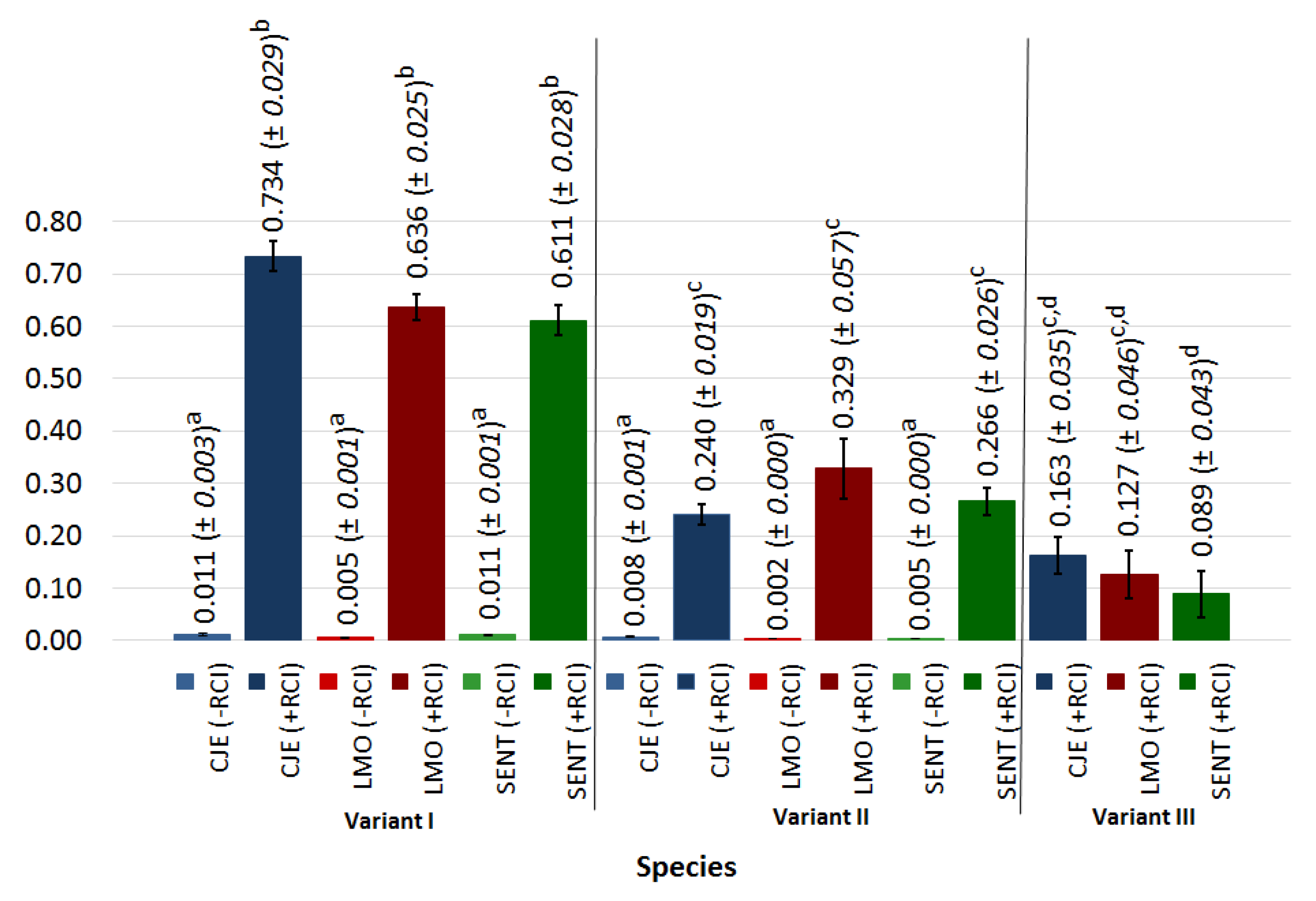
3.3. Assessment of the Effectiveness of Surface Disinfection Using Radiant Catalytic Ionization during Bacterial Attachment (Experimental Variant I)

In the case of experimental variant I, the average number of bacteria reisolated from the glass surface after 12 h contact with the meat sample ranged from 4.332 log CFU × cm−2 for S. Enteritidis strains (from 4.182 log CFU × cm−2 for S. Enteritidis strain 2 to 4.448 log CFU × cm−2 for S. Enteritidis strain 1) to 4.837 log CFU × cm−2 for L. monocytogenes strains (from 4.823 log CFU × cm−2 for L. monocytogenes strains ATTC and 2 to 4.864 log CFU × cm−2 for L. monocytogenes strain 1). The differences were not statistically significant (Figure 2). In all cases, no significant differences were noted between strains of the tested species. After 24 h, the bacteria count on the glass surface not subjected to RCI treatment remained at a similar level. The average number of bacteria reisolated from the glass surface for C. jejuni, L. monocytogenes, and S. Enteritidis was 4.681 log CFU × cm−2, 4.810 log CFU × cm−2, and 4.285 log CFU × cm−2, respectively (Figure 2). The number of C. jejuni varied from 4.492 log CFU × cm−2 (strain 2) to 4.778 log CFU × cm−2 (strain 1), L. monocytogenes from 4.795 log CFU × cm−2 (strain ATCC) to 4.838 log CFU × cm−2 (strain 1), and S. Enteritidis from 4.138 CFU × cm−2 (strain 2) to 4.401 log CFU × cm−2 (strain 1). Application of RCI for 24 h resulted in a statistically significant decrease of C. jejuni (varied from 1.192 CFU × cm−2 for strain 2 to 1.308 CFU × cm−2 for ATCC strain), L. monocytogenes (varied from 1.678 CFU × cm−2 for ATCC strain to 1.811 CFU × cm−2), and S. Enteritidis (varied from 1.646 CFU × cm−2 for ATCC strain to 1.708 CFU × cm−2 for strain 1). The average number of bacteria reisolated in this experimental variant was 1.258 log CFU × cm−2 for C. jejuni, 1.764 log CFU × cm−2 for L. monocytogenes, and 1.683 log CFU × cm−2 for S. Enteritidis (Figure 2). The differences between species were not statistically significant.
The calculated reduction rates of the tested bacteria during experimental variant I are presented in Figure 3. The reduction rate of the bacteria reisolated from the glass surface without RCI treatment was not higher than 0.012 and no significant differences between strains and species were found. On the contrary, RCI most efficiently reduced the number of C. jejuni (0.734). For L. monocytogenes, the reduction rate was 0.636 and for S. Enteritidis, 0.611. The differences calculated for tested species were not statistically significant.
The absolute RCI efficiency coefficient (E) values for the experimental variant I ranged from 0.584 (S. Enteritidis strain 2) to 0.725 (C. jejuni strain 1). The differences between strains and species were not statistically significant (Table 1).

3.4. Assessment of Effectiveness of Surface Disinfection Using Radiant Catalytic Ionization during Biofilm Maturation (Experimental Variant II)

The average number of bacteria reisolated from the glass surface, after 72 h contact with the contaminated meat sample, for C. jejuni was 4.729 log CFU × cm−2 (Figure 4). The greatest number of bacteria for C. jejuni strains was noted for the reference strain (5.478 log CFU × cm−2) and was significantly higher than the values obtained for the other strains (4.355 log CFU × cm−2—strain 1, 4.354 log CFU × cm−2—strain 2) The initial number of L. monocytogenes and S. Enteritidis isolated from the contaminated glass surface ranged from 6.082 log CFU × cm−2 (strain 2) to 6.258 log CFU × cm−2 (ATTC strain) and 5.082 log CFU × cm−2 (for strain 1) to 5.213 log CFU × cm−2 (strain 2), respectively. The average number of bacteria for L. monocytogenes and S. Enteritidis was 6.176 log CFU × cm−2 and 5.162 log CFU × cm−2, respectively. No statistically significant differences between the individual strains of both species were observed (Figure 4).
As presented in Figure 4, no significant difference between the initial bacteria number on the glass and the bacteria number isolated from the non-treated with RCI surface was observed. For C. jejuni, the bacteria number ranged from 4.358 log CFU × cm−2 (C. jejuni strain 1) to 5.352 (C. jejuni strain ATCC). For L. monocytogenes, the values ranged from 6.241 log CFU × cm−2 (L. monocytogenes strain 1) to 6.011 log CFU × cm−2 (L. monocytogenes strain 2). For S. Enteritidis, the number of bacteria reisolated from the glass surface not treated with RCI ranged from 5.120 log CFU × cm−2 (S. Enteritidis ATCC strain) to 5.225 log CFU × cm−2 (S. Enteritidis strain 2). The average number of bacteria reisolated from the biofilm of C. jejuni, L. monocytogenes, and S. Enteritidis was 4.691 log CFU × cm−2, 6.164 log CFU × cm−2, and 3.786 log CFU × cm−2, respectively. Significant differences between C. jejuni, L. monocytogenes, and S. Enteritidis were noted (Figure 4). The 24 h application of radiant catalytic ionization caused a statistically significant decrease of the bacteria number, as compared with the non-RCI-treated surface. The average bacteria number after 24 h RCI application was 3.724, 4.145, and 2.761, for C. jejuni, L. monocytogenes, and S. Enteritidis, respectively. The count of C. jejuni ranged from 3.393 log CFU × cm−2 (ATCC strain) to 3.668 log CFU × cm−2 (strain 2), L. monocytogenes from 3.587 log CFU × cm−2 (strain 2) to 4.585 log CFU × cm−2 (ATCC strain), and S. Enteritidis from 3.614 log CFU × cm−2 (for strain 2) to 3.880 log CFU × cm−2 (strain 1). Significant differences between strains of the same species were observed in the case of L. monocytogenes reference strain and strain 2. The reduction of the S. Enteritidis number was significantly higher than that observed for C. jejuni and L. monocytogenes (Figure 4).
The reduction of bacteria number (R) in the biofilm formed on the non-RCI-treated glass surface ranged from 0.002 to 0.008. High effectiveness of RCI application was confirmed by the obtained reduction rate values, ranging from 0.240 (C. jejuni) to 0.329 (L. monocytogenes). No significant differences were observed between different species (Figure 3).
The highest value of the absolute RCI efficiency coefficient (0.409) was noted for L. monocytogenes strain 2; however, it was not statistically significantly higher than those calculated for two other strains of this species. On the contrary, absolute RCI efficiency coefficients calculated for C. jejuni strains 1 and 2 (0.173 and 0.150, respectively) were statistically significantly lower, as compared with C. jejuni reference strain (0.375), L monocytogenes strains 1 and 2, and S. Enteritidis strain 2 (0.306, 0.409, and 0.302, respectively). No significant differences in the RCI absolute efficiency coefficient were observed for S. Enteritidis strains (Table 1).

3.5. Assessment of Effectiveness of Surface Disinfection Using Radiant Catalytic Ionization during Biofilm Formation (Experimental Variant III)

The average number of bacteria reisolated from the glass surface not treated with RCI was 5.472 log CFU × cm−2, 6.124 log CFU × cm−2, and 5.162 log CFU × cm−2 for C. jejuni, L. monocytogenes, and S. Enteritidis, respectively (Figure 5). The number of bacteria for C. jejuni ranged from 5.526 log CFU × cm−2 (ATCC strain) to 5.439 log CFU × cm−2 (strain 2), for L. monocytogenes from 6.060 log CFU × cm−2 (strain 2) to 6.166 log CFU × cm−2 (ATCC strain), and for S. Enteritidis from 5.091 log CFU × cm−2 (ATCC strain) to 5.211 log CFU × cm−2 (strain 2). The application of radiant catalytic ionization resulted in a decrease of bacteria number, but not statistically significant. The average number of bacteria for C. jejuni, L. monocytogenes, and S. Enteritidis was 4.602 log CFU × cm−2, 5.366 log CFU × cm−2, and 4.647 log CFU × cm−2, respectively. No significant differences between RCI-treated and non-treated samples were observed for all species (Figure 5).
The highest bacteria number reduction rate after RCI treatment was observed for C. jejuni strains (0.163) in variant III. L monocytogenes and S. Enteritidis reduction rates were 0.127 and 0.089, respectively. The differences in the bacteria number reduction rate between species were not statistically significant (Figure 3).
The coefficient of absolute RCI efficiency calculated for the tested bacteria varied from 0.049 (L. monocytogenes strain 1) to 0.171 (C. jejuni strain 2). The E values for C. jejuni strains were statistically significantly higher than those obtained for the remaining strains, excluding L. monocytogenes reference strain (0.119) and S. Enteritidis strain 1 (0.102). The absolute RCI efficiency coefficients of L. monocytogenes and S. Enteritidis strains were not statistically significantly different (Table 1).
An overall analysis of E coefficient showed, for all the tested strains, statistically significantly higher values in the experimental variant I (Table 1). Comparable and not statistically different coefficients for variants II and III were observed for C. jejuni strains 1 and 2, L. monocytogenes reference strain, and S. Enteritidis strain 2. For the rest of the tested strains, the E values were significantly higher when the variant II was applied. The highest reduction of bacteria number after application of RCI was achieved in variant I, whereas the lowest was found in variant III (Figure 3).

4. Discussion

The elimination of pathogenic microorganisms from food contact surfaces is of great concern for the food industry. Since many foodborne human pathogens are able to form biofilm on various surfaces, widely used in the food processing environment, the risk of food cross-contamination during production increases. Many pathogenic bacteria are known to adhere to, and subsequently, form biofilm on the food and food contact surfaces, such as stainless steel, plastic, glass, or rubber [8,11,12,26,27,31,32]. In the present study, all the tested species formed biofilm on the glass surface after 72 h at 4 °C. The strongest biofilm was observed for S. Enteritidis. According to Manijeh et al. [12], the number of cells in the biofilm formed on the glass surface by S. Enteritidis ranged from 104 log CFU × cm−2 after 2 h to 107 log CFU × cm−2 after 20 h of adhesion at 35 °C. In our study, the number of S. Enteritidis after 72 h at 4 °C ranged from 8.74 to 8.86 log CFU × cm−2.
Currently, a wide spectrum of conventional chemical methods can be applied for the efficient eradication of pathogens from food contact surfaces. Nonetheless, the toxicity of chemical agent residues and the potential increase of microbial resistance to the applied chemicals encourage researchers to search for new techniques of safe and effective pathogen inactivation. Alternative processes, including the control of quorum sensing, the use of phages, and the application of essential oils (EO), biosanitizers, bacteriocines, blue laser light, ozone, pulsed electric field, high pressure technologies or electrolyzed water, are currently at various stages of development [21,22,31,33]. The important alternative antimicrobial technology is ionizing radiation. The most commonly used forms for decontamination purposes are gamma rays, high energy electrons and X-rays; however, a number of modern methods based on this type of radiation, that is, cold plasma or binary ionization technology (BIT), are still intensively investigated [34,35,36,37,38]. Radiant catalytic ionization (RCI) is one of these novel techniques and, according to the results of many research studies, appears to be a reasonable alternative to the conventional decontamination procedures. The great advantage of RCI, in contrast to gamma rays, X-rays, and high energy electrons, is the possibility of its application to household refrigeration appliances, industrial cold rooms, and rooms with low indoor temperature, intended for raw or processed food product storage.
The antimicrobial efficiency of RCI is attributed to the cell damaging effect of oxidative gases, including ozone and peroxide, generated by the RCI cell [25,28]. The inhibition of pathogenic microorganisms on various surfaces was confirmed by our previous studies [8,26,27]. The reduction rate of the L. monocytogenes number in biofilms formed on different surfaces (rubber, milled rock tiles, polypropylene) varied from 3.92% to 70.10%, whereas in the case of stainless steel AISI 304 and lacquered veneer surfaces, up to 95% of L. monocytogenes and S. Enteritidis were reduced [26]. Saini et al. [39] achieved 4.37 log CFU reduction of the L. monocytogenes number on stainless steel coupons after 15 min of photohydroionization (PHI) treatment. According to Ortega et al. [25], the reduction of different microorganisms, including foodborne pathogens, on stainless steel surfaces subjected to 24 h of RCI treatment reached at least 90%. RCI efficacy against Salmonella on food contact surfaces and animal food products was also confirmed [28,40]. The present research showed the inhibition of C. jejuni, L. monocytogenes, and S. Enteritidis strains on a glass surface after RCI treatment. To assess the RCI effect exclusively on bacteria, the absolute reduction rate coefficient (E) was calculated. The highest E values for all species ranged from 0.584 to 0.725 and were obtained after 24 h of RCI application to the surface contaminated for 12 h (experimental variant I). On the contrary, a significant reduction was not observed on the glass surface covered with the contaminated meat sample during 72 h exposure of RCI (variant III). In this case, the meat sample placed on the glass surface created a favorable environment for bacterial growth, protecting the microorganisms from the harmful effect of RCI.
The efficiency of radiant catalytic ionization is determined by a variety of factors, including type and physiological state of microorganisms, properties of the contaminated surface, environmental conditions, and parameters of the ionization process [26,41,42]. The sensitivity to irradiation is species- and strain-dependent and may be associated with the origin of the strain, the metabolic state of cells, and the ability to form biofilm [26]. The results obtained by Yang et al. [40] suggest less sensitivity to photohydroionization of the non-antimicrobial resistant (non-AMR) Salmonella strain compared to AMR Salmonella. Spore-forming bacteria and viruses are more resistant to radiant catalytic ionization [42]. According to Kang et al. [43], the structural differences of Gram-positive and Gram-negative bacteria affect the resistance to ionization. It was found that S. Typhimurium inoculated in pork jerky was more sensitive to EB-irradiation combined with the leek extract treatment than L. monocytogenes. On the contrary, in the study by Osaili et al. [42], L. monocytogenes showed higher radiation sensitivity than Salmonella spp. Skowron et al. [27] noted a similar level of L. monocytogenes and S. Enteritidis reduction after RCI treatment. Mannozzi et al. [28] observed lower sensitivity of Escherichia coli, as compared to Listeria innocua and S. Typhimurium after 60 min of RCI treatment; however, after 90 min no differences were reported. In the present study, in most cases, we observed no significant differences in the resistance to RCI exposure between C. jejuni, L. monocytogenes, and S. Enteritidis. No statistically significant differences in the bacteria number reduction rate were noted for the strains of the same species, except for C. jejuni in the experimental variant II.
The time of ionization affects the irradiation efficiency and, generally, the greater reduction is associated with the longer time exposure [25,28,44]. According to Grinshpun et al. [45], approximately 75% of B. subtilis spores were inactivated in 10 min, and about 90% after 30 min of photocatalytic oxidation. Ortega et al. [25] noted that RCI reduced microbial populations on stainless steel surfaces within 2 h and the greatest reduction was achieved after a 24 h treatment. The study of Yang et al. [40] showed no differences in Salmonella reduction resulting from the extension of photohydroionization time from 15 to 60 s. Ionization time applied in our research ranged from 24 to 72 h. However, the longer exposition to RCI was applied to the surface covered with the contaminated meat sample during the whole experiment.
The lower effectiveness of disinfection agents against bacterial biofilm compared to planktonic forms is a well-documented and undoubtful fact [26,32,46,47,48,49]. It is speculated that the biofilm extracellular polymeric substances (EPSs) may serve as the primary target of ROS and protect the associated bacteria cells [32]. Biofilm maturity is considered as an important factor affecting bacteria cell sensitivity to ionization. Biofilm formation is a four-stage biological process. In the initial stage, bacteria attach to the surface and next they proliferate, form microcolonies, and finally detach from the mature biofilm structure [50]. According to Sommers [51], ionizing radiation is more efficient against bacteria in the initial phase of attachment than in mature biofilm. The D10 values for most food-borne pathogens are higher in the stationary phase of growth than in the log phase. Niemira [32] found that reduced susceptibility to ionization was achieved for the mature 24 h biofilm and no further differences in radiation sensitivity were observed for 48 h and 72 h samples. In the present study, the highest effectiveness of RCI treatment was observed also against the bacteria at the initial stage of biofilm formation—bacterial attachment (variant I). The reduction of bacteria number in the mature biofilm, formed on the glass surface after 72 h contact with the contaminated meat sample (variant II), was significantly lower.
The research proves the RCI efficiency against bacteria on a glass surface. Nevertheless, the state of bacterial cells plays an important role. Satisfying results of the current research on the inhibiting effect of RCI on microorganisms on the materials used in the food industry support the need for further studies for the implementation of this method on an industrial scale.

Author Contributions

Conceptualization, K.S., E.W.-Z., and K.J.S.; methodology, K.S., K.J.S., and J.K.-P.; validation, K.S. and J.B.-K.; formal analysis, K.S. and E.W.-Z.; investigation, K.J.S., N.W., and K.G.-B.; resources, J.B.-K., N.W., and K.G.-B.; writing—original draft preparation, K.J.S., J.K.-P., and J.B.-K.; writing—review and editing, K.S., K.G.-B., and E.W.-Z.; visualization, J.K.-P. and N.W.; supervision, K.S. and E.G.-K.; project administration, K.S.; funding acquisition, E.W.-Z. and E.G.-K. All authors have read and agreed to the published version of the manuscript.

Funding

This research was funded by the Nicolaus Copernicus University with funds from the maintenance of the research potential of the Department of Microbiology DS-UPB, city, country.

Conflicts of Interest

The authors declare no conflict of interest.

References

  1. EFSA. The European Union One Health 2018 Zoonoses Report. Available online: https://www.efsa.europa.eu/en/efsajournal/pub/5926 (accessed on 21 December 2019).
  2. Desai, A.N.; Anyoha, A.; Madoff, L.C.; Lassmann, B. Changing epidemiology of Listeria monocytogenes outbreaks, sporadic cases, and recalls globally: A review of ProMED reports from 1996 to 2018. Int. J. Infect. Dis. 2019, 84, 48–53. [Google Scholar] [CrossRef] [PubMed] [Green Version]
  3. Smith, A.M.; Tau, N.P.; Smouse, S.L.; Allam, M.; Ismail, A.; Ramalwa, N.R.; Disenyeng, B.; Ngomane, M.; Thomas, J. Outbreak of Listeria monocytogenes in South Africa, 2017-2018: Laboratory Activities and Experiences Associated with Whole-Genome Sequencing Analysis of Isolates. Foodborne Pathog. Dis. 2019, 16, 524–530. [Google Scholar] [CrossRef] [PubMed] [Green Version]
  4. Das, A. Listeriosis in Australia—January to July 2018. Glob. Biosecurity 2019, 1, 150–158. [Google Scholar] [CrossRef]
  5. WHO. Available online: https://www.who.int/csr/don/16-september-2019-listeriosis-spain/en/ (accessed on 21 December 2019).
  6. Food Safety News. Available online: https://www.foodsafetynews.com/2019/10/german-meat-plant-linked-to-listeria-outbreak-stops-production/ (accessed on 21 December 2019).
  7. Carpentier, B.; Cerf, O. Review--Persistence of Listeria monocytogenes in food industry equipment and premises. Int. J. Food Microbiol. 2011, 31, 1–8. [Google Scholar] [CrossRef] [PubMed]
  8. Skowron, K.; Wiktorczyk, N.; Kwiecińska-Piróg, J.; Sękowska, A.; Wałecka-Zacharska, E.; Gospodarek-Komkowska, E. Elimination of Klebsiella pneumoniae NDM from the air and selected surfaces in hospital using radiant catalytic ionization. Lett. Appl. Microbiol. 2019, 69, 333–338. [Google Scholar] [CrossRef] [PubMed]
  9. Saraiva, C.; García Díez, J.; Fontes, M.; Esteves, A. Modeling the Behavior of Listeria monocytogenes in Meat. List. Monocytogenes 2018, 25. [Google Scholar] [CrossRef] [Green Version]
  10. Zuber, I.; Lakicevic, B.; Pietzka, A.; Milanov, D.; Djordjevic, V.; Karabasil, N.; Teodorovic, V.; Ruppitsch, W.; Dimitrijevic, M. Molecular characterization of Listeria monocytogenes isolates from a small-scale meat processor in Montenegro, 2011–2014. Food Microbiol. 2019, 79, 116–122. [Google Scholar] [CrossRef]
  11. Russo, P.; Hadjilouka, A.; Beneduce, L.; Capozzi, V.; Paramithiotis, S.; Drosinos, E.H.; Spano, G. Effect of different conditions on Listeria monocytogenes biofilm formation and removal. Czech. J. Food Sci. 2018, 36, 208–214. [Google Scholar] [CrossRef] [Green Version]
  12. Manijeh, M.A.; Mohammad, J.M.; Roha, K.K. Biofilm Formation by Salmonella enteritidis on Food Contact Surfaces. J. Biol. Sci. 2008, 8, 502–505. [Google Scholar]
  13. Hue, O.; Le Bouquin, S.; Laisney, M.J.; Allain, V.; Lalande, F.; Petetin, I.; Rouxel, S.; Quesne, S.; Gloaguen, P.Y.; Picherot, M.; et al. Prevalence of and risk factors for Campylobacter spp. contamination of broiler chicken carcasses at the slaughterhouse. Food Microbiol. 2010, 27, 992–999. [Google Scholar] [CrossRef]
  14. Connerton, I.F.; Connerton, P.L. Foodborne Diseases, 3rd ed.; Elsevier Science Publishing Co Inc.: San Diego, CA, USA, 2017; pp. 209–221. [Google Scholar]
  15. Igwaran, A.; Okoh, A.I. Human campylobacteriosis: A public health concern of global importance. Heliyon 2019, 5, e02814. [Google Scholar] [CrossRef] [PubMed]
  16. Jensen, L.K.; Bjarnsholt, T.; Kragh, K.N.; Aalbæk, B.; Henriksen, N.L.; Blirup, S.A.; Pankoke, K.; Petersen, A.; Jensen, H.E. In vivo gentamicin susceptibility test for prevention of bacterial biofilms in bone tissue and on implants. Antimicrob Agents Chemother. 2019, 63, e01889-18. [Google Scholar] [CrossRef] [PubMed] [Green Version]
  17. Stewart, P.S. Animicrobial tolorance in biofilms. Microbiol. Spectr. 2015, 3, MB-0010-2014. [Google Scholar] [CrossRef] [Green Version]
  18. Wu, H.; Moser, C.; Wang, H.Z.; Hoiby, N.; Song, Z.J. Strategies for combating bacterial biofilm infections. Int. J. Oral Sci. 2015, 7, 1–7. [Google Scholar] [CrossRef] [Green Version]
  19. Pearce, P.; Song, B.; Skinner, D.J.; Mok, R.; Hartmann, R.; Singh, P.K.; Jeckel, H.; Oishi, J.; Drescher, K.; Dunkel, J. Flow-induced symmetry breaking in growing bacterial biofilms. Phys. Rev. Lett. 2019, 20, 258101. [Google Scholar] [CrossRef]
  20. Flemming, H.C.; Wingender, J.; Szewzyk, U.; Steinberg, P.; Rice, S.A.; Kjelleberg, S. Biofilms: An emergent form of bacterial life. Nat. Rev. Microbiol. 2016, 14, 563–575. [Google Scholar] [CrossRef]
  21. Giaouris, E.; Heir, E.; Hébraud, M.; Chorianopoulos, N.; Langsrud, S.; Møretrø, T.; Habimana, O.; Desvaux, M.; Renier, S.; Nychas, G.J. Attachment and biofilm formation by foodborne bacteria in meat processing environments: Causes, implications, role of bacterial interactions and control by alternative novel methods. Meat Sci. 2014, 97, 298–309. [Google Scholar] [CrossRef]
  22. Rupel, K.; Zupin, L.; Ottaviani, G.; Iris Bertani, I.; Martinelli, V.; Porrelli, D.; Vodret, S.; Roman Vuerich, R.; Daniel Passos da Silva, D.; Bussani, R.; et al. Blue laser light inhibits biofilm formation in vitro and in vivo by inducing oxidative stress. NPJ Biofilms Microbiomes 2019, 29, 1–11. [Google Scholar] [CrossRef]
  23. Diehl, J.F. Food irradiation-Past, present and future. Rad. Phys. Chem. 2002, 63, 211–215. [Google Scholar] [CrossRef]
  24. Lado, B.H.; Yousef, A.E. Alternative food-preservation technologies: Efficacy and mechanisms. Microbes Infect 2002, 4, 433–440. [Google Scholar] [CrossRef]
  25. Ortega, M.T.; Franken, L.J.; Hatesohl, P.R.; Marsden, J.L. Efficacy of ecoquest radiant catalytic ionization cell and breeze at ozone generator at reducing microbial populations on stainless steel surfaces. J. Rapid Methods Autom Microbiol. 2007, 15, 359–368. [Google Scholar] [CrossRef]
  26. Skowron, K.; Grudlewska, K.; Krawczyk, A.; Gospodarek- Komkowska, E. The effectiveness of radiant catalytic ionization in inactivation of Listeria monocytogenes planktonic and biofilm cells from food and food contact surfaces as a method of food preservation. J. Appl. Microbiol. 2018, 124, 1493–1505. [Google Scholar] [CrossRef] [PubMed]
  27. Skowron, K.; Grudlewska, K.; Kwiecińska-Piróg, J.; Gryń, G.; Śrutek, M.; Gospodarek-Komkowska, E. Efficacy of radiant catalytic ionization to reduce bacterial populations in air and on different surfaces. Sci. Total Environ. 2018, 610–611, 111–120. [Google Scholar] [CrossRef] [PubMed]
  28. Mannozzi, J.T.; Filbert, V.J.; Mackay, W.J.; Fulford, D.E.; Steele, C.W. Evaluation of radiant catalytic ionization in reducing Escherichia coli, Listeria innocua and Salmonella typhimurium on represantative food contact surfaces. Texas J. Sci. 2018, 70, 6. [Google Scholar] [CrossRef]
  29. Vatansever, F.; de Melo, W.C.; Avci, P.; Vecchio, D.; Sadasivam, M.; Gupta, A.; Chandran, R.; Karimi, M.; Parizotto, N.A.; Yin, R.; et al. Antimicrobial strategies centered around reactive oxygen species--bactericidal antibiotics, photodynamic therapy, and beyond. FEMS Microbiol. Rev. 2013, 37, 955–989. [Google Scholar] [CrossRef] [Green Version]
  30. Dryden, M. Reactive oxygen species: A novel antimicrobial. Int. J. Antimicrob Agents 2018, 51, 299–303. [Google Scholar] [CrossRef]
  31. Soni, K.A.; Nannapaneni, R. Removal of Listeria monocytogenes biofilms with bacteriophage P100. J. Food Prot. 2010, 73, 1519–1524. [Google Scholar] [CrossRef]
  32. Niemira, B.A. Irradiation sensitivity of planktonic and biofilm-associated Escherichia coli O157:H7 isolates is influenced by culture conditions. Appl. Environ. Microbiol. 2007, 73, 3239–3244. [Google Scholar] [CrossRef] [Green Version]
  33. Vidács, A.; Kerekes, E.; Rajkó, R.; Petkovits, T.; Alharbi, N.S.; Khaled, J.M.; Vágvölgyi, C.; Krisch, J. Optimization of essential oil-based natural disinfectants against Listeria monocytogenes and Escherichia coli biofilms formed on polypropylene surfaces. J. Mol. Liq. 2018, 255, 257–262. [Google Scholar] [CrossRef]
  34. Higgins, S.E.; Wolfenden, A.D.; Bielke, L.R.; Pixley, C.M.; Torres-Rodriguez, A.; Vicente, J.L.; Bosseau, D.; Neighbor, N.; Hargis, B.M.; Tellez, G. Application of Ionized Reactive Oxygen Species for Disinfection of Carcasses, Table Eggs, and Fertile Eggs. J. Appl. Poult. Res. 2005, 14, 716–720. [Google Scholar] [CrossRef] [Green Version]
  35. Misra, N.N.; Jo, C. Applications of cold plasma technology for microbiological safety in meat industry. Trends Food Sci. Technol. 2017, 64, 74–86. [Google Scholar] [CrossRef]
  36. Yang, S.; Sadekuzzaman, M.; Ha Do, S. Reduction of Listeria monocytogenes on chicken breasts by combined treatment with UV-C light and bacteriophage ListShield. LWT Food Sci. Technol. 2017, 86, 193–200. [Google Scholar] [CrossRef]
  37. Ritter, A.C.; Santi, L.; Vannini, L.; Beys-da-Silva, W.O.; Gozzi, G.; Yates, J.; Ragni, L.; Brandelli, A. Comparative proteomic analysis of foodborne Salmonella Enteritidis SE86 subjected to cold plasma treatment. Food Microbiol. 2018, 76, 310–318. [Google Scholar] [CrossRef] [PubMed]
  38. Roh, S.H.; Lee, S.Y.; Park, H.H.; Lee, E.S.; Min, S.C. Effects of the treatment parameters on the efficacy of the inactivation of Salmonella contaminating boiled chicken breast by in-package atmospheric cold plasma treatment. Int. J. Food Microbiol. 2019, 293, 24–33. [Google Scholar] [CrossRef] [PubMed]
  39. Saini, J.K.; Marsden, J.L.; Getty, K.J.; Fung, D.Y. Advanced oxidation technology with photohydroionization as a surface treatment for controlling Listeria monocytogenes on stainless steel surfaces and ready-to-eat cheese and turkey. Foodborne Pathog. Dis. 2014, 11, 295–300. [Google Scholar] [CrossRef] [PubMed]
  40. Yang, X.; Kalchayanand, N.; Belk, K.E.; Wheeler, T.L. Photohydroionization Reduces Shiga Toxin-Producing Escherichia coli and Salmonella on fresh beef with minimal effects on meat quality. Meat Muscle Biol. 2019, 3, 105–115. [Google Scholar] [CrossRef] [Green Version]
  41. Chung, H.; Lee, N.; Jo, C.; Shin, D.; Byun, M. Use of gamma irradiation for inactivation of pathogens inoculated into Kimbab, steamed rice rolled by dried laver. Food Control 2007, 18, 108–112. [Google Scholar] [CrossRef]
  42. Osaili, T.M.; Al-Nabulsi, A.A.; Aljaafreh, T.F. Use of gamma radiation for inactivating Salmonella spp., Escherichia coli O157:H7 and Listeria monocytogenes in tahini halva. Int. J. Food Microbiol. 2018, 2, 20–25. [Google Scholar] [CrossRef]
  43. Kang, M.; Kim, H.-J.; Jayasena, D.D.; Bae, S.Y.; Yong, I.H.; Lee, M.; Jo, C. Effects of combined treatments of electron-beam irradiation and addition of Leek (Allium tuberosum) extract on reduction of pathogens in pork jerky. Foodborne Pathog. Dis. 2012, 9, 1083–1087. [Google Scholar] [CrossRef]
  44. Gabriel, A.A.; Vera, D.D.; Lazo, O.M.Y.; Azarcon, V.B.; De Ocampo, C.G.; Marasigan, J.C.; Sandel, G.T. Ultraviolet-C inactivation of Escherichia coli O157:H7, Listeria monocytogenes, Pseudomonas aeruginosa, and Salmonella enterica in liquid egg white. Food Control 2017, 73, 1303–1309. [Google Scholar] [CrossRef]
  45. Grinshpun, S.A.; Adhikari, A.; Honda, T.; Kim, K.Y.; Toivola, M.; Rao, K.S.; Reponen, T. Control of aerosol contaminants in indoor air: Combining the particle concentration reduction with microbial inactivation. Environ. Sci. Technol. 2007, 41, 606–612. [Google Scholar] [CrossRef] [PubMed]
  46. Kim, Y.; Kim, H.; Beuchat, L.R.; Ryu, J.H. Development of non-pathogenic bacterial biofilms on the surface of stainless steel which are inhibitory to Salmonella enterica. Food Microbiol. 2018, 69, 136–142. [Google Scholar] [CrossRef] [PubMed]
  47. Fagerlund, A.; Langsrud, S.; Heir, E.; Mikkelsen, M.I.; Møretrø, T. Biofilm Matrix Composition Affects the Susceptibility of Food Associated Staphylococci to Cleaning and Disinfection Agents. Front. Microbiol. 2016, 7, 856. [Google Scholar] [CrossRef] [PubMed] [Green Version]
  48. Pang, X.Y.; Yang, Y.S.; Yuk, H.G. Biofilm formation and disinfectant resistance of Salmonella sp. in mono- and dual-species with Pseudomonas aeruginosa. J. Appl. Microbiol. 2017, 123, 651–660. [Google Scholar] [CrossRef] [PubMed]
  49. Rodríguez-Melcón, C.; Riesco-Peláez, F.; García-Fernández, C.; Alonso-Calleja, C.; Capita, R. Susceptibility of Listeria monocytogenes planktonic cultures and biofilms to sodium hypochlorite and benzalkonium chloride. Food Microbiol. 2019, 82, 533–540. [Google Scholar] [CrossRef] [PubMed]
  50. Chen, L.; Wen, Y. The role of bacterial biofilm in persistent infections and control strategies. Int. J. Oral Sci. 2011, 3, 66–73. [Google Scholar] [CrossRef] [PubMed] [Green Version]
  51. Sommers, C.H. Irradiation of minimally processed meats. In Microbial Safety of Minimally Processed Foods; Novak, J.S., Sapers, G.M., Juneja, V.K., Eds.; CRC Press: Boca Raton, FL, USA, 2003; pp. 301–318. [Google Scholar]
Figure 1. The average number of bacteria recovered from the biofilm (CJE—Campylobacter jejuni strains, LMO—Listeria monocytogenes strains, SENT—Salmonella Enteritidis strains; a,b,c—values marked with different letters differ statistically significantly).
Applsci 10 01364 g001
Figure 2. The average number of Campylobacter jejuni (CJE), L. monocytogenes (LMO), and Salmonella Enteritidis (SENT) isolated from the contaminated meat sample (M), glass surface after contact with the meat sample (I), non-radiant catalytic ionization (RCI)-treated (-RCI) glass surface, RCI-treated (RCI+) glass surface in experimental variant I (a,b,c—values marked with different letters differ statistically significantly).
Applsci 10 01364 g002
Figure 3. The reduction rate of Campylobacter jejuni (CJE), Listeria monocytogenes (LMO), and Salmonella Enteritidis (SENT) on non-RCI-treated (RCI-) and RCI-treated (RCI+) glass surfaces in experimental variants I, II, and III (a,b,c—values marked with different letters differ statistically significantly).
Applsci 10 01364 g003
Figure 4. The average number of Campylobacter jejuni (CJE), Listeria monocytogenes (LMO), and Salmonella Enteritidis (SENT) isolated from the contaminated meat sample (M), glass surface after contact with the meat sample (I), non-RCI-treated (-RCI) glass surface, RCI-treated (RCI+) glass surface in the experimental variant II (a,b,c—values marked with different letters differ statistically significantly).
Applsci 10 01364 g004
Figure 5. The average number of Campylobacter jejuni (CJE), Listeria monocytogenes (LMO), and Salmonella Enteritidis (SENT) isolated from the contaminated meat sample (M), non-RCI-treated (-RCI) glass surface, RCI-treated (RCI+) glass surface in the experimental variant III (a,b,c—values marked with different letters differ statistically significantly).
Applsci 10 01364 g005
Table 1. The E coefficient ratio values.
Experimental VariantStrainCoefficient of Absolute RCI Efficiency (E)Mean Value of Coefficient of Absolute RCI Efficiency (E)
ICJE ATCC0.720 (± 0.081)* a0.723 (±0.323) a
CJE 10.725 (± 0.125) a
CJE 20.723 (± 1.188) a
LMO ATCC0.647 (± 0.089) a0.629 (± 0.181) a
LMO 10.622 (± 0.155) a
LMO 20.620 (± 0.217) a
SENT ATCC0.612 (± 0.121) a0.600 (± 0.076) a
SENT 10.605 (± 0.061) a
SENT 2 0.584 (± 0.110) a,b
IICJE ATCC0.375 (± 0.041) c0.232 (± 0.011) b
CJE 10.173 (± 0.014) d
CJE 20.150 (± 0.030)d
LMO ATCC0.266 (± 0.040) c.d0.329 (± 0.061) b
LMO 10.306 (± 0.084) c
LMO 20.409 (± 0.110) b,c
SENT ATCC0.250 (± 0.051) c,d0.261 (± 0.024) b
SENT 10.231 (± 0.022) c.d
SENT 20.302 (± 0.058) c
IIICJE ATCC0.155 (± 0.017) d0.162 (± 0.005) c
CJE 10.162 (± 0.008) d
CJE 20.171 (± 0.039) d
LMO ATCC0.119 (± 0.016) d,e0.087 (± 0.022) c
LMO 10.049 (± 0.009) e
LMO 20.092 (± 0.014) e
SENT ATCC0.083 (± 0.038) e0.088 (± 0.028) c
SENT 10.102 (± 0.015) d,e
SENT 20.079 (± 0.025) e
*—standard deviation; a,b,c—values marked with different letters differ statistically significantly.

