Article

Establishment of Effective Callus Induction in the Economically Important Brown Seaweed Ecklonia cava

by
Jin-Hwa Lee
1,†,
Khawaja Muhammad Imran Bashir
1,2,†,
Gabriel Tirtawijaya
3,4,
Bertoka Fajar Surya Perwira Negara
5,6 and
Jae-Suk Choi
1,*
1
Department of Seafood Science and Technology, The Institute of Marine Industry, Gyeongsang National University, Tongyeong 53064, Republic of Korea
2
German Engineering Research and Development Center for Life Science Technologies in Medicine and Environment, Busan 46742, Republic of Korea
3
Faculty of Biotechnology, University of Surabaya, Surabaya 60292, Indonesia
4
Seafood Research Center, Industry Academy Cooperation Foundation (IACF), Silla University, Busan 49277, Republic of Korea
5
Department of Food Biotechnology, College of Medical and Life Sciences, Silla University, Busan 46958, Republic of Korea
6
Department of Marine Science, University of Bengkulu, Bengkulu 38371, Indonesia
*
Author to whom correspondence should be addressed.
These authors contributed equally to this work.
Appl. Sci. 2024, 14(8), 3480; https://doi.org/10.3390/app14083480
Submission received: 29 February 2024 / Revised: 18 April 2024 / Accepted: 19 April 2024 / Published: 20 April 2024
(This article belongs to the Section Marine Science and Engineering)

Abstract

:
The edible brown seaweed, Ecklonia cava, is highly valued for its bioactive compounds, and is widely used in food supplements and functional foods. The increasing demand for this seaweed in the food industry emphasizes the necessity for sustainable cultivation practices. This study focused on inducing callus in the meristem and stipe of E. cava using different culture media: Provasoli’s enriched seawater medium (PESI), enriched artificial seawater medium (ESAW), artificial enriched seawater medium (ASP2), or Von Stosch’s enriched seawater medium (VS). Various abiotic stress factors (photoperiod, agar concentration, and temperature), growth regulators, carbon sources, polyamines, and plasma treatments were explored for their impact on callus induction. Both stipe and meristem explants developed callus within three to six weeks across all media except ASP2. Callus development was favored at temperatures between 8 to 13 °C and in the absence of light. Stipe explants showed a higher callus induction rate (up to 65.59 ± 6.24%) compared to meristem (up to 57.53 ± 8.32%). Meristem explants showed optimal callus induction in PESI medium with a low concentration of indole-3-acetic acid (IAA; 40.93 ± 8.65%). However, higher concentrations of IAA and 1-naphthaleneacetic acid (NAA) reduced meristem callus induction. Stipe showed high induced-callus (up to 50.37 ± 5.17%) in PESI medium with low concentrations of IAA, NAA, and 6-benzylaminopurine (BAP). Both stipe and meristem explants induced largest callus at 2% sucrose, but higher carbon source concentrations reduced callus induction. Spermine (Spm) at 1 µM resulted in high induced calluses; however, increasing Spm concentrations decreased callus induction. This tissue culture technique not only supports mass cultivation of E. cava, but also holds potential for extending to other seaweed species, contributing to the sustainability of seaweed stocks for the food industry.

1. Introduction

Seaweeds encompass a diverse array of marine species [1,2], showing remarkable adoptability to challenging environmental conditions. In response to these adversities, seaweeds produce allelochemicals which contribute to their ability to compete for space, resist pathogenic microorganisms and predators, and hinder the establishment of epiphytes [3,4]. Notably, seaweeds have been associated with various biological activities that offer health-promoting benefits related to human skin, immunity, and growth [5,6,7,8]. These attributes position seaweed as a valuable source for pharmaceuticals, nutraceuticals, and cosmeceuticals [5,9,10]. The seaweed market is expected to undergo significant growth, with projections indicating a 39.8% increase, reaching USD 24.98 billion from 2021 to 2028 [11]. Recognizing the potential of seaweed as a source of biomass in the pharmaceutical, food, and chemical industries, there is pressing need to adopt sustainable seaweed cultivation practices, with tissue culture emerging as a key strategy.
Seaweeds can undergo in vitro cultivation through various methods, including (1) micropropagation, (2) protoplast isolation and regeneration, or (3) callus induction [12]. Among these techniques, micropropagation, which involves meristem or somatic embryogenesis, stands out as one of the most commonly employed methods for in vitro propagation of macroalgae [12,13]. Tissue culture presents a sustainable approach for fostering tissue development and enhancing quality in seaweeds. The controlled cultivation of seaweed tissues has the potential to maximize biomass production and stimulate the synthesis of desired compounds [14]. Particularly vital for species like Ecklonia cava, characterized by limited wild stocks, tissue culture serves as a strategic response to challenges posed by climate change, pollution, and escalating demand, ensuring the availability of stocks. Tissue culture of seaweeds can be achieved through direct regeneration from explant tissues or indirectly through callus induction. While callus culture is a well-established technique in tissue culture engineering for terrestrial plants, its application in seaweed culture remains underdeveloped, despite the recognition of seaweeds for various applications such as functional foods, nutraceuticals, and pharmaceuticals.
The brown seaweed, E. cava, found only in Japan, Korea’s Jeju Island, and Busan [15,16,17], has been associated with various physiological benefits, including antioxidant, antibacterial, anti-thrombotic, anti-diabetic, anti-hypertensive, anti-obesity, and anti-inflammatory properties, making it a potential raw material for functional foods [18,19,20,21,22,23]. However, industrialization has been limited to a few seaweed species from various genera that are suitable for cultivation and harvesting. The cultivation of E. cava is particularly challenging due to farming difficulties and environmental issues such as microplastic and radioactive pollution, ocean desertification, and resource depletion. Countermeasures are essential, especially considering that Korea’s Incheon-Gyeonggi coast and the Nakdong river estuary rank second to third globally in microplastic concentration [24,25]. While indoor culture technology has advanced for land crops and mushrooms, tissue culture research for seaweeds, especially E. cava, is limited, with only the report on callus culture by Kawashima and Tokuda [26], examining the impact of collection time on callus development. The limited research on this species may be attributed to the restricted distribution of E. cava resources, mainly in Korea and Japan. Considering the escalating marine pollution and global interest in the safety of marine resources, we aim to develop callus culture for E. cava as a natural, year-round, and cost-effective food and pharmaceutical material, safe from marine pollution.
Callus induction, a critical initial stage of proliferation and growth, is influenced by various abiotic factors. Seaweed callus induction is triggered by tissue wounding and changes in the physical environment [27], with different seaweed groups displaying varied responses to abiotic conditions [14,28]. Factors such as light irradiance, temperature, media composition, growth regulators, CO2 levels, temperature, nutrient absorption, osmolarity, nutrient absorption, salinity, photosynthesis, and culture medium composition influence callus induction [14,29,30,31,32,33,34,35,36,37,38,39,40,41,42,43,44,45,46,47,48,49,50,51]. Earlier studies have investigated the effects of different abiotic parameters on various marine algae species, including red algae like Gracilariopsis and Gelidium, brown algae such as Dictyota and Undaria, and green algae like Cladophora and Ulva [29,30,31,32,33,34,35,36,37,38,39,40,41,42,43,44,45,46,47,48,49,50,51]. Additionally, studies have examined the effects of radiation, carposporophyte culture, protoplast isolation, callus ontogeny, tissue culture, gametogenesis induction, clonal propagation, and epigenetic variations [52,53,54,55,56].
Despite the importance of abiotic factors in callus induction, research on this aspect in seaweed culture is limited. While some studies have explored the influence of abiotic factors on seaweed callus induction [14,57,58], to the best of our knowledge, except one study focusing on the impact of collection time [26], no other studies have reported their impact on tissue culture or callus induction in E. cava. Therefore, in this study, we not only focused on callus induction in E. cava, but also investigated the impact of abiotic stresses on callus development. This study would provide basis for establishing the liquid suspension culture of E. cava to mass-produce the secondary metabolites, mainly phlorotannins, which have been proven for their antibacterial, antioxidant, anti-inflammatory, anti-proliferative, anti-tumor, anti-diabetic, anti-adipogenic, anti-allergic, and radio-protective effects [59].

2. Materials and Methods

2.1. Sample Collection

Fresh and dark brown thalli of Ecklonia cava were collected from Gijang, Busan, Republic of Korea (GPS coordinates: 35°15′34″ N 129°15′02″ E). Thalli were transported in a portable icebox to Tongyeong, Republic of Korea, and pre-processed on the same day.

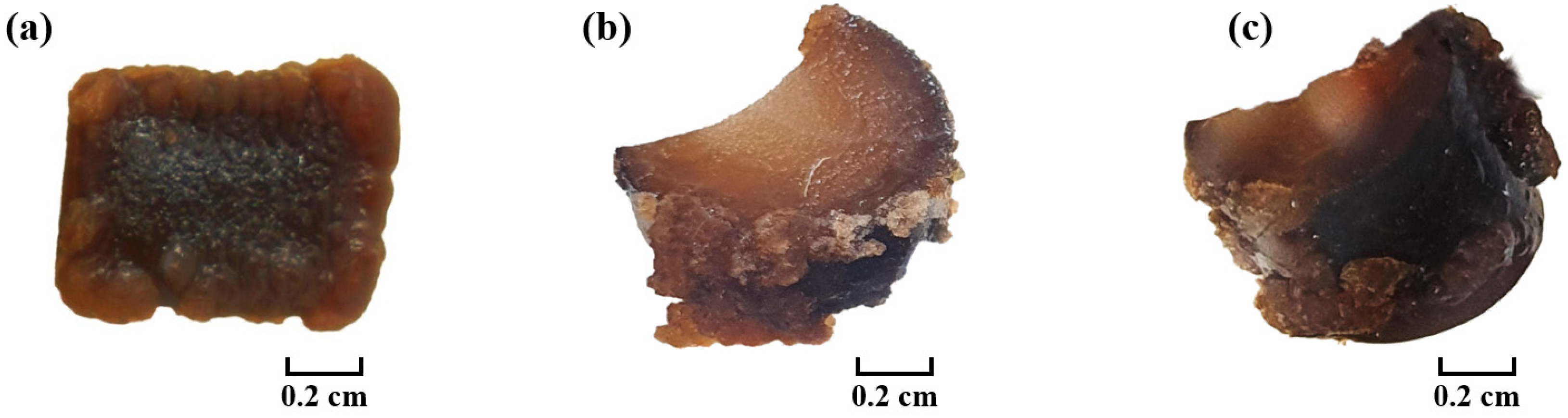
2.2. Pre-Processing of E. cava Thalli

Fresh and dark brown thalli of E. cava were selected for the tissue culture experiment. Thalli were wiped with sterile paper towels (Wypall, Yuhan-Kimberly Co., Ltd., Seoul, Republic of Korea), washed twice with autoclaved seawater, and immersed sequentially in autoclaved seawater containing 1% povidone-iodine (Green Pharmaceutical Co., Ltd., Jincheon, Republic of Korea) and 2% triton X-100 (Samchun Pure Chemical Co., Ltd., Pyeongtaek, Republic of Korea) for 3 min each. After rinsing and washing, thalli were treated with an antibiotic mixture: Kanamycin (0.1 g L−1; K1377; Sigma-Aldrich, St. Louis, MO, USA), Ampicillin (0.1 g L−1; A9518; Sigma), Streptomycin (0.2 g L−1; S9137; Sigma), Neomycin (20 mg L−1; N1876; Sigma), and Nystatin (1.5 mg L−1; N4014; Sigma) for 30 min at 12 °C to prevent contamination.

2.3. Experimental Conditions

Four culture media were employed: Provasoli’s enriched seawater medium (PESI) [60], enriched artificial seawater medium (ESAW) [61], artificial enriched seawater medium (ASP-2) [62], and Von Stosch’s enriched seawater medium (VS) [63]. Meristem and stipe were used for callus induction. Each section was cut into 1 × 1 cm2 (L × W) pieces, treated with a 10× antibiotics mixture for 30 min at 12 °C, and placed on agar media in a growth chamber (Multi-Room Incubator; LMI-3004PL, Daihan Labtech Co., Ltd., Namyangju, Republic of Korea) for callus development. Six to eight explants were inoculated on each agar plate, and callus formation was confirmed under a microscope (Routine Microscopes; CX33; Evidient Co., Ltd., Shinjuku-ku, Tokyo, Japan). Various treatments to optimize callus induction were performed as described in Table 1. The plant growth regulators, carbon sources, and polyamines were purchased from Sigma-Aldrich, St. Louis, MO, USA.

2.4. Plasma Treatment

Meristem and stipe explants were prepared as discussed in Section 2.2 and Section 2.3, and then subjected to plasma treatment.

2.4.1. Indirect Plasma Treatment

A 40 mL autoclaved seawater in a Petri dish was treated with a plasma generator provided by KRIBB (Korea Research Institute of Bioscience and Biotechnology, Daejeon, Republic of Korea) for 5, 10, 40, or 60 s, respectively, following the procedure by Bian et al. [64]. Prepared explants were socked in plasma–treated autoclaved seawater for 20–22 h and then cultured on agar PESI medium in a growth chamber (LMI-3004PL; Daihan Labtech) for eight weeks at 12 °C in the dark.

2.4.2. Direct Plasma Treatment

Explants were placed in a beaker filled with autoclaved seawater and incubated for 20–22 h, and then cultured on agar PESI medium. Plasma treatment durations were 5, 10, 30, or 60 s, followed by incubation in a growth chamber (LMI-3004PL; Daihan Labtech) for eight weeks at 12 °C in the dark.

2.5. Statistical Analysis

Ten different replicates were utilized per experiment (n = 10). Initially, the percentage values underwent arcsin-square root transformation prior to statistical analysis. Subsequently, statistical significance was assessed using a one-way or multiple-way analysis of variance (ANOVA) conducted with IBM SPSS Statistics version 29.0 (IBM Corp., Armonk, NY, USA). Post hoc tests were conducted using Tukey’s Honestly Significant Difference (HSD) test. Statistical significance was set at p ≤ 0.05. The experimental results were presented as percentage values in the figures.

3. Results and Discussion

During preliminary experiments, poor callus development was observed in liquid media. Therefore, only agar media were used for callus induction and subsequent cultures, aligning with the findings of Kawashima and Tokuda [26]. Callus growth was monitored for up to eight weeks, and both stipe and meristem explants displayed callus development within three to six weeks. The calluses, appearing light brown, exhibited gradual volume increases over the inoculation period and featured rod-shaped filamentous structures (Figure 1 and Figure 2), reminiscent of those observed by Kawashima and Tokuda [26]. The optimal callus induction parameters were determined under various conditions, and the impact of abiotic factors on callus development was explored. Callus induction rate (%) was calculated based on the total number of induced calluses per total number of explants multiplied by 100.

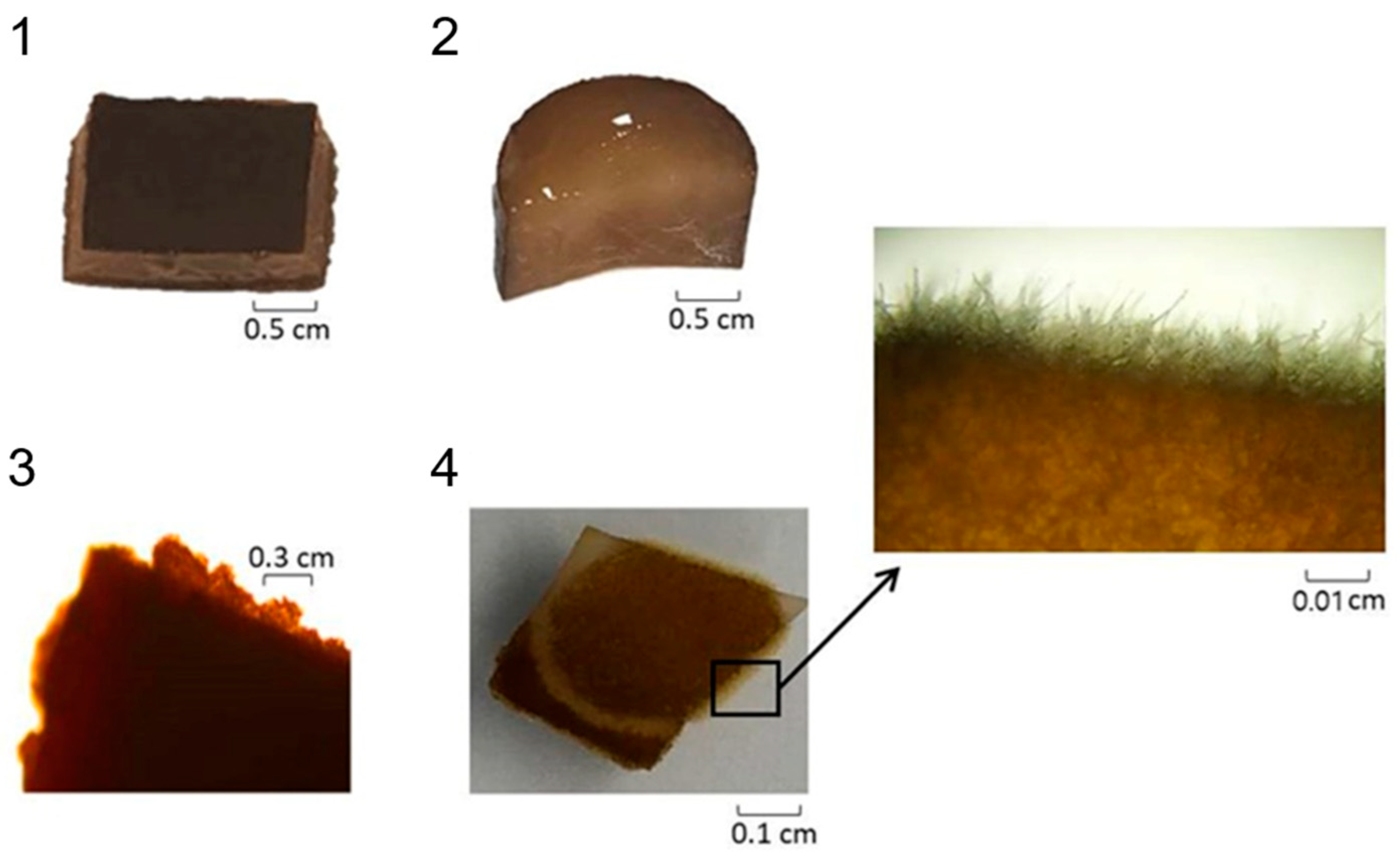
3.1. Effect of Media Type

Figure 3 presents the callus induction rates (%) observed with different culture media, and Figure 4 illustrates selected calluses induced from meristem and stipe explants of E. cava. The highest callus induction (31.73 ± 4.41% in stipe tissue and 38.28 ± 8.37% in meristem tissue) was observed on PESI medium, leading to its selection for subsequent experiments. The addition of potassium iodide in PESI medium [60] may have enhanced callus formation, aligning with the findings of Kawashima and Tokuda [26]. No callus growth was observed on ASP2 medium, possibly due to the presence of nitrilotriacetic acid and mannitol, acting as potential toxins [26,65] and increasing osmotic pressure of media [66], respectively.

3.2. Effect of Agar Concentration

Figure 5 shows the results of callus induction in meristem and stipe using PESI solid medium with different agar concentrations (1.2% and 1.5%). The highest callus induction rate (39.85 ± 8.27%) was observed in stipe at 1.5% agar concentration, followed by meristem (33.44 ± 6.50%) at 1.5% agar and 18 °C. Subsequent experiments utilized 1.5% agar concentration.

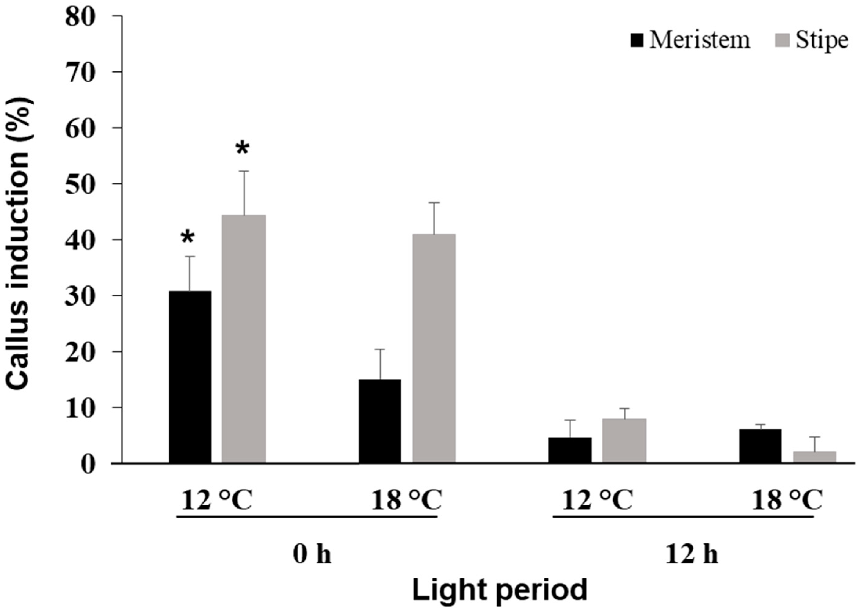
3.3. Effect of Photoperiod and Temperature

Environmental elements, including factors like photoperiod and light intensity, play a crucial role in the growth of algae. However, their influence varies among species and is contingent upon the specific product under investigation [67,68]. Additionally, light significantly impacts the growth and morphogenesis of callus by influencing the rate of cell division in various plants, directly regulating plant growth and development [69]. The impact of photoperiod (0 h and 12 h light) and temperatures (12 °C and 18 °C) on callus induction in meristem and stipe is shown in Figure 6. The highest callus induction (44.30 ± 6.28%) occurred in stipe at 0 h photoperiod and 12 °C. Optimal development of E. cava callus was observed at 12 °C, consistent with the findings of Kawashima and Tokuda [26]. Higher callus development was observed in complete darkness (0 h provision of light), possibly due to heterotrophic culture conditions overcoming growth inhibition challenges in light and aeration-dependent algal growth [70]. Subsequent experiments were conducted at 12 °C in dark.

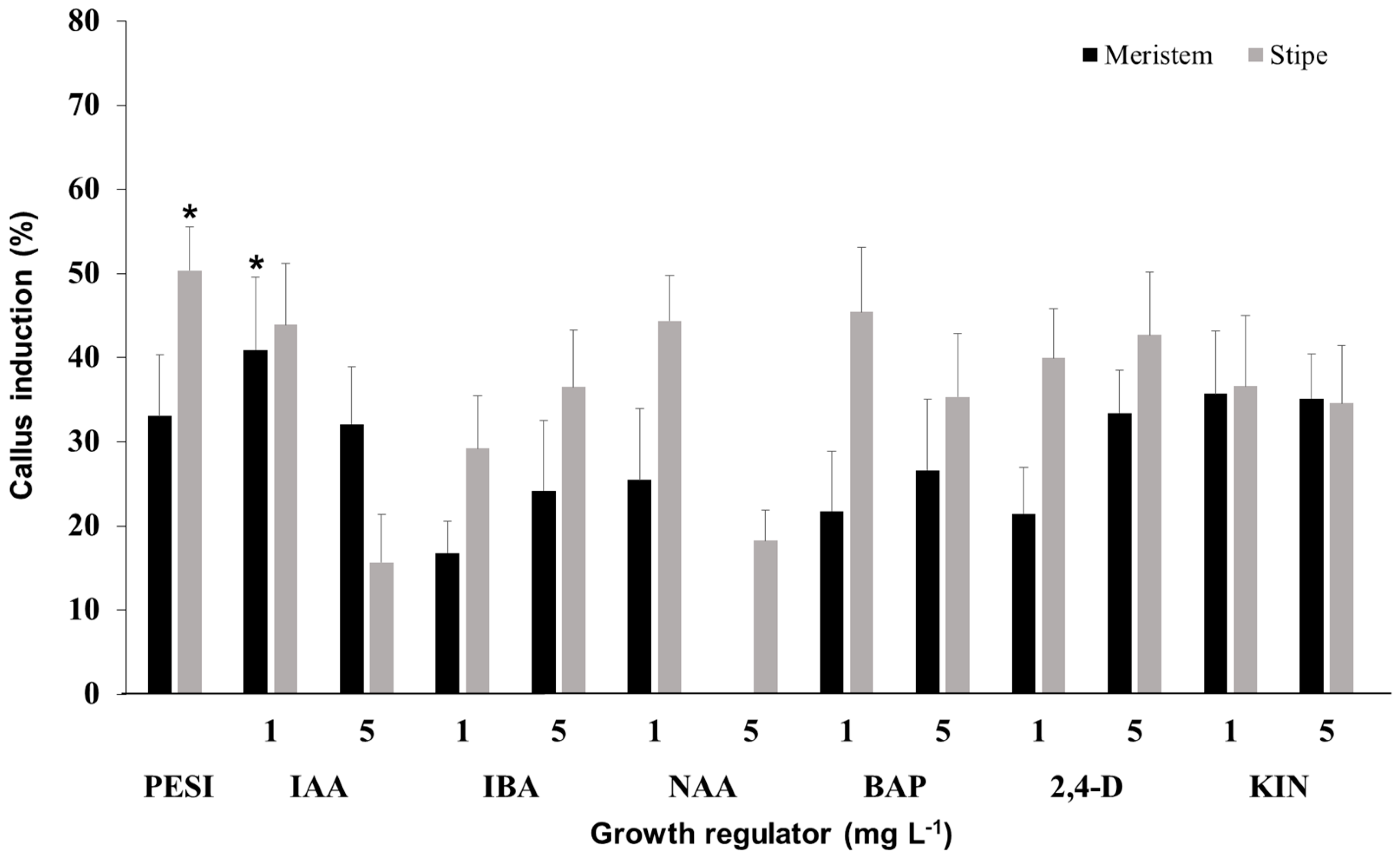
3.4. Effect of Growth Regulator

Plant growth regulators (PGRs) are incorporated into the basal growth medium of cell cultures to stimulate and regulate plant development [71,72,73]. These PGRs govern cell division in undifferentiated cells [74] and prompt callogenesis, leading to subsequent callus proliferation. Callus induction in meristem and stipe was observed in PESI solid medium with different growth regulators: indole-3-acetic acid (IAA), indole-3-butyric acid (IBA), 1-naphthaleneacetic acid (NAA), 6-benzylaminopurine (BAP), 2,4-dichlorophenoxyacetic acid (2,4-D), and kinetin (KIN) at concentrations of 1 or 5 mg L−1 at 12 °C (Figure 7). The selected calluses developed from meristem and stipe tissues are shown in Figure 8. The highest callus induction (50.37 ± 5.17%) in stipe occurred in standard PESI solid medium without growth regulators. Meanwhile, the highest callus induction rate (40.93 ± 8.65%) in meristem tissue was observed in PESI medium containing 1 mg L−1 IAA. Further experiments for meristem calluses were performed in PESI agar medium containing IAA, while stipe explants were cultured on standard PESI solid medium.

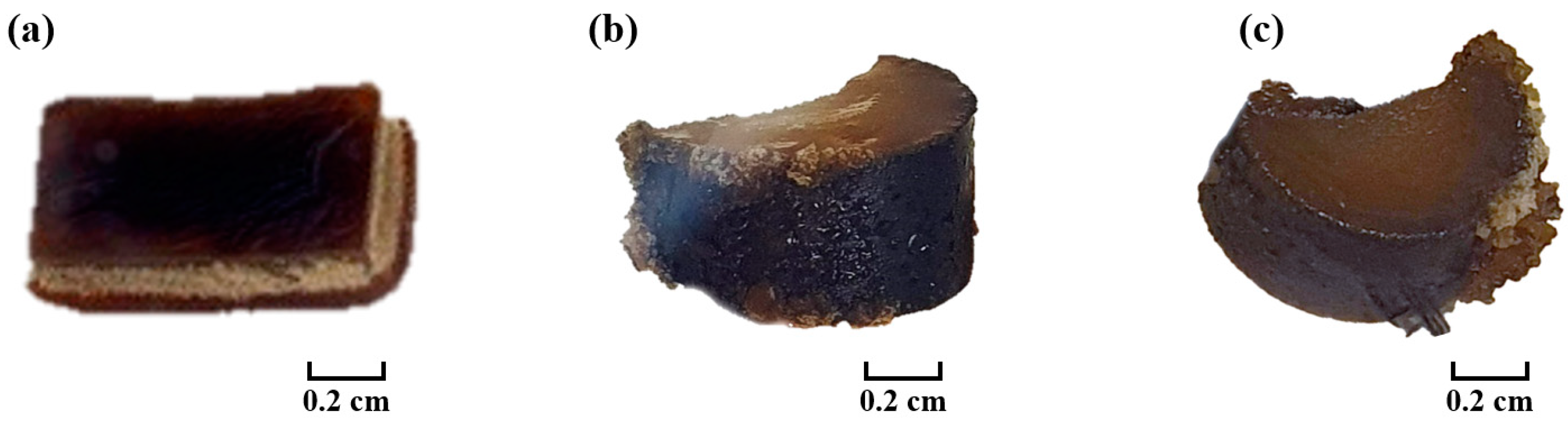
3.5. Effect of Carbon Source

Sugars are essential for biomass accumulation, serving as a vital energy source due to typically diminished photosynthetic activity in in vitro growing tissues. They also contribute to supply of carbon for biosynthetic processes and cell wall synthesis [75]. Moreover, sugars function as signal molecules that can either repress or activate plant genes, as noted by Tognetti et al. [76]. Callus induction in meristem and stipe was observed in PESI solid medium with different carbon sources (glucose, lactose, galactose, fructose, sucrose, or sorbitol) at 12 °C in dark (Figure 9). The selected calluses induced from meristem and stipe tissues are shown in Figure 10. Meristem tissue exhibited higher callus induction rates in PESI medium containing sucrose (51.42 ± 5.05%) or glucose (43.60 ± 7.88%) compared to standard PESI medium (40.31 ± 17.41%). Stipe tissue displayed an overall higher rate of callus induction in PESI medium supplemented with 2% sucrose (58.48 ± 5.66%) or glucose (53.10 ± 7.15%). It could be due to the factor that the inclusion of organic carbon sources may alleviate stresses arising from heterotrophic conditions in algal growth [40]. Furthermore, sucrose proves to be a relatively economic choice compared to other carbon sources. Consequently, subsequent experiments were performed in PESI medium containing 2% sucrose at 12 °C in the dark.

3.6. Effect of Polyamine

Polyamines, including spermine (Spm), putrescine (Put), and spermidine (Spd), represent a category of low molecular weight aliphatic nitrogenous organic compounds containing two or more amino groups [77]. These compounds are associated with various biological processes, including tissue growth, cell division, and cell differentiation [78]. Polyamines have been linked to higher callus induction and growth [79], while also playing a significant role in responding to both biotic and abiotic stresses [80]. Previous studies have indicated an increase in arginine decarboxylase (ADC) activity in rice seedlings when exposed to salinity [81]. Similarly, Wang and Liu [82] observed an increase in ADC and S-adenosylmethionine decarboxylase (SAMDC) expression in citrus embryogenic callus under high salinity and both low and high temperatures. Furthermore, various abiotic stresses have been shown to trigger the up regulation of SAMDC expression at the transcriptional level in transgenic tobacco plants [83]. Moreover, Zhou et al. [78] observed higher free polyamine levels and the expression of polyamine biosynthesis enzyme genes in young rice spikelets under heat stress, thereby increasing endogenous Spd and Spm levels. This correlation was associated with higher yield and resistance to heat stress, providing insights for rice production under high temperatures. This involvement in stress response is just one aspect of their intricate physiological functions.
Callus induction in meristem and stipe was monitored in PESI solid medium with different polyamines (Spm, Put, Spd) at 12 °C in dark (Figure 11). The selected calluses are shown in Figure 12. Overall, an increase in callus induction was observed in meristem tissue when culture medium was supplemented with 1 µM Spm (60.55 ± 3.05%). Except for this, all other conditions did not show a favorable impact on callus induction in meristem and stipe of E. cava compared to the callus development in standard PESI medium.

3.7. Effect of Plasma Treatment

The impact of plasma treatment on callus induction in meristem and stipe is shown in Figure 13. The selected calluses are shown in Figure 14. Indirect plasma treatment showed callus induction in both meristem and stipe, while direct plasma treatment on meristem explants did not yield callus induction. Indirect or direct plasma treatment failed to enhance callus induction rate, as the highest development occurred at 0 s plasma treatment in both meristem (57.53 ± 5.19%) and stipe (65.59 ± 6.24%). Therefore, based on the findings of this study, plasma treatment is not recommended for callus development in E. cava. However, further research exploring alternative plasma techniques may reveal different outcomes.

4. Conclusions

Callus, often referred to as the stem cell of a plant, represents an undifferentiated cell mass capable of unlimited proliferation and re-differentiation under favorable conditions. The establishment of callus-based seaweed culture technology holds the key to the mass production and industrialization of seaweeds that pose challenges in cultivation or collection, such as E. cava. The improved method for callus induction in E. cava involves culturing in PESI solid medium with 1.5% agar and 2% sucrose at 12 °C in the dark. Specifically for stipe explants, it is recommended to omit growth regulators, while for meristem, the use of 1 mg L−1 IAA and 1 µM Spm is advisable. Although plasma treatment did not yield favorable results in our study, exploring different plasma techniques may offer alternative outcomes. The establishment of E. cava callus cultures holds significant promise for research purposes and for addressing seed stock supply for mariculture and bioactive compound production. The callus induction technique developed in this study could streamline the mass cultivation of E. cava and other beneficial seaweed species, paving the way for the development of a callus-based smart farming technology. This advancement contributes to the cultivation of E. cava and other commercially valuable seaweed species for functional food and pharmaceutical materials.

Author Contributions

Conceptualization, J.-S.C. and K.M.I.B.; methodology, G.T., B.F.S.P.N. and J.-H.L.; software, K.M.I.B.; validation, K.M.I.B., G.T. and J.-S.C.; formal analysis, J.-H.L., K.M.I.B. and G.T.; investigation, J.-H.L., K.M.I.B. and G.T.; resources, J.-S.C.; data curation, J.-H.L., K.M.I.B., G.T. and J.-S.C.; writing—original draft preparation, J.-H.L. and K.M.I.B.; writing—review and editing, K.M.I.B. and J.-S.C.; visualization, K.M.I.B. and J.-S.C.; supervision, J.-S.C.; project administration, J.-S.C.; funding acquisition, J.-S.C. All authors have read and agreed to the published version of the manuscript.

Funding

This work has supported by the National Research Foundation of Korea (NRF) grant funded by the Korea government (MSIT) (No. NRF-2021R1A2C2093826).

Institutional Review Board Statement

Not applicable.

Informed Consent Statement

Not applicable.

Data Availability Statement

Data supporting the findings of this study are contained within the article.

Conflicts of Interest

The authors declare no conflicts of interest.

Abbreviations

2,4-D: 2,4-dichlorophenoxyacetic acid; ANOVA: Analysis of variance; ASP2: Artificial enriched seawater medium; BAP: 6-benzylaminopurine; ESAW: Enriched artificial seawater medium; IAA: Indole-3-acetic acid; IBA: Indole-3-butyric acid; KIN: Kinetin; KRIBB: Korea Research Institute of Bioscience and Biotechnology; NAA: 1-naphthaleneacetic acid; PESI: Provasoli´s enriched seawater medium; PGRs: Plant growth regulators; Put: Putrescine; S.E.: Standard error; Spd: Spermidine; Spm: Spermine; VS: Von Stosch’s enriched seawater medium.

References

  1. Stiger-Pouvreau, V.; Zubia, M. Macroalgal diversity for sustainable biotechnological development in French tropical overseas terri Stiger-Pouvreau, V.; Zubia, M. Macroalgal diversity for sustainable biotechnological development in French tropical overseas territories. Bot. Mar. 2020, 63, 17–41. [Google Scholar] [CrossRef]
  2. Sanches, P.F.; Pellizzari, F.; Horta, P.A. Multivariate analyses of Antarctic and sub-Antarctic seaweed distribution patterns: An evaluation of the role of the Antarctic Circumpolar Current. J. Sea Res. 2016, 110, 29–38. [Google Scholar] [CrossRef]
  3. Bedoux, G.; Bourgougnon, N. Bioactivity of secondary metabolites from macroalgae. In The Algae World. Cellular Origin, Life in Extreme Habitats and Astrobiology; Sahoo, D., Seckbach, J., Eds.; Springer: Dordrecht, The Netherlands, 2015; Volume 26, pp. 391–401. [Google Scholar]
  4. Budzałek, G.; Śliwińska-Wilczewska, S.; Wiśniewska, K.; Wochna, A.; Bubak, I.; Latała, A.; Wiktor, J.M. Macroalgal defense against competitors and herbivores. Int. J. Mol. Sci. 2021, 22, 7865. [Google Scholar] [CrossRef] [PubMed]
  5. Tanna, B.; Mishra, A. Nutraceutical potential of seaweed polysaccharides: Structure, bioactivity, safety, and toxicity. Compr. Rev. Food Sci. Food Saf. 2019, 18, 817–831. [Google Scholar] [CrossRef] [PubMed]
  6. Biris-Dorhoi, E.S.; Michiu, D.; Pop, C.R.; Rotar, A.M.; Tofana, M.; Pop, O.L.; Socaci, S.A.; Farcas, A.C. Macroalgae—A sustainable source of chemical compounds with biological activities. Nutrients 2020, 12, 3085. [Google Scholar] [CrossRef] [PubMed]
  7. Mehdi, A.; Ali, H.; Mehdi, A. Seaweed proteins as a source of bioactive peptides. Curr. Pharm. Des. 2021, 27, 1342–1352. [Google Scholar]
  8. Meinita, M.D.N.; Harwanto, D.; Choi, J.-S. Seaweed exhibits therapeutic properties against chronic disease: An overview. Appl. Sci. 2022, 12, 2638. [Google Scholar] [CrossRef]
  9. Antony, T.; Chakraborty, K. Pharmacological properties of seaweeds against progressive lifestyle diseases. J. Aquat. Food Prod. Technol. 2019, 28, 1092–1104. [Google Scholar] [CrossRef]
  10. Ganesan, A.R.; Tiwari, U.; Rajauria, G. Seaweed nutraceuticals and their therapeutic role in disease prevention. Food Sci. Hum. Wellness 2019, 8, 252–263. [Google Scholar] [CrossRef]
  11. Fortune Business Insights. Commercial Seaweed Market Size, Share & COVID-19 Impact Analysis, by Type (Red Seaweed, Brown Seaweed, and Green Seaweed), form (Flakes, Powder, and Liquid), End-Uses (Food & Beverages, Agricultural Fertilizer, Animal Feed Additives, Pharmaceutical, and Cosmetics & Personal Care), and Regional Forecast. 2021–2028. Available online: https://www.fortunebusinessinsights.com/industry-reports/commercial-seaweed-market-100077 (accessed on 26 March 2022).
  12. Obando, J.M.C.; dos Santos, T.C.; Martins, R.C.C.; Teixeira, V.L.; Barbarino, E.; Cavalcanti, D.N. Current and promising applications of seaweed culture in laboratory conditions. Aquaculture 2022, 560, 738596. [Google Scholar] [CrossRef]
  13. Kawai, H.; Motomura, T.; Okuda, K. Isolation and purification techniques for macroalgae. In Algal Culturing Techniques; Academic Press: Cambridge, MA, USA, 2005; Volume 133. [Google Scholar]
  14. Tirtawijaya, G.; Negara, B.F.S.P.; Lee, J.-H.; Cho, M.-G.; Kim, H.K.; Choi, Y.-S.; Lee, S.-H.; Choi, J.-S. The Influence of abiotic factors on the induction of seaweed callus. J. Mar. Sci. Eng. 2022, 10, 513. [Google Scholar] [CrossRef]
  15. Kang, J.W. Illustrated Encyclopedia of Fauna and Flora of Korea: Marine Algae; Samhwa Press: Seoul, Republic of Korea, 1968; pp. 147–148. [Google Scholar]
  16. Lee, Y.; Kang, S.A. Catalogue of the Seaweeds in Korea; Jeju National University Press: Jeju, Republic of Korea, 2001; pp. 107–108. [Google Scholar]
  17. Asanka Sanjeewa, K.K.; Fernando, I.P.S.; Kim, S.-Y.; Kim, W.-S.; Ahn, G.; Jee, Y.; Jeon, Y.-J. Ecklonia cava (Laminariales) and Sargassum horneri (Fucales) synergistically inhibit the lipopolysaccharide-induced inflammation via blocking NF-κB and MAPK pathways. Algae 2019, 34, 45–56. [Google Scholar] [CrossRef]
  18. Kang, C.; Jin, Y.B.; Lee, H.; Cha, M.; Sohn, E.T.; Moon, J. Brown alga Ecklonia cava attenuates type 1 diabetes by activating AMPK and Akt signaling pathways. Food Chem. Toxicol. 2010, 48, 509–516. [Google Scholar] [CrossRef] [PubMed]
  19. Wijesekara, I.; Yoon, N.Y.; Kim, S.K. Phlorotannins from Ecklonia cava (Phaeophyceae): Biological activities and potential health benefits. Biofactors 2010, 36, 408–414. [Google Scholar] [CrossRef] [PubMed]
  20. Wijesinghe, W.A.J.P.; Jeon, Y.J. Exploiting biological activities of brown seaweed Ecklonia cava for potential industrial applications: A review. Int. J. Food Sci. Nutr. 2012, 63, 225–235. [Google Scholar] [CrossRef] [PubMed]
  21. Shin, T.; Ahn, M.; Hyun, J.W.; Kim, S.H.; Moon, C. Antioxidant marine algae phlorotannins and radioprotection: A review of experimental evidence. Acta Histochem. 2014, 116, 669–674. [Google Scholar] [CrossRef] [PubMed]
  22. Barde, S.R.; Sakhare, R.S.; Kanthale, S.B.; Chandak, P.G.; Jamkhande, P.G. Marine bioactive agents: A short review on new marine antidiabetic compounds. Algae 2015, 14, 15. [Google Scholar] [CrossRef]
  23. Sanjeewa, K.K.A.; Kim, E.A.; Son, K.T.; Jeon, Y.J. Bioactive properties and potentials cosmeceutical applications of phlorotannins isolated from brown seaweeds: A review. J. Photochem. Photobiol. B 2016, 162, 100–105. [Google Scholar] [CrossRef] [PubMed]
  24. Chae, D.-H.; Kim, I.-S.; Kim, S.-K.; Song, Y.K.; Shim, W.J. Abundance and distribution characteristics of microplastics in surface seawaters of the Incheon/Kyeonggi coastal region. Arch. Environ. Contam. Toxicol. 2015, 69, 269–278. [Google Scholar] [CrossRef]
  25. Hurley, R.; Woodward, J.; Rothwell, J.J. Microplastic contamination of river beds significantly reduced by catchment-wide flooding. Nat. Geosci. 2018, 11, 251–257. [Google Scholar] [CrossRef]
  26. Kawashima, Y.; Tokuda, H. Callus formation in Ecklonia cava Kjellman (Laminariales, Phaeophyta). Hydrobiologia 1990, 204, 375–380. [Google Scholar] [CrossRef]
  27. Kumar, G.R.; Reddy, C.R.K.; Jha, B. Callus induction and thallus regeneration from callus of phycocolloid yielding seaweeds from the Indian coast. J. Appl. Phycol. 2007, 19, 15–25. [Google Scholar] [CrossRef]
  28. Mansoor, S.; Bashir, K.M.I.; Mohibbullah, M.; Meinita, M.D.N.; Khan, M.N.A.; Sohn, J.-H.; Choi, J.-S. Nutritional and health promoting perspectives of Monostroma spp. (Chlorophyta): A systematic review. J. Appl. Phycol. 2024; in press. [Google Scholar]
  29. Gordillo, F.J.; Dring, M.J.; Savidge, G. Nitrate and phosphate uptake characteristics of three species of brown algae cultured at low salinity. Mar. Ecol. Prog. Ser. 2002, 234, 111–118. [Google Scholar] [CrossRef]
  30. Nishihara, G.N.; Mori, Y.; Terada, R.; Noro, T. A simplified method to isolate and cultivate, Laurencia brongniartii J. Agardh (Rhodophyta, Ceramiales) from Kagoshima, Japan. Aquac. Sci. 2004, 52, 1–10. [Google Scholar]
  31. Steen, H. Effects of reduced salinity on reproduction and germling development in Sargassum muticum (Phaeophyceae, Fucales). Eur. J. Phycol. 2004, 39, 293–299. [Google Scholar] [CrossRef]
  32. Zou, D.H.; Gao, K.S. Comparative mechanisms of photosynthetic carbon acquisition in Hizikia fusiforme under submersed and emersed conditions. Acta Bot. Sin. 2004, 46, 1178–1185. [Google Scholar]
  33. Hayashi, L.; Yokoya, N.S.; Kikuchi, D.M.; Oliveira, E.C. Callus induction and micropropagation improved by colchicine and phytoregulators in Kappaphycus alvarezii (Rhodophyta, Solieriaceae). J. Appl. Phycol. 2008, 20, 653–659. [Google Scholar] [CrossRef]
  34. Kyaw, S.P.P.; Wai, M.K.; Nyunt, T.; Aye, M.M.; Soe-Htun, U. Effects of light intensity on the formation and growth of the secondary branches of Dictyota adnata zanardini grown in different salinity and temperature regimes. J. Myanmar Acad. Arts Sci. 2009, 7, 321–332. [Google Scholar]
  35. Xu, Z.; Zou, D.; Gao, K. Effects of elevated CO2 and phosphorus supply on growth, photosynthesis and nutrient uptake in the marine macroalga Gracilaria lemaneiformis (Rhodophyta). Bot. Mar. 2010, 53, 123–129. [Google Scholar] [CrossRef]
  36. Scherner, F.; Ventura, R.; Barufi, J.B.; Horta, P.A. Salinity critical threshold values for photosynthesis of two cosmopolitan seaweed species: Providing baselines for potential shifts on seaweed assemblages. Mar. Environ. Res. 2013, 91, 14–25. [Google Scholar] [CrossRef]
  37. Terada, R.; Inoue, S.; Nishihara, G.N. The effect of light and temperature on the growth and photosynthesis of Gracilariopsis chorda (Gracilariales, Rhodophtya) from geographically separated locations of Japan. J. Appl. Phycol. 2013, 25, 1863–1872. [Google Scholar] [CrossRef]
  38. Wei, X.; Shuai, L.; Lu, B.; Wang, S.; Chen, J.; Wang, G. Effects of temperature and irradiance on filament development of Grateloupia turuturu (Halymeniaceae, Rhodophyta). J. Appl. Phycol. 2013, 25, 1881–1886. [Google Scholar] [CrossRef]
  39. Mandal, S.K.; Ajay, G.; Monisha, N.; Malarvizhi, J.; Temkar, G.; Mantri, V.A. Differential response of varying temperature and salinity regimes on nutrient uptake of drifting fragments of Kappaphycus alvarezii: Implication on survival and growth. J. Appl. Phycol. 2015, 27, 1571–1581. [Google Scholar] [CrossRef]
  40. Torres, P.B.; Chow, F.; Santos, D.Y. Growth and photosynthetic pigments of Gracilariopsis tenuifrons (Rhodophyta, Gracilariaceae) under high light in vitro culture. J. Appl. Phycol. 2005, 27, 1243–1251. [Google Scholar] [CrossRef]
  41. Terada, R.; Vo, T.D.; Nishihara, G.N.; Shioya, K.; Shimada, S.; Kawaguchi, S. The effect of irradiance and temperature on the photosynthesis and growth of a cultivated red alga Kappaphycus alvarezii (Solieriaceae) from Vietnam, based on in situ and in vitro measurements. J. Appl. Phycol. 2016, 28, 457–467. [Google Scholar] [CrossRef]
  42. Endo, H.; Okumura, Y.; Sato, Y.; Agatsuma, Y. Interactive effects of nutrient availability, temperature, and irradiance on photosynthetic pigments and color of the brown alga Undaria pinnatifida. J. Appl. Phycol. 2017, 29, 1683–1693. [Google Scholar] [CrossRef]
  43. Han, T.; Qi, Z.; Huang, H.; Liao, X.; Zhang, W. Nitrogen uptake and growth responses of seedlings of the brown seaweed Sargassum hemiphyllum under controlled culture conditions. J. Appl. Phycol. 2018, 30, 507–515. [Google Scholar] [CrossRef]
  44. Yuniarti, L.S.; Sri, A.; Happy, N.; Muhammad, F. Concentration of liquid pes media on the growth and photosynthetic pigments of seaweeds Cotonii propagule (Kappaphycus alvarezii Doty) through tissue culture. Russ. J. Agric. Soc. Econ. Sci. 2018, 75, 133–144. [Google Scholar]
  45. Mo, V.T.; Cuong, L.K.; Tung, H.T.; Van Huynh, T.; Nghia, L.T.; Khanh, C.M.; Lam, N.N.; Nhut, D.T. Somatic embryogenesis and plantlet regeneration from the seaweed Kappaphycus striatus. Acta Physiol. Plant. 2020, 42, 104. [Google Scholar] [CrossRef]
  46. Wijayanto, A.; Widowati, I.; Winanto, T. Domestication of red seaweed (Gelidium latifolium) in different culture media. Indones. J. Mar. Sci. Ilmu Kelaut. 2020, 25, 39–44. [Google Scholar] [CrossRef]
  47. Aris, M.; Muchdar, F.; Labenua, R. Study of seaweed Kappaphycus alvarezii explants growth in the different salinity concentrations. J. Ilm. Perikan. Kelaut. 2021, 13, 97–105. [Google Scholar] [CrossRef]
  48. Öztaşkent, C.; Ak, İ. Effect of LED light sources on the growth and chemical composition of brown seaweed Treptacantha barbata. Aquac. Int. 2021, 29, 193–205. [Google Scholar] [CrossRef]
  49. Cai, Y.; Li, G.; Zou, D.; Hu, S.; Shi, X. Rising nutrient nitrogen reverses the impact of temperature on photosynthesis and respiration of a macroalga Caulerpa lentillifera (Ulvophyceae, Caulerpaceae). J. Appl. Phycol. 2021, 33, 1115–1123. [Google Scholar] [CrossRef]
  50. Nauer, F.; Borburema, H.D.; Yokoya, N.S.; Fujii, M.T. Effects of ocean acidification on growth, pigment contents and antioxidant potential of the subtropical Atlantic red alga Hypnea pseudomusciformis Nauer, Cassano & MC Oliveira (Gigartinales) in laboratory. Rev. Bras. Bot. 2021, 44, 69–77. [Google Scholar]
  51. Avila-Peltroche, J.; Won, B.Y.; Cho, T.O. An improved protocol for protoplast production, culture, and whole plant regeneration of the commercial brown seaweed Undaria pinnatifida. Algal Res. 2022, 67, 102851. [Google Scholar] [CrossRef]
  52. Asensi, A.; Gall, E.A.; Marie, D.; Billot, C.; Dion, P.; Kloareg, B. Clonal propagation of Laminaria digitata (Phaeophyceae) sporophytes through a diploid cell-filament suspension. J. Phycol. 2001, 37, 411–417. [Google Scholar] [CrossRef]
  53. Zhang, Q.S.; Qu, S.C.; Cong, Y.Z.; Luo, S.J.; Tang, X.X. High throughput culture and gametogenesis induction of Laminaria japonica gametophyte clones. J. Appl. Phycol. 2008, 20, 205–211. [Google Scholar] [CrossRef]
  54. Mussio, I.; Rusig, A.M. Morphogenetic responses from protoplasts and tissue culture of Laminaria digitata (Linnaeus) JV Lamouroux (Laminariales, Phaeophyta): Callus and thalloid-like structures regeneration. J. Appl. Phycol. 2009, 21, 255–264. [Google Scholar] [CrossRef]
  55. Gupta, V.; Bijo, A.J.; Kumar, M.; Reddy, C.R.K.; Jha, B. Detection of epigenetic variations in the protoplast-derived germlings of Ulva reticulata using methylation sensitive amplification polymorphism (MSAP). Mar. Biotechnol. 2012, 14, 692–700. [Google Scholar] [CrossRef]
  56. Luhan, M.R.J.; Mateo, J.P. Clonal production of Kappaphycus alvarezii (Doty) Doty in vitro. J. Appl. Phycol. 2017, 29, 2339–2344. [Google Scholar] [CrossRef]
  57. Reddy, C.R.K.; Jha, B.; Fujita, Y.; Ohno, M. Seaweed micropropagation techniques and their potentials: An overview. J. Appl. Phycol. 2007, 20, 159–167. [Google Scholar]
  58. Baweja, P.; Sahoo, D.; García-Jiménez, P.; Robaina, R.R. Review: Seaweed tissue culture as applied to biotechnology: Problems, achievements and prospects. Phycol. Res. 2009, 57, 45–58. [Google Scholar] [CrossRef]
  59. Kumar, L.R.G.; Paul, P.T.; Anas, K.K.; Tejpal, C.S.; Chatterjee, N.S.; Anupama, T.K.; Mathew, S.; Ravishankar, C.N. Phlorotannins-bioactivity and extraction perspectives. J. Appl. Phycol. 2022, 34, 2173–2185. [Google Scholar] [CrossRef] [PubMed]
  60. Tatewaki, M. Formation of a crustaceous sporophyte with unilocular sporangia in Scytosiphon lomentaria. Phycologia 1966, 6, 62–66. [Google Scholar] [CrossRef]
  61. Berges, J.A.; Franklin, D.J.; Harrison, P.J. Evolution of an artificial seawater medium: Improvements in enriched seawater, artificial water over the past two decades. J. Phycol. 2001, 37, 1138–1145. [Google Scholar] [CrossRef]
  62. Provasoli, L.; McLaughlin, J.J.A.; Droop, M.R. The development of artificial media for marine algae. Arch. Mikrobiol. 1957, 25, 392–428. [Google Scholar] [CrossRef] [PubMed]
  63. Gargiulo, G.M.; Genovese, G.; Morabito, M.; Culoso, F.; De Masi, F. Sexual and asexual reproduction in a freshwater population of Bangia atropurpurea (Bangiales, Rhodophyta) from eastern Sicily (Italy). Phycologia 2001, 40, 88–96. [Google Scholar] [CrossRef]
  64. Bian, J.Y.; Guo, X.Y.; Lee, D.H.; Sun, X.R.; Liu, L.S.; Shao, K.; Kwon, T. Non-thermal plasma enhances rice seed germination, seedling development, and root growth under low-temperature stress. Appl. Biol. Chem. 2024, 67, 2. [Google Scholar] [CrossRef]
  65. Anderson, R.L.; Bishop, W.E.; Campbell, R.L. A review of the environmental and mammalian toxicology of nitrilotriacetic acid. CRC Crit. Rev. Toxicol. 1985, 15, 1–102. [Google Scholar] [CrossRef]
  66. El-Bahr, M.K.; Abd EL-Hamid, A.; Matter, M.A.; Shaltout, A.; Bekheet, S.A.; El-Ashry, A.A. In vitro conservation of embryogenic cultures of date palm using osmotic mediated growth agents. J. Genet. Eng. Biotechnol. 2016, 14, 363–370. [Google Scholar] [CrossRef]
  67. Chen, G.-Q.; Chen, F. Growing phototrophic cells without light. Biotechnol. Lett. 2006, 28, 607–616. [Google Scholar] [CrossRef] [PubMed]
  68. Engin, I.K.; Cekmecelioglu, D.; Yücel, A.M. Heterotrophic growth and oil production from Micractinium sp. ME05 using molasses. J. Appl. Phycol. 2018, 30, 3483–3492. [Google Scholar] [CrossRef]
  69. Afshari, R.T.; Angoshtari, R.; Kalantari, S. Effects of light and different plant growth regulators on induction of callus growth in rapeseed (Brassica napus L.) genotypes. Plant Omics 2011, 4, 60–67. [Google Scholar]
  70. Bashir, K.M.I.; Mansoor, S.; Kim, N.R. Effect of organic carbon sources and environmental factors on cell growth and lipid content of Pavlova lutheri. Ann. Microbiol. 2019, 69, 353–368. [Google Scholar] [CrossRef]
  71. Gaspar, T.; Keveks, C.; Penel, C.; Greppin, H.; Reid, D.M.; Thorpe, T.A. Plant hormones and plant growth regulators in plant tissue culture. In Vitro Cell. Dev. Biol. Plant 1996, 32, 272–289. [Google Scholar] [CrossRef]
  72. Phillips, G.C.; Garda, M. Plant tissue culture media and practices: An overview. Plant 2019, 55, 242–257. [Google Scholar] [CrossRef]
  73. Martinez, M.E.; Jorquera, L.; Poirrier, P.; Díaz, K.; Chamy, R. Effect of the carbon source and plant growth regulators (PGRs) in the induction and maintenance of an in vitro callus culture of Taraxacum officinale (L.) Weber Ex F.H. Wigg. Agronomy 2021, 11, 1181. [Google Scholar] [CrossRef]
  74. Coenen, C.; Lomax, T.L. Auxin-cytokinin interactions in higher plants: Old problems and new tools. Trends Plant Sci. 1997, 2, 351–355. [Google Scholar] [CrossRef] [PubMed]
  75. Praveena, C.; Veeresham, C. Multiple shoot regeneration and effect of sugars on growth and nitidine accumulation in shoot cultures of Toddalia asiatica. Pharmacogn. Mag. 2014, 10, S480. [Google Scholar]
  76. Tognetti, J.A.; Pontis, H.G.; Martínez-Noël, G.M.A. Sucrose signaling in plants: A world yet to be explored. Plant Signal Behav. 2013, 8, e23316. [Google Scholar]
  77. Chen, D.; Shao, Q.; Yin, L.; Younis, A.; Zheng, B. Polyamine function in plants: Metabolism, regulation on development, and roles in abiotic stress responses. Front. Plant Sci. 2019, 9, 1945. [Google Scholar] [CrossRef] [PubMed]
  78. Zhou, R.; Hu, Q.; Pu, Q.; Chen, M.; Zhu, X.; Gao, C.; Cao, Y. Spermidine enhanced free polyamine levels and expression of polyamine biosynthesis enzyme gene in rice spikelets under heat tolerance before heading. Sci. Rep. 2020, 10, 8976. [Google Scholar] [CrossRef] [PubMed]
  79. Eren, B.; Türkoğlu, A.; Haliloğlu, K.; Demirel, F.; Nowosad, K.; Özkan, G.; Niedbała, G.; Pour-Aboughadareh, A.; Bujak, H.; Bocianowski, J. Investigation of the influence of polyamines on mature embryo culture and DNA methylation of wheat (Triticum aestivum L.) using the machine learning algorithm method. Plants 2023, 12, 3261. [Google Scholar] [CrossRef] [PubMed]
  80. Alcázar, R.; Tiburcio, A.F. Plant polyamines in stress and development: An emerging area of research in plant sciences. Front. Plant Sci. 2014, 5, 319. [Google Scholar] [CrossRef] [PubMed]
  81. Quinet, M.; Ndayiragije, A.; Lefèvre, I.; Lambillotte, B.; Dupont-Gillain, C.C.; Lutts, S. Putrescine differently influences the effect of salt stress on polyamine metabolism and ethylene synthesis in rice cultivars differing in salt resistance. J. Exp. Bot. 2010, 61, 2719–2733. [Google Scholar] [CrossRef] [PubMed]
  82. Wang, J.; Liu, J.H. Change in free polyamine contents and expression profiles of two polyamine biosynthetic genes in citrus embryogenic callus under abiotic stresses. Biotechnol. Biotechnol. Equip. 2009, 23, 1289–1293. [Google Scholar] [CrossRef]
  83. Wi, S.J.; Kim, W.T.; Park, K.Y. Overexpression of carnation S-adenosylmethionine decarboxylase gene generates a broad-spectrum tolerance to abiotic stresses in transgenic tobacco plants. Plant Cell Rep. 2006, 25, 1111–1121. [Google Scholar] [CrossRef]
Figure 1. Preparation of E. cava for callus induction. (1) Collected E. cava, (2) E. cava meristem, (3) E. cava stipe.
Applsci 14 03480 g001
Figure 2. Callus induction in E. cava on PESI solid medium. (1) Meristem explant (1 × 1 cm2), (2) Stipe explant (1 × 1 cm2), (3) induced meristem callus under microscope (4×), (4) induced meristem fibrous callus under microscope (4×).
Applsci 14 03480 g002
Figure 3. Effect of media type on callus induction. Values are means ± standard errors (S.E.) of 10 individual replicates per experiment (n = 10). PESI: Provasoli´s enriched seawater medium; ESAW: Enriched artificial seawater medium; ASP2: Artificial enriched seawater medium; VS: Von Stosch’s enriched seawater medium. *: Indicates significant differences using Tukey’s HSD test using a one-way ANOVA test at p < 0.05.
Applsci 14 03480 g003
Figure 4. Callus induction in meristem (a) and stipe (b,c) on PESI solid medium supplemented with 1.5% agar.
Applsci 14 03480 g004
Figure 5. Effect of agar concentration on callus induction. Values are means ± standard errors (S.E.) of 10 individual replicates per experiment (n = 10). *: Indicates significant differences using Tukey’s HSD test using a one-way ANOVA test at p < 0.05.
Applsci 14 03480 g005
Figure 6. Effect of photoperiod and temperature on callus induction. Values are means ± standard errors (S.E.) of 10 individual replicates per experiment (n = 10). *: Indicates significant differences using Tukey’s HSD test using a multiple-way ANOVA test at p < 0.05. The temperature alone had no statistically significant effect (p = 0.089) on callus induction in stipe explant. However, the photoperiod alone (p = 0.002) and the interaction between photoperiod (0 h) and temperature (12 °C) showed a significant effect (p = 0.049) on callus induction in meristem when checked using a multiple-way ANOVA test using SPSS.
Applsci 14 03480 g006
Figure 7. Effect of growth regulators on callus induction. Values are means ± standard errors (S.E.) of 10 individual replicates per experiment (n = 10). IAA: Indole-3-acetic acid; IBA: Indole-3-butyric acid; NAA: 1-naphthaleneacetic acid; BAP: 6-benzylaminopurine; 2,4-D: 2,4-dichlorophenoxyacetic acid; KIN: Kinetin. *: Indicates significant differences using Tukey’s HSD test using a multiple-way ANOVA test at p = 0.0312 for stipe and p = 0.047 for meristem.
Applsci 14 03480 g007
Figure 8. Callus induction in meristem (a) on PESI medium supplemented with 1 mg L−1 of IAA, and stipe (b,c) on standard PESI solid medium without containing growth regulators.
Applsci 14 03480 g008
Figure 9. Effect of carbon sources on callus induction. Values are means ± standard errors (S.E.) of 10 individual replicates per experiment (n = 10). *: Indicates significant differences using Tukey’s HSD test using a multiple-way ANOVA test at p < 0.05.
Applsci 14 03480 g009
Figure 10. Callus induction in meristem (a) and stipe (b,c) on PESI solid medium supplemented with 2% sucrose.
Applsci 14 03480 g010
Figure 11. Callus induction in PESI medium supplemented with different polyamines. Values are means ± standard errors (S.E.) of 10 individual replicates per experiment (n = 10). Spm: Spermine; Put: Putrescine; Spd: Spermidine. *: Indicates significant differences using Tukey’s HSD test using a multiple-way ANOVA test at p < 0.05.
Applsci 14 03480 g011
Figure 12. Callus induction in meristem (a) on PESI solid medium supplemented 1 µM SPM, and in stipe (b,c) on standard PESI solid medium without containing polyamines.
Applsci 14 03480 g012
Figure 13. Effect of plasma treatment on callus induction on PESI medium. Values are means ± standard errors (S.E.) of 10 individual replicates per experiment (n = 10). *: Indicates significant differences using Tukey’s HSD test using a multiple-way ANOVA test at p < 0.05.
Applsci 14 03480 g013
Figure 14. Callus induction in direct (a) for 5 s and indirect for 60 s, (b,c) plasma treated stipe explants cultured on standard PESI solid medium.
Applsci 14 03480 g014
Table 1. Experimental conditions for callus induction in explants from E. cava meristem and stipe.
ParameterExperimental Conditions
Effect of culture medium
  • Explants were cultured on PESI, ESAW, ASP2, or VS solid medium supplemented with 1.5% agar.
  • Growth was monitored at 12 °C with a light period of 12 h for eight weeks.
Effect of agar concentration
  • Explants were cultured on PESI solid medium containing 1.2% or 1.5% agar.
  • Growth was monitored at 12 °C for eight weeks.
Effect of photoperiod and
temperature
  • Explants were cultured on PESI solid medium containing 1.5% agar. Varying photoperiods (0 h or 12 h light period at a light intensity of 160 μmol m−2 s−1 using a fluorescent lamp; 36 W; FPL36EX-D/C, Ilshin Vitson Co., Ltd., Namyangju, Republic of Korea) and temperatures (12 °C or 18 °C) were tested.
  • Growth was monitored for eight weeks.
Effect of growth regulator
  • Explants were cultured on PESI solid medium containing 1.5% agar. Different plant growth regulators: IAA (1003530010), IBA (I5386), NAA (N0640), BAP (B3408), 2,4-D (D70724) or KIN (48130), were added at concentrations up to 5 mg L−1.
  • Growth was monitored at 12 °C in the dark for eight weeks.
Effect of carbon source
  • Explants were cultured on PESI solid medium containing 1.5% agar.
  • Different carbon sources: glucose (PHR1000), lactose (PHR1025), galactose (PHR1206), fructose (F0127), sucrose (S0389), or sorbitol (PHR1006), were added at concentrations up to 5%.
  • The medium for meristem explants was supplemented with 1 mg L−1 IAA.
  • Growth was monitored at 12 °C in the dark for eight weeks.
Effect of polyamine
  • Explants were cultured on PESI solid medium containing 1.5% agar and 2% sucrose. Different polyamines: Spm (85590), Put (51799), or Spd (S0266), were added at concentrations up to 1000 µM.
  • The medium for meristem explants was supplemented with 1 mg L−1 IAA.
  • Growth was monitored at 12 °C in the dark for eight weeks.
Effect of plasma treatment
  • Explants were directly or indirectly treated with plasma.
  • Explants were cultured on PESI solid medium containing 1.5% agar and 2% sucrose. The medium for meristem explants was supplemented with 1 mg L−1 IAA and 1 µM Spm.
  • Growth was monitored at 12 °C in the dark for eight weeks.
PESI: Provasoli’s enriched seawater medium; ESAW: Enriched artificial seawater medium; ASP2: Artificial enriched seawater medium; VS: Von Stosch’s enriched seawater medium; IAA: Indole-3-acetic acid; IBA: Indole-3-butyric acid; NAA: 1-naphthaleneacetic acid; BAP: 6-benzylaminopurine; 2,4-D: 2,4-dichlorophenoxyacetic acid; KIN: Kinetin; Spm: Spermine; Put: Putrescine; Spd: Spermidine.
Disclaimer/Publisher’s Note: The statements, opinions and data contained in all publications are solely those of the individual author(s) and contributor(s) and not of MDPI and/or the editor(s). MDPI and/or the editor(s) disclaim responsibility for any injury to people or property resulting from any ideas, methods, instructions or products referred to in the content.

