Article

Main Leaf Polyphenolic Components of Berry Color Variant Grapevines and Their Acclimative Responses to Sunlight Exposure

1
Department of Plant Biology, University of Pécs, 6 Ifjúság, Pécs 7624, Hungary
2
Department of Applied Chemistry, Corvinus University of Budapest, 29-43 Villányi, Budapest 1118, Hungary
3
Cellular Imaging Laboratory, Institute of Plant Biology, Biological Research Centre, Hungarian Academy of Sciences, P.O. Box 521, Szeged 6701, Hungary
4
Research Institute for Viticulture and Oenology, University of Pécs, 4 Pázmány P, Pécs 7632, Hungary
*
Author to whom correspondence should be addressed.
Appl. Sci. 2015, 5(4), 1955-1969; https://doi.org/10.3390/app5041955
Submission received: 12 March 2015 / Revised: 12 June 2015 / Accepted: 23 June 2015 / Published: 18 December 2015

Abstract

:
Grapevine leaf synthesizes a wide variety of bioactive secondary metabolites, including polyphenols, which are also key components in ensuring development and growth of the whole plant even under adverse environmental conditions. Our study evaluates the nonanthocyanin polyphenolic composition in grapevine leaves of three varieties of Gohér conculta (Vitis vinifera L.) native to Hungary. A high performance liquid chromatography (HPLC) system including a diode array detector (DAD) coupled to a time-of-flight mass spectrometer (q-TOFMS) was successfully applied to profile intact glycoconjugate forms in samples. In-source fragmentation was utilized in order to provide structural information on the compounds. Using this method, the presence of 16 polyphenolic metabolites were confirmed, and eight of them were subjected to further quantification in sun acclimated and half shaded leaves. Intracellular microimaging detected accumulation of flavonols in cell nuclei, cell wall and chloroplasts. Our findings demonstrated that Gohér conculta—a special grapevine taxon of our viticultural heritage with berry color variants—is a suitable model to study the interaction between genetic and environmental factors in determination of grapevine phenolic composition.

Graphical Abstract

1. Introduction

Grapevine is capable of synthetizing a wide variety of phenolic compounds, such as phenolic acids (hydroxybenzoic and hydroxycinnamic acids), stilbenes and flavonoids, with important protecting functions against biotic and abiotic stresses. Flavonoids act among others as antioxidants and UV-protectants and have important health protecting effects by consumption of berries, wine or leaf extracts [1,2,3,4,5,6]. The flavonoid composition tends to be specific, mainly determined by genetic factors, but their amount is highly dependent on viticultural and environmental factors such as light, temperature, and water status; furthermore on insect and pathogen attacks or phenology [7,8,9,10]. Acclimation to sunlight includes a variety of metabolic responses [11]. Among these, flavonoids provide multiple roles in photoprotection as potential UV absorbers and as antioxidants [12,13,14,15]. According to their micro-localization in leaf tissues, flavonoids are expected to aid both physical (screening) or chemical (antioxidant) defense functions. The possibility of direct solar UV-screening by flavonoids is supported by their presence in epidermal cell vacuoles and trichomes [16,17,18]. On the other hand, the importance of UV-shielding has been challenged by a generally better absorption of lower energy UV-A, than of potentially more damaging UV-B by flavonoids [19], in favor of potential antioxidant functions [20]. The positive relationship between sunlight exposure and increased flavonol accumulation have been noticed by several researchers studying berry flavonoids and their biosynthetic pathways [21,22,23], while grapevine leaf flavonoids as important participants in acclimation processes of the plant are less studied.
The main objective of our work was to analyze and localize the leaf polyphenolics of grapevine varieties in terms of effect of natural sunshade conditions in the canopy, using a high performance liquid chromatography and accurate mass, high-resolution mass spectrometry as well as laser scanning microscopy. Grapevine (Vitis vinifera L.) leaves are rich in polyphenols and thus provide an excellent specimen for these studies. Traditionally, the growing method (see in Section 3.1) consistently provides a relatively stable shaded condition for leaves developing inside the canopy as opposed to fully sun exposed ones. Gohér white, Gohér altering and Gohér red, belonging to the Gohér conculta, show very close genetic relationship [24], but differ in the regulation of the anthocyanin biosynthesis in both their leaves and berries. However, the nonanthocyanin polyphenolic composition of the leaves of these varieties is unstudied. Since genetic differences among phenotypes may render the importance of environmental factors on phenolics production moot [25], these varieties offer an amenable model to determine the importance of genetic and environmental factors influencing grapevine phenolic composition. Nevertheless, the native varieties bring diversity; therefore, their characterization is critical. Good adaptation to local environmental conditions could contribute to unique composition, which finally reflects in sensory properties of grape and in prepared wines. The results of this study give information on the accumulation and localization of nonanthocyanin phenolic compounds in the leaves of three grapevine varieties native to Hungary.

2. Experimental Section

2.1. Plant Material, Light Conditions

Gohér varieties (V. vinifera L.)—G. white, G. altering, G. red—have been grown for centuries in the Carpathian Basin. According to Németh [26], their taxonomic classification is convarietas pontica, subconvarietas balcanica, provarietas mesocarpa, subprovarietas hungarica. Among the berry color variants, mainly Gohér white played important role in Aszú and Essencia production, but it almost disappeared with the phylloxera crisis in the 1880s. Recently, they can be found in grapevine collections, and experiments are under way to reintroduce them. Healthy, mature leaves of Gohér varieties were harvested from less sun exposed, shady core of the plant and direct sunshine exposed eastern parts of the canopy during the mid-growing season—23 and 28 July 2013, at 10:00–11:00 a.m.—in the autochthonous grapevine collection of the Research Institute for Viticulture and Oenology at University of Pécs. The sun exposed leaves were harvested from the eastern part of the canopy, where leaves received full sunlight during morning time until noon. The varieties are grafted on Teleki 5C (V. berlandieri X V. riparia) rootstock, in mid-high cordon vertical shoot positioning training system, with shoot topping of the canopy at pea berry size phenological stage, pruning level 8 buds per m2. Typical PAR conditions of sun and half shade leaves were 1800–2000 µmol photons m−2 s−1 and 500–600 µmol photons m−2 s−1, respectively. PAR was measured using a Cole Parmer radiometer (Cole-Parmer Instrument Co. Ltd., London, UK).

2.2. Reagents

Acetonitrile and methanol (Prolabo HiPerSolv, VWR International, Radnor, PA, USA) used were super gradient grade. Formic acid (~98% for mass spectrometry) was obtained from Fluka (Sigma-Aldrich, St. Luis, MO, USA). Crystalline reference substances of caftaric acid, quercetin-3-O-rutinoside, quercetin-3-O-galactoside, quercetin-3-O-glucuronide, quercetin-3-O-glucoside, kaempferol-3-O-rutinoside, kaempferol-3-O-glucoside, kaempferol-3-O-glucuronide and daidzein (purity ≥ 95% based on HPLC-UV) were obtained from Extrasynthese (Genay, France). A Milli-Q ultrapure water system (Merck Millipore, Billerica, MA, USA) was used throughout the study to obtain high purity water.

2.3. Sample Preparation

The plant material was air dried in the dark at room temperature and powdered using a household grinder. Pulverized samples were stored in dark at room temperature until use. An amount of 150 mg pulverized leaf sample was weighed in three replicates into PP tubes, and 10 mL 60% aqueous MeOH containing 1% formic acid and 50 µL of 1000 µg mL−1 daidzein surrogate standard was added to each tube. Samples were extracted for 40 min in an ultrasonic bath (<35 °C at the end). Extracts were centrifuged for 10 min at 8000 g, and 2.5 mL supernatant was transferred to another PP tube and diluted to 50 mL with water containing 0.1% formic acid. Finally, samples were filtered through a 0.22 µm PTFE membrane before injecting 5 μL to the HPLC.

2.4. Chromatographic Separation

Chromatographic separation was carried out on a Phenomenex Kinetex C18, 4.6 × 150 mm, 2.6 μm column (Torrance, CA, USA) using an Agilent 1200 series high performance liquid chromatography (HPLC) system (Waldbronn, Germany). For the elution, 0.1% (v/v) formic acid in water (mobile phase A) and 0.1% (v/v) formic acid in acetonitrile (mobile phase B) were used as solvents at a flow rate of 500 μL/min. The gradient program started at 10% B, and after 5 min of isocratic run, solvent B was increased linearly and reached 45% at 35 min and then 100% at 40 min. Finally, 100% B was kept constant for 5 min.

2.5. HPLC-ESI-qTOFMS Analysis

The HPLC system including a diode array detector (DAD) was coupled to an Agilent (Santa Clara, CA, USA) 6530 quadrupole—time-of-flight mass spectrometer (q-TOFMS), which was equipped with a dual spray ESI source. The q-TOFMS was used with the following operation parameters: capillary voltage, ±4000 V; nebulizer pressure, 40 psig; drying gas flow rate, 13 L/min; gas temperature, 350 °C. During these experiments, fragmentor voltage was triggered automatically between 160 V and 210 V in positive mode and 140 and 240 V in negative mode. The lower value represents mild conditions in order to minimize in-source fragmentation, while the higher one is to foster in-source fragmentation. Full-scan mass spectra in the range of m/z 50–1100 were recorded at 1.5 spectra/s scanning speed at all times during the chromatographic run. The instrument performed the internal mass calibration automatically, using an automated calibrant delivery system, which introduces the flow from the outlet of the chromatograph together with a low-flow (approximately 10 μL/min) of a calibrating solution. The solution contains the internal reference masses of HP-921 (hexakis-(1H,1H,3H-tetrafluoro-pentoxy)-phosphazene) and purine. Protonated molecules of purine ([C5H5N4]+ at m/z 121.0509) and HP-0921 ([C18H19O6N3P3F24]+ at m/z 922.0098) were used as reference masses in positive ion mode, while deprotonated purine at m/z 119.0363 and the formic acid adduct of HP-0921 ([C19H19O8N3P3F24] at m/z 966.000725) were used for the same purpose in negative ion mode. The DAD was acquiring data in the range of 200–800 nm in 2 nm steps at 0.5 spectra/s acquisition speed. Quantification of identified compounds was carried out based on high-resolution MS data using reference standards and the standard addition calibration technique. Caftaric acid was quantified in negative ion mode using the [M − H] ion, whereas all remaining compounds were quantified in positive ion mode based on their [M + H]+ ions. For this purpose, the MS was used in TOF only mode and was set into the extended dynamic range instrument state. Daidzein, which was added to all standards at equal quantities as a surrogate standard, was used for quality control purposes (e.g., checking chromatographic retention time reproducibility and sensitivity of the MS instrument in each run).

2.6. Confocal Laser Scanning Microscopy (CLSM) and Flavonoid Fluorescence Detection

A square region cut out from the middle part of freshly harvested leaves (Figure 1) was stained with 0.1% (w/v) 2-aminoethyl diphenyl boric acid (Naturstoff-reagent, NS) in phosphate buffer (pH 6.8) [12]. Leaf pieces for optical longitudinal sectioning were soaked for 15 min in NS. Confocal laser scanning microscopy was performed using Olympus Fluoview FV1000 confocal laser scanning microscope (Olympus Life Science Europa GmbH, Hamburg, Germany). Microscope configuration was the following: objective lens PLSAPO 20× (dry, NA: 0.75), optical section thickness in z direction was 2.3 µm/slice; sampling speed: 4 µs/pixel; line averaging: 2×); scanning mode: sequential unidirectional; excitation: 488 nm; laser transmissivity: 5%; main dichroic beamsplitter: DM405/488; intermediate dichroic beamsplitter: SDM 560; NS-conjugated flavonoids were detected between 510–610 nm and chlorophyll autofluorescence was detected between 680-780 nm. To reduce image saturation, 6.4% (Figure 1) less PMT detector voltage was used during NS-conjugated flavonoid detection of sun leaves. Composite images were prepared using “import image sequence” and “make montage” functions of ImageJ software (version 1.41; National Institutes of Health: Bethesda, MD, USA).

2.7. Statistics

Six leaves from direct sunshine exposed eastern parts of the canopy and six ones from half shaded parts inside the canopy were collected on different days from three different plants. Average values and standard deviation (SD) data were calculated using Microsoft Excel 2010 software. The significance of differences was assessed using Student’s t-tests.

3. Results and Discussion

3.1. Identification of Major Polyphenols

The mass spectrometric profiling of polyphenols was based on the method previously developed by Abranko [27]. Briefly, compounds separated by HPLC were passed through an on-line coupled diode array detector (DAD) before entering the ESI-MS system. The Q-TOF mass spectrometer was used in TOF mode and in-source fragmentation was utilized in order to provide structural information on the compounds. This approach offers an advantage compared to real tandem MS experiments, where precursor ion is selected and then subjected to fragmentation. In this ‘pseudo MS/MS’ approach, fragmentation information is obtained simultaneously on unlimited numbers of compounds without requiring any preliminary selection and isolation of the suspected ions. Tentative identification of major polyphenol compounds was based on combined UV and MS data. Background corrected MS peak spectra of the UV peaks obtained at 280 and 330 nm were investigated on first attempt. Tentative identification of compounds was primarily based on the evaluation of stacked extracted ion chromatograms (EICs) (with ±5 mDa mass window selection) of selected predefined diagnostic ions. In addition, the UV spectra of indicated compounds, which are indicative of classes of compounds, were also applied as an additional piece of information for tentative identification. As a result of profiling, 16 putative polyphenol compounds could be detected in the studied Gohér leaf samples (Table A1). Eight of these 16 compounds, which provided remarkable peaks in most samples also with UV detection and therefore were considered as major compounds, were subjected to further confirmation using reference standards. As a result, 8 polyphenols namely, trans-caftaric acid (tR = 6.3 min), quercetin-3-O-rutinoside (tR = 19.2 min), quercetin-3-O-galactoside (tR = 19.7 min), quercetin-3-O-glucuronide (tR = 19.8 min), quercetin-3-O-glucoside (tR = 20.0 min.), kaempferol-3-O-rutinoside (tR = 21.1 min), kaempferol-3-O-glucoside (tR = 21.2 min) and kaempferol-3-O-glucuronide (tR = 22.0 min) could be identified. The remaining eight tentatively identified compounds were considered as minor compounds, thus they were not subjected to any further investigation. The eight identified polyphenols are among the principal nonanthocyanin polyphenols of grape [28]. The priority of quercetin- and kaempferol-derivatives of grapevine leaves (Vitis vinifera L.) has been found in the variety Silvaner, where the only flavonoids resulted by acidic hydrolysis were these aglycones analyzed by HPLC [18]. Quercetin- and kaempferol-glycosides proved to also be the main leaf flavonoids in Pinot noir leaves [8]. Quercetin and kaempferol content in leaf extracts of Vitis vinifera [4] and Vitis labrusca [2] were measured in order to study their health protecting properties. However, the major flavonols in muscadine leaves were myricetin-derivatives [29]. Regarding phenolic acid content in grapevine leaves, Monagas [4] identified trans-caftaric acid as the only hydroxycinnamic acid derivative in leaf extracts, whereas the presence of caffeic and chlorogenic acids and their derivatives were detected in grapevine leaves by Kosar [3]. Some papers analyzed secondary metabolites from both leaves and berries. Taware and coworkers’ [30] results indicated that there was a positive correlation between healthy leaf and berry phenolic content of three grape varieties. Studies on total amount of phenolics in different grapevine parts showed that leaves contained significantly higher amount of these metabolites than berries [31,32], while detailed phenolic profiling proved the same main components in leaves and berries [29]. Therefore our study in leaves will be important from the point of berry phenolics and wine quality.

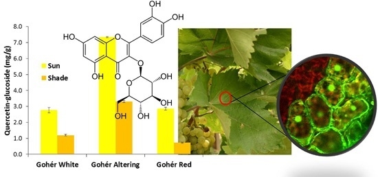
3.2. Changes in Leaf Polyphenolic Composition

In the forthcoming experiment, we studied the phytochemical acclimation strategies of grapevine leaves grown in full sunlight and in half shade, corresponding to 3–4-times lower photosynthetically active radiation (PAR) conditions. According to the literature [8,18], quercetin- and kaempferol-derivatives proved to be the main leaf flavonoids in Vitis vinifera, but their dependence on natural light conditions has not been studied yet in detail. For that purpose, quantification of quercetin-3-O-glucuronide, quercetin-3-O-glucoside, quercetin-3-O-galactoside, quercetin-3-O-rutinoside, kaempferol-3-O-glucuronide, kaempferol-3-O-glucoside, kaempferol-3-O-rutinoside, and trans-caftaric acid was carried out based on MS data and results are given in Table 1. The most predominant compound in the varieties was quercetin-3-O-glucuronide, followed by trans-caftaric acid. Looking at the pattern of flavonols, quercetin-3-O-glucuronide was followed by quercetin-3-O-glucoside, both of which were eminently more abundant than the remaining flavonol components. Similarly to our results, quercetin-3-O-glucuronide was the most abundant flavonol followed by the quercetin-3-O-glucoside in Pinot noir leaves [8]. Total amount of detected quercetin-derivatives were significantly higher than that of kaempferol-derivatives in both leaf types. Amount of polyphenols detected was significantly different between sun and half shade leaves, except for quercetin-glucuronide, kaempferol-glucuronide and kaempferol-rutinoside in Gohér white (Table 1). All other compounds were light responsive, but to a different extent. Accumulation of quercetin-glucoside, kaempferol-glucoside and quercetin-galactoside was the most prominent in full sunlight exposed samples. In contrast, the most abundant quercetin-derivative, quercetin-glucuronide showed only 1.3-fold (G. altering) and 1.4-fold (G. red) increase in sun leaves. The hydroxycinnamate trans-caftaric acid accumulated significantly (1.07-fold in G. white, 1.36-fold in G. altering, and 1.88-fold in G. red) in the varieties due to full sunlight. Regarding total amount of polyphenolics detected, significant differences were found among varieties when comparing their leaves in the same exposure as well as when comparing their sun leaves to half shade ones. Gohér altering was the variety with the highest polyphenolic content, followed by Gohér white and Gohér red. The only non-significant difference was found between sun leaves of Gohér white and Gohér red (Table 2). Gohér red accumulated mostly these compounds exposed to full sunlight, followed by Gohér altering and Gohér white (1.78-fold, 1.50-fold and 1.24-fold higher amount in sun leaves than in half shade leaves, respectively, calculated based on total polyphenol amount presented in Table 1).
Table 1. Polyphenolic composition of sun and half shade leaves of Gohér varieties. Values are given as the average of three replicates ± SD. T-values from a paired Student’s-t-tests are in Table A2.
Compounds Leaf Polyphenolic Profile [mg g−1] Dry Weight
Gohér WhiteGohér AlteringGohér Red
SunShadeSunShadeSunShade
Phenolic acid
trans-caftaric acid4.00 ± 0.075*3.73 ± 0.046 8.46 ± 0.314*6.20 ± 0.2453.66 ± 0.071*1.94 ± 0.075
Flavonol-glycosides
Quercetin-glucuronide8.87 ± 0.377 8.02 ± 0.39418.52 ± 0.386*14.42 ± 0.1738.42 ± 0.177*6.19 ± 0.102
Quercetin-glucoside2.77 ± 0.168*1.20 ± 0.0547.35 ± 0.036*3.30 ± 0.0592.86 ± 0.091*0.73 ± 0.030
Quercetin-rutinoside0.64 ± 0.044*0.43 ± 0.0311.65 ± 0.018*1.06 ± 0.0330.50 ± 0.020*0.13 ± 0.004
Quercetin-galactoside0.59 ± 0.042*0.26 ± 0.0171.81 ± 0.036*0.73 ± 0.0200.57 ± 0.010*0.13 ± 0.005
Kaempferol-glucuronide0.56 ± 0.0250.56 ± 0.0271.69 ± 0.048*1.31 ± 0.0240.43 ± 0.006*0.24 ± 0.007
Kaempferol-glucoside0.53 ± 0.031*0.21 ± 0.0111.76 ± 0.027*0.54 ± 0.0050.46 ± 0.013*0.11 ± 0.004
Kaempferol-rutinoside0.18 ± 0.0080.17 ± 0.0100.73 ± 0.010*0.42 ± 0.0110.13 ± 0.006*0.07 ± 0.003
Total amount18.14 ± 0.771 14.58 ± 0.590 41.97 ± 0.876 27.98 ± 0.57117.02 ± 0.3939.54 ± 0.229
* indicates significant (df = 4, p ˂ 0.05) differences between sun and half shade leaves of the same variety.
Table 2. Comparison of total polyphenolic contents in Gohér varieties. Data are actual t-values, corresponding to the total amount data in Table 1, for analysis between grapevine varieties within sun exposure. Significant differences at p ˂ 0.05 indicated in bold.
Sun LeavesG. WhiteG. Altering
G. Altering39.13
G. Red2.5258.32
Shade LeavesG. WhiteG. Altering
G. Altering−34.86
G. Red13.9885.45
Shade LeavesSun Leaves
G. WhiteG. AlteringG. Red
G. White6.4150.627.01
G. Altering−20.7030.56−56.21
G. Red18.8674.0952.83
Kolb [18] revealed the effect of visible and UV-light on accumulation of polyphenolics using greenhouse and filtered illumination conditions. While the amount of hydroxycinnamic acids highly increased by strong visible light, the amount of flavonoids increased primarily by UV-B irradiation. The roles of main leaf polyphenolics of different plant species in natural sunlight acclimation have been studied under Mediterranean climate, which revealed significant differences in acclimation strategies [12,13]. In our case, the varieties were grown under natural sunlight conditions characteristic to mainland temperate climate summers. Despite of the highly similar viticultural and environmental factors and the close genetic background of studied varieties, they showed differences not only in quantity of detected polyphenolics, but also in their response to full sunlight.

3.3. Changes in Leaf Flavonoid Distribution

In order to study the functional role of flavonoids in natural sunlight acclimation of grapevine leaf, confocal laser scanning microscopy (CLSM) was used for micro-localization of these secondary metabolites. Leaves of Gohér altering have been chosen for analysis, because it displayed the highest flavonoid content. Leaves were pre-treated with the Naturstoff reagent (NS, see 3.6 for details) to enhance fluorescence of flavonols. The adaxial side of lamina directly facing sunlight irradiation were used in microscopy analyses. Significant changes were found in flavonoid content of epidermal layer, with markedly high accumulation of NS-conjugated flavonoids in cell nuclei and cell walls in sun leaves compared to half shade leaves (Figure 1, insets). Similar changes were observed in the palisade parenchyma cells and in their chloroplasts (Figure 1).
Studies have shown that flavonoids are associated with the chloroplast envelope membrane and may limit the diffusion of reactive oxygen species out of the chloroplast [33,34,35]. It has also been shown in different plants that they are capable of quenching hydroxyl radicals, hence protecting cells against nuclear DNA damage [36,37]. Flavonoids indeed may protect membranes by enhancing membrane rigidity and therefore preserving them against oxidative damage [38,39]. In our case, there was no vacuolar fluorescence in the palisade cells of the grapevine, while Agati [40,41] successfully detected NS-conjugated flavonoids in the vacuoles of mesophyll cells in Ligustrum vulgare under 100% natural sunlight, but failed to visualize flavonoids in other cellular compartments. They detected light-responsive quercetin and luteolin derivatives, while hydroxycinnamates, which serve specific UV-B screening functions [42], and monohydroxy-B-ring flavonoids were unresponsive to excess light. Our study showed similar results in so far as flavonoid derivatives accumulated to a greater extent than trans-caftaric acid did, which provides support for their antioxidant role prior to screening properties in photoprotection. This conclusion is further corroborated by distribution of NS-conjugated flavonoids, not only in the epidermis, but also in the mesophyll. L. vulgare and Phillyrea latifolia with the same flavonoid composition, but with different morpho-anatomical traits, showed different metabolic plasticity in the acclimation process to high solar radiation, namely quercetin- and luteolin-glycosides accumulated to a much greater extent in L. vulgare than in P. latifolia, and in different microlocations [43]. Together with the dihydroxy-B-ring substituted quercetin-glycosides, trihydroxy myricetin-glycosides accumulated significantly in two Mediterranean shrubs (Myrtus communis, Pistacia lentiscus) in response to different abiotic stresses (light, salt) [13]. Our studies confirm that changes in the flavonoid content of the leaves, and their micro-localization in the leaf tissues, have a key role in the acclimation mechanisms of the grapevine, even in a less contrasting light environment.
Figure 1. Laser scanning confocal microscopy images of sun exposed (A) and half shaded (B) Gohér altering leaves with NS-conjugated flavonoid (green) and chlorophyll (red) fluorescence (merged). Circles highlight three representative mesophyll cells from palisade parenchyma regions of sun and half shade leaves. Note the difference in green fluorescence intensity of NS-conjugated flavonoids in these comparable regions (to reduce image saturation, detector sensitivity was reduced 6.4% to capture sun leaf’s intense green fluorescence emission). Insets show lumen of upper epidermis cells with nuclear labeling (arrows). Red chlorophyll fluorescence appears yellow when superimposed with green signals. Scale bar is 30 µm.
Applsci 05 01955 g001

4. Conclusions

To our best knowledge, this study was the first to detect the main glycoconjugate forms of the native Gohér conculta and their detailed quantitative reactions to local irradiance. The microlocalization analysis proved the antioxidant role of flavonols in the leaf tissue. We can conclude that grapevine leaves exposed to full sunlight, significantly activated the biosynthesis of light-responding secondary metabolites in order to avoid the harmful effects of light-stress and to maintain their optimal photosynthetic capacity. Although the three Gohér varieties are genetically very similar, demonstrated by SSR markers, they showed differences in their basic phenolic quantity and in their acclimation response as well. Therefore, accumulation of polyphenols in grapevine leaves depends on both autonomous and sunlight regulated pathways. Our work reports preliminary results, and further studies should include designed experiments to statistically evaluate biological variability for a more complete picture of environmental roles, such as sunlight, in the production of these secondary metabolites. On the other hand, to better recognize the acclimation strategies used by these genetically closely related grapevine varieties, our further studies will focus on the regulation of polyphenolic biosynthetic pathways as well.

Acknowledgments

We wish to thank É. Hideg for her valuable advices and contribution to the management of the experimental plan. The research was supported by the Hungarian Scientific Grant Agency (OTKA-K101430 and OTKA-PD100506). The present scientific contribution is dedicated to the 650th anniversary of the foundation of the University of Pécs, Hungary.

Author Contributions

M.K. and G.J. conceived the original ideas and designed the experiments with the collaboration of P.T.; F.A. performed the microscopic studies; L.A., K.C. and N.P. performed the chromatographic experiments and analyzed the data; the manuscript was, for the most part, written by M.K., L.A. and F.A. with contributions from all co-authors.

Conflicts of Interest

The authors declare no conflict of interest.

Appendix

Table A1. Observed diagnostic ions of polyphenols found in the Gohér leaf samples.
Retention Time, minUV maxMS PolaritySupposed Compound and Annotation of Diagnostic IonsElemental Composition of Diagnostic Ions (Ion Formula)Expected m/zError, ppm
6.32325Caftaric acidaC13H11O9311.0409−0.32
Caffeic acid residueC9H7O4179.0350.83
Caffeic acid residue-CO2C8H7O2135.0452−1.02
Tartaric acid residueC4H5O6149.00920.31
6.94325Caftaric acid isomerC13H11O9311.04092.77
Caffeic acid residueC9H7O4179.0354.29
Caffeic acid residue-CO2C8H7O2135.04525.53
Tartaric acid residueC4H5O6149.00924.99
[[2M − H]C26H23O18623.0890.33
10.91315Coutaric acidC13H11O8295.04594.34
Tartaric acid residueC4H5O6149.00926.85
Coumaric acid residueC9H7O3163.04019.46
17.3260, 360+Myr − Hx 1
MyrC15H11O8319.04480.91
Myr + HxC21H21O13481.09773.53
Myr + Hx + NaC21H20O13Na503.07963.68
17.47265, 360+Myr − Hx 2
MyrC15H11O8319.0448−1.35
Myr + HxC21H21O13481.0977 2.23
Myr + Hx + NaC21H20O13Na503.0796 3.68
19.23260, 360+Que − 3-O-rutinosidea
QueC15H11O7303.05−3.1
Que + dHxC21H21O11449.1079−1.02
Que + HxC21H21O12465.1028−2.69
Que + dHx + HxC27H31O16611.1607−0.93
Que + dHx + Hx + NaC27H30O16Na633.1432−1.43
19.68260, 360+Que − 3-O-galactosidea
QueC15H11O7303.054.00
Que + HxC21H21O12465.10285.15
Que + Hx + NaC21H20O12Na487.08535.66
19.81255, 360+Que − 3-O-glucuronidea
QueC15H11O7303.05−5.66
Que + HxaC21H19O13479.082 4.19
Que + Hxa + NaC21H18O13Na501.064−4.68
19.95255, 360+Que − 3-O-glucosidea
QueC15H11O7303.05−1.52
Que + HxC21H21O12465.1028−0.56
Que + Hx + NaC21H20O12Na487.0853 0.70
21.05260, 350+Kae − 3-O-rutinosidea
KaeC15H11O6287.0555.64
Kae + HxC21H21O11449.10787.34
Kae + Hx + dHxC27H31O15595.16578.05
Kae + Hx + dHx + NaC27H30O15Na617.14828.32
21.23260, 350+Kae − 3-O-glucosidea
KaeC15H11O6287.0553.56
Kae + HxC21H21O11449.10784.17
Kae + Hx + NaC21H20O11Na471.08985.34
21.97260, 350+Kae − Hx 2
KaeC15H11O6287.0553.24
Kae + HxC21H21O11449.10784.83
Kae + Hx + NaC21H20O11Na471.08984.84
21.99260, 350+Kae − 3-O-glucuronidea
KaeC15H11O6287.055−3.32
Kae + HxaC21H19O12463.0871−2.22
Kae + Hxa + NaC21H18O12Na485.0694.95
21.47265, 350+Isr + Hx + dHx
IsrC16H13O7317.06563.50
Isr + HxC22H23O12479.1184−2.70
Isr + Hx + dHxC28H33O16625.17633.46
Isr + Hx + dHx + NaC28H32O16Na647.15835.30
22.12265, 350+Isr + Hx 1
IsrC16H13O7317.06562.52
Isr + HxC22H23O12479.1184−5.85
Isr + Hx + NaC22H22O12Na501.1003−1.29
22.44265, 350+Isr + Hx 2
IsrC16H13O7317.06561.31
Isr + HxC22H23O12479.11841.87
Isr + Hx + NaC22H22O12Na501.10033.84
a Compound was identified with reference standards. Myr: myricetin, Que: quercetin, Kae: kaempferol, Isr: isorhamnetin, Hx: hexoside, dHx: deoxyhexoside, Hxa: hexosic acid.
Table A2. Actual t-values corresponding to the polyphenolic composition of sun and half shade leaves of Gohér varieties. Significant differences at p ˂ 0.05 indicated in bold.
Sun / Shade Leaves
Polyphenolic CompoundsGohér
White
Gohér
Altering
Gohér
Red
trans-caftaric-acid5.319.8328.75
Quercetin-glucuronide2.6916.7918.88
Quercetin-glucoside15.43101.0738.55
Quercetin-rutinoside6.7427.3331.73
Quercetin-galactoside12.4144.9668.00
Kaempferol-glucuronide−0.1512.1135.50
Kaempferol-glucoside16.9877.3446.24
Kaempferol-rutinoside2.2135.3315.19

References

  1. Berli, F.; D’Angelo, J.; Cavagnaro, B.; Bottini, R.; Wuilloud, R.; Silva, M.F. Phenolic composition in grape (Vitis vinifera L. cv. Malbec) ripened with different solar UV-B radiation levels by capillary zone electroforesis. J. Agric. Food Chem. 2008, 56, 2892–2898. [Google Scholar] [CrossRef] [PubMed]
  2. Dani, C.; Oliboni, L.S.; Agostini, F.; Funchal, C.; Serafini, L.; Henriques, J.A.; Salvador, M. Phenolic content of grapevine leaves (Vitis labrusca var. Bordo) and its neuroprotective effect against peroxide damage. Toxicol. In Vitro 2010, 24, 148–153. [Google Scholar] [CrossRef] [PubMed]
  3. Kosar, M.; Küpeli, E.; Malyer, H.; Uylaser, V.; Türkben, C.; Baser, K.H. Effect of brining on biological activity of leaves of Vitis vinifera L. (cv. Sultani Cekirdeksiz) from Turkey. J. Agric. Food Chem. 2007, 55, 4596–4603. [Google Scholar] [CrossRef] [PubMed]
  4. Monagas, M.; Hernández-Ledesma, B.; Gómez-Cordovés, C.; Bartolomé, B. Commercial dietary ingredients from Vitis vinifera L. leaves and grape skins: Antioxidant and chemical characterization. J. Agric. Food Chem. 2006, 54, 319–327. [Google Scholar] [CrossRef] [PubMed]
  5. Iacopini, P.; Baldi, M.; Storchi, P.; Sebastiani, L. Catechin, epicatechin, quercetin, rutin and resveratrol in red grape: Content, in vitro antioxidant activity and interactions. J. Food Compos. Anal. 2008, 21, 589–598. [Google Scholar] [CrossRef]
  6. Leifert, W.R.; Abeywardena, M.Y. Cardioprotective actions of grape polyphenols. Nutr. Res. 2008, 28, 729–737. [Google Scholar] [CrossRef] [PubMed]
  7. Markham, K.R. Flavones, Flavonols and their glycosides. Methods in Plant Biochemistry; Harborne, J.B., Ed.; Academic Press Ltd.: London, UK, 1989; Volume 1, pp. 197–235. [Google Scholar]
  8. Schoedl, K.; Schuhmacher, R.; Forneck, A. Studying the polyphenols of grapevine leaves according to age and insertion level under controlled conditions. Sci. Hortic. 2012, 141, 37–41. [Google Scholar] [CrossRef]
  9. Chacón, J.L.; García, E.; Martínez, J.; Romero, R.; Gómez, S. Impact of the vine water status on the berry and seed phenolic composition of ‘Merlot’ (Vitis vinifera L.) cultivated in a warm climate: Consequence for the style of wine. Vitis 2009, 48, 7–9. [Google Scholar]
  10. Downey, M.O.; Dokoozlian, N.K.; Krstic, M.P. Cultural practice and environmental impacts on the flavonoid composition of grapes and wine: A review of recent research. Am. J. Enol. Viticul. 2006, 57, 257–268. [Google Scholar]
  11. Ramakrishna, A.; Ravishankar, G.A. Influence of abiotic stress signals on secondary metabolites in plants. Plant Signal Behav. 2011, 6, 1720–1731. [Google Scholar] [PubMed]
  12. Tattini, M.; Galardi, C.; Pinelli, P.; Massai, R.; Remorini, D.; Agati, G. Differential accumulation of flavonoids and hydroxycinnamates in leaves of Ligustrum vulgare under excess light and drought stress. New Phytol. 2004, 163, 547–561. [Google Scholar] [CrossRef]
  13. Tattini, M.; Remorini, D.; Pinelli, P.; Agati, G.; Saracini, E.; Traversi, M.L.; Massai, R. Morpho-anatomical, physiological and biochemical adjustments in response to root zone salinity stress and high solar radiation in two Mediterranean evergreen schrubs, Myrtus communis and Pistacia lentiscus. New Phytol. 2006, 170, 779–794. [Google Scholar] [CrossRef] [PubMed]
  14. Majer, P.; Neugart, S.; Krumbein, A.; Schreiner, M.; Hideg, É. Singlet oxygen scavenging by leaf flavonoids contributes to sunlight acclimation in Tilia platyphyllos. Environ. Exp. Bot. 2014, 100, 1–9. [Google Scholar] [CrossRef]
  15. Di Ferdinando, M.; Brunetti, C.; Agati, G.; Tattini, M. Multiple functions of polyphenols in plants inhabiting unfavorable Mediterranean areas. Environ. Exp. Bot. 2014, 103, 107–116. [Google Scholar] [CrossRef]
  16. Rozema, J.; van de Staaij, J.; Björn, L.O.; Caldwell, M.M. UV-B as an environmental factor in plant life: Stress and regulation. Trends. Ecol. Evol. 1997, 12, 22–28. [Google Scholar] [CrossRef]
  17. Hutzler, P.; Fischbach, R.; Heller, W.; Jungblut, T.P.; Reuber, S.; Schmitz, R.; Veit, M.; Weissenbck, G.; Schnitzler, J.P. Tissue localization of phenolic compounds in plants by confocal laser scanning microscopy. Exp. Bot. 1998, 49, 953–965. [Google Scholar] [CrossRef]
  18. Kolb, C.A.; Kaser, M.A.; Kopecky, J.; Zotz, G.; Riederer, M.; Pfündel, E.E. Effects of natural intensities of visible and ultraviolet radiation on epidermal ultraviolet screening and photosynthesis in grape leaves. Plant Physiol. 2001, 127, 863–875. [Google Scholar] [CrossRef] [PubMed]
  19. Götz, M.; Albert, A.; Stitch, S.; Heller, W.; Scherb, H.; Krins, A.; Langebartels, C.; Seidlitz, H.K.; Ernst, D. PAR modulation of the UV-dependent levels of flavonoid metabolites in Arabidopsis thaliana (L.) Heynh. leaf rosettes: Cumulative effects after a whole vegetative growth period. Protoplasma 2010, 243, 95–103. [Google Scholar] [CrossRef] [PubMed]
  20. Brunetti, C.; Di Ferdinando, M.; Fini, A.; Pollastri, S.; Tattini, M. Flavonoids as Antioxidants and Developmental Regulators: Relative Significance in Plants and Humans. Int. J. Mol. Sci. 2013, 14, 3540–3555. [Google Scholar] [CrossRef] [PubMed]
  21. Cortell, J.M.; Kennedy, J.A. Effect of shading on accumulation of flavonoid compounds in (Vitis vinifera L.) Pinot noir fruit and extraction in a model system. J. Agric. Food Chem. 2006, 55, 8510–8520. [Google Scholar] [CrossRef] [PubMed]
  22. Matus, J.T.; Loyola, R.; Vega, A.; Pena-Neira, A.; Bordeu, E.; Arce-Johnson, P.; Alcalde, J.A. Post-veraison sunlight exposure induces MYB-mediated transcriptional regulation of anthocyanin and flavonol synthesis in berry skins of Vitis vinifera. J. Exp. Bot. 2009, 60, 853–867. [Google Scholar] [CrossRef] [PubMed]
  23. Figueiredo-González, M.; Martínez-Carballo, E.; Cancho-Grande, B.; Santiago, J.L.; Martínez, M.C.; Simal-Gándara, J. Pattern recognition of three Vitis vinifera L. red grapes varieties based on anthocyanin and flavonol profiles, with correlations between their biosynthesis pathways. Food Chem. 2012, 13, 9–19. [Google Scholar] [CrossRef]
  24. Galbács, Z.; Molnár, S.; Halász, G.; Kozma, P.; Hoffmann, S.; Kovács, L.; Veres, A.; Galli, Z.; Szőke, A.; Heszky, L.; et al. Identification of grapevine cultivars using microsatellite-based DNA barcodes. Vitis 2009, 48, 17–24. [Google Scholar]
  25. Castellarin, S.D.; di Gaspero, G.; Marconi, R.; Nonis, A.; Peterlunger, E.; Paillard, S.; Adam-Blondon, A.F.; Testolin, R. Colour variation in red grapevines (Vitis vinifera L.): Genomic organisation, expression of flavonoid 3′-hydroxylase, flavonoid 3′,5′-hydroxylase genes and related metabolite profiling of red cyanidin-/blue delphinidin-based anthocyanins in berry skin. BMC Genomics 2006, 7. [Google Scholar] [CrossRef] [PubMed]
  26. Németh, M. Ampelographic Album: Cultivated Grapevine Varieties II, 1st ed.; Agricultural Press: Budapest, Hungary, 1970; pp. 69–70. [Google Scholar]
  27. Abrankó, L.; Garcia-Reyes, J.F.; Molina-Diaz, A. Systemic bottom-up approach for flavonoid derivative screening in plant material using liquid chromatography high-resolution mass spectrometry. Anal. Bioanal. Chem. 2012, 403, 995–1006. [Google Scholar] [CrossRef] [PubMed]
  28. Flamini, R.; Traldi, P. Grape and Wine Polyphenols. In Mass Spectrometry in Grape and Wine Chemistry; Desiderio, D.M., Nibbering, N.M.M., Eds.; Wiley & Sons Inc.: Hoboken, NJ, USA, 2010; Part II; pp. 163–214. [Google Scholar]
  29. Pastrana-Bonilla, E.; Akoh, C.C.; Sellappan, S.; Krewer, G. Phenolic content and antioxidant capacity of muscadine grapes. J. Agric. Food Chem. 2003, 51, 5497–5503. [Google Scholar] [CrossRef] [PubMed]
  30. Taware, P.B.; Dhumal, K.N.; Oulkar, D.P.; Patil, S.H.; Banerjee, K. Phenolic alterations in grape leaves, berries and wines due to foliar and cluster powdery mildew infections. Int. J. Pharm. Biol. Sci. 2010, 1, 1–14. [Google Scholar]
  31. Balik, J.; Kyselakova, M.; Vrchotova, N.; Triska, J.; Kumsta, M.; Veverka, J.; Hic, P.; Totusek, J.; Lefernova, D. Relations between polyphenols content an antioxidant activity in vine grapes and leaves. Chez. J. Food Sci. 2008, 26, 25–32. [Google Scholar]
  32. Doshi, P.; Adsule, P.; Banerjee, K. Phenolic composition and antioxidant activity in grapevine parts and berries (Vitis vinifera L.) cv. Kishmish Chornyi (Sharad Seedless) during maturation. Int. J. Food Sci. Technol. 2006, 41, 1–9. [Google Scholar] [CrossRef]
  33. Mullineaux, P.; Karpinski, S. Signal transduction in response to excess light: Getting out of the chloroplast. Curr. Opin. Plant Biol. 2002, 5, 43–48. [Google Scholar] [CrossRef]
  34. Hatier, J.H.B.; Gould, K.S. Foliar anthocyanins as modulators of stress signals. J. Theor. Biol. 2008, 253, 625–627. [Google Scholar] [CrossRef] [PubMed]
  35. Agati, G.; Azzarello, E.; Pollastri, S.; Tattini, M. Flavonoids as antioxidants in plants: Location and functional significance. Plant Sci. 2012, 196, 67–76. [Google Scholar] [CrossRef] [PubMed]
  36. Polster, J.; Dithmar, H.; Burgemeister, R.; Friedemann, G.; Feucht, W. Flavonoids in plant nuclei: Detection by laser microdissection and pressure catapulting (LMPC), in vivo staining, and UV-visible spectroscopic titration. Physiol. Plantarum. 2006, 126, 163–174. [Google Scholar] [CrossRef]
  37. Pérez-Gregorio, M.R.; Regueiro, J.; González-Barreiro, C.; Rial-Otero, R.; Simal-Gándara, J. Changes in antioxidant flavonoids during freeze-drying of red onions and subsequent storage. Food Cont. 2011, 22, 1108–1113. [Google Scholar] [CrossRef]
  38. Inoue, K. Emerging roles of the chloroplast outer envelope membrane. Trends Plant Sci. 2011, 16, 550–557. [Google Scholar] [CrossRef] [PubMed]
  39. Erlejman, A.G.; Verstraiten, S.V.; Graga, C.G.; Oteiza, P.I. The interaction of flavonoids with membranes: Potential determinants of flavonoid antioxidant effects. Free Radical Res. 2004, 38, 1311–1320. [Google Scholar] [CrossRef] [PubMed]
  40. Agati, G.; Stefano, G.; Biricolti, S.; Tattini, M. Mesophyll distribution of ‘antioxidant’ flavonoid glycosides in Ligustrum vulgare leaves under contrasting sunlight irradiance. Ann. Bot-London 2009, 104, 853–861. [Google Scholar] [CrossRef] [PubMed]
  41. Agati, G.; Biricolti, S.; Guidi, L.; Ferrini, F.; Fini, A.; Tattini, M. The biosynthesis of flavonoids is enhanced similarly by UV radiation and root zone salinity in L. vulgare leaves. J. Plant Phys. 2011, 168, 204–212. [Google Scholar] [CrossRef] [PubMed]
  42. Harborne, J.B.; Williams, C.A. Advances in flavonoid research since 1992. Phytochem 2000, 55, 481–504. [Google Scholar] [CrossRef]
  43. Tattini, M.; Guidi, L.; Morassi-Bonzi, L.; Pinelli, P.; Remorini, D.; Degl’Innocenti, E.; Giordano, C.; Massai, R.; Agati, G. On the role of flavonoids in the integrated mechanisms of response of Ligustrum vulgare and Phillyrea latifolia to high solar radiation. New Phytol 2005, 167, 457–470. [Google Scholar] [CrossRef] [PubMed]

