Article

The Enhancing Effects of Amelogenin Exon 5-Encoded Peptide from Enamel Matrix Derivative on Odontoblast-Like KN-3 Cells

1
Department of Periodontology, Osaka Dental University, Osaka 573-1121 Japan
2
Department of Endodontics, Osaka Dental University, Osaka 573-1121, Japan
3
Department of Oral Pathology, Osaka Dental University, Osaka 573-1121, Japan
*
Author to whom correspondence should be addressed.
Appl. Sci. 2018, 8(10), 1890; https://doi.org/10.3390/app8101890
Submission received: 30 August 2018 / Accepted: 8 October 2018 / Published: 11 October 2018
(This article belongs to the Section Applied Biosciences and Bioengineering)

Abstract

:

Featured Application

Amelogenin exon 5 could have potential for application in dental pulp capping.

Abstract

Enamel matrix derivative (EMD) is applied for periodontal therapy. We created a synthetic amelogenin peptide (SP) derived from EMD, and have previously investigated the biological function of SP. However, it is unknown whether SP affects odontoblastic differentiation. In this study, we tested the effects of SP in the odontoblast-like cells, KN-3 cells. KN-3 cells were cultured with SP (0 to 1000 ng/mL) and then cultured for 3, 8, 24, or 48 h in order to determine the effects of SP on cell proliferation and detect its optimum concentration. KN-3 cells were treated with SP in odontogenic differentiation medium cultured for 3 or 7 days. Odontogenic markers were measured by the detection of alkaline phosphatase (ALP) activity and dentin sialo phosphoprotein (DSPP) expression, the calcified nodule formation, and calcium deposition. The addition of SP significantly promoted cell proliferation at 100 ng/mL, generating the greatest change in cell proliferation. SP also showed increased odontogenic expression markers and mineralization. These results suggest that SP, derived from EMD, could have potential for application in dental pulp capping.

Graphical Abstract

1. Introduction

Enamel matrix derivative (EMD) can induce the formation of hard tissue, such as alveolar bone and cementum tissue [1,2]. EMD is applied for periodontal therapy and bone regeneration. We previously showed that subcutaneous injections of EMD can induce the growth of cartilage tissue and eosinophilic round bodies (ERBs) [3]. We further analyzed these ERBs by using MALDI-TOF, and found fragments of exon 5 of amelogenin.
We synthesized a 7-amino acid (WYQNMIR) peptide based on these fragments and tested whether the synthetic peptide (SP) would behave similarly to EMD [4]. We found that the SP could induce bone-like tissue formation in artificial periodontal defects in rats [5,6]. Moreover, we found that SP could enhance the cell proliferation of periodontal ligament (PDL) cells [7] and enhance osteoblastic differentiation in mesenchymal stem cells (MSCs) [8,9,10]. These findings also could help to clarify the biological functions of amelogenin exon 5.
EMD induces anti-EMD antibodies [11]. Only peptides of greater than approximately ten residues (or >5 kDa) can function as antigens [12,13]. SP is 7 amino acids long, with a mass of 1118 Da. Therefore, SP has no effect with respect to inducing an immunological response.
Dental caries, tooth fractures, and other types of dental trauma induce tooth loss. Direct pulp treatment requires materials that protect the pulp tissue but induce hard tissue formation, in order to repair and maintain dental pulp tissues [14,15,16,17,18,19]. This requirement has led to the design and introduction of new, bioactive agents for dental pulp tissue engineering materials.
To investigate dental pulp tissue regeneration, a rat odontoblast-like cell line, KN-3, was created by Prof. Kitamura and Prof. Nishihara [20]. KN-3 cells showed high levels of odontogenic expression and the ability to form calcium nodules [21]. KN-3 cells have also been used as an authentic control to study the differentiation of induced pluripotent stem (iPS) cells and embryonic stem (ES) cells into odontoblast-like cells [22,23].
The biological response to SP regarding odontogenic differentiation has not yet been investigated. In the present study, we evaluated the odontogenic effects of SP in KN-3 cells.

2. Materials and Methods

2.1. Cell Culture

The rat odontoblast-like cell, KN-3, was provided by Prof. Chiaki Kitamura and Prof. Tatsuji Nishimura (Kyushu Dental College). KN-3 cells were maintained as described previously. Twenty KN-3 cells were cultured in normal medium comprising Eagle’s minimal essential medium (α-MEM) with 10% FBS (Gibco BRL, Life Technologies, Grand Island, NY, USA), 500 U/mL penicillin, 500 µg/mL streptomycin, and 25 µg/mL amphotericin B (Nacalai Tesque, Kyoto, Japan). For differentiation assays, KN-3 cells were incubated in medium containing 50 µM l-ascorbic acid 2-phosphate (Nacalai) and 10 mM β-glycerophosphate (Wako Pure Chemical Industries Ltd., Tokyo, Japan).

2.2. Cell Proliferation Assay

KN-3 cells were cultured in normal medium. After 24 h, the medium was changed to normal culture medium containing varying concentrations of SP (0 to 1000 ng/mL); KN-3 cells were incubated for 3, 8, 24, or 48 h. Cell proliferation was measured via the amount of formazan. We measured the absorbance and analyzed results using the SoftMax Pro software (Molecular Devices, Sunnyvale, CA, USA).

2.3. Morphological Analysis

KN-3 cells were incubated in normal culture medium containing SP (0 to 1000 ng/mL) for 48 h. The images were analyzed using the all-in-one fluorescence microscope (BZ-II, Keyence Corporation, Osaka, Japan).

2.4. Alkaline Phosphatase (ALP) Activity Assay

KN-3 cells were cultured with osteogenic medium for 7 days, and measurements were made at days 3 and 7. After cells were washed using PBS and lysed using 0.2% Triton X-100 (Sigma-Aldrich, St. Louis, MO, USA), the activity of ALP was investigated by one-step p-nitro phenyl phosphate (pNPP) (Pierce Biotechnology Inc., Rockford, IL, USA). ALP activity was normalized to the quantity of DNA. DNA content was investigated by DNA assay kit (Invitrogen, Paisley, UK). Data were investigated using the SoftMax Pro software.

2.5. Extracellular Matrix Mineralization

For measurements of calcium production, KN-3 cells were melted using 10% formic acid, and then calcium deposition (Ca) was investigated by calcium detection kit (Wako). For qualitative histology, cells were then stained using 1% alizarin red. Calcified nodules were captured using a BZ-II microscope (Keyence).

2.6. Quantitative Real-Time Polymerase Chain Reaction (PCR)

RNA was obtained using a kit (RNeasy Mini Kit; Qiagen, Venlo, The Netherlands). and then the extracted RNA was transcribed into cDNA using a kit (Prime Script Reagent kit, Takara, Kyoto, Japan). mRNA expression was examined by real-time polymerase chain reaction (PCR) assay. The expression of dentin sialoprotein (DSPP; Rn02132391_s1) was investigated in accordance with standard protocols.

2.7. Immunofluorescence Staining

KN-3 cells were fixed by 70% ethanol. The cells were treated using 0.2% Triton X-100, after which they were blocked using 3% bovine serum albumin (BSA) and then incubated using mouse anti-rat DSPP antibody (Santa Cruz Biotechnology, Santa Cruz, CA, USA). After cells were washed with phosphate-buffered saline (PBS), they were incubated using a fluorescence-labeled secondary anti-mouse antibody (Santa Cruz). The samples were stained using DAPI solution (Dojindo Laboratory, Kumamoto, Japan). Images were captured using fluorescence microscope.

2.8. Statistical Analysis

Statistical analysis was performed by one-way analysis of variance (ANOVA) followed by the Bonferroni post-hoc test using IBM SPSS. Significant differences (p < 0.05) were determined.

3. Results

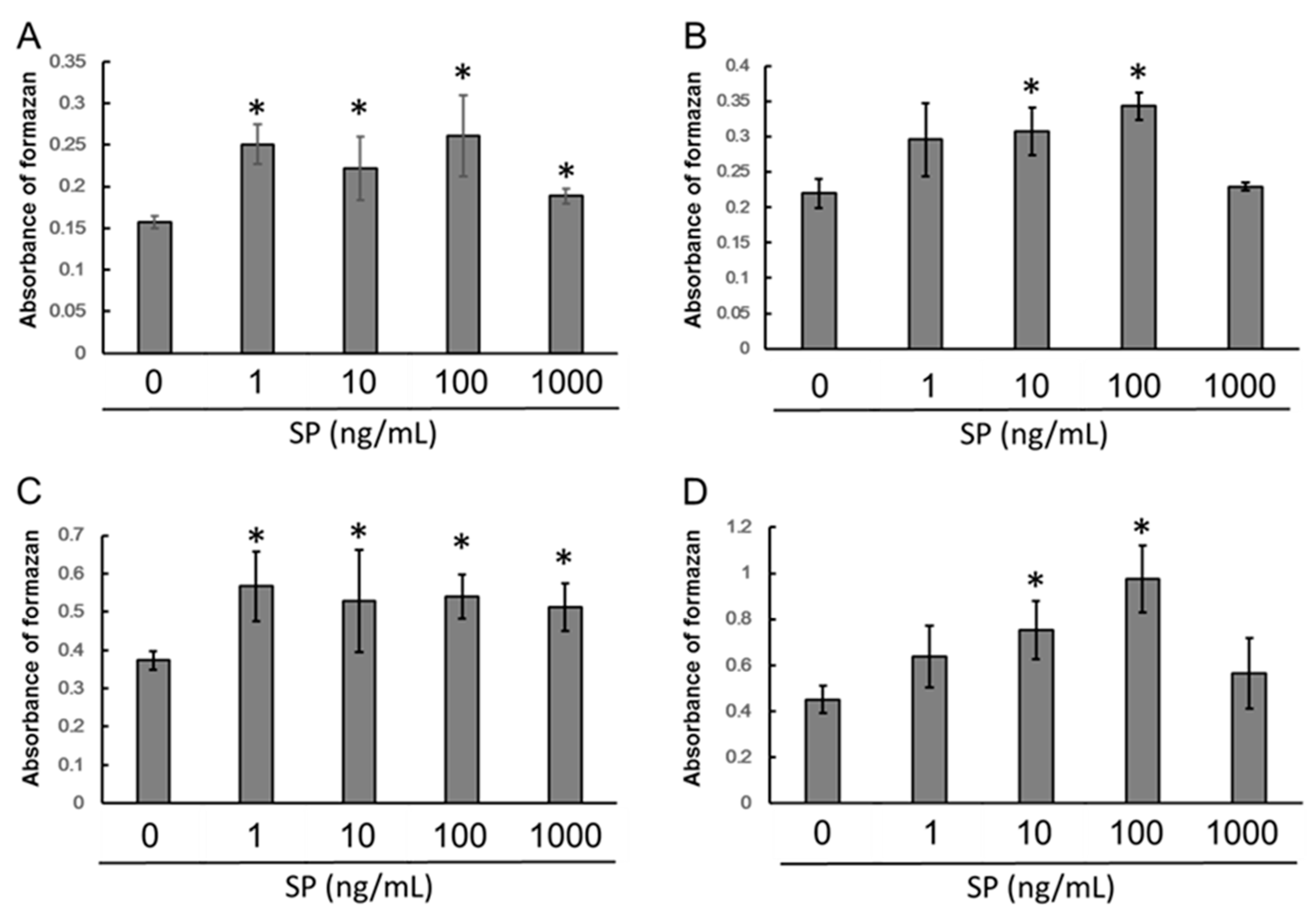
3.1. Cell Proliferation

We first tested varying concentrations of SP on KN-3 cells to determine an effective concentration. We found that 100 ng/mL of SP significantly promoted KN-3 proliferation at 8, 24, 48, and 72 h (Figure 1A, p < 0.05). Based on these results, we chose 100 ng/mL SP as the optimal concentration for subsequent experiments.

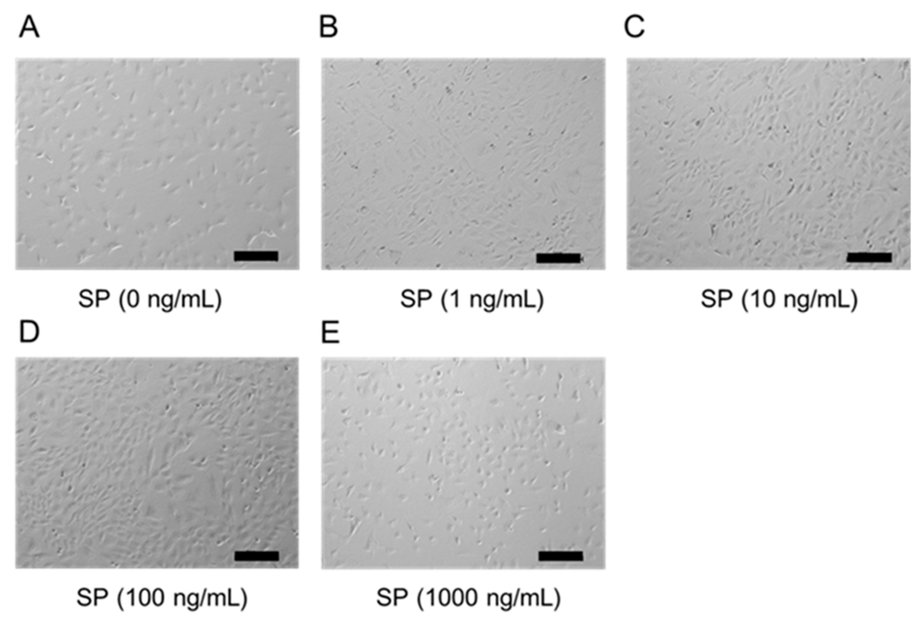
3.2. Cell Morphology
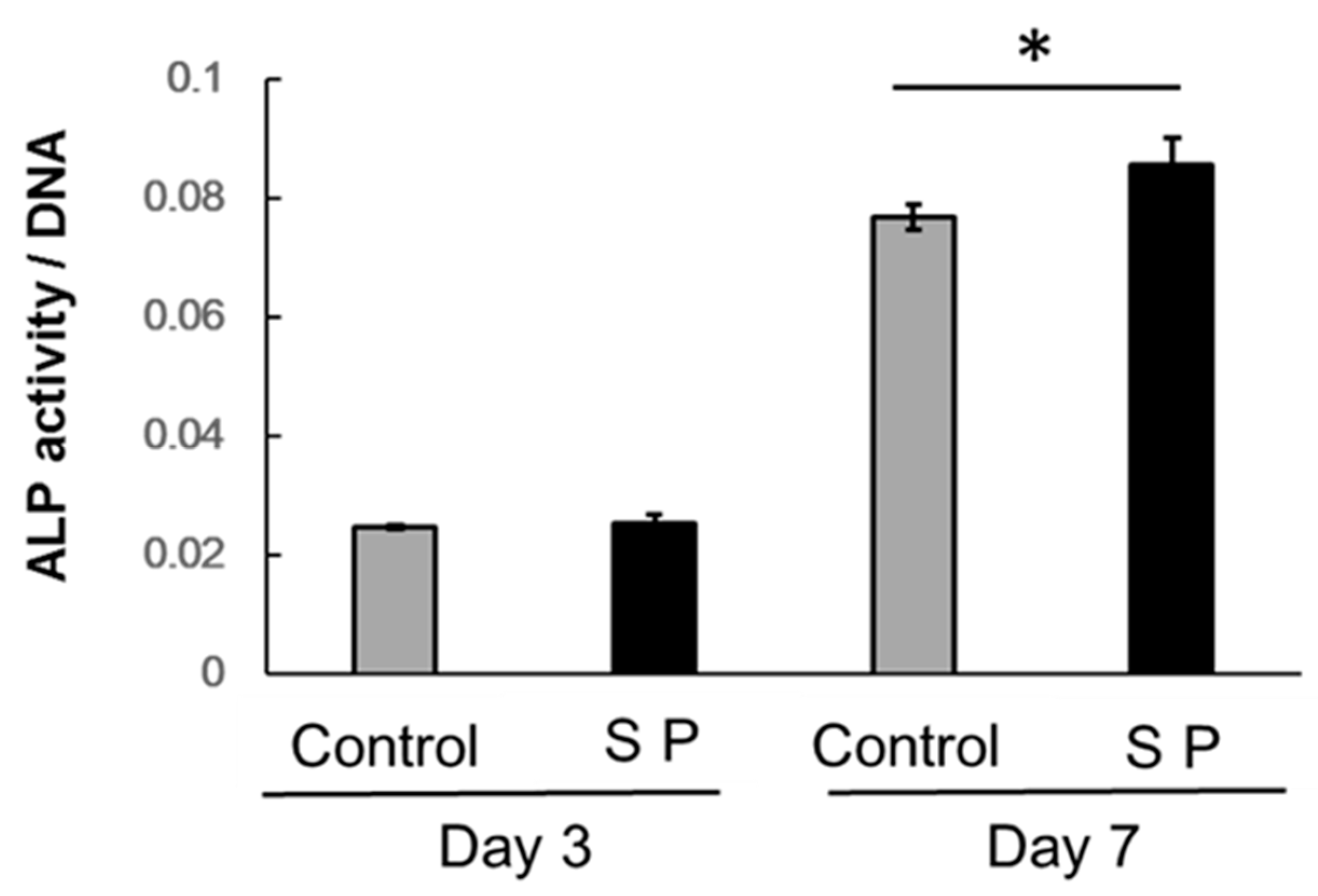
In the SP control group (0 ng/mL SP), the cell morphology exhibited round-shaped morphology. On the other hand, the cell morphology exhibited spindle-shaped morphology in the SP 100 ng/mL group. Figure 2 indicates that the effect of SP on cell morphology.

3.3. ALP Activity

ALP activity in the SP-treated group was significantly increased after 7 days, compared with cells solely treated with differentiation media (Figure 3; p < 0.05).

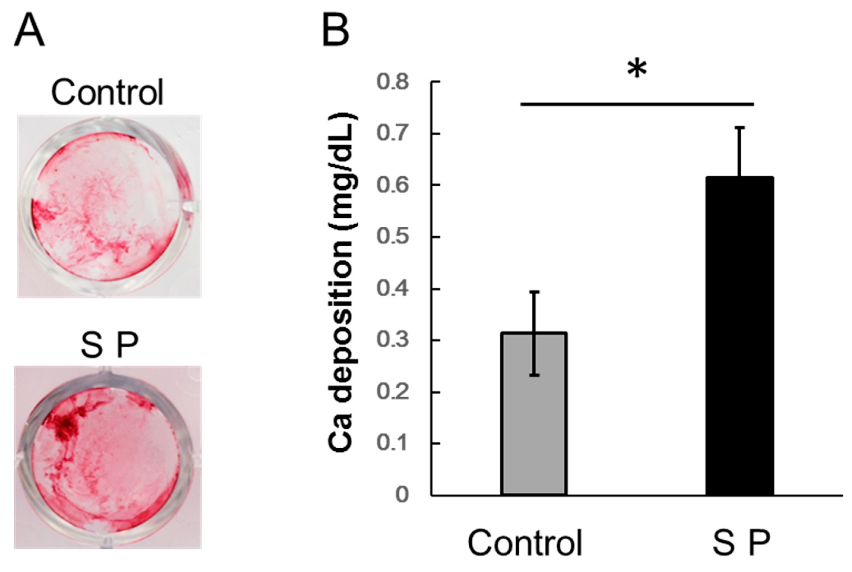
3.4. Extracellular Matrix Mineralization

Calcified nodules were increased in the SP-treated group. Calcium deposition in the SP-treated group significantly increased after 7 days (p < 0.05). Figure 4 indicates that the effect of SP on extracellular matrix mineralization in KN-3 cells. Figure 4A shows the effect of SP on calcified nodule formation by Alizarin red staining. Figure 4B shows the effect of SP on extracellular matrix mineralization.

3.5. mRNA Expression of Dentin Sialoprotein (DSPP)

DSPP was significantly enhanced in the SP group at 1, 3, and 5 h (p < 0.05). Figure 5 indicates that the effect of SP on mRNA expression of DSPP.

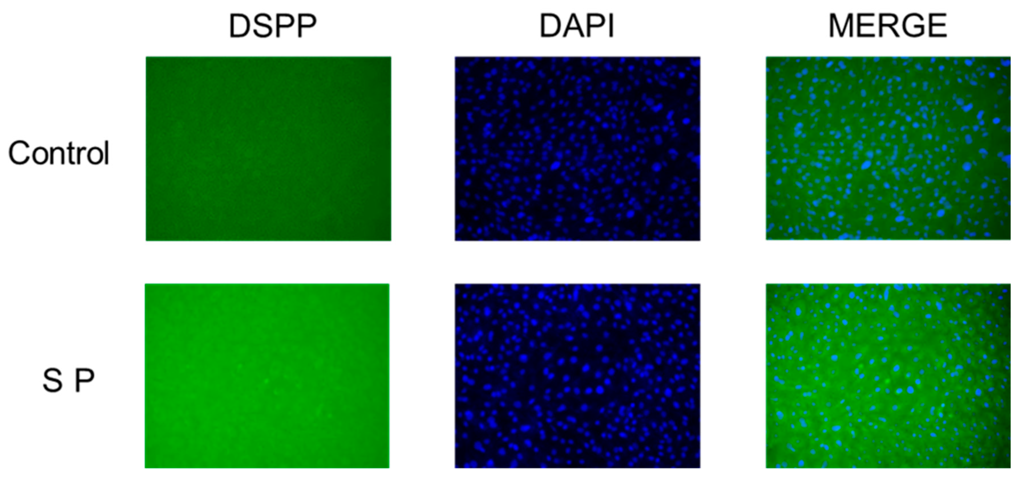
3.6. Immunofluorescence Expression of DSPP

Sigillary of mRNA expression, the fluorescent intensity of DSPP by immunofluorescence staining was enhanced in the SP group. Figure 6 indicates that the effect of SP on immunofluorescence expression of DSPP in KN-3 cells.

4. Discussion

In this study, we found that SP enhances the proliferation and odontogenic differentiation of KN-3 cells.
EMD and amelogenin peptide promote the proliferation of dental tissue cells, such as PDL fibroblasts and bone marrow stromal cells (BMSCs) [24,25]. In a previous study, we showed that SP, derived from EMD, can also promote cell proliferation of human BMSCs [8]. However, the effects of SP on proliferation in odontoblasts had not been previously investigated.
We showed that SP promotes KN-3 proliferation at 100 ng/mL, similar to the optimal concentration for PDL stem cells [10]. Therefore, we further examined the odontogenic differentiation at this concentration of SP (100 ng/mL).
ALP is considered to be an odontogenic marker [26]. We found that SP enhanced ALP activity, which is similar to the effect of EMD in hard tissues and the effect of SP in KN-3 cells. Therefore, SP can also induce odontogenic differentiation in KN-3 cells.
We quantified calcium levels in cultures of KN-3 cells by using alizarin red staining and investigation of extracellular matrix calcium deposition. Alizarin red staining is performed for the detection of calcified nodules formed by osteoblasts or odontoblasts [27]. We qualitatively and quantitatively determined changes in mineralization in response to treatment with SP. We found that SP enhanced mineralized nodule formation stained with Alizarin red and calcium deposition in cultures treated for 7 days, compared with untreated cultures. Our previous study showed that SP can also promote mineralization of PDL stem cells [16]. Our results suggest that SP can be used for the dentin formation for earlier protection of dental pulp tissue.
We also investigated the effect of SP on odontogenic differentiation. We found that it impacted the levels of DSPP, which is a noncollagenous dentin matrix protein that is known as an early-stage marker of odontoblastic differentiation [28,29]. KN-3 cells cultured for 1–12 h expressed high levels of DSPP [30]. This exhibited that KN-3 cells have the ability to differentiate into odontoblasts. In this study, we showed that DSPP expression was upregulated by SP in KN-3 cells. These results indicate that SP promotes odontoblastic differentiation.

5. Conclusions

We found that SP, an amelogenin peptide derived from EMD, can enhance the proliferation, odontogenic differentiation, and formation of calcified nodules of KN-3 cells in vitro. Our findings suggest that SP could be a new biomaterial in dental pulp therapy. Moreover, the present study partially clarified the function of amelogenin exon 5 in odontogenesis.

Author Contributions

Conceptualization, H.K. and Y.T.; Methodology, K.I., Y.R., Y.-W.T., and Y.-C.C.; Writing—original draft preparation, H.K., Data curation, M.S. and R.T.; Supervision, K.T.; Project administration, Y.T. and M.U.

Funding

This research was funded by a Grant-in-Aid for Scientific Research from the Japan Society for the Promotion of Science (Nos. 16K 20476, 16K 11617, 16K 11847, and 17K 11818).

Acknowledgments

We acknowledge the contribution of Chiaki Kitamura and Tatsuji Nishihara for KN-3 cell providing.

Conflicts of Interest

The authors declare no conflict of interest.

References

  1. Hammarström, L. Enamel matrix, cementum development and regeneration. J. Clin. Periodontol. 1997, 24, 658–668. [Google Scholar] [CrossRef] [PubMed]
  2. Hammarström, L.; Heijl, L.; Gestrelius, S. Periodontal regeneration in a buccal dehiscence model in monkeys after application of enamel matrix proteins. J. Clin. Periodontol. 1997, 24, 669–677. [Google Scholar] [CrossRef] [PubMed]
  3. Kim, N.H.; Tominaga, K.; Tanaka, A. Analysis of eosinophilic round bodies formed after injection of enamel matrix derivative into the backs of rats. J. Periodontol. 2005, 76, 1934–1941. [Google Scholar] [CrossRef] [PubMed]
  4. Hida, T.; Tominaga, K.; Tanaka, A. Tissue Reaction to synthetic oligopeptide derived from enamel matrix derivative in rats. Oral Sci. Int. 2010, 7, 26–33. [Google Scholar] [CrossRef]
  5. Noguchi, M.; Tominaga, K.; Tanaka, A.; Ueda, M. Hard tissue formation induced by synthetic oligopeptide derived from an enamel matrix derivative. Oral Med. Pathol. 2012, 16, 75–80. [Google Scholar] [CrossRef]
  6. Miki, H.; Tominga, K.; Takahashi, T.; Tanaka, A.; Umeda, M. Comparison of the influence of a new synthetic peptide and enamel matrix derivative in the early wound healing process of artificial periodontal tissue defects in rats. Jpn. J. Conserv. Dent. 2018, 61, 17–29. [Google Scholar]
  7. Kawanaka, A.; Tominaga, K.; Tanaka, A. Effect of peptide derived from Emdogain on human periodontal ligament fibroblasts. J. Osaka Dent. Univ. 2009, 43, 111–117. [Google Scholar]
  8. Katayama, N.; Kato, H.; Taguchi, Y.; Tanaka, A.; Umeda, M. The effects of synthetic oligopeptide derived from enamel matrix derivative on cell proliferation and osteoblastic differentiation of human mesenchymal stem cells. Int. J. Mol. Sci. 2014, 15, 14026–14043. [Google Scholar] [CrossRef] [PubMed]
  9. Yasui, N.; Taguchi, Y.; Tanaka, A.; Ueda, M.; Umeda, M. Biological effects of emdogain-derived oligopeptides on rat bone marrow cells in vitro. J. Oral Tissue Eng. 2012, 9, 126–135. [Google Scholar]
  10. Kato, H.; Katayama, N.; Taguchi, Y.; Tominaga, K.; Umeda, M.; Tanaka, A. A synthetic oligopeptide derived from enamel matrix derivative promotes the differentiation of human periodontal ligament stem cells into osteoblast-like cells with increased mineralization. J. Periodontol. 2013, 84, 1476–1483. [Google Scholar] [CrossRef] [PubMed]
  11. Yuan, K.; Hsu, C.W.; Tsai, W.H. The induction and possible subsequent effect of human antibodies against porcine enamel matrix derivative. J. Periodontol. 2006, 77, 1355–1361. [Google Scholar] [CrossRef] [PubMed]
  12. Shinnick, T.M.; Sutcliffe, J.G.; Green, N.; Lerner, R.A. Synthetic peptide immunogens as vaccines. Ann. Rev. Microbiol. 1983, 37, 425–446. [Google Scholar] [CrossRef] [PubMed]
  13. Lerner, R.A. Tapping the immunological repertoire to produce antibodies of predetermined specificity. Nature 1982, 299, 593–596. [Google Scholar] [CrossRef] [PubMed]
  14. Bergenholtz, G.; Mjör, I.A.; Cotton, W.R.; Hanks, C.T.; Kim, S.; Torneck, C.D.; Trowbridge, H.O. The biology of dentin and pulp. Consensus report. J. Dent. Res. 1985, 64, 631–633. [Google Scholar] [CrossRef] [PubMed]
  15. Couve, E. Ultrastructural changes during the life cycle of human odontoblasts. Arch. Oral Biol. 1986, 31, 643–651. [Google Scholar] [CrossRef]
  16. Bergenholtz, G. Advances since the paper by Zander and Glass (1949) on the pursuit of healing methods for pulpal exposures: Historical perspectives. Oral Surg. Oral Med. Oral Pathol. Oral Radiol. 2005, 100, S102–S108. [Google Scholar] [CrossRef] [PubMed]
  17. Pashley, D.H. Dynamics of the pulpo-dentin complex. Crit. Rev. Oral Biol. Med. 1996, 7, 104–133. [Google Scholar] [CrossRef] [PubMed]
  18. Zander, H.A.; Glass, R.L. The healing of phenolized pulp exposures. Oral Surg. Oral Med. Oral Pathol. 1949, 2, 803–810. [Google Scholar] [CrossRef]
  19. Nomiyama, K.; Kitamura, C.; Tsujisawa, T.; Nagayoshi, M.; Morotomi, T.; Terashita, M.; Nishihara, T. Effects of lipopolysaccharide on newly established rat dental pulp-derived cell line with odontoblastic properties. J. Endod. 2007, 33, 1187–1191. [Google Scholar] [CrossRef] [PubMed]
  20. Washio, A.; Kitamura, C.; Morotomi, T.; Terashita, M.; Nishihara, T. Possible involvement of Smad signaling pathways in induction of odontoblastic properties in KN-3 cells by bone morphogenetic protein-2: A growth factor to induce dentin regeneration. Int. J. Dent. 2012, 2012, 258469. [Google Scholar] [CrossRef] [PubMed]
  21. Hase, N.; Ozeki, N.; Hiyama, T.; Yamaguchi, H.; Kawai, R.; Kondo, A.; Nakata, K.; Mogi, M. Products of dentin matrix protein-1 degradation by interleukin-1β-induced matrix metalloproteinase-3 promote proliferation of odontoblastic cells. Biosci. Trends 2015, 9, 228–236. [Google Scholar] [CrossRef] [PubMed] [Green Version]
  22. Hiyama, T.; Ozeki, N.; Mogi, M.; Yamaguchi, H.; Kawai, R.; Nakata, K.; Kondo, A.; Nakamura, H. Matrix metalloproteinase-3 in odontoblastic cells derived from ips cells: Unique proliferation response as odontoblastic cells derived from ES cells. PLoS ONE 2013, 8, e83563. [Google Scholar] [CrossRef] [PubMed]
  23. Cattaneo, V.; Rota, C.; Silvestri, M.; Piacentini, C.; Forlino, A.; Gallanti, A.; Rasperini, G.; Cetta, G. Effect of enamel matrix derivative on human periodontal fibroblasts: Proliferation, morphology and root surface colonization. An in vitro study. J. Periodontal Res. 2003, 38, 568–574. [Google Scholar] [CrossRef] [PubMed]
  24. Guida, L.; Annunziata, M.; Carinci, F.; Di Feo, A.; Passaro, I.; Oliva, A. In vitro biologic response of human bone marrow stromal cells to enamel matrix derivative. J. Periodontol. 2007, 78, 2190–2196. [Google Scholar] [CrossRef] [PubMed]
  25. Taguchi, Y.; Yasui, N.; Takahashi, S.; Tominaga, K.; Kato, H.; Komasa, S.; Shida, M.; Hayashi, H.; Tanaka, A.; Umeda, M. Hard tissue formation by human periodontal ligament fibroblast Cells treated with an emdogain-derived oligopeptide in vitro. J. Hard Tissue Biol. 2012, 21, 375–384. [Google Scholar] [CrossRef]
  26. Weinreb, M.; Shinar, D.; Rodan, G.A. Different pattern of alkaline phosphatase, osteopontin, and osteocalcin expression in developing rat bone visualized by in situ hybridization. J. Bone Miner. Res. 1990, 5, 831–842. [Google Scholar] [CrossRef] [PubMed]
  27. Magne, D.; Bluteau, G.; Lopez-Cazaux, S.; Weiss, P.; Pilet, P.; Ritchie, H.H.; Daculsi, G.; Guicheux, J. Development of an odontoblast in vitro model to study dentin mineralization. Connect. Tissue Res. 2004, 45, 101–108. [Google Scholar] [CrossRef] [PubMed] [Green Version]
  28. Hao, J.; Zou, B.; Narayanan, K.; George, A. Differential expression patterns of the dentin matrix proteins during mineralized tissue formation. Bone 2004, 34, 921–932. [Google Scholar] [CrossRef] [PubMed]
  29. Sreenath, T.; Thyagarajan, T.; Hall, B.; Longenecker, G.; D’Souza, R.; Hong, S.; Wright, J.T.; MacDougall, M.; Sauk, J.; Kulkarni, A.B. Dentin sialophosphoprotein knockout mouse teeth display widened predentin zone and develop defective dentin mineralization similar to human dentinogenesis imperfecta type III. J. Biol. Chem. 2003, 278, 24874–24880. [Google Scholar] [CrossRef] [PubMed]
  30. Yeom, K.H.; Ariyoshi, W.; Okinaga, T.; Washio, A.; Morotomi, T.; Kitamura, C.; Nishihara, T. Platelet-rich plasma enhances the differentiation of dental pulp progenitor cells into odontoblasts. Int. Endod. J. 2016, 49, 271–278. [Google Scholar] [CrossRef] [PubMed]
Figure 1. Effect of synthetic peptide (SP) on KN-3 cell proliferation. KN-3 cells were treated with 0 to 1000 ng/mL SP diluted in 100 µL culture medium. Cell proliferation was measured at 3, 8, 24, and 48 h. Significant differences (*, p > 0.05) were determined in comparison with the control (0 ng/mL SP). (A) 3 h, (B) 8 h, (C) 24 h, (D) 48.
Applsci 08 01890 g001
Figure 2. Effect of SP on cell morphology in KN-3 cells. Cell morphology in KN-3 cells treated with SP. Cells were cultured with SP (0 to 1000 ng/mL) for 48 h. Scale bar indicates 200 µm. (A) SP (0 ng/mL), (B) SP (1 ng/mL), (C) SP (10 ng/mL), (D) SP (100 ng/mL), (E) SP (1000 ng/mL).
Applsci 08 01890 g002
Figure 3. Effect of SP on alkaline phosphatase (ALP) activity. Confluent cells were treated with differentiation medium for 7 days (*, p > 0.05), and changes in ALP activity were investigated at days 3 and 7.
Applsci 08 01890 g003
Figure 4. Effect of SP on mineralization in KN-3 cells. (A) Alizarin Red staining for detection of calcified nodule formation, (B) Measurement of extracellular matrix calcium deposition.
Applsci 08 01890 g004
Figure 5. Effect of SP on odontoblastic differentiation in KN-3 cells. Quantitative real-time PCR analysis of dentin sialoprotein (DSPP) in KN-3 cells at 1, 3, 6, and 24 h.
Applsci 08 01890 g005
Figure 6. KN-3 cells treated with or without SP were incubated with anti-DSPP monoclonal antibody. Cells were incubated with a fluorescence-labeled secondary antibody. The samples were stained with DAPI solution. Scale bar = 100 µm.
Applsci 08 01890 g006

