Enhanced Photocatalytic Efficiency of TiO2 Membrane Decorated with Ag and Au Nanoparticles

1
State Key Laboratory of Silicate Materials for Architectures, Wuhan University of Technology, Wuhan 430070, China
2
School of Chemistry, Chemical Engineering and Life Science, Wuhan University of Technology, Wuhan 430070, China
*
Author to whom correspondence should be addressed.
Appl. Sci. 2018, 8(6), 945; https://doi.org/10.3390/app8060945
Submission received: 9 May 2018
/
Revised: 25 May 2018
/
Accepted: 25 May 2018
/
Published: 7 June 2018
(This article belongs to the Special Issue Gold Nanoparticles for Catalytic Applications)
Abstract
:Featured Application
The photocatalysis could be used for water treatment and other environmental engineering.
Abstract
Ag and Au nanoparticles (NPs) were decorated on the surface of TiO2 membranes by two methods, i.e., hydrothermal synthesis and photoreduction. The size of Ag and Au NPs on the surface of TiO2 membranes was dependent on the method of preparation and varied from 2 nm–10 nm. The photocatalytic performance of the TiO2 particle, TiO2 membrane and the Ag/Au-decorated TiO2 membrane was tested for the catalytic degradation of Rhodamine B (RhB) and Escherichia coli (E. coli) under irradiation of visible light. The experiment results showed that both Ag- and Au-decorated TiO2 membranes exhibited excellent photocatalytic activity in the visible light region. Among the prepared materials, Ag-decorated TiO2 membranes prepared by photoreduction showed the highest activity, which could be attributed to the local surface plasmon resonance (LSPR) effect of the noble metal.
In recent years, many thin film materials, including carbon film, graphene and thin SnO2 membranes, have been studied and have shown excellent physical and chemical properties [1,2,3]. Various TiO2 nanomaterials have also been reported, such as TiO2 nanopowders, nanowires, nanotubes, nanoflowers, etc. [4,5,6]. Among them, TiO2 membrane have gained considerable attention due to their outstanding catalytic performance, which could be attributed to the large surface area and low thickness of the material [7,8]. TiO2 nanoparticles (NPs) with a large specific area have higher photocatalytic efficiency because the photocatalytic process occurs on the surface.
High photocatalytic efficiency is the main evaluation method for photocatalysts, which is important to degrade organics in short time periods [9,10]. According to the research, photocatalysts with low photocatalytic efficiency degraded organics within a couple of hours, such as nitrogen-doped TiO2 thin film [11], TiO2 nanotubes [12] and BiOCl nanowire arrays [13]; high photocatalytic efficiency like that of BiOCl membranes could completely degrade RhB within 2 min under UV light [14].
Further modification of TiO2 can improve its response to visible light, inhibit the recombination of photogenerated carriers and, consequently, enhance the photocatalytic performance. Various materials, including metals (Au, Ag, Pt, Cu), non-metals (C, N, S), metal oxides (WO3, Fe2O3, SiO2), etc., have been used to dope TiO2 [8,15,16,17]. These doping materials can produce vacancies and interstitial or substitutional defects that change the photocatalytic properties of TiO2. The band gap of TiO2 is narrowed notably after doping with noble metals (Au, Ag, Pt) [18]. Because the noble metals have lower Fermi levels than TiO2, the photogenerated electrons of the conduction band will not recombine with the holes, but instead transfer to the noble metal particles on the surface of TiO2 [19,20,21,22]. Hence, the photogenerated electrons and holes are effectively separated, and the photocatalytic activity is improved [23,24,25]. Moreover, the noble metal NPs can more easily attract organic molecules than semiconductors. Therefore, the noble metal NPs play the role of the electron carrier in the system of TiO2 doped with noble metals, which is an effective method to improve the photocatalytic properties of TiO2.
Herein, we report a TiO2 membrane decorated with Ag and Au NPs, which exhibited extremely high catalytic efficiency. The TiO2 membrane was prepared by a hydrothermal method [26]. A certain amount (8 mL) of TiCl3 was mixed with ethylene glycol (30 mL), and the mixture was stirred for 7 h at room temperature until the reaction completed. Then, doubly-distilled water (1 mL) was added, and the mixture was transferred into a stainless steel reactor and kept at 150 °C for 4 h. The products were separated by centrifugation at 16,000 rpm, washed with ethanol three times and dried in a vacuum at 50 °C for 48 h. TiO2 membrane was decorated with Ag and Au NPs by the hydrothermal and photoreduction methods, respectively. The mixture of the TiO2 membrane (0.4 mg) and AgNO3 or HAuCl4 (0.015 mol/L, 0.4 mL) was processed by 365-nm UV light or hydrothermal reaction (180 °C, 6 h). The final products were centrifuged at 10,000 rpm, washed with doubly-distilled water three times and dried in a vacuum at 50 °C for 24 h. The sample descriptions are given in Table 1.
TiO2 membranes decorated with Ag and Au NPs were observed and characterized by transmission electron microscope (TEM, Talos F200S, Thermo fisher, Waltham, MA, USA) and X-ray diffraction (XRD, PHILIPS P W 3O4O/60X′PertPRO, Amsterdam, The Netherlands). Scanning electron microscope energy dispersive spectromete (SEM EDS, JSM-7500F, JEOL Ltd., Tokyo, Japan) analysis showed the real composition of the prepared samples, as given in Table 1. Ultraviolet-visible light (UV-Vis) diffuse reflectance spectroscopy was measured on a Perkin Elmer UV-Vis spectrometer (Lambda 750 S, Perkin Elmer, Waltham, MA, USA) equipped with a Labsphere RSA-PE-20 integration sphere. The electrochemical behaviors of the TiO2 membranes, SAg and SAu, were examined by electrochemical impedance spectroscopy (EIS, RST5200F, shiruisi Instrument, Zhengzhou, China). The photocatalytic performance of the TiO2 membranes, SAg and SAu, was evaluated by the degradation reaction of Rhodamine B (RhB). A Xe lamp with optical filters (14 V, 14–21 A, visible light) was used as the light source, and the distance between the lamp and the interface of the solution was 12 cm (0.1 W/m2). The test was carried out by adding the prepared photocatalyst (0.4 mg) to the solution of RhB (5 mg/L, 250 mL) and measuring the absorbance at 552 nm (i.e., the maximum absorption wavelength of RhB) by UV-visible spectrophotometry every 5 min. The concentration of RhB in the solution was calculated from the absorbance, and the photocatalytic degradation efficiency was calculated as follows:
where C0 is the initial concentration of RhB and Ct is the concentration of RhB after an irradiation time of t. Escherichia coli (E. coli) cells were treated by SAg under visible light irradiation (0.1 W/m2) for 10 min. Then, the morphology of the native and treated E. coli cells was examined by SEM. The cells were fixed according to the standard protocol by reduction with glutaraldehyde and oxidation with OsO4, then dehydrated stepwise with ethanol of increasing concentration (50–100%).
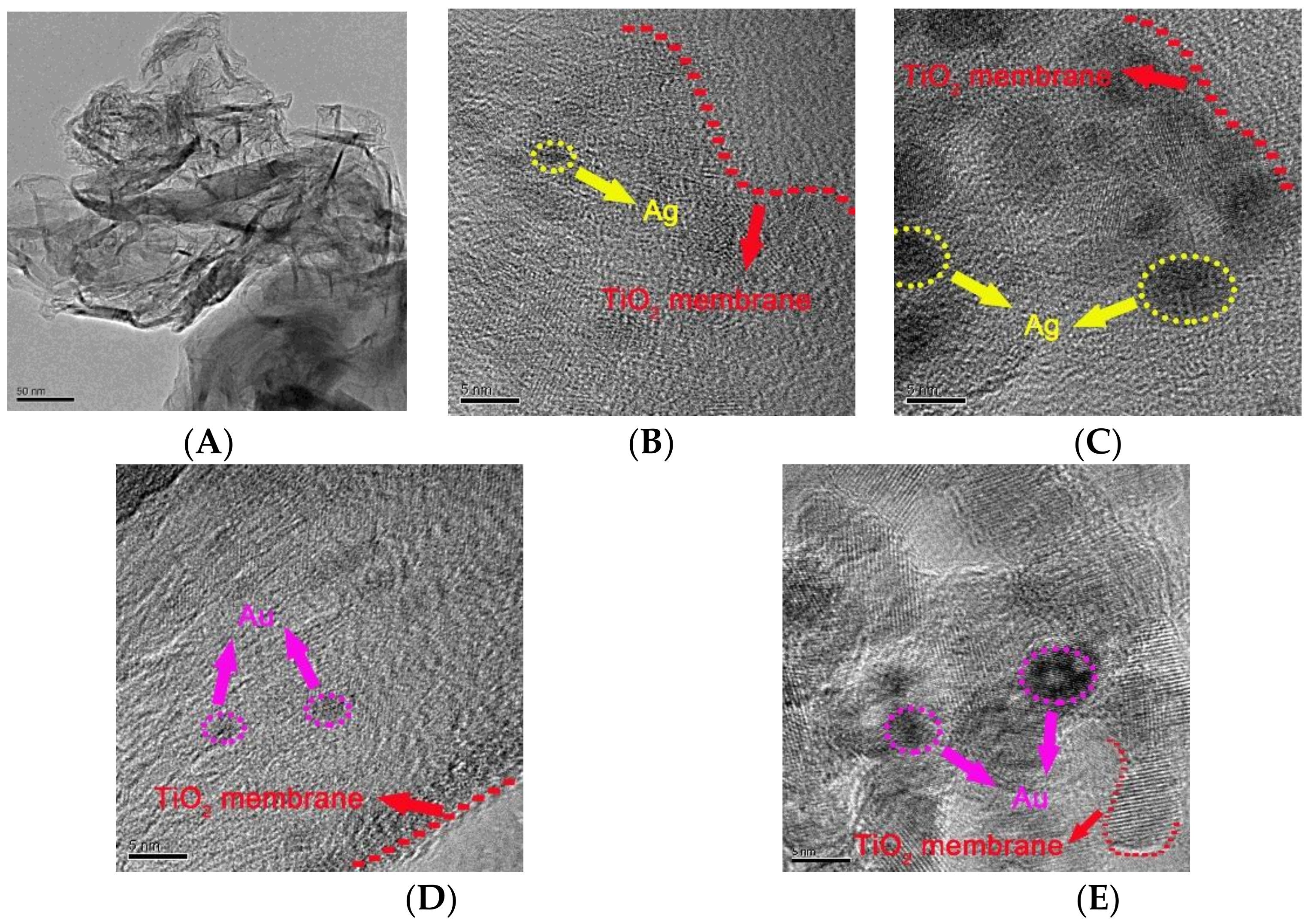
The prepared TiO2 membrane was decorated with Ag/Au NPs by either hydrothermal synthesis or photoreduction. The morphology and structure of the resulting material were observed and characterized by TEM. Figure 1 shows the diameter histogram and the TEM images of the SAg and SAu samples prepared under different conditions.
The ultrathin TiO2 membrane was two atoms thick and Ag or Au NPs were dispersed on the membrane surface, as shown in Figure 1. The particle size ranged from 2 nm–10 nm. The particle size of the decorated Ag was the smallest (ca. 2 nm) when SAg was prepared by photoreduction with ultraviolet light (Figure 1). Similarly, the particle size of the decorated Au was also the smallest (ca. 2 nm) in SAu prepared by photoreduction with ultraviolet light (Figure 2D). Hydrothermal synthesis at 180 °C furnished Au particles of ca. 10 nm in size on the TiO2 membrane. Ag or Au NPs prepared by the photoreduction method were dispersed evenly within the TiO2 network. However, the NPs aggregated together with hydrothermal synthesis. Generally, Ag and Au NPs were successfully decorated on the TiO2 membrane.
The SAg and SAu samples exhibited some adsorption in the visible light region as shown in Supplementary Material S1, which could be attributed to the local surface plasmon resonance (LSPR) of the Ag and Au NPs. The LSPR effect on the surface of noble metal NPs is caused by the collective shock of conductive electrons generated by electromagnetic radiation. The size and shape of the noble metal NPs can change the surface density of the electromagnetic field and are thus important factors for the LSPR effect [27]. Obviously, the SAg sample prepared by photoreduction had a strong absorption in the visible light region. For TiO2, the absorption in the UV region resulted only from its wide band gap (3.2 eV). After the Ag and Au NPs were introduced, the light response range of TiO2 clearly extended to the visible region. The Ag and Au NPs on the TiO2 membrane could transfer free elections generated by the LSPR effect to the valence band of TiO2 and consequently enhance the photocatalytic activity of TiO2 under visible light [28,29,30].
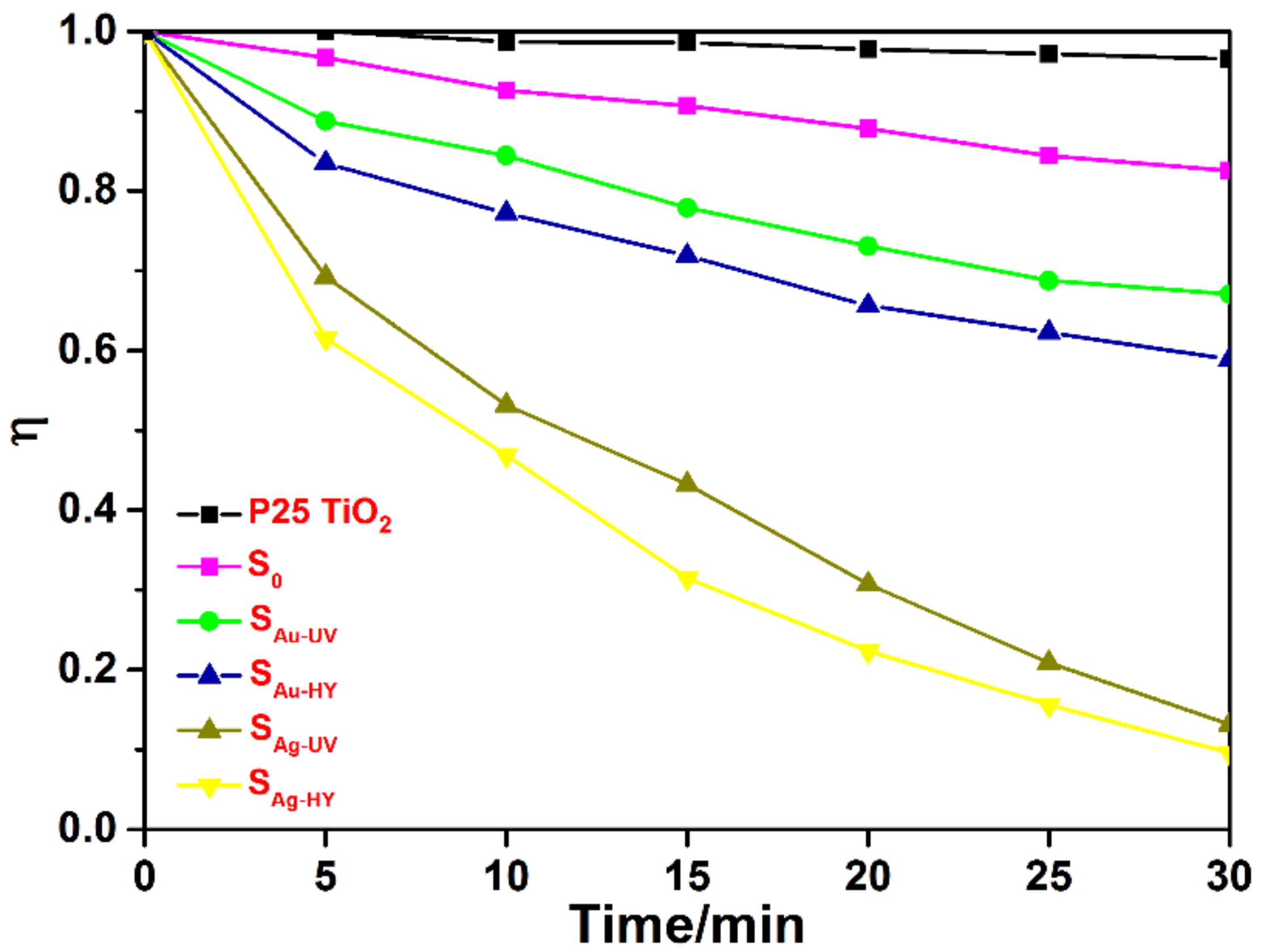
Figure 2 shows the photocatalytic degradation of RhB with P25 (TiO2 particle with size 25 nm) and the decorated TiO2 membrane. The degradation efficiency of RhB was evaluated by the first order reaction, which is presented in Supplementary Material S2. The catalytic efficiency of photocatalysts was ranked as SAg-UV > SAg-HY > SAu-UV > SAu-HY > S0 > SP25. When P25 was used as the catalyst, the degradation of RhB under irradiation of visible light was very limited. Nevertheless, the degradation efficiency was improved notably when the TiO2 membrane was used in the photodegradation reaction. The decorated TiO2 membrane enhanced the degradation efficiency further. SAg exhibited higher catalytic efficiency than SAu. Besides, SAg/SAu prepared by UV irradiation exhibited higher degradation efficiency than hydrothermal synthesis. In addition, the recycling experiment of SAg-UV showed that it could be used over 50 times and that the photocatalytic efficiency of SAg-UV had not reduced significantly, which is presented in Supplementary Material S3.
Mechanistically, in the photocatalytic reaction, photogenerated electrons and holes on the TiO2 membrane work as the active species to decomposing organic compounds. The existence of Ag and Au NPs on the TiO2 membrane forms a Schottky junction on the surface of TiO2, which promotes charge separation and improves the quantum efficiency. Generally, Ag shows a stronger LSPR effect than Au. In addition, the absorption range of TiO2 extended to the visible light region due to the LSPR effect of the Ag and Au NPs. Since the LSPR sensitization effect of plasmonic NPs depends mainly on their size and shape, the LSPR effect is strengthened when the size of the Ag and Au NPs increases [31,32]. Hence, the SAu and SAg samples prepared by photoreduction showed higher efficiency in the photodegradation of RhB. During this process, RhB was first broken down into benzoic acid and phthalic acid and then into ethanedioic acid, while the organic carbon content remain unchanged [33]. Finally, the RhB was degraded into small molecules, such as CO2, NOx and H2O, and finally, oxidized completely, with the organic carbon content decreased significantly [34,35,36].
The degradation of RhB by photocatalysts has been reported by many groups. TiO2 and Fe2O3 co-doped graphene aerogel can remove 97.7% RhB within 60 min under visible light irradiation [37]. SiO2@C-doped TiO2 hollow spheres can degrade 61.7% RhB after 100 min under UV irradiation [38]. Ag/TiO2 composites can degrade 80% RhB after 120 min under UV irradiation [39]. Compared with the above photocatalysts, the Ag decorated TiO2 membrane can almost degrade 90% RhB within 30 min under visible light irradiation. Therefore, Ag/Au-decorated TiO2 membranes showed excellent photodegradation efficiency.
The prepared TiO2 membrane was characterized as being twos atoms thick [26]. The specific surface area was greatly increased compared with P25 TiO2. The target molecules were more easily absorbed on TiO2 membrane. The degradation of the target molecules experienced a dynamic adsorption-photocatalysis-desorption process. The electron and hole produced under irradiation reacted with the target molecules on the enlarged surface; thus, the recombination of the electron and hole was also inhibited. The process is presented in Figure 3. As a result, the photocatalytic efficiency of the TiO2 membrane was enhanced significantly compared to P25 TiO2.
The photocatalytic mechanism is demonstrated below with SAg as an example. The SAu has photoresponse in the visible light region due to the LSPR effect of Au NPs. Analyzing by EIS, the Au NP-doped TiO2 membrane represented lower resistances than the pure TiO2 membrane, which could make the charge transfer at the interface more rapid and effective. The charges on the surface of Au NPs are subjected to collective shock under irradiation of visible light and become free elections, which results in a strengthened local electric field. Under the local electric field, free electrons then rapidly jump to the conduction band (CB) of TiO2 and overcome the Schottky junction, which results in the separation of photogenerated electrons and holes and thus improves quantum efficiency. Generally, pure TiO2 has a wide band gap. Under light irradiation, the excitation of an electron would take place if the light energy is greater than the band gap of the catalysts. The wide band gap of pure TiO2 is known to be 3.2 eV. By using Mott−Schottky plots and XPS spectra, the band gap from the valence band to the conduction band for Ag-decorated TiO2 deceased to 2.6 eV. Therefore, the material showed photocatalytic activity under visible light irradiation. The photogenerated electrons can react with the O2 on the surface of catalyst to form ·O2−, and the holes of Au can combine with H2O on the catalyst surface to form ·OH, both of which are strongly oxidative. These oxidative species can then oxidize the RhB molecule on the surface of Au NPs.
The photocatalysis degradation is divided into three stages. They are the formation of both the hole and electron under light excitation, the production of radical species and the reaction of the radical species with the targets (pollutants, bacteria, etc.). Electron paramagnetic resonance (EPR) has confirmed the separation of the hole and electron [40]. The production of radical species along time was also proven by the different adduct species detected with EPR [41]. The degradation product of the targets was detected by some groups [42]. The silver content is closely related to the separation of the hole and electron and the production of radical species, as described above. As a result, silver will affect photocatalysis and degradation efficiency significantly.
Au or Ag NPs experience an evolution during the photocatalytic process. A moderate growth of particle size was observed. From the statistics of the TEM images, the particle size of Ag and Au NPs increased from 2.3 ± 0.1 nm–13 ± 0.3 nm and from 2.4 ± 0.1 nm–17 ± 0.4 nm after photocatalysis, respectively. On the other hand, the photocatalytic activity did not change in the catalyst recyclability test (Supplementary Material S3), showing that Au or Ag NPs maintain a dominant metallic state.
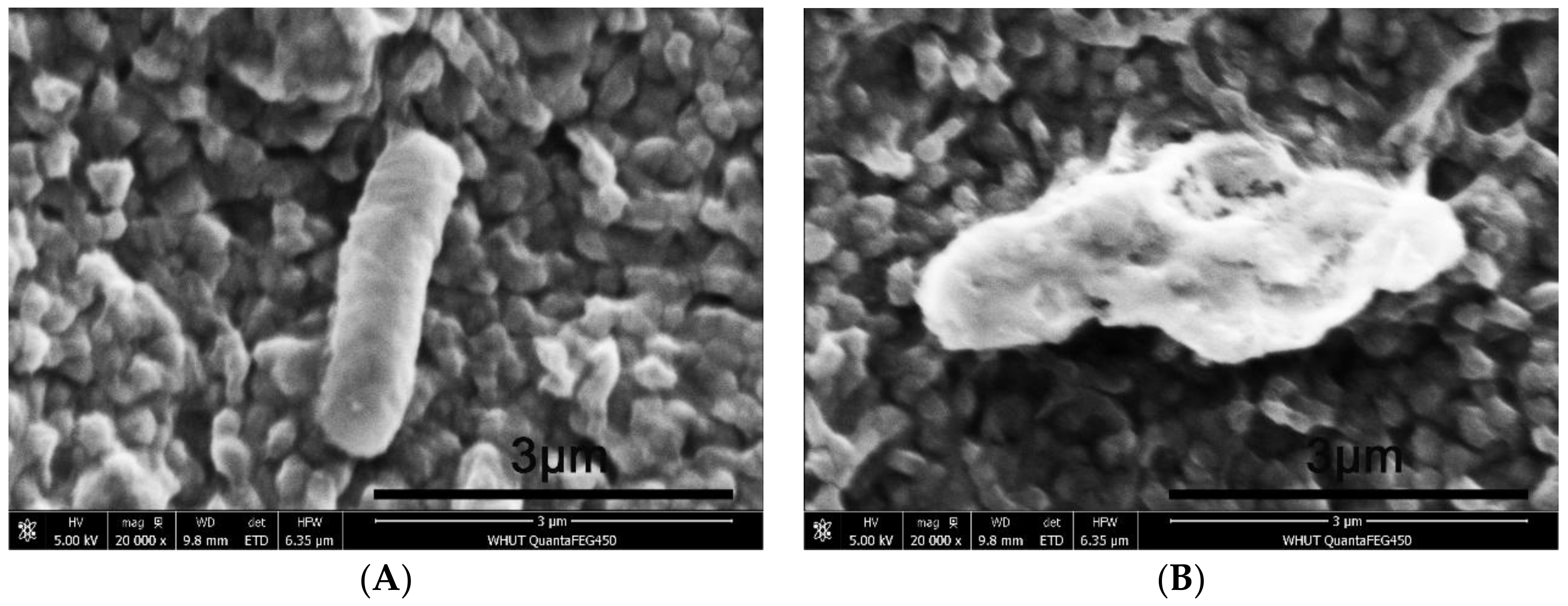
The surface morphology of the E. coli cells was examined by SEM. Figure 4A shows that the native E. coli cells had a rod-like structure with regular wrinkles, which was changed after the photocatalytic degradation. Figure 4B shows that some depressions appeared on the surface of cells treated with P25 TiO2, and irregular wrinkles appeared on the cells treated with SAu.
The results suggested that the cell surface was damaged and the cell membrane structure was altered. The outer membrane of Gram-negative bacteria is composed of lipopolysaccharide (LPS), peptidoglycan and phospholipid layers. The mechanical strength of the cell membrane depends on peptidoglycan, and LPS is responsible for the surface structure and morphology [43,44,45]. The beta 1,4-link between the two amino sugars of N-acetyl glucose amine and N-acetyl acid forms the LPS. By connecting four peptide side chains on the N-acetyl cell wall acid molecules, the peptides in between are again joined by bridges to form a reticulate structure that is very strong. Therefore, the cell membrane plays a vital role in maintaining cell morphology. The outermost layer of the cell membrane is LPS, which is exposed to the environment and subjected to the attack of free radicals during photocatalytic degradation. The cells still maintained the original rod shape after treatment with P25 TiO2 and SAu, which indicated that LPS was damaged, but peptidoglycan remained intact. The peptidoglycan layer and the outer membrane form a strong network to maintain the morphology of the cell [46,47]. In contrast, after treatment with SAg, the E. coli cells exhibited a fusiformis structure, and the structure of the native cells was destroyed, which indicated that the outer membrane and the peptidoglycan layer were fully damaged, but the inner membrane remained intact. In addition, E. coli photo-killing analysis was done by E. coli inactivation, suggesting that the E. coli treated with SAg inactivation was decreased within 3.5 h under visible light, which is presented in Supplementary Material S4.
The outstanding performance of the TiO2 membrane as a photocatalyst stems from the material’s large surface area and small thickness. However, the quantum efficiency of the TiO2 membrane is not high. Decorating the TiO2 membrane with Ag and Au NPs can extend the photo response to a broader range and improve the photocatalytic properties. In this work, the TiO2 membrane was prepared by hydrothermal synthesis, and then, Ag and Au NPs were decorated on the surface of the TiO2 membrane by hydrothermal synthesis or photoreduction. The size of the Ag and Au NPs prepared by photoreduction was ca. 2 nm and the smallest among all material samples(Supplementary Material S5). The size of the noble metal NPs is important for the local surface plasmon resonance (LSPR) sensitization and affects the quantum efficiency. Under visible light, SAg and SAu showed excellent photocatalytic performance. Silver NPs can produce a stronger LSPR effect than Au NPs [31,32]. The TiO2 membranes decorated with Ag NPs were found to have good photocatalytic properties in the degradation of RhB and E. coli under irradiation of visible light and presumably may find use in the photocatalytic degradation of more organic compounds and microorganisms.
Supplementary Materials
The following are available online at https://www.mdpi.com/2076-3417/8/6/945/s1. Figure S1. UV-Vis diffuse reflectance spectroscopy of S0, SAg-UV, SAg-HY, SAu-UV, SAu-HY and P25 TiO2. Table S2. Degradation rate constant of Rhodamine B. Figure S3. Recycling test of the SAg-UV during the degradation of RhB under visible light irradiation. Figure S4. E. coli inactivation with SAg treatment. Figure S5. The evolution of Au or Ag NPs’ particle size after the photocatalytic process. The particle size of Ag and Au NPs increased from 2.3 ± 0.1 nm–13 ± 0.3 nm and from 2.4 ± 0.1 nm–17 ± 0.4 nm, respectively.
Author Contributions
W.Z. and F.W. conceived and designed the experiments; Y.G. performed the experiments; P.L. and Y.G. analyzed the data; F.W. contributed reagents/materials/analysis tools; W.Z. and Y.G. wrote the paper.
Acknowledgments
This work was financially supported by the National Natural Science Foundation of China (Grant No. 51502222).
Conflicts of Interest
The authors declare no conflict of interest.
References
- Brilot, A.F.; Chen, J.Z.; Cheng, A.; Pan, J.; Harrison, S.C.; Potter, C.S.; Carragher, B.; Henderson, R.; Grigorieff, N. Beam-induced motion of vitrified specimen on holey carbon film. J. Struct. Biol. 2012, 177, 630–637. [Google Scholar] [CrossRef] [PubMed] [Green Version]
- Di Valentin, C.; Pacchioni, G.; Selloni, A.; Livraghi, S.; Giamello, E. Characterization of Paramagnetic Species in N-Doped TiO2 Powders by EPR Spectroscopy and DFT Calculations. J. Phys. Chem. B 2005, 109, 11414–11419. [Google Scholar] [CrossRef] [PubMed]
- Brennan, B.; Spencer, S.J.; Belsey, N.A.; Faris, T.; Cronin, H.; Silva, S.R.P.; Sainsbury, T.; Gilmore, I.S.; Stoeva, Z.; Pollard, A.J. Structural, chemical and electrical characterisation of conductive graphene-polymer composite films. Appl. Surf. Sci. 2017, 403, 403–412. [Google Scholar] [CrossRef]
- Mor, G.K.; Carvalho, M.A.; Varghese, O.K.; Pishko, M.V.; Grimes, C.A. A room-temperature TiO2-nanotube hydrogen sensor able to self-clean photoactively from environmental contamination. J. Mater. Res. 2004, 19, 628–634. [Google Scholar] [CrossRef]
- Lei, Y.; Zhang, L.D.; Meng, G.W.; Li, G.H. Preparation and photoluminescence of highly ordered TiO2 nanowire arrays. Appl. Phys. Lett. 2001, 78, 1125–1127. [Google Scholar] [CrossRef]
- Xia, H.R.; Peng, C.; Li, J.; Sun, W.T.; Ai, G.; Peng, L.M. Large-scale floated single-crystalline TiO2 flower-like films: synthesis details and applications. RSC Adv. 2013, 3, 17668–17671. [Google Scholar] [CrossRef]
- Xiang, G.; Li, T.; Zhuang, J.; Wang, X. Large-scale synthesis of metastable TiO2(B) membrane with atomic thickness and their photocatalytic properties. Chem. Commun. 2010, 46, 6801–6803. [Google Scholar] [CrossRef] [PubMed]
- Choi, H.; Stathatos, E.; Dionysiou, D.D. Photocatalytic TiO2 films and membranes for the development of efficient wastewater treatment and reuse systems. Desalination 2015, 202, 199–206. [Google Scholar] [CrossRef]
- Ma, R.; Islam, M.J.; Reddy, D.A.; Kim, T.K. Transformation of CeO2, into a mixed phase CeO2/Ce2O3, nanohybrid by liquid phase pulsed laser ablation for enhanced photocatalytic activity through Z-scheme pattern. Ceram. Int. 2016, 42, 18495–18502. [Google Scholar] [CrossRef]
- Islam, M.J.; Kim, H.K.; Reddy, D.A.; Kim, Y.; Ma, R.; Baek, H.; Kim, J.; Kim, T.K. Hierarchical BiOI nanostructures supported on a metal organic framework as efficient photocatalysts for degradation of organic pollutants in water. Dalton Trans. 2017, 46, 6013–6023. [Google Scholar] [CrossRef] [PubMed]
- Irie, H.; Washizuka, S.; Watanabe, Y.; Kako, T.; Hashimoto, K. Photoinduced Hydrophilic and Electrochemical Properties of Nitrogen-Doped TiO2 Films. J. Electrochem. Soc. 2005, 152, E351–E356. [Google Scholar] [CrossRef]
- Chen, Y.; Shi, L.Y.; Yuan, S. Photoelectrocatalytic Degradation of Methylene Blue by TiO2 Nanotube. J. Inorg. Mater. 2009, 24, 680–684. [Google Scholar] [CrossRef]
- Wu, S.; Wang, C.; Cui, Y.; Wang, T.; Huang, B.; Zhang, X.; Qin, X.; Brault, P. Synthesis and photocatalytic properties of BiOCl nanowire arrays. Mater. Lett. 2010, 64, 115–118. [Google Scholar] [CrossRef]
- Ye, L.; Zan, L.; Tian, L.; Peng, T.; Zhang, J. The {001} Facets-Dependent High Photoactivity of BiOCl Membrane. Chem. Commun. 2011, 47, 6951–6953. [Google Scholar] [CrossRef] [PubMed]
- Wang, C.; Zhou, Y.; Ge, M.; Xu, X.; Zhang, Z.; Jiang, J.Z. Large-scale synthesis of SnO2 membrane with high lithium storage capacity. J. Am. Chem. Soc. 2010, 132, 46–47. [Google Scholar] [CrossRef] [PubMed]
- Kumar, S.G.; Devi, L.G. Review on modified TiO2 photocatalysis under UV/Visible light: selected results and related mechanisms on interfacial charge carrier transfer dynamics. J. Phys. Chem. A 2011, 115, 13211–13241. [Google Scholar] [CrossRef] [PubMed]
- Daghrir, R.; Drogui, P.; Robert, D. Modified TiO2 for Environmental Photocatalytic Applications: A Review. Ind. Eng. Chem. Res. 2013, 52, 3581–3599. [Google Scholar] [CrossRef]
- Finazzi, E.; Valentin, C.D.; Pacchioni, G.; Selloni, A. Excess electron states in reduced bulk anatase TiO2: Comparison of standard GGA, GGA+U, and hybrid DFT calculations. J. Chem. Phys. 2008, 129, 035108. [Google Scholar] [CrossRef] [PubMed]
- Reddy, D.A.; Choi, J.; Lee, S.; Kim, T.K. Synthesis of heterostructured Ag@AgI/ZnS microspheres with enhanced photocatalytic activity and selective separation of methylene blue from mixture dyes. J. Taiwan Inst. Chem. Eng. 2016, 66, 200–209. [Google Scholar] [CrossRef]
- Islam, M.J.; Reddy, D.A.; Han, N.S.; Choi, J.; Song, J.K.; Kim, T.K. An oxygen-vacancy rich 3D novel hierarchical MoS2/BiOI/AgI ternary nanocomposite: Enhanced photocatalytic activity through photogenerated electron shuttling in a Z-scheme manner. Phys. Chem. Chem. Phys. 2016, 18, 24984–24993. [Google Scholar] [CrossRef] [PubMed]
- Islam, M.J.; Reddy, D.A.; Choi, J.; Kim, T.K. Surface oxygen vacancy assisted electron transfer and shuttling for enhanced photocatalytic activity of a Z-scheme CeO2–AgI nanocomposite. RSC Adv. 2016, 6, 19341–19350. [Google Scholar] [CrossRef]
- Rahimi, N.; Pax, R.A.; Gray, E.M. Review of functional titanium oxides. I: TiO2 and its modifications. Prog. Solid State Chem. 2016, 44, 86–105. [Google Scholar] [CrossRef]
- Lee, S.; Reddy, D.A.; Kim, T.K. Well-wrapped reduced graphene oxide nanosheets on Nb3O7(OH) nanostructures as good electron collectors and transporters for efficient photocatalytic degradation of rhodamine B and phenol. RSC Adv. 2016, 6, 37180–37188. [Google Scholar] [CrossRef]
- Reddy, D.A.; Choi, J.; Lee, S.; Ma, R.; Kim, T.K. Green Synthesis of AgI Nanoparticle–Functionalized Reduced Graphene Oxide Aerogels with Enhanced Catalytic Performance and Facile Recycling. RSC Adv. 2015, 5, 67394–67404. [Google Scholar] [CrossRef]
- Islam, M.J.; Reddy, D.A.; Ma, R.; Kim, Y.; Kim, T.K. Reduced-graphene-oxide-wrapped BiOI-AgI heterostructured nanocomposite as a high-performance photocatalyst for dye degradation under solar light irradiation. Solid State Sci. 2016, 61, 32–39. [Google Scholar] [CrossRef]
- Liu, P.; Zhao, Y.; Qin, R.; Mo, S.; Chen, G.; Gu, L.; Chevrier, D.M.; Zhang, P.; Guo, Q.; Zang, D. Photochemical route for synthesizing atomically dispersed palladium catalysts. Science 2016, 352, 797–800. [Google Scholar] [CrossRef] [PubMed]
- Sarina, S.; Waclawik, E.R.; Zhu, H. ChemInform Abstract: Photocatalysis on Supported Gold and Silver Nanoparticles under Ultraviolet and Visible Light Irradiation. Cheminform 2013, 44, 1814–1833. [Google Scholar] [CrossRef]
- Zhou, X.; Liu, G.; Yu, J.; Fan, W. Surface plasmon resonance-mediated photocatalysis by noble metal-based composites under visible light. J. Mater. Chem. 2012, 22, 21337–21354. [Google Scholar] [CrossRef]
- Bera, S.; Ji, E.L.; Rawal, S.B.; Wan, I.L. Size-dependent plasmonic effects of Au and Au@SiO2 nanoparticles in photocatalytic CO2 conversion reaction of Pt/TiO2. Appl. Catal. B Environ. 2016, 199, 55–63. [Google Scholar] [CrossRef]
- Choi, J.; Reddy, D.A.; Islam, M.J.; Ma, R.; Kim, T.K. Self-assembly of CeO2, nanostructures/reduced graphene oxide composite aerogels for efficient photocatalytic degradation of organic pollutants in water. J. Alloys Compd. 2016, 688, 527–536. [Google Scholar] [CrossRef]
- Zhang, X.; Chen, Y.L.; Liu, R.S.; Tsai, D.P. Plasmonic photocatalysis. Rep. Prog. Phys. 2013, 76, 046401. [Google Scholar] [CrossRef] [PubMed]
- Zheng, Z.; Huang, B.; Qin, X.; Zhang, X.; Dai, Y.; Whangbo, M.H. Facile in situ synthesis of visible-light plasmonic photocatalysts M@TiO2 (M = Au, Pt, Ag) and evaluation of their photocatalytic oxidation of benzene to phenol. J. Mater. Chem. 2011, 21, 9079–9087. [Google Scholar] [CrossRef]
- Li, J.Y.; Ma, W.H.; Lei, P.X.; Zhao, P.C. Detection of intermediates in the TiO2-assisted photodegradation of Rhodamine B under visible light irradiation. J. Environ. Sci. 2007, 19, 892–896. [Google Scholar] [CrossRef]
- Reddy, D.A.; Ma, R.; Choi, M.Y.; Kim, T.K. Reduced graphene oxide wrapped ZnS–Ag2S ternary composites synthesized via hydrothermal method: Applications in photocatalyst degradation of organic pollutants. Appl. Surf. Sci. 2015, 324, 725–735. [Google Scholar] [CrossRef]
- Reddy, D.A.; Lee, S.; Choi, J.; Park, S.; Ma, R.; Yang, H.; Kim, T.K. Green synthesis of AgI-reduced graphene oxide nanocomposites: Toward enhanced visible-light photocatalytic activity for organic dye removal. Appl. Surf. Sci. 2015, 341, 175–184. [Google Scholar] [CrossRef]
- Choi, J.; Reddy, D.A.; Kim, T.K. Enhanced photocatalytic activity and anti-photocorrosion of AgI Nanostructures by coupling with graphene-analogue boron nitride nanosheets. Ceram. Int. 2015, 41, 13793–13803. [Google Scholar] [CrossRef]
- Zhang, J.J.; Qi, P.; Zheng, X.C.; Liu, P.; Guan, X.X.; Zheng, G.P. Three-dimensional Fe2O3-TiO2-graphene aerogel nanocomposites with enhanced adsorption and visible light-driven photocatalytic performance in the removal of RhB dyes. J. Ind. Eng. Chem. 2017, 61, 407–415. [Google Scholar] [CrossRef]
- Zhang, Y.; Chen, J.; Hua, L.; Li, S.; Zhang, X.; Sheng, W.; Cao, S. High photocatalytic activity of hierarchical SiO2@C-doped TiO2 hollow spheres in UV and visible light towards degradation of RhB. J. Hazard. Mater. 2017, 340, 309–318. [Google Scholar] [CrossRef] [PubMed]
- Liang, H.; Jia, Z.; Zhang, H.; Wang, X.; Wang, J. Photocatalysis oxidation activity regulation of Ag/TiO2, composites evaluated by the selective oxidation of RhB. Appl. Surf. Sci. 2017, 422, 1–10. [Google Scholar] [CrossRef]
- Munoz-Batista, M.J.; Fontelles-Carceller, O.; Ferrer, M.; Fernández-García, M.; Kubacka, A. Disinfection capability of ag/g-c3n4 composite photocatalysts under UV and visible light illumination. Appl. Catal. B Environ. 2016, 183, 86–95. [Google Scholar] [CrossRef]
- Hernández, J.V.; Coste, S.; Murillo, A.G.; Romo, F.C.; Kassiba, A. Effects of metal doping (Cu, Ag, Eu) on the electronic and optical behavior of nanostructured TiO2. J. Alloys Compd. 2017, 710, 355–363. [Google Scholar] [CrossRef]
- Baran, E.; Yazıcı, B. Preparation and characterization of poly (3-hexylthiophene) sensitized Ag doped TiO2 nanotubes and its carrier density under solar light illumination. Thin Solid Films 2017, 627, 82–93. [Google Scholar] [CrossRef]
- Torres, A.; Ruales, C.; Pulgarin, C.; Aimable, A.; Bowen, P.; Sarria, V.; Kiwi, J. Innovative High Surface Area CuO Pretreated Cotton Effective in Bacterial Inactivation under Visible Light. ACS Appl. Mater. Interfaces 2010, 2, 2547–2552. [Google Scholar] [CrossRef] [PubMed]
- Rengifo-Herrera, J.A.; Pierzchała, K.; Sienkiewicz, A.; Forro, L.; Kiwi, J.; Pulgarin, C. Abatement of organics and Escherichia coli by N, S co-doped TiO2 under UV and visible light. Implications of the formation of singlet oxygen (1O2) under visible light. Appl. Catal. B Environ. 2009, 88, 398–406. [Google Scholar] [CrossRef]
- Kubacka, A.; Munoz-Batista, M.J.; Ferrer, M.; Fernández-García, M. UV and visible light optimization of anatase TiO2, antimicrobial properties: Surface deposition of metal and oxide (Cu, Zn, Ag) species. Appl. Catal. B Environ. 2013, 140–141, 680–690. [Google Scholar] [CrossRef]
- Liu, P.; Duan, W.; Wang, Q.; Li, X. The damage of outer membrane of Escherichia coli in the presence of TiO2 combined with UV light. Colloids Surf. B Biointerfaces 2010, 78, 171–176. [Google Scholar] [CrossRef] [PubMed]
- Muñoz-Batista, M.J.; Ferrer, M.; Fernández-García, M.; Kubacka, A. Abatement of organics and Escherichia coli, using CeO2-TiO2, composite oxides: Ultraviolet and visible light performances. Appl. Catal. B Environ. 2014, 154–155, 350–359. [Google Scholar] [CrossRef]
Figure 1.
TEM images of the TiO2 membrane and those decorated with Ag or Au NPs. (A) TiO2 membrane; (B) TiO2 membrane decorated with Ag NPs by UV irradiation; (C) TiO2 membrane decorated with Ag NPs by hydrothermal synthesis; (D) TiO2 membrane decorated with Au NPs by UV irradiation; (E) TiO2 membrane decorated with Ag NPs by hydrothermal synthesis.

Figure 2.
Photocatalytic degradation of RhB by TiO2 membrane decorated with Ag and Au NPs.

Figure 3.
Scheme of the mechanism of inhibiting the recombination of electrons and holes by TiO2 membranes and enhancing the separation of electrons and holes.
Figure 3.
Scheme of the mechanism of inhibiting the recombination of electrons and holes by TiO2 membranes and enhancing the separation of electrons and holes.

Figure 4.
SEM images of the native (A) and treated E. coli cells (B) with a scale bar (3 μm).


Table 1.
Processes for TiO2 membranes.
| Name | Sample Description | Process Method | Ti (At%) | Ag (At%) | Au (At%) |
|---|---|---|---|---|---|
| S0 | TiO2 membrane | -- | -- | -- | |
| SAg-UV | TiO2 membrane with Ag | UV | 48.2 | 1.1 | -- |
| SAg-HY | TiO2 membrane with Ag | hydrothermal reaction | 47.7 | 2.0 | -- |
| SAu-UV | TiO2 membrane with Au | UV | 48.1 | -- | 1.2 |
| SAu-HY | TiO2 membrane with Au | hydrothermal reaction | 47.6 | -- | 2.3 |
© 2018 by the authors. Licensee MDPI, Basel, Switzerland. This article is an open access article distributed under the terms and conditions of the Creative Commons Attribution (CC BY) license (http://creativecommons.org/licenses/by/4.0/).
Share and Cite
MDPI and ACS Style
Gao, Y.; Zhang, W.; Liu, P. Enhanced Photocatalytic Efficiency of TiO2 Membrane Decorated with Ag and Au Nanoparticles. Appl. Sci. 2018, 8, 945. https://doi.org/10.3390/app8060945
AMA Style
Gao Y, Zhang W, Liu P. Enhanced Photocatalytic Efficiency of TiO2 Membrane Decorated with Ag and Au Nanoparticles. Applied Sciences. 2018; 8(6):945. https://doi.org/10.3390/app8060945
Chicago/Turabian StyleGao, Yining, Wenqin Zhang, and Peng Liu. 2018. "Enhanced Photocatalytic Efficiency of TiO2 Membrane Decorated with Ag and Au Nanoparticles" Applied Sciences 8, no. 6: 945. https://doi.org/10.3390/app8060945
Note that from the first issue of 2016, this journal uses article numbers instead of page numbers. See further details here.