Article

Optimizing a Simple Natural Dye Production Method for Dye-Sensitized Solar Cells: Examples for Betalain (Bougainvillea and Beetroot Extracts) and Anthocyanin Dyes

by
María José García-Salinas
* and
María Jesús Ariza
Department of Chemistry and Physics, Applied Physics Area, University of Almería, 04120 Almería, Spain
*
Author to whom correspondence should be addressed.
Appl. Sci. 2019, 9(12), 2515; https://doi.org/10.3390/app9122515
Submission received: 27 April 2019 / Revised: 5 June 2019 / Accepted: 18 June 2019 / Published: 20 June 2019

Abstract

:

Featured Application

After reviewing literature and showing our own results, this paper summarizes some steps to extract, characterize and use natural dyes, proposing a protocol to obtain and work with these dyes. As an example of application, we use dye-sensitized solar cells (DSSCs). Dyes are the ‘booster’ of this sustainable energy source, and our exhaustive study contributes to the knowledge of dyes, and thus to improve cells’ environmentally friendly design and performance.

Abstract

We present a study about the sensitizers extracted from natural resources. This paper focuses on how to select, extract and characterize natural dyes, giving some guides to establish a protocol for the whole process of fabricating and using these dyes. The influence of the extraction solvent and method, and of parameters such as pH are analyzed. Also, dye precursor and dye extract stability have been studied, as well as how the dye adsorbs onto substrates and the effect of mixing or concentrating the extracts. Results concerning betalain pigments present in bougainvillea and beetroot extracts, and anthocyanins in eggplant extracts, analyzed by using UV-Vis spectrometry, are included. As an example of application, we report procedures intended to test and enhance the dye potential as a main component of dye-sensitized solar cells (DSSCs). DSSCs mimic nature’s photosynthesis and have some advantages like an easy and low-cost fabrication procedure. Their efficiency depends on its design and fabrication process and also on the different components involved. Hence, optimizing each component is essential to achieve the best performance, and thus the dye used as a sensitizer is crucial. We fabricate cells by using a simple procedure: As the interest is focused on the sensitizer, the same consecutive steps are followed, varying only the dye extract. Among all the natural-dyes tested, beetroot extract reaches up to 0.47% cell efficiency, which is near the highest values found in literature for this pigment.

Graphical Abstract

1. Introduction

Dyes from bio-resources or natural dyes were the first ones used by human beings, and nowadays, ecological and health concerns have made natural dyes preferable to synthetic ones in different fields such as cosmetic, pharmaceutical, textile, or food industries [1]. Most natural dyes are found in the roots, barks, leaves, bracts, flowers, skins, and shells of plants. These plant pigments are classified into four major categories [1,2,3]: (i) Tetrapyrroles, such as green chlorophylls; (ii) Carotenoids, usually red, orange or yellow; (iii) Flavonoids, of which red, purple or blue anthocyanins are an important subgroup; and (iv) Betalains (yellow betaxanthins and red-purple betacyanins). The most abundant pigments are chlorophylls, followed by carotenoids. Carotenoids can coexist with other families, but betalains are incompatible with anthocyanins.
These natural colorants have been of interest in different fields and applications [1]. Particularly interesting is the use of natural dyes as sensitizers in dye-sensitized solar cells (DSSCs) [4,5,6], their main advantages being a simple extraction procedure, low cost, wide availability, and their environmentally friendly nature [7,8,9]. For almost three decades, DSSCs have been considered a focus line of renewable energies, as a promising simple alternative power source. The increase in the number of publications reflects the interest on the subject [10]. The cell efficiency has been steadily improving due to extensive efforts in numerous experimental investigations and theoretical or computational studies, but there are many areas that deserve further research [11] before considering these cells as a feasible commercial product [12]. The DSSC device is an ensemble of various materials that undergo different interactions and processes, and researchers have focused on the study, modification or innovation of each component. For example, the dye or sensitizer, responsible for solar energy caption, should possess certain features in order to effectively play its role for an efficient performance. The research community has attempted to find the best sensitizer and has developed different types of them: Ru-complex dyes, metal-free organic dyes, quantum dot sensitizers, perovskite-based sensitizers, and natural dyes [6,13,14]. Dye molecules are adsorbed onto a nano-structured layer of a wide band gap semiconductor (i.e., TiO2) to harvest photons. If these solar cells are to be considered a as a green energy generator, the more “natural” the dye, the more “green” the device. For NDSSCs (DSSCs using natural dyes), the number of publications, scarce in the decade of 2000, has increased since 2009 (Figure 1). As an example of the still alive controversy, the early work by Zhou [15] studied 20 natural dyes and Narayan [3], only a few months later, published a review pointing out the requirements of photosensitizers, why the NDSSC efficiency is low and further recommendations. Something similar occurs nowadays; reviews have been published stating recent progress in natural dyes [16,17] but there are still challenges in the mass production of NDSSCs [12,16]. So this subject is still of interest to researches, i.e., conferences and symposiums are being held, and since last year, almost 200 papers related to “natural dyes and DSSC” have been published, both from the experimental and theoretical points of view, aimed to improve devices and understanding of the different materials and phenomena involved. In general, and some examples are cited next, most papers include firstly experimental details about dyes. Pigments are obtained from a great variety of plants, some animals or minerals by different methods, and then are characterized through UV–Vis and FTIR spectrophotometry [18,19,20]. Further purification, combinations, analysis, and use constitute the body of the studies, together with new ideas about cell implementation. Researches have focused on the influence of different factors on the final performance, such as: solvent for extraction and adsorption [21,22,23], extraction time [18], pH and temperature during dye adsorption onto the nanoporous film [24], dipping time [25], dye mixing or co-sensitization [26,27,28], together with “tandem” cells [28], film thickness [22,29] etc. Some papers include also theoretical calculations or simulations [24,30,31], for example, adsorption isotherms of a dye onto TiO2 film are fitted by models established through statistical physics treatment [24] or chlorophyll is used as a reference dye to simulate absorption spectra and theoretically calculate ground and excited state properties [30]. There have also been some interesting ideas related to the improvement of cell efficiency. Due to its nature or different chemical structure, chlorophyll dyes performed different in different liquid electrolytes [32] and this could be related to iodide mobilities [31]. Exposing the photo anode 10 min to microwave frequency enhances electrode characteristics and efficiency and absorption of dye [33]. Studying the recombination processes for different natural dyes, the most efficient dyes can be discriminated [34]. To obtain dyes, a strategy to induce anthocyanins synthesis in in-vitro cultures, has been presented: Stress induced by nutrient deficiencies leads to the production of secondary metabolites such as anthocyanins [35]. Also, to improve the efficiency and anchoring ability of natural dye, two algal buffer layers (sodium alginate and spirulina) are introduced previously to dye (anthocyanin) sensitization [36]. Firstly, the TiO2 film is treated with a layer of sodium alginate to help in producing more hydroxyl groups on the surface, improving the dye concentration in the film. Secondly, an algal layer made of Spirulina is sensitized above the film (with alginate) for uniquely enhancing the anthocyanin efficiency in DSSC.
In this paper, we summarize, according to our expertise, the main advisable steps to obtain natural pigments without complicated chemical procedures. We review previous works and give some examples of our results regarding dye extraction and characterization and also their use as sensitizers for NDSSCs performance, as an application use of betalain (bougainvillea and beetroot extracts) and anthocyanin (eggplant extracts) dyes. The following three phases include the steps of the whole procedure. (i) Dyes preparation and characterization: select natural raw vegetable pigment and select the simplest and best extraction solution and method as well as characterize the resulting dye solutions. (ii) Exam dye solutions behaviour: how they adsorb onto substrates, how stable they are, what the role of pH is, solutions mixtures, etc. (iii) NDSSC tests: Optimize a simple and economical cell fabrication method and measure cell performance varying the dye with all the other parameters fixed.

2. Materials and Methods

Nanocrystalline TiO2 (Aeroxide® P-25, Evonik Degussa GmbH, Essen, Germany), nitric acid (65%, Panreac, Castellar del Vallès, Spain), H2PtCl6 (Aldrich, Merck, Darmstadt, Germany), Iodine (99.9%, Aldrich, Merck), LiI (99.99%, Aldrich, Merck), Triton X-100 (Merck), 4-tert-butylpyridine (96%, Aldrich, Merck), ethanol absolute (J. T. Baker, Phillipsburg, NJ, USA) and 3-metoxypropionitrile (≥98%, Aldrich, Merk) were used as received. H2O was purified up to <3 µS/cm by a Milli-Q system provided by Atapa (Seville, Spain).
Conducting glass plates (FTO) (F-doped SnO2, sheet resistance 11–13 Ω/sq, Nippon Sheet Glass, Osaka, Japan) were used as substrates for depositing TiO2 porous films after being cleaned (sonication in a 70% Isopropanol solution for 10 min) and activated (fired on air at 420 °C for 30 min). A Scotch adhesive tape (50 µm thickness) was used as the masking material for doctor-blading and a 50 µm Surlyn-film from Dupont was used to seal the cell and fix the gap between both electrodes.
The raw vegetable material to extract natural dyes was obtained locally and easily. Anthocyanins dyes from eggplant peels were extracted as described elsewhere [37]. Betalain dyes from the colored leaves (bracts) of a local bougainvillea plant were extracted as follows [38]. The leaves were harvested, selected, dried at 70 °C, and crushed to obtain a colored powder which was stored in the dark. Afterwards, 2 g of the powder was dispersed in different solutions (Table 1) and stirred for 24 h before the solid residue was eliminated by centrifugation (2 × 15 min. 4000 rpm + 50 min. 5000 rpm). The extracted dye was stored at 4 °C until being used. Betalains obtained from beetroot followed the same procedure, dispersing 2 g of boiled root in the Solutions. Clean pollen (mainly flavonoids) was used as collected, dispersing also 2 g in the solutions. Finally, 100 g of ground coffee in 1L of water was stirred at 80 °C for 6 h.
Reflectance, transmittance and absorption spectra from solid materials (bougainvillea bracts) were obtained with a LICOR LI-1800 spectroradiometer. The dye extracts UV–Vis absorption spectra in dilution and onto TiO2 were recorded using a high-resolution spectrophotometer HR4000 (Ocean Optics), following a systematic procedure for all measurements. Absorbed intensity data in all figures are in arbitrary units.
DSSC fabrication followed the method proposed by Ito and collaborators [39,40]. In summary, commercial TiO2 nanoparticles (15% wt) were stirred in ethanol (61% wt) and diluted nitric acid (pH 3–4, 24% wt) for 12 h at room temperature to obtain a paste of 150 g/l. Two drops of Triton X-100 were added to 20 ml of paste, which was stirred for a further 12 h to obtain the final paste used in this work. The electrolyte was a solution containing 0.05 M I2, 0.5 M LiI, 0.5 M 4-tert-butylpyridine in 3-metoxypropionitrile [38]. Photovoltaic experiments were performed directly under solar radiation in Almeria (Spain), working with similar light irradiation in all tests: 660 W/m2 (admitting 6% error tolerance). This light source is natural, available in our geographical area, and quite in tune with the possible final applications of devices based on DSSCs. However, some disadvantages like meteorological changes, have to be taken into account. A calibrated high performance thermopile-based pyranometer (Kipp&Zonnen, CMP 11) from CIESOL (Solar Energy Research Center, UAL-PSA, Almería, Spain) was used as a reference, measuring continuously the direct solar radiation. Digital source meters were used to measure the I-V curves without any external bias. To determine the electrical parameters, at least three cells for each natural dye extract were built, and I-V curves were measured to verify the reliability of data and the whole fabrication method. The detailed procedures for cell assembly and measurements are described elsewhere [37,38,41]

3. Results and Discussion

3.1. Dyes Preparation and Characterization

In a first approach to natural dyeing, at least three main steps should be followed. (i) Select natural raw vegetable pigment. As mentioned before, there are four main pigment families and several important pigments in each of them. The pigments can be found in leaves, roots, bracts, flowers… and to select one or more pigments we have to consider the geographical area, availability, and extraction methods that should be applied. (ii) Try several and select the simplest and best extraction solution and method. Once we have the raw vegetable material, we choose economical and environmentally friendly solvents; consider different solvent combinations; test time spent in solutions, time and centrifugation velocity, filtering processes, etc. (iii) Characterize the resulting dye solutions. The solutions can be characterized initially by UV-Vis spectroscopy and the spectra could be compared to reference artificial dyes like N719 to check the dye’s potential. The spectra are also important to identify pigments or pigment families and to decide whether an extraction solution or method works.
To tackle this first phase, we have used different vegetable raw material: pollen, coffee grains, beetroot, bougainvillea bracts with different colors, and eggplant peels, all of them easily available. Solvent influence on natural dye extraction has been dealt with previously [21,22,23,42]. In our case, after a previous sequence of tests with different solvents and procedures, four simple and economical extraction methods have been selected using HCl, acetone, water and ethanol. As it is summarized in Table 1, the four Solutions prepared and numbered from 1 to 4 are: (1) acetone/water 32/68 (vol/vol); (2) 0.1 M HCl in ethanol; (3) 0.1 M HCl in water; and (4) ethanol/water 80/20 (vol/vol). After previous simple preparation (select, dry, crush), the vegetable material in the solution was stirred (800 rpm) for 24 h and the residue was eliminated by centrifugation. See Refs [37,38,41] for details. Different filtering processes and different concentrations have also been tested before determining dye extract spectra. Then, after a previous basic study, some of the materials can be ruled out, and the procedure continues only for the a priori good candidates for DSSC sensitizers, finding finally that Solution 1 works better for betalains and Solutions 2 and 3 (much lower pH) go well for anthocyanins [37]. These results are summarized next.
Coffee extract showed a spectrum with increasing absorbance towards lower wavelength, and peaks for 450 nm and below. Similar results have already been shown [42], but in our case the spectra were noisy in the UV values, with several peaks also affected by the presence of a caffeine-band around 275 nm [43]. Recently, Setiawan and col. [44] extracted anthocyanins from coffee bean peels. The extract was used as the natural dye, and the solid waste after the extraction process was carbonated and used to prepare counter electrodes with different thicknesses. In spite of the fact that some previous results support coffee as a dye with not bad efficiency (i.e., 0.33%, as mentioned by Zhou et al. [15]), in this work, it was decided not to continue with it, since it is not a local natural product in our region.
Something similar happened to pollen. Figure 2 shows spectra of pollen extracts with Solutions 1 and 2. This figure also includes spectral photon density, Φλ, calculated from the standard ASTM G173-03 reference solar spectrum derived from SMARTS v. 2.9.2 and provided by Plataforma Solar de Almería (PSA-CIEMAT, Almería, Spain). These data provide information to analyze dyes’ spectra. Since TiO2 absorbs at wavelengths below 387.5 nm (semiconductor bad gap Eg = 3.2 eV), there is an optical window of interest for dyes in the visible region, from 400 to 750 nm, where the higher the wavelength the higher the number of photons despite them having less energy. Roughly speaking, a dye is presumed to work better when having wide absorption near red wavelengths.
The spectrum of N719, the most widely-used artificial dye for DSSCs, is also included in Figure 2 as a reference. As can be seen, N719 has a peak around 400 nm and a wide peak around 550 nm, both of which are in this spectral region. The wavelengths may change slightly, as they depend on dye concentration and pH [37].
The spectrum obtained for pollen with Solution 1 (pollen 1) showed a wide and noisy peak around 350 nm, as was also found by Masaya [45]. The spectrum obtained with Solution 2 gives two main peaks at 424 and 456 nm, then absorbance diminishes and slightly increases again for 718 nm up to a scarce 20% of the main peaks. Flavonoids can account for 2–5% of pollen dry weight [2], but the specific compounds depend on geographical area [46], and FT-IR spectroscopy or spectral fluorescence analysis should be required to identify them [47,48]. This would complicate our analysis. Besides, the main peak is found for wavelengths far below the optimum interval of interest. For these reasons, and also because no high efficiencies for pollen dyes have been reported in literature, pollen extracts were not finally used as NDSSC sensitizers.
The dye solutions extracted from other vegetable materials were also studied by UV-Vis spectroscopy and results will be shown next. Clearly, the spectra contain pigments from two families: flavonoids (anthocyanins, in eggplant extracts) and belatains (in extracts from beetroot and different colored bougainvillea bracts).

3.1.1. Flavonoids

In general, when working with anthocyanins, alcoholic solvents are used [23,35,49,50,51,52,53,54]. For example, anthocyanin extracts from five fruits were studied by Teoli and coworkers [54]. To determine the best extraction protocol, they used five different solvent solutions and measured the anthocyanins content, finding that the best solvent was ethanol 70%.
For our eggplant extracts, only good results have been obtained with acidic solutions: Solutions 2 and 3 (data and discussion already published [37]). The spectra showed the presence of anthocyanins with absorbance peaks at 520–550 nm [37], related to the presence of nasunin [7,49]. After the whole study, it was found that solvent Solution 2 gave the highest cell efficiency.

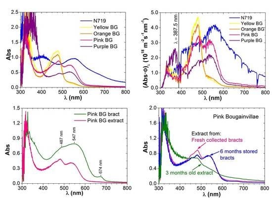
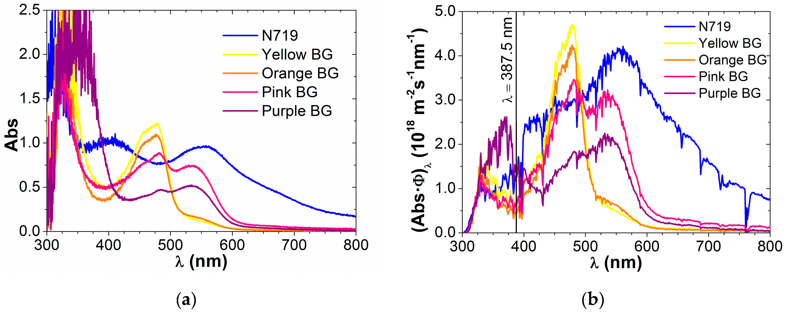
3.1.2. Betalains from Bougainvillea Bracts

The effect of solvent on the extraction of betalains has also been tested in literature [55]. Acetone [56] and acid water [57] have been used as solvents for bougainvillea extracts, although other solvents are possible, i.e., methanol [58] or ethanol [59] solutions, and of course, any of the extracts may undergo further purification [60].
In our group, all the four different solvents listed in Table 1 have been tested in previous works. For instance, the spectra of dye Solutions 1 and 2 are compared with N719 dye spectra in the work by Maldonado and co-workers [38]. In our work, the best Solution is 1 (acetone/water). This best solvent and the corresponding method have been subsequently applied to four different-colored (yellow, orange, pink, and purple) bougainvillea (BG) bracts. The UV-Vis spectra of the four extracts obtained are compared in Figure 3a, which also includes N719 spectrum for reference. Quantitative data, wavelength and intensity of the main peaks found are listed in Table 2.
The bougainvillea extracts absorption spectra show two sets of peaks around 480 nm (Peak 1 in Table 2) and 540 nm (Peak 2). These peaks are due to betalain pigments. Betalains can be divided in two groups: (i) betacyanins, red-purple, with light adsorption peaks or bands on 530–540 nm (for example betanidin, which is the smallest betacyanyn with a peak at 542 nm, or betanin with a peak at 535 nm); and (ii) betaxanthins (orange, adsorption on lower wavelengths, for example, indicaxantin has a peak around 482 nm) [3,16,59]. Peak 2 due to betacyanins appears for a wavelength near the maxima of the N-719 dye, which supports the use of these natural dye extracts.
Regarding the different bougainvillea varieties, pink and purple spectra are similar, except that for the pink variety, peak 1 is more intense than peak 2, while peak 2 is a bit higher than 1 in purple bougainvillea, which means that the relative concentration of both pigments change in these two varieties. Orange and yellow extracts have almost the same spectral results, with a main peak 1 around 477 nm wavelength, related to indicaxanthin (yellow-orange). The noticeable absence of betanin gives the yellow-orange color to these extracts. These varieties only show a shoulder probably related to betanidin (542 nm). There are scarce publications for orange BG, and some of the published spectra are different to Figure 3a even using acetone as a solvent [56], showing thus the influence not only of the solvent but also of the particular extraction method. A specific experimental and theoretical study of these betalain pigments, obtained from red beet-roots, can be found in the work by Oprea and col. [61].
The absorbed intensity leads to a quantitative estimation of pigment concentration considering the molar absorbance of indicaxanthin and betanin [57,60]. Applying the formulas in Reference [57] to our experimental results, we find the pigment-relative concentrations listed in Table 3.
Attending to these values, for purple and pink BG, both pigments corresponding to peaks 1 and 2 are present in the extract in a similar percentage, the relative concentration of indicaxanthin (47.3%) being slightly lower than the betanin concentration (52.7%) for purple BG, and the opposite for pink BG (57.3% indicaxanthin, 42.7% betanin), thus confirming the previous visual inspection. On the other hand, for yellow and orange BG dyes, betaxanthins (Peak 1) are present in more than 90%, and betanin is scarce. These relative concentrations may vary with aging or pH, as will be shown later. Isah et al. studied dyes from red bougainvillea as a function of pH [57], and they obtained for water extract (pH 5.7), concentration values of 15.17 μM betaxanthin (57.4%) and 11.26 μM betanin (42.6%), very close to our values for pink BG obtained with water-acetone solvent at similar pH.
Figure 3b shows the products of all BG dye spectra and the sun spectrum (the photon flux plotted in Figure 2), giving an idea of the total number of photons which can be converted in electrons by a NDSSC device using these dyes. Comparing with N719, the absorbance of BG dyes is poor at wavelengths above 600 nm, but it is quite good between 400 and 600 nm. The solar spectrum has a maximum of photons at 675 nm and enhances the dye absorbance peaks between 400 and roughly 750 nm. The total number of visible photons (Nϕ) potentially absorbed for every BG dye can be obtained by integrating Figure 3b curves in the range 387 (TiO2 limit) to 750 nm. Results for every BG variety are listed in the last column of Table 3 in percentages relative to N ϕ obtained for N719. According to these values, the pink BG extract is the best bougainvillea dye and purple BG is the less convenient variety for NDSSCs applications, although factors such as the dye absorption onto the TiO2 semiconductor and dye stability in the device would obviously also determine the final solar cell performance.

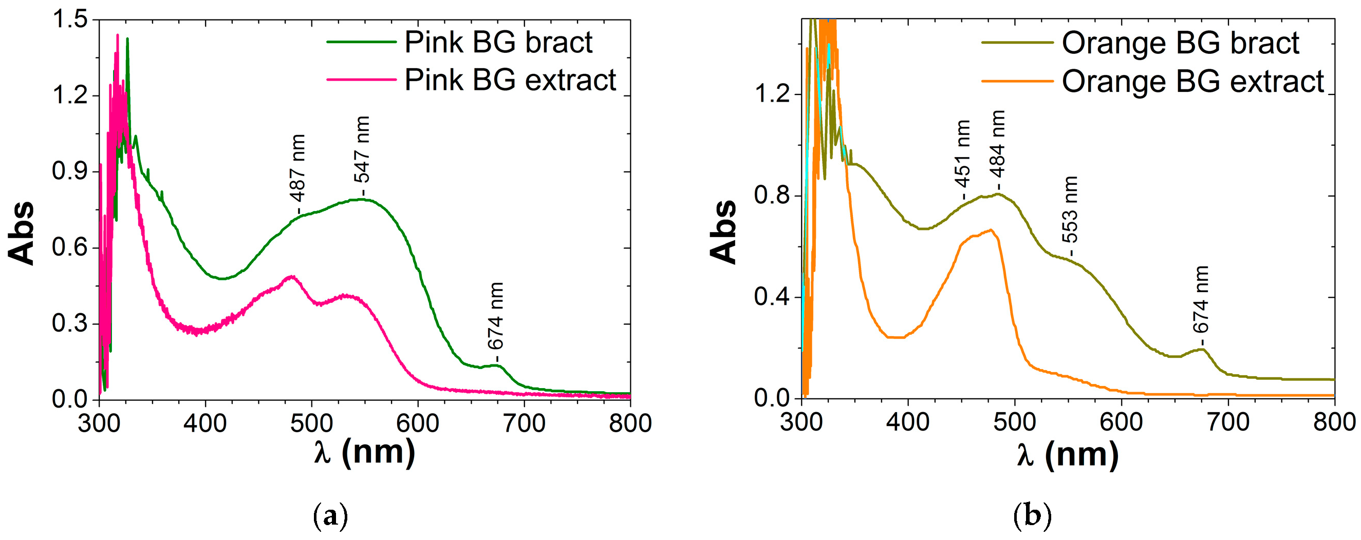
Comparison of Dye Extracts and Dry Bracts Absorption Spectra

Before concluding the dye characterization phase for bougainvillea, we have interest in measuring the spectra of dry-colored bracts and comparing them with the corresponding dye solution extract spectra.
Regarding the results for dry bracts, reflectance, transmittance and absorption spectra obtained with the spectroradiometer are quite similar for orange and yellow bracts, and on the other hand, for pink and purple as well. Then the absorption spectrum obtained for a bract with the spectroradiometer, is compared with the UV-Vis absorption spectrum of the corresponding dye extract.
Figure 4a,b show the comparison for pink and orange BG respectively. From both a and b graphs, it can be concluded that: (i) The shape of both bract and extract spectra are similar, although the peaks corresponding to the main betalains pigments are of course more evident in the extract than in the solid sample. (ii) Besides, the spectra peaks in the dye solution appear in general at lower wavelengths (see Table 2). Chromophores containing electrons with low activation energies (responsible of absorption peaks) change with media, pH, or even with temperature, and consequently the amount of energy absorbed by those functional groups also changes, moving the position of the main peaks, in this case, to higher energies. iii) Finally, a remarkable fact is that the chlorophyll peak (around 670 nm, [62]) of the bract diminishes or even disappears in dye solution, probably because chlorophyll molecules present in bracts are eliminated when the dye is extracted following our process with acetone solvent solutions, as it is known that ethanol has been preferably used when the pigment of interest is chlorophyll [55,62].

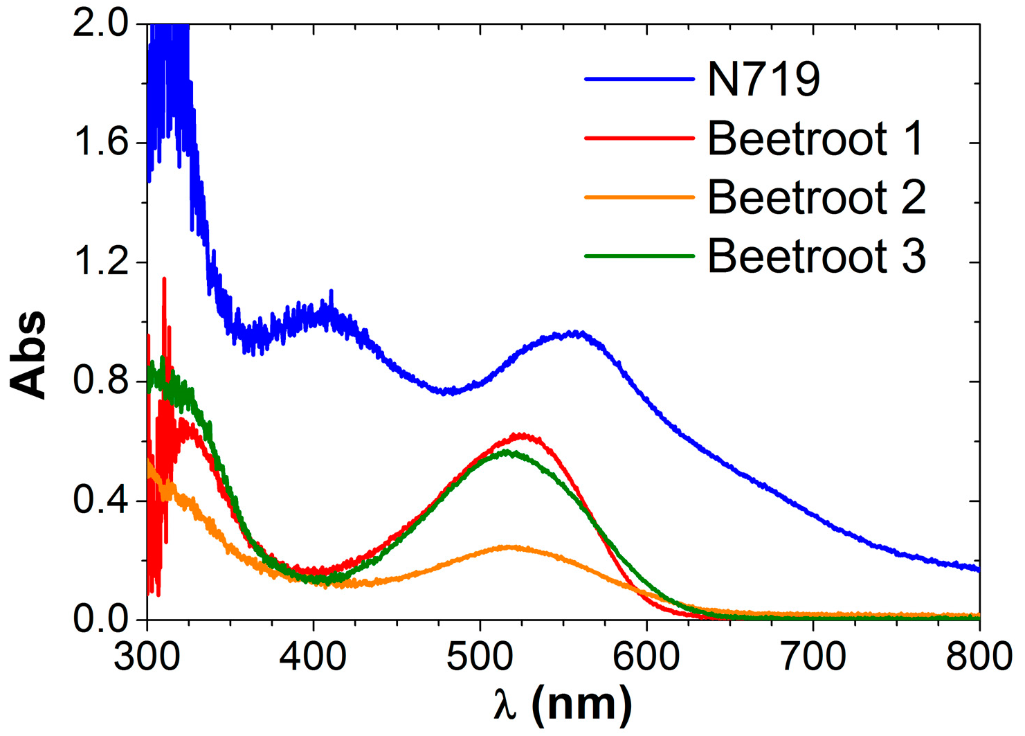
3.1.3. Betalains from Beetroots

Ethanol has been mainly used as an extraction solvent for beetroots [63,64,65]. However, we have obtained that Solutions 1, 2 and 3 all give good results. The three extracted dyes show an adsorption band around 520 nm (see Figure 5 and Table 2), comparable to the 533 nm peak of N719, so these natural extracts from beetroots could be eligible for sensitizing the NDSSCs.
It is noticeable that extract with Solution 2 (using ethanol) presents lower absorbance. In this case, the extremely low pH of the extract (pH = 1.7) may play an important role, as stated in previous works. Sengupta et al. [64] showed UV-Vis spectra of ethanol beetroot extracts with varying pH. In the pH range 6–9, the dyes showed similar pattern absorption, with the two peaks corresponding to betalain pigments, although their intensity and relative intensity within each pH varied. However, the absorbance fell drastically and the spectrum changed in shape at extreme pH levels of 3 and 12.
As we have prepared the three extracts with the same procedure, less concentration of pigment in Beetroot 2 is a priori presumed, which, apart from other considerations, would give a less efficient cell. This can be checked in Table 4: among NDSSCs using beetroot extracts, the number 2 is the less efficient. However, the Beetroot 3 dye with a similar absorbance peak to Beetroot 1, gives also lower efficiency than 1. Thus, the absorbance is not the only determinant factor: pH (pH Beetrrot 1 = 5.6; pH Beetroot 3 = 1.4) and the solvent [55] may influence the process of pigment anchoring to the semiconductor surface, which should also be analyzed in each particular dye use.

3.2. Exam Dyes Behavior

Once the extracts have been selected, and before using, it is advisable to have a deeper knowledge of the dye solutions, their stability, and how they absorb onto substrates. So, for example, considering DSSC, in this second phase we propose to study: (i) How does the dye solution adsorb onto semiconductor NDSSC film? (ii) What about mixing different dyes? (iii) How stable is the dye extract? (iv) What’s the role of pH, temperature or pigment concentration? The main results from this second phase summarize as follows.

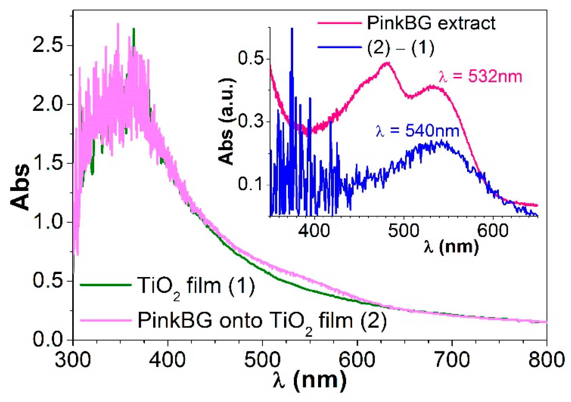
3.2.1. How the Dye Solution Adsorbs onto Semiconductor DSSC Film

The dye must have at least one functional group that helps the dye molecules to attach on the surface of the semiconductor, so the chemical structure of natural dyes ([7,16]) must be considered. Anthocyanin’s basic structure has –OH groups favoring bonding to TiO2 [53]. Betalains have also anchor groups, such as carboxyl (–COOH), capable of binding to the atoms of the semiconductor. There are several binding modes and sites which are influenced by several factors like dye’s structure, total anchoring groups, energy levels, pH, substrate pre-treatment, etc. [61]
Some works include absorption spectra of dyes onto TiO2 film [20,21,27,32,49,50,55,59,66,67,68,69,70,71]. Calogero and col. published UV-Vis absorption spectra of anthocyanins and betalains onto TiO2 [67]. Torchani and col. showed UV-Vis absorption spectra of spinach, beet, red cabbage and other natural dyes, and also the spectra of these dyes adsorbed on TiO2 [69]. Adedokun and col. studied the influence of solvent on dye adsorption onto TiO2. Also, Park and col. published studies of dye absorption on TiO2 (in terms of concentration) and how the cell efficiency depends on the amount of adsorbed dye [71]. However, Jun et al. [32] showed that a high adsorption rate does not necessarily indicate a highly efficient solar energy conversion; the electrolyte used was more influential.
The dye-onto-TiO2 UV-Vis spectra show enhanced light photon absorption and in addition, when TiO2 contribution is removed, absorption peaks (or shoulders) identifiable with pigments can be seen. Our group has already published data about eggplant extracts (anthocyanins) adsorbed onto TiO2, detecting the pigment main peak around 530 nm, after removing the TiO2 contribution [37]. In general, we find that the spectra of the extracts onto TiO2 are quite similar to that of the TiO2 film alone, but with a slight increase in absorbance and in some cases a modification of shape, as reported also in other works [68,69]. In this paper, Figure 6 shows how betalain extract from pink bougainvillea adsorbs onto TiO2 film. In the inset of Figure 6, the peak corresponding to betanin (~540 nm) is clearly seen when TiO2 film contribution is removed from the spectra of the dye absorbed onto the semiconductor nanostructure (blue line in the inset). The indicaxantin peak (~480 nm) is not specifically detected, probably due to the higher absorption of the TiO2 for these lower wavelengths, but a small hump can be seen in its place.

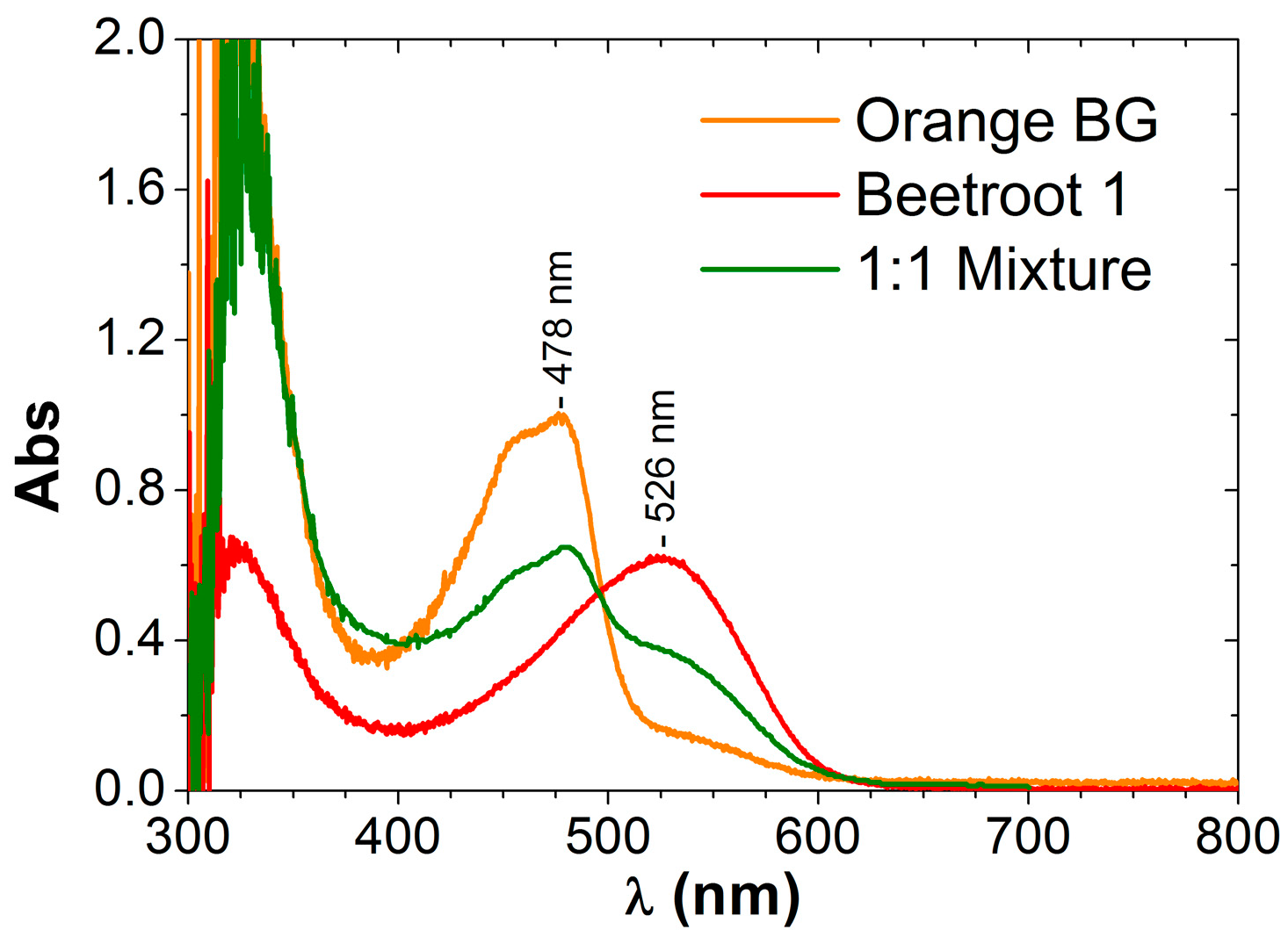
3.2.2. Mixing Different Dyes: Co-sensitization

The absorption spectrum of a single dye hardly follows the whole solar radiation spectrum (see Figure 2 and Figure 3). Mixtures of several dyes with different absorption spectra are often prepared to obtain the maximum absorption within the visible and infra-red region. For DSSCs applications, these mixtures co-sensitize the device to increase the global absorption using the widest wavelength range possible and thus maximizing the efficiency.
Works including the study of natural dye mixtures show different approaches [6,26,27,28,64,72,73,74,75]. Chang and col. used chlorophyll from bougainvillea leaves and anthocyanins from blueberries [72], finding that the efficiency of the cell improved when using a 1:1 dye mixture (0.62% chlorophyll, 0.64% anthocyanins and 0.75% dye mixture). Something similar was found later by Kimpa and col.: DSSCs were prepared using anthocyanins dyes (flame three flower), chlorophyll dyes (from pawpaw leaves) and a 1:1 mixture of both, the latter having a higher conversion efficiency [73]. Kumar and co-workers [74] studied four pigments, one of each family, and six 1:1 mixtures. EIS measurements showed higher electron recombination lifetime for co-sensitized cells (i.e., individual dyes gave cell efficiencies of 0.11% and 0.64%, while 1:1 co-sensitized cell achieved 1.14%).
We have also studied mixtures of different betalain dye solutions and in different proportions: orange and pink bougainvillea extracts (1:1, 1:3 and 3:1) and orange bougainvillea extract with beetroot extract 1:1 both with Solution 1. Figure 7 shows UV-Vis spectrum of the latter mixture. In general, when mixing dyes that belong to the same family, we can conclude that the mixture gives an intermediate spectrum with absorption peaks or bands for the same wavelengths as the individual dyes, but with less absorption, as there is less concentration of each individual component. Regarding cell performance, we do not obtain the synergistic effect of the two dyes, probably because both dyes belong to the same family, and thus compete for a place to attach to the semiconductor. Scarce works report an intermediate efficiency for the co-sensitized cell. For example, when mixing anthocyanins from different sources, an efficiency of the co-sensitized cell below the average of individual cell is obtained [76]. This paper led us to test whether co-sensitized cells mixing dyes from betalains family behaved similar to co-sensitized cells with anthocyanins or achieved a higher efficiency. In our case, efficiency of the co-sensitized cells mixing betalain dyes is bit more than the average of the single sensitized cells efficiency for the example in Figure 7. (see Table 4). The publications that report a notable increase in efficiency mix different dye families, i.e., betalain (beetroot) & chlorophyll (spinach) [64], chlorophyll & anthocyanins [72,73,75] or chlorophyll & betacyanins [74].

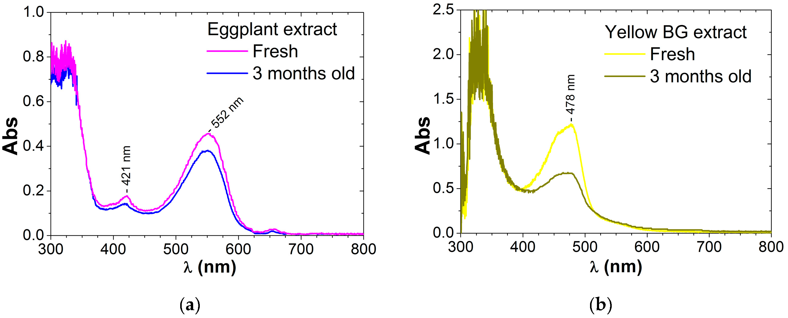
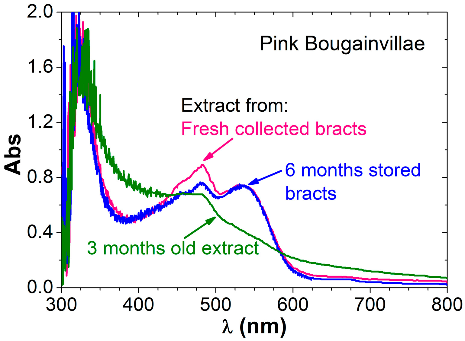
3.2.3. How Stable the Dye Extract is

Some researchers have published studies of DSSC stability or lifetime [8,12], showing how cell performance does not significantly change [60] (i.e., betacyanin dye cell was found stable for 12 weeks [63]) or diminishes with time [70,76,77,78].
Our group has also tested cell performance, keeping the cell working continuously for 350 h, with the aim to test not the dye, but the electrolyte. It was found that the cell with volatile electrolytes lasted for about 80 h, and when fresh electrolyte was injected, the cell could recover at 70% of the initial efficiency for almost 100 h. However, for cells with non-volatile electrolytes (i.e., ionic liquids), the efficiency kept constant at the beginning but diminished to 80% after 175 h and to 40% after 250 h [79].
In this work here, we have focused on dye extract stability, not the entire cell, just by recording UV-Vis spectra after a certain time, and the stability is not the same for the different extracts.
Anthocyanins can be affected by age, sugars, organic acids, co-pigments, etc. These molecules are susceptible to degradation under the influence of many factors, including temperature or basic pH in the presence of oxygen [2,80,81]. For example, Askar and col. [80] find degradation of the extract in 3 h of about 5% for the most stable extract or 20% for the less one. They measure degradation as the decrease of absorbance peaks in the dark. In contrast, our eggplant extract, with the extracting method and Solution 2, containing nasunin [37,82], seems to be stable for months even without any kind of purification, just keeping it preserved from air and light. Figure 8a compares UV-Vis absorption spectra of a freshly prepared eggplant extract and of the same extract 3 months later. The spectra shape is the same and the absorption decreases by only 16% in the main peak at 550 nm. For this extract, pH is very acid, below 2 in all the samples. The influence of pH on anthocyanins absorption spectra has been previously reported [81,83]; the lower the pH, the lower the wavelength of the main absorbance peak and the more stable the dye.
Regarding betalains pigments, on the one hand, both orange and yellow bougainvillea extracts behave similarly. Figure 8b shows an example of two UV-Vis spectra of a yellow BG extract: recently prepared (yellow plot) and 3 months later (dark green plot). The spectrum after 3 months is essentially the same, still with indicaxantin peak (absorption around 470–480 nm), but the absorbance is 44% lower. Therefore, it is preferable to make dye extract just before cell fabrication, due to possible degradation with time.
On the other hand, it has been found that pink and violet bougainvillea extracts quickly degrade into less efficient compounds (see Figure 9, green line). Absorbance of peaks initially in the range [450–480] nm diminishes 24% after less than 3 months (this is consistent with the previous results for yellow-orange extracts), and the betanin peak initially around 530 nm is only a shoulder with an intensity fall of 44%. This peak gives the red-violet color, and it is also visually obvious that betanin degrades with time, as the dye solution becomes brown. Some results already published [60] state that bougainvillea solutions are stable for at least 12 months, but in this case, the solutions had been previously purified. The maximum stability of betalains is reached for pH between 5 and 6 and it is known that betanin at neutral pH (534 nm) converts to betanidin (540 nm) by β-glucosidase at acid pH, and in general when decreasing pH, λmax decreases [84]. Besides, betalains degrade in the presence of light and oxygen and also with increasing temperature [85].
To compensate for this disadvantage of bougainvillea extract, we tried to preserve dry bracts. We verified that pigments responsible for light absorption can be successfully extracted from bracts that have been kept stored for several months and adequately preserved from air and light exposure. Figure 9 also shows UV-Vis spectra of dye extracts made from a) bracts just after having been collected (pink line) and b) bracts preserved for 6 months (blue line). Pigments extracted seem to be the same, since the same absorbance peaks appear and the intensity of betanin peak is the same, although the overall absorbance is slightly lower for curve b, and thus the relative concentration of the two pigments slightly changes with respect to fresh collected bracts (see Table 3), being the percentages 52% indicaxanthin, and 48% betanin for the extract from 6-months-stored bracts. So we conclude that bracts can be collected, dried and preserved for a long time to be used later to get the dye extracts.

3.2.4. Role of Other Parameters Like pH or Pigment Concentration

Some publications have studied the influence of diverse parameters on dye extraction, absorption, stability, etc. [64,86]. Wongcharee studied the effects of extracting temperature and solvent and also the effect of pH on DSSC efficiency [76]. Anthocyanin extracts from five fruits were studied by Teoli and coworkers [54]. Different pH values were also tested. UV-Vis spectra of the extracts showed an increase in the wavelength of the maximum absorbance peak with increasing pH (also found in Refs. [80,83]), and the highest conversion efficiency was obtained using a pH 4 dye solution. Suyitno and col. studied papaya-leaf dye and the influence of pH, finding the most efficient and stable cell for pH 3.5 [70].
In our group, we have not detected a strong pH influence on spectral data. Extracts of eggplant gave different peak positions even having similar pH values [37]. Bougainvillea extracts all have similar acid pH values (~5). For beetroot extracts, the lower the pH, the lower the wavelength (around 520 nm) for the main betalain pigment peak, as Solutions 1 to 3 with pH 5.6, 1.7 and 1.4 presented the peak for wavelengths 523, 519 and 516 nm, respectively. Similar pH dependence for betalains has been previously found in the literature [84].
Scarce authors concentrate the dyes [28], and in the case that they do, it is usually a previous step to refinement or purification [63]. In our works, simply concentrating dye solutions does not apparently change the spectral data, nor cell performance, although FF increases about 20%, as can be checked in Table 4.

3.3. NDSSC Tests

After this previous exhaustive study of natural-dye solutions, an example of using is presented. This last step is to prepare the cells and check how the dyes work measuring cell performance varying the dye and with all the other parameters fixed. Under equal preparation and irradiation conditions, the efficiencies of NDSSCs with different extracts are measured. DSSC fabrication and test method is thus a simple and optimized procedure consisting of always the same consecutive steps, from paste preparation to cell assembly and photovoltaic measurements: The only differences are due to the different dye solutions used, as the aim of the experiments is to study the dye influence on cell efficiency. Table 4 summarizes some of our results.
Efficiency values are similar to those obtained in our previous works [37,38,41] and in literature, see for example some tables comparing NDSSC and references therein [3,7,8,9,13,14,15,16,60]. Artificial dye N719 gives higher efficiency than natural dyes, but lower than values found in literature. This is due to our cell fabrication methodology, which was selected for its simplicity to test the natural dyes and not to maximize the efficiency [40].
Among all the natural dyes listed in Table 4, the highest efficiencies are obtained when using beetroot extract with Solution 1, reaching up to 0.47%, among the highest values in literature for this pigment (i.e., raw 0.12%—purified 0.4% [61]; 0.11% [77] or 0.19% [84] and finally Hernandez-Martinez and col. obtained 0.68% to 0.89% optimizing electrodes preparation [78]). Regarding the other beetroot extracts, the one with Solution 3, even having a similar spectrum and pigment concentration as the former, gives lower efficiency. This may be due to the very low pH, which limits –COOH anchoring to the semiconductor surface. Solution 2 behaves similarly, and, in addition, the absorbance spectrum shows the lowest pigment concentration, so in this case the efficiency is even lower.
Eggplant extracts gave low efficiencies compared with already published data (i.e., 0.48% [82]). Other works obtain lower efficiency for eggplant (0.15%) compared to other natural dyes, and attribute it to more pronounced recombination reactions for eggplant dye than for the other dyes [34].
The different bougainvillea extracts achieved similar efficiencies (0.16% to 0.19%) no matter what bract color, which is due to the presence of different betalain groups and in different proportions. Red-purple bracts extracts gave also efficiencies of the same order or up to (0.29 ± 0.02) % in our previous works, and the small differences found were due to other factors like film thickness, surfactant used in paste preparation, and porosity of the semiconductor film [38,41].
The fill factor does not change noticeably when varying the dye, although in our experiments the highest FF (0.62) has been found for eggplant extracts, while the lowest FF (0.35) is obtained for cells made with beetroot extract.
We have also concentrated some of the dyes, but no clear increase or change in efficiencies has been seen. In Table 4, we give as an example the cell efficiency when using orange bougainvillea extract concentrated 10% wt. Efficiency does not change, although the fill factor does increase from 0.43 (initial extract) to 0.52 (concentrated).

4. Conclusions

Based on our experience and after doing a wide literature review, we propound a general protocol to optimize a simple natural dye production method, without complicating the chemical procedures, with the aim of enhancing the dye potential to whatever subsequent use. In particular, as a possible application, NDSSCs performance is considered.
In conclusion, the protocol consists of these main and minimum advisable steps to obtain and test natural pigments: (i) Dye solutions preparation and characterization by UV-Vis spectrometry: Select natural raw vegetable pigment, preferably to be obtained locally and easily, and select the simplest and best extraction solution and method. (ii) Exam dye solutions behavior i.e., how they adsorb onto semiconductor film, how stable they are, or what is the influence of different parameters on extraction and adsorption. And finally (iii) NDSSC tests: As an example of application of the previous exhaustive study, we have designed a simple and economical cell fabrication method and measured cell performance in environmental conditions varying the dye and with all the other parameters fixed. The novelty of this approach is that it gives a previous deeper knowledge of the dye, crucial to the final NDSSC performance.
We have successfully employed this protocol with betalain pigments (a series of four bougainvillea dyes and three beetroot dyes) and anthocyanins (three eggplant dyes). The previous UV-Vis spectra analysis gives useful information about:
  • The best extraction solution and method: for betalains, Solution 1 (acetone/water, moderately acid), for anthocyanins, Solutions 2 and 3 (very acidic solutions).
  • The relative concentration of different pigment molecules for the different extracts.
  • Some candidate vegetable material can be a priori selected or ruled out by inspection of the extract spectra. Besides, comparison with the corresponding spectra of dry raw material performed with a spectroradiometer, gives reliable information about the dye molecules which can be potentially extracted. We show the example of bougainvillea-colored bracts spectra.
  • The dye stability or ageing: Anthocyanins are more stable and betalain molecules degrade, but conservation of the dry precursor (collected bracts) for at least 6 months is possible.
  • How the dye adsorbs onto photoelectrode film, and the effect of mixing dye extracts. In this paper, we focus on mixing dyes from the betalain family to complement results in the bibliography.
For the selected cell fabrication method, NDSSC tests show that for anthocyanins the best performances were obtained with Solutions 2 and 3, but with very low values compared to literature. In this work, betalains give higher efficiencies than anthocyanins. For betalains, the best extract is obtained with Solution 1, as initially found from UV-Vis spectrometry analysis. Within the betalain family, for bougainvillea dyes, similar performances were obtained for the different varieties, and for beetroots the best result reached up to 0.47% cell efficiency, which is among the highest values in literature.

Author Contributions

Conceptualization, M.J.G.-S.; Methodology, M.J.G.-S.; Investigation, M.J.G.-S.; Resources, M.J.A.; Writing—Original Draft Preparation, M.J.G.-S.; Writing—Review & Editing, M.J.G.-S. and M.J.A.; Visualization, M.J.A.; Supervision, M.J.G.-S. and M.J.A.; Project Administration, M.J.A.; Funding Acquisition, M.J.A. Both authors have read and approved the manuscript.

Funding

The research work leading to this article received ERDF funds from the Spanish government within the framework of the SOLTERMIN project (Ref. ENE2017-83973-R) of the Ministerio de Economía, Industria y Competitividad (Spanish Ministry of Economy, Industry and Competitiveness).

Acknowledgments

The authors sincerely thank S. Rosiek and J. Alonso from CIESOL (Solar Energy Research Center, UAL-PSA, Almería, Spain); and J. Fernández-Reche from Plataforma Solar de Almería (PSA-CIEMAT, Almería, Spain); S. Galera and J. Barbero are acknowledged for the technical support.

Conflicts of Interest

“The authors declare no conflict of interest.”.

References

  1. Mohd, Y.; Mohd, S.; Faqeer, M. Natural Colorants: Historical, Processing and Sustainable Prospects. Nat. Prod. Bioprospect. 2017, 7, 123–145. [Google Scholar] [CrossRef] [Green Version]
  2. Miller, R.; Owens, S.J.; Rørslett, B. Plants and colour: Flowers and pollination. Opt. Laser Technol. 2011, 43, 282–294. [Google Scholar] [CrossRef]
  3. Narayan, M.R. Review: Dye sensitized solar cells based on natural photosensitizers. Renew. Sust. Energ. Rev. 2012, 16, 208–215. [Google Scholar] [CrossRef]
  4. Sugathan, V.; John, E.; Sudhakar, K. Recent improvements in dye sensitized solar cells: A review. Renew. Sust. Energ. Rev. 2015, 52, 54–64. [Google Scholar] [CrossRef]
  5. Sharma, S.; Siwach, B.; Ghoshal, S.K.; Mohan, D. Dye sensitized solar cells: From genesis to recent drifts. Renew. Sust. Energ. Rev. 2017, 70, 529–537. [Google Scholar] [CrossRef]
  6. Mohiuddin, O.; Obaidullah, M.; Sabah, C. Improvement in dye sensitized solar cells from past to present. Opt. Quant. Electron. 2018, 50, 377. [Google Scholar] [CrossRef]
  7. Hug, H.; Bader, M.; Mair, P.; Glatzel, T. Biophotovoltaics: Natural pigments in dye-sensitized solar cells. Appl. Energ. 2014, 115, 216–225. [Google Scholar] [CrossRef] [Green Version]
  8. Ludin, N.A.; Mahmoud, A.M.A.A.; Mohamad, A.B.; Amir, H.K.A.; Sopian, K.; Karim, N.S.A. Review on the development of natural dye photosensitizer for dye-sensitized solar cells. Renew. Sust. Energ. Rev. 2014, 31, 386–396. [Google Scholar] [CrossRef]
  9. Shalini, S.; Balasundara Prabhu, R.; Prasanna, S.; Mallick, T.K.; Senthilarasu, S. Review on natural dye sensitized solar cells: Operation, materials and methods. Renew. Sust. Energ. Rev. 2015, 51, 1306–1325. [Google Scholar] [CrossRef]
  10. Gong, J.; Sumathy, K.; Qiao, Q.; Zhou, Z. Review on dye-sensitized solar cells (DSSCs): Advanced techniques and research trends. Renew. Sust. Energ. Rev. 2017, 68, 234–246. [Google Scholar] [CrossRef]
  11. Ye, M.; Wen, X.; Wang, M.; Iocozzia, J.; Zhang, N.; Lin, C.; Lin, Z. Recent advances in dye-sensitized solar cells: From photoanodes, sensitizers and electrolytes to counter electrodes. Mater. Today 2015, 18, 155–162. [Google Scholar] [CrossRef]
  12. Mozaffari, S.; Nateghi, M.R.; Zarandi, M.B. An overview of the challenges in the commercialization of dye sensitized solar cells. Renew. Sust. Energ. Rev. 2017, 71, 675–686. [Google Scholar] [CrossRef]
  13. Chawla, P.; Tripathi, M. Novel improvements in the sensitizers of dye-sensitized solar cells for enhancement in efficiency—A review. Int. J. Energy Res. 2015, 39, 1579–1596. [Google Scholar] [CrossRef]
  14. Shalini, S.; Balasundaraprabhu, R.; Satish-Kumar, T.; Prabavathy, N.; Senthilarasu, S.; Prasanna, S. Status and outlook of sensitizers/dyes used in dye sensitized solar cells (DSSC): A review. Int. J. Energy Res. 2016, 40, 1303–1320. [Google Scholar] [CrossRef]
  15. Zhou, H.; Wu, L.; Gao, Y.; Ma, T. Dye-sensitized solar cells using 20 natural dyes as sensitizers. J. Photoch. Photobio. A 2011, 219, 188–194. [Google Scholar] [CrossRef]
  16. Kumara, N.T.R.N.; Lim, A.; Lim, S.M.; Petra, M.I.; Ekanayake, P. Recent progress and utilization of natural pigments in dye sensitized solar cells: A review. Renew. Sust. Energ. Rev. 2017, 78, 301–317. [Google Scholar] [CrossRef]
  17. Richhariya, G.; Kumar, A.; Tekasakul, P.; Gupta, B. Natural dyes for dye sensitized solar cell: A review. Renew. Sust. Energ. Rev. 2017, 69, 705–718. [Google Scholar] [CrossRef]
  18. Gu, P.; Yang, D.; Zhu, X.; Sun, H.; Li, J. Fabrication and characterization of dye-sensitized solar cells based on natural plants. Chem. Phys. Lett. 2018, 693, 16–22. [Google Scholar] [CrossRef]
  19. Mahmoud, A.M.A.A.; Ludin, N.A.; Mohamad, A.B.; Kadhum, A.A.H.; Mukhlus, A. Application of dyes extracted from Alternanthera dentata leaves and Musa acuminata bracts as natural sensitizers for dye-sensitized solar cells. Spectrochim. Acta A Mol. Biomol. Spectrosc. 2018, 192, 487–498. [Google Scholar] [CrossRef]
  20. Maurya, I.C.; Singh, S.; Neetu; NeetuGupta, A.K.; Srivastava, P.; Bahadur, L. Dye-sensitized solar cells employing extracts from four cassia flowers as natural sensitizers: Studies on dye ingredient effect on photovoltaic performance. J. Electron. Mater. 2018, 47, 225–232. [Google Scholar] [CrossRef]
  21. Adedokun, O.; Sanusi, Y.K.; Awodugba, A.O. Solvent dependent natural dye extraction and its sensitization effect for dye sensitized solar cells. Optik 2018, 174, 497–507. [Google Scholar] [CrossRef]
  22. Calogero, G.; Barichello, J.; Citro, I.; Mariani, P.; Vesce, L.; Bartolotta, A.; Di Carlo, A.; Di Marco, G. Photoelectrochemical and spectrophotometric studies on dye-sensitized solar cells (DSCs) and stable modules (DSCMs) based on natural apocarotenoids pigments. Dyes Pigments 2018, 155, 75–83. [Google Scholar] [CrossRef]
  23. Sanjay, P.; Deepa, K.; Madhavan, J.; Senthil, S. Optical, spectral and photovoltaic characterization of natural dyes extracted from leaves of Peltophorum pterocarpum and Acalypha amentacea used as sensitizers for ZnO based dye sensitized solar cells. Opt. Mater. 2018, 83, 192–199. [Google Scholar] [CrossRef]
  24. Manaa, M.B.; Bouaziz, N.; Schmaltz, B.; Van, F.T.; Lamine, A.B. Study of the effect of variation in temperature and pH on the adsorption process of natural Gardenia yellow dye into TiO2 mesoporous for dye sensitized solar cells using the statistical physics formalism: Physicochemical and thermodynamic investigation. Micropor. Mesopor. Mat. 2018, 270, 82–92. [Google Scholar] [CrossRef]
  25. Gu, P.; Yang, D.; Zhu, X.; Sun, H.; Li, J. Performance of dye-sensitized solar cells based on natural dyes. Opt. Quant. Electron. 2018, 50, 223. [Google Scholar] [CrossRef]
  26. Tahir, D.; Satriani, W.; Gareso, P.L.; Abdullah, B. Dye sensitized solar cell (DSSC) with natural dyes extracted from Jatropha leaves and purple Chrysanthemum flowers as sensitizer. IOP Conf. Ser. J. Phys. Conf. Ser. 2018, 979, 012056. [Google Scholar] [CrossRef]
  27. Rajkumar, S.; Nirmal Kumar, M.; Suguna, K.; Muthulakshmi, S.; Ashok Kumar, R. Enhanced performance of dye-sensitized solar cells using natural cocktail dye as sensitizer. Optik 2019, 178, 224–230. [Google Scholar] [CrossRef]
  28. Hosseinnezhad, M.; Rouhani, S.; Gharanjig, K. Extraction and application of natural pigments for fabrication of green dye-sensitized solar cells. Opto. Electron. Rev. 2018, 26, 165–171. [Google Scholar] [CrossRef]
  29. Yildiz, Z.K.; Atilgan, A.; Atli, A.; Özel, K.; Altinkaya, C.; Yildiz, A. Enhancement of efficiency of natural and organic dye sensitized solar cells using thin film TiO2 photoanodes fabricated by spin-coating. J. Photochem. Photobiol. A 2019, 368, 23–29. [Google Scholar] [CrossRef]
  30. Liu, Q.; Gao, N.; Liu, D.; Liu, J.; Li, Y. Structure and photoelectrical properties of natural photoactive dyes for solar cells. Appl. Sci. 2018, 8, 1697. [Google Scholar] [CrossRef]
  31. Cassone, G.; Calogero, G.; Sponer, J.; Saija, F. Mobilities of iodide anions in aqueous solutions for applications in natural dye-sensitized solar cells. Phys. Chem. Chem. Phys. 2018, 20, 13038–13046. [Google Scholar] [CrossRef] [PubMed]
  32. Jun, H.K.; Arof, A.K. Performance of natural dyes as sensitizer in dye-sensitized solar cells employing LiBOB-based liquid electrolyte. Ionics 2018, 24, 915–922. [Google Scholar] [CrossRef]
  33. Swathi, K.E.; Jinchu, I.; Sreelatha, K.S.; Sreekala, C.O.; Menon, S.K. Effect of microwave exposure on the photo anode of DSSC sensitized with natural dye. IOP Conf. Ser. Mater. Sci. Eng. 2018, 310, 012141. [Google Scholar] [CrossRef]
  34. Tractz, G.T.; Viomar, A.; Dias, B.V.; de Lima, C.A.; Banczek, E.P.; da Cunha, M.T.; Antunes, S.R.M.; Rodrigues, P.R.P. Recombination Study of Dye Sensitized Solar Cells with Natural Extracts. J. Braz. Chem. Soc. 2019, 30, 371–378. [Google Scholar] [CrossRef]
  35. Lucioli, S.; Di Bari, C.; Forni, C.; Di Carlo, A.; Barrajón-Catalán, E.; Micol, V.; Nota, P.; Teoli, F.; Matteocci, F.; Frattarelli, A.; et al. Anthocyanic pigments from elicited in vitro grown shoot cultures of Vaccinium corymbosum L., cv. Brigitta Blue, as photosensitizer in natural dye-sensitized solar cells (NDSSC). J. Photochem. Photobiol. B 2018, 188, 69–76. [Google Scholar] [CrossRef]
  36. Prabavathy, N.; Shalini, S.; Balasundaraprabhu, R.; Velauthapillai, D.; Prasanna, S.; Balaji, G.; Muthukumarasamy, N. Algal buffer layers for enhancing the efficiency of anthocyanins extracted from rose petals for natural dye-sensitized solar cell (DSSC). Int. J. Energy Res. 2018, 42, 790–801. [Google Scholar] [CrossRef]
  37. Galera-Moreno, S.; González-Galindo, E.; Ariza, M.J.; García-Salinas, M.J. Influence of natural dyes in low-cost dye-sensitized solar cell’s efficiency. In Materials and Technologies for Energy Efficiency; Méndez-Vilas, A., Ed.; BrownWalker Press: Boca Ratón, FL, USA, 2015; pp. 48–52. [Google Scholar]
  38. Maldonado-Valdivia, A.I.; Galindo, E.G.; Ariza, M.J.; Garcia-Salinas, M.J. Surfactant influence in the performance of titanium dioxide photoelectrodes for dye-sensitized solar cells. Sol. Energy 2013, 91, 263–272. [Google Scholar] [CrossRef]
  39. Ito, S.; Kitamura, T.; Wada, Y.; Yanagida, S. Facile fabrication of mesoporous TiO2 electrodes for dye solar cells: Chemical modification and repetitive coating. Sol. Energy Mater. Sol. Cells 2003, 76, 3–13. [Google Scholar] [CrossRef]
  40. Ito, S.; Murakami, T.N.; Comte, P.; Liska, P.; Grätzel, C.; Nazeeruddin, M.K.; Grätzel, M. Fabrication of thin film dye sensitized solar cells with solar to electric power conversion efficiency over 10%. Thin Solid Films 2008, 516, 4613–4619. [Google Scholar] [CrossRef]
  41. Galindo, E.G.; Ariza, M.J.; De las Nieves, F.J.; García-Salinas, M.J. Effects of multilayer coating and calcination procedures on the morphology of dye-sensitized solar cell semiconductor photoelectrodes. Thin Solid Films 2015, 590, 230–240. [Google Scholar] [CrossRef]
  42. Syafinar, R.; Gomesh, N.; Irwanto, M.; Fareq, M.; Irwan, Y.M. Potential of Purple Cabbage, Coffee, Blueberry and Turmeric as Nature Based Dyes for Dye Sensitized Solar Cell (DSSC). Energy Procedia 2015, 79, 799–807. [Google Scholar] [CrossRef] [Green Version]
  43. Belay, A.; Ture, K.; Redi, M.; Asfaw, A. Measurement of caffeine in coffee beans with UV/vis spectrometer. Food Chem. 2008, 108, 310–315. [Google Scholar] [CrossRef]
  44. Setiawan, T.; Subekti, W.Y.; Nur’Adya, S.S.; Ilmiah, K.; Ulfa, S.M. Performance of natural dye and counter electrode from robusta coffee beans peel waste for fabrication of dye-sensitized solar cell (DSSC). IOP Conf. Ser.: Mat. Sci. Eng. 2018, 299, 012073. [Google Scholar] [CrossRef]
  45. Kawase, M.; Takahashi, M. Chemical composition of sporopollenin in magnolia grandiflora (Magnoliaceae) and hibiscus syriacus (Malvaceae). Grana 1995, 34, 242–245. [Google Scholar] [CrossRef]
  46. Di Paola-Naranjo, R.D.; Sánchez-Sánchez, J.; González-Paramás, A.M.; Rivas-Gonzalo, J.C. Liquid chromatographic–mass spectrometric analysis of anthocyanin composition of dark blue bee pollen from Echium plantagineum. J. Chromatogr. A 2004, 1054, 205–210. [Google Scholar] [CrossRef] [PubMed]
  47. Urbanczyk, J.; Fernandez-Casado, M.A.; Díaz, T.E.; Heras, P.; Infante, M.; Borrego, A.G. Spectral fluorescence variation of pollen and spores from recent peat-forming plants. Int. J. Coal Geol. 2014, 131, 263–273. [Google Scholar] [CrossRef] [Green Version]
  48. Mularczyk-Oliwa, M.; Bombalska, A.; Kaliszewski, M.; Włodarski, M.; Trafny, A.; Kopczyński, K.; Kwaśny, M.; Szpakowska, M.; Zbieta, E. Comparison of fluorescence spectroscopy and FTIR in differentiation of plant pollens. Spectrochim. Acta A Mol. Biomol. Spectrosc. 2012, 97, 246–254. [Google Scholar] [CrossRef] [PubMed]
  49. Calogero, G.; Di Marco, G.; Caramori, S.; Cazzanti, S.; Argazzi, R.; Bignozzi, C.A. Natural dye sensitizers for photoelectrochemical cells. Energy Environ. Sci. 2009, 2, 1162–1172. [Google Scholar] [CrossRef]
  50. Shanmugam, V.; Manoharan, S.; Anandan, S.; Murugan, R. Performance of dye-sensitized solar cells fabricated with extracts from fruits of ivy gourd and flowers of red frangipani as sensitizers. Spectrochim. Acta A Mol. Biomol. Spectrosc. 2013, 104, 35–40. [Google Scholar] [CrossRef] [PubMed]
  51. Gholamrezaei, S.; Salavati-Niasari, M. Natural sensitizer for low cost dye sensitized solar cell based on Strontium Titanate nanoparticles. J. Mater. Sci. Mater. Electron. 2016, 27, 2467–2472. [Google Scholar] [CrossRef]
  52. Sashank, T.V.S.S.P.; Manikanta, B.; Pasula, A. Fabrication and experimental investigation on dye sensitized solar cells using titanium dioxide nano particles. Mater. Today Proc. 2017, 4, 3918–3925. [Google Scholar] [CrossRef]
  53. Liao, S.K.; Chang, Y.H.; Wu, C.T.; Lai, Y.R.; Chen, W.Y. Fabrication of anthocyanin-sensitized nanocrystalline titanium dioxide solar cells using supercritical carbon dioxide. J. CO2 Util. 2017, 21, 513–520. [Google Scholar] [CrossRef]
  54. Teoli, F.; Lucioli, S.; Nota, P.; Frattarelli, A.; Matteocci, F.; Di Carlo, A.; Caboni, E.; Forni, C. Role of pH and pigment concentration for natural dye-sensitized solar cells treated with anthocyanin extracts of common fruits. J. Photoch. Photobio. A 2016, 316, 24–30. [Google Scholar] [CrossRef]
  55. Mahmoud, A.M.A.A.; Mohamad, A.B.; Kadhum, A.A.H.; Ludin, N.A. Effect of solvents on the extraction of natural pigments and adsorption onto TiO2 for dye-sensitized solar cell applications. Spectrochim. Acta A Mol. Biomol. Spectrosc. 2015, 138, 130–137. [Google Scholar] [CrossRef]
  56. Maabong, K.; Muiva, C.M.; Monowe, P.; Sathiaraj, T.S.; Hopkins, M.; Nguyen, L.; Malungwa, K.; Thobega, M. Natural pigments as photosensitizers for dye-sensitized solar cells with TiO2 thin films. Int. J. Renew. Sustain. Energy Res. 2015, 5, 54–60. [Google Scholar]
  57. Isah, K.U.; Ahmadu, U.; Idris, A.; Kimpa, M.I.; Uno, U.E.; Ndamitso, M.M.; Alu, N. Betalain pigments as natural photosensitizers for dye-sensitized solar cells: The effect of dye pH on the photoelectric parameters. Mater. Renew. Sustain. Energy 2015, 4, 39. [Google Scholar] [CrossRef]
  58. Kumar, S.N.A.; Ritesh, S.K.; Sharmila, G.; Muthukumaran, C. Extraction optimization and characterization of water soluble red purple pigment from floral bracts of Bougainvillea glabra. Arab. J. Chem. 2017, 10, 2145–2150. [Google Scholar] [CrossRef]
  59. Damit, D.N.F.P.; Galappaththi, K.; Lim, A.; Petra, M.I.; Ekanayake, P. Formulation of water to ethanol ratio as extraction solvents of Ixora coccinea and Bougainvillea glabra and their effect on dye aggregation in relation to DSSC performance. Ionics 2017, 23, 485–495. [Google Scholar] [CrossRef]
  60. Hernandez-Martinez, A.R.; Estevez, M.; Vargas, S.; Quintanilla, F.; Rodriguez, R. New dye-sensitized solar cells obtained from extracted bracts of bougainvillea Glabra and Spectabilis Betalain Pigments by Different Purification Processes. Int. J. Mol. Sci. 2011, 12, 5565–5576. [Google Scholar] [CrossRef]
  61. Oprea, C.I.; Dumbravă, A.; Enache, I.; Georgescu, A.; Gîrţu, M.A. A combined experimental and theoretical study of natural betalain pigments used in dye-sensitized solar cells. J. Photochem. Photobio. A 2012, 240, 5–13. [Google Scholar] [CrossRef]
  62. Shanmugam, V.; Manoharan, S.; Sharafali, A.; Anandan, S.; Murugan, R. Green grasses as light harvesters in dye sensitized solar cells. Spectrochim. Acta A Mol. Biomol. Spectrosc. 2015, 135, 947–952. [Google Scholar] [CrossRef] [PubMed]
  63. Tripathi, M.; Upadhyay, R.; Pandey, A. Novel dye based photoelectrode for improvementof solar cell conversion efficiency. Appl. Sol. Energy 2013, 49, 54–57. [Google Scholar] [CrossRef]
  64. Sengupta, D.; Mondal, B.; Mukherjee, K. Visible light absorption and photo-sensitizing properties of spinach leaves and beetroot extracted natural dyes. Spectrochim. Acta A 2015, 148, 85–92. [Google Scholar] [CrossRef]
  65. Hemmatzadeh, R.; Mohammadi, A. Improving optical absorptivity of natural dyes for fabrication of efficient dye-sentitized solar cells. J. Theor. Appl. Phys. 2013, 7, 57. [Google Scholar] [CrossRef]
  66. Senthil, T.S.; Muthukumarasamy, N.; Velauthapillai, D.; Agilan, S.; Thambidurai, M.; Balasundaraprabhu, R. Natural dye (cyaniding 3-O-glucoside) sensitized nanocrystalline TiO2 solar cell fabricated using liquid electrolyte/quasi-solid-state polymer electrolyte. Renew. Energy 2011, 36, 2484–2488. [Google Scholar] [CrossRef]
  67. Calogero, G.; Yum, J.H.; Sinopoli, A.; Di Marco, G.; Grätzel, M.; Nazeeruddin, M.K. Anthocyanins and betalains as light-harvesting pigments for dye-sensitized solar cells. Sol. Energy 2012, 86, 1563–1575. [Google Scholar] [CrossRef]
  68. Hemalatha, K.V.; Karthick, S.N.; Justin-Raj, C.; Hong, N.Y.; Kim, S.K.; Kim, H.-J. Performance of Kerria japonica and Rosa chinensis flower dyes as sensitizers for dye-sensitized solar cells. Spectrochim. Acta A Mol. Biomol. Spectrosc. 2012, 96, 305–309. [Google Scholar] [CrossRef]
  69. Torchani, A.; Gharbi, R.; Fathallah, M. Study of natural dyes for sensitized solar cells applications. Sens. Transducers 2014, 27, 185–189. [Google Scholar]
  70. Suyitno, S.; Saputra, T.J.; Supriyanto, A.; Arifin, Z. Stability and efficiency of dye-sensitized solar cells based on papaya-leaf dye. Spectrochim. Acta A Mol. Biomol. Spectrosc. 2015, 148, 99–104. [Google Scholar] [CrossRef]
  71. Park, K.H.; Kim, T.Y.; Park, J.Y.; Jin, E.M.; Yim, S.H.; Choi, D.Y.; Lee, J.W. Adsorption characteristics of gardenia yellow as natural photosensitizer for dye-sensitized solar cells. Dyes Pigments 2013, 96, 595–601. [Google Scholar] [CrossRef]
  72. Chang, H.; Kao, M.J.; Chen, T.L.; Fuo, H.G.; Choand, K.C.; Lin, X.P. Natural sensitizer for dye-sensitized solar cells using three layers of photoelectrode thin films with a Schottky barrier. Am. J. Eng. Appl. Sci. 2011, 4, 214–222. [Google Scholar] [CrossRef]
  73. Kimpa, M.I.; Momoh, M.; Isah, K.U.; Yahya, H.N.; Ndamitso, M.M. Photoelectric characterization of dye sensitized solar cells using natural dye from pawpaw leaf and flame tree flower as sensitizers. Mater. Sci. Appl. 2012, 3, 281–286. [Google Scholar] [CrossRef]
  74. Kumar, K.A.; Subalakshmi, K.; Senthilselvan, J. Effect of mixed valence state of titanium on reduced recombination for natural dye-sensitized solar cell applications. J. Solid State Electrochem. 2016, 20, 1921–1932. [Google Scholar] [CrossRef]
  75. Chang, H.; Lo, Y.J. Pomegranate leaves and mulberry fruit as natural sensitizers for dye-sensitized solar cells. Sol. Energy 2010, 84, 1833–1837. [Google Scholar] [CrossRef]
  76. Wongcharee, K.; Meeyoo, V.; Chavadej, S. Dye-sensitized solar cell using natural dyes extracted from rosella and blue pea flowers. Sol. Energy Mater. Sol. Cell 2007, 91, 566–571. [Google Scholar] [CrossRef]
  77. Torchani, A.; Saadaoui, S.; Gharbi, R.; Fathallah, M. Sensitized solar cells based on natural dyes. Curr. Appl. Phys. 2015, 15, 307–312. [Google Scholar] [CrossRef]
  78. Hernandez-Martinez, A.R.; Estevez, M.; Vargas, S.; Rodriguez, R. Stabilized conversion efficiency and dye-sensitized solar cells from beta vulgaris pigment. Int. J. Mol. Sci. 2013, 14, 4081–4093. [Google Scholar] [CrossRef]
  79. Galindo, E.G. Estudio de la Dinámica de Liquidos Ionicos Confinados en Medios Nanoporosos Mediante Experimentos y Simulaciones. Aplicación a las Celulas Solares de Colorante. Ph.D. Thesis, University of Almeria, Almeria, Spain, 2015. [Google Scholar]
  80. Askar, K.A.; Alsawad, Z.H.; Khalaf, M.N. Evaluation of the pH and thermal stabilities of rosella anthocyanin extracts under solar light. Beni Suef Univ. J. Basic Appl. Sci. 2015, 4, 262–268. [Google Scholar] [CrossRef] [Green Version]
  81. Norman, M.; Bartczak, P.; Zdarta, J.; Ehrlich, H.; Jesionowski, T. Anthocyanin dye conjugated with Hippospongia communis marine demosponge skeleton and its antiradical activity. Dyes Pigments 2016, 134, 541–552. [Google Scholar] [CrossRef]
  82. Calogero, G.; Di Marco, G. Red Sicilian orange and purple eggplant fruits as natural sensitizers for dye-sensitized solar cells. Sol. Energy Mater. Sol. Cell 2008, 92, 1341–1346. [Google Scholar] [CrossRef]
  83. Vankar, P.S.; Shukla, D. Natural Dyeing with Anthocyanins from Hibiscus rosa sinensis Flowers. J. Appl. Polym. Sci. 2011, 122, 3361–3368. [Google Scholar] [CrossRef]
  84. Zhang, D.; Lanier, S.M.; Downing, J.A.; Avent, J.L.; Lum, J.; McHale, J.L. Betalain pigments for dye-sensitized solar cells. J. Photochem. Photobiol. A 2008, 195, 72–80. [Google Scholar] [CrossRef]
  85. Azeredo, H.M.C. Betalains: Properties, sources, applications and stability—A review. Int. J. Food Sci. Technol. 2009, 44, 2365–2376. [Google Scholar] [CrossRef]
  86. Chien, C.Y.; Hsu, B.D. Optimization of the dye-sensitized solar cell with anthocyanin as photosensitizer. Sol. Energy 2013, 98, 203–211. [Google Scholar] [CrossRef]
Figure 1. Evolution of the number of publications published per year obtained from literature searching using “natural dye” and “solar cell” topics (data source ISI Web of Science).
Applsci 09 02515 g001
Figure 2. Spectra of pollen extracts with Solutions 1 and 2 and artificial dye N719 spectrum. Right y axis: Spectral photon density, Φλ, calculated from the standard ASTM G173-03 reference solar spectrum.
Applsci 09 02515 g002
Figure 3. (a) UV-Vis spectra of the four bougainvillea extracts obtained. N719 spectrum is also included for comparison. (b) Products of all dye spectra in Figure 3a and the sun spectrum (Φλ) in Figure 2.
Applsci 09 02515 g003
Figure 4. Dry bract and dye extract spectra for: (a) pink bougainvillea; (b) orange bougainvillea.
Applsci 09 02515 g004
Figure 5. Betalains from beetroots: three pigment extracts with solvent solutions 1, 2 and 3.
Applsci 09 02515 g005
Figure 6. Absorption spectrum of TiO2 film (green line 1) and pink bougainvillea extract adsorbed onto TiO2 film (pink line 2). Inset: main peak of adsorbed dye onto TiO2 film after removing film contribution (2-1, blue line) compared to pink bougainvillea extract solution (magenta line).
Applsci 09 02515 g006
Figure 7. UV-Vis spectrum of dye mixture (green line) 1:1 orange bougainvillea extract with beetroot extract, both with Solution 1. Spectra of individual dyes are also included.
Applsci 09 02515 g007
Figure 8. Extracts stability: (a) Eggplant extract with Solution 2: fresh, recently prepared (purple line) and 3 months later, preserved from air and light (blue line); (b) Yellow bougainvillea extract with Solution 2: recently prepared (yellow line) and 3 months old (dark green line).
Applsci 09 02515 g008
Figure 9. UV-Vis spectra of pink bougainvillea extract 3 months after preparation (green line). UV-Vis spectra of recently prepared dye extracts made from (i) bracts just after been collected (red data) and (ii) bracts preserved for 6 months (blue data).
Applsci 09 02515 g009
Table 1. Solutions used for extracting the dye from vegetable raw materials.
Extraction MethodAcetone
(% vol.)
Ethanol
(% vol.)
HCl
(M)
H2O
(% vol.)
pH
Range
Solution 132------685.0–5.6
Solution 2---Solvent0.1---1.6–1.7
Solution 3------0.1Solvent1.3–1.4
Solution 4---80---206.5–7.0
Table 2. Wavelength and intensity of the main absorbance peaks of the UV-Vis spectra shown in Figure 3, Figure 4 and Figure 5.
Spectrum (Figure)Peak Typeλ (nm)Abs
N719 (3)Peak 14051.03
Peak 25530.96
Yellow BG (3)Shoulder 14551.10
Peak 14781.21
Shoulder 25430.12
Orange BG (3)Shoulder 14551.01
Peak 14781.08
Shoulder 25430.16
Pink BG (3)Shoulder 14510.74
Peak 14820.88
Peak 25350.74
Purple BG (3)Shoulder 14540.40
Peak 14840.47
Peak 25350.52
Pink BG bract (4)Shoulder 14870.72
Peak 25470.79
Peak 36740.13
Orange BG bract (4)Shoulder 14510.76
Peak 14840.81
Shoulder 25530.54
Peak 36740.19
Beetroot 1 (5)Peak 15260.62
Beetroot 2 (5)Peak 15190.24
Beetroot 3 (5)Peak 15160.56
Table 3. Estimation of concentration (μmol) and relative percentage of betaxanthins (absorption Peak 1 at ~482 nm) and betanin (Peak 2 at ~536 nm) molecules for the four bougainvillea extracts according to Ref. [57]. The last column lists the total number of photons absorbed between 387 and 750 nm by the natural dyes in relative percentages with respect to the absorption of N719 reference dye.
Peak 1[Betaxanthins]Peak 2[Betanin]Nϕ
AbsμM(%)AbsμM(%)(%)
Purple BG0.477.247.30.528.052.734.7
Pink BG0.8815.257.30.7411.442.753.2
Orange BG1.0824.590.90.162.59.135.0
Yellow BG1.2127.993.80.121.86.238.7
Table 4. Performance of DSSC prepared with natural dyes: Average and maximum efficiency and fill factors.
DSSC Dyeηav (%)ηmax (%)FFavFFmax
N-7192.3 ± 0.53.2350.44 ± 0.080.553
Eggplant 2 [37]0.090 ± 0.0060.1010.51 ± 0.060.623
Eggplant 3 [37]0.069 ± 0.0110.0830.54 ± 0.020.568
Pink bougainvillea 0.19 ± 0.070.2500.51 ± 0.080.595
Orange Bougainvillea0.19 ± 0.020.2150.427 ± 0.0130.448
Concentrated orange BG0.188 ± 0.0120.2020.521 ± 0.0160.542
Yellow Bougainvillea0.162 ± 0.0170.1850.39 ± 0.040.451
Beetroot 10.32 ± 0.120.4670.41 ± 0.080.469
Beetroot 20.08 ± 0.040.1120.35 ± 0.060.414
Beetroot 30.17 ± 0.060.2290.35 ± 0.030.392
1:1 Orange BG + Beetroot 10.269 ± 0.0040.2740.55 ± 0.020.563

