Article

In Vitro Antioxidant Activity and In Vivo Topical Efficacy of Lipid Nanoparticles Co-Loading Idebenone and Tocopheryl Acetate

by
Lucia Montenegro
1,*,
Concetta Maria Messina
2,
Simona Manuguerra
2,
Ludovica Maria Santagati
1,
Lorella Pasquinucci
3,
Rita Turnaturi
3,
Carmela Parenti
4,
Rosaria Arena
2 and
Andrea Santulli
2,5
1
Department of Drug Sciences, Pharmaceutical Technology Section, University of Catania, Viale A. Doria 6, 95125 Catania, Italy
2
Dipartimento di Scienze della Terra e del Mare DiSTeM, Laboratorio di Biochimica Marina ed Ecotossicologia, Università degli Studi di Palermo, Via G. Barlotta 4, 91100 Trapani, Italy
3
Department of Drug Sciences, Medicinal Chemistry Section, University of Catania, Viale A. Doria 6, 95125 Catania, Italy
4
Department of Drug Sciences, Pharmacology and Toxicology Section, University of Catania, Viale A. Doria 6, 95125 Catania, Italy
5
Consorzio Universitario della Provincia di Trapani, Istituto di Biologia marina, Via G. Barlotta 4, 91100 Trapani, Italy
*
Author to whom correspondence should be addressed.
Appl. Sci. 2019, 9(5), 845; https://doi.org/10.3390/app9050845
Submission received: 26 December 2018 / Revised: 20 February 2019 / Accepted: 22 February 2019 / Published: 27 February 2019
(This article belongs to the Section Nanotechnology and Applied Nanosciences)

Abstract

:

Featured Application

Development of topical formulations based on lipid nanoparticles for the treatment of skin aging and diseases involving cutaneous dryness and scaling such as atopic and exfoliative dermatitis and psoriasis.

Abstract

Idebenone (IDE) is a strong antioxidant that has been proposed for the treatment of skin disorders, including skin ageing. Unfavorable physico-chemical properties make IDE a poor skin permeant where effectiveness could be improved by its loading into suitable delivery systems such as solid lipid nanoparticles (SLN) and nanostructured lipid carriers (NLC). In this work, we designed novel IDE-loaded NLC containing tocopheryl acetate (VitE) as a liquid component to obtain a synergic effect between IDE and VitE. The resulting NLC showed small particle sizes (24–42 nm), low polydispersity indices (<0.300), good stability, and were assessed for their in vitro antioxidant activity and in vivo topical effects. IDE-loaded SLN and NLC showed a high antioxidant activity in in vitro assays (DPPH and reducing power method) and provided a similar and significant protection from oxidative stress of fibroblast cells, HS-68, exposed to UV light. After a two-week topical treatment of human volunteers with gels containing IDE-loaded SLN or NLC, a similar increase in skin hydration was observed, while IDE NLC reduced skin pigmentation to a greater extent than IDE SLN. These results suggest that co-loading IDE and VitE into NLC could be a promising strategy to obtain topical formulations with improved photo-protection.

1. Introduction

In the last decades, solid lipid nanoparticles (SLNs) have been widely investigated as carriers for topical drug delivery [1,2,3,4,5,6,7]. These colloidal systems, consisting of a solid lipid matrix stabilized by different types of surfactants, have been proposed for skin delivery of various active ingredients including antifungal, anticancer, anti-inflammatory, and antioxidant agents [8,9,10,11,12,13,14,15]. The interest in SLNs arises from their many advantages compared to other colloidal carriers such as possibility of incorporating both hydrophilic and lipophilic drugs, ability to achieve drug-controlled release and targeting, thus improving drug topical bioavailability while reducing systemic side effects [1,4]. However, the highly ordered structure of the solid lipid matrix of SLN could result in low incorporation of lipophilic drugs and drug leakage during storage. To overcome these drawbacks, liquid lipids were mixed together with solid lipids to obtain a less packed lipid matrix, leading to a second generation of lipid nanoparticles defined as nanostructured lipid carriers (NLC) [2,3,6]. To obtain this last type of nanocarrier, a variety of oils without any intrinsic pharmacological activity has been investigated. However, to improve NLC effectiveness, oils that have a synergic activity with the loaded drug could be used together with solid lipids to form the NLC core.
Previously, we evaluated in vitro skin permeation of idebenone (IDE), an analogue of coenzyme Q10 with strong antioxidant activity [16], from SLNs with different compositions [17]. We observed an interesting IDE-targeting effect in the upper skin layers from SLNs prepared using oleth-20 as a surfactant and cetyl palmitate as the solid lipid. These SLNs showed good stability during storage at room temperature but limited IDE loading.
In this work, we designed IDE-loaded nanoparticles (NLC) containing tocopheryl acetate (vitamin E acetate, VitE) as a liquid component to obtain both a synergic effect between IDE and VitE and to improve IDE loading. Vitamin E is a well-known radical scavenger agent with skin photo-protective properties [18] for which topical beneficial effects are well known [19].
To evaluate the effect of co-loading vitamin E acetate and IDE in the matrix of lipid nanoparticles, we assessed technological properties, in vitro antioxidant activity, and cytotoxic and photo-protective effects of IDE loaded SLN and NLC on human fibroblast cells HS-68. In addition, we evaluated the effects of topical application on human volunteers of gel formulations containing IDE-loaded SLN and NLC.

2. Materials and Methods

2.1. Materials

Glyceryl oleate (Tegin O®, GO), cetyl palmitate (CP), methylisothiazolinone and methylchloroisothiazolinone (Acnibio AC®), and triethanolamine (TEA) were bought from ACEF (Fiorenzuola D’Arda, Italy). Idebenone (IDE) was obtained from Carbosynth (Berkshire, UK). Polyoxyethylene-20-oleyl ether (Brij 98®, Oleth-20) and tocopheryl acetate (VitE) was obtained from Farmalabor (Canosa di Puglia, Italy). Potassium ferricyanide and trichloroacetic acid were bought from Carlo Erba reagents (Milano, Italy). Gallic acid, iron (III) chloride (FeCl3), 2,2-Diphenyl-1-picrylhydrazyl (DPPH), human skin fibroblasts (Sigma® HS68), Dulbecco’s Modified Eagle’s Medium (DMEM), fetal bovine serum, glutamine, and penicillin–streptomycin were obtained from Merck KGaA (Darmstadt, Germany).

2.2. Preparation of Lipid Nanoparticles (SLN and NLC)

Lipid nanoparticles (SLN and NLC) were prepared using the phase inversion temperature (PIT) method [20], and their composition is illustrated in Table 1. The aqueous phase consisted of deionized water containing preservatives (Acnibio AC® 0.05% w/w and Kemipur 100® 0.35 % w/w) to prevent microbial growth during storage. Unloaded and drug-loaded lipid nanocarriers were prepared using the same procedure. Briefly, after heating the oil phase and aqueous phase separately at 90 °C, the aqueous phase was added dropwise to the oil phase under vigorous stirring (700 rpm). While cooling down to room temperature, at the phase inversion temperature (PIT), the colloidal suspension turned clear and the PIT value was recorded using a conductivity meter (model 525, Crison, Modena, Italy). The resulting samples were stored in airtight jars at room temperature in the dark until used.

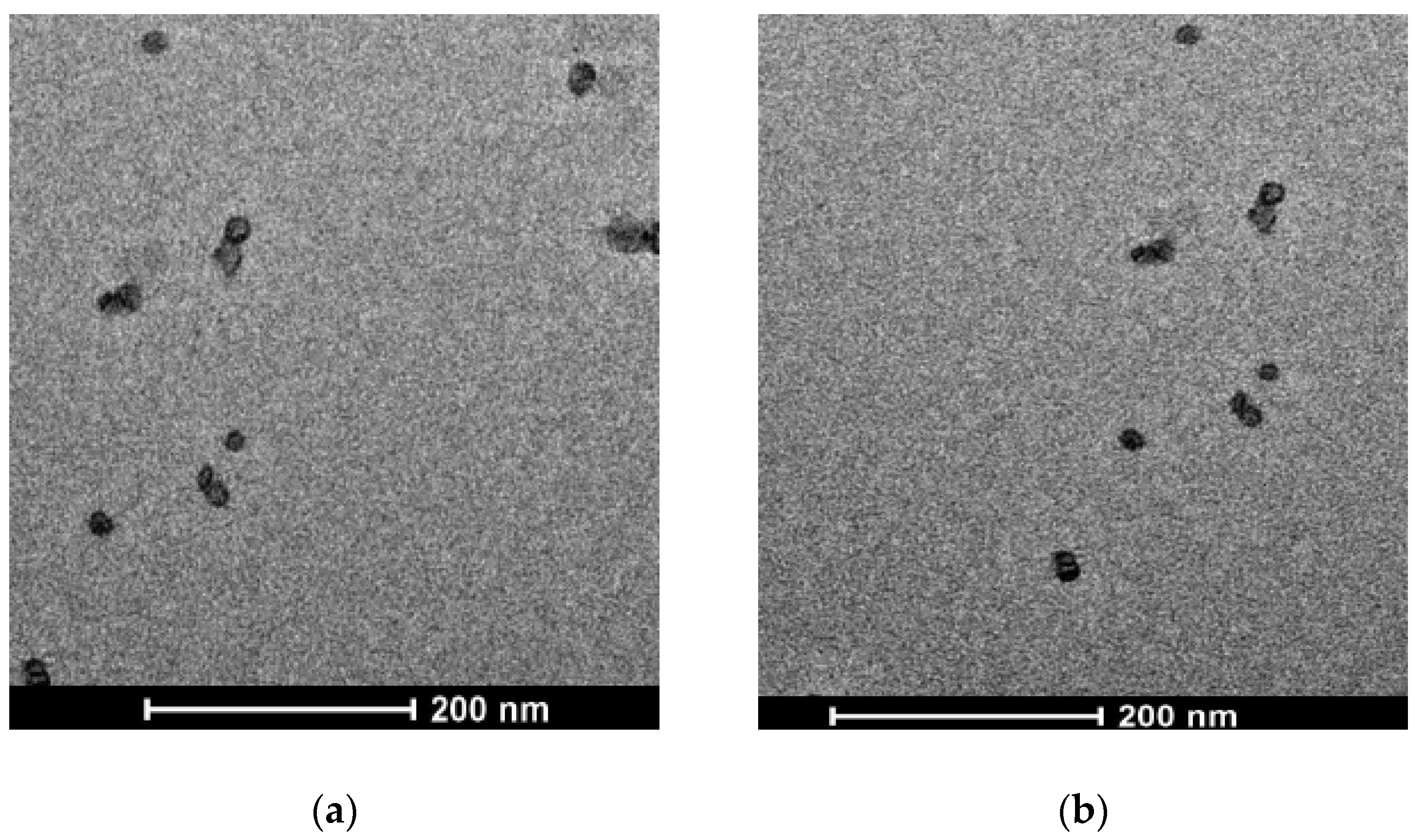
2.3. Transmission Electron Microscopy (TEM)

The morphology of SLN and NLC was assessed by transmission electron microscopy (TEM) analysis. Samples of each colloidal suspension (5 μL) were placed on a Formvar (200-mesh) copper grid (TAAB Laboratories Equipment, Berks, UK). After sample absorption, the excess was removed by filter paper, and a drop of 2% (w/w) aqueous solution of uranyl acetate was added. Then, the surplus was removed, and the sample was allowed to dry at room temperature. Analyses were performed with a transmission electron microscope (model JEM 2010, Jeol, Peabody, MA, USA) operating at an acceleration voltage of 200 KV.

2.4. Photon Correlation Spectroscopy (PCS)

The mean particle size and the size distribution (polydispersity index, PDI) of each colloidal suspension were determined by dynamic light scattering (DLS) using a Zetasizer Nano ZS90 (Malvern Instruments, Malvern, UK) with a 4 mW laser diode at 670 nm and scattering light at 90°.
All samples were analyzed after dilution (1:5, sample:distilled water) and allowed to reach 25 °C for 2 min prior to the analysis. All measurements were carried out in triplicate, and the results were expressed as mean ± SD. ζ-potential was determined by laser Doppler velocimetry using the same Zetasizer reported above. All samples were diluted with KCl 1 mM (pH 7.0) before analysis [21].

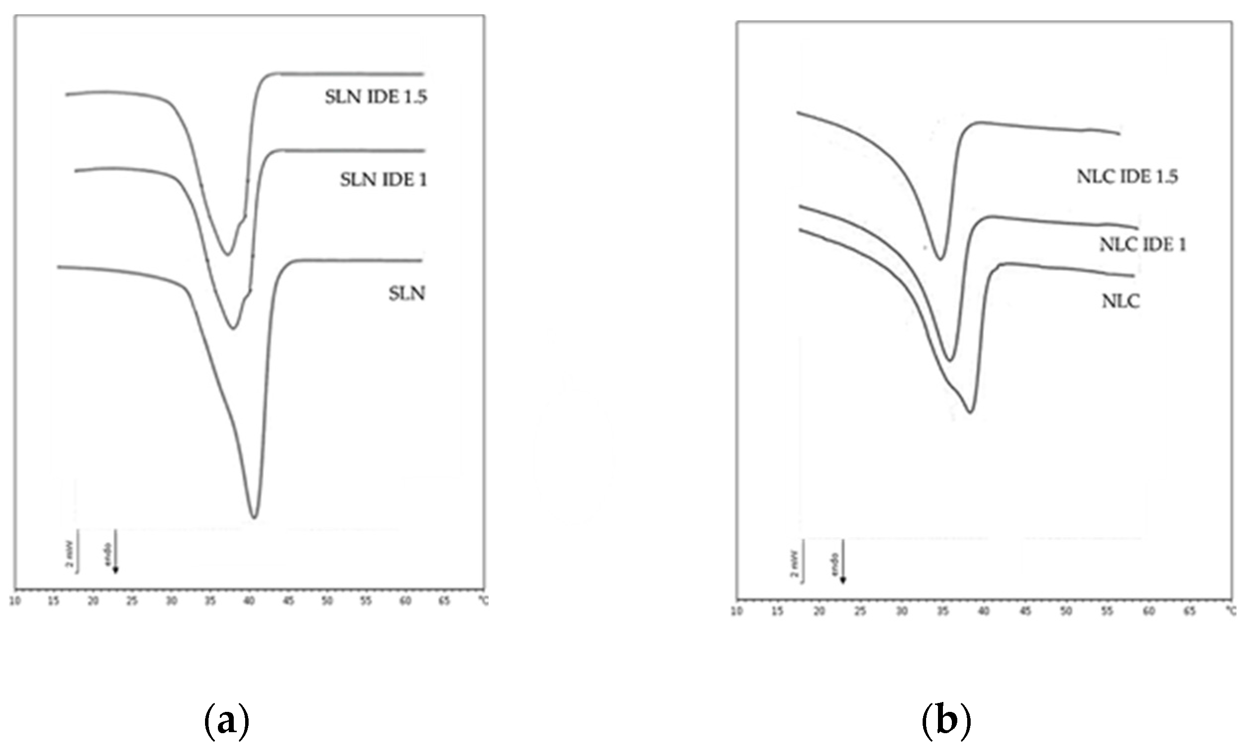
2.5. Differential Scanning Calorimetry (DSC)

SLN and NLC samples were analyzed by DSC using a Mettler TA STARe instrument (Mettler Toledo, Greifensee, Switzerland) equipped with a DSC 822e cell and a Mettler STARe V8.10 software. Indium and palmitic acid (purity ≥ 99.95% and ≥ 99.5%, respectively; Fluka, Buchs, Switzerland) were used to calibrate the instrument. For each sample, 100 µL was sealed into a calorimetric pan, and DSC analyses were carried out by heating the sample from 5 to 65 °C (rate: 2 °C/min) and then cooled from 65 to 5 °C (rate: 4 °C/min) for at least three times. A pan filled with 100 µL of the aqueous phase used for lipid nanocarrier preparation was used as the reference. To calculate the sample crystallinity index (CI), the melting enthalpy of the lipid in the nanocarriers was expressed as a percentage of the melting enthalpy of the bulk lipid cetyl palmitate, which was considered totally crystalline as reported in previous works [22,23]. VitE CI was taken equal to zero as this compound was liquid at room temperature and did not show any melting peak. Each experiment was run in triplicate.

2.6. Stability Tests

Samples of each colloidal suspension were stored at room temperature, protected from light exposure, for two months. Particle sizes, PDI, and ζ-potential values were measured at intervals (24 h, one week, two weeks, one month, and two months).

2.7. Reducing Power

The reducing power of the samples under investigation was determined according to the method by Falleh [24]. Sample solutions at different concentrations (300 µL) were mixed with phosphate buffer (300 µL, 0.2 M, pH 6.6) and potassium ferricyanide [K3Fe(CN)6] (300 µL, 1%); the mixture was incubated at 50 °C for 20 min. A portion (300 µL) of trichloroacetic acid (10%) was added to the mixture, which was then centrifuged at 3000 rpm for 10 min. The upper layer of solution (300 µL) was mixed with distilled water (300 µL) and FeC13 (600 µL, 0.1%), and the absorbance was measured at 700 nm against gallic acid as the authentic standard. An increased absorbance of the reaction mixture indicated increased reducing power. The EC50 value (mg mL−1) is the effective concentration of the extract at which the absorbance was 0.5 and was obtained from linear regression analysis [24].

2.8. DPPH Radical Scavenging Activity

In this assay, gallic acid was used as a positive control. Different aliquots of sample were taken, and the volume was made to 1.0 mL with ethanol. The reaction was started by the addition of a 1.0 mL solution of 200 µM DPPH solution in ethanol. The reaction mixture was kept at room temperature for 30 min, and the absorbance was measured at 517 nm [25]. Scavenging activity was determined by the following equation:
Scavenging activity = [1 − (Absorbance sample/Absorbance control)] × 100

2.9. Cell Culture

Human skin fibroblasts (Sigma® HS68) were cultured as monolayers in Dulbecco’s Modified Eagle’s Medium (DMEM) supplemented with 10% fetal bovine serum, 2 mM glutamine, and 100 µg/mL penicillin–streptomycin, and incubated in a humidified atmosphere at 5% CO2, 95% air, and 37 °C. All cell culture methods were performed under sterile conditions using a grade II flow hood.

2.10. Evaluation of Cytotoxicity and Photo-Protective Effect in Fibroblast Cell Line HS-68

Confluent cells were trypsinized and seeded in a 96-well plate at a concentration of 7 × 10³ cells/well and incubated for 24 h. Preliminary experiments on fibroblast cells were directed toward the evaluation of effects of different doses of IDE on cell vitality and the individuation of the adequate range of compound concentrations to perform experiments of oxidative stress induction. Different concentrations of IDE (10–100 µg/mL dissolved in 0.1% ethanol) were added to previously seeded cells on a 96-well plate and left to incubate for 24 h, and afterward, the viability of cells was assessed. Subsequently, cells were treated for 24 h with an amount of IDE-loaded SLN and NLC corresponding to the highest IDE concentration that did not lead to cytotoxic effects (25 µg/mL). Then, the cells were exposed to UV radiation (lamp UVB KW 254 nm) and left to incubate at 37 °C for 30 minutes, according to Saija et al. [26] The viability of cells was measured using the MTT method according to Mossman [27]. Results were expressed as a percentage of viable cells in respect to the negative control. Each experiment of viability was carried out in triplicate.

2.11. Gel Preparation

To prepare gel formulations, Carbopol Ultrez 21 (0.8 % w/w) was used as a rheological additive, and TEA (1.00% w/w) was used as a neutralizing agent. Carbopol Ultrez 21 was dispersed in the aqueous phase consisting of deionized water containing 0.35% w/w Kemipur 100 and 0.05% Acnibio AC (gel C), unloaded SLN (gel SLN), SLN IDE1 (gel SLN IDE1), SLN IDE1.5 (gel SLN IDE 1.5), unloaded NLC (gel NLC) NLC IDE1 (gel NLC IDE1), and NLC IDE1.5 (gel NLC IDE1.5). Owing to IDE’s poor water solubility, we could not prepare a gel containing only free IDE as a control, not even by adding low amounts of solubilizing agents to the vehicle. Full polymer hydration was obtained by maintaining the mixture in the dark at room temperature for 24 h. Formulations were made viscous by adding TEA (1.00 % w/w) drop by drop under slow mixing to avoid air bubble formation. All gels were stored in airtight jars at room temperature and in the dark to protect from photo-degradation until use. These gels were inspected for any changes in their organoleptic properties during storage for one month at room temperature and in the dark.

2.12. In Vivo Evaluation of Gel Formulations

In vivo topical efficacy under in use conditions of unloaded and IDE-loaded SLN and NLC was assessed according to a protocol previously described [11,12,28]. Fourteen female subjects with average photo-aging were selected (age range: 45–63 years) and experiments were performed according to the rules of the Declaration of Helsinki of 1975, revised in 2008. The local ethics committee declared that this type of study did not require any approval due to its nature and the safety of the formulations under investigation.
All volunteers provided their written informed consent after being informed of the nature of the study. Four different gels, randomly chosen among the seven prepared gels, were given to each volunteer in non-transparent jars labeled with alphanumeric codes (double blind study).
The volunteers were instructed to apply each product (about 2 mg/cm2) onto different areas over the back of their hands, twice a day, morning and evening, for two weeks, while refraining from applying other products. Measurements of skin hydration and pigmentation were performed using the specific probes of the instrument Soft Plus (Callegari Srl, Parma, Italy) under controlled temperature (22 ± 1 °C) and humidity (35 ± 5%) conditions. Baseline values were determined prior to applying the products. Skin hydration was evaluated by capacity measurements in the range of 0–100 u.c. (arbitrary units) with a resolution of 1 u.c. and 5% precision. Skin pigmentation was analyzed using a probe equipped with a double wavelength reflectance photometer (λ1 = 875 nm, λ2 = 660 nm), and values were obtained in the range of 0–100 u.c., with a resolution of 1 u.c. and 5% precision.
Each measurement was performed in triplicate and the values were expressed as mean ± S.D.

2.13. Statistical Analyses

The statistical differences were evaluated for each parameter by the analysis of variance (ANOVA). The differences among the mean values were subjected to the Student–Newman–Keuls test (SNK). Before analysis, the degree of heterogeneity was assessed by Cochran’s test [29]. Data were processed for one-way ANOVA using Statistica (version 8.0, Statsoft, Inc., Oklahoma).

3. Results and Discussion

3.1. Lipid Nanoparticle Characterization
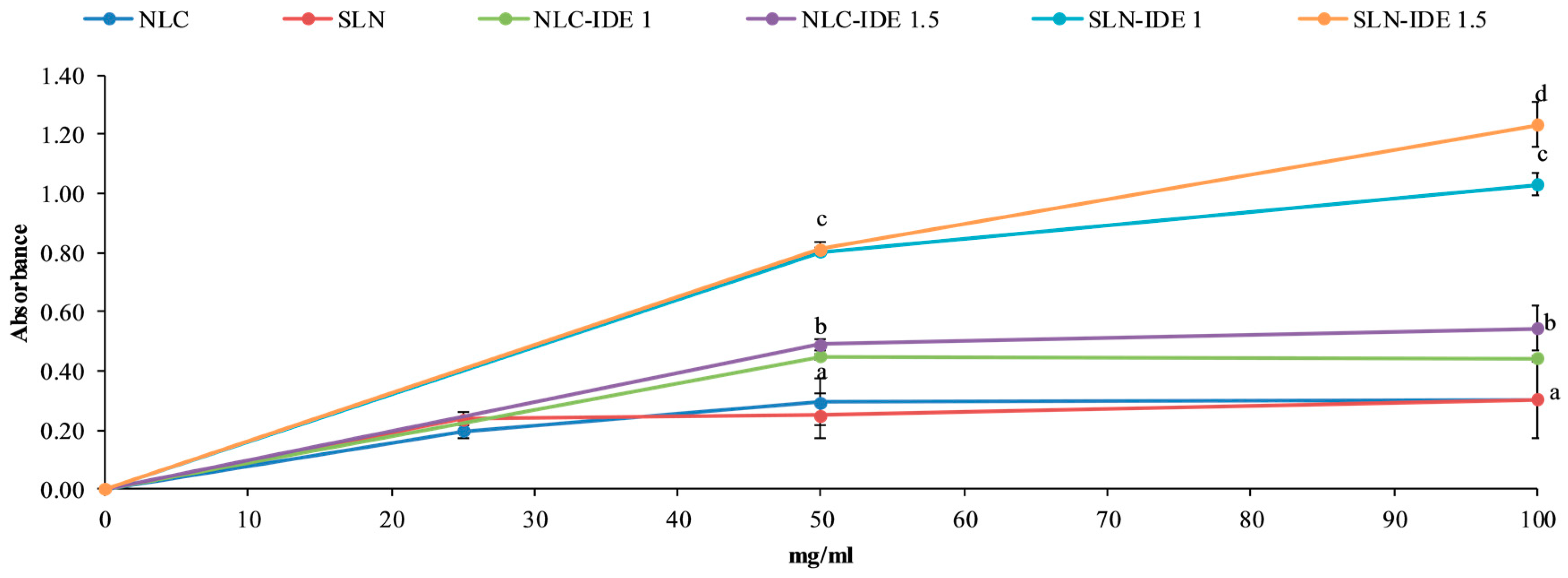
As shown in Table 2, unloaded and IDE-loaded lipid nanoparticles, prepared by the PIT method, showed small particle sizes (range: 23–42 nm), which were not significantly affected by IDE or VitE loading. PDI values were lower than 0.300, suggesting the presence of a homogeneous population of nanoparticles in the investigated samples. TEM analyses of the lipid nanocarriers under investigation showed roughly spherical nanoparticles (Figure 1). As this technique gave information only about the external surface of the nanocarrier, similar images were obtained for all nanoparticles. Therefore, we reported as an example in Figure 1 TEM pictures of only SLN IDE1 and NLC IDE1.
Although all these lipid nanoparticles showed low ζ-potential values (range: −1.56/−2.32 mV), they proved stable after storage for two months at room temperature as no change of particle size and PDI values were observed (data not shown). In addition, we did not observe any sign of drug precipitation during storage. Similar low ζ-potential vales were obtained in previous works on SLN with the same composition, and their good stability was attributed to their high PIT values [17,20,30].
IDE loading into SLN and NLC was determined as the maximum amount of drug that could be incorporated in the carriers leading to a clear colloidal system, as previously reported [17,20,30]. IDE loading into SLN and NLC was 1.5 and 2%, respectively. The higher IDE loading into NLC could be attributed to the presence of the oil component that could favor both IDE solubilization in the lipid matrix of the nanoparticles and a less ordered packing of the NLC core.
DSC analyses of unloaded and IDE-loaded SLN and NLC pointed out different calorimetric behaviors of these nanocarriers (Figure 2). As already reported in previous studies, unloaded SLN showed a transition temperature (Tm) at 42 °C that was about 12 °C lower than that of the bulk lipid (cetyl palmitate), thus indicating the solid nature of the lipid in the nanocarrier matrix [31,32,33]. Unloaded SLN had a crystallinity index (CI) of 66%, which is in accordance with the results obtained in previous studies [12,23,34].
In the thermogram of unloaded NLC, we observed a Tm value lower than that of unloaded SLN that suggested a lesser crystalline structure of the nanocarrier core in the presence of the liquid lipid VitE. As expected, unloaded NLC had a CI (59%) lower than that of unloaded SLN.
Loading 1% w/w IDE in these nanocarriers led to changes in the thermograms of the resulting lipid nanocarriers. In particular, a small shoulder at a temperature higher than Tm appeared in the DSC curve of SLN loaded with 1% w/w IDE, while the shoulder in the thermogram of unloaded NLC disappeared when 1% w/w IDE was loaded in these nanocarriers.
These results suggest that a stronger interaction of IDE with the lipid matrix of NLC is likely due to the presence of the liquid component VitE. Loading IDE 1% w/w into SLN and NLC reduced the CI of these nanocarriers (60% for SLN IDE1 and 55% for NLC IDE1). This behavior has already been reported in literature (after incorporating different active compounds in lipid nanocarriers) [12,35].
The incorporation of IDE 1.5% w/w into SLN and NLC provided DSC curves similar to those obtained from loading IDE 1% w/w (data not shown), and CI was not significantly affected for NLC but was lower (57%) for SLN, thus supporting the hypothesis that NLC allowed a better incorporation of IDE in the matrix of lipid nanoparticles.

3.2. Reducing Power

The reducing capacity of a compound indicates its potential antioxidant activity, as the presence of reductants (such as antioxidant substances) in the samples causes the reduction of the Fe3+/ferricyanide complex to the ferrous form. Therefore, Fe2+ can be monitored by measuring the formation of Perl’s Prussian blue at 700 nm [36]. Figure 3 depicts the reducing power of the different percentages of IDE loaded in SLN and NLC using the potassium ferricyanide reduction method, as reported in the literature [24]. The reducing power of SLN after loading 1% or 1.5% w/w IDE increased with the increase of the sample concentrations and showed a statistically significant greater value compared to IDE-loaded NLC (p < 0.05).

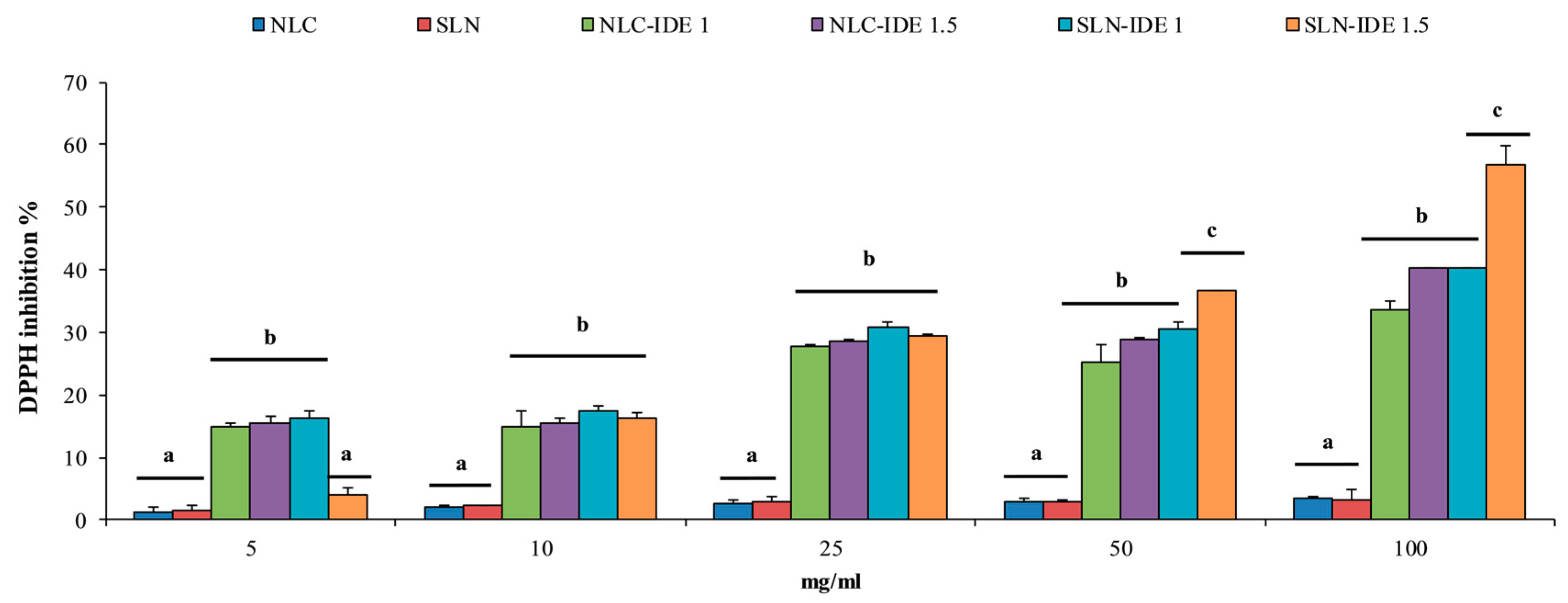
3.3. Free Radical Scavenging Activity

1,1-Diphenyl-2-picrylhydrazyl (DPPH) is a stable nitrogen-centered free radical that may convert into 1,1-diphenyl-2-picrylhydrazine, a diamagnetic molecule, upon one-electron reduction. DPPH has a strong absorption at 516 nm that decreases after the addition of reducing equivalents [37]. Experiments with sample concentrations in the range 5–100 mg/mL showed a decrease of DPPH absorbance (Figure 4); in particular, SLN IDE1.5 had the highest activity at 100 mg/mL that was significantly different from IDE-loaded NLC and SLN IDE1. The test showed greater antioxidant activity of IDE-loaded SLN compared to IDE-loaded NLC, thus confirming the results obtained with the potassium ferricyanide reduction method. The lower antioxidant activity of IDE-loaded NLC in comparison to IDE-loaded SLN could be due to the stronger interaction of IDE with the lipid matrix of NLC, which reduced IDE release from the nanoparticles and hence the antioxidant activity of the resulting colloidal system.

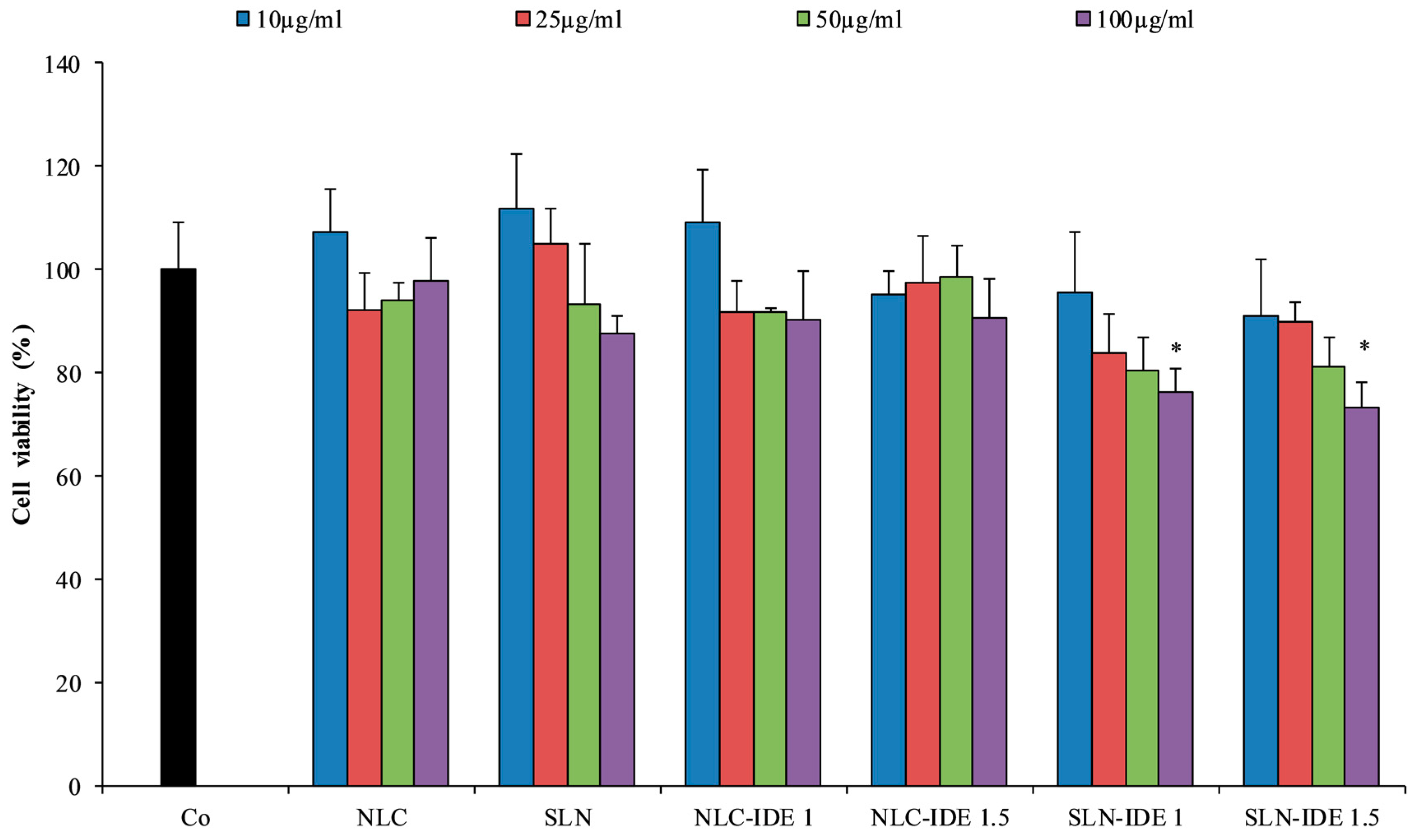
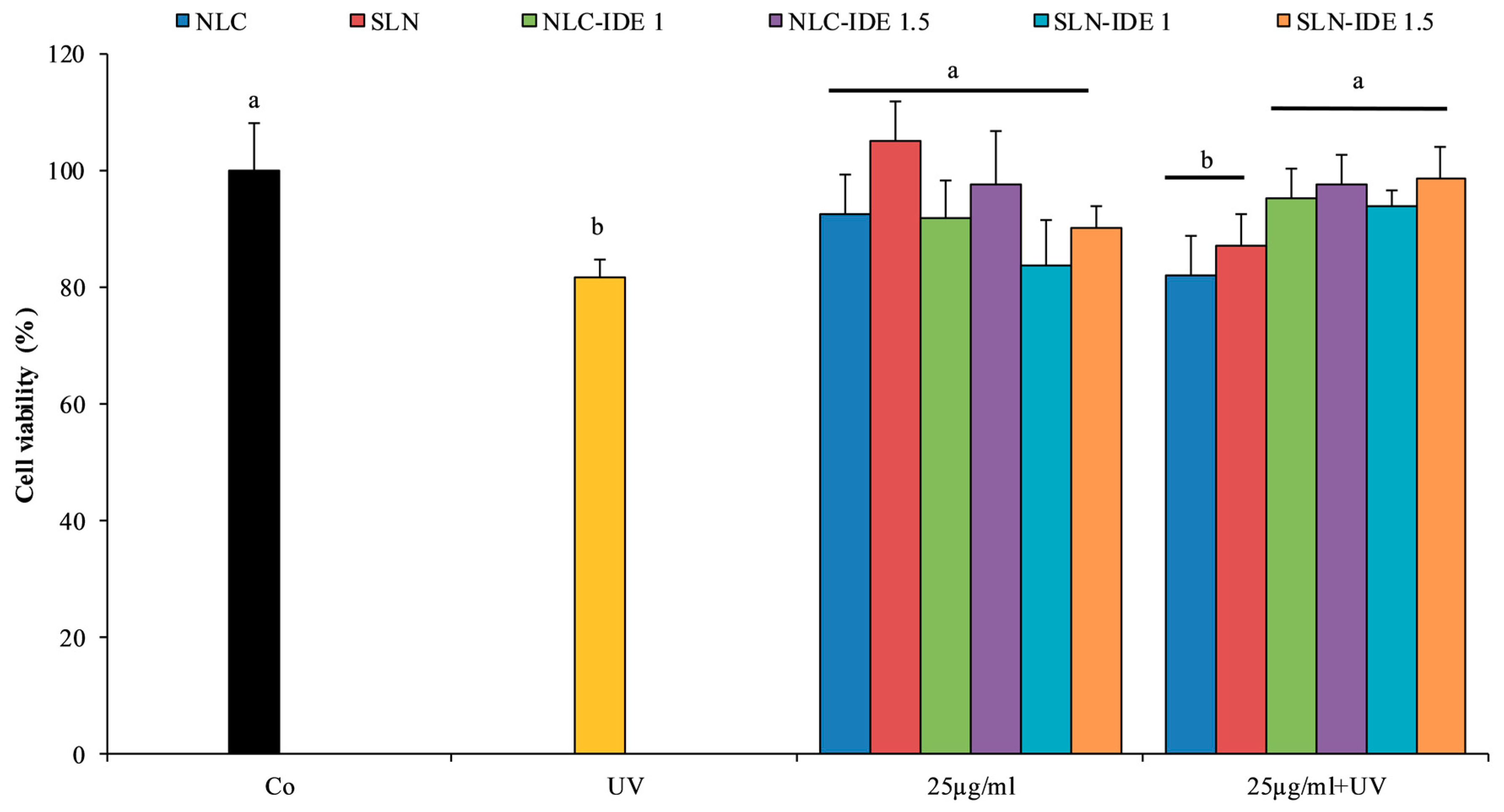
3.4. Evaluation of Cytotoxicity and Photo-Protective Effect in Fibroblast Cell Line HS-68

From a toxicological point of view, safety issues arise from nanomaterials with at least one dimension smaller than 100 nm; therefore, SLNs are generally regarded as safe nanocarriers as they commonly show particle sizes bigger than 100 nm and consist of well-tolerated ingredients [2,38]. As far as SLN topical application is concerned, Weyenberg et al. [39] reported that SLNs with positive surface charge or containing stearic acid as a solid lipid showed significant cytotoxicity on mouse fibroblast cells, HaCaT keratinocyte cells, or J774 macrophage cells used as model systems to evaluate skin toxicity. On the contrary, Ridolfi et al. [40] did not observe any cytotoxicity in fibroblasts and keratinocytes cells after treatment with SLN containing different lipid matrices.
In this work, we assessed the cytotoxic effects of the lipid nanocarriers under investigation containing different percentages of IDE on human skin HS68 fibroblast cells.
Figure 5 shows the response of fibroblast cells exposed to increasing concentrations of different IDE-loaded SLN and NLC. Results are expressed as a percentage of viable cells compared to the control. Cell viability evaluations on sample concentrations in the range 10–100 µg/mL revealed that the highest non-toxic concentration was 25 µg/mL, and for this reason, the experiment of induction of oxidative stress was carried out with non-toxic concentrations of the samples. Figure 6 shows that the exposure of cells to UV radiation, which were able to induce toxicity in this cell line in these experimental conditions [26], determined a significant decrease of vitality, as calculated by the MTT test (p < 0.05). On the contrary, treating the cells with 25 µg/mL of different IDE nanocarriers before the exposure to UV light resulted in a notable photoprotective effect, as cell viability increased in a dose-dependent manner compared to unprotected cells.

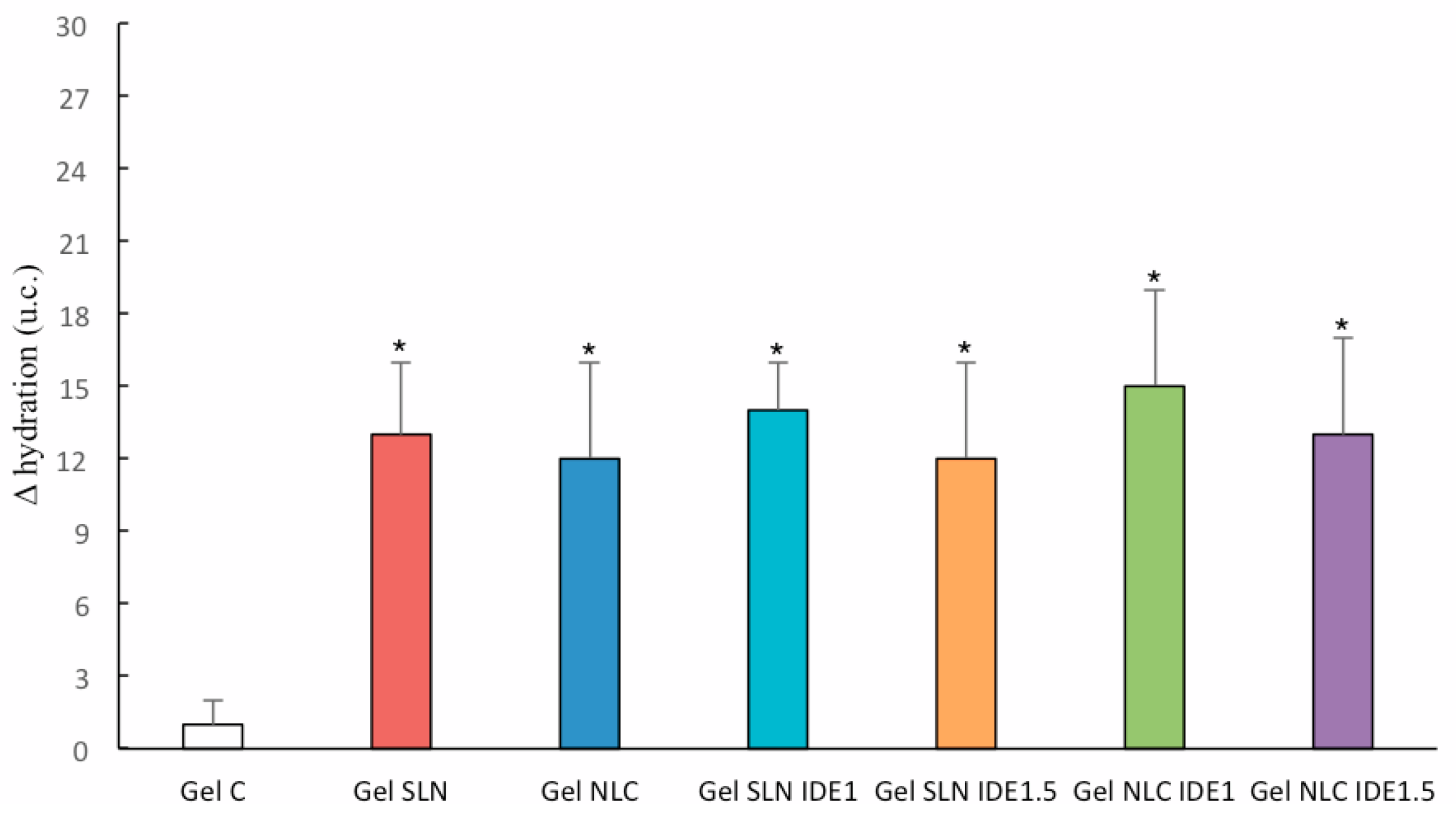
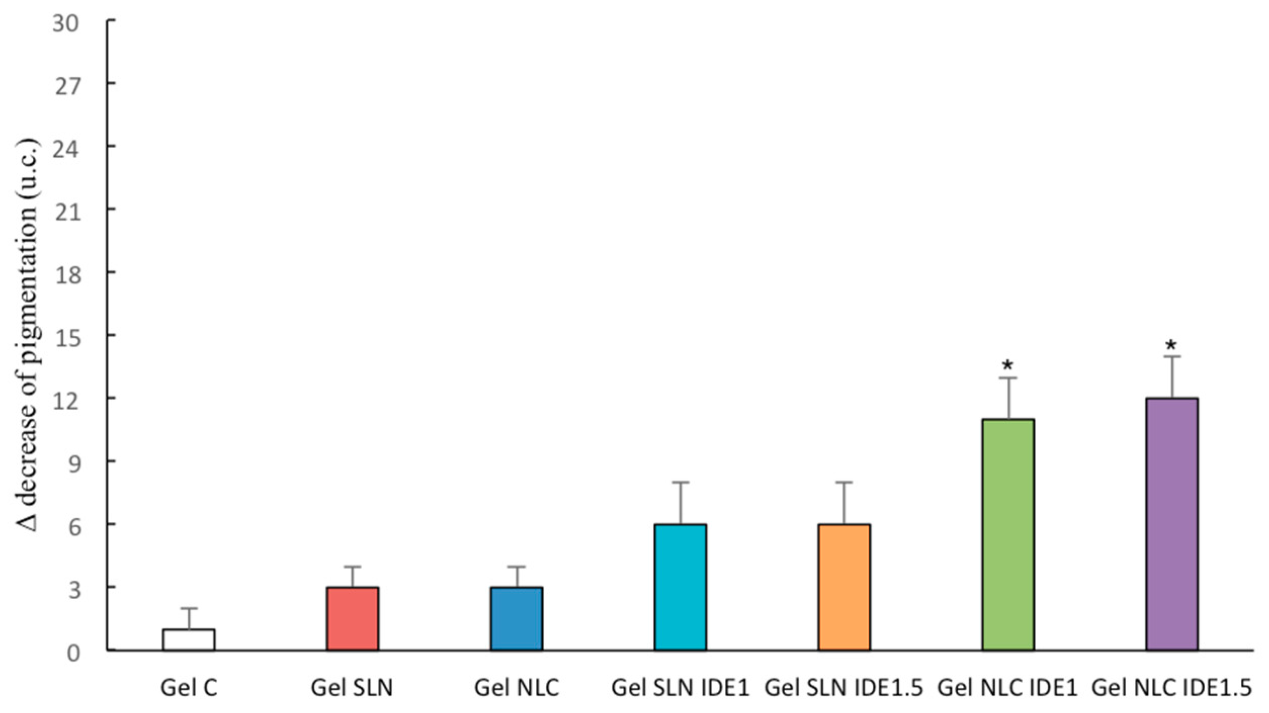
3.5. In Vivo Topical Effects

Topical application of gel formulations containing unloaded SLN and NLC on human volunteers resulted in an increase of skin hydration compared to the control vehicle (gel without lipid nanocarriers, Figure 7). These results were in accordance with previous studies performed on similar formulations and were attributed to the well-known occlusive properties of these lipid nanocarriers [11,12,41]. Loading different percentages of IDE into these lipid nanocarriers did not provide any significant increase of skin hydration compared to unloaded SLN and NLC.
On the contrary, the incorporation of IDE into SLN and NLC provided a decrease in skin pigmentation after treating human volunteers with gels containing such lipid nanocarriers for two weeks (Figure 8). In particular, both IDE-loaded SLN (SLN IDE1 and SLN IDE1.5) led to similar Δ pigmentation values that were higher than those obtained from applying the control gel or gels containing unloaded SLN and NLC. A further decrease of skin pigmentation was observed from loading IDE into NLC is likely due to the presence of tocopheryl acetate in these lipid nanocarriers as the photo-protective effects of this vitamin are well-known [42]. Other authors pointed out an increase of photo-protective efficacy in albino mice after loading IDE into NLC [43], thus supporting the hypothesis that NLC could be regarded as suitable carriers for the development of topical formulations with photo-protective activity. In our study, the in vivo topical effects of IDE-loaded SLN and NLC were not dependent on IDE concentration in these lipid nanocarriers. Therefore, we have planned further studies to investigate ID- loaded SLN and NLC interactions with skin tissue and the effects of lipid nanocarrier composition on the photo-protective activity of such nanocarriers.

4. Conclusions

IDE-loaded NLC containing tocopheryl acetate as the liquid lipid showed good technological properties (small particle sizes, low polydispersity indices, and good stability at room temperature). In vitro antioxidant assays (DPPH and reduction power) showed that, at the highest non-toxic concentration determined by the MTT test, the antioxidant activity of NLC containing different percentages of IDE (1.0 and 1.5 % w/w) was similar to that of SLN loaded with the same percentages of IDE. Analogously, similar photo-protective effects of IDE-loaded NLC and SLN were observed on HS68 fibroblast cells after exposure to UV light. The topical efficacy of these lipid nanocarriers loading IDE was evaluated on human volunteers from gel vehicles. The results of in vivo studies highlighted a greater photo-protective activity of IDE-loaded NLC compared to IDE-loaded SLN, thus suggesting a synergic effect between IDE and tocopheryl acetate. Therefore, co-loading IDE and tocopheryl acetate into NLC could be regarded as a promising approach to develop topical formulations with improved photo-protective efficacy.

Author Contributions

Conceptualization, L.M.; methodology, L.M., C.M.M., and A.S.; validation, R.T., L.P., and C.P.; formal analysis, C.M.M., S.M., R.A., R.T., L.P., and C.P.; investigation, L.S. and R.A.; resources, L.M., C.M.M., and A.S.; writing—original draft preparation, L.M., C.M.M., S.M., and R.A.; writing—review and editing, L.M., C.M.M., and A.S.; visualization, L.M.; supervision, L.M.; project administration, L.M.

Funding

This research received no external funding.

Conflicts of Interest

The authors declare no conflict of interest.

References

  1. Mehnert, W.; Mäder, K. Solid lipid nanoparticles: Production, characterization and applications. Adv. Drug Deliv. Rev. 2001, 47, 165–196. [Google Scholar] [CrossRef]
  2. Pardeike, J.; Hommoss, A.; Müller, RH. Lipid nanoparticles (SLN, NLC) in cosmetic and pharmaceutical dermal products. Int. J. Pharm. 2009, 366, 70–84. [Google Scholar] [CrossRef] [PubMed]
  3. Montenegro, L.; Lai, F.; Offerta, A.; Sarpietro, M.G.; Micicchè, L.; Maccioni, A.M.; Valenti, D.; Fadda, A.M. From nanoemulsions to nanostructured lipid carriers: A relevant development in dermal delivery of drugs and cosmetics. J. Drug Deliv. Sci. Technol. 2016, 32, 100–112. [Google Scholar] [CrossRef]
  4. Müller, R.H.; Mäder, K.; Gohla, S. Solid lipid nanoparticles (SLN) for controlled drug delivery—A review of the state of the art. Eur. J. Pharm. Biopharm. 2000, 50, 161–177. [Google Scholar] [CrossRef]
  5. Nair, R.; Arun Kumar, K.S.; Vishnu Priya, K.; Sevukarajan, M. Recent advances in solid lipid nanoparticle based drug delivery systems. J. Biomed. Sci. Res. 2011, 3, 368–384. [Google Scholar]
  6. Schäfer-Korting, M.; Mehnert, W.; Korting, H.C. Lipid nanoparticles for improved topical application of drugs for skin diseases. Adv. Drug Deliv. Rev. 2007, 59, 427–443. [Google Scholar] [CrossRef] [PubMed]
  7. Montenegro, L. Nanocarriers for skin delivery of cosmetic antioxidants. J. Pharm. Pharmacogn. Res. 2014, 2, 73–92. [Google Scholar]
  8. Dasgupta, S.; Ghosh, SK.; Ray, S.; Mazumder, B. Solid lipid nanoparticles (SLNs) gels for topical delivery of aceclofenac in vitro and in vivo evaluation. Curr. Drug. Deliv. 2013, 10, 656–666. [Google Scholar] [CrossRef] [PubMed]
  9. Khallaf, R.A.; Salem, H.F.; Abdelbary, A. 5-Fluorouracil shell-enriched solid lipid nanoparticles (SLN) for effective skin carcinoma treatment. Drug Deliv. 2016, 23, 3452–3460. [Google Scholar] [CrossRef] [PubMed]
  10. Madan, J.R.; Khude, P.A.; Dua, K. Development and evaluation of solid lipid nanoparticles of mometasone furoate for topical delivery. Int. J. Pharm. Investig. 2014, 4, 60–64. [Google Scholar] [CrossRef] [PubMed]
  11. Montenegro, L.; Pasquinucci, L.; Zappalà, A.; Chiechio, S.; Turnaturi, R.; Parenti, C. Rosemary Essential Oil-Loaded Lipid Nanoparticles: In Vivo Topical Activity from Gel Vehicles. Pharmaceutics 2017, 9, 48. [Google Scholar] [CrossRef] [PubMed]
  12. Montenegro, L.; Parenti, C.; Turnaturi, R.; Pasquinucci, L. Resveratrol-Loaded Lipid Nanocarriers: Correlation between In Vitro Occlusion Factor and In Vivo Skin Hydrating Effect. Pharmaceutics 2017, 9, 58. [Google Scholar] [CrossRef] [PubMed]
  13. Trombino, S.; Mellace, S.; Cassano, R. Solid lipid nanoparticles for antifungal drugs delivery for topical applications. Ther. Deliv. 2016, 7, 639–647. [Google Scholar] [CrossRef] [PubMed]
  14. Tupal, A.; Sabzichi, M.; Ramezani, F.; Kouhsoltani, M.; Hamishehkar, H. Dermal delivery of doxorubicin-loaded solid lipid nanoparticles for the treatment of skin cancer. J. Microencapsul. 2016, 33, 372–380. [Google Scholar] [CrossRef] [PubMed]
  15. Montenegro, L. Lipid-based nanoparticles as carriers for dermal delivery of antioxidants. Curr. Drug Metab. 2017, 18, 469–480. [Google Scholar] [CrossRef] [PubMed]
  16. Montenegro, L.; Turnaturi, R.; Parenti, C.; Pasquinucci, L. Idebenone: Novel strategies to improve its systemic and local efficacy. Nanomaterials 2018, 8, 87. [Google Scholar] [CrossRef] [PubMed]
  17. Montenegro, L.; Sinico, C.; Castangia, I.; Carbone, C.; Puglisi, G. Idebenone-loaded solid lipid nanoparticles for drug delivery to the skin: In vitro evaluation. Int. J. Pharm. 2012, 434, 169–174. [Google Scholar] [CrossRef] [PubMed]
  18. Thiele, J.J.; Ekanayake-Mudiyanselage, S. Vitamin E in human skin: Organ-specific physiology and considerations for its use in dermatology. Mol. Aspects Med. 2007, 2007 28, 646–667. [Google Scholar] [CrossRef]
  19. Keen, M.A.; Hassan, I. Vitamin E in dermatology. Indian Dermatol. Online J. 2016, 7, 311–315. [Google Scholar] [CrossRef] [PubMed]
  20. Montenegro, L.; Campisi, A.; Sarpietro, M.G.; Carbone, C.; Acquaviva, R.; Raciti, G.; Puglisi, G. In vitro evaluation of idebenone-loaded solid lipid nanoparticles for drug delivery to the brain. Drug Dev. Ind. Pharm. 2011, 37, 737–746. [Google Scholar] [CrossRef] [PubMed]
  21. Montenegro, L.; Trapani, A.; Latrofa, A.; Puglisi, G. In vitro evaluation on a model of blood brain barrier of idebenone-loaded solid lipid nanoparticles. J. Nanosci. Nanotechnol. 2012, 12, 330–337. [Google Scholar] [CrossRef] [PubMed]
  22. Radomska-Soukharev, A. Stability of lipid excipients in solid lipid nanoparticles. Adv. Drug Deliv. Rev. 2007, 59, 411–418. [Google Scholar] [CrossRef] [PubMed]
  23. Sarpietro, M.G.; Accolla, M.L.; Puglisi, G.; Castelli, F.; Montenegro, L. Idebenone loaded solid lipid nanoparticles: Calorimetric studies on surfactant and drug loading effects. Int. J. Pharm. 2014, 471, 69–74. [Google Scholar] [CrossRef] [PubMed]
  24. Falleh, H.; Ksouri, R.; Medini, F.; Guyot, S.; Abdelly, C.; Magné, C. Antioxidant activity and phenolic composition of the medicinal and edible halophyte Mesembryanthemum edule L. Ind. Crop. Prod. 2011, 34, 1066–1071. [Google Scholar] [CrossRef]
  25. Manuguerra, S.; Caccamo, L.; Mancuso, M.; Arena, R.; Rappazzo, A.C.; Genovese, L.; Santulli, A.; Messina, C.M.; Maricchiolo, G. The antioxidant power of horseradish, Armoracia rusticana, underlies antimicrobial and antiradical effects, exerted in vitro. Natl. Prod. Res. 2018. [Google Scholar] [CrossRef] [PubMed]
  26. Saija, A.; Tomaino, A.; Trombetta, D.; Pellegrino, M.L.; Tita, B.; Messina, C.; Bonina, F.P.; Rocco, C.; Nicolosi, G.; Castelli, F. In vitro antioxidant and photoprotective properties and interaction with model membranes of three new quercetin esters. Eur. J. Pharm. Biopharm. 2003, 56, 167–174. [Google Scholar] [CrossRef]
  27. Mossman, T. Rapid colorimetric assay for cellular growth and survival: Application to proliferation and cytotoxicity assays. J. Immunol. Methods 1983, 65, 55. [Google Scholar] [CrossRef]
  28. Montenegro, L.; Panico, A.M.; Santagati, L.M.; Siciliano, E.A.; Intagliata, S.; Modica, M.N. Solid lipid nanoparticles loading idebenone ester with pyroglutamic acid: In vitro antioxidant activity and in vivo topical efficacy. Nanomaterials 2019, 9, 43. [Google Scholar] [CrossRef] [PubMed]
  29. Underwood, A.J. Experiments in Ecology: Their Logical Design and Interpretation Using Analysis of Variance; Cambridge University Press: Cambridge, UK, 1997. [Google Scholar]
  30. Montenegro, L.; Modica, M.N.; Salerno, L.; Panico, A.M.; Crascì, L.; Puglisi, G.; Romeo, G. In vitro antioxidant activity of idebenone derivative-loaded solid lipid nanoparticles. Molecules 2017, 22, 887. [Google Scholar] [CrossRef] [PubMed]
  31. Montenegro, L.; Sarpietro, M.G.; Ottimo, S.; Puglisi, G.; Castelli, F. Differential scanning calorimetry studies on sunscreen loaded solid lipid nanoparticles prepared by the phase inversion temperature method. Int. J. Pharm. 2011, 415, 301–306. [Google Scholar] [CrossRef] [PubMed]
  32. Lee, M.K.; Lim, S.J.; Kim, C.K. Preparation, characterization and in vitro cytotoxicity of paclitaxel-loaded sterically stabilized solid lipid nanoparticles. Biomaterials 2007, 28, 2137–2146. [Google Scholar] [CrossRef] [PubMed]
  33. Montenegro, L.; Castelli, F.; Sarpietro, M.G. Differential scanning calorimetry analyses of idebenone-loaded solid lipid nanoparticles interactions with a model of bio-membrane: A comparison with in vitro skin permeation data. Pharmaceuticals 2018, 11, 138. [Google Scholar] [CrossRef] [PubMed]
  34. Montenegro, L.; Ottimo, S.; Puglisi, G.; Castelli, F.; Sarpietro, M.G. Idebenone loaded solid lipid nanoparticles interact with biomembrane models: Calorimetric evidence. Mol. Pharm. 2012, 9, 2534–2541. [Google Scholar] [CrossRef] [PubMed]
  35. zur Mühlen, A.; Schwarz, C.; Mehnert, W. Solid lipid nanoparticles (SLN) for controlled drug delivery—Drug release and release mechanism. Eur. J. Pharm. Biopharm. 1998, 45, 149–155. [Google Scholar] [CrossRef]
  36. Chung, Y.C.; Chang, C.T.; Chao, W.W.; Lin, C.F.; Chou, S.T. Antioxidative activity and safety of the 50% ethanolic extract from red bean fermented by Bacillus subtilis IMR-NK1. J. Agric. Food Chem. 2002, 50, 2454–2458. [Google Scholar] [CrossRef] [PubMed]
  37. Blois, M.S. Antioxidant determinations by the use of stable free radical. Nature 1958, 181, 1199–1200. [Google Scholar] [CrossRef]
  38. Doktorovova, S.; Souto, E.B.; Silva, A.M. Nanotoxicology applied to solid lipid nanoparticles and nanostructured lipid carriers—A systematic review of in vitro data. Eur. J. Pharm. Biopharm. 2014, 87, 1–18. [Google Scholar] [CrossRef] [PubMed]
  39. Weyenberg, W.; Filev, P.; Van den Plas, D.; Vandervoort, J.; De Smet, K.; Sollie, P.; Ludwig, A. Cytotoxicity of submicron emulsions and solid lipid nanoparticles for dermal application. Int. J. Pharm. 2007, 337, 291–298. [Google Scholar] [CrossRef] [PubMed]
  40. Ridolfi, D.M.; Marcato, P.D.; Machado, D.; Silva, R.A.; Justo, G.Z.; Duran, N. In vitro cytotoxicity assays of solid lipid nanoparticles in epithelial and dermal cells. J. Phys. Conf. Ser. 2012, 304, 012032. [Google Scholar] [CrossRef]
  41. Wissing, S.A.; Müller, R.H. The influence of solid lipid nanoparticles on skin hydration and viscoelasticity-in vivo study. Eur. J. Pharm. Biopharm. 2003, 56, 67–72. [Google Scholar] [CrossRef]
  42. Krol, E.S.; Kramer-Stickland, K.A.; Liebler, D.C. Photoprotective actions of topically applied vitamin E. Drug Metab. Rev. 2000, 32, 413–420. [Google Scholar] [CrossRef] [PubMed]
  43. Salunkhe, S.S.; Bhatia, N.M.; Pokharkar, V.B.; Thorat, J.D.; Bhatia, M.S. Topical delivery of Idebenone using nanostructured lipid carriers: Evaluations of sun-protection and anti-oxidant effects. J. Pharm. Investig. 2013, 43, 287–303. [Google Scholar] [CrossRef]
Figure 1. Transmissions electron microscopy (TEM) images of SLN IDE1 (a) and NLC IDE1 (b). IDE = idebenone, SLN = solid lipid nanoparticles, NLC = nanostructured lipid carriers.
Applsci 09 00845 g001
Figure 2. Calorimetric curves, in heating mode, of the following lipid nanocarriers: (a) unloaded solid lipid nanoparticles (SLN), SLN loaded with idebenone (IDE) 1% w/w (SLN IDE 1), SLN loaded with idebenone (IDE) 1.5% w/w (SLN IDE 1.5); (b) unloaded nanostructured lipid carriers (NLC), nanostructured lipid carriers loaded with IDE 1% w/w (NLC IDE 1), nanostructured lipid carriers loaded with IDE 1.5% w/w (NLC IDE 1.5).
Applsci 09 00845 g002
Figure 3. Reducing power of SLN, NLC, and samples loaded with 1% and 1.5% w/w of idebenone (IDE). The letters b, c, and d indicate a significant difference (p < 0.05) between samples loaded with different concentrations of IDE and the samples of unloaded NLC and SLN (indicated with the letter a). NLC = unloaded nanostructured lipid carrier; SLN = unloaded solid lipid nanoparticles; NLC-IDE 1 = NLC loaded with IDE 1% w/w; SLN-IDE 1 = SLN loaded with IDE 1% w/w; NLC-IDE 1.5 = NLC loaded with IDE 1.5% w/w; SLN-IDE 1.5 = SLN loaded with IDE 1.5% w/w.
Applsci 09 00845 g003
Figure 4. Antioxidant activity of IDE-loaded SLN and IDE-loaded NLC (5-100 mg/mL) indicated as percent (%) of DPPH radical inhibition. The letters b and c indicate, for each concentration, a significant difference (p < 0.05.) between the samples loaded with 1% or 1.5% w/w of idebenone (IDE) and the samples of unloaded SLN and NLC (indicated with the letter a). NLC = unloaded nanostructured lipid carrier; SLN = unloaded solid lipid nanoparticles; NLC-IDE 1 = NLC loaded with IDE 1% w/w; SLN-IDE 1 = SLN loaded with IDE 1% w/w; NLC-IDE 1.5 = NLC loaded with IDE 1.5% w/w; SLN-IDE 1.5 = SLN loaded with IDE 1.5% w/w.
Applsci 09 00845 g004
Figure 5. Effect of increased doses of idebenone (IDE)-loaded solid lipid nanoparticles (SLN) and nanostructured lipid carriers (NLC) on the percentage of vitality of HS68 fibroblast cells, determined by MTT (each data is presented as mean ± SD; n=12). Statistical analysis for the comparison: * p < 0.05 vs. Co. Co = control (no treatment), NLC = unloaded nanostructured lipid carrier, SLN = unloaded solid lipid nanoparticles, NLC-IDE 1 = NLC loaded with IDE 1% w/w; SLN-IDE 1 = SLN loaded with IDE 1% w/w; NLC-IDE 1.5 = NLC loaded with IDE 1.5% w/w, SLN-IDE 1.5 = SLN loaded with IDE 1.5% w/w.
Applsci 09 00845 g005
Figure 6. Effect of IDE-loaded SLN and NLC on the vitality percentage of HS68 fibroblast cells exposed to UV radiation: control cells not treated with compounds and not exposed to UV (Co); cells treated with UV radiation alone (UV); cells treated with 25 µg/mL of IDE-loaded SLN or NLC and not exposed to UV (25 µg/mL); cells pre-treated with IDE-loaded SLN or NLC (25µg/mL) and subsequently exposed to UV radiation (25 µg/mL + UV). Each data is presented as mean ± SD (n = 12). Different superscript letters indicate a significant difference compared to the control (p < 0.05).
Applsci 09 00845 g006
Figure 7. Differences in skin hydration values (Δ hydration) obtained after a two-week in vivo topical treatment with the gels under investigation. Statistical analysis for the comparison: * p < 0.05 vs. gel C. Gel C = control gel (without lipid nanocarriers), Gel SLN = gel containing unloaded solid lipid nanoparticles (SLN), Gel NLC = gel containing unloaded nanostructured lipid carriers, Gel SLN IDE1 = gel containing idebenone (IDE) 1% w/w loaded SLN, Gel IDE1.5 = gel containing IDE 1.5 % w/w loaded SLN, Gel NLC IDE1 = gel containing NLC loaded with IDE 1% w/w, Gel NLC IDE1.5 = gel containing NLC loaded with IDE 1.5 % w/w.
Applsci 09 00845 g007
Figure 8. Decrease of skin pigmentation values (Δ pigmentation) obtained after a two-week in vivo topical treatment with the gels under investigation. Statistical analysis for the comparison: * p < 0.05 vs. Gel C, Gel SLN, Gel NLC, Gel SLN IDE1, and Gel SLN IDE1.5. Gel C = control gel (without lipid nanocarriers), Gel SLN = gel containing unloaded solid lipid nanoparticles (SLN), Gel NLC = gel containing unloaded nanostructured lipid carriers, Gel SLN IDE1 = gel containing idebenone (IDE) 1% w/w loaded SLN, Gel IDE1.5 = gel containing IDE 1.5 % w/w loaded SLN, Gel NLC IDE1 = gel containing NLC loaded with IDE 1% w/w, Gel NLC IDE1.5 = gel containing NLC loaded with IDE 1.5 % w/w.
Applsci 09 00845 g008
Table 1. Composition of the oil phase of lipid nanoparticles. GO = glyceryl oleate; CP = cetyl palmitate; VitE = tocopheryl acetate, IDE = idebenone, SLN = solid lipid nanoparticles, NLC = nanostructured lipid carriers.
CodeIngredients (% w/w) *
Oleth-20GOCPVitEIDE
SLN9.005.007.00--
SLN IDE19.005.007.00-1.00
SLN IDE1.59.005.007.00-1.50
NLC 9.005.006.001.00-
NLC IDE19.005.006.001.001.00
NLC IDE1.59.005.006.001.001.50
* Water phase q.s. 100.00%
Table 2. Mean size (size), polydispersity index (PDI), zeta potential (ζ), and phase inversion temperature (PIT) of unloaded and IDE-loaded SLN and NLC. IDE = idebenone, SLN = solid lipid nanoparticles, NLC = nanostructured lipid carriers. Values are expressed as mean ± standard deviation (SD).
CodeSize ± SD (nm)PDI ± SDζ ± SD (mV)PIT (°C)
SLN36.40 ± 0.300.260 ± 0.010−1.56 ± 0.4878
SLN IDE123.55 ± 1.080.266 ± 0.023−2.02 ± 0.5469
SLN IDE 1.542.33 ± 0.290.230 ±0.031−2.01 ± 0.3377
NLC28.78 ± 0.900.235 ± 0.030−2.00 ± 0.2485
NLC IDE126.76 ± 0.330.178 ± 0.041−1.99 ± 0.3378
NLC IDE1.527.33 ± 0.550.198 ± 0.023−2.32 ± 0.6371

