The Detection of Long-Chain Bio-Markers Using Atomic Force Microscopy

Jet Propulsion Laboratory, California Institute of Technology, 4800 Oak Grove Dr., Pasadena, CA 91109, USA
Appl. Sci. 2019, 9(7), 1280; https://doi.org/10.3390/app9071280
Submission received: 15 February 2019
/
Revised: 14 March 2019
/
Accepted: 22 March 2019
/
Published: 27 March 2019
(This article belongs to the Special Issue Surfaced Enhanced Raman Scattering (SERS) in Disease Diagnosis)
Abstract
:The detection of long-chain biomolecules on mineral surfaces is presented using an atomic force microscope (AFM). This is achieved by using the AFM’s ability to manipulate molecules and measure forces at the pico-newton scale. We show that a highly characteristic force-distance signal is produced when the AFM tip is used to detach long-chain molecules from a surface. This AFM force spectroscopy method is demonstrated on bio-films, spores, fossils and mineral surfaces. The method works with AFM imaging and correlated tip enhanced infrared spectroscopy. The use of AFM force spectroscopy to detect this class of long chain bio-markers has applications in paleontology, life detection and planetary science.
1. Introduction
Scanning probe microscopes are fundamental tools in microscopy and nanotechnology. The most widely utilized of the probe microscopes is the atomic force microscope (AFM) that was first described in 1986 by Binnig, Gerber and Quate [1]. With an AFM, a tip mounted on a micro-fabricated cantilever is scanned over the surface and the interaction between the tip and the substrate is detected by monitoring the deflection of the cantilever [2]. These microscopes are remarkable for their ability to image individual atoms or molecules. Related AFM technologies have been developed that broadly include molecular manipulation, force measurement, and tip-mediated spectroscopy. The small size, low power requirements and non-vacuum operation make the AFM a suitable instrument for in situ planetary science missions. Consequently, AFM instruments have been sent to Mars aboard the Phoenix lander mission and an AFM is currently on the Rosetta comet mission [3,4].
The AFM cantilever-stylus mechanism is an inherently sensitive force sensor working down to the pico-newton force range. This force detection capability of the AFM has been harnessed to measure the interaction of the tip with surfaces and individual molecules. The force-distance signal of these AFM tip interactions is known as force spectroscopy (AFM-FS) and provides additional sensing capability to the AFM. This enables the AFM to measure localized hardness, friction and the study of molecular interactions with the AFM tip. In addition, the AFM tip-cantilever mechanism has been used as a localized photo-thermal detector for infrared spectroscopy and to mediate surface enhanced Raman spectroscopy.
In this work, the AFM is applied to the detection of long chain molecules on mineral surfaces. Long-chain molecules have been suggested as a class of biomarkers [5,6,7]. This includes long chain polymers such as intra-cellular polymers (proteins, nucleotides, polysaccharides) and extra-cellular bio-films. Using this rationale, the detection of long chain molecules provides evidence for biotic, fossilized and possible pre-biotic molecules—particularly when used in context with other analytical methods and sample history.
The basic concept for the detection of long chain molecules using AFM-FS is illustrated in Figure 1. The deflection of the AFM cantilever is monitored as a function of distance. As the tip approaches from a long distance, there is no force (deflection) on the cantilever. Once the tip is in proximity to the surface, there are short range van der Waal attractive (negative) forces on the tip and ultimately the cantilever is positively deflected when full contact is made with the surface. When in contact with the surface, molecules on the surface may adhere to the AFM tip [8]. As the tip is pulled away from the surface, molecular interactions produce adhesion forces on the tip until detachment of the molecule. The detachment is readily apparent in the force-distance plot. The presence of absorbed small molecules, such as water, produces comparatively short range forces and abrupt detachment in the force-distance curve.
A crucial aspect of this work is that long chain molecules will produce highly characteristic force-distance plots. The tip detachment signal is at significantly greater distances when long chain molecules are present on a mineral surface. These mineral surfaces can be planetary rocks, meteorites or fossils. This approach is an extension of previous work, where AFM-FS has been used to measure the length of purified polymers that have one end chemically tethered to the surface [9]. The AFM-FS has also been used to detect the forces associated with the unraveling protein polymers [10,11,12]. Force spectroscopy has also been used to measure the molecular weight distribution of synthetic polymers chemically attached (tethered) to a surface [13,14].
The use of AFM force spectroscopy has been expanded in this work to include non-tethered biomolecules on mineral surfaces without chemical surface preparation. While the precise molecular length is not directly measured without tethering the end of the molecule to the surface, the presence of long chain, high molecular weight molecules can be readily inferred. A molecular length distribution can be measured and the lower bound of the polymer length can be determined by the detachment length revealed in the force-distance plots. Long-chain polymers may show detachment signals that are hundreds of nanometers away from the surface depending on the molecular length. In contrast, mineral surfaces without such organic films are distinct and show relatively sharp, short-range detachment in the force-distance plots. This AFM-FS approach is demonstrated here to work with standard AFM instruments and tip enhanced spectroscopy. Bio-films, fossils and mineral surfaces were evaluated to demonstrate the use of an AFM-FS to discriminate biotic from abiotic residues on mineral surfaces.
2. Materials and Methods
The experiments were performed using a Digital Instruments Nanoscope 4 AFM (DI, Santa Barbara, CA, USA) and a Bruker Icon AFM system (Bruker, Santa Barbara, CA, USA). The AFM approach and retract sampling rates were 6300 nm/s. The force is a product of the deflection of cantilever and its spring constant. Silicon nitride cantilevers were used with spring constants in the range 0.14–0.16 N/m (Bruker FESP tip, 225 microns, 80 KHz). It should be noted the distance of the detachment signal from the surface is the measurement goal, the absolute detachment force is, therefore, not directly measured or precisely calibrated.
The spores samples consisted of Bacillus Atrophaeus (ATCC 9372, previously known as Bacillus Subtilis var. Niger) were purchased as suspensions of 1010 spores/ml (Raven Biological Laboratories, Omaha, NE, USA). The suspensions were diluted 1 to 100 in deionized water evaporated onto a quartz substrate. Samples of granular, lysed Chlorella Pyrenoidosa algae were obtained from Sun Chlorella (Torrance, CA, USA) and were dispersed in water and cast on quartz microscope slides. The test fossil was a Miocene age, selenitic domical stromatolite from evaporitic gypsum deposits formed about 5.33–7.25 million years ago. The stromatolite fossil was previously analyzed and shown to contain complex organic residues [15].
3. Results
3.1. Abiotic Mineral Surfaces
The discrimination of bio-polymer coatings and pure minerals was tested in a series of samples. A pure mineral was first run to show the force curve of a surface without biological materials present. As an analog of a clean mineral, Figure 2 shows the AFM-FS of a quartz surface. The AFM-FS force distance curve for quartz is qualitatively similar to other minerals observed by others [16]. This includes mica, basalt, palagonite, calcite, gypsum, glass and graphite. It should be noted that hydration layers may dominate the surface of many inorganic, minerals and this has been recently reviewed [17]. While the magnitude of detachment force is influenced by surface water layers, the detachment distance is relatively short range (<1 nm) relative to the chain length of bio-molecules.
3.2. Biological Samples
3.2.1. Spores and Algae
The biological samples tested include spore surfaces and lysed algae samples known to contain long chain organic residues. In order to test a biological surface with a complex set of proteins, a spore surface was first selected for analysis. Spores of Bacillus atrophaeus were deposited on quartz substrates from deionized water suspensions and evaporated under dry nitrogen. The proteinaceous surfaces of these spores were readily probed using AFM-FS. Detailed analysis of these surfaces was previously reported and number of morphogenetic proteins are known to be present in the outer spore coating [18,19]. For comparison, a lysed algae surface (Chlorella pyrenoidosa) was similarly examined using samples that were cast onto microscope slides. Figure 3 shows a comparison of the AFM-FS for the spore surface and algae samples with quartz as a reference. The presence of bio-polymers on the spore surface is clearly revealed in the force plot (Figure 3b) where the detachment distances are detected from 10 nanometers to greater than 100 nanometers away from the surface. The algae sample produces broad, long range force distance signals from detachment of a complex mixture of polymers (Figure 3c).
3.2.2. Analysis of a Fossil Sample Using Atomic Force Microscope-Force Spectroscopy (AFM-FS) and Tip-Enhanced Infrared Spectroscopy
A fossil sample was analyzed to illustrate how AFM-FS can be applied to detecting biomarkers in a mineral matrix. The test fossil, described in the experiment section, is a stromatolite that has morphological and chemical complexity formed by interactions between microorganisms, gypsum deposition, and gypsum crystal growth. In such structures the evidence for biological origins is apparent due to the presence of microfossils, the spatial distribution of organics, and the stromatolite morphology [15].
Since the AFM-FS mode interrogates a surface at one location at a time, it is useful to select areas that are likely to contain organics. To accomplish this microscopic site selection, the AFM’s optical targeting microscope is used to find regions that are amenable to AFM imaging and have morphological interest. In addition, AFM imaging using lateral force or phase contrast imaging can be used to localize soft organic coated surfaces [2]. As additional confirmation for the presence of organic residues, the AFM can be used to remove residues onto the AFM tip for enhanced infrared micro-analysis using methods previously described [20].
The stromatolite fossil was analyzed using successive steps in order to detect organics and bio-polymers: 1. Optical microscopy to target the location for AFM analysis; 2. AFM imaging with friction contrast modes to locate soft areas on the fossil; 3. AFM-FS to detect the presence of long chain polymers; 4. Correlated infrared spectroscopy for detection of organic functional groups removed onto the AFM tip.
Using the above targeting approach, Figure 4 shows AFM-FS plot from the surface of the stromatolite fossil. This provides evidence for long-chain polymers remaining in this fossil sample.
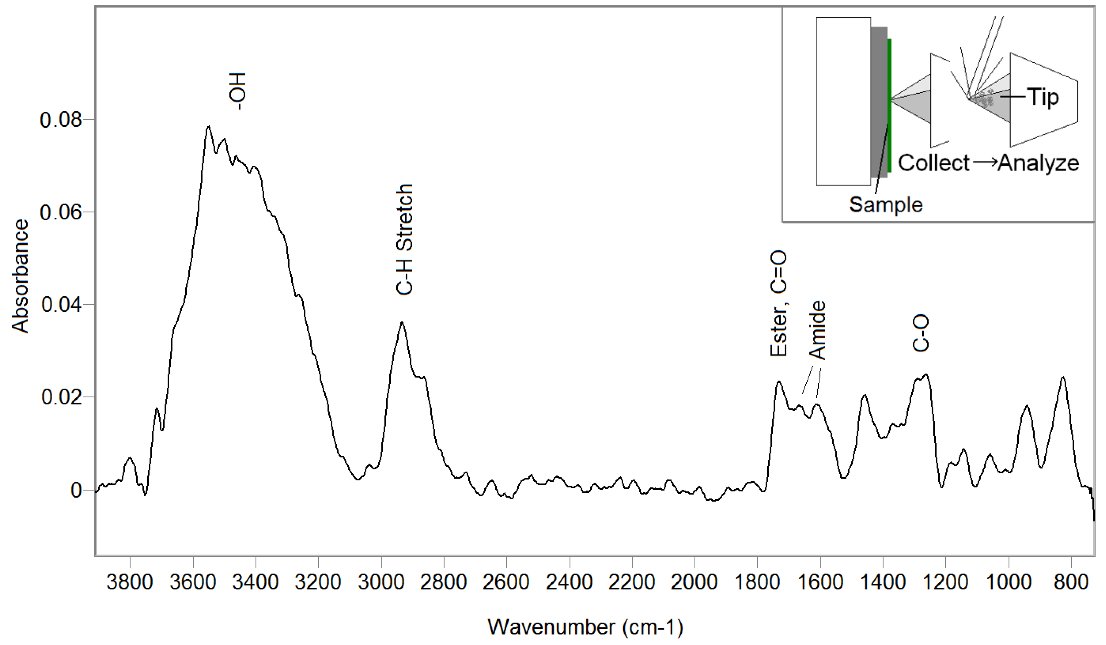
As further confirmation of the presence of bio-organic residue, Figure 5 shows the infrared spectrum of organic material removed from the fossil surfaces. This was collected onto the AFM tip from the same region that was probed using AFM-FS. The infrared spectrum was acquired from the AFM tip coated with the sample removed from the surface. The infrared spectrum shows functional groups including hydrocarbon, esters, amide I and amide II.
The evidence of proteinaceous residue is provided by both the AFM-FS and the amide bands in the infrared spectrum. The detachment length of the detected polymers is in the range of 10 nm to 160 nm, with a grouping in the 10 nm to 60 nm range. Dividing this length by the average length of 0.38 nm per amino acid and multiplying by an average molecular weight of 110 daltons per amino acid, gives a molecular weight range of approximately 2.9 to 45 kilodaltons detected on the fossil surface [14]. This provides qualitative evidence of bio-polymers on the surface. Quantitatively this is a measure of the lower bound of molecular length (or molecular weight) since the polymer attachment is at a random location along the molecular strand.
4. Discussion
The uniqueness of the force detachment signal for long chain organic polymers on mineral surfaces will now be considered. A potential interference could be from inorganic molecules that are linear, long chain, flexible, polymeric strands present in a mineral matrix. However there are very few naturally occurring inorganic materials with these properties. The most common naturally occurring, linear chain, inorganic polymer is elemental sulfur. However, below the melting point (114 °C) polymeric sulfur is unstable and forms of rhombic S8 [21]. Contamination is a problem with all trace organic detection methods. This is particularly true for the analysis of fossils or for detecting organics using spacecraft instruments. Experiments applying this new method for detecting long-chain bio-molecules need to include controls and sample blanks to assure organic residues were not added to the sample during collection.
The use of AFM-FS to study biomolecules that are not chemically attached (tethered) to a surface is a new approach that is useful for studying bio-molecules on fossils or planetary mineral surfaces. The distinctive molecular detachment signal adds a new feature for detecting signs life or pre-biotic compounds. The use of atomic force microscopy to analyze mineral surfaces for organic residues and long-chain bio-polymers has been demonstrated. A variety of long chain polymers on mineral surfaces have been analyzed. This includes spore surface proteins, algae and fossils containing known bio-organic residue. AFM force spectroscopy produces a unique force-curve that is indicative of the presence of polymer chains. This is quite distinct from abiotic mineral surfaces. This approach may be incorporated with other methods for additional chemical information. The tip-enhanced infrared spectroscopy of removed residues provides additional chemical information. It should be noted that a similar application of tip-enhanced Raman spectroscopy could be used to provide spectrochemical analysis [22].
The detection of the chemical signatures of life is a challenge for both in situ planetary exploration and for the analysis of terrestrial fossils [6]. Many schemes have been proposed ranging from the detection of specific chemistry found in terrestrial life to measuring general characteristics such as molecular chirality. A goal is to find markers for biological activity that are general enough without being obscured by abiotic background chemistry. Claims about detecting life or pre-biotic molecules in the oldest fossils, meteorites or in planetary environments are inherently controversial and require multiple levels of proof. Obviously, no single approach is definitive. The new method presented here is extremely sensitive (single molecule detection) and is relatively non-destructive so it may operate in a suite of other analytical methods.
AFM instruments have flown on two space missions, the Phoenix MECA experiment and the Rosetta MIDAS experiment. The implementation of AFM instruments equipped with force spectroscopy on future spacecraft missions may use existing designs with relatively minor modifications of the software to enable force spectroscopy measurements. This work demonstrates a new application of the AFM that is relevant to astrobiology missions. Lastly, this type of analysis in a laboratory setting could be useful for the analysis of fossils or meteorites.
Funding
The work described in this paper was carried out at the Jet Propulsion Laboratory, California Institute of Technology, through an agreement with the National Aeronautics and Space Administration, California Institute of Technology. Government sponsorship acknowledged © 2019 California Institute of Technology.
Acknowledgments
I would like to thank Abigail Allwood for providing the stromatolite sample.
Conflicts of Interest
The author declares no conflict of interest.
References
- Binnig, G.; Quate, C.F.; Gerber, C. Atomic force microscope. Phys. Rev. Lett. 1986, 56, 930. [Google Scholar] [CrossRef] [PubMed]
- Giessibl, F.J. Advances in atomic force microscopy. Rev. Mod. Phys. 2003, 75, 949. [Google Scholar] [CrossRef]
- Pike, W.T.; Staufer, U.; Hecht, M.H.; Goetz, W.; Parrat, D.; Sykulska-Lawrence, H.; Vijendran, S.; Madsen, M.B. Quantification of the dry history of the Martian soil inferred from in situ microscopy. Geophys. Res. Lett. 2011, 38. [Google Scholar] [CrossRef] [Green Version]
- Riedler, W.; Torkar, K.; Jeszenszky, H.; Romstedt, J.; Alleyne, H.S.; Arends, H.; Barth, W.; Biezen, J.V.; Butler, B.; Ehrenfreund, P.; et al. MIDAS–The micro-imaging dust analysis system for the Rosetta Mission. Space Sci. Rev. 2007, 128, 869–904. [Google Scholar] [CrossRef]
- Lovelock, J.E. A physical basis for life detection experiments. Nature 1965, 207, 568–570. [Google Scholar] [CrossRef] [PubMed]
- Georgiou, C.D.; Deamer, D.W. Lipids as universal biomarkers of extraterrestrial life. Astrobiology 2014, 14, 541–549. [Google Scholar] [CrossRef] [PubMed]
- Westall, F.; Steele, A.; Toporski, J.; Walsh, M.; Allen, C.; Guidry, S.; McKay, D.; Gibson, E.; Chafetz, H. Polymeric substances and biofilms as biomarkers in terrestrial materials: Implications for extraterrestrial samples. J. Geophys. Res. Planet. 2000, 105, 24511–24527. [Google Scholar] [CrossRef] [Green Version]
- Zlatanova, J.; Lindsay, S.M.; Leuba, S.H. Single molecule force spectroscopy in biology using the atomic force microscope. Prog. Biophys. Mol. Boil. 2000, 74, 37–61. [Google Scholar] [CrossRef]
- Hugel, T.; Seitz, M. The study of molecular interactions by AFM force spectroscopy. Macromol. Rapid Commun. 2001, 22, 989–1016. [Google Scholar] [CrossRef]
- Clausen-Schaumann, H.; Seitz, M.; Krautbauer, R.; Gaub, H.E. Force spectroscopy with single bio-molecules. Curr. Opin. Chem. Biol. 2000, 4, 524–530. [Google Scholar] [CrossRef]
- Broedel, S.E., Jr. The I27RS8 Protein as a Reference for Force Extension Experiments; thena Enzyme Systems™ Tech Bulletin #4; Athena Environmental Sciences, Inc.: Baltimore, MD, USA, 2014. [Google Scholar]
- Rief, M.; Gautel, M.; Oesterhelt, F.; Fernandez, J.M.; Gaub, H.E. Reversible unfolding of individual titin immunoglobulin domains by AFM. Science 1997, 276, 1109–1112. [Google Scholar] [CrossRef] [PubMed]
- Goodman, D.; Kizhakkedathu, J.N.; Brooks, D.E. Molecular weight and polydispersity estimation of adsorbing polymer brushes by atomic force microscopy. Langmuir 2004, 20, 3297–3303. [Google Scholar] [CrossRef] [PubMed]
- Carrion-Vazquez, M.; Marszalek, P.E.; Oberhauser, A.F.; Fernandez, J.M. Atomic force microscopy captures length phenotypes in single proteins. Proc. Natl. Acad. Sci. USA 1999, 96, 11288–11292. [Google Scholar] [CrossRef] [PubMed] [Green Version]
- Leite, F.L.; Riul, A.; Herrmann, P.S. Mapping of adhesion forces on soil minerals in air and water by atomic force spectroscopy (AFS). J. Adhes. Sci. Technol. 2003, 17, 2141–2156. [Google Scholar] [CrossRef]
- Zhang, Z.; Ryu, S.; Ahn, Y.; Jang, J. Molecular features of hydration layers probed by atomic force microscopy. Phys. Chem. Chem. Phys. 2018, 20, 30492–30501. [Google Scholar] [CrossRef] [PubMed]
- Plomp, M.; Leighton, T.J.; Wheeler, K.E.; Pitesky, M.E.; Malkin, A.J. Bacillus atrophaeus outer spore coat assembly and ultrastructure. Langmuir 2005, 21, 10710–10716. [Google Scholar] [CrossRef] [PubMed]
- Carroll, A.M.; Plomp, M.; Malkin, A.J.; Setlow, P. Protozoal digestion of coat-defective Bacillus subtilis spores produces “rinds” composed of insoluble coat protein. Appl. Environ. Microbiol. 2008, 74, 5875–5881. [Google Scholar] [CrossRef] [PubMed]
- Allwood, A.C.; Burch, I.W.; Rouchy, J.M.; Coleman, M. Morphological biosignatures in gypsum: Diverse formation processes of Messinian (~6.0 Ma) gypsum stromatolites. Astrobiology 2013, 13, 870–886. [Google Scholar] [CrossRef] [PubMed]
- Anderson, M.S. Infrared spectroscopy with an atomic force microscope. Appl. Spectrosc. 2000, 54, 349–352. [Google Scholar] [CrossRef]
- Raj, G. Advanced Inorganic Chemistry Vol-2; Krishna Prakashan Media: Meerut, Uttar Pradesh, India, 2010; p. 116. [Google Scholar]
- Anderson, M.S. Locally enhanced Raman spectroscopy with an atomic force microscope. Appl. Phys. Lett. 2000, 76, 3130–3132. [Google Scholar] [CrossRef]
Figure 1.
An illustration of how the force-displacement plot can be used to reveal the presence of long-chain molecules. If long-chain molecules are present, there is a characteristic detachment signal that can be hundreds of nanometers away from the surface depending on the molecular length.
Figure 1.
An illustration of how the force-displacement plot can be used to reveal the presence of long-chain molecules. If long-chain molecules are present, there is a characteristic detachment signal that can be hundreds of nanometers away from the surface depending on the molecular length.

Figure 2.
Atomic force microscope-force spectroscopy (AFM-FS) detachment plot of quartz showing the deflection of the cantilever as a function of distance. There is an abrupt force change when the tip is detached from the surface as it is withdrawn.
Figure 2.
Atomic force microscope-force spectroscopy (AFM-FS) detachment plot of quartz showing the deflection of the cantilever as a function of distance. There is an abrupt force change when the tip is detached from the surface as it is withdrawn.

Figure 3.
AFM-FS detachment plots comparing a clean mineral surface and complex biological surfaces: (a) quartz reference surface; (b) spore surface revealing the presence of multiple protein strands; (c) a lysed algae surface showing the presence of a complex mixture of polymeric strands.
Figure 3.
AFM-FS detachment plots comparing a clean mineral surface and complex biological surfaces: (a) quartz reference surface; (b) spore surface revealing the presence of multiple protein strands; (c) a lysed algae surface showing the presence of a complex mixture of polymeric strands.

Figure 4.
An AFM-FS detachment plot of a Miocene age selenitic domical stromatolite revealing the presence of a number of bio-polymer residues.
Figure 4.
An AFM-FS detachment plot of a Miocene age selenitic domical stromatolite revealing the presence of a number of bio-polymer residues.

Figure 5.
The infrared spectrum of the organic material from the fossil collected onto the AFM tip. This shows a mixture of bio-organic functional groups including hydrocarbon, esters, and amides.
Figure 5.
The infrared spectrum of the organic material from the fossil collected onto the AFM tip. This shows a mixture of bio-organic functional groups including hydrocarbon, esters, and amides.

© 2019 by the author. Licensee MDPI, Basel, Switzerland. This article is an open access article distributed under the terms and conditions of the Creative Commons Attribution (CC BY) license (http://creativecommons.org/licenses/by/4.0/).
Share and Cite
MDPI and ACS Style
Anderson, M.S. The Detection of Long-Chain Bio-Markers Using Atomic Force Microscopy. Appl. Sci. 2019, 9, 1280. https://doi.org/10.3390/app9071280
AMA Style
Anderson MS. The Detection of Long-Chain Bio-Markers Using Atomic Force Microscopy. Applied Sciences. 2019; 9(7):1280. https://doi.org/10.3390/app9071280
Chicago/Turabian StyleAnderson, Mark S. 2019. "The Detection of Long-Chain Bio-Markers Using Atomic Force Microscopy" Applied Sciences 9, no. 7: 1280. https://doi.org/10.3390/app9071280
Note that from the first issue of 2016, this journal uses article numbers instead of page numbers. See further details here.