
Molecular Properties of β-Carotene Oxygenases and Their Potential in Industrial Production of Vitamin A and Its Derivatives


1
Department of Integrative Bioscience and Biotechnology, Konkuk University, Seoul 05029, Korea
2
School of Biological Sciences and Technology, Chonnam National University, Gwangju 61186, Korea
3
Wild Plants Industrialization Research Division, Baekdudaegan National Arboretum, Bonghwa 36209, Korea
*
Authors to whom correspondence should be addressed.
Antioxidants 2022, 11(6), 1180; https://doi.org/10.3390/antiox11061180
Submission received: 10 May 2022
/
Revised: 11 June 2022
/
Accepted: 13 June 2022
/
Published: 16 June 2022
(This article belongs to the Special Issue 10th Anniversary of Antioxidants—Review Collection)
Abstract
:β-Carotene 15,15′-oxygenase (BCO1) and β-carotene 9′,10′-oxygenase (BCO2) are potential producers of vitamin A derivatives, since they can catalyze the oxidative cleavage of dietary provitamin A carotenoids to retinoids and derivative such as apocarotenal. Retinoids are a class of chemical compounds that are vitamers of vitamin A or are chemically related to it, and are essential nutrients for humans and highly valuable in the food and cosmetics industries. β-carotene oxygenases (BCOs) from various organisms have been overexpressed in heterogeneous bacteria, such as Escherichia coli, and their biochemical properties have been studied. For the industrial production of retinal, there is a need for increased production of a retinal producer and biosynthesis of retinal using biocatalyst systems improved by enzyme engineering. The current review aims to discuss BCOs from animal, plants, and bacteria, and to elaborate on the recent progress in our understanding of their functions, biochemical properties, substrate specificity, and enzyme activities with respect to the production of retinoids in whole-cell conditions. Moreover, we specifically propose ways to integrate BCOs into retinal biosynthetic bacterial systems to improve the performance of retinal production.
1. Introduction
Vitamin A, an essential nutrient for humans, is a fat-soluble natural precursor of signaling molecules and chromophores [1,2,3]. It belongs to the retinoid category, which is a class of chemical compounds that are either derivatives of vitamin A or are chemically related to it. Retinol, retinal, and retinoic acid are alcohol, aldehyde, and oxidized forms of vitamin A, respectively [4]. Vitamin A and its derivatives are used in pharmaceuticals, nutraceuticals, animal feed additives, functional foods, antioxidants, anti-aging, and anti-wrinkle cosmetic products [5,6,7]. Since humans cannot synthesize vitamin A, they obtain it only through diet [8]. Vitamin A can be synthesized from carotenoids, such as α-carotene, β-carotene, γ-carotene, β-cryptoxanthin, β-apo-carotenal, and β-apocarotenol, by carotenoid cleavage enzymes, including apo-carotenoid oxygenase (ACO), viviparous14 (VP14), neither inactivation nor after potential B (NinaB), and β-carotene oxygenase (BCO) [9,10]. Carotenoid cleavage enzymes constitute a family of enzymes that can metabolize carotenoids. They are classified into central cleavage enzymes and eccentric cleavage enzymes based on the position of asymmetric cleavage site. BCOs, which are known to play a key role in metabolizing carotenoids in vertebrates, are further classified into two types, namely β-carotene 15,15′-oxygenase (BCO1; EC 1.13.11.63) and β-carotene 9′,10′-oxygenases (BCO2; EC 1.14.99), based on the position of cleavage site on β-carotene (Figure 1). BCO1 is important as a retinoid producer, acting on the β-carotene 15,15′-double bond and producing two molecules of all-trans-retinal, while BCO2 acts on the β-carotene 9′,10′-double bond and produces ionone and apo-10′-carotenal (Figure 1), which can be further cleaved by BCO1 to produce other retinoids, such as retinaldehyde, retinol, and retinoic acid [8,11,12,13].
Retinoids, including retinal, retinol, and retinoic acid, are produced via chemical synthesis or enzymatic conversion. Chemical synthesis-based commercial production of retinal is performed by acidification/hydrolysis of retinal isomeric mixtures to produce retinol [4,14,15]. However, the chemical process has some disadvantages, such as involving multiple purification steps, by-product formation, and chemical waste formation [4]. On the other hand, the enzymatic conversion of β-carotene to retinal, using BCO, has some advantages, such as being an eco-friendly process and having reduced by-product generation due to the substrate-specificity of the enzyme. BCO is expressed in several tissues in humans, and its high enzymatic activity has been quantified in homogenates of liver and intestine [16,17,18,19,20]. In addition, BCOs from various organisms, such as animals, plants, and bacteria, have been reported as potential retinal producers (Table 1).
In this review, we summarize the biochemical properties and substrate specificities of BCOs and discuss the enzymatic bioconversion of β-carotene to retinal using these enzymes. Further, we propose several ways of efficient retinal production by bioengineering of BCOs and by optimizing the metabolic pathway responsible for retinal synthesis.
2. Biochemical Properties of BCO1 and BCO2
The characteristics of BCO1 and BCO2 from various organisms are summarized in Table 1. BCO1 derived from different sources (hereafter, BCO1s) are mainly found in animals, such as mammals [8,20,26,27,28,29,30,33,34,35], chicken [22,23,24,25], Caenorhabditis elegans [21,44], and uncultured marine bacterium [32]. BCO2 derived from different sources (hereafter, BCO2s) have been reported in a wide range of organisms, including animals [11,15,38,39,40], Saccharomyces cerevisiae [42], and plants such as apple [36], Arabidopsis thaliana [37], chrysanthemum [36], osmanthus [36], potato [41], rose [36], and saffron [43]. Most BCOs show optimal enzymatic activity at 28–40 °C and pH 7.4–8.0 (Table 1). However, human BCO1 shows optimal activity at a slightly acidic pH of 6.5 [27], and BCO2 from S. cerevisiae shows optimal activity at 45 °C [42].
The molecular weights of BCO1 subunits are approximately 60–65 kDa, and those of BCO2 subunits are approximately 50–64 kDa (Table 1). However, BCO1 derived from pig has a molecular weight of 156 kDa [34], and is larger in size than other BCOs. This result is not consistent with porcine genomes encoding BCO1 of about 65 kDa. On the contrary, the subunit of BCO1 derived from uncultured marine bacterium has the smallest molecular weight (38 kDa) among all BCOs, which is probably because BCO1 from uncultured marine bacterium is an enzyme belonging to bacterioopsin-related protein (Brp) or bacteriorhodopsin-related protein-like homolog protein (Blh), unlike other BCOs [32]. BCO1s show various associated forms, including monomers [23,27], dimers [21,32], and tetramers [24,30], whereas BCO2 from humans has been reported to exist only as a tetramer [15]. BCO1 from chicken exists as both a monomer and a tetramer, according to two different studies, even though it was expressed from the same gene. This might have been due to the synthesis of proteins by different host expression systems, such as baby hamster kidney (BHK) and Escherichia coli cells [23,24].
Metal ions play an important role in aiding the activities of several types of enzymes. Fe2+ has been suggested to affect the activity of BCO by binding to histidine residues at its active site [45,46]. Fe2+ has also been reported to have a strong impact on the activity of BCO1s and BCO2s. However, only BCO2 from S. cerevisiae showed an increase in activity with Ca2+ and a decrease in activity with Fe2+ [42]. Furthermore, this enzyme was not inhibited by EDTA treatment, suggesting that it is not a metalloprotein, unlike other BCOs.
3. Substrate Specificities of BCO1 and BCO62
BCOs exhibit various enzymatic properties with a large variation in activity depending on their origin, substrate, and the systems used for expression and purification. Therefore, based on quantitative comparison across BCOs, reported in different case studies, we have summarized the activities of BCO1s and BCO2s toward β-carotene and those with respect to various substrates, such as β-carotene, α-carotene, γ-carotene, lutein, β-cryptoxanthin, β-apo-4′-carotenal, β-apo-8′-carotenal, and β-apo-10′-carotenal, along with the relevant expression and purification methods (Table 2). Since the substrates of BCOs are generally very hydrophobic, their activity may depend on the substrate delivery methods such as detergent, solvent, and micellization. However, the difference in activity according to the substrate delivery method can be accurately compared under the same assay method. Therefore, in this review, only the values for activities are compared, except for a discussion of the substrate delivery methods.
The specific activity and catalytic efficiency (kcat/Km) of BCO1 derived from uncultured marine bacterium for β-carotene were the highest among all BCO1s [32] that were expressed in E. coli. BCO1 derived from chicken, and expressed in E. coli, showed a higher specific activity than that expressed in BHK cells, indicating that activity of BCO seems to be affected by enzyme expression conditions such as the expression vector and host cells. For example, under the pBAD system, BCO activity was approximately 20 times higher than under the pET system [24,25]. On the other hand, purified human-derived BCO1, expressed in SF9 cells showed a higher specific activity and catalytic efficiency than that expressed in E. coli cells [8,29]. BCO1s from chicken [24] and uncultured marine bacterium [32] had the highest kcat/Km values for β-carotene compared to the values for any other substrate, but BCO1 derived from human had the highest catalytic efficiency for lycopene [8]. BCO1 from uncultured marine bacterium had the highest kcat/Km value for β-carotene among all BCO1s [32]. Quantitative activities of purified BCO2s have only been reported in case of ferret [39], humans [15], and S. cerevisiae [42], which are fewer than reports on BCO1s. BCO2 from S. cerevisiae displayed the highest specific activity and kcat/Km for β-carotene than for other substrates [42].
Overall, BCOs derived from microorganisms, such as uncultured marine bacterium and S. cerevisiae, showed higher activity levels than those derived from animals, suggesting that they can be easily applied in biological engineering, with microbial cells as hosts, for the improvement of enzyme expression and performance in future.
4. Retinal Production by Biological Methods
Retinal is used by the food, cosmetics, and pharmaceutical industries. Its industrial production involves chemical synthesis via acid or base reduction, which has some disadvantages, such as use of expensive reagents, complex purification steps, generation of toxic chemical waste, and formation of undesired by-products [4]. Thus, efficient biological methods, using enzymes and microbial cells, for retinal production have been studied, considering that they are more cost-effective and environment-friendly than chemical methods [47,48,49,50].
Retinal production by enzymes or metabolically engineered cells is summarized in Table 3. BCO1 from various sources, such as mouse [51], human [52], chicken [24], fruit fly [53], uncultured marine bacterium [32], and pig [34], have been used for the enzymatic production of retinal. Among those, uncultured marine bacterium-derived BCO1 showed the highest retinal production through optimization of reaction conditions such as type and concentration of solvent and detergent for the formation of micelles, and concentrations of enzyme and β-carotene. This enzyme, which produced 181 mg/L retinal, was produced from 350 mg/L β-carotene at pH 8.0, 40 °C, 2.4% Tween 20 (w/v), and 15 U/mL enzyme with a conversion yield of 52% and a productivity of 9.1 mg/L/h [14]. On the other hand, chicken-derived BCO had the highest conversion yield (60%) at pH 8.0 and 37 °C in the presence of 0.64 mg/mL Tween80 and 0.2 U/mL enzyme, which produced 3.2 mg/L retinal production from 5.37 mg/L β-carotene with a productivity of 1.06 mg/L/h [24].
Retinal has also been produced by metabolically engineered cells such as E. coli and S. cerevisiae. To produce retinal using metabolically engineered E. coli, the methylerythritol 4-phosphate (MEP) pathway and mevalonate (MVA) pathway can be adopted with BCOs. Lee et al. produced up to 600 mg/L retinal using metabolically engineered E. coli [54], which is the highest retinal production among the engineered E. coli cells. Additionally, Zhang et al. developed a “plug-n-play” platform, which can simultaneously produce retinol, retinal, and various apocarotenoids by modifying apocarotenoid genetic module [58]. However, E. coli might not be the most desirable host cell for industrial production of retinal, owing to possible endotoxin contamination of the final product. The other candidate host, S. cerevisiae, is relatively superior for the industrial production of retinal due to easy genetic manipulation, robust compatibility for large-scale fermentation, and expression of the MVA pathway, which has a relatively high flux towards isoprenoids. Recently, Sun et al. developed an engineered S. cerevisiae with BCO1 to produce vitamin A, which consisted of retinal and retinol, via a xylose fed-batch fermentation [60]. The engineered S. cerevisiae produced 2094 mg/L retinal from xylose via β-carotene biosynthetic pathway with a productivity of 13.1 mg/L/h, which is the highest production of retinal in vitamin A synthesis [60], indicating that biological engineering systems can be used for sustainable bioprocesses for the industrial production of retinal using BCOs.
5. Proposed Roles of Amino Acids in BCOs
To identify the critical amino acid residues involved in BCO activity, structural analyses using homology modeling and site-directed mutagenesis have been performed. Recently, crystal structures of enzymes annotated as putative BCOs from C. elegans were reported; however, the enzymes did not exhibit any activity [21]. Moreover, a new crystal structure of the carotenoid cleavage enzyme from Candidatus Nitrosotalea devanaterra was determined recently [61]. However, this enzyme showed no activity toward bicyclic carotenoids such as β-carotene. Since the crystal structure of the active BCO enzyme has not been determined yet, a homology model of BCO1 was built based on the ACO crystal structure [45,62]. BCOs are metal-dependent enzymes, and most of them prefer Fe2+ for their enzymatic activity (except BCO1 from S. cerevisiae that prefers Ca2+). Fe2+ is coordinated by four strictly conserved His residues with three Glu residues, which involve the formation of second coordination sphere via hydrogen bonds (Figure 2, show the three-dimensional structure of BCO1 from C. elegans).
The critical residues in carotenoid cleavage enzymes are His and Glu, which are conserved the most of carotenoid cleavage enzymes [61]. The His and Glu residues in the predicted active site of BCO were mutated, resulting in loss of enzyme activity [45]. The four His residues, as metal-binding residues, were all conserved in the reported BCOs (Figure 3a), and in BCO1 from uncultured marine bacterium they were absolutely conserved as those of putative bacterial BCOs from Halobacterium sp., Halobacterium marismortui, and Halobacterium walsbyi [32] (Figure 3b). Kim et al. conducted site-directed mutagenesis for the His into Ala residues of uncultured marine bacterium (specific strain No. 66A03) in order to identify the role of these critical residues. Replacements of His21, His78, His188, and His192 by Ala showed significantly decreased activities (to below 5%) compared to the activity of wild-type enzyme [32]. On the other hand, one of the three Glu residues is replaced by Asp in some BCO2s from plants, such as apple, A. thaliana, chrysanthemum, osmanthus, and rose (Figure 3a), indicating that negatively charged amino acids may be involved in BCO activity.
The roles of other amino acid residues in BCO1 were suggested by Poliakov et al. They conducted mutagenesis to determine the role of aromatic residues (especially Tyr), which are the nearby acidic residues such as Glu, using the homology model of BCO1 from mouse that was built based on ACO structure [62]. Replacements of Tyr235 by Leu and Tyr326 by Glu showed decreased enzyme activity toward β-carotene, and the two Tyr residues were proposed to possibly be involved in enzyme stabilization via cation-π interactions between them, with the β-carotene cationic intermediate playing an important role in the reaction mechanism [62].
Despite the efforts to understand the roles of amino acid residues of BCO, only those involved in metal coordination and BCO stabilization have been reported so far. For industrial applications of retinal biosynthesis, BCOs should be further engineered, through the determination of new crystal structures and identification of critical residues for increased activity.
6. Future Perspectives of BCO in Synthetic Bacteria
Many challenges in retinal production have been addressed using metabolically engineered E. coli cells. Several aspects have been considered for the efficient production of carotenoid, which can be used as a substrate for BCOs, from glucose or glycerol via the endogenous MEP pathway and exogenous MVA pathway in metabolically engineered cells [63,64,65,66,67,68]. However, studies on the engineering of BCOs, which are crucial enzymes for catalyzing the last step of the retinal biosynthetic pathway, are still lacking. As mentioned above, BCOs have been successfully derived from animals, plants, and a few bacteria (Table 1), and need to be engineered to maximize the conversion of β-carotene to retinal. The catalytic properties of native BCOs can be improved via directed evolution, structure-based mutagenesis, and de-novo designing of the protein structure [69] (Figure 4). Directed evolution techniques require large libraries and efficient screening to select BCOs with the desired activity (Figure 4A). The DNA shuffling method can be used for genetic engineering of BCOs from different organisms to alter the conformation or orientation of the entire enzyme, including that of the active site, and obtain BCOs capable of efficient production of retinal. The library for DNA shuffling can be easily expanded by BCO genes from various organisms, which can efficiently obtain engineered BCOs with the desired activity [70] (Figure 4A). Structure-based mutagenesis, based on molecular modelling and rational considerations with respect to known structures of BCOs, can also be used to change the amino acid residues of the catalytic site (Figure 4B). Recently, advances in synthetic biology have enabled de-novo design and synthesis of proteins that have new functions and activities. Since the known BCO, expressed by microbial cells, lacks efficient and high-turnover retinal production, de-novo design will be a novel alternative to enhance the industrial production of retinal (Figure 4C). However, the designed components should be biocompatible with natural biochemical processes and pathways, and not interfere with cellular function [71].
Another engineering strategy for improving retinal production is the metabolon system, which can be considered for the optimization of upstream metabolic pathway for carotenoid bioconversion. Metabolons, which are complexes that use scaffold proteins to form multi-enzyme catalysts, have recently attracted much attention due to their ability for synergistic and simultaneous action of different types of enzymes in metabolic pathways (Figure 4D). They enhance metabolic flux using pushing and pulling mechanisms, which can also be applied to carotenoid production. Moreover, metabolons reduce metabolic burden by increasing the efficiency of intermediate transfer, in turn reducing intermediate toxicity and increasing product yield. Recently, affibody scaffolds formed via noncovalent interactions have been applied to improve sesquiterpene production in S. cerevisiae [72], and zinc-finger motifs on DNA scaffold systems have been used to improve the metabolic efficiency of the lycopene biosynthesis pathway [73]. The SpyTag/SpyCatcher system and its derivatives, which are covalently conjugated systems, have become the most versatile and widely used enzyme-assembly systems for diverse applications [74,75]. The design of cascade reactions using scaffolds can be applied to the MEP pathway for the efficient production of retinal. Furthermore, increasing the level of co-factors such as ATP and NADPH, which play important roles as donors of phosphate and electrons, respectively, are crucial for metabolic pathways, so that they can be used to increase the efficiency of enzymes involved in the MVA pathway for carotenoid production [76]. Levels of NADPH can be increased by enhancing metabolic flux through the pentose phosphate pathway via knock out of genes encoding glucose 6-phosphate isomerase and PEP carboxylase [77]. Increasing and replenishing the co-factor pools (NADPH, ATP, etc.) can be an effective strategy to enhance the rate of retinal biosynthesis, since they form the major intracellular energy source for many biological reactions (Figure 4E).
Finally, the construction and culture of BCO-driven retinal-producing strains are still in their early stages. Thus, further optimization of fermentation operations (such as fed-batch cultivation) and media, and improvement of strains, may increase the current titer of retinal produced by E. coli. Advances in enzymatic engineering and metabolically engineered cells have already provided many advantages in industrial processes, but there are still few attempts to utilize BCOs for the retinal production. We expect future studies in the design and engineering of BCOs, and the associated pathways will accelerate improvements in retinal production on an industrial scale process.
7. Conclusions
BCOs are important biocatalysts in the production of retinal. The current review summarizes the biochemical properties and substrate specificities of BCOs and the production of retinal using these enzymes. Retinal can be produced from metabolically engineered cells via enhancing the MVA or MEP pathway. BCOs are the final enzymes in retinal production. Thus, they should be engineered to improve their enzymatic performance. In this review, we propose several ways of enzyme engineering of BCOs and optimization of the metabolic pathway responsible for retinal synthesis. Bioengineering of BCOs can be the key to high-level retinal production. Furthermore, engineered cells, and an improved via synthetic biological approach, should be developed for the industrial production of retinal.
Author Contributions
Conceptualization, K.-C.S., M.-J.S., Y.-S.K. and S.-J.Y.; data curation, K.-C.S., M.-J.S., Y.-S.K. and S.-J.Y.; funding acquisition, K.-C.S., M.-J.S., Y.-S.K. and S.-J.Y.; project administration, K.-C.S., M.-J.S., Y.-S.K. and S.-J.Y.; writing—original draft, K.-C.S., M.-J.S., Y.-S.K. and S.-J.Y.; writing—reviewing and editing, K.-C.S., M.-J.S., Y.-S.K. and S.-J.Y. All authors have read and agreed to the published version of the manuscript.
Funding
This paper was supported by the Enzyme engineering for next generation biorefinery (NRF-2022M3J5A1056169 and NRF-2022M3J5A1085239) from National Research Foundation (NRF) and NRF grant (NRF-2020R1C1C1004178 and NRF-2022R1C1C2003774) supported by the Korean Ministry of Science and ICT.
Acknowledgments
This paper was supported by the KU Research Professor Program of Konkuk University.
Conflicts of Interest
The authors declare no conflict of interest.
References
- Blaner, W.S. Chapter 5—Vitamin A and Provitamin A Carotenoids. Present Knowledge in Nutrition, 11th ed.; Wiley-Blackwell: Cambridge, MA, USA, 2020; pp. 73–91. [Google Scholar]
- Corbo, J.C. Vitamin A1/A2 chromophore exchange: Its role in spectral tuning and visual plasticity. Dev. Biol. 2021, 475, 145–155. [Google Scholar] [CrossRef] [PubMed]
- Carazo, A.; Macakova, K.; Matousova, K.; Krcmova, L.K.; Protti, M.; Mladenka, P. Vitamin A Update: Forms, Sources, Kinetics, Detection, Function, Deficiency, Therapeutic Use and Toxicity. Nutrients 2021, 13, 1703. [Google Scholar] [CrossRef] [PubMed]
- Kim, Y.S.; Oh, D.K. Biotransformation of carotenoids to retinal by carotenoid 15,15′-oxygenase. Appl. Microbiol. Biotechnol. 2010, 88, 807–816. [Google Scholar] [CrossRef] [PubMed]
- Fisher, G.J.; Voorhees, J.J. Molecular mechanisms of retinoid actions in skin. FASEB J. 1996, 10, 1002–1013. [Google Scholar] [CrossRef] [Green Version]
- De Luca, L.M. Retinoids and their receptors in differentiation, embryogenesis, and neoplasia. FASEB J. 1991, 5, 2924–2933. [Google Scholar] [CrossRef]
- Semba, R.D. Vitamin A as “anti-infective” therapy, 1920–1940. J. Nutr. 1999, 129, 783–791. [Google Scholar] [CrossRef] [Green Version]
- Dela Sena, C.; Narayanasamy, S.; Riedl, K.M.; Curley, R.W., Jr.; Schwartz, S.J.; Harrison, E.H. Substrate specificity of purified recombinant human β-carotene 15,15′-oxygenase (BCO1). J. Biol. Chem. 2013, 288, 37094–37103. [Google Scholar] [CrossRef] [Green Version]
- Sui, X.; Kiser, P.D.; Lintig, J.; Palczewski, K. Structural basis of carotenoid cleavage: From bacteria to mammals. Arch. Biochem. Biophys. 2013, 539, 203–213. [Google Scholar] [CrossRef] [Green Version]
- Babino, D.; Golczak, M.; Kiser, P.D.; Wyss, A.; Palczewski, K.; von Lintig, J. The Biochemical Basis of Vitamin A3 Production in Arthropod Vision. ACS Chem. Biol. 2016, 11, 1049–1057. [Google Scholar] [CrossRef] [Green Version]
- Kiefer, C.; Hessel, S.; Lampert, J.M.; Vogt, K.; Lederer, M.O.; Breithaupt, D.E.; von Lintig, J. Identification and characterization of a mammalian enzyme catalyzing the asymmetric oxidative cleavage of provitamin A. J. Biol. Chem. 2001, 276, 14110–14116. [Google Scholar] [CrossRef] [Green Version]
- Amengual, J.; Widjaja-Adhi, M.A.K.; Rodriguez-Santiago, S.; Hessel, S.; Golczak, M.; Palczewski, K.; von Lintig, J. Two carotenoid oxygenases contribute to mammalian provitamin A metabolism. J. Biol. Chem. 2013, 288, 34081–34096. [Google Scholar] [CrossRef] [PubMed] [Green Version]
- Jin, Y.; Yu, Y.; Zhang, C.; Li, S.; Zhang, X.; Li, F. Characterization and function analysis of the β-carotene oxygenase-like genes in carotenoids metabolism of the ridgetail white prawn Exopalaemon carinicauda. Front. Physiol. 2020, 11, 745. [Google Scholar] [CrossRef]
- Kim, Y.S.; Park, C.S.; Oh, D.K. Retinal production from β-carotene by β-carotene 15,15′-dioxygenase from an unculturable marine bacterium. Biotechnol. Lett. 2010, 32, 957–961. [Google Scholar] [CrossRef] [PubMed]
- Kim, Y.S.; Yeom, S.J.; Oh, D.K. Production of β-apo-10′-carotenal from β-carotene by human β-carotene-9′,10′-oxygenase expressed in E. coli. Biotechnol. Lett. 2011, 33, 1195–1200. [Google Scholar] [CrossRef]
- Olson, J.A.; Hayaishi, O. The enzymatic cleavage of β-carotene into vitamin A by soluble enzymes of rat liver and intestine. Proc. Natl. Acad. Sci. USA 1965, 54, 1364–1370. [Google Scholar] [CrossRef] [Green Version]
- Goodman, D.S.; Huang, H.S. Biosynthesis of Vitamin a with Rat Intestinal Enzymes. Science 1965, 149, 879–880. [Google Scholar] [CrossRef]
- Lakshmanan, M.R.; Chansang, H.; Olson, J.A. Purification and properties of carotene 15,15′-dioxygenase of rabbit intestine. J. Lipid Res. 1972, 13, 477–482. [Google Scholar] [CrossRef]
- Devery, J.; Milborrow, B.V. β-Carotene-15,15′-dioxygenase (EC 1.13.11.21) isolation reaction mechanism and an improved assay procedure. Br. J. Nutr. 1994, 72, 397–414. [Google Scholar] [CrossRef] [Green Version]
- During, A.; Nagao, A.; Hoshino, C.; Terao, J. Assay of β-carotene 15,15′-dioxygenase activity by reverse-phase high-pressure liquid chromatography. Anal. Biochem. 1996, 241, 199–205. [Google Scholar] [CrossRef]
- Pan, W.M.; Zhou, Y.L.; Wang, J.; Dai, H.E.; Wang, X.; Liu, L. Structural and functional analysis of nonheme iron enzymes BCMO-1 and BCMO-2 from Caenorhabditis elegans. Front. Mol. Biosci. 2022, 9, 844453. [Google Scholar] [CrossRef]
- Wyss, A.; Wirtz, G.; Woggon, W.; Brugger, R.; Wyss, M.; Friedlein, A.; Bachmann, H.; Hunziker, W. Cloning and expression of β,β-carotene 15,15′-dioxygenase. Biochem. Biophys. Res. Commun. 2000, 271, 334–336. [Google Scholar] [CrossRef] [PubMed]
- Wyss, A.; Wirtz, G.M.; Woggon, W.D.; Brugger, R.; Wyss, M.; Friedlein, A.; Riss, G.; Bachmann, H.; Hunziker, W. Expression pattern and localization of β,β-carotene 15,15′-dioxygenase in different tissues. Biochem. J. 2001, 354, 521–529. [Google Scholar] [CrossRef] [PubMed]
- Kim, Y.S.; Oh, D.K. Substrate specificity of a recombinant chicken β-carotene 15,15′-monooxygenase that converts β-carotene into retinal. Biotechnol. Lett. 2009, 31, 403–408. [Google Scholar] [CrossRef]
- Garcia-Lopez, E.; Gonzalez-Gallardo, A.; Antaramian, A.; Gonzalez-Davalos, M.L.; Shimada, A.; Varela-Echavarria, A.; Mora, O. In vitro conversion of β-carotene to retinal in bovine rumen fluid by a recombinant β-carotene-15, 15′-monooxygenase. Int. J. Vitam. Nutr. Res. 2012, 82, 94–103. [Google Scholar] [CrossRef] [PubMed]
- Singh, H.; Cama, H.R. Enzymatic cleavage of carotenoids. Biochim. Biophys. Acta 1974, 370, 49–61. [Google Scholar] [CrossRef]
- Yan, W.; Jang, G.F.; Haeseleer, F.; Esumi, N.; Chang, J.; Kerrigan, M.; Campochiaro, M.; Campochiaro, P.; Palczewski, K.; Zack, D.J. Cloning and characterization of a human β,β-carotene-15,15′-dioxygenase that is highly expressed in the retinal pigment epithelium. Genomics 2001, 72, 193–202. [Google Scholar] [CrossRef]
- dela Sena, C.; Riedl, K.M.; Narayanasamy, S.; Curley, R.W.; Schwartz, S.J.; Harrison, E.H. The human enzyme that converts dietary provitamin A carotenoids to vitamin A is a dioxygenase. J. Biol. Chem. 2014, 289, 13661–13666. [Google Scholar] [CrossRef] [Green Version]
- Kowatz, T.; Babino, D.; Kiser, P.; Palczewski, K.; von Lintig, J. Characterization of human β,β-carotene-15,15′-monooxygenase (BCMO1) as a soluble monomeric enzyme. Arch. Biochem. Biophys. 2013, 539, 214–222. [Google Scholar] [CrossRef] [Green Version]
- Lindqvist, A.; Andersson, S. Biochemical properties of purified recombinant human β-carotene 15,15′-monooxygenase. J. Biol. Chem. 2002, 277, 23942–23948. [Google Scholar] [CrossRef] [Green Version]
- Kelly, M.E.; Ramkumar, S.; Sun, W.; Colon Ortiz, C.; Kiser, P.D.; Golczak, M.; von Lintig, J. The Biochemical Basis of Vitamin A Production from the Asymmetric Carotenoid β-Cryptoxanthin. ACS Chem. Biol. 2018, 13, 2121–2129. [Google Scholar] [CrossRef]
- Kim, Y.S.; Kim, N.H.; Yeom, S.J.; Kim, S.W.; Oh, D.K. In vitro characterization of a recombinant Blh protein from an uncultured marine bacterium as a β-carotene 15,15′-dioxygenase. J. Biol. Chem. 2009, 284, 15781–15793. [Google Scholar] [CrossRef] [PubMed] [Green Version]
- Redmond, T.M.; Gentleman, S.; Duncan, T.; Yu, S.; Wiggert, B.; Gantt, E.; Cunningham, F.X., Jr. Identification, expression, and substrate specificity of a mammalian β-carotene 15,15′-dioxygenase. J. Biol. Chem. 2001, 276, 6560–6565. [Google Scholar] [CrossRef] [Green Version]
- Yu, D.; Zhang, R.; Wang, Y.; Zou, D.; Li, T.; Tang, H.; Jiang, L.; Wang, L. Purification of β-carotene 15,15′-monooxygenase from pig intestine and its enzymatic hydrolysis of pigment in soybean oil. Int. J. Food Sci. 2019, 54, 480–489. [Google Scholar] [CrossRef]
- Lakshman, M.R.; Okoh, C. Enzymatic conversion of all-trans-β-carotene to retinal. Methods Enzymol. 1993, 214, 256–269. [Google Scholar] [PubMed]
- Huang, F.C.; Molnar, P.; Schwab, W. Cloning and functional characterization of carotenoid cleavage dioxygenase 4 genes. J. Exp. Bot. 2009, 60, 3011–3022. [Google Scholar] [CrossRef] [PubMed] [Green Version]
- Holger, S.; Kurtzer, R.; Eisenreich, W.; Schwab, W. The carotenase AtCCD1 from Arabidopsis thaliana is a dioxygenase. J. Biol. Chem. 2006, 281, 9845–9851. [Google Scholar]
- Dela Sena, C.; Sun, J.; Narayanasamy, S.; Riedl, K.M.; Yuan, Y.; Curley, R.W., Jr.; Schwartz, S.J.; Harrison, E.H. Substrate specificity of purified recombinant chicken β-carotene 9′,10′-oxygenase (BCO2). J. Biol. Chem. 2016, 291, 14609–14619. [Google Scholar] [CrossRef] [Green Version]
- Hu, K.Q.; Liu, C.; Ernst, H.; Krinsky, N.I.; Russell, R.M.; Wang, X.D. The biochemical characterization of ferret carotene-9′,10′-monooxygenase catalyzing cleavage of carotenoids in vitro and in vivo. J. Biol. Chem. 2006, 281, 19327–19338. [Google Scholar] [CrossRef] [Green Version]
- Lindqvist, A.; He, Y.G.; Andersson, S. Cell type-specific expression of β-carotene 9′,10′-monooxygenase in human tissues. J. Histochem. Cytochem. 2005, 53, 1403–1412. [Google Scholar] [CrossRef]
- Bruno, M.; Beyer, P.; Al-Babili, S. The potato carotenoid cleavage dioxygenase 4 catalyzes a single cleavage of β-ionone ring-containing carotenes and non-epoxidated xanthophylls. Arch. Biochem. Biophys. 2015, 572, 126–133. [Google Scholar] [CrossRef]
- Wei, T.; Jia, B.; Huang, S.; Yang, K.; Jia, C.; Mao, D. Purification and characterization of a novel β-carotene-9′,10′-oxygenase from Saccharomyces cerevisiae ULI3. Biotechnol. Lett. 2015, 37, 1993–1998. [Google Scholar] [CrossRef] [Green Version]
- Rubio-Moraga, A.; Rambla, J.L.; Fernandez-de-Carmen, A.; Trapero-Mozos, A.; Ahrazem, O.; Orzaez, D.; Granell, A.; Gomez-Gomez, L. New target carotenoids for CCD4 enzymes are revealed with the characterization of a novel stress-induced carotenoid cleavage dioxygenase gene from Crocus sativus. Plant Mol. Biol. 2014, 86, 555–569. [Google Scholar] [CrossRef] [PubMed]
- Goodman, D.S.; Huang, H.S.; Shiratori, T. Mechanism of the biosynthesis of vitamin A from β-carotene. J. Biol. Chem. 1966, 241, 1929–1932. [Google Scholar] [CrossRef]
- Poliakov, E.; Gentleman, S.; Cunningham, F.X., Jr.; Miller-Ihli, N.J.; Redmond, T.M. Key role of conserved histidines in recombinant mouse β-carotene 15,15′-monooxygenase-1 activity. J. Biol. Chem. 2005, 280, 29217–29223. [Google Scholar] [CrossRef] [PubMed] [Green Version]
- Kloer, D.P.; Ruch, S.; Al-Babili, S.; Beyer, P.; Schulz, G.E. The structure of a retinal-forming carotenoid oxygenase. Science 2005, 308, 267–269. [Google Scholar] [CrossRef] [Green Version]
- Bornscheuer, U.T.; Huisman, G.W.; Kazlauskas, R.J.; Lutz, S.; Moore, J.C.; Robins, K. Engineering the third wave of biocatalysis. Nature 2012, 485, 185–194. [Google Scholar] [CrossRef]
- Bornscheuer, U.T. Biocatalysis: Successfully Crossing Boundaries. Angew. Chem. Int. Ed. Engl. 2016, 55, 4372–4373. [Google Scholar] [CrossRef] [Green Version]
- Sheldon, R.A.; Brady, D. Broadening the Scope of Biocatalysis in Sustainable Organic Synthesis. ChemSusChem 2019, 12, 2859–2881. [Google Scholar] [CrossRef]
- Wu, S.; Snajdrova, R.; Moore, J.C.; Baldenius, K.; Bornscheuer, U.T. Biocatalysis: Enzymatic Synthesis for Industrial Applications. Angew. Chem. Int. Ed. Engl. 2021, 60, 88–119. [Google Scholar] [CrossRef]
- Kim, Y.S.; Kim, N.H.; Kim, H.J.; Lee, J.K.; Kim, S.W.; Oh, D.K. Effective production of retinal from β-carotene using recombinant mouse β-carotene 15,15′-monooxygenase. Appl. Microbiol. Biotechnol. 2007, 76, 1339–1345. [Google Scholar] [CrossRef]
- Kim, N.H.; Kim, Y.S.; Kim, H.J.; Oh, D.K. Optimized formation of detergent micelles of β-carotene and retinal production using recombinant human β,β-carotene 15,15′-monooxygenase. Biotechnol. Prog. 2008, 24, 227–231. [Google Scholar] [CrossRef] [PubMed]
- von Lintig, J.; Vogt, K. Filling the gap in vitamin A research. Molecular identification of an enzyme cleaving β-carotene to retinal. J. Biol. Chem. 2000, 275, 11915–11920. [Google Scholar] [CrossRef] [PubMed] [Green Version]
- Lee, J.H.; Choi, J.G.; Kim, Y.S.; Kim, K.R.; Kim, S.W.; Oh, D.K. Enhancement of retinal production by supplementing the surfactant Span 80 using metabolically engineered Escherichia coli. J. Biosci. Bioeng. 2012, 113, 461–466. [Google Scholar] [CrossRef] [PubMed]
- Jang, H.J.; Yoon, S.H.; Ryu, H.K.; Kim, J.H.; Wang, C.L.; Kim, J.Y.; Oh, D.K.; Kim, S.W. Retinoid production using metabolically engineered Escherichia coli with a two-phase culture system. Microb. Cell. Fact. 2011, 10, 59. [Google Scholar] [CrossRef] [Green Version]
- Jang, H.J.; Ha, B.K.; Zhou, J.; Ahn, J.; Yoon, S.H.; Kim, S.W. Selective retinol production by modulating the composition of retinoids from metabolically engineered E. coli. Biotechnol. Bioeng. 2015, 112, 1604–1612. [Google Scholar] [CrossRef]
- Choi, B.H.; Hwang, H.J.; Lee, J.E.; Oh, S.H.; Hwang, J.S.; Lee, B.Y.; Lee, P.C. Microbial production of retinyl palmitate and its application as a cosmeceutical. Antioxidants 2020, 9, 1130. [Google Scholar] [CrossRef]
- Zhang, C.; Chen, X.; Lindley, N.D.; Too, H.P. A “plug-n-play” modular metabolic system for the production of apocarotenoids. Biotechnol. Bioeng. 2018, 115, 174–183. [Google Scholar] [CrossRef]
- Hu, Q.; Zhang, T.; Yu, H.; Ye, L. Selective biosynthesis of retinol in S. cerevisiae. Bioresour. Bioprocess. 2022, 9, 22. [Google Scholar] [CrossRef]
- Sun, L.; Kwak, S.; Jin, Y.S. Vitamin A production by engineered Saccharomyces cerevisiae from xylose via two-phase in situ extraction. ACS Synth. Biol. 2019, 8, 2131–2140. [Google Scholar] [CrossRef]
- Daruwalla, A.; Zhang, J.; Lee, H.J.; Khadka, N.; Farquhar, E.R.; Shi, W.; von Lintig, J.; Kiser, P.D. Structural basis for carotenoid cleavage by an archaeal carotenoid dioxygenase. Proc. Natl. Acad. Sci. USA 2020, 117, 19914–19925. [Google Scholar] [CrossRef]
- Poliakov, E.; Gentleman, S.; Chander, P.; Cunningham, F.X., Jr.; Grigorenko, B.L.; Nemuhin, A.V.; Redmond, T.M. Biochemical evidence for the tyrosine involvement in cationic intermediate stabilization in mouse β-carotene 15, 15′-monooxygenase. BMC Biochem. 2009, 10, 31. [Google Scholar] [CrossRef] [PubMed] [Green Version]
- Yoon, S.H.; Kim, J.E.; Lee, S.H.; Park, H.M.; Choi, M.S.; Kim, J.Y.; Lee, S.H.; Shin, Y.C.; Keasling, J.D.; Kim, S.W. Engineering the lycopene synthetic pathway in E. coli by comparison of the carotenoid genes of Pantoea agglomerans and Pantoea ananatis. Appl. Microbiol. Biotechnol. 2007, 74, 131–139. [Google Scholar] [CrossRef] [PubMed]
- Yoon, S.H.; Park, H.M.; Kim, J.E.; Lee, S.H.; Choi, M.S.; Kim, J.Y.; Oh, D.K.; Keasling, J.D.; Kim, S.W. Increased β-carotene production in recombinant Escherichia coli harboring an engineered isoprenoid precursor pathway with mevalonate addition. Biotechnol. Prog. 2007, 23, 599–605. [Google Scholar] [CrossRef] [PubMed]
- Liu, H.; Wang, Y.; Tang, Q.; Kong, W.; Chung, W.J.; Lu, T. MEP pathway-mediated isopentenol production in metabolically engineered Escherichia coli. Microb. Cell Fact. 2014, 13, 135. [Google Scholar] [CrossRef] [PubMed] [Green Version]
- Zhang, C.; Seow, V.Y.; Chen, X.; Too, H.P. Multidimensional heuristic process for high-yield production of astaxanthin and fragrance molecules in Escherichia coli. Nat. Commun. 2018, 9, 1858. [Google Scholar] [CrossRef] [Green Version]
- Chatzivasileiou, A.O.; Ward, V.; Edgar, S.M.; Stephanopoulos, G. Two-step pathway for isoprenoid synthesis. Proc. Natl. Acad. Sci. USA 2019, 116, 506–511. [Google Scholar] [CrossRef] [Green Version]
- Kim, M.J.; Noh, M.H.; Woo, S.; Lim, H.G.; Jung, G.Y. Enhanced lycopene production in Escherichia coli by expression of two MEP pathway enzymes from Vibrio sp. Dhg. Catalysts 2019, 9, 1003. [Google Scholar] [CrossRef] [Green Version]
- Ferrando, J.; Solomon, L.A. Recent progress using de novo design to study protein structure, design and binding interactions. Life 2021, 11, 225. [Google Scholar] [CrossRef]
- Zhang, T.; Wu, Z.F.; Chen, H.; Wu, Q.; Tang, Z.Z.; Gou, J.B.; Wang, L.H.; Hao, W.W.; Wang, C.M.; Li, C.M. Progress in strategies for sequence diversity library creation for directed evolution. Afr. J. Biotechnol. 2010, 9, 9277–9285. [Google Scholar]
- Grayson, K.J.; Anderson, J.L.R. Designed for life: Biocompatible de novo designed proteins and components. J. R. Soc. Interface 2018, 15, 20180472. [Google Scholar] [CrossRef]
- Tippmann, S.; Anfelt, J.; David, F.; Rand, J.M.; Siewers, V.; Uhlen, M.; Nielsen, J.; Hudson, E.P. Affibody scaffolds improve sesquiterpene production in Saccharomyces cerevisiae. ACS Synth. Biol. 2017, 6, 19–28. [Google Scholar] [CrossRef] [PubMed]
- Xu, X.; Tian, L.Q.; Tang, S.S.; Xie, C.J.; Xu, J.L.; Jiang, L. Design and tailoring of an artificial DNA scaffolding system for efficient lycopene synthesis using zinc-finger-guided assembly. J. Ind. Microbiol. Biot. 2020, 47, 209–222. [Google Scholar] [CrossRef] [PubMed]
- Hatlem, D.; Trunk, T.; Linke, D.; Leo, J.C. Catching a SPY: Using the SpyCatcher-SpyTag and related systems for labeling and localizing bacterial proteins. Int. J. Mol. Sci. 2019, 20, 2119. [Google Scholar] [CrossRef] [PubMed] [Green Version]
- Keeble, A.H.; Howarth, M. Power to the protein: Enhancing and combining activities using the Spy toolbox. Chem. Sci. 2020, 11, 7281–7291. [Google Scholar] [CrossRef] [PubMed]
- Wu, Y.; Yan, P.; Li, Y.; Liu, X.; Wang, Z.; Chen, T.; Zhao, X. Enhancing β-Carotene Production in Escherichia coli by Perturbing Central Carbon Metabolism and Improving the NADPH Supply. Front. Bioeng. Biotechnol. 2020, 8, 585. [Google Scholar] [CrossRef]
- Jung, J.; Lim, J.H.; Kim, S.Y.; Im, D.K.; Seok, J.Y.; Lee, S.J.V.; Oh, M.K.; Jung, G.Y. Precise precursor rebalancing for isoprenoids production by fine control of gapA expression in Escherichia coli. Metab. Eng. 2016, 38, 401–408. [Google Scholar] [CrossRef]
Figure 1.
Enzymatic cleavage of β-carotene. Routes of formation of retinal (left) by β-carotene 15,15′-oxygenase (BCO1) and that of apo-carotenal and ionone (right) by β-carotene 9′,10′-oxygenase (BCO2).
Figure 1.
Enzymatic cleavage of β-carotene. Routes of formation of retinal (left) by β-carotene 15,15′-oxygenase (BCO1) and that of apo-carotenal and ionone (right) by β-carotene 9′,10′-oxygenase (BCO2).

Figure 2.
Active site in molecular model of BCO1. The three-dimensional structure was adopted from C. elegans BCO1 (PDB no. 7WH0).
Figure 2.
Active site in molecular model of BCO1. The three-dimensional structure was adopted from C. elegans BCO1 (PDB no. 7WH0).
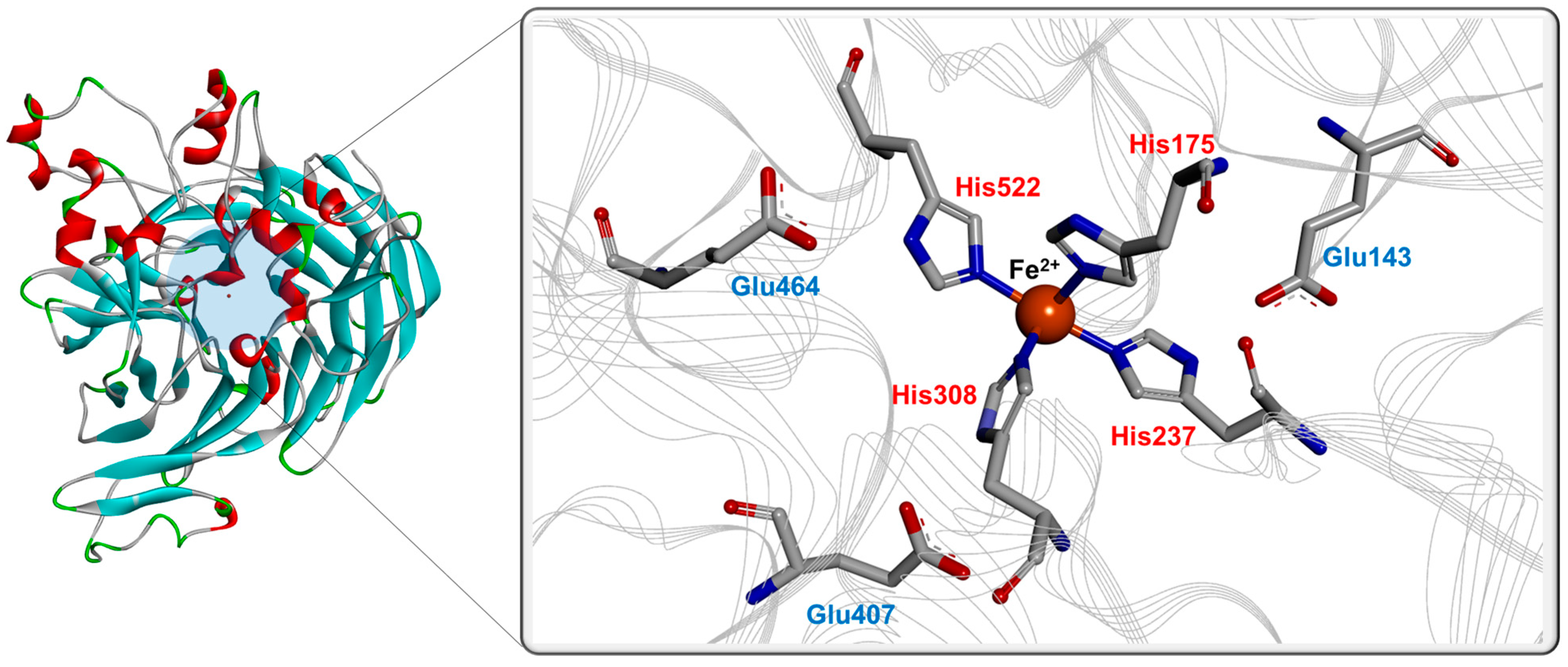
Figure 3.
Sequence alignments of BCOs. (a) Alignment of BCOs from mammals, chicken, C. elegans, and plants. The numbers for amino acids are represented based on BCO1 from C. elegans. (b) Alignment of bacterial BCOs, adapted from previous report [32]. The numbers for amino acids are represented based on BCO1 from uncultured marine bacterium 66A03. Abbreviation: UMB, uncultured marine bacterium. The UniProt numbers for BCOs are as follows: C. elegans, Q9U2E4; chicken, Q9I993; human, Q9HAY6; mouse, Q9JJS6; apple, A9Z0V8; A. thaliana, O65572; chicken, E1C8E0; chrysanthemum, B0FLM7; ferret, Q6QT07; human, Q9BYV7; mouse, Q99NF1; osmanthus, B0FLM9; rose, B0FLM8; saffron, A0SE36; UMB 66A03, Q4PNI0; UMB HF10, A4GIC0; Halobacterium sp., Q9HNE6; H. marismortui, Q5V0N2; and H. walsbyi, Q18DH3. Residues involved in the metal coordination and the formation of second coordination sphere via hydrogen bonds are represented by the red and blue colors, respectively.
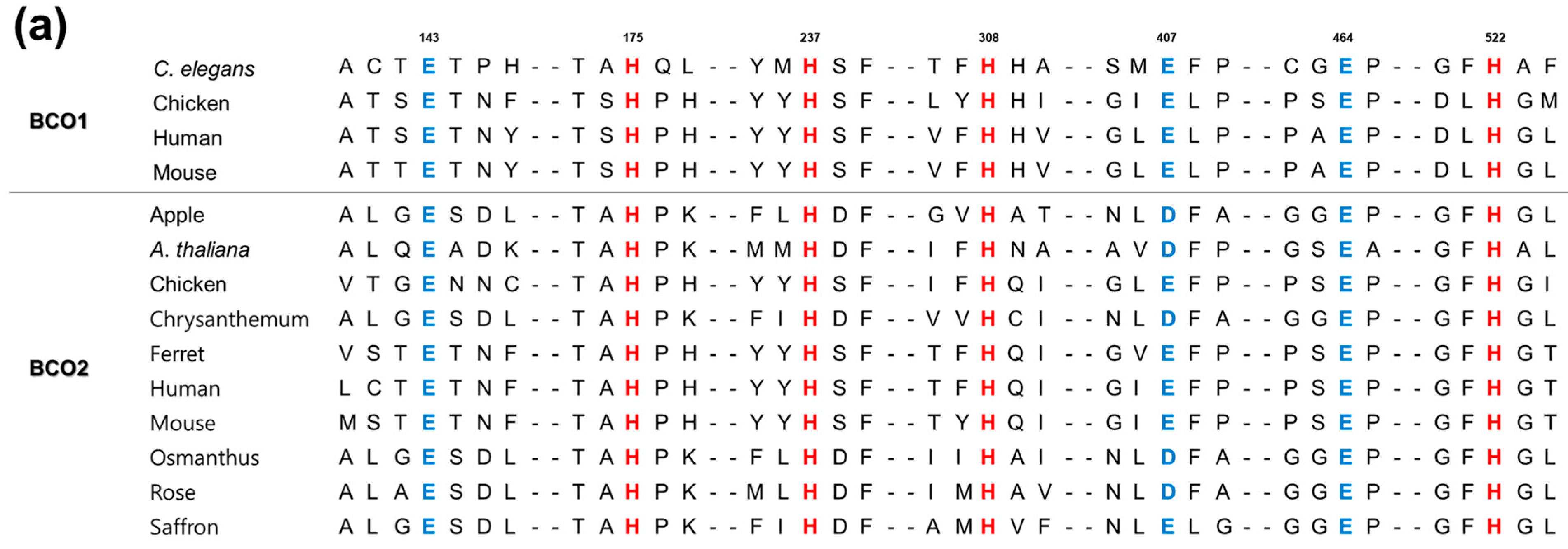

Figure 4.
Strategies for the improvement of retinal production via protein engineering of BCO by directed evolution (A), structure-based mutagenesis (B), d novo design of BCOs (C), optimization of metabolic pathway (MVA pathway, left; MEP pathway, right) for retinal production (top) through substrate channeling system (D), and co-factor regeneration system to increase the co-factor levels (E). Abbreviation for the compounds: G-3-P, glucose-3-phosphate; HMG-CoA, 3-hydroxy-3-methylglutaryl CoA; MVA, mevalonic acid; MVAP, phosphomevalonate; MVAPP, diphosphomevalonate; IPP, isopentenyl pyrophosphate; DMAPP, dimethylallyl pyrophosphate; GPP, geranyl pyrophosphate; FPP, farnesyl pyrophosphate; GGPP, geranylgeranyl pyrophosphate; DXP, 1-deoxy-d-xylulose-5-phosphate; MEP, methylerythritol phosphate; CDP-ME, 4-diphosphocytidyl-2-C-methyl-d-erythritol; CDP-ME2P, 4-diphosphocytidyl-2-C-methyl-d-erythritol 2-phosphate; MECDP, 2C-methyl-d-erythritol 2,4-cyclodiphosphate; HMBPP, 1-hydroxy-2-methyl-2-butenyl 4-diphosphate. The genes encode the following enzymes: atoB/phaA, acetoacetyl-CoA synthase; mvaS, HMG-CoA synthase; mvaA, HMG-CoA reductase; mvaK1, mevalonate kinase; mvaK2, phosphomevalonate kinase; mavD, mevalonate-5-diphosphate decarboxylase; idi, IPP isomerase; ispA, FPP synthase; crtE, GGPP synthase, crtB, phytoene synthase; crtI, phytoene desaturase; crtY, lycopene cyclase; BCO1, β-carotene 15,15′-oxygenase; BCO2, β-carotene 9,10′-oxygenase; dxs, 1-deoxy-d-xylulose-5-phosphate synthase; dxs, 1-deoxy-d-xylulose-5-phosphate reductoisomerase; cms, 4-diphosphocytidyl-2C-methyl-d-erythritol synthase, mecps, 2C-methyl-d-erythriol-2,4-cyclodiphosphate synthase. Dashed arrow indicates multi-enzymatic steps.


Table 1.
Biochemical properties of BCO1 and BCO2.
| Type /Organism | Accession No. | Optimum Temperature (°C) | Optimum pH | Molecular Weight (kDa) | Associated Form | Metal Ion | References | |
|---|---|---|---|---|---|---|---|---|
| Subunit | Native | |||||||
| BCO1 | ||||||||
| Caenorhabditis elegansR | Q9U2E4 | 37 | 7.5 | – | – | dimer | – | [21] |
| ChickenR | Q9I993 | – | – | 60 | – | – | – | [22] |
| ChickenR | 37 | 8.0 | 60 | ≈50 | monomer | – | [23] | |
| ChickenR | 37 | 8.0 | 60 | 240 | tetramer | Fe2+ | [24] | |
| ChickenR | 37 | – | 64 | – | – | – | [25] | |
| Guinea pigN | – | 37 | 8.5 | – | – | – | Fe2+ | [26] |
| HumanR | Q9HAY6 | 37 | 6.5 | 65 | – | monomer | Fe2+ | [27] |
| HumanR | 37 | 8.0 | – | – | – | – | [28] | |
| HumanR | 37 | 8.0 | – | – | – | – | [8] | |
| HumanR | 28 | 7.6 | – | – | – | – | [29] | |
| HumanR | 37 | 7.5–8.0 | ≈64 | ≈230 | tetramer | Fe2+ | [30] | |
| HumanR | 30 | 7.0 | – | – | dimer | Fe2+ | [31] | |
| Uncultured marine bacteriumR | Q4PNI0 | 40 | 8.0 | 32 | 64 | dimer | Fe2+ | [32] |
| MouseR | Q9JJS6 | 37 | 8.0 | 65 | – | – | Fe2+ | [33] |
| PigN | – | 37 | 8.0 | 156 | – | – | – | [34] |
| RatN | – | – | 7.5–8.0 | 62 | 62 | monomer | – | [35] |
| RatN | – | 37 | 8.0 | – | – | – | – | [20] |
| BCO2 | ||||||||
| AppleR | A9Z0V8 | 30 | ≈7.4 | – | – | – | – | [36] |
| Arabidopsis thalianaR | O65572 | 30 | ≈7.4 | – | – | – | – | [37] |
| ChickenR | E1C8E0 | 37 | 8.0 | 64 | – | – | Fe2+ | [38] |
| ChrysanthemumR | B0FLM7 | 30 | ≈7.4 | – | – | – | – | [36] |
| FerretR | Q6QT07 | 37 | 8.5 | ≈64 | – | – | Fe2+ | [39] |
| HumanR | Q9BYV7 | – | – | 64 | – | – | – | [40] |
| HumanR | 37 | 8.0 | 58 | 240 | tetramer | Fe2+ | [15] | |
| MouseR | Q99NF1 | 37 | 8.0 | – | – | – | – | [11] |
| MouseR | 30 | 7.0 | – | – | dimer | Fe2+ | [31] | |
| OsmanthusR | B0FLM9 | 30 | ≈7.4 | – | – | – | – | [36] |
| PotatoN | – | 28 | 7.8 | – | – | – | Fe2+ | [41] |
| RoseR | B0FLM8 | 30 | ≈7.4 | – | – | – | – | [36] |
| Saccharomyces cerevisiaeN | – | 45 | 8.0 | 50 | – | – | Ca2+ | [42] |
| SaffronR | A0SE36 | 30 | – | – | – | – | – | [43] |
N native enzyme; R recombinant enzyme.
Table 2.
Summary of substrate specificities of BCOs.
| Type /Organism | Expression | Substrate | Specific Activity (nmol/min/mg) | kcat/Km (min−1 mM−1) | References |
|---|---|---|---|---|---|
| BCO1 | |||||
| Chicken | pBAD system in E. coli cellsP | β-carotene | 6.8 | [25] | |
| Chicken | SFV system in BHK cellsH | β-carotene | 0.04 | [22] | |
| Chicken | SFV system in BHK cells | β-carotene | 0.04 | [23] | |
| Chicken | pET system in E. coli cellsH | β-carotene | 0.32 | 64.0 | [24] |
| α-carotene | 1.3 | ||||
| γ-carotene | 0.7 | ||||
| β-cryptoxanthin | 7.1 | ||||
| β-apo-4′-carotenal | 4.9 | ||||
| β-apo-8′-carotenal | 6.0 | ||||
| Guinea pig | Extraction from intestineP | β-carotene | 0.03 | [26] | |
| lutein | 0.02 | ||||
| β-apo-10′-carotenal | 0.02 | ||||
| Human | expression in SF9 cells | β-carotene | 18.5 | [29] | |
| Human | expression in SF9 cells | β-carotene | 93.0 | [30] | |
| β-cryptoxanthin | 19.0 | ||||
| Human | pET system in E. coli cellsH | β-carotene | 2.20 | 6.10 | [8] |
| α-carotene | 1.81 | ||||
| β-cryptoxanthin | 1.43 | ||||
| lycopene | 8.85 | ||||
| β-apo-8′-carotenal | 2.72 | ||||
| Uncultured marine bacterium | pET system in E. coli cellsH | β-carotene | 45.0 | 97.0 | [32] |
| α-carotene | 3.60 | ||||
| γ-carotene | 0.55 | ||||
| β-cryptoxanthin | 28.0 | ||||
| β-apo-4′-carotenal | 4.20 | ||||
| Mouse | pBAD system in E. coli cellsH | β-carotene | 0.39 | [33] | |
| Rat | extraction from intestine | β-carotene | 0.012 | [20] | |
| BCO2 | |||||
| Chicken | pET system in E. coli cellsH | β-carotene | 0.49 | [38] | |
| Human | pET system in E. coli cellsH | β-carotene | 80.0 | [15] | |
| S. cerevisiae | extraction from microbial cells | β-carotene | 380.0 | 77.0 | [42] |
| α-carotene | 49.0 | ||||
| lutein | 10.0 |
P partially purified enzyme; H purification of his-tagged protein.
Table 3.
Production of retinal using BCO1 and metabolically engineered cells.
| Biocatalyst | Source/Host | Substrate (mg/L) | Product (mg/L) | Reaction Time (h) | Conversion Yield (%) | Productivity (mg/L/h) | References |
|---|---|---|---|---|---|---|---|
| BCO1 | Mouse | 200 | 72 | 2 | 36 | 4.8 | [51] |
| Human | 200 | 98 | 16 | 49 | 6.13 | [52] | |
| Chicken | 5.37 | 3.2 | 16 | 60 | 1.06 | [24] | |
| Fruit fly | 0.17 | 0.13 | 2 | 18 | 0.065 | [53] | |
| Uncultured Marine bacterium | 350 | 181 | 20 | 52 | 9.1 | [14] | |
| Pig | 44 | 14.65 | 20 | 33.3 | 0.73 | [34] | |
| Metabolically engineered cells | E. coli | – | 600 | 33 | – | 18 | [54] |
| E. coli | – | 67 | 72 | – | 0.93 | [55] | |
| E. coli | – | 7 | 72 | – | 0.1 | [56] | |
| E. coli | – | 2.26 | 36 | – | 0.062 | [57] | |
| E. coli | – | 5.1 | 50 | – | 0.102 | [58] | |
| S. cerevisiae | – | 221.37 | 20 | – | 11.065 | [59] | |
| S. cerevisiae | – | 2094 | 160 | – | 13.1 | [60] |
Publisher’s Note: MDPI stays neutral with regard to jurisdictional claims in published maps and institutional affiliations. |
© 2022 by the authors. Licensee MDPI, Basel, Switzerland. This article is an open access article distributed under the terms and conditions of the Creative Commons Attribution (CC BY) license (https://creativecommons.org/licenses/by/4.0/).
Share and Cite
MDPI and ACS Style
Shin, K.-C.; Seo, M.-J.; Kim, Y.-S.; Yeom, S.-J. Molecular Properties of β-Carotene Oxygenases and Their Potential in Industrial Production of Vitamin A and Its Derivatives. Antioxidants 2022, 11, 1180. https://doi.org/10.3390/antiox11061180
AMA Style
Shin K-C, Seo M-J, Kim Y-S, Yeom S-J. Molecular Properties of β-Carotene Oxygenases and Their Potential in Industrial Production of Vitamin A and Its Derivatives. Antioxidants. 2022; 11(6):1180. https://doi.org/10.3390/antiox11061180
Chicago/Turabian StyleShin, Kyung-Chul, Min-Ju Seo, Yeong-Su Kim, and Soo-Jin Yeom. 2022. "Molecular Properties of β-Carotene Oxygenases and Their Potential in Industrial Production of Vitamin A and Its Derivatives" Antioxidants 11, no. 6: 1180. https://doi.org/10.3390/antiox11061180
Note that from the first issue of 2016, this journal uses article numbers instead of page numbers. See further details here.