
Oxidative Stress: A Suitable Therapeutic Target for Optic Nerve Diseases?


Department of Ophthalmology, University Medical Center, Johannes Gutenberg University Mainz, Langenbeckstrasse 1, 55131 Mainz, Germany
*
Authors to whom correspondence should be addressed.
Antioxidants 2023, 12(7), 1465; https://doi.org/10.3390/antiox12071465
Submission received: 27 June 2023
/
Revised: 17 July 2023
/
Accepted: 18 July 2023
/
Published: 20 July 2023
(This article belongs to the Special Issue Oxidative-Stress in Human Diseases—2nd Edition)
Abstract
:Optic nerve disorders encompass a wide spectrum of conditions characterized by the loss of retinal ganglion cells (RGCs) and subsequent degeneration of the optic nerve. The etiology of these disorders can vary significantly, but emerging research highlights the crucial role of oxidative stress, an imbalance in the redox status characterized by an excess of reactive oxygen species (ROS), in driving cell death through apoptosis, autophagy, and inflammation. This review provides an overview of ROS-related processes underlying four extensively studied optic nerve diseases: glaucoma, Leber’s hereditary optic neuropathy (LHON), anterior ischemic optic neuropathy (AION), and optic neuritis (ON). Furthermore, we present preclinical findings on antioxidants, with the objective of evaluating the potential therapeutic benefits of targeting oxidative stress in the treatment of optic neuropathies.
1. Introduction
Optic nerve diseases encompass a wide range of disorders characterized by optic nerve atrophy, resulting from the loss of retinal ganglion cells (RGCs) and leading to sight-threatening conditions [1,2,3]. These pathologies include:
- Anterior ischemic optic neuropathies (AION): This category includes arteritic forms, like giant cell arteritis (GCA), which has a pooled prevalence of approximately 51.74 in 100,000 for individuals over the age of 50 [15]. Nonarteritic forms have a reported prevalence of approximately 102.87 in 100,000 in the general population over the age of 40 in the Republic of Korea [16];
- Infiltrative optic neuropathies, such as leukemic optic neuropathy, which presents in approximately 16% and 18% of all chronic and acute leukemia cases, respectively [28];
Glaucoma is the most prevalent optic nerve disease worldwide (Figure 1) [6,7]. LHON has a low estimated prevalence for complete penetrance cases [8,9,10,11,12], while the prevalence is much higher for carriers of mutation variants in the general population [13,14]. The common underlying feature in all optic nerve diseases is the damage and loss of RGCs and their axons, which gradually leads to optic nerve degeneration [3,33,34]. RGCs have high energy requirements and are particularly susceptible to alterations in their energy supply, mainly generated in the mitochondria through the electron transport chain (ETC) [35]. Oxidative stress plays a pivotal role in the pathophysiology of optic nerve diseases such as glaucoma, LHON, and AION. Imbalances between reactive oxygen species (ROS) as well as reactive nitrogen species (RNS) generation and antioxidant systems lead to reactive species overproduction, adenosine triphosphate (ATP) insufficiency, irreversible cellular injuries, and ultimately RGC loss [3,36,37,38,39,40,41,42,43,44,45].
Considering the high global prevalence of glaucoma and the limited treatment options available for most optic neuropathies [33], it becomes essential to explore new research avenues and investigate novel therapeutic approaches. This review aims to shed light on the role of oxidative damage in the pathophysiology of optic nerve diseases. Specifically, we provide an overview of the current understanding of oxidative stress as a key pathological factor and explore its potential as a viable therapeutic target. We will especially focus on four of the most prevalent and clinically challenging optic nerve diseases, glaucoma, LHON, AION, and ON. Ultimately, our goal is to identify novel curative strategies that may pave the way for improved treatment options in these conditions.
2. Anatomy and Perfusion of the Visual Pathway
The optic nerve, also known as the second cranial nerve [46], is composed of thin (0.1 µm) and lengthy (~50 mm) RGC axons that extend from the retina to the lateral geniculate nucleus, resulting in a soma/axon ratio of approximatively 1:10,000 [35]. Within the retinal layers, these axons merge to form the retinal nerve fiber layer [47], which runs parallel to the superficial blood vessels. The inner retina, including the outer plexiform layer through the nerve fiber layer, is supplied by the central retinal artery. On the other hand, the avascular outer retina, consisting of the outer nuclear layer and photoreceptors, receives its blood supply through diffusion from the choriocapillaris. The choriocapillaris is nourished by short posterior ciliary arteries, which branch from the ophthalmic artery [48,49]. Notably, retinal oxygenation exhibits variability depending on light or dark conditions due to the different oxygen demand of rods and cones. In humans, rods, which are responsible for vision in low light, are present in approximately 120 million, while cones number around 6 million [50]. Consequently, retinal oxygen consumption is reduced by half in the presence of light, attributed to the decreased activity of rods compared to cones [50].
Optic nerve axons account for approximately 38% of all axons within the central nervous system [51]. Around 1.2 million RGC axons converge to form the optic nerve head (ONH), also referred to as the optic papilla or optic disc. The ONH exhibits a brighter central depression known as the optic cup [51,52,53,54]. Blood supply to the ONH in humans is provided by the arterial circle of Zinn–Haller [48].
The optic nerve can be divided into four compartments: the intraocular segment (1–2 mm), which includes the retinal nerve fiber layer (RNFL) and extends from the ONH to the lamina cribrosa; the intraorbital segment (25–30 mm), which spans from the retrobulbar tract to the optic canal; the intracanalicular segment (5–9 mm); and the intracranial segment (9–10 mm), which extends from the optic canal to the optic chiasm [51]. Four distinct regions can be identified within the optic nerve head: the nerve fiber layer, the prelaminar region, the lamina cribrosa, and the retrolaminar region [47]. The lamina cribrosa serves as a supportive structure for the RGC axons within the ONH [55,56] and consists of approximately 200–300 porous apertures through which the optic nerve passes from the sclera into the retrobulbar cavity [51]. Deformations of the lamina cribrosa may indicate RGC loss and can signify the initial stages of glaucomatous optic neuropathy [57]. Once beyond the lamina cribrosa, the optic nerve becomes myelinated by oligodendrocytes, increasing its diameter from 1–2 mm to 3–4 mm [51]. The intraorbital, intracanalicular, and intracranial segments receive their blood supply from the posterior ciliary arteries as well as the circle of Willis [58].
The two optic nerves converge at the optic chiasm, where nerve fibers originating from the nasal retina of each eye cross over to join the temporal fibers of the contralateral eye [47,59,60]. The blood supply to the chiasm is provided by the circle of Willis [47,52]. From the chiasm, the RGC axons continue their course into the optic tract, which receives perfusion from the posterior communicating and internal carotid artery [52]. Within the optic tract, the nerve fibers undergo rearrangement to align with their corresponding positions in the lateral geniculate nucleus [59]. Fibers carrying visual information from the right visual field project to the left cerebral hemisphere and vice versa [59]. In the lateral geniculate nucleus, the RGC axons synapse with the second-order neurons of the visual pathway, organized in six layers consisting mainly of large and small neurons [47]. Some fibers from the optic tract also synapse with the olivary pretectal nucleus, regulating the pupillary light reflex [52,61]. Additionally, RGC axons containing melanopsin terminate in the suprachiasmatic nucleus, a crucial center for controlling the circadian rhythms [47,61].
The large and small axons of the lateral geniculate nucleus form optic radiation, which initially projects anteriorly and then turns posteriorly, terminating in the occipital lobe where the visual cortex (Brodmann area 17) is located [47,59]. These regions receive blood supply from branches of the internal carotid artery (lateral geniculate nucleus and optic radiation) and the posterior cerebral artery (visual cortex) [52]. In Figure 2, the perfusion and the anatomy of the visual pathway are illustrated.
An interesting aspect of the optic nerve anatomy is the presence of an unmyelinated portion in the RGCs [35]. In the unmyelinated compartment, specifically in the intraocular segment of the optic nerve, potential signals cannot be transmitted through saltatory conduction due to the absence of myelin [62]. To compensate for this limitation and enable rapid transmission, RGCs generate higher quantities of ATP in their axons to repolarize the plasma membrane [63]. Mitochondrial bidirectional transport (antero- and retrograde) along the axons plays a crucial role in this process. These organelles move toward regions with high energy demands, such as the unmyelinated portions, and ATP gradients are believed to guide this transport [35]. This mechanism may explain the specific vulnerability of RGCs to mitochondrial dysfunction, leading to the triggering of ROS production in a vicious cycle [44,63].
In this context, it is important to highlight the trophic role of myelin in the optic nerve sheath. Myelin has been shown to play a vital role in supplying nutrients to the axon, as the entire mitochondrial respiratory chain has been detected in the myelin sheath of the optic nerve [64]. This finding provides possible explanations for the link between myelin loss and axonal degeneration observed in neuropathies or demyelinating disorders [64]. A decrease in myelin-related mitochondrial respiration may be one of the main triggers responsible for neurodegenerative events [65]. With aging, there is a loss of structural integrity in the myelin sheath, which can subsequently lead to axonal deterioration [66]. This process may underlie various neurodegenerative pathologies, including Alzheimer’s disease, as recent studies have suggested [67]. Therefore, an intriguing future therapeutic approach for neurodegenerative disorders involves improving the integrity of the myelin sheath, which could potentially slow disease progression [67]. In this regard, the comanipulation of microglia and a specific signaling pathway, such as the G protein-coupled receptor 17 pathway in oligodendrocyte precursor cells, has been shown to induce robust myelination and promote axonal regeneration following injury [68]. Exploring these avenues may also offer new therapeutic perspectives for other neurodegenerative diseases, including optic neuropathies [69].
3. General Mechanisms of Nitro-Oxidative Stress in the Optic Nerve
3.1. Generation of Reactive Oxygen and Nitrogen Species
Mitochondria are vital intracellular organelles responsible for essential chemical reactions that produce energy substrates [70,71]. In addition to their various cellular functions, such as modulating intracellular calcium levels, synthesizing nucleotides, lipids, and amino acids, and regulating apoptosis, mitochondria also generate ROS [70,72,73,74]. ROS, at basal levels, serve as critical mediators of signaling pathways, including hypoxic and inflammatory pathways [44,70,75,76]. The fundamental function of mitochondria is to regulate oxygen metabolism and produce energy in the form of ATP [70,71,77]. The electron transport chain (ETC) within the inner mitochondrial membrane plays a central role in this process [78]. Despite the efficiency of oxidative phosphorylation, electron leaks can occur, leading to the direct interaction of electron carriers with molecular oxygen (O2) in the mitochondrial matrix. This interaction results in the donation of electrons and the generation of superoxide (O2•−) [71,77,79,80]. While mitochondria are recognized as the main source of ROS in the cell, other significant sources include the enzymatic activities of nitric oxide synthase (NOS) and nicotinamide adenine dinucleotide phosphate (NADPH) oxidase (NOX) [3,79,81,82]. NOS generates nitric oxide (NO), while NOX (comprising seven isoforms: NOX1, -2, -3, -4, -5, DUOX1, -2) transfers electrons from cytosolic NADPH to molecular O2, generating O2•− [83,84].
NO is a free radical that plays a pivotal role in various physiological functions [85]. It serves as a regulator of vascular tone [86,87,88]. Additionally, NO acts as a signaling molecule in neurotransmission and as a regulator of gene transcription [89,90,91,92,93,94]. The production of NO is facilitated by the activity of NOS, an enzyme that has three isoforms: neuronal NOS (nNOS or NOS I); inducible NOS (iNOS or NOS II); and endothelial NOS (eNOS or NOS III) [85,95]. NO rapidly and spontaneously reacts with O2•− through a “diffusion-limited reaction” [96,97]. As a result, a highly damaging RNS termed peroxynitrite (ONOO−) is generated [85,96,97]. Peroxynitrite contributes to the pathogenesis of diverse retinal disorders, being also newly proposed as a critical factor in the pathogenesis of glaucoma [98,99,100].
3.2. Oxidative Damage and Antioxidant Defense Systems
ROS and RNS play a physiological role in cellular responses to hypoxia, cell proliferation, cell death, inflammation, or infection [44,76]. Immune cells, such as phagocytes, produce ROS, which provide reactions necessary for an appropriate killing of pathogens [101,102]. Due to endogenous or exogenous trigger factors, the balance between pro- and antioxidant systems can be critically undermined, resulting in nitro-oxidative stress. In this context, radicals begin to compete for paired electrons with intracellular substrates [103], creating oxidative damage. Oxidative injuries are recognized to be a crucial player in the pathogenesis of a variety of pathologies, including ocular diseases [36,104,105,106,107]. At the biomolecular level, three general forms of injuries caused by reactive species can be distinguished: DNA lesions [108,109], protein alterations [110,111], and lipid peroxidation [96,112]. The consequences of DNA damage are modifications in the expression of proteins and the altered regulation of fundamental activities, like oxidative phosphorylation, according to the vicious cycle theory [113,114,115]. In this context, mitochondrial ROS also induce activation of the nod-like receptor family pyrin domain-containing 3 (Nlrp3) inflammasome, a key factor in pyroptotic cell death during inflammation [116].
Antioxidant systems are responsible for defending cells and tissues from the damaging impact of reactive species, which are constantly produced as a “by-product” of oxidative phosphorylation but also serve, at basal levels, physiological functions [76]. Enzymatic antioxidants comprise SOD, catalase (CAT), glutathione peroxidase (GPX), glutathione-S-transferase (GST), heme oxygenase (HO), peroxiredoxin, and thioredoxin [103,117,118,119,120,121,122].
Nonenzymatic antioxidants can be classified into direct and indirect agents. Direct antioxidants react with ROS or RNS, “being sacrificed in the process of their antioxidant actions” [123,124]. Free radical scavengers are, for example, glutathione (GSH) [125], carotenoids [126], vitamin C (ascorbic acid) [127], and vitamin E (α-tocopherol) [128]. Alternatively, indirect antioxidants are molecules, such as vitamin C, that upregulate antioxidant proteins, for example, via the nuclear factor erythroid-2-related factor 2 (Nrf2) [123,127] or molecules, like α-lipoic acid [129]. Examples of antioxidant compounds adsorbed with the food are resveratrol and betulinic acid [98,130,131].
3.3. Oxidative Stress in Retinal Ganglion Cells
The retina belongs to the metabolically most active organs in the human body [132] and requires a relatively large amount of energy substrates [133], which makes it particularly vulnerable to energy insufficiency [134]. Oxygen supply is essential for retinal function [135], and its consumption occurs very rapidly, like in the brain [136,137,138]. Hence, conditions that can modify the supply of molecules, such as O2, necessary for the production of energy substrates, like ATP, may rapidly generate significant damage in RGCs due to their susceptibility to oxygen deficiency (Figure 3). Thus, an appropriate blood supply via retrobulbar and retinal vessels is crucial for the proper function of RGCs. Studies in retrobulbar blood vessels reported that ROS blunted endothelial function partially by reducing the contribution of the NOS pathway to endothelium-dependent vasodilation [139]. Likewise, moderately elevated IOP induced endothelial dysfunction in retinal arterioles together with RGC loss [140,141]. Zadeh et al. found in apolipoprotein E (ApoE)-deficient mice that hypercholesterolemia caused oxidative stress and endothelial dysfunction in retinal arterioles but did neither lead to increased ROS levels in the RGC layer nor to loss of RGCs, indicative of compensatory effects [142]. In contrast, a study in pigs reported that after only 12 min of ocular ischemia and 20 h of reperfusion, endothelial dysfunction, retinal edema, and RGC loss occurred [143]. ROS generation due to ischemia/reperfusion (I/R) injury is reported to be caused by diverse enzymes involved in the regulation of oxidative metabolism, such as NOX2, xanthine oxidase (XO), uncoupled eNOS, and by ETC dysfunction [143,144,145,146]. Hyperglycemia was also described to be a cause of endothelial dysfunction and oxidative stress in the retina [147,148,149] via the involvement of NOX2 due to the activation of the receptor of an advanced glycation end product (RAGE)-, mitogen-activated protein kinase (MAPK)-, polyol-, protein kinase C (PKC)-, renin–angiotensin system (RAS) signaling pathways [150,151,152,153,154,155].
4. Oxidative Stress in Individual Optic Nerve Diseases
4.1. Glaucoma
4.1.1. General Aspects
The term “glaucoma” encompasses a group of optic nerve diseases that share similar morphological characteristics but may have diverse origins. They represent the most common optic nerve disorders [34,156,157], characterized by alterations in the ONH, which include a decrease in the neuroretinal rim and an enlargement of the optic cup, leading to deformation of the lamina cribrosa [34,158]. As a result, in the early stages, typical visual field defects, which follow the arcuate pattern corresponding to the arrangement of nerve fiber bundles, can be observed [159]. Elevated intraocular pressure (IOP) is described as the primary risk factor, which can result from pathological resistance to aqueous humor drainage through the trabecular meshwork (TM), causing mechanical stress and compression on the axonal fibers and the ONH [160,161,162]. Glaucomatous disorders are classified into primary forms, where there is no associated ocular disease, and secondary forms, where known coexisting pathological processes (such as uveitis, neovascularization, trauma, or lens-related conditions) lead to IOP elevation and thus to the development of glaucomatous damage [34,160,163]. Primary and secondary forms are further divided into open-angle glaucoma, which among others includes a subtype known as normal tension glaucoma (NTG) and angle-closure glaucoma, in relation to the angle between the iris and cornea in the anterior chamber [34,164,165]. Primary open-angle glaucoma (POAG), the worldwide major form of glaucoma, describes a disorder where, under conditions of an open chamber angle, structural damage to the optic nerve emerge silently, gradually, and chronically [160]. While high IOP values can be a contributing factor in this disorder, it is not always present in POAG. Elevated IOP is defined as a pressure value that exceeds the 97.5th percentile for the population under consideration and is commonly assumed to be a value higher than 21 mmHg [34,163]. However, it is important to note that elevated IOP is recognized as a risk factor but not a diagnostic criterion for glaucoma [160,163]. In fact, approximately 30–90% of POAG cases have IOP values below the 21 mmHg cutoff, which is considered NTG. The prevalence of NTG varies significantly geographically [166]. Additionally, the term “ocular hypertension” (OHT) refers to a condition where elevated IOP is detected without evidence of glaucomatous optic neuropathy [34]. Angle-closure glaucoma is considered an ocular emergency resulting from anatomical contact between the iris and lens. This contact can cause obstruction of the aqueous humor outflow leading to a condition known as pupillary block, present in approximately 90% of cases of primary angle-closure glaucoma (PACG) [165,167]. In this context, a sudden and significant increase in IOP occurs and can reach values as high as 60 to 80 mmHg [164]. PACG is estimated to affect approximately 26% of the glaucoma population and is responsible for about half of the cases of glaucoma-related visual loss worldwide [168,169,170].
The main risk factors for POAG include elevated IOP, advanced age, black race, myopia, and a positive family history [163]. On the other hand, risk factors for PACG include hyperopia, shallow central anterior chamber depth, shallower limbal anterior chamber depth, anteriorly positioned lens, increased lens thickness, small corneal diameter, and steep corneal curvature, as well as high IOP values, advanced age, Asian ethnicity, and female gender [168,171,172]. Elevated IOP is the only identified modifiable risk factor [173], and reducing IOP is currently the only proven method to treat glaucoma effectively [171,174]. IOP has been considered a central factor in both the “mechanical” and “vascular” theories proposed to explain the etiopathogenesis of glaucoma. According to the mechanical theory, elevated IOP leads to the compression and deformation of RGC axons and the optic nerve, ultimately resulting in cell death due to a reduced or blocked axoplasmic flow and a deficit of cellular supply elements [175]. On the other hand, the vascular theory suggests that decreased perfusion to the optic nerve causes ischemia in RGCs, leading to neurodegenerative damage [161]. A reduced blood supply may be a consequence of IOP-related compression or the functional deficits of blood or vessels that supply the laminar regions of the ONH [176]. Elevated IOP can be a result, for example, of pathological resistance in the aqueous humor drainage process through the TM [162].
The primary goal of antiglaucomatous drugs is to lower IOP to a target level, which is an acceptable range individually set to prevent disease progression [177]. Antiglaucomatous drugs are generally administered topically as eye drops and can be categorized into different classes based on their mechanism of action. Prostaglandin analogues, such as latanoprost, increase uveoscleral and trabecular outflow, while β-blockers (e.g., timolol), α2-adrenoceptor agonists (e.g., brimonidine), and carbonic anhydrase inhibitors (e.g., dorzolamide) reduce aqueous humor production [173]. Additionally, α2- adrenoceptor agonists, like brimonidine, clonidine, and epinephrine, can also increase trabecular outflow, similar to prostaglandins [173]. Miotic agents, such as pilocarpine, widen the chamber angle, and osmotically active drugs, like mannitol, increase water removal from the eye through the systemic circulation when administered intravenously (IV) [173]. Moreover, pilocarpine was reported to provide a neuroprotective effect through the activation of muscarinic receptors [178]. In the case of PACG, the primary treatment is peripheral iridotomy, a laser procedure that creates a full thickness opening in the iris to relieve pupillary blockage [179]. Additionally, osmotic substances, like mannitol, or carbonic anhydrase inhibitors, such as acetazolamide, can be administered intravenously [173].
The economic implications of glaucoma have been extensively studied worldwide, highlighting its significance as a critical social problem. Cost-of-illness studies have shed light on the financial impact of glaucoma. For instance, a study conducted in the UK in 2002 reported that over £300 million was spent on glaucoma-related expenses [180]. Similarly, an Australian review analyzed published randomized trials and population-based studies since 1985, projecting that the total costs (including direct, indirect, and costs related to the loss of well-being) for POAG in Australia would increase from AUD 1.9 billion in 2005 to AUD 4.3 billion in 2025 [181]. Examining the indirect costs of glaucoma, a study from the UK investigated whether glaucoma can be a risk factor for falls. The study found that between 2012 and 2018, 11.7% of hospital admissions for falls in a national health service hospital were in patients with a secondary diagnosis of glaucoma [182]. Furthermore, a recent retrospective cohort conducted in the US, extrapolating data from 2015 to 2017 and based on a database of patients with POAG and ocular hypertension (OHT), compared these two conditions. The study revealed that advanced stage POAG was associated with a higher risk of falls compared to OHT. Additionally, it was found that the annual eye-related outpatient costs for POAG patients were higher (median: USD 516) than for OHT patients (median: USD 344). Among patients with POAG, those in advanced stages exhibited even higher annual eye-related costs (median: USD 639) compared to those in moderate (median: USD 546) and mild (median: USD 476) stages [183]. Similarly, a previous multicenter study conducted in Germany between 2009 and 2010, involving 2 university hospitals and 13 ophthalmological practices, reported that direct costs for therapies were higher for glaucoma compared to OHT and were directly associated with disease progression. The study highlighted that additional treatment changes necessitated by uncontrolled IOP were the major contributors to the increased costs associated with glaucoma. The study concluded that by effectively managing IOP over the long term and avoiding advanced disease stages, both disease progression and associated costs could be reduced [184]. In summary, publications on this topic consistently emphasize that early first-line therapies play a vital role in reducing the economic burden of glaucoma. By preventing disease progression to advanced stages, these early interventions can help minimize the need for more expensive and sometimes less effective treatments, ultimately alleviating the economic strain imposed by glaucoma [185].
4.1.2. Redox Parameters and Oxidative Stress Biomarkers in Glaucoma
Numerous studies have been conducted to gather evidence of altered redox status in glaucoma by examining oxidant or antioxidant levels in various samples, including blood, plasma, serum, aqueous humor, and the TM. Common systemic parameters used to assess oxidative stress include total antioxidant status (TAS), total antioxidant capacity (TAC), biological antioxidant potential (BAP), total reactive antioxidant potential (TRAP), and total oxidant status (TOS). Biomarkers of oxidative stress, such as 8-hydroxy-2′-deoxyguanosine (8-OHdG), poly-(adenosine diphosphate-ribose)-polymerase (PARP1), oxoguanine-DNA-glycosylase (OGG1), and malondialdehyde (MDA), are indicative of DNA oxidative damage, base excision repair activity, and lipid peroxidation.
An Indian study examining redox-related DNA damage in POAG patients reported elevated levels of 8-OHdG levels in both plasma and aqueous humor. They also observed a negative correlation between 8-OHdG levels and PARP1 and OGG1 levels, which were concurrently lower [186]. Increased levels of 8-OHdG were also found in the TM of patients with POAG, correlating with more severe visual field defects [187,188].
TAC levels were found to be lower in the serum and aqueous humor of glaucoma patients, while MDA levels were increased [189]. Decreased levels of TAS [190] and TRAP [179] were also observed in the aqueous humor of glaucoma patients. Lower TAS levels were additionally found in the plasma samples of individuals with POAG, further associated with an increasing cup-to-disc ratio, which indicates cup–disc enlargement and retinal ganglion cell loss [191]. Studies on serum samples of glaucoma patients reported lower BAP levels, correlating with lower RGC density in young males [192], more advanced visual field loss [193], or higher IOP [194].
A meta-analysis on the redox parameters in glaucoma revealed a general increase in oxidative stress parameters in both serum and aqueous humor, with MDA being the most significant biomarker, suggesting its potential clinical utility [195]. Antioxidative markers were found to be lower in serum, while aqueous humor showed an increase in antioxidant defense, indicating a possible compensatory response to oxidative stress [195]. Similarly, a recent meta-analysis on oxidative stress markers in glaucoma highlighted lower TAS levels in blood and elevated levels of antioxidant enzymes, such as SOD, CAT, and GPX, in the aqueous humor [196]. This systematic review also compared patients with POAG to those with exfoliation glaucoma, the most common secondary form of POAG, characterized by the deposition of fibrillar material in the anterior segment [197,198]. It reported no increase in antioxidant defense in the aqueous humor of exfoliation glaucoma patients, along with a decrease in TAS levels in the blood [196].
RNS, including NO, were found to be increased in the aqueous humor of patients with POAG and PACG [199]. A Spanish study examining both the TM and the aqueous humor of POAG patients reported an upregulation in iNOS and downregulation in eNOS in trabecular meshwork cells (TMCs). They also observed increased levels of MDA in the aqueous humor, corresponding to an increase in visual field defects [200]. The authors of this study suggested that the increased production of NO induced by iNOS may play a role in the process of RGC death, potentially leading to an elevation in MDA levels in the aqueous humor [200].
4.1.3. Oxidative Stress in the Pathogenesis of Glaucoma
To gain a better understanding on the role of reactive species in glaucoma, it is important to discuss the anatomy and function of the TM and the detrimental impact of ROS on these structures. The TM is located in the sclerocorneal angle and consists of three layers: the uveal TM, the corneoscleral TM, and the juxtacanalicular TM, also known as the cribriform TM region. This region is adjacent to the Schlemm’s canal, which allows for the drainage of aqueous humor into the episcleral veins [201]. The TM plays a crucial role in regulating IOP by controlling the outflow of aqueous humor. It allows for the turnover of aqueous humor through drainage into superficial veins toward the conjunctiva [201]. Any alterations in TM permeability, such as changes in TM structure, can result in increased resistance to aqueous humor outflow, leading to elevated IOP. Studies focusing on TMCs have investigated how increased IOP may develop in glaucoma patients. These studies have shown that the oxidative balance in aqueous humor, which has been found to be altered in individuals with glaucoma [179,202], can impact the structure and function of TMCs, particularly the TM endothelial cells lining the Schlemm’s canal [203]. Elevated levels of ROS in the aqueous humor can trigger changes in the TM [162]. This can occur through the fusion of TMCs, leading to trabecular thickening [204]. Additionally, ROS can induce TMC apoptosis, resulting in TM disruption [203]. Alterations in the extracellular matrix (ECM) can also occur, affecting the adhesion of endothelial TMCs to the ECM and causing the TM to collapse [205]. In all of the cases, increased resistance to aqueous humor circulation occurs, leading to elevated IOP [203]. In Figure 4, an anatomical overview of the described organs, with focus on the anterior segment of the eye, is presented.
Oxidative damage contributes to axonal injuries, leading to the death of RGCs [37]. RGC loss may occur as a result of a process involving ROS, elevated IOP, mechanical compression on RGC axons, or vascular compression of optic-nerve-perfusing vessels [40]. Our previous research in mice has demonstrated that already moderately elevated IOP can impair vascular autoregulation and cause endothelial dysfunction in the retina, which is associated with an upregulation in NOX2 [140]. Additionally, high hydrostatic pressure and ischemia have been found to stimulate the release of tumor necrosis factor α (TNF-α) from glial cells, activating apoptotic signaling pathways in RGCs [206]. TNF-α released by active astrocytes, microglia, and Müller glial cells is one of the most significant proinflammatory cytokines in glial neuroinflammation, mitochondrial dysfunction, and oxidative stress [207]. Moreover, TNF-α can induce apoptosis through caspase-8 activation [207]. Mitochondrial dysfunction also plays a pivotal role in glaucomatous optic disorders. Increased IOP has been shown to damage mitochondria, leading to mitochondrial fission and alterations in the expression of the OPA1 gene [208,209]. Via ROS-induced activation of the transcription factor nuclear factor “kappa-light-chain-enhancer” of activated B-cells (NF-kB), mitochondria indirectly contribute to the initiation or amplification of glial neuroinflammation processes [208], resulting in the production of inflammatory cytokines. Furthermore, oxidized mitochondrial DNA and mitochondrial fragments released from microglia can activate inflammasomes [208]. Oxidative stress also disrupts the glutamate/glutamine metabolism, leading to the neurotoxic extracellular accumulation of glutamate [40]. Glutamate is released from dying RGCs and activated glia, and the dysfunctional glial cells are unable to adequately buffer excessive extracellular glutamate [40,210,211]. Additionally, oxidized substrates, such as advanced glycation end products (AGEs) and oxidized low density lipoproteins (oxLDLs) can act as “antigenic stimuli”, inducing ROS production, triggering NF-kB activation, glial activation, neuroinflammation, and apoptosis [40,208]. In this context, the overproduction of ROS activates the apoptosis signal-regulating kinase 1 (ASK-1) [212,213], which, in turn, activates the p38/c-Jun N-terminal kinase (JNK)/extracellular signal-regulated kinase (ERK) axis. This axis triggers the mitochondrial apoptotic pathway, characterized by the release of the Bcl-2-associated X-protein (Bax) and cytochrome c into the cytosol, followed by apoptosome formation (cytochrome c/apoptotic protease-activating factor 1(Apaf-1)/caspase-9), ultimately leading to caspase-3 activation [212,214,215]. Moreover, hydrogen peroxide has been described to activate the phosphoinositide 3-kinase (PI3K)/Ak strain transforming (Akt) axis and reduce the intracellular concentration of the mammalian target of rapamycin (mTor), promoting NF-kB activation [216].
Apart from elevated IOP, other potential risk factors have been investigated in the context of NTG. The “Collaborative Normal-Tension Glaucoma Study” (1998) examined the effectiveness of IOP-lowering therapy in patients with NTG and concluded that although reducing IOP can have a positive impact, it cannot prevent disease progression [217,218]. Recent reviews on NTG have focused on the multifactorial nature of the disease, considering various risk factors implicated in its pathogenesis, including translaminar cribrosa pressure difference, fractal dimensions, neurovascular coupling, vascular dysregulation, endothelial dysfunction, ocular perfusion pressure, and oxidative stress [166,218]. Regarding vascular pathological aspects, Leung and Tham described NTG as part of a larger group of disorders known as small vessel diseases [166]. They focused on the correlation between NTG and cerebral silent infarcts detected in patients with NTG [219,220,221]. CSI can lead to hypoperfusion of the optic nerve head and hypoperfusion in the optic nerve head, increasing the risk of NTG [166]. Additionally, arterial hypotonia, especially during night time when blood pressure is reduced, may result in lower ocular perfusion pressure and decreased blood supply to the ONH [218]. In this context, it is relevant to consider another potential primary injury to RGCs independent of elevated IOP: tissue hypoxia, which can be associated with pathogenic mechanisms in glaucoma [37]. During hypoxia, hypoxia-inducible factor 1-alpha (HIF-1α) induces the transcription of various genes, including those encoding the vascular endothelial growth factor (VEGF), heme oxygenase-1 (HO-1), and inducible nitric oxide synthase (iNOS), aiming to increase oxygen supply to the affected tissue [222,223]. However, HIF-1α also upregulates NOX, which generates ROS, creating a vicious cycle of HIF-1α overexpression and ROS production [224,225]. The presence of increased HIF-1α expression regions in the retina and optic nerve of glaucoma patients was confirmed by Tezel and Wax, further supporting the pathogenic role of hypoxia in glaucoma [226]. Hypoxic conditions primarily cause energy depletion and disrupt ionic homeostasis, leading to increased ROS generation and inflammation [37,227], mediated by the overproduction of ROS via NOX2 [228] and other sources, such as XO and cyclooxygenase (COX) [229]. Additionally, activated glia release TNF-α, which, among other effects, activates NF-kB, intensifying glial activation, neuroinflammation, and apoptosis through caspase-mediated or caspase-independent signaling pathways [207,230].
Reviewing the literature on the pathogenesis of glaucoma (Figure 5), it becomes evident that the pathways leading to RGC loss are redundant, intersect, and overlap, making it challenging to identify a single initial cause-and-effect factor [40]. Ultimately, the outcome appears to be a combination of “vicious cycles”, in which inflammation and ROS interconnect and amplify each other.
4.1.4. Interplay between Mitochondrial and ER Stress in TMCs and RGCs
Mechanical and vascular stress in glaucoma contribute to mitochondrial dysfunction [210], which leads to calcium imbalance and interconnects with endoplasmic reticulum (ER) stress, resulting in energy impairment and subsequent ROS generation. Numerous studies have explored the relationship between the endoplasmic reticulum and mitochondria in the pathogenesis of glaucoma. Understanding the main processes of this connection can help trace intracellular pathways that contribute to TM dysfunction and RGC death.
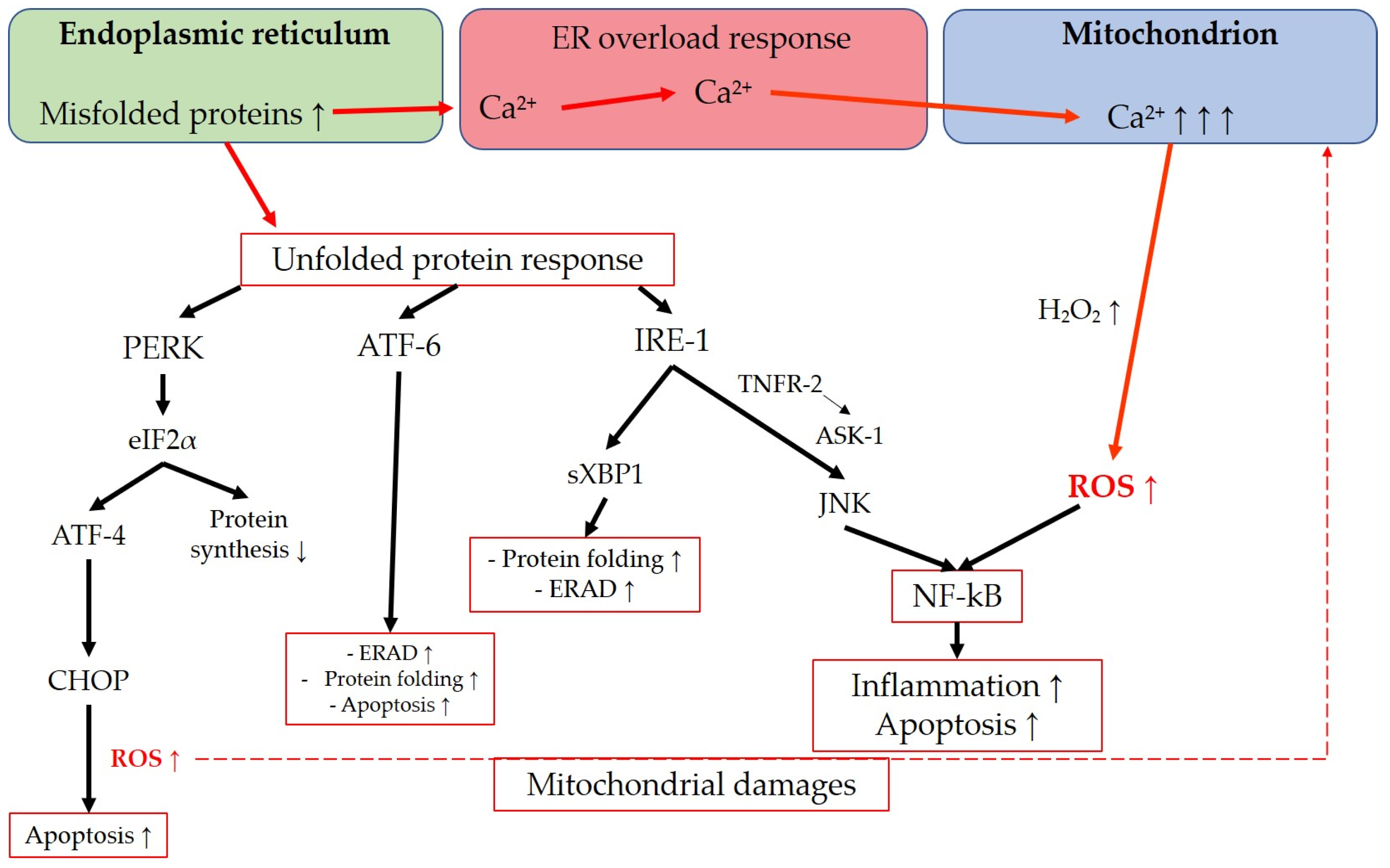
The endoplasmic reticulum is an intracellular organelle with multiple functions, including lipid synthesis, calcium storage, and protein processing. It regulates protein folding to ensure proper functionality, facilitates protein transportation, and detects misfolded proteins, which are then retained in the ER for degradation [231,232]. Various conditions, such as hypoxia, oxidative stress, viral infections, nutrient depletion, protein mutations, impaired glycosylation, or disrupted disulfide bond formation, can interfere with the ER’s physiological functions. These disturbances can lead to ER saturation and the accumulation of misfolded proteins in the ER lumen, resulting in endoplasmic reticulum stress [233,234,235]. ER stress activates multiple signaling pathways aimed at restoring cellular homeostasis [231,234]. The unfolded protein response (UPR) is triggered in response to ER stress and encompasses a range of signaling mechanisms designed to reduce protein synthesis, enhance protein folding, and increase protein degradation [236]. Additionally, the ER-associated degradation (ERAD) system is responsible for retro-transporting misfolded proteins from the ER lumen to the cytosol for clearance through the ubiquitin-proteasome system [234,237]. The UPR and ERAD are two independent quality-control mechanisms that can interact to eliminate misfolded proteins and maintain protein folding homeostasis [237]. The UPR can lead to either cell survival or apoptosis [235]. Prolonged activation of the UPR pathways can shift the balance toward cell death [234]. Three main UPR regulators have been extensively described: activating transcription factor (ATF)-6, inositol-requiring protein 1 (IRE1), and protein kinase RNA-like endoplasmic reticulum kinase (PERK). These transmembrane proteins with ER lumen domains serve as sensors of ER stress [235]. ATF-6 activates cascades to enhance the ER folding capacity, promote clearance of misfolded proteins through ERAD, and may also have a proapoptotic effect [231,235,238]. IRE1 initiates a pathway involving spliced X-box binding protein-1 (sXBP1), which promotes ERAD components, ER folding proteins, and autophagy [231]. Furthermore, IRE1 activates JNK through tumor necrosis factor receptor 2 (TNRF-2) and ASK1, leading to apoptosis [235], as well as inflammation via NF-kB induction [239]. PERK activates eukaryotic initiation factor 2α (eIF2α), which downregulates overall protein translation, indirectly protecting the cell from protein misfolding [235]. PERK also upregulates activating transcription factor 4 (ATF-4), which triggers the CCAAT/enhancer-binding protein (C/EBP) homologous protein (CHOP), exerting a proapoptotic function [240]. In this regard, Han et al. demonstrated that a sustained activation of ATF-4 and CHOP increases protein synthesis and leads to cell death through oxidative stress and ATP depletion [236]. These findings align with previous research showing that CHOP deletion in multiple mouse models of diabetes reduces oxidative stress [241]. A noteworthy aspect regarding the connection between ER stress and ROS generation is the ER overload response, which is activated when there is a high concentration of misfolded proteins in the ER lumen [234]. During the ER overload response, a significant amount of calcium ions (Ca2+) may be released from the ER, possibly through Ca2+ release channels, such as inositol 1,4,5-trisphosphate receptor (IP3R) or ryanodine receptor (RyR) [231,234]. This process can result in increased Ca2+ uptake from the ER to mitochondria, leading to the abnormal production of H2O2 and disruption of the ETC, ultimately causing mitochondrial dysfunction [231,242]. Moreover, UPR-related signaling can activate the endoplasmic reticulum oxidoreductin 1 (ERO1) and NOX, which are involved in oxidative protein folding under normal physiological conditions. However, in the context of ER stress, their activation can contribute to ROS production in the stressed ER [231,243]. Lastly, ROS generation resulting from both ER and mitochondrial dysfunction can activate NF-kB, a key transcription factor involved in inflammation and cell proliferation [244]. The main transductions in the interconnection between ER and mitochondria are summarized in Figure 6.
ER-related oxidative stress has been reported in both TMCs and RGCs in the context of glaucomatous optic neuropathy. Studies on TMCs have demonstrated the involvement of the PERK-eIF2α-ATF4-CHOP cascade in glaucomatous TM, both in human and murine cells, highlighting the activation of this pathway in glaucoma [245,246,247,248]. Similarly, various studies have described the implication of the PERK-eIF2α-ATF4-CHOP pathway in RGC loss [249,250,251,252]. The recent literature has highlighted the potential of targeting the PERK-eIF2α-ATF4-CHOP pathway as a therapeutic approach to prevent CHOP-related oxidative stress and apoptosis, thereby mitigating the TM structure and function loss and potentially reducing IOP elevation. Notably, in a recent study conducted by Gao et al., the protective effect of valdecoxib, a selective COX-2 inhibitor commonly used in the treatment of conditions such as osteoarthritis and rheumatoid arthritis was assessed against apoptosis induced by ER stress. The study demonstrated that valdecoxib inhibits the ATF4-CHOP pathway in “I/R-induced glaucoma-like” damaged cells, providing potential insights into its therapeutic efficacy in glaucoma management [253].
4.2. Therapeutic Perspectives in Glaucoma
4.2.1. Therapeutic Potential of Natural Compounds in Glaucoma
Naturally occurring antioxidant compounds offer a diverse range of potential therapeutic options. One such compound is resveratrol, a polyphenol found in peanuts, berries, grapes, and red wine, known for its anti-inflammatory and antioxidant properties in conditions like cancer, neurodegeneration, and aging [254]. In human glaucomatous TMCs, resveratrol has been reported to reduce the expression of proinflammatory molecules, such as IL-1α and iNOS, while increasing the production of NO by elevating eNOS expression. These effects contribute to its beneficial antioxidant effects in the TM [255]. Resveratrol is also an activator of suirtin1 (SIRT1), a nuclear NAD+-dependent deacetylase, highly relevant for the regulation of several antioxidant genes, performed by triggering the Nrf2/ARE (antioxidant response elements) pathway [256,257]. In an experimental rat model of glaucoma, resveratrol was shown to delay the loss of RGCs [258]. Additionally, studies by Ye and Meng have demonstrated that resveratrol protects RGCs from H2O2-induced apoptosis by inhibiting proteins involved in MAPK cascades (p38, JNK, and ERK) and activating antioxidant enzymes such as SOD, CAT, and GSH [259].
Another naturally occurring antioxidant molecule with promising characteristics is curcumin. In chronic high IOP rat models, curcumin has been shown to decrease ROS formation and prevent activation of apoptotic pathways by downregulating caspase-3, Bax, and cyt c [260]. Moreover, in an ex vivo mouse model of optic nerve cut, curcumin has been found to preserve RGC survival by preventing MAPK activation and inhibiting caspase-9 and caspase-3 activation [261].
Spermidine, a natural polyamine present in mushrooms and soybeans, possesses antioxidant properties and has demonstrated the ability to improve RGC loss and visual impairment in murine NTG models [262]. The same study also observed that spermidine treatment in murine optic nerve injury models promoted RGC survival by suppressing the ASK-1/p38/MAPK apoptotic pathway and inhibiting iNOS, particularly in microglial cells [263].
Flavonoids, known for their free radical scavenging properties, have been extensively studied. Extracts from the Gingko biloba L. plant, which contain over 70 different flavonoids, have shown interactions with apoptotic pathway proteins such as p53, Bax, Bcl-2, caspase-3, and caspase-9 [264]. These interactions suggest that Gingko biloba extracts may reduce RGC damage in glaucoma by inhibiting H2O2-related apoptosis through pathways involving p53, Bax/Bcl-2, and caspase-3/9 [265]. A dedicated clinical trial evaluated the use of oral antioxidants for glaucoma, comparing extracts of Gingko biloba with α-tocopherol for 3 months (NCT01544192), and completed phase III, reporting no clear clinical benefit for the use of Gingko biloba [240]. Another flavonoid, coenzyme Q10, a natural lipophilic compound, has demonstrated in murine glaucoma models its ability to reduce glutamate excitotoxicity and oxidative stress, promoting RGC survival and preventing apoptotic cell death by decreasing Bax expression and increasing the Bcl-2 associated agonist of cell death (Bad) protein expression [266]. Currently, a clinical trial (NCT03611530) is underway to evaluate the effect of a medical solution containing coenzyme Q10 and vitamin E on patients with primary open-angle glaucoma (POAG) [267].
The α-lipoic acid (ALA) is a naturally occurring antioxidant molecule found in various fruits and vegetables, as well as in the heart or liver of animals [268]. In murine glaucoma models (DBA/2J), Inman et al. demonstrated that ALA reduces oxidative stress and upregulates antioxidant agents such as HO-1 and NOS, possibly through the activation of Nrf2 [268]. In a more recent prospective case-control study, an ALA-based solution (also containing taurine, vitamins C and E, lutein, zeaxanthin, zinc, copper, and docosahexaenoic acid) was found to increase TAS and to decrease MDA, a marker of lipid peroxidation, in the plasma of patients with POAG [269].
Additionally, vitamin B3, also known as niacin, has been studied as a potential new treatment for glaucoma due to its antioxidant properties [270]. A study conducted in Korea revealed that patients with NTG have a lower intake of niacin compared to other nutrients, suggesting a possible association between vitamin B3 deficiency and NTG risk [271]. Williams et al. administrated nicotinamide (an amide form of niacin) to DBA/2J mice and demonstrated both preventive and therapeutic effects against the development of glaucoma, preserving the age-dependent reduction in nicotinamide adenine dinucleotide (NAD), a crucial molecule for mitochondrial health [272]. In a recent randomized controlled trial involving 57 patients with glaucoma, nicotinamide supplementation was shown to improve inner retinal function [273].
Currently, there is an ongoing randomized controlled trial (NCT04784234) evaluating a mixture of curcumin, Ginkgo biloba extract, alpha-lipoic acid, coenzyme Q10, and other naturally occurring compounds in 100 patients with POAG. The expected completion date for this study is the end of 2023.
4.2.2. NOX Inhibitors
A novel and promising class of antioxidant medications for glaucoma are NOX inhibitors. These compounds offer a new strategy to counteract glaucomatous damage by preserving RGCs from the detrimental effects of neuroinflammation and glial activation, potentially complementing traditional IOP-lowering therapies [274]. Among these compounds, GKT137831, also known as setanaxib, is particularly noteworthy. It acts as a dual inhibitor of NOX1 and NOX4 and has demonstrated protective effects against retinal inflammation and ischemia by reducing hypoxia-induced ROS generation [275]. Another intriguing molecule in this class is GLX7013114, a specific NOX4 inhibitor. In a study involving rats with α-amino-3-hydroxy-5-methyl-4-isoxazolepropionic acid (AMPA)-induced retinal excitotoxicity, intravitreal injections of GLX7013114 were found to attenuate glial activation [276]. Moreover, a recent publication reviewed the role of NOX and NOX inhibitors in glaucoma, highlighting the significant relationship between NOX4 and TGF-β in the fibrotic changes observed in glaucomatous TMCs, which contribute to functional impairment and elevated IOP [274,277]. These NOX inhibitors offer exciting prospects in antioxidant therapy for optic nerve diseases, as they target underlying mechanisms beyond IOP regulation, aiming to protect RGCs and to mitigate neuroinflammatory processes. Continued research in this field holds promise for the development of innovative treatments for glaucoma.
4.2.3. Exploring Nrf2 Activation for Antioxidant Therapy in Glaucoma
Exciting new prospects in antioxidant therapy for glaucoma involve the use of Nrf2 activators, which have the potential to provide benefits through their antioxidant and anti-inflammatory properties. One example is trimetazidine, an anti-ischemic medication that has been shown to activate the Nrf2-HO-1 pathway, leading to the inhibition of RGC apoptosis [278]. Astaxanthin (AST), a potent natural antioxidant found in microalgae and seafood, such as lobster, is another noteworthy compound in this context [279,280]. In a rat model of elevated IOP, AST has been demonstrated to reduce apoptotic pathways [281]. In mouse models of NTG, AST administration inhibited RGC degeneration and suppressed RGC loss [282]. Li et al. showed that AST activates Nrf2 and HO-1 in RGCs of mouse models, resulting in a decrease in RGC loss in glaucoma [283]. Sestrin2, a stress-induced protein with antioxidant properties, has also shown promise in activating Nrf2 by inhibiting the kelch-like ECH-associated protein 1 (Keap-1), thus protecting against RGC apoptosis in mouse tissues [284]. Additionally, eye drops containing metformin, an antidiabetic drug, have been demonstrated to preserve against fibrosis following glaucoma filtration surgery in rodent models by activating the AMP-activated protein kinase (AMPK)/Nrf2 signaling pathway [285]. A recent study in mice demonstrated that sustained intraocular release of erythropoietin can counteract glaucomatous pathogenic processes by reducing superoxide levels in the retina, upregulating antioxidant agents, and activating the Nrf2/ARE pathway through MAPK signaling [286].
4.2.4. Rho Kinase Inhibitors in Glaucoma Treatment
The class of Rho-associated protein kinase (ROCK) inhibitors has shown promise in glaucoma treatment. One notable ROCK inhibitor is netarsudil, which has been demonstrated to reduce fibrosis in the TM, leading to improved aqueous humor outflow and lowered IOP [287]. It has received clinical approval for use in the United States (2017) and Europe (2019) as a 0.02% ophthalmic solution for once-daily topical application [288]. Another ROCK inhibitor, Y-27632, has been shown to upregulate antioxidant enzymes such as CAT and to partially reduce ROS generation [289]. In addition to its beneficial effects on the TM, Y-27632 promotes phagocytosis in glaucomatous TM cells, leading to IOP reduction [290]. Ripasudil, also known as Rho kinase inhibitor K-115, has been evaluated in porcine retinal arterioles and shown to induce endothelium-independent relaxation and inhibit endothelin-1 activity. These findings highlight its potential as a potential antiglaucomatous medication [291]. The development of Rho kinase inhibitors offers a promising avenue for glaucoma treatment. By targeting the ROCK pathway, these inhibitors improve TM function, reduce fibrosis, and potentially lower IOP. Continued research and clinical investigations will further elucidate their efficacy and safety for glaucoma patients.
4.2.5. ER Stress Antagonists: Potential Antioxidant Drugs
Another subgroup of potential antioxidant drug includes compounds that antagonize ER stress. Among these compounds, 4-phenylbutyric acid (4-PBA) has been the subject of several studies. Originally used in urea cycle disorders and later in the treatment of cystic fibrosis in the 1990s [292,293], phenylbutyrate has been reported to attenuate ROS production in activated microglia [294]. Additionally, 4-PBA has been found to reduce oxidative stress caused by a high-fat diet or acute ammonia challenge by counteracting ER stress [295]. Furthermore, 4-PBA has shown the ability to decrease ER stress and prevent disease phenotypes in murine glaucoma models [296]. Importantly, it has also been demonstrated to lower IOP by promoting ECM degradation through the activation of matrix metalloproteinase (MMP)-9 [297].
4.2.6. Other Antioxidant Molecules
In addition to the previously mentioned compounds, several other antioxidant molecules have shown potential therapeutic effects in glaucoma.
For example, edaravone, a medication primarily used against stroke and known for its free radical scavenging properties [298] was described to suppress pJNK/p38 proapoptotic pathways in glaucoma models [43], preserving RGCs from death [299,300].
Valproic acid (VPA), a widely used antiepileptic drug, has been shown to upregulate antioxidant enzymes, such as SOD, CAT, and GPX, while inhibiting apoptotic pathways in rodent models of retinal ischemia–reperfusion injuries [301]. In murine models of NTG, VPA has been found to decrease oxidative stress and enhance RGC survival through a pathway associated with ERK [302]. Additionally, a recent Swedish study using retina explant models suggested that VPA possesses anti-inflammatory properties by reducing the expression of proinflammatory cytokines and attenuating microglial changes, highlighting its potential as an antineuroinflammatory drug in retinal diseases [303]. A randomized controlled trial assessing the effectiveness of VPA in glaucoma patients reported an improvement in visual acuity among patients with advanced glaucoma [304].
N-acetylcysteine, a molecule with radical scavenger properties originally used as antidote in cases of paracetamol overdose and more recently, as a mucolytic agent in bronchopulmonary disorders [305] has shown promise in mitigating retinal oxidative stress caused by high IOP when combined with brimonidine therapy in rat models of OHT [306]. It has also been demonstrated to target the HIF-1α pathway via BNIP3 (Bcl2 interacting protein 3) and the PI3K/Akt/mTOR pathway, thereby preventing hypoxia–mimetic induced autophagy in RGCs [307]. In murine models of NTG, N-acetylcysteine suppressed oxidative stress and autophagy in RGCs and increased levels of glutathione [308].
Rapamycin, a macrolide antibiotic with antineurodegenerative and neuroprotective properties described in Alzheimer’s and Parkinson’s diseases [309,310], has been found to enhance RGC survival in rodent glaucoma models. It achieves this by inhibiting the production of NO and TNF-α in microglia through the modulation of NF-kB activity and by maintaining Akt phosphorylation to inhibit RGC apoptosis [311].
Geranylgeranylacetone (GGA), a molecule used in the therapy of gastric ulcers and known for its antioxidant features, has been shown to reduce oxidative stress in the retina by triggering thioredoxin and heat shock proteins (Hsp)-72, thereby protecting against apoptosis [312]. In murine models of NTG, GGA has been demonstrated to decrease RGC loss through the upregulation of Hsp-70 and a reduction in caspase-3 and -9 activities [313].
These findings highlight the potential of these antioxidant molecules in providing neuroprotection and preserving retinal health in glaucoma.
4.3. Leber’s Hereditary Optic Neuropathy
4.3.1. General Aspects: Genetics, Clinical Presentation, and Current Therapeutic Options
Leber’s hereditary optic neuropathy (LHON) is a relatively rare disease compared to glaucoma, but it is considered the most frequent mitochondrial DNA (mtDNA) disorder [314]. It follows a maternal inheritance pattern and primarily affects young males, typically presenting between the ages of 15 and 35 [32]. The most prevalent mutations associated with LHON are m.3460G>A, m.11778G>A, and m.14484T>C, which account for approximately 90–95% of all cases [13]. These mutations affect the protein subunits of complex I in the mitochondrial respiratory chain [315]. Among these mutations, m.11778G>A is most frequently observed in Northern Europe, Australia, and Japan [8,12,316,317], while m.14484T>C is more common among the French-Canadian population [318,319,320]. Studies examining the complete penetrance of LHON have reported varying prevalence estimates in Europe, ranging from 1 in 30,000 to 1 in 54,000, approximately 0.00002% of the population [8,9,10,11,12]. A comprehensive Australian review, which accounted for incomplete disease penetrance in variant carriers and used a well-characterized population-based control cohort to minimize sampling bias, estimated a prevalence of 1 in 800 or approximately 0.00125% [13]. Similarly, another study using a similar methodology concluded that LHON prevalence in the general population exceeds 1 in 1000 [14]. These findings suggest that most LHON mutations remain silent until unknown triggers precipitate their conversion from asymptomatic mutation carriers to symptomatic individuals [321]. Indeed, only around 50% of males and 10% of females harboring a pathogenic mtDNA mutation effectively develop the disease [315]. LHON also exhibits a gender bias, with males being more likely to be affected than females [315]. This gender predisposition has been associated with X-linked susceptibility loci [322,323]. Other studies have explored potential factors that may explain the incomplete penetrance and have found that smoking and excessive alcohol consumption are more common in individuals with LHON compared to asymptomatic carriers [324]. Additionally, it has been suggested that vitamin B12 (cobalamin) deficiency could accelerate the symptomatology in LHON carriers [325] and that regular screening for vitamin B12 levels may be considered in LHON carriers and patients [314].
Clinically, LHON is highly disabling and leads to subacute bilateral irreversible vision loss [315]. A recent study examining the quality of life in 17 LHON patients from different countries (Germany, UK, France, and the US) described several daily life challenges in, for example, physical capabilities, interpersonal relationships, work and recreational activities, that significantly impact their well-being [326]. A cost-of-illness study focusing on inherited retinal disorders (IRD) estimated that in 2019, the overall costs associated with LHON amounted to USD 84–200 million and CAD 10–42 million in the US and Canada, respectively [327]. Moreover, healthcare costs, including therapy expenses, represented only 7% and 2% of the total costs in the US and Canada, respectively [327].
It is widely acknowledged that the treatment of LHON should ideally begin within one year from the onset of visual loss [321,328]. In relation to disease progression, it is divided into three stages starting from the onset [329]:
- Subacute phase (<6 months): During this phase, patients commonly experience blurred vision and impaired color perception without pain, and their pupillary reflex remains unaffected [315]. Approximately 75% of cases initially experience visual loss in one eye, with the contralateral eye becoming affected within a few weeks [315,330]. Fundoscopy may reveal axonal loss in the papillomacular bundle and circumpapillary telangiectasias. Perimetry often demonstrates typical centrocecal or central scotomas [315,330,331]. Optical coherence tomography (OCT) may show swelling of the peripapillary RNFL. Magnetic resonance imaging (MRI) is commonly performed for differential diagnosis [330];
- Dynamic phase (6–12 months): During this stage, fundoscopic signs such as telangiectasias and RNFL edema gradually regress [330];
- Chronic phase (>12 months): In the chronic phase, there is a further decline in visual acuity and visual field loss. Fundoscopic examinations may reveal optic nerve head atrophy, while OCT may indicate thinning of the RNFL [330].
An innovative therapeutic approach for LHON has also been developed in the realm of gene therapy. An example of this is lenadogene nolparvovec (Lumevoq©, Gensight Biologics SA, Paris, France), which was administered via intravitreal injections and designed to treat LHON caused by the m.11778G>A variant [332]. This drug utilizes a replication-defective, single-stranded DNA recombinant adeno-associated virus vector of serotype 2. It carries a codon-optimized complementary DNA encoding the human wild-type subunit protein that is affected in the m.11778G>A variant [332]. The viral vector is designed to deliver the therapeutic gene into the targeted cells, aiming to enable them to produce the NADH dehydrogenase 4 (ND4) enzyme, which can cause restoration of the ETC [333,334].
However, it is important to note that this drug has been withdrawn by the EMA as of 20 April 2023, as it was found to be ineffective in improving outcomes for patients with LHON (source: EMA website, https://www.ema.europa.eu/en/medicines/human/withdrawn-applications/lumevoq (accessed on 17 July 2023).
An international consensus has established that idebenone (IDE), a synthetic analogue of coenzyme Q10 (COQ10), is the first disease-specific antioxidative drug authorized by the European Medicines Agency in 2015. It has been shown to provide benefits when administered during the subacute or dynamic stages of LHON at a dosage of 900 mg/day. However, it is not recommended for use during the chronic phase [328,329,335].
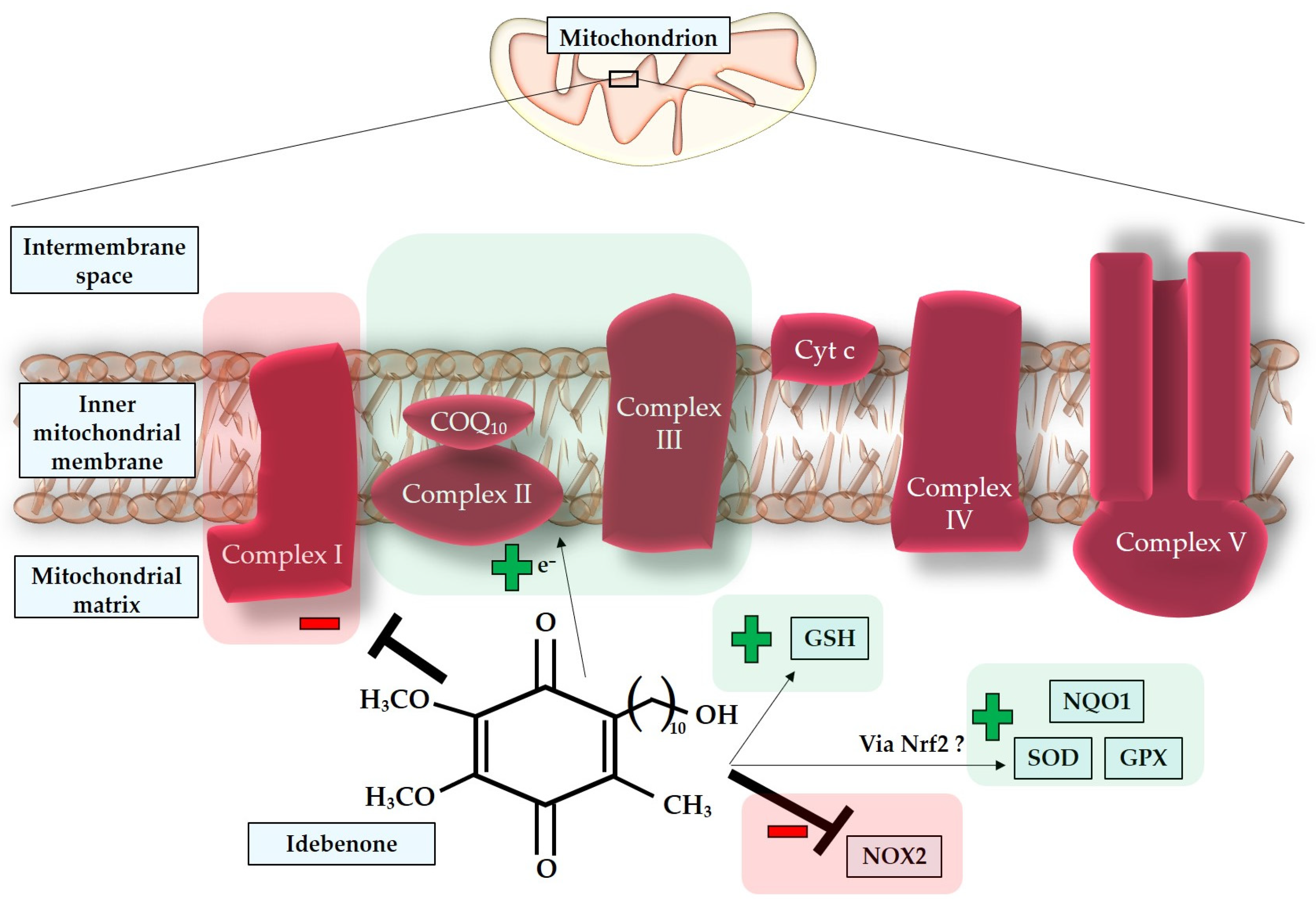
Extensive research has been conducted to discover medications that can restore the ETC, which is crucial for ATP synthesis and maintaining a normal redox status [321]. In this context, IDE functions as an electron carrier within the mitochondrial ETC, facilitating electron transfer from complex II to complex III and promoting ATP synthesis [336]. However, it is important to note that Jaber and Polster described IDE as a pro-oxidative molecule that also inhibits complex I [336]. Additionally, Gueven et al. extensively reviewed the pro-oxidative properties of IDE and questioned the actual antioxidative activity of this drug [337]. They compared studies that detected low nanomolar concentrations of IDE in target tissues for only a short period of time with the majority of publications on IDE, which reported the need for micromolar concentrations to achieve an antioxidative effect [338,339,340]. One possible explanation for the apparent contradiction regarding the low bioavailability of IDE is the suggestion of an indirect antioxidant effect through the inhibition of NOX2, which normally produces damaging ROS, such as superoxide [341]. IDE may also activate different signaling pathways that increase the activity of SOD, NAD(P)H quinone oxidoreductase 1 (NQO1), glutathione (GSH), and glutathione peroxidase (GPX), possibly through Nrf2, a transcription factor that regulates the expression of SOD, NQO1, and GPX. Another proposed explanation for the rapid pharmacokinetics of IDE focuses on the possibility that some of its metabolites, such as 6-(9-carboxynonyl)-2,3-dimethoxy-5-methyl-1,4-benzoquinone (QS10), can also provide therapeutic effects [337]. In support of this hypothesis, a previous Italian study concluded that QS10, like IDE, can bypass the defect in complex I and, unlike IDE, can replace the endogenous coenzyme Q10 (CoQ10), potentially exhibiting even greater activity than IDE in diseases caused by complex I defects or CoQ10 deficiency [342]. In Figure 7, direct and indirect effects of idebenone are represented.
4.3.2. Oxidative Stress in the Pathogenesis of Leber’s Hereditary Optic Neuropathy
Mitochondrial DNA mutations in LHON lead to a defective complex I, resulting in impaired ETC activity [343]. Studies on fibroblast and cybrid mitochondria affected by LHON mutations have shown varying levels of mitochondrial aerobic respiration alterations depending on the specific mutations [344]. Decreased respiration rates have been reported as approximately 20–28% for m.3460G>A, 30–36% for m.11778G>A, and 10–15% for m.14484T>C [344]. Despite the impaired oxidative phosphorylation and reduced ATP production associated with complex I dysfunction, it is proposed that LHON partially compensates for these deficiencies through glycolysis, an alternative energetic pathway observed in human tissues. This compensation is suggested to maintain the total cellular ATP concentration despite the severe decrease in complex I-related ATP synthesis [343,345]. Hence, additional processes need to be considered to explain the clinical manifestation of the disease. Carelli et al. proposed that decreased pH resulting from defective complex I affects redox sites during aerobic respiration, leading to the overproduction of ROS [345]. Studies on cells carrying LHON mutations have reported increased levels of oxidative stress biomarkers, such as 8-OHdG, the reduced activity of antioxidant systems, including glutathione reductase and Mn-SOD, and elevated levels of oxidized glutathione (GSSG) [346,347,348]. Lin et al. demonstrated in a murine model of mtDNA mutations that LHON mutations cause a systemic elevation in ROS production, with chronic elevation in ROS generation observed in synaptosomes, optic nerves, and RGCs, suggesting a more significant damaging role of oxidative stress compared to ATP depletion [349]. Another study in mice aimed at establishing a murine model of LHON found that a mutation in the subunit ND4 of complex I led to mitochondrial structure disassembly, increased ROS levels, ONH swelling, and RGC apoptosis [350]. Additionally, an alternative hypothesis suggests that altered permeability transition pores, possibly due to Ca2+ and ROS-mediated depolarization of the mitochondrial membrane, may play a role in LHON. These opened permeability transition pores in cybrid cells carrying LHON mutations could facilitate the release of cytochrome c from mitochondria to the cytosol, activating the apoptotic pathway [351].
Dysfunctional complex I in LHON may lead to sensitization of permeability transition pores primarily due to increased ROS generation and cytosolic Ca2+ overload. The reduced ATP supply to the Ca2+ pumps in the ER membranes may contribute to the intracellular Ca2+ overload by impairing Ca2+ uptake from the cytosol to the ER [351]. Consequently, the death of RGCs in LHON is likely influenced by the altered redox status and intracellular Ca2+ overload, with an emphasis on the role of ROS in triggering proapoptotic pathways (Figure 8) [349,352,353,354,355,356]. Apoptosis in LHON has been described as mediated by caspases, including the Fas-induced pathway involving caspase-8 [355,356]. The activation of caspase-8 leads to the cleavage of protein Bid (BH3 interacting-domain death agonist), a component of the Bcl-2 family, indirectly causing the release of cytochrome c from mitochondria. Cytochrome c then binds to Apaf-1, along with caspase-9, forming a complex called the “apoptosome.” The apoptosome subsequently activates caspase-3, which is responsible for cellular disassembly and apoptosis. Additionally, caspase-independent pathways have been observed in LHON, involving the release of cytochrome c, AIF (apoptosis-inducing factor), and EndoG (endonuclease G) into the cytosol [357]. These apoptotic processes contribute to the demise of RGCs in LHON.
4.4. Therapeutic Perspectives in Leber’s Hereditary Optic Neuropathy
α-Tocotrienol quinone, also known as EPI-743, is a synthetic molecule classified as a para-benzoquinone. It targets NQO1 and leads to the replenishment of intracellular glutathione stores, thereby increasing antioxidant capabilities [358]. In vitro studies have shown that EPI-743 is over one thousand-fold more effective than IDE in protecting cells from oxidative stress [359]. An investigation into the effect of EPI-743 in LHON demonstrated disease progression arrest and the reversal of vision loss in four out of five patients treated with EPI-743 within 4 months of the onset of visual loss [360]. Another study involving 12 patients with active phase LHON concluded that EPI-743 stabilized or improved visual function in approximately 70% of the treated eyes [361]. However, larger trials in the LHON population are needed to further validate these findings. Currently, EPI-743 is in phase II of various clinical trials (NCT01370447; NCT04378075; and NCT02352896) aimed at evaluating its effectiveness in primary mitochondrial disorders [362].
Elamipretide, also known as MTP-131, Bendavia, or SS-31, is a relatively new mitochondria-targeting peptide that has been shown to protect RGCs from oxidative stress-induced apoptosis [363]. Other interesting molecules that modulate mitochondrial redox status and enhance mitochondrial biogenesis include sonlicromanol (also known as KH176) and KL1333 [362]. KL1333 is an NAD+ modulator that improves the NAD+/NADH ratio and has been demonstrated to decrease lactate and ROS levels while increasing ATP synthesis in fibroblasts from patients with MELAS (mitochondrial encephalopathy, lactic acidosis, and stroke-like episodes) [364].
These emerging therapeutic options provide potential avenues for the treatment of LHON by targeting oxidative stress and mitochondrial dysfunction.
4.5. Anterior Ischemic Optic Neuropathy
4.5.1. General Aspects: Prevalence, Clinical Presentation, and Current Therapies
Among optic nerve disorders, anterior ischemic optic neuropathies stand out as one of the most complex and significant conditions. These conditions are classified into anterior and posterior optic neuropathies (AION and PION), distinguished by the presence or absence of disc edema, respectively [365]. They are further categorized into arteritic and nonarteritic diseases based on the presence or absence of vasculitis, which results in reduced perfusion to the optic nerve head [366]. Nonarteritic anterior optic neuropathy (NA-AION) is the most common acute optic nerve disease in patients older than 50 years [367]. Clinical presentations include sudden acute unilateral vision loss without pain. However, effective treatments for these conditions remain challenging [16,367,368]. Giant cell arteritis (GCA) is the most common subtype of arteritic anterior ischemic optic neuropathy (A-AION). High intravenous corticosteroid doses serve as the first-line therapy for GCA [369]. It is crucial to initiate rapid treatment once GCA is suspected [370]. A systematic review predicts that by 2050, over 3 million individuals in Europe, North America, and Oceania will suffer from giant cell arteritis primarily due to aging [371]. Additionally, the estimated economic burden resulting from visual disability caused by GCA in the United States is expected to exceed USD 76 billion by 2050 [371]. Clinically, GCA manifests in individuals over 50 years old, presenting classic symptoms such as severe headaches, jaw claudication, cutaneous allodynia, and, in the majority of patients, additional symptoms like fever, weight loss, depression, and night sweats [370]. Despite the administration of high steroid doses, including intravenous methylprednisolone 1000 mg/day for 3 days, followed by a maintenance dose of 1 mg/kg of prednisone [372], the chances of visual recovery in GCA-related visual loss remain very low [373]. Furthermore, the long-term use of steroids is associated with various side effects and rebound syndrome, while GCA is prone to relapse [374]. Consequently, there is a significant research need for new therapeutic strategies, and immunomodulating therapies have emerged as promising avenues in the treatment of GCA. One example of an immunomodulating therapy is tocilizumab (TCZ), an interleukin-6 (IL-6) receptor antibody. TCZ has already received approval from the National Institute for Health and Care Excellence and NHS England for the treatment of refractory GCA [375]. However, the use of TCZ is restricted due to its known side effects, including alterations in liver enzymes and cholesterol levels, gastrointestinal perforation, infections, headaches, and arterial hypertension [376,377]. In the field of immunomodulation, several new immunoglobulins are currently undergoing clinical trials for the treatment of GCA [378].
Nonarteritic anterior ischemic optic neuropathies (NA-AION) occur as a result of events that lead to hypoperfusion of the optic nerve head. A recent comprehensive meta-analysis revealed several risk factors for NA-AION, including male gender, hypertension, hyperlipidemia, diabetes mellitus, coronary heart disease, sleep apnea, factor V Leiden heterozygosity, and a history of cardiovascular medication use [379]. Pathogenetically, Hayreh SS. described that NA-AION involves hypoperfusion of the optic nerve head, leading to hypoxia of the retinal ganglion cell axons. This, in turn, results in the stasis of the axoplasmic flow and the generation of swollen axons [366]. Consequently, optic disc edema occurs, causing compression of the capillaries supplying the optic nerve head, creating a vicious cycle. The primary trigger for the initial hypoperfusion of the optic nerve head is typically a transient drop in blood pressure, often occurring during sleep, such as nocturnal arterial hypotension or hypotension after sleeping during the day. Severe occlusion of the internal carotid or ophthalmic artery rarely causes ocular ischemia in NA-AION cases [366]. While embolic lesions can be an occasional cause of NA-AION, they result in more extensive and permanent damage to the optic nerve head compared to the hypotensive type [366]. The clinical onset of NA-AION is sudden and typically affects a single eye, resulting in vision loss without pain, often discovered by patients upon waking in the morning [380]. Fundoscopy and perimetry are essential diagnostic tools for NA-AION. The Goldmann perimeter, in particular, helps detect classic visual field defects in NA-AION. A study on 312 consecutive eyes reported central scotomas in nearly half of the cases, with the absolute inferior nasal defect being the most commonly detected [381].
Therapeutic options for NA-AION remain a challenging clinical issue. The effectiveness of steroids, whether administered orally or intravenously, is a subject of intensive debate [382]. A large nonrandomized cohort study in the United States involving 696 eyes, spanning from 1973 to 2000, suggested that systemic steroid treatment with oral prednisone (80 mg per day) during the acute phase may increase the probability of visual acuity and visual field improvement after six months [383]. However, due to the nonrandomized nature of this study and potential biases, the findings should be interpreted with caution [384]. Katz and Trobe extensively reviewed possible treatment strategies for NA-AION in both conservative and surgical fields and concluded that optic nerve fenestration surgery is ineffective and potentially harmful, while the efficacy of steroids remains uncertain [385]. In an Indian randomized controlled trial involving 38 patients with NA-AION, Saxena et al. found that oral prednisolone at 80 mg per day reduced the duration of disc edema and improved the electrophysiological parameters of the optic nerve but did not result in a visual acuity benefit after six months [386]. One potential clinical approach is the use of α2-adrenergic agonists, such as brimonidine, in the acute phase of AION through topical application. A study investigating the neuroprotective effect of brimonidine on anterior optic neuropathy in rodents (rAION) demonstrated a decrease in RGC loss in mice treated with brimonidine in the acute phase, possibly through a reduction in VEGF-A and HIF-1α expression [387]. These findings align with previous studies that also demonstrated the effectiveness of brimonidine in the acute phase of rAION [388,389]. In summary, the significant lack of effective treatments for NA-AION and its relatively high prevalence underscore the urgent need for research in novel therapeutic approaches. Exploring the antioxidative branch of treatment holds promising potential to make significant contributions in this regard. By focusing on antioxidant strategies, new therapeutic avenues can be explored to address the challenges posed by NA-AION.
4.5.2. Oxidative Stress in the Pathogenesis of Anterior Ischemic Optic Neuropathy
GCA is an autoimmune disorder characterized by granulomatous infiltration involving T-cells and activated macrophages, including multinucleated giant cells, primarily observed in the vascular wall of the aorta and its main branches [370,390,391,392]. Vascular aging is considered one of the critical risk factors for GCA, as oxidative stress is believed to play a significant role in its pathogenesis [370]. This is likely attributed to age-associated mitochondrial dysfunction, leading to an increased ROS generation in endothelial cells and vascular smooth muscle cells [391,393,394]. A study on GCA patients has demonstrated the presence of neutrophils in the vascular lumen and adventitia tissues surrounding the external lamina elastica of temporal arteries, producing high levels of extracellular ROS [395]. Additionally, systemic oxidative stress parameters, such as TAC and MDA levels, along with intracellular leukocyte ROS levels, were found to be elevated in GCA patients [396], and the findings support the hypothesis that reactive species contribute to the pathophysiology of GCA by inducing vascular stress in large and medium-sized vessels.
In the pathogenesis of NA-AION, damage may arise as a consequence of hypoxia in the optic nerve head. Cellular hypoxia resulting from glucose and oxygen depletion, followed by reoxygenation during the ischemia/reperfusion (I/R) process, can lead to an overproduction of reactive species [397,398,399,400]. It has been proposed that systemic corticosteroids are effective in the acute phase of NA-AION, as they suppress NF-kB activation during inflammation [383]. NF-kB is a transcription factor that can be triggered by ROS-related cascades, as described in the pathogenesis of glaucoma [40,208,401]. ROS have been shown to cause immune-mediated neuronal injuries, disruption of the blood–optic nerve barrier, apoptosis, and autophagy through damage to DNA, proteins, and lipids [402,403]. Studies have revealed that I/R injuries in ischemic neurons lead to somatic autophagy of axonal mitochondria, resulting in increased retrograde movement and decreased anterograde movement of mitochondria, ultimately reducing the functional mitochondria available for ATP synthesis [403]. Our laboratory’s study on short-term ischemia in porcine models demonstrated hypoxia-related alterations characterized by the upregulation of HIF-1α, VEGF-A, NOX2, iNOS, and high levels of ROS [143]. Supporting these findings, a study on NOX2-deficient murine models (NOX2−/−) showed that these mice displayed neuroprotection in retinal I/R injury scenarios [228].
Interestingly, a Turkish study investigating plasma samples from 18 newly diagnosed NA-AION patients found no significant differences in systemic oxidative stress parameters, including TOS and TAS, when compared to healthy controls [402]. However, genetic investigations have revealed an association between NA-AION and a loss-of-function deletion in the gene GSTM1, which encodes one of the three isoforms of the antioxidative enzyme glutathione-S-transferase (GST) [404,405]. Additionally, another study reported a higher frequency of mitochondrial DNA mutations in NA-AION patients compared to controls, suggesting that mitochondrial dysfunction may be a risk factor for NA-AION [406].
Based on the current literature and the pivotal role of hypoxia in NA-AION, some studies have provided evidence that conditions leading to increased ROS generation may contribute to the risk of developing NA-AION. These findings highlight the potential involvement of ROS in the pathogenesis of this disease.
4.6. Therapeutic Perspectives in Anterior Ischemic Optic Neuropathy
4.6.1. Giant Cell Arteritis
Nuclear sirtuins, such as the before-mentioned SIRT1, are proteins that potentially have an antioxidative impact on GCA. These enzymes inhibit inflammation and oxidative stress by transcriptionally repressing various inflammation-related genes [407,408]. A recent study observed SIRT1 downregulation and subsequent ROS generation in individuals with GCA, suggesting a potential therapeutic effect of SIRT1 activators [396]. While immunomodulating molecules have been extensively investigated due to the autoimmune nature of GCA, there is growing scientific interest in exploring new antioxidant molecules such as nuclear sirtuins.
4.6.2. Nonarteritic Anterior Ischemic Optic Neuropathy
An investigation using rAION mice and intravitreal injections of the Rho-kinase inhibitor, E212, immediately after optic nerve infarction resulted in increased SOD activity, decreased ROS levels, reduced oxidative stress, and preservation of the blood–retinal barrier [409]. Our laboratory conducted a study on mice with I/R retinal injuries and found that betulinic acid, a natural compound found in plane bark, leaves, and fruit peel, preserved vascular function, attenuated ROS formation, and upregulated SOD and HO-1 [131]. Oroxylin A, a bioactive flavonoid extracted from Scutellariae baicalensis Georgi, showed potential protective effects in rAION by activating Nrf2, increasing HO-1 and NQO1 activity, promoting RGC survival, maintaining RNFL thickness, and exhibiting anti-inflammatory effects by reducing IL-6 and increasing TGF-β levels [410]. N-butylidenephthalide, another molecule, demonstrated a neuroprotective role in rAION by inhibiting the NF-κB signaling pathway [411]. Vitamin B3 (niacin) was found to provide neuroprotection from oxidative stress in rAION by activating Nrf2, increasing SOD expression, inhibiting mitochondrial apoptosis, and reducing the inflammatory response through NF-κB inhibition [368].
Geranylgeranylacetone (GGA), previously mentioned as a promising molecule for glaucoma therapies, was also tested in murine models of I/R retinal injury and demonstrated a decrease in RGC death by inhibiting the p38 MAPK apoptotic pathway [412]. In a more recent study, GGA was shown to induce an increase in Hsp70 levels, inhibiting glial activation, autophagy, and apoptosis [413]. Astaxanthin, previously identified as a potential antiglaucomatous drug, possesses antioxidant and antiapoptotic properties. Lin et al. found that astaxanthin preserved visual function, increased RGC survival, and inhibited apoptosis by blocking the Akt pathway in rAION [414]. Resveratrol, previously investigated for its potential in preventing RGC loss in glaucoma, has also been studied in I/R injuries. It was found to attenuate glial activation and RGC death by suppressing HIF-1α-VEGF-A upregulation and activating the downregulation of the PI3K/Akt pathway [415]. Resveratrol also protects RGCs from ischemia-related injuries by increasing Opa1 expression [416]. Our recent research demonstrated that resveratrol prevents vascular dysfunction and RGC death in I/R-induced murine models, possibly through the inhibition of the I/R-related upregulation of NOX2 [98]. These findings highlight potential therapeutic approaches for AION by targeting oxidative stress, inflammation, and mitochondrial dysfunction.
4.7. Optic Neuritis
4.7.1. General Aspects
Optic neuritis (ON) is an idiopathic inflammatory demyelinating disease of the optic nerve [417,418], which can be associated with the neuromyelitis optica [419] or can also appear as initial manifestation of multiple sclerosis, a chronic demyelinating disorder of the central nervous system [420,421]. In addition, reported risk factors for ON are granulomatous diseases, infections, and autoimmune pathologies [422]. In the pathogenesis of ON are involved activated T-cells, which releasing an abnormal volume of inflammatory cytokines, elicit demyelination and disruption of the blood–optic nerve barrier and lead to the loss of retinal ganglion cells and ONH atrophy [44,419,423,424,425]. ON is described as the major cause of acute optic nerve disorder in young patients [419], being commonly observed between 20 and 45 years and predominantly in women, with a ratio of 3:1 [17,420,426]. Its clinical presentation can occur through single or multiple episodes and usually consists of a sudden and unilateral loss of visual acuity, afferent pupillary impairment, orbital pain by eye movements and altered color perception, in the presence or absence of disc edema [427]. Diagnostically, fundoscopy and OCT help to detect ONH-anomalies and RNFL thinning, respectively. A study estimated that OCT can reveal thinning of the RNFL, with an average reduction of 33% compared to controls and an average reduction of 27% between impaired and unimpaired eyes of the same patient [428]. Furthermore, it has been reported that RNFL thinning is visible in 85% of all patients suffering from ON within 3–6 months after the acute phase [429]. In addition, perimetry can evidence visual field loss limited to the nerve fiber bundle region, with paracentral, partial arcuate, or arcuate defects [430]. A cranial MRI scan is performed for differential diagnosis and to evaluate the association with multiple sclerosis [420]. Current standard therapies of ON include high intravenous steroid doses (1000 mg IV methylprednisolone) during acute phases, which were reported to facilitate visual recovery, whereas oral prednisone alone is not recommended, as it failed to show comparable improvements [431]. However, a systematic review examining six clinical trials globally, including 750 patients affected by ON, did not evidence beneficial effects neither through intravenous nor via oral steroid therapy, compared with the placebo groups, in terms of the visual field, visual acuity, and contrast sensitivity outcomes [432]. Alternatively, the use of intravenous immunoglobulins in acute ON, which may be considered in steroid-refractory cases [433], failed anyway to provide benefits for generalized practice [434,435].
4.7.2. Oxidative Stress in the Pathogenesis of Optic Neuritis
Various investigations succeeded to provide evidence on an altered redox status in the context of ON, using experimental autoimmune encephalomyelitis (EAE) models [411,436,437,438] and an animal model originally established for multiple sclerosis, often employed in experiments aimed to study optic neuritis [427]. Moreover, studies on patients affected by ON showed significant ROS-related anomalies, such as an elevated fraction of oxidized thiol [439] or reduced bilirubin serum levels [440], indicators of high ROS generation and low antioxidant status, respectively. As mentioned before, similar to NA-AION and LHON, ON loss-of-function deletions in the gene coding for the antioxidative enzyme glutathione-S-transferase were also detected [405], therefore emphasizing a decreased antioxidant activity as a possible risk factor. As we previously highlighted in glaucoma, an elevated ROS generation and active inflammatory grade are deeply interconnected and tend to trigger and intensify each other in a process where NF-kB and cytokines are fundamental pathomechanistic players [441]. In relation to ON, increased ROS are responsible for myelin phagocytosis and stimulate changes in the permeability of the blood–brain barrier, favoring migration and infiltration of active T-cells, which, in turn, reflects in abnormal inflammation and augmented ROS production [439]. Oxidative damage may possibly be an initiator for demyelination and neurodegeneration [439], which finally drives to RGC loss [417].
More large studies designed to interpret the multiple aspects in the pathogenesis of ON, including the role of oxidative stress, may deliver new knowledge for the design of more effective pharmacological drugs to treat this disease.
4.8. Therapeutic Perspectives in Optic Neuritis
While a prompt administration of anti-inflammatory drugs during the acute phase of optic neuritis (ON) may help preserve vision [442], the use of steroids alone has not been shown to protect the optic nerve from inflammatory demyelination or prevent degeneration of retinal ganglion cells (RGCs) [417,443]. Therefore, antioxidant approaches have been explored to support nerve fiber integrity by counteracting inflammation and demyelination, thus preventing RGC death [417,444]. In preclinical studies on ON models, several major antioxidant targets have been investigated, including the activation of Nrf2 and the suppression of ROS-related proapoptotic signaling, such as blocking ASK-1 and the pJNK/p38 pathway.
Dimethyl fumarate, a Nrf2 activator that has been licensed for the treatment of relapsing-remitting multiple sclerosis in the US since 2013, has shown promising results in experimental autoimmune encephalomyelitis (EAE) models, reducing the severity and relapses of optic neuritis and preserving RGCs from cell death [444]. Gypenosides, an extract from Gynostemma pentaphyllum, have demonstrated beneficial antioxidant effects on the retina [445]. In a study on murine models of optic neuritis, gypenosides led to a decrease in iNOS and COX2 expression while activating Nrf2, resulting in the upregulation of HO and GPX, along with free radical scavenging and anti-inflammatory activities [446]. α-Lipoic acid, which potentially activates Nrf2 [268], has been investigated in EAE models, showing increased RGC survival and anti-inflammatory effects [447]. However, a dedicated trial on 31 patients with ON (NCT01294176) reported good tolerability after oral supplementation (600 mg twice a day) of α-lipoic acid [448] but did not show significant benefits in retinal nerve fiber layer thickness after 24 weeks compared to the placebo group [449]. Consequently, the trial ended in phase I due to insufficient recruitment [450]. As previously mentioned, SIRT1, known to activate Nrf2, has also been studied [417]. In an EAE study, an adeno-associated virus vector was utilized as a delivery vehicle to enhance SIRT1 expression, demonstrating that the selective upregulation of SIRT1 promotes RGC survival and preserves axons from demyelination through intravitreal injection [417]. Another investigation explored the use of matrine, an extract from the herb Radix Sophorae Flavescentis, in EAE models. Matrine induced an overexpression of SIRT1, the subsequent upregulation of Nrf2, and collectively resulted in mitochondrial biosynthesis, reduced ROS formation, and the suppression of inflammation and demyelination [451].
The inhibition of ASK-1 also showed encouraging preclinical results in mitigating oxidative-stress-induced RGC apoptosis. The ASK-1 inhibitor MSC2032964A has been demonstrated to alleviate neuroinflammation and diminish optic nerve demyelination in EAE murine models [452]. Spermidine, previously mentioned as a potential antioxidant for glaucoma, also suppresses the ASK-1/p38/pJNK pathway [263] and has been reported to reduce RGC apoptosis in EAE mice [453].
A recent publication investigated the effects of edaravone, previously mentioned, in cultures and mice with neuromyelitis optica spectrum disorder and found that it promoted remyelination through activation of the mTOR complex I (mTORC1) signaling pathway [454]. Currently, there is an ongoing trial (NCT05540262) that aims to assess the impact of edaravone in patients with optic neuritis who are positive for aquaporin-4 antibodies. The trial is expected to be completed by the end of 2024.
5. Other Rare Optic Neuropathies
5.1. Traumatic Optic Neuropathies
5.1.1. General Characteristics
Craniofacial traumas can directly or indirectly insult the optic nerve, leading to so-called traumatic optic neuropathies (TONs) [455,456]. Direct traumas are caused by injuries to the optic nerve through the intracranial fragmentation of bones or also through contusion, causing anatomical disruption [457]. Indirect traumas occur when compression and disruption of pial vessels cause a reduction in the vascular perfusion in the optic nerve [458,459]. Moreover, a deformation of the skull at the optic canal, and more specifically at its intracranial end, was reported to also be responsible for optic nerve injuries in indirect traumas [460]. The most reported causes of TON are vehicle accidents, bicycle accidents, falls, assaults, and sport injuries [24]. Indirect TON is more common than direct forms and occurs in 0.5–5% of all closed head injuries and 2.5% of all midfacial fractures [22,461,462]. In terms of diagnosis, a brain MRI scan is typically conducted [456]. Therapeutically, reported options for the treatment of traumatic optic neuropathy (TON) include high-dose corticosteroids or surgical decompressions [456]. However, both of these treatment approaches are accompanied by controversies due to the pharmacological side effects of steroids or the potential complications associated with surgery [456,463]. Consequently, exploring neuroprotective strategies to prevent optic nerve damage represents a promising curative option with substantial potential for TON [456].
An emerging and intriguing area of research for traumatic and inflammatory optic nerve disorders is neuromodulation based on biomedical ultrasound stimulation. This field is continuously evolving, not only for the retina but also for other ocular structures, such as the cornea and ciliary body [464]. Moreover, it extends beyond ophthalmology, finding applications in fields like cardiology and the peripheral nervous system [465,466,467,468,469]. Retinal ultrasound stimulation is a promising therapeutic approach for TON [470]. Some studies reported that curative strategies based on ultrasound may prevent oxidative-stress-induced damage [471,472], even on retinal pigmental epithelial cells in vitro [473]. In cases of complete RGC degeneration resulting from severe optic nerve trauma, ultrasound neuromodulation may offer a potential noninvasive approach to restore vision. High-frequency ultrasound can biochemically initiate localized neuronal responses, triggering electrically evoked cortical potentials along the visual pathway [470,474]. Implantable treatments, including a retinal stimulating piezo-array, are also being explored as wireless retinal prostheses capable of eliciting visual percepts in cases of irreversible visual loss [475]. A study conducted on rabbits demonstrated that an intraneural electrode array positioned in the intracranial segment of the optic nerve can generate selective activation patterns in the visual cortex, sparking interest in the potential use of optic nerve prostheses for total retinal detachment cases [474]. Another intriguing possibility is direct stimulation of the visual cortex bypassing the visual pathway, which was recently explored using hybrid noninvasive ultrasound techniques [476].
5.1.2. Pathogenesis of Traumatic Optic Neuropathies: Role of Oxidative Stress and Ca2+
In murine models, traumatic optic neuropathy (TON) has been shown to be associated with RGC loss, axonal degeneration, and visual impairment [477]. The pathomechanisms potentially involved in RGC death include the disruption of the blood–brain barrier, leading to the infiltration of activated immune cells, such as macrophages and neutrophils. These immune cells produce a significant amount of reactive oxygen species (ROS) and cytokines, further activating microglia and causing damage to mitochondria and the endoplasmic reticulum [478,479,480]. Numerous studies on murine and rodent models of TON have demonstrated an overproduction of ROS [481,482,483], as well as a decrease in the antioxidant activity of SOD [470] and CAT [484,485]. Additionally, mitochondrial anomalies [486] and disturbances in Ca2+ fluxes, which can occur concomitantly with ROS formation, have been described [485]. Notably, the progression of damage in TON has been associated with intracellular Ca2+ influx, observed after stretching axonal myelinated fibers of the optic nerve, resulting in the dissolution of the axonal cytoskeleton [487]. An insightful review discussed the interplay between endoplasmic reticulum (ER) stress and oxidative stress in the pathophysiology of TON, highlighting the involvement of these two pathogenic factors in optic nerve trauma-induced RGC loss [488]. In this context, a study by Hu et al. on optic nerve crush models revealed an upregulation of the ER stress mediator protein CHOP as a consequence of optic nerve injury, further demonstrating that its deletion can prevent RGC death [489]. Consistent with these findings, other investigations on models of traumatic brain injury have reported the overexpression of CHOP and induction of ER stress, contributing to neurodegenerative processes following neurotrauma [490,491,492]. In summary, in line with existing literature, after an optic nerve insult, disruption of the blood–optic nerve barrier may allow the infiltration of activated immune cells, resulting in the generation of high concentrations of ROS and proinflammatory mediators. These events ultimately lead to axonal degeneration, myelin damage, and culminate in mitochondrial and ER stress, further exacerbating inflammation and triggering apoptotic RGC death.
5.1.3. Potential Antioxidants for Traumatic Optic Neuropathies
Numerous naturally occurring compounds have been tested for their antioxidant properties in models of traumatic optic neuropathy (TON). For example, the ethanol extract of Echium amoenum L. demonstrated antioxidant and anti-inflammatory effects in optic nerve crush models. It was shown to inhibit glutamate-induced ROS formation, reduce NF-kB activity, and decrease microglial activation and optic nerve damage [493]. Similarly, the ethanol extract of Lithospermum erythrorhizon in optic nerve crush models was found to decrease ROS generation and lower the levels of proapoptotic proteins, such as caspase-3 [494]. In experimental neurotrauma murine models, a high vitamin E diet exhibited neuroprotective properties by preventing RGC death, reducing ROS levels, and suppressing inflammasome activation [484]. Neuroglobin, a mammalian heme protein with antioxidant features, was reported to enhance RGC survival and promote optic axon regeneration in TON murine models [495,496,497].
Moreover, synthetic compounds have also been investigated in TON models. Carvedilol, a nonselective β-adrenoreceptor blocker, was found to inhibit iNOS expression, ASK-1, and the p38/MAPK pathways, subsequently reducing RGC apoptosis [498]. Polydopamine is a polymer synthetically obtained through oxidation of the neurotransmitter dopamine [499] and a major pigment of the naturally occurring eumelanin [500]. Polydopamine-based nanoreactors were demonstrated to scavenge ROS in RGCs after optic nerve injury in mice, as well as to mitigate microglial activation and to suppress ROS-related RGC apoptosis [501]. Galantamine is an FDA-licensed compound used in the treatment of Alzheimer’s disease, which primarily acts as an acetylcholinesterase inhibitor but also has antioxidant [502,503] and anti-inflammatory features [504]. This molecule was reported to protect against visual function deficiency, attenuate oxidative stress and inflammation, and inhibit axonal degeneration in TON models [505]. Finally, the previously reported resveratrol, which can activate SIRT1 and subsequently, Nrf2 [256,257], also appears to possess curative effects in TON models. In fact, this compound showed protective effects on RGCs in optic nerve crush models, attenuating oxidative stress [506]. In line with these findings, a recent investigation revealed that the regulation of the SIRT1/mTORC1 axis in the microglia has the potential to mitigate optic-nerve-crush-induced RGC death [507].
Taken together, many publications have gathered evidence on promising preclinical results in animal models of optic nerve trauma. However, an important factor to be considered for the practical use of antioxidants after optic nerve trauma in humans is the prompt administration of the drug after diagnosis [478]. A delay in this context may result in unsuccessful effects of the medication due to the early activation of intracellular downstream transductions [478]. Thus, a combination of antioxidants and other medications with possibly different targets may enhance the probability of effectiveness in contrasting damage after optic nerve injuries [478]. Further investigations are warranted to test the efficacy and safety of these molecules.
5.2. Compressive and Stretch Optic Neuropathies
5.2.1. General Features
Graves orbitopathy (GO) can cause dysthyroid optic neuropathy (DON), which is due to optic nerve compression (found in over 90% of cases) through the extension of extraocular muscles, orbital fat expansion and interstitial edema [508], or alternatively, optic nerve stretching without compression (described in less than 10% of cases) [509]. DON is recognized as the most common form of compressive optic neuropathy, and its main reported risk factors comprise advanced age, male gender, smoking, and diabetes mellitus [509]. This disorder clinically manifests with central vision defects, altered color perception, relative afferent pupillary defect, and mostly and typically through protrusio bulbi, also known as exophthalmos, due to widening of the extraocular muscles, which leads to restricted ocular motility [509]. The diagnostic features include (1) fundoscopy, which can evidence compression-induced disc edema; (2) perimetry, which help detect central scotomas; and (3) OCT, which can show RNFL thinning. Further, cranial MRI or CT scans may help to differentiate compressive from stretch conditions [510]. The gold standard therapy in DON caused by compression is a high intravenous dose of methylprednisolone (0.5–1 g for 3 days), eventually followed by surgical decompression in the case of insufficient corticosteroid response [511]. In the case of DON caused by stretching, the surgical option becomes the first-line treatment [509]. Other reported therapeutic options are biological drugs, such as teprotumumab and tocilizumab, and orbital radiotherapy [511]. Although first-line therapies are largely assumed to be effective to control symptoms and to prevent vision loss in DON, deterioration of the optic nerve function, relapses of DON, and corticosteroid side effects or contraindications, induce the search for new curative options [511].
5.2.2. Oxidative Stress in the Pathogenesis of Dysthyroid Optic Neuropathy
DON is a consequence of GO, an autoimmune inflammatory disorder, in which through the stimulation of autoantibodies and interactions with activated T-cells, orbital fibroblasts are triggered and induce diverse intracellular transductions, such as the MAPK pathway with downstream NF-kB activation, as well as the PI3K/Akt axis, collectively inducing the abnormal generation of proinflammatory cytokines, such as IL-1α, IL-1β, IL-6, IL-8, macrophage chemoattractant protein-1 (MCP-1), and transforming growth factor (TGF)-β [508,512]. Moreover, orbital fibroblasts differentiate into adipocytes and myofibroblasts, further producing glycosaminoglycan, a component of connective tissues, responsible, through its deposition, for muscle enlargement, together with hyaluronic acid [513]. Investigations have also demonstrated that indicators of oxidative damage are increased in blood and urine, as well as in the orbital fibroblasts of patients with GO. In the blood of patients affected by GO, elevated levels of H2O2 were demonstrated as well as of lipid hydroperoxides (ROOHs) and the further enhanced activities of SOD and CAT, while GPX activity was reduced [514]. Biomarkers of oxidative stress, such as 8-OHdG and MDA, as well as intracellular ROS were elevated in the orbital fibroblasts of patients with GO compared to healthy controls [515]. High concentrations of MDA and 8-OHdG were also found in the tears of patients with GO, particularly in the active phases of the disease [516]. Another study reported that positive urinary 8-OHdG correlates with disease activity and that smoking as risk factor has a higher influence on the elevation of 8-OHdG [517]. In this regard, an in vitro investigation on the orbital fibroblasts of patients affected by GO, exposed to cigarette smoke extracts, showed that fibroblasts react to cigarette smoke components through the aberrant inducement of oxidative stress, as well as through the elevation of TGF-β and IL-1 [518]. These studies globally evidenced that oxidative stress is present in GO and possibly indicate that ROS have a role in the pathophysiology of this disorder. In this context, hypotheses on the impact of ROS in the pathogenesis of GO were formulated and proven in some studies. For example, O2•− was suggested to trigger a retro-ocular fibroblast proliferation in patients with GO [519]. In addition, H2O2 was shown to stimulate the production of TGF-β and IL-1 in GO orbital fibroblasts [520].
Altogether, from the existing literature, we can extrapolate that ROS, IL-1, and TGF-β play critical roles in the stimulation and progression of inflammation, in the fibroblastic differentiation, and in the deposition of connective tissue components in GO, which collectively lead to remodeling events that cause compression of the optic nerve in DON [521].
5.2.3. Antioxidant Candidates in Dysthyroid Optic Neuropathy
Some molecules with antioxidant characteristics showed encouraging results in dedicated investigations for mild stages of GO [508]. A relevant example is represented by selenium, a natural compound with antioxidant and anti-inflammatory properties [522], present in nuts, shrimps, eggs, meat, and cereals, among others [523]. This molecule was assessed in the orbital fibroblasts of patients with GO and was shown to inhibit fibroblastic proliferation and release proinflammatory cytokines, such as TNF-α, and the ROS-induced production of hyaluronic acid [524]. In line with these findings, another in vitro investigation on orbital fibroblasts affected by GO confirmed the suppressing action of selenium against ROS formation, hyaluronan production, and the release of inflammatory mediators [525]. A dedicated trial on 159 patients with mild GO tested the potential curative effects of selenium supplementations and proved an improvement in symptoms as well as quality of life, together with a slowed-down disease progression after selenium-based therapy, ultimately recommending a six-month treatment through selenium supplementation in patients with mild GO of short duration [526,527]. However, the administration of selenium should be considered in relation to selenium intake from the diet, which, in turn, varies in accordance with different geographical locations [523]. Hence, the detection of selenium concentrations in serum should occur preliminary to prevent a high selenium intake, which may also produce side effects [521,528].
Quercetin, an antioxidant flavonoid found, for example, in fruits and vegetables and used in traditional Chinese medicine [529], was reported to decrease ROS formation as well as to inhibit adipogenesis in primary cultured orbital fibroblasts obtained from GO patients exposed to cigarette smoke extracts [530]. In addition to the demonstrated antioxidative effect, quercetin was also described as a significant anti-inflammatory molecule in orbital fibroblasts affected by GO, suppressing IL-1-related inflammation as well as hyaluronan formation and adipogenetic processes [531].
Other natural compounds which also showed an effective antioxidant activity in orbital fibroblasts from patients with GO in preclinical studies are ascorbic acid in combination with N-acetyl-L-cysteine and melatonin [532] and β-carotene [533]. Furthermore, pentoxifylline was tested in preclinical [534] and clinical investigations [535,536], showing the promising effects contrasting the glycosaminoglycan production from orbital fibroblasts in both. However, the same trial which the confirmed curative effects of selenium in 159 patients with GO, also tested pentoxifylline and did not conclude an equivalent effectiveness for this class of molecules in GO [526].
The before-mentioned resveratrol was also investigated in fibroblasts with GO, showing a decrease in ROS levels and suppressing adipogenesis in vitro [537].
Additionally, synthetic existing compounds were tested as potential antioxidants in GO. In this regard, allopurinol in combination with nicotinamide showed promising results in vitro, reducing ROS formation in fibroblasts [538], as well as in clinical trials, positive impacting on the severity of the disorder [539]. Enalapril, a widespread antihypertensive drug, also possesses antioxidant features [540,541] and in vitro, displayed decreased cell proliferation and reduced hyaluronic acid levels in both the orbital fibroblasts affected by GO and the control fibroblasts [542]. A dedicated trial confirmed encouraging preclinical results in 12 patients with mild GO treated with enalapril for 6 months, concluding a beneficial impact on the clinical course and disease progression [543].
After reviewing existing publications on this theme, it appears reasonable that antioxidants may be considered a promising additional therapy in combination with corticosteroids to improve the quality of life of patients and slow down disease progression, as well as reduce the severity of the inflammation in GO [521].
5.3. Infiltrative Optic Neuropathies
Infiltrative processes of the optic nerve likewise generate RGC degeneration. An example is the leukemic optic neuropathy, mainly associated with acute lymphoblastic leukemia [544,545,546]. The involvement of the optic nerve in leukemic cases remains, however, a rare circumstance, which may mostly appear in relapses and late in the disease course [547,548]. The optic nerve head affected by infiltration may dangerously mimic a disc edema, a pathological sign detected by a large variety of different optic neuropathies [549]. In a fundoscopic examination, ONH can show irregular and nodular conformations [549]. Visual impairment is typically due to the neoplastic infiltration of ONH, which causes nerve fibers and vascular compressions [550]. Diagnostically, an MRI scan of the brain is essential for early detection and to possibly rapidly begin an appropriate therapy [551]. Pathogenetically, in relation to the possible direct involvement of oxidative stress in the pathophysiology of these pathologies, the current literature presents no dedicated studies on the argument.
5.4. Congenital Anomalies of the Optic Nerve
Optic nerve congenital anomalies also constitute a subgroup of optic neuropathies and include, for example, optic nerve hypoplasia and optic disc colobomas [552,553], the latter with a reported prevalence in children of ~8.9 in 100,000 [554]. Visual impairment in this large group of disorders may appear isolated or as part of a systemic malformation syndrome [555]. Congenital visual defects or visual loss are commonly associated with this devastating class of disorders [555]. No studies were performed to analyze the pathogenetic roles of ROS in these diseases.
5.5. Nutritional and Toxic Optic Neuropathies
Nutritional deficits, such as in case of vitamin B12 (cyanocobalamin), B1 (thiamine), or B9 (folic acid) deficiency, as well as intoxications, caused, for example, by ethambutol, amiodarone, or antibiotics, such as chloramphenicol [556,557,558], may cause optic nerve disorders. The prevalence of these pathologies is variable and typically depends on social, economic, geographical, and historical factors [31,32]. Some examples are the optic neuropathy in prisoners of the Japanese during World War II [559], the Cuban epidemic optic neuropathy [560], and the Tanzanian epidemic optic neuropathy [561], all of which were caused by metabolic deficiencies. Interestingly, nutritional optic neuropathies are currently becoming increasingly common as a consequence of bariatric surgery as well as strict vegetarian and vegan diets, thereby awaking scientific interest [32]. Nutritional optic neuropathies are usually caused by the deficiency of molecules which are crucial for the normal functionality of mitochondria, such as diverse subtypes of vitamin B [562]. Both intoxications or vitamin deficiencies usually manifest with optic disc pallor and anomalies in the papillomacular nerve fiber bundle and symptomatically through reduced color perception and detectable visual field defects, corresponding to the central or cecocentral scotomas, as observed in cases of LHON [556,562]. For example, toxic optic neuropathies caused by a chronic intake of chloramphenicol can mimic an acute stage of LHON [558]. Chloramphenicol can suppress mitochondrial protein synthesis, inducing an alteration in the mitochondrial structure and subsequently, dysfunction, with decreasing ATP and increasing ROS, which collectively reflect in LHON-like symptoms [563].
Possible treatments in these disorders obviously depend on the etiology. Indeed, nutritional optic neuropathies can be treated through vitamin B supplementations [564,565]. Toxic optic neuropathies should be treated rapidly after diagnosis through stopping the drug therapy in the case of pharmacological etiology and stopping smoking or alcohol consumption [557]. In this context, the dedicated studies also demonstrated the effectiveness of erythropoietin in improving visual acuities in patients affected by methanol-related toxic optic neuropathies [566,567].
6. Conclusions and Future Directions
Optic nerve disorders pose significant challenges due to their prevalence, severity, and economic implications for patients and the healthcare system. The development and proposal of new effective curative strategies are crucial in addressing these conditions. The field of antioxidative research has garnered increasing scientific interest, with numerous studies demonstrating promising preclinical results. Oxidative stress has emerged as a potential therapeutic target in animal models of glaucoma, LHON, AION, and ON. Clinical translations of these findings have already begun, as evidenced by the licensing of idebenone for LHON. However, considering the relative novelty of these potential antioxidants and the unknown aspects of their tolerability in humans, the careful planning of clinical studies is necessary. The high prevalence of glaucoma worldwide suggests that the cost-effective use of new antioxidant medications is feasible. Nonetheless, large-scale trials are imperative to ensure the applicability and long-term safety of these treatments for a large population of patients. The severity and rarity of conditions such as LHON may prompt exploration of new pharmacological avenues, including the antioxidant approach, as demonstrated by the development of idebenone [329]. Similarly, the lack of effective drugs for AION highlights the need to explore new directions, including the “antioxidant opportunity”.
Despite several positive findings in preclinical investigations, many potential antioxidant drugs have failed to progress beyond phase II in the corresponding clinical trials [568,569,570]. Currently, only a limited number of clinical studies are planned in the antioxidant field. Recent studies have examined the reasons for these failures, which include the low bioavailability of antioxidants, limited tissue targeting, poor antioxidant capacity, and high drug dosing that may be toxic for human use [571]. Additionally, some clinical investigations may have been limited by slow disease progression, such as in the case of glaucoma, resulting in long follow-up periods and a lack of sensitive biomarkers [568]. Significant efforts are currently being made to develop efficient drug delivery systems to enhance the translational success of potential antioxidants [445,446]. Alternative clinical trial designs, such as adaptive clinical trials, offer flexibility by allowing modifications based on predefined criteria [572,573].
In conclusion, the findings of our review have compiled significant preclinical evidence supporting the potential of targeting oxidative stress as a therapeutic approach in animal models of optic nerve diseases. These results highlight the importance of further advancing clinical trials in this research area, as they hold immense potential to bridge the current gap between preclinical and translational applications. By implementing and refining clinical trials, we can move closer to harnessing the benefits of oxidative stress modulation for the treatment of optic nerve diseases, ultimately improving patient outcomes and advancing therapeutic strategies in this field.
Author Contributions
Conceptualization, F.B. and A.G.; writing—original draft preparation, F.B.; writing—review and editing, A.G., E.W.B. and N.P.; visualization, F.B.; supervision, A.G. All authors have read and agreed to the published version of the manuscript.
Funding
This research received no external funding.
Institutional Review Board Statement
Not applicable.
Informed Consent Statement
Not applicable.
Data Availability Statement
Not applicable.
Conflicts of Interest
The authors declare no conflict of interest.
References
- Riordan-Eva, P. Clinical assessment of optic nerve disorders. Eye Lond. 2004, 18, 1161–1168. [Google Scholar] [CrossRef]
- Van Stavern, G.P.; Newman, N.J. Optic neuropathies. An overview. Ophthalmol. Clin. N. Am. 2001, 14, 61–71, viii. [Google Scholar]
- Sanz-Morello, B.; Ahmadi, H.; Vohra, R.; Saruhanian, S.; Freude, K.K.; Hamann, S.; Kolko, M. Oxidative Stress in Optic Neuropathies. Antioxidants 2021, 10, 1538. [Google Scholar] [CrossRef] [PubMed]
- Tham, Y.C.; Li, X.; Wong, T.Y.; Quigley, H.A.; Aung, T.; Cheng, C.Y. Global prevalence of glaucoma and projections of glaucoma burden through 2040: A systematic review and meta-analysis. Ophthalmology 2014, 121, 2081–2090. [Google Scholar] [CrossRef]
- Stingl, J.V.; Wagner, F.M.; Liebezeit, S.; Baumgartner, R.; Spät, H.; Schuster, A.K.; Prokosch, V.; Grehn, F.; Hoffmann, E.M. Long-Term Efficacy and Safety of Modified Canaloplasty Versus Trabeculectomy in Open-Angle Glaucoma. Life 2023, 13, 516. [Google Scholar] [CrossRef] [PubMed]
- Zhang, N.; Wang, J.; Li, Y.; Jiang, B. Prevalence of primary open angle glaucoma in the last 20 years: A meta-analysis and systematic review. Sci. Rep. 2021, 11, 13762. [Google Scholar] [CrossRef] [PubMed]
- Flaxman, S.R.; Bourne, R.R.A.; Resnikoff, S.; Ackland, P.; Braithwaite, T.; Cicinelli, M.V.; Das, A.; Jonas, J.B.; Keeffe, J.; Kempen, J.H.; et al. Global causes of blindness and distance vision impairment 1990-2020: A systematic review and meta-analysis. Lancet Glob. Health 2017, 5, e1221–e1234. [Google Scholar] [CrossRef] [Green Version]
- Mascialino, B.; Leinonen, M.; Meier, T. Meta-analysis of the prevalence of Leber hereditary optic neuropathy mtDNA mutations in Europe. Eur. J. Ophthalmol. 2012, 22, 461–465. [Google Scholar] [CrossRef]
- Puomila, A.; Hämäläinen, P.; Kivioja, S.; Savontaus, M.L.; Koivumäki, S.; Huoponen, K.; Nikoskelainen, E. Epidemiology and penetrance of Leber hereditary optic neuropathy in Finland. Eur. J. Hum. Genet. 2007, 15, 1079–1089. [Google Scholar] [CrossRef]
- Rosenberg, T.; Nørby, S.; Schwartz, M.; Saillard, J.; Magalhães, P.J.; Leroy, D.; Kann, E.C.; Duno, M. Prevalence and Genetics of Leber Hereditary Optic Neuropathy in the Danish Population. Investig. Ophthalmol. Vis. Sci. 2016, 57, 1370–1375. [Google Scholar] [CrossRef]
- Spruijt, L.; Kolbach, D.N.; de Coo, R.F.; Plomp, A.S.; Bauer, N.J.; Smeets, H.J.; de Die-Smulders, C.E. Influence of mutation type on clinical expression of Leber hereditary optic neuropathy. Am. J. Ophthalmol. 2006, 141, 676–682. [Google Scholar] [CrossRef] [PubMed]
- Yu-Wai-Man, P.; Griffiths, P.G.; Brown, D.T.; Howell, N.; Turnbull, D.M.; Chinnery, P.F. The epidemiology of Leber hereditary optic neuropathy in the North East of England. Am. J. Hum. Genet. 2003, 72, 333–339. [Google Scholar] [CrossRef] [PubMed] [Green Version]
- Watson, E.C.; Davis, R.L.; Ravishankar, S.; Copty, J.; Kummerfeld, S.; Sue, C.M. Low disease risk and penetrance in Leber hereditary optic neuropathy. Am. J. Hum. Genet. 2023, 110, 166–169. [Google Scholar] [CrossRef] [PubMed]
- Mackey, D.A.; Ong, J.S.; MacGregor, S.; Whiteman, D.C.; Craig, J.E.; Lopez Sanchez, M.I.G.; Kearns, L.S.; Staffieri, S.E.; Clarke, L.; McGuinness, M.B.; et al. Is the disease risk and penetrance in Leber hereditary optic neuropathy actually low? Am. J. Hum. Genet. 2023, 110, 170–176. [Google Scholar] [CrossRef]
- Li, K.J.; Semenov, D.; Turk, M.; Pope, J. A meta-analysis of the epidemiology of giant cell arteritis across time and space. Arthritis Res. Ther. 2021, 23, 82. [Google Scholar] [CrossRef]
- Lee, J.Y.; Park, K.A.; Oh, S.Y. Prevalence and incidence of non-arteritic anterior ischaemic optic neuropathy in South Korea: A nationwide population-based study. Br. J. Ophthalmol. 2018, 102, 936–941. [Google Scholar] [CrossRef] [PubMed]
- Toosy, A.T.; Mason, D.F.; Miller, D.H. Optic neuritis. Lancet Neurol. 2014, 13, 83–99. [Google Scholar] [CrossRef]
- Braithwaite, T.; Subramanian, A.; Petzold, A.; Galloway, J.; Adderley, N.J.; Mollan, S.P.; Plant, G.T.; Nirantharakumar, K.; Denniston, A.K. Trends in Optic Neuritis Incidence and Prevalence in the UK and Association With Systemic and Neurologic Disease. JAMA Neurol. 2020, 77, 1514–1523. [Google Scholar] [CrossRef]
- Rodriguez, M.; Siva, A.; Cross, S.A.; O’Brien, P.C.; Kurland, L.T. Optic neuritis: A population-based study in Olmsted County, Minnesota. Neurology 1995, 45, 244–250. [Google Scholar] [CrossRef]
- Percy, A.K.; Nobrega, F.T.; Kurland, L.T. Optic neuritis and multiple sclerosis. An epidemiologic study. Arch. Ophthalmol. 1972, 87, 135–139. [Google Scholar] [CrossRef]
- Cockerham, G.C.; Goodrich, G.L.; Weichel, E.D.; Orcutt, J.C.; Rizzo, J.F.; Bower, K.S.; Schuchard, R.A. Eye and visual function in traumatic brain injury. J. Rehabil. Res. Dev. 2009, 46, 811–818. [Google Scholar] [CrossRef] [PubMed]
- Karimi, S.; Arabi, A.; Ansari, I.; Shahraki, T.; Safi, S. A Systematic Literature Review on Traumatic Optic Neuropathy. J. Ophthalmol. 2021, 2021, 5553885. [Google Scholar] [CrossRef] [PubMed]
- Pirouzmand, F. Epidemiological trends of traumatic optic nerve injuries in the largest Canadian adult trauma center. J. Craniofac. Surg. 2012, 23, 516–520. [Google Scholar] [CrossRef] [PubMed]
- Miller, N.R. Traumatic Optic Neuropathy. J. Neurol. Surg. B Skull Base 2021, 82, 107–115. [Google Scholar] [CrossRef]
- Blandford, A.D.; Zhang, D.; Chundury, R.V.; Perry, J.D. Dysthyroid optic neuropathy: Update on pathogenesis, diagnosis, and management. Expert Rev. Ophthalmol. 2017, 12, 111–121. [Google Scholar] [CrossRef] [Green Version]
- Neigel, J.M.; Rootman, J.; Belkin, R.I.; Nugent, R.A.; Drance, S.M.; Beattie, C.W.; Spinelli, J.A. Dysthyroid optic neuropathy. The crowded orbital apex syndrome. Ophthalmology 1988, 95, 1515–1521. [Google Scholar] [CrossRef]
- Bartalena, L.; Piantanida, E.; Gallo, D.; Lai, A.; Tanda, M.L. Epidemiology, Natural History, Risk Factors, and Prevention of Graves’ Orbitopathy. Front. Endocrinol. 2020, 11, 615993. [Google Scholar] [CrossRef]
- Kincaid, M.C.; Green, W.R. Ocular and orbital involvement in leukemia. Surv. Ophthalmol. 1983, 27, 211–232. [Google Scholar] [CrossRef]
- Patel, L.; McNally, R.J.; Harrison, E.; Lloyd, I.C.; Clayton, P.E. Geographical distribution of optic nerve hypoplasia and septo-optic dysplasia in Northwest England. J. Pediatr. 2006, 148, 85–88. [Google Scholar] [CrossRef]
- Tear Fahnehjelm, K.; Dahl, S.; Martin, L.; Ek, U. Optic nerve hypoplasia in children and adolescents; prevalence, ocular characteristics and behavioural problems. Acta Ophthalmol. 2014, 92, 563–570. [Google Scholar] [CrossRef]
- Jefferis, J.M.; Hickman, S.J. Treatment and Outcomes in Nutritional Optic Neuropathy. Curr. Treat. Options Neurol. 2019, 21, 5. [Google Scholar] [CrossRef] [PubMed]
- Roda, M.; di Geronimo, N.; Pellegrini, M.; Schiavi, C. Nutritional Optic Neuropathies: State of the Art and Emerging Evidences. Nutrients 2020, 12, 2653. [Google Scholar] [CrossRef]
- Levin, L.A. Neuroprotection in Optic Neuropathy. Asia Pac. J. Ophthalmol. Phila. 2018, 7, 246–250. [Google Scholar] [CrossRef]
- Casson, R.J.; Chidlow, G.; Wood, J.P.; Crowston, J.G.; Goldberg, I. Definition of glaucoma: Clinical and experimental concepts. Clin. Exp. Ophthalmol. 2012, 40, 341–349. [Google Scholar] [CrossRef] [PubMed] [Green Version]
- Yu, D.-Y.; Cringle, S.J.; Balaratnasingam, C.; Morgan, W.H.; Yu, P.K.; Su, E.-N. Retinal ganglion cells: Energetics, compartmentation, axonal transport, cytoskeletons and vulnerability. Prog. Retin. Eye Res. 2013, 36, 217–246. [Google Scholar] [CrossRef] [PubMed]
- Ruan, Y.; Jiang, S.; Musayeva, A.; Gericke, A. Oxidative Stress and Vascular Dysfunction in the Retina: Therapeutic Strategies. Antioxidants 2020, 9, 761. [Google Scholar] [CrossRef]
- Tezel, G. Oxidative stress in glaucomatous neurodegeneration: Mechanisms and consequences. Prog. Retin. Eye Res. 2006, 25, 490–513. [Google Scholar] [CrossRef] [Green Version]
- Mozaffarieh, M.; Flammer, J. New insights in the pathogenesis and treatment of normal tension glaucoma. Curr. Opin. Pharmacol. 2013, 13, 43–49. [Google Scholar] [CrossRef]
- Aslan, M.; Dogan, S.; Kucuksayan, E. Oxidative stress and potential applications of free radical scavengers in glaucoma. Redox Rep. 2013, 18, 76–87. [Google Scholar] [CrossRef] [Green Version]
- Chrysostomou, V.; Rezania, F.; Trounce, I.A.; Crowston, J.G. Oxidative stress and mitochondrial dysfunction in glaucoma. Curr. Opin. Pharmacol. 2013, 13, 12–15. [Google Scholar] [CrossRef]
- Saccà, S.C.; Izzotti, A. Focus on molecular events in the anterior chamber leading to glaucoma. Cell. Mol. Life Sci. 2014, 71, 2197–2218. [Google Scholar] [CrossRef] [PubMed]
- Langbøl, M.; Saruhanian, S.; Baskaran, T.; Tiedemann, D.; Mouhammad, Z.A.; Toft-Kehler, A.K.; Jun, B.; Vohra, R.; Bazan, N.G.; Kolko, M. Increased Antioxidant Capacity and Pro-Homeostatic Lipid Mediators in Ocular Hypertension—A Human Experimental Model. J. Clin. Med. 2020, 9, 2979. [Google Scholar] [CrossRef] [PubMed]
- Masuda, T.; Shimazawa, M.; Hara, H. Retinal Diseases Associated with Oxidative Stress and the Effects of a Free Radical Scavenger (Edaravone). Oxid. Med. Cell. Longev. 2017, 2017, 9208489. [Google Scholar] [CrossRef] [PubMed] [Green Version]
- Kang, E.Y.; Liu, P.K.; Wen, Y.T.; Quinn, P.M.J.; Levi, S.R.; Wang, N.K.; Tsai, R.K. Role of Oxidative Stress in Ocular Diseases Associated with Retinal Ganglion Cells Degeneration. Antioxidants 2021, 10, 1948. [Google Scholar] [CrossRef]
- Harvey, J.P.; Sladen, P.E.; Yu-Wai-Man, P.; Cheetham, M.E. Induced Pluripotent Stem Cells for Inherited Optic Neuropathies-Disease Modeling and Therapeutic Development. J. Neuroophthalmol. 2022, 42, 35–44. [Google Scholar] [CrossRef]
- Freddi, T.d.A.L.; Ottaiano, A.C. The Optic Nerve: Anatomy and Pathology. In Seminars in Ultrasound, CT and MRI; WB Saunders: Philadelphia, PA, USA, 2022; Volume 43, pp. 378–388. [Google Scholar] [CrossRef]
- De Moraes, C.G. Anatomy of the visual pathways. J. Glaucoma 2013, 22 (Suppl. S5), S2–S7. [Google Scholar] [CrossRef]
- Anderson, D.R. Vascular supply to the optic nerve of primates. Am. J. Ophthalmol. 1970, 70, 341–351. [Google Scholar] [CrossRef]
- Linsenmeier, R.A.; Zhang, H.F. Retinal oxygen: From animals to humans. Prog. Retin. Eye Res. 2017, 58, 115–151. [Google Scholar] [CrossRef]
- Arden, G.B.; Wolf, J.E.; Tsang, Y. Does dark adaptation exacerbate diabetic retinopathy?: Evidence and a linking hypothesis. Vis. Res. 1998, 38, 1723–1729. [Google Scholar] [CrossRef] [Green Version]
- Selhorst, J.B.; Chen, Y. The optic nerve. Semin. Neurol. 2009, 29, 29–35. [Google Scholar] [CrossRef]
- Prasad, S.; Galetta, S.L. Chapter 1—Anatomy and physiology of the afferent visual system. In Handbook of Clinical Neurology; Kennard, C., Leigh, R.J., Eds.; Elsevier: Amsterdam, The Netherlands, 2011; Volume 102, pp. 3–19. [Google Scholar]
- Almazroa, A.; Burman, R.; Raahemifar, K.; Lakshminarayanan, V. Optic Disc and Optic Cup Segmentation Methodologies for Glaucoma Image Detection: A Survey. J. Ophthalmol. 2015, 2015, 180972. [Google Scholar] [CrossRef] [PubMed] [Green Version]
- Dasgupta, S.; Mukherjee, R.; Dutta, K.; Sen, A. Deep learning based framework for automatic diagnosis of glaucoma based on analysis of focal notching in the optic nerve head. arXiv 2021, arXiv:2112.05748. [Google Scholar]
- Wilczek, M. The lamina cribrosa and its nature. Br. J. Ophthalmol. 1947, 31, 551–565. [Google Scholar] [CrossRef] [PubMed] [Green Version]
- Emery, J.M.; Landis, D.; Paton, D.; Boniuk, M.; Craig, J.M. The lamina cribrosa in normal and glaucomatous human eyes. Trans. Am. Acad. Ophthalmol. Otolaryngol. 1974, 78, Op290–Op297. [Google Scholar] [PubMed]
- Quigley, H.A.; Hohman, R.M.; Addicks, E.M.; Massof, R.W.; Green, W.R. Morphologic changes in the lamina cribrosa correlated with neural loss in open-angle glaucoma. Am. J. Ophthalmol. 1983, 95, 673–691. [Google Scholar] [CrossRef] [PubMed]
- Hayreh, S.S. Orbital vascular anatomy. Eye Lond. 2006, 20, 1130–1144. [Google Scholar] [CrossRef] [Green Version]
- McCaa, C.S. The eye and visual nervous system: Anatomy, physiology and toxicology. Environ. Health Perspect. 1982, 44, 1–8. [Google Scholar] [CrossRef]
- Juan, J.S.; Ana, I.R.; Rosa De, H.; Elena, S.-G.; Pilar, R.; José, A.F.-A.; Inés, L.-C.; Blanca, R.; Alberto, T.; José, M.R. Anatomy of the Human Optic Nerve: Structure and Function. In Optic Nerve; Felicia, M.F., Ed.; IntechOpen: Rijeka, Croatia, 2018; Chapter 2. [Google Scholar]
- Hattar, S.; Liao, H.W.; Takao, M.; Berson, D.M.; Yau, K.W. Melanopsin-containing retinal ganglion cells: Architecture, projections, and intrinsic photosensitivity. Science 2002, 295, 1065–1070. [Google Scholar] [CrossRef] [Green Version]
- Wang, L.; Dong, J.; Cull, G.; Fortune, B.; Cioffi, G.A. Varicosities of intraretinal ganglion cell axons in human and nonhuman primates. Investig. Ophthalmol. Vis. Sci. 2003, 44, 2–9. [Google Scholar] [CrossRef] [Green Version]
- Bristow, E.A.; Griffiths, P.G.; Andrews, R.M.; Johnson, M.A.; Turnbull, D.M. The Distribution of Mitochondrial Activity in Relation to Optic Nerve Structure. Arch. Ophthalmol. 2002, 120, 791–796. [Google Scholar] [CrossRef]
- Bartolucci, M.; Ravera, S.; Garbarino, G.; Ramoino, P.; Ferrando, S.; Calzia, D.; Candiani, S.; Morelli, A.; Panfoli, I. Functional Expression of Electron Transport Chain and FoF1-ATP Synthase in Optic Nerve Myelin Sheath. Neurochem. Res. 2015, 40, 2230–2241. [Google Scholar] [CrossRef] [PubMed]
- Ravera, S.; Panfoli, I. Role of myelin sheath energy metabolism in neurodegenerative diseases. Neural Regen. Res. 2015, 10, 1570–1571. [Google Scholar] [CrossRef] [PubMed]
- Bowley, M.P.; Cabral, H.; Rosene, D.L.; Peters, A. Age changes in myelinated nerve fibers of the cingulate bundle and corpus callosum in the rhesus monkey. J. Comp. Neurol. 2010, 518, 3046–3064. [Google Scholar] [CrossRef] [PubMed] [Green Version]
- Depp, C.; Sun, T.; Sasmita, A.O.; Spieth, L.; Berghoff, S.A.; Nazarenko, T.; Overhoff, K.; Steixner-Kumar, A.A.; Subramanian, S.; Arinrad, S.; et al. Myelin dysfunction drives amyloid-β deposition in models of Alzheimer’s disease. Nature 2023, 618, 349–357. [Google Scholar] [CrossRef]
- Wang, J.; He, X.; Meng, H.; Li, Y.; Dmitriev, P.; Tian, F.; Page, J.C.; Lu, Q.R.; He, Z. Robust Myelination of Regenerated Axons Induced by Combined Manipulations of GPR17 and Microglia. Neuron 2020, 108, 876–886.e4. [Google Scholar] [CrossRef]
- Wong, K.A.; Benowitz, L.I. Retinal Ganglion Cell Survival and Axon Regeneration after Optic Nerve Injury: Role of Inflammation and Other Factors. Int. J. Mol. Sci. 2022, 23, 10179. [Google Scholar] [CrossRef]
- Serasinghe, M.N.; Chipuk, J.E. Mitochondrial Fission in Human Diseases. Handb. Exp. Pharmacol. 2017, 240, 159–188. [Google Scholar] [CrossRef]
- Cadenas, E.; Davies, K.J. Mitochondrial free radical generation, oxidative stress, and aging. Free Radic. Biol. Med. 2000, 29, 222–230. [Google Scholar] [CrossRef]
- Walsh, C.; Barrow, S.; Voronina, S.; Chvanov, M.; Petersen, O.H.; Tepikin, A. Modulation of calcium signalling by mitochondria. Biochim. Biophys. Acta 2009, 1787, 1374–1382. [Google Scholar] [CrossRef] [Green Version]
- Ward, P.S.; Thompson, C.B. Signaling in control of cell growth and metabolism. Cold Spring Harb. Perspect. Biol. 2012, 4, a006783. [Google Scholar] [CrossRef] [Green Version]
- Elkholi, R.; Renault, T.T.; Serasinghe, M.N.; Chipuk, J.E. Putting the pieces together: How is the mitochondrial pathway of apoptosis regulated in cancer and chemotherapy? Cancer Metab. 2014, 2, 16. [Google Scholar] [CrossRef] [PubMed] [Green Version]
- Hamanaka, R.B.; Chandel, N.S. Mitochondrial reactive oxygen species regulate cellular signaling and dictate biological outcomes. Trends Biochem. Sci. 2010, 35, 505–513. [Google Scholar] [CrossRef] [PubMed] [Green Version]
- Shu, D.Y.; Chaudhary, S.; Cho, K.-S.; Lennikov, A.; Miller, W.P.; Thorn, D.C.; Yang, M.; McKay, T.B. Role of Oxidative Stress in Ocular Diseases: A Balancing Act. Metabolites 2023, 13, 187. [Google Scholar] [CrossRef] [PubMed]
- Turrens, J.F. Mitochondrial formation of reactive oxygen species. J. Physiol. 2003, 552, 335–344. [Google Scholar] [CrossRef] [PubMed]
- Kühlbrandt, W. Structure and function of mitochondrial membrane protein complexes. BMC Biol. 2015, 13, 89. [Google Scholar] [CrossRef] [PubMed] [Green Version]
- Zhao, R.Z.; Jiang, S.; Zhang, L.; Yu, Z.B. Mitochondrial electron transport chain, ROS generation and uncoupling (Review). Int. J. Mol. Med. 2019, 44, 3–15. [Google Scholar] [CrossRef] [Green Version]
- Brand, M.D. Mitochondrial generation of superoxide and hydrogen peroxide as the source of mitochondrial redox signaling. Free Radic. Biol. Med. 2016, 100, 14–31. [Google Scholar] [CrossRef]
- Pizzino, G.; Irrera, N.; Cucinotta, M.; Pallio, G.; Mannino, F.; Arcoraci, V.; Squadrito, F.; Altavilla, D.; Bitto, A. Oxidative Stress: Harms and Benefits for Human Health. Oxid. Med. Cell. Longev. 2017, 2017, 8416763. [Google Scholar] [CrossRef] [Green Version]
- Valko, M.; Leibfritz, D.; Moncol, J.; Cronin, M.T.D.; Mazur, M.; Telser, J. Free radicals and antioxidants in normal physiological functions and human disease. Int. J. Biochem. Cell Biol. 2007, 39, 44–84. [Google Scholar] [CrossRef]
- Meza, C.A.; La Favor, J.D.; Kim, D.H.; Hickner, R.C. Endothelial Dysfunction: Is There a Hyperglycemia-Induced Imbalance of NOX and NOS? Int. J. Mol. Sci. 2019, 20, 3775. [Google Scholar] [CrossRef] [Green Version]
- Taurone, S.; Ralli, M.; Artico, M.; Madia, V.N.; Scarpa, S.; Nottola, S.A.; Maconi, A.; Betti, M.; Familiari, P.; Nebbioso, M.; et al. Oxidative stress and visual system: A review. EXCLI J. 2022, 21, 544–553. [Google Scholar] [CrossRef] [PubMed]
- Forstermann, U.; Sessa, W.C. Nitric oxide synthases: Regulation and function. Eur. Heart J. 2012, 33, 829–837, 837a–837d. [Google Scholar] [CrossRef] [PubMed] [Green Version]
- Kannenkeril, D.; Bosch, A.; Kolwelter, J.; Jung, S.; Striepe, K.; Ott, C.; Delles, C.; Schmieder, R.E. Dependency of flow-mediated vasodilatation from basal nitric oxide activity. Clin. Physiol. Funct. Imaging 2021, 41, 310–316. [Google Scholar] [CrossRef] [PubMed]
- Tibballs, J. The role of nitric oxide (formerly endothelium-derived relaxing factor-EDRF) in vasodilatation and vasodilator therapy. Anaesth. Intensive Care 1993, 21, 759–773. [Google Scholar] [CrossRef] [Green Version]
- Simonsen, U.; Rodriguez-Rodriguez, R.; Dalsgaard, T.; Buus, N.H.; Stankevicius, E. Novel approaches to improving endothelium-dependent nitric oxide-mediated vasodilatation. Pharmacol. Rep. 2009, 61, 105–115. [Google Scholar] [CrossRef]
- Sanders, K.M.; Ward, S.M. Nitric oxide and its role as a non-adrenergic, non-cholinergic inhibitory neurotransmitter in the gastrointestinal tract. Br. J. Pharmacol. 2019, 176, 212–227. [Google Scholar] [CrossRef] [Green Version]
- Rand, M.J.; Li, C.G. Nitric oxide as a neurotransmitter in peripheral nerves: Nature of transmitter and mechanism of transmission. Annu. Rev. Physiol. 1995, 57, 659–682. [Google Scholar] [CrossRef]
- Vincent, S.R. Nitric oxide: A radical neurotransmitter in the central nervous system. Prog. Neurobiol. 1994, 42, 129–160. [Google Scholar] [CrossRef]
- Falak, N.; Imran, Q.M.; Hussain, A.; Yun, B.W. Transcription Factors as the “Blitzkrieg” of Plant Defense: A Pragmatic View of Nitric Oxide’s Role in Gene Regulation. Int. J. Mol. Sci. 2021, 22, 522. [Google Scholar] [CrossRef]
- Harari, O.; Liao, J.K. Inhibition of MHC II gene transcription by nitric oxide and antioxidants. Curr. Pharm. Des. 2004, 10, 893–898. [Google Scholar] [CrossRef] [Green Version]
- Campbell, S.C.; Richardson, H.; Ferris, W.F.; Butler, C.S.; Macfarlane, W.M. Nitric oxide stimulates insulin gene transcription in pancreatic beta-cells. Biochem. Biophys. Res. Commun. 2007, 353, 1011–1016. [Google Scholar] [CrossRef] [PubMed]
- Gunnett, C.A.; Lund, D.D.; Chu, Y.; Brooks, R.M., 2nd; Faraci, F.M.; Heistad, D.D. NO-dependent vasorelaxation is impaired after gene transfer of inducible NO-synthase. Arterioscler. Thromb. Vasc. Biol. 2001, 21, 1281–1287. [Google Scholar] [CrossRef] [PubMed] [Green Version]
- Pacher, P.; Beckman, J.S.; Liaudet, L. Nitric oxide and peroxynitrite in health and disease. Physiol. Rev. 2007, 87, 315–424. [Google Scholar] [CrossRef] [PubMed] [Green Version]
- Huie, R.E.; Padmaja, S. The reaction of no with superoxide. Free Radic. Res. Commun. 1993, 18, 195–199. [Google Scholar] [CrossRef]
- Chronopoulos, P.; Manicam, C.; Zadeh, J.K.; Laspas, P.; Unkrig, J.C.; Göbel, M.L.; Musayeva, A.; Pfeiffer, N.; Oelze, M.; Daiber, A.; et al. Effects of Resveratrol on Vascular Function in Retinal Ischemia-Reperfusion Injury. Antioxidants 2023, 12, 853. [Google Scholar] [CrossRef]
- Lei, Y.; Gao, Y.; Song, M.; Cao, W.; Sun, X. Peroxynitrite is a novel risk factor and treatment target of glaucoma. Nitric Oxide 2020, 99, 17–24. [Google Scholar] [CrossRef]
- Cantó, A.; Olivar, T.; Romero, F.J.; Miranda, M. Nitrosative Stress in Retinal Pathologies: Review. Antioxidants 2019, 8, 543. [Google Scholar] [CrossRef] [Green Version]
- Roos, D. Chronic Granulomatous Disease. Methods Mol. Biol. 2019, 1982, 531–542. [Google Scholar] [CrossRef] [Green Version]
- Agita, A.; Alsagaff, M.T. Inflammation, Immunity, and Hypertension. Acta Med. Indones. 2017, 49, 158–165. [Google Scholar]
- Hsueh, Y.J.; Chen, Y.N.; Tsao, Y.T.; Cheng, C.M.; Wu, W.C.; Chen, H.C. The Pathomechanism, Antioxidant Biomarkers, and Treatment of Oxidative Stress-Related Eye Diseases. Int. J. Mol. Sci. 2022, 23, 1255. [Google Scholar] [CrossRef]
- Cheung, E.C.; Vousden, K.H. The role of ROS in tumour development and progression. Nat. Rev. Cancer 2022, 22, 280–297. [Google Scholar] [CrossRef] [PubMed]
- Kattoor, A.J.; Pothineni, N.V.K.; Palagiri, D.; Mehta, J.L. Oxidative Stress in Atherosclerosis. Curr. Atheroscler. Rep. 2017, 19, 42. [Google Scholar] [CrossRef] [PubMed]
- Singh, A.; Kukreti, R.; Saso, L.; Kukreti, S. Oxidative Stress: A Key Modulator in Neurodegenerative Diseases. Molecules 2019, 24, 1583. [Google Scholar] [CrossRef] [Green Version]
- Smallwood, M.J.; Nissim, A.; Knight, A.R.; Whiteman, M.; Haigh, R.; Winyard, P.G. Oxidative stress in autoimmune rheumatic diseases. Free Radic. Biol. Med. 2018, 125, 3–14. [Google Scholar] [CrossRef] [PubMed]
- Fleming, A.M.; Burrows, C.J. Chemistry of ROS-mediated oxidation to the guanine base in DNA and its biological consequences. Int. J. Radiat. Biol. 2022, 98, 452–460. [Google Scholar] [CrossRef] [PubMed]
- Fleming, A.M.; Burrows, C.J. Interplay of Guanine Oxidation and G-Quadruplex Folding in Gene Promoters. J. Am. Chem. Soc. 2020, 142, 1115–1136. [Google Scholar] [CrossRef]
- Jiang, M.; Zhao, X.M.; Jiang, Z.S.; Wang, G.X.; Zhang, D.W. Protein tyrosine nitration in atherosclerotic endothelial dysfunction. Clin. Chim. Acta 2022, 529, 34–41. [Google Scholar] [CrossRef]
- Xiao, H.; Jedrychowski, M.P.; Schweppe, D.K.; Huttlin, E.L.; Yu, Q.; Heppner, D.E.; Li, J.; Long, J.; Mills, E.L.; Szpyt, J.; et al. A Quantitative Tissue-Specific Landscape of Protein Redox Regulation during Aging. Cell 2020, 180, 968–983.e24. [Google Scholar] [CrossRef]
- Radi, R.; Beckman, J.S.; Bush, K.M.; Freeman, B.A. Peroxynitrite-induced membrane lipid peroxidation: The cytotoxic potential of superoxide and nitric oxide. Arch. Biochem. Biophys. 1991, 288, 481–487. [Google Scholar] [CrossRef]
- Hiona, A.; Leeuwenburgh, C. The role of mitochondrial DNA mutations in aging and sarcopenia: Implications for the mitochondrial vicious cycle theory of aging. Exp. Gerontol. 2008, 43, 24–33. [Google Scholar] [CrossRef] [Green Version]
- Sanz, A.; Caro, P.; Gómez, J.; Barja, G. Testing the vicious cycle theory of mitochondrial ROS production: Effects of H2O2 and cumene hydroperoxide treatment on heart mitochondria. J. Bioenerg. Biomembr. 2006, 38, 121–127. [Google Scholar] [CrossRef] [PubMed]
- Kaarniranta, K.; Pawlowska, E.; Szczepanska, J.; Jablkowska, A.; Blasiak, J. Role of Mitochondrial DNA Damage in ROS-Mediated Pathogenesis of Age-Related Macular Degeneration (AMD). Int. J. Mol. Sci. 2019, 20, 2374. [Google Scholar] [CrossRef] [PubMed] [Green Version]
- Wang, Y.; Shi, P.; Chen, Q.; Huang, Z.; Zou, D.; Zhang, J.; Gao, X.; Lin, Z. Mitochondrial ROS promote macrophage pyroptosis by inducing GSDMD oxidation. J. Mol. Cell. Biol. 2019, 11, 1069–1082. [Google Scholar] [CrossRef] [PubMed] [Green Version]
- Ali, S.S.; Ahsan, H.; Zia, M.K.; Siddiqui, T.; Khan, F.H. Understanding oxidants and antioxidants: Classical team with new players. J. Food Biochem. 2020, 44, e13145. [Google Scholar] [CrossRef]
- Kurutas, E.B. The importance of antioxidants which play the role in cellular response against oxidative/nitrosative stress: Current state. Nutr. J. 2016, 15, 71. [Google Scholar] [CrossRef] [Green Version]
- Singhal, S.S.; Singh, S.P.; Singhal, P.; Horne, D.; Singhal, J.; Awasthi, S. Antioxidant role of glutathione S-transferases: 4-Hydroxynonenal, a key molecule in stress-mediated signaling. Toxicol. Appl. Pharmacol. 2015, 289, 361–370. [Google Scholar] [CrossRef] [Green Version]
- Schipper, H.M.; Song, W.; Zukor, H.; Hascalovici, J.R.; Zeligman, D. Heme oxygenase-1 and neurodegeneration: Expanding frontiers of engagement. J. Neurochem. 2009, 110, 469–485. [Google Scholar] [CrossRef]
- Rhee, S.G. Overview on Peroxiredoxin. Mol. Cells 2016, 39, 1–5. [Google Scholar] [CrossRef] [Green Version]
- Lu, J.; Holmgren, A. The thioredoxin antioxidant system. Free Radic. Biol. Med. 2014, 66, 75–87. [Google Scholar] [CrossRef]
- Dinkova-Kostova, A.T.; Talalay, P. Direct and indirect antioxidant properties of inducers of cytoprotective proteins. Mol. Nutr. Food Res. 2008, 52 (Suppl. S1), S128–S138. [Google Scholar] [CrossRef]
- Engwa, G.A. Free Radicals and the Role of Plant Phytochemicals as Antioxidants Against Oxidative Stress-Related Diseases. In Phytochemicals—Source of Antioxidants and Role in Disease Prevention; IntechOpen: London, UK, 2018. [Google Scholar]
- Yadav, A.; Mishra, P.C. Modeling the activity of glutathione as a hydroxyl radical scavenger considering its neutral non-zwitterionic form. J. Mol. Model. 2013, 19, 767–777. [Google Scholar] [CrossRef] [PubMed]
- Stahl, W.; Sies, H. Antioxidant activity of carotenoids. Mol. Aspects Med. 2003, 24, 345–351. [Google Scholar] [CrossRef] [PubMed]
- Gęgotek, A.; Skrzydlewska, E. Ascorbic acid as antioxidant. Vitam. Horm. 2023, 121, 247–270. [Google Scholar] [CrossRef] [PubMed]
- Lee, G.Y.; Han, S.N. The Role of Vitamin E in Immunity. Nutrients 2018, 10, 1614. [Google Scholar] [CrossRef] [PubMed] [Green Version]
- Rochette, L.; Ghibu, S.; Richard, C.; Zeller, M.; Cottin, Y.; Vergely, C. Direct and indirect antioxidant properties of α-lipoic acid. Mol. Nutr. Food Res. 2013, 57, 114–125. [Google Scholar] [CrossRef] [PubMed]
- Xia, N.; Daiber, A.; Förstermann, U.; Li, H. Antioxidant effects of resveratrol in the cardiovascular system. Br. J. Pharmacol. 2017, 174, 1633–1646. [Google Scholar] [CrossRef] [PubMed] [Green Version]
- Musayeva, A.; Unkrig, J.C.; Zhutdieva, M.B.; Manicam, C.; Ruan, Y.; Laspas, P.; Chronopoulos, P.; Göbel, M.L.; Pfeiffer, N.; Brochhausen, C.; et al. Betulinic Acid Protects from Ischemia-Reperfusion Injury in the Mouse Retina. Cells 2021, 10, 2440. [Google Scholar] [CrossRef]
- Ito, Y.A.; Di Polo, A. Mitochondrial dynamics, transport, and quality control: A bottleneck for retinal ganglion cell viability in optic neuropathies. Mitochondrion 2017, 36, 186–192. [Google Scholar] [CrossRef]
- Joyal, J.S.; Gantner, M.L.; Smith, L.E.H. Retinal energy demands control vascular supply of the retina in development and disease: The role of neuronal lipid and glucose metabolism. Prog. Retin. Eye Res. 2018, 64, 131–156. [Google Scholar] [CrossRef]
- Casson, R.J.; Chidlow, G.; Crowston, J.G.; Williams, P.A.; Wood, J.P.M. Retinal energy metabolism in health and glaucoma. Prog. Retin. Eye Res. 2021, 81, 100881. [Google Scholar] [CrossRef]
- Wangsa-Wirawan, N.D.; Linsenmeier, R.A. Retinal Oxygen: Fundamental and Clinical Aspects. Arch. Ophthalmol. 2003, 121, 547–557. [Google Scholar] [CrossRef] [PubMed]
- Cohen, L. Relationships between visual function and metabolism. In Biochemistry of the Eye; Graymore, C.N., Ed.; Academic Press: New York, NY, USA; London, UK, 1965; pp. 36–50. [Google Scholar]
- Anderson, B.; Saltzman, H.A. Retinal oxygen utilization measured by hyperbaric blackout. Arch. Ophthalmol. 1964, 72, 792–795. [Google Scholar] [CrossRef]
- Ames, A., III. Energy requirements of CNS cells as related to their function and to their vulnerability to ischemia: A commentary based on studies on retina. Can. J. Physiol. Pharmacol. 1992, 70, S158–S164. [Google Scholar] [CrossRef] [PubMed]
- Birk, M.; Baum, E.; Zadeh, J.K.; Manicam, C.; Pfeiffer, N.; Patzak, A.; Helmstadter, J.; Steven, S.; Kuntic, M.; Daiber, A.; et al. Angiotensin II Induces Oxidative Stress and Endothelial Dysfunction in Mouse Ophthalmic Arteries via Involvement of AT1 Receptors and NOX2. Antioxidants 2021, 10, 1238. [Google Scholar] [CrossRef] [PubMed]
- Gericke, A.; Mann, C.; Zadeh, J.K.; Musayeva, A.; Wolff, I.; Wang, M.; Pfeiffer, N.; Daiber, A.; Li, H.; Xia, N.; et al. Elevated Intraocular Pressure Causes Abnormal Reactivity of Mouse Retinal Arterioles. Oxid. Med. Cell. Longev. 2019, 2019, 9736047. [Google Scholar] [CrossRef] [PubMed] [Green Version]
- Wang, M.; Liu, H.; Xia, N.; Li, H.; van Beers, T.; Gericke, A.; Prokosch, V. Intraocular Pressure-Induced Endothelial Dysfunction of Retinal Blood Vessels Is Persistent, but Does Not Trigger Retinal Ganglion Cell Loss. Antioxidants 2022, 11, 1864. [Google Scholar] [CrossRef]
- Zadeh, J.K.; Zhutdieva, M.B.; Laspas, P.; Yuksel, C.; Musayeva, A.; Pfeiffer, N.; Brochhausen, C.; Oelze, M.; Daiber, A.; Xia, N.; et al. Apolipoprotein E Deficiency Causes Endothelial Dysfunction in the Mouse Retina. Oxid. Med. Cell. Longev. 2019, 2019, 5181429. [Google Scholar] [CrossRef] [Green Version]
- Zadeh, J.K.; Garcia-Bardon, A.; Hartmann, E.K.; Pfeiffer, N.; Omran, W.; Ludwig, M.; Patzak, A.; Xia, N.; Li, H.; Gericke, A. Short-Time Ocular Ischemia Induces Vascular Endothelial Dysfunction and Ganglion Cell Loss in the Pig Retina. Int. J. Mol. Sci. 2019, 20, 4685. [Google Scholar] [CrossRef] [Green Version]
- Granger, D.N.; Kvietys, P.R. Reperfusion injury and reactive oxygen species: The evolution of a concept. Redox Biol. 2015, 6, 524–551. [Google Scholar] [CrossRef] [Green Version]
- Abramov, A.Y.; Scorziello, A.; Duchen, M.R. Three Distinct Mechanisms Generate Oxygen Free Radicals in Neurons and Contribute to Cell Death during Anoxia and Reoxygenation. J. Neurosci. 2007, 27, 1129–1138. [Google Scholar] [CrossRef] [Green Version]
- Ono, T.; Tsuruta, R.; Fujita, M.; Aki, H.S.; Kutsuna, S.; Kawamura, Y.; Wakatsuki, J.; Aoki, T.; Kobayashi, C.; Kasaoka, S.; et al. Xanthine oxidase is one of the major sources of superoxide anion radicals in blood after reperfusion in rats with forebrain ischemia/reperfusion. Brain Res. 2009, 1305, 158–167. [Google Scholar] [CrossRef] [PubMed] [Green Version]
- Dauth, A.; Breborowicz, A.; Ruan, Y.; Tang, Q.; Zadeh, J.K.; Bohm, E.W.; Pfeiffer, N.; Khedkar, P.H.; Patzak, A.; Vujacic-Mirski, K.; et al. Sulodexide Prevents Hyperglycemia-Induced Endothelial Dysfunction and Oxidative Stress in Porcine Retinal Arterioles. Antioxidants 2023, 12, 388. [Google Scholar] [CrossRef] [PubMed]
- Giurdanella, G.; Lazzara, F.; Caporarello, N.; Lupo, G.; Anfuso, C.D.; Eandi, C.M.; Leggio, G.M.; Drago, F.; Bucolo, C.; Salomone, S. Sulodexide prevents activation of the PLA2/COX-2/VEGF inflammatory pathway in human retinal endothelial cells by blocking the effect of AGE/RAGE. Biochem. Pharmacol. 2017, 142, 145–154. [Google Scholar] [CrossRef] [PubMed]
- Hein, T.W.; Xu, W.; Xu, X.; Kuo, L. Acute and Chronic Hyperglycemia Elicit JIP1/JNK-Mediated Endothelial Vasodilator Dysfunction of Retinal Arterioles. Investig. Ophthalmol. Vis. Sci. 2016, 57, 4333–4340. [Google Scholar] [CrossRef]
- Yan, S.D.; Schmidt, A.M.; Anderson, G.M.; Zhang, J.; Brett, J.; Zou, Y.S.; Pinsky, D.; Stern, D. Enhanced cellular oxidant stress by the interaction of advanced glycation end products with their receptors/binding proteins. J. Biol. Chem. 1994, 269, 9889–9897. [Google Scholar] [CrossRef]
- Dehdashtian, E.; Mehrzadi, S.; Yousefi, B.; Hosseinzadeh, A.; Reiter, R.J.; Safa, M.; Ghaznavi, H.; Naseripour, M. Diabetic retinopathy pathogenesis and the ameliorating effects of melatonin; involvement of autophagy, inflammation and oxidative stress. Life Sci. 2018, 193, 20–33. [Google Scholar] [CrossRef]
- Pang, B.; Li, M.; Song, J.; Li, Q.W.; Wang, J.; Di, S.; Tong, X.L.; Ni, Q. Luo Tong formula attenuates retinal inflammation in diabetic rats via inhibition of the p38MAPK/NF-κB pathway. Chin. Med. 2020, 15, 5. [Google Scholar] [CrossRef] [Green Version]
- Lazzara, F.; Fidilio, A.; Platania, C.B.M.; Giurdanella, G.; Salomone, S.; Leggio, G.M.; Tarallo, V.; Cicatiello, V.; De Falco, S.; Eandi, C.M.; et al. Aflibercept regulates retinal inflammation elicited by high glucose via the PlGF/ERK pathway. Biochem. Pharmacol. 2019, 168, 341–351. [Google Scholar] [CrossRef]
- Tarr, J.M.; Kaul, K.; Chopra, M.; Kohner, E.M.; Chibber, R. Pathophysiology of diabetic retinopathy. ISRN Ophthalmol. 2013, 2013, 343560. [Google Scholar] [CrossRef] [Green Version]
- Lee, S.R.; An, E.J.; Kim, J.; Bae, Y.S. Function of NADPH Oxidases in Diabetic Nephropathy and Development of Nox Inhibitors. Biomol. Ther. Seoul 2020, 28, 25–33. [Google Scholar] [CrossRef]
- Nakazawa, T.; Fukuchi, T. What is glaucomatous optic neuropathy? Jpn. J. Ophthalmol. 2020, 64, 243–249. [Google Scholar] [CrossRef] [PubMed]
- Fortune, B.; Grzybowski, A. Glaucomatous or Non-glaucomatous Optic Neuropathy-It Is a Question? Am. J. Ophthalmol. 2022, 234, A5–A7. [Google Scholar] [CrossRef] [PubMed]
- Burgoyne, C. The morphological difference between glaucoma and other optic neuropathies. J. Neuroophthalmol. 2015, 35 (Suppl. S1), S8–S21. [Google Scholar] [CrossRef] [PubMed] [Green Version]
- Anderson, D.R.; Patella, V.M. Automated Static Perimetry, 2nd ed.; Mosby: Saint Louis, MO, USA, 1999. [Google Scholar]
- Foster, P.J.; Buhrmann, R.; Quigley, H.A.; Johnson, G.J. The definition and classification of glaucoma in prevalence surveys. Br. J. Ophthalmol. 2002, 86, 238–242. [Google Scholar] [CrossRef] [Green Version]
- McMonnies, C. Reactive oxygen species, oxidative stress, glaucoma and hyperbaric oxygen therapy. J. Optom. 2018, 11, 3–9. [Google Scholar] [CrossRef]
- Nita, M.; Grzybowski, A. The Role of the Reactive Oxygen Species and Oxidative Stress in the Pathomechanism of the Age-Related Ocular Diseases and Other Pathologies of the Anterior and Posterior Eye Segments in Adults. Oxid. Med. Cell. Longev. 2016, 2016, 3164734. [Google Scholar] [CrossRef] [Green Version]
- Kroese, M.; Burton, H. Primary open angle glaucoma. The need for a consensus case definition. J. Epidemiol. Community Health 2003, 57, 752–754. [Google Scholar] [CrossRef]
- Khazaeni, B.; Khazaeni, L. Acute Closed Angle Glaucoma. In StatPearls; StatPearls Publishing: Treasure Island, FL, USA, 2022. [Google Scholar]
- Flores-Sánchez, B.C.; Tatham, A.J. Acute angle closure glaucoma. Br. J. Hosp. Med. Lond. 2019, 80, C174–C179. [Google Scholar] [CrossRef]
- Leung, D.Y.L.; Tham, C.C. Normal-tension glaucoma: Current concepts and approaches-A review. Clin. Exp. Ophthalmol. 2022, 50, 247–259. [Google Scholar] [CrossRef]
- Patel, K.; Patel, S. Angle-closure glaucoma. Disease-a-Month 2014, 60, 254–262. [Google Scholar] [CrossRef]
- Wright, C.; Tawfik, M.A.; Waisbourd, M.; Katz, L.J. Primary angle-closure glaucoma: An update. Acta Ophthalmol. 2016, 94, 217–225. [Google Scholar] [CrossRef]
- Quigley, H.A.; Broman, A.T. The number of people with glaucoma worldwide in 2010 and 2020. Br. J. Ophthalmol. 2006, 90, 262–267. [Google Scholar] [CrossRef] [PubMed] [Green Version]
- Quigley, H.A. Number of people with glaucoma worldwide. Br. J. Ophthalmol. 1996, 80, 389–393. [Google Scholar] [CrossRef] [PubMed] [Green Version]
- Weinreb, R.N.; Aung, T.; Medeiros, F.A. The pathophysiology and treatment of glaucoma: A review. JAMA 2014, 311, 1901–1911. [Google Scholar] [CrossRef] [Green Version]
- Zhang, Y.; Zhang, Q.; Thomas, R.; Li, S.Z.; Wang, N.L. Development of angle closure and associated risk factors: The Handan eye study. Acta Ophthalmol. 2022, 100, e253–e261. [Google Scholar] [CrossRef]
- Schuster, A.K.; Erb, C.; Hoffmann, E.M.; Dietlein, T.; Pfeiffer, N. The Diagnosis and Treatment of Glaucoma. Dtsch. Arztebl. Int. 2020, 117, 225–234. [Google Scholar] [CrossRef]
- Boland, M.V.; Ervin, A.M.; Friedman, D.S.; Jampel, H.D.; Hawkins, B.S.; Vollenweider, D.; Chelladurai, Y.; Ward, D.; Suarez-Cuervo, C.; Robinson, K.A. Comparative effectiveness of treatments for open-angle glaucoma: A systematic review for the U.S. Preventive Services Task Force. Ann. Intern. Med. 2013, 158, 271–279. [Google Scholar] [CrossRef]
- Flammer, J.; Orgül, S.; Costa, V.P.; Orzalesi, N.; Krieglstein, G.K.; Serra, L.M.; Renard, J.-P.; Stefánsson, E. The impact of ocular blood flow in glaucoma. Prog. Retin. Eye Res. 2002, 21, 359–393. [Google Scholar] [CrossRef]
- Downs, J.C.; Roberts, M.D.; Burgoyne, C.F. Mechanical environment of the optic nerve head in glaucoma. Optom. Vis. Sci. 2008, 85, 425–435. [Google Scholar] [CrossRef] [Green Version]
- Prum, B.E., Jr.; Rosenberg, L.F.; Gedde, S.J.; Mansberger, S.L.; Stein, J.D.; Moroi, S.E.; Herndon, L.W., Jr.; Lim, M.C.; Williams, R.D. Primary Open-Angle Glaucoma Preferred Practice Pattern(®) Guidelines. Ophthalmology 2016, 123, P41–P111. [Google Scholar] [CrossRef] [PubMed] [Green Version]
- Tan, P.P.; Yuan, H.H.; Zhu, X.; Cui, Y.Y.; Li, H.; Feng, X.M.; Qiu, Y.; Chen, H.Z.; Zhou, W. Activation of muscarinic receptors protects against retinal neurons damage and optic nerve degeneration in vitro and in vivo models. CNS Neurosci. Ther. 2014, 20, 227–236. [Google Scholar] [CrossRef] [PubMed]
- Ferreira, S.M.; Lerner, S.F.; Brunzini, R.; Evelson, P.A.; Llesuy, S.F. Oxidative stress markers in aqueous humor of glaucoma patients. Am. J. Ophthalmol. 2004, 137, 62–69. [Google Scholar] [CrossRef]
- Rouland, J.F.; Berdeaux, G.; Lafuma, A. The economic burden of glaucoma and ocular hypertension: Implications for patient management: A review. Drugs Aging 2005, 22, 315–321. [Google Scholar] [CrossRef]
- Dirani, M.; Crowston, J.G.; Taylor, P.S.; Moore, P.T.; Rogers, S.; Pezzullo, M.L.; Keeffe, J.E.; Taylor, H.R. Economic impact of primary open-angle glaucoma in Australia. Clin. Exp. Ophthalmol. 2011, 39, 623–632. [Google Scholar] [CrossRef] [PubMed]
- McGinley, P.; Ansari, E.; Sandhu, H.; Dixon, T. The cost burden of falls in people with glaucoma in National Health Service Hospital Trusts in the UK. J. Med. Econ. 2020, 23, 106–112. [Google Scholar] [CrossRef] [Green Version]
- Shih, V.; Parekh, M.; Multani, J.K.; McGuiness, C.B.; Chen, C.-C.; Campbell, J.H.; Miller-Ellis, E.; Olivier, M.M.G. Clinical and Economic Burden of Glaucoma by Disease Severity: A United States Claims-Based Analysis. Ophthalmol. Glaucoma 2021, 4, 490–503. [Google Scholar] [CrossRef] [PubMed]
- Lorenz, K.; Wolfram, C.; Breitscheidel, L.; Shlaen, M.; Verboven, Y.; Pfeiffer, N. Direct cost and predictive factors for treatment in patients with ocular hypertension or early, moderate and advanced primary open-angle glaucoma: The CoGIS study in Germany. Graefes Arch. Clin. Exp. Ophthalmol. 2013, 251, 2019–2028. [Google Scholar] [CrossRef]
- Varma, R.; Lee, P.P.; Goldberg, I.; Kotak, S. An assessment of the health and economic burdens of glaucoma. Am. J. Ophthalmol. 2011, 152, 515–522. [Google Scholar] [CrossRef] [Green Version]
- Mohanty, K.; Dada, R.; Dada, T. Oxidative DNA damage and reduced expression of DNA repair genes: Role in primary open angle glaucoma (POAG). Ophthalm. Genet. 2017, 38, 446–450. [Google Scholar] [CrossRef]
- Izzotti, A.; Saccà, S.C.; Longobardi, M.; Cartiglia, C. Mitochondrial Damage in the Trabecular Meshwork of Patients With Glaucoma. Arch. Ophthalmol. 2010, 128, 724–730. [Google Scholar] [CrossRef] [Green Version]
- Saccà, S.C.; Pascotto, A.; Camicione, P.; Capris, P.; Izzotti, A. Oxidative DNA Damage in the Human Trabecular Meshwork: Clinical Correlation in Patients With Primary Open-Angle Glaucoma. Arch. Ophthalmol. 2005, 123, 458–463. [Google Scholar] [CrossRef] [Green Version]
- Nucci, C.; Di Pierro, D.; Varesi, C.; Ciuffoletti, E.; Russo, R.; Gentile, R.; Cedrone, C.; Pinazo Duran, M.D.; Coletta, M.; Mancino, R. Increased malondialdehyde concentration and reduced total antioxidant capacity in aqueous humor and blood samples from patients with glaucoma. Mol. Vis. 2013, 19, 1841–1846. [Google Scholar] [PubMed]
- Zanon-Moreno, V.; Garcia-Medina, J.J.; Gallego-Pinazo, R.; Vinuesa-Silva, I.; Moreno-Nadal, M.A.; Pinazo-Duran, M.D. Antioxidant Status Modifications by Topical Administration of Dorzolamide in Primary Open-Angle Glaucoma. Eur. J. Ophthalmol. 2009, 19, 565–571. [Google Scholar] [CrossRef] [PubMed]
- Abu-Amero, K.K.; Kondkar, A.A.; Mousa, A.; Osman, E.A.; Al-Obeidan, S.A. Decreased total antioxidants in patients with primary open angle glaucoma. Curr. Eye Res. 2013, 38, 959–964. [Google Scholar] [CrossRef]
- Asano, Y.; Himori, N.; Kunikata, H.; Yamazaki, M.; Shiga, Y.; Omodaka, K.; Takahashi, H.; Nakazawa, T. Age- and sex-dependency of the association between systemic antioxidant potential and glaucomatous damage. Sci. Rep. 2017, 7, 8032. [Google Scholar] [CrossRef] [PubMed] [Green Version]
- Tanito, M.; Kaidzu, S.; Takai, Y.; Ohira, A. Association between systemic oxidative stress and visual field damage in open-angle glaucoma. Sci. Rep. 2016, 6, 25792. [Google Scholar] [CrossRef] [Green Version]
- Tanito, M.; Kaidzu, S.; Takai, Y.; Ohira, A. Correlation between Systemic Oxidative Stress and Intraocular Pressure Level. PLoS ONE 2015, 10, e0133582. [Google Scholar] [CrossRef] [Green Version]
- Benoist d’Azy, C.; Pereira, B.; Chiambaretta, F.; Dutheil, F. Oxidative and Anti-Oxidative Stress Markers in Chronic Glaucoma: A Systematic Review and Meta-Analysis. PLoS ONE 2016, 11, e0166915. [Google Scholar] [CrossRef]
- Tang, B.; Li, S.; Cao, W.; Sun, X. The Association of Oxidative Stress Status with Open-Angle Glaucoma and Exfoliation Glaucoma: A Systematic Review and Meta-Analysis. J. Ophthalmol. 2019, 2019, 1803619. [Google Scholar] [CrossRef] [Green Version]
- Aboobakar, I.F.; Allingham, R.R. Genetics of exfoliation syndrome and glaucoma. Int. Ophthalmol. Clin. 2014, 54, 43–56. [Google Scholar] [CrossRef]
- Ritch, R.; Schlötzer-Schrehardt, U. Exfoliation syndrome. Surv. Ophthalmol. 2001, 45, 265–315. [Google Scholar] [CrossRef] [PubMed]
- Tsai, D.-C.; Hsu, W.-M.; Chou, C.-K.; Chen, S.-J.; Peng, C.-H.; Chi, C.-W.; Ho, L.L.-T.; Liu, J.-H.; Chiou, S.-H. Significant Variation of the Elevated Nitric Oxide Levels in Aqueous Humor from Patients with Different Types of Glaucoma. Ophthalmologica 2002, 216, 346–350. [Google Scholar] [CrossRef] [PubMed]
- Fernández-Durango, R.; Fernández-Martínez, A.; García-Feijoo, J.n.; Castillo, A.; de la Casa, J.M.N.; García-Bueno, B.; Pérez-Nievas, B.G.; Fernández-Cruz, A.; Leza, J.C. Expression of Nitrotyrosine and Oxidative Consequences in the Trabecular Meshwork of Patients with Primary Open-Angle Glaucoma. Investig. Ophthalmol. Vis. Sci. 2008, 49, 2506–2511. [Google Scholar] [CrossRef] [PubMed] [Green Version]
- Llobet, A.; Gasull, X.; Gual, A. Understanding trabecular meshwork physiology: A key to the control of intraocular pressure? News Physiol. Sci. 2003, 18, 205–209. [Google Scholar] [CrossRef] [PubMed] [Green Version]
- Sorkhabi, R.; Ghorbanihaghjo, A.; Javadzadeh, A.; Rashtchizadeh, N.; Moharrery, M. Oxidative DNA damage and total antioxidant status in glaucoma patients. Mol. Vis. 2011, 17, 41–46. [Google Scholar]
- Saccà, S.C.; Izzotti, A.; Rossi, P.; Traverso, C. Glaucomatous outflow pathway and oxidative stress. Exp. Eye Res. 2007, 84, 389–399. [Google Scholar] [CrossRef]
- Hogg, P.; Calthorpe, M.; Batterbury, M.; Grierson, I. Aqueous humor stimulates the migration of human trabecular meshwork cells in vitro. Investig. Ophthalmol. Vis. Sci. 2000, 41, 1091–1098. [Google Scholar]
- Zhou, L.; Li, Y.; Yue, B.Y. Oxidative stress affects cytoskeletal structure and cell-matrix interactions in cells from an ocular tissue: The trabecular meshwork. J. Cell. Physiol. 1999, 180, 182–189. [Google Scholar] [CrossRef]
- Tezel, G.; Wax, M.B. Increased production of tumor necrosis factor-alpha by glial cells exposed to simulated ischemia or elevated hydrostatic pressure induces apoptosis in cocultured retinal ganglion cells. J. Neurosci. 2000, 20, 8693–8700. [Google Scholar] [CrossRef] [Green Version]
- Tezel, G. TNF-alpha signaling in glaucomatous neurodegeneration. Prog. Brain Res. 2008, 173, 409–421. [Google Scholar] [CrossRef] [Green Version]
- Tezel, G. Molecular regulation of neuroinflammation in glaucoma: Current knowledge and the ongoing search for new treatment targets. Prog. Retin. Eye Res. 2022, 87, 100998. [Google Scholar] [CrossRef] [PubMed]
- Ju, W.K.; Kim, K.Y.; Lindsey, J.D.; Angert, M.; Duong-Polk, K.X.; Scott, R.T.; Kim, J.J.; Kukhmazov, I.; Ellisman, M.H.; Perkins, G.A.; et al. Intraocular pressure elevation induces mitochondrial fission and triggers OPA1 release in glaucomatous optic nerve. Investig. Ophthalmol. Vis. Sci. 2008, 49, 4903–4911. [Google Scholar] [CrossRef] [PubMed]
- Tezel, G.; Fourth ARVO/Pfizer Ophthalmics Research Institute Conference Working Group. The role of glia, mitochondria, and the immune system in glaucoma. Investig. Ophthalmol. Vis. Sci. 2009, 50, 1001–1012. [Google Scholar] [CrossRef] [PubMed]
- Martin, K.R.; Levkovitch-Verbin, H.; Valenta, D.; Baumrind, L.; Pease, M.E.; Quigley, H.A. Retinal glutamate transporter changes in experimental glaucoma and after optic nerve transection in the rat. Investig. Ophthalmol. Vis. Sci. 2002, 43, 2236–2243. [Google Scholar]
- Mantzaris, M.D.; Bellou, S.; Skiada, V.; Kitsati, N.; Fotsis, T.; Galaris, D. Intracellular labile iron determines H2O2-induced apoptotic signaling via sustained activation of ASK1/JNK-p38 axis. Free Radic. Biol. Med. 2016, 97, 454–465. [Google Scholar] [CrossRef] [PubMed]
- Harada, C.; Nakamura, K.; Namekata, K.; Okumura, A.; Mitamura, Y.; Iizuka, Y.; Kashiwagi, K.; Yoshida, K.; Ohno, S.; Matsuzawa, A.; et al. Role of apoptosis signal-regulating kinase 1 in stress-induced neural cell apoptosis in vivo. Am. J. Pathol. 2006, 168, 261–269. [Google Scholar] [CrossRef] [Green Version]
- Kitsati, N.; Mantzaris, M.D.; Galaris, D. Hydroxytyrosol inhibits hydrogen peroxide-induced apoptotic signaling via labile iron chelation. Redox Biol. 2016, 10, 233–242. [Google Scholar] [CrossRef] [Green Version]
- Di Marzo, N.; Chisci, E.; Giovannoni, R. The Role of Hydrogen Peroxide in Redox-Dependent Signaling: Homeostatic and Pathological Responses in Mammalian Cells. Cells 2018, 7, 156. [Google Scholar] [CrossRef] [Green Version]
- Chang, Y.-S.; Chang, Y.-C.; Chen, P.-H.; Li, C.-Y.; Wu, W.-C.; Kao, Y.-H. MicroRNA-100 Mediates Hydrogen Peroxide-Induced Apoptosis of Human Retinal Pigment Epithelium ARPE-19 Cells. Pharmaceuticals 2021, 14, 314. [Google Scholar] [CrossRef]
- Collaborative Normal-Tension Glaucoma Study Group. The effectiveness of intraocular pressure reduction in the treatment of normal-tension glaucoma. Am. J. Ophthalmol. 1998, 126, 498–505. [Google Scholar] [CrossRef]
- Killer, H.E.; Pircher, A. Normal tension glaucoma: Review of current understanding and mechanisms of the pathogenesis. Eye Lond. 2018, 32, 924–930. [Google Scholar] [CrossRef] [PubMed]
- Stroman, G.A.; Stewart, W.C.; Golnik, K.C.; Curé, J.K.; Olinger, R.E. Magnetic resonance imaging in patients with low-tension glaucoma. Arch. Ophthalmol. 1995, 113, 168–172. [Google Scholar] [CrossRef]
- Ong, K.; Farinelli, A.; Billson, F.; Houang, M.; Stern, M. Comparative study of brain magnetic resonance imaging findings in patients with low-tension glaucoma and control subjects. Ophthalmology 1995, 102, 1632–1638. [Google Scholar] [CrossRef] [PubMed]
- Suzuki, J.; Tomidokoro, A.; Araie, M.; Tomita, G.; Yamagami, J.; Okubo, T.; Masumoto, T. Visual field damage in normal-tension glaucoma patients with or without ischemic changes in cerebral magnetic resonance imaging. Jpn. J. Ophthalmol. 2004, 48, 340–344. [Google Scholar] [CrossRef] [PubMed]
- Rupin, A.; Paysant, J.; Sansilvestri-Morel, P.; Lembrez, N.; Lacoste, J.M.; Cordi, A.; Verbeuren, T.J. Role of NADPH oxidase-mediated superoxide production in the regulation of E-selectin expression by endothelial cells subjected to anoxia/reoxygenation. Cardiovasc. Res. 2004, 63, 323–330. [Google Scholar] [CrossRef] [PubMed]
- Mittal, M.; Roth, M.; König, P.; Hofmann, S.; Dony, E.; Goyal, P.; Selbitz, A.C.; Schermuly, R.T.; Ghofrani, H.A.; Kwapiszewska, G.; et al. Hypoxia-dependent regulation of nonphagocytic NADPH oxidase subunit NOX4 in the pulmonary vasculature. Circ. Res. 2007, 101, 258–267. [Google Scholar] [CrossRef]
- Kleikers, P.W.; Wingler, K.; Hermans, J.J.; Diebold, I.; Altenhöfer, S.; Radermacher, K.A.; Janssen, B.; Görlach, A.; Schmidt, H.H. NADPH oxidases as a source of oxidative stress and molecular target in ischemia/reperfusion injury. J. Mol. Med. Berl. 2012, 90, 1391–1406. [Google Scholar] [CrossRef]
- Kietzmann, T.; Görlach, A. Reactive oxygen species in the control of hypoxia-inducible factor-mediated gene expression. Semin. Cell. Dev. Biol. 2005, 16, 474–486. [Google Scholar] [CrossRef]
- Tezel, G.; Wax, M.B. Hypoxia-inducible factor 1alpha in the glaucomatous retina and optic nerve head. Arch. Ophthalmol. 2004, 122, 1348–1356. [Google Scholar] [CrossRef] [Green Version]
- Banasiak, K.J.; Xia, Y.; Haddad, G.G. Mechanisms underlying hypoxia-induced neuronal apoptosis. Prog. Neurobiol. 2000, 62, 215–249. [Google Scholar] [CrossRef]
- Yokota, H.; Narayanan, S.P.; Zhang, W.; Liu, H.; Rojas, M.; Xu, Z.; Lemtalsi, T.; Nagaoka, T.; Yoshida, A.; Brooks, S.E.; et al. Neuroprotection from retinal ischemia/reperfusion injury by NOX2 NADPH oxidase deletion. Investig. Ophthalmol. Vis. Sci. 2011, 52, 8123–8131. [Google Scholar] [CrossRef] [PubMed]
- Rieger, J.M.; Shah, A.R.; Gidday, J.M. Ischemia-reperfusion injury of retinal endothelium by cyclooxygenase- and xanthine oxidase-derived superoxide. Exp. Eye Res. 2002, 74, 493–501. [Google Scholar] [CrossRef]
- Tezel, G.; Yang, X. Caspase-independent component of retinal ganglion cell death, in vitro. Investig. Ophthalmol. Vis. Sci. 2004, 45, 4049–4059. [Google Scholar] [CrossRef] [PubMed]
- Bhattarai, K.R.; Riaz, T.A.; Kim, H.-R.; Chae, H.-J. The aftermath of the interplay between the endoplasmic reticulum stress response and redox signaling. Exp. Mol. Med. 2021, 53, 151–167. [Google Scholar] [CrossRef] [PubMed]
- Kroeger, H.; Chiang, W.-C.; Felden, J.; Nguyen, A.; Lin, J.H. ER stress and unfolded protein response in ocular health and disease. FEBS J. 2019, 286, 399–412. [Google Scholar] [CrossRef] [PubMed] [Green Version]
- Hiramatsu, N.; Chiang, W.C.; Kurt, T.D.; Sigurdson, C.J.; Lin, J.H. Multiple Mechanisms of Unfolded Protein Response-Induced Cell Death. Am. J. Pathol. 2015, 185, 1800–1808. [Google Scholar] [CrossRef]
- Hurley, D.J.; Normile, C.; Irnaten, M.; O’Brien, C. The Intertwined Roles of Oxidative Stress and Endoplasmic Reticulum Stress in Glaucoma. Antioxidants 2022, 11, 886. [Google Scholar] [CrossRef]
- Lin, J.H.; Walter, P.; Yen, T.S.B. Endoplasmic Reticulum Stress in Disease Pathogenesis. Annu. Rev. Pathol. Mech. Dis. 2008, 3, 399–425. [Google Scholar] [CrossRef]
- Han, J.; Back, S.H.; Hur, J.; Lin, Y.H.; Gildersleeve, R.; Shan, J.; Yuan, C.L.; Krokowski, D.; Wang, S.; Hatzoglou, M.; et al. ER-stress-induced transcriptional regulation increases protein synthesis leading to cell death. Nat. Cell. Biol. 2013, 15, 481–490. [Google Scholar] [CrossRef] [Green Version]
- Hwang, J.; Qi, L. Quality Control in the Endoplasmic Reticulum: Crosstalk between ERAD and UPR pathways. Trends Biochem. Sci. 2018, 43, 593–605. [Google Scholar] [CrossRef]
- Chen, A.C.-H.; Burr, L.; McGuckin, M.A. Oxidative and endoplasmic reticulum stress in respiratory disease. Clin. Transl. Immunol. 2018, 7, e1019. [Google Scholar] [CrossRef] [PubMed]
- Kaneko, M.; Niinuma, Y.; Nomura, Y. Activation Signal of Nuclear Factor-κB in Response to Endoplasmic Reticulum Stress is Transduced <i>via</i> IRE1 and Tumor Necrosis Factor Receptor-Associated Factor 2. Biol. Pharm. Bull. 2003, 26, 931–935. [Google Scholar] [CrossRef] [PubMed] [Green Version]
- Anne-Marie, B.; Beatriz, V.; Ana Isabel, J.; Covadonga, P. Managing Intraocular Pressure: Innovation in Glaucoma Management. In Glaucoma; Parul, I., Ed.; IntechOpen: Rijeka, Croatia, 2016; Chapter 6. [Google Scholar]
- Song, B.; Scheuner, D.; Ron, D.; Pennathur, S.; Kaufman, R.J. Chop deletion reduces oxidative stress, improves beta cell function, and promotes cell survival in multiple mouse models of diabetes. J. Clin. Investig. 2008, 118, 3378–3389. [Google Scholar] [CrossRef] [PubMed] [Green Version]
- Booth, D.M.; Enyedi, B.; Geiszt, M.; Várnai, P.; Hajnóczky, G. Redox Nanodomains Are Induced by and Control Calcium Signaling at the ER-Mitochondrial Interface. Mol. Cell. 2016, 63, 240–248. [Google Scholar] [CrossRef] [Green Version]
- Sciarretta, S.; Zhai, P.; Shao, D.; Zablocki, D.; Nagarajan, N.; Terada, L.S.; Volpe, M.; Sadoshima, J. Activation of NADPH oxidase 4 in the endoplasmic reticulum promotes cardiomyocyte autophagy and survival during energy stress through the protein kinase RNA-activated-like endoplasmic reticulum kinase/eukaryotic initiation factor 2α/activating transcription factor 4 pathway. Circ. Res. 2013, 113, 1253–1264. [Google Scholar] [CrossRef]
- Pahl, H.L.; Baeuerle, P.A. The ER-overload response: Activation of NF-κB. Trends Biochem. Sci. 1997, 22, 63–67. [Google Scholar] [CrossRef]
- Ying, Y.; Xue, R.; Yang, Y.; Zhang, S.X.; Xiao, H.; Zhu, H.; Li, J.; Chen, G.; Ye, Y.; Yu, M.; et al. Activation of ATF4 triggers trabecular meshwork cell dysfunction and apoptosis in POAG. Aging Albany NY 2021, 13, 8628–8642. [Google Scholar] [CrossRef]
- Kasetti, R.B.; Patel, P.D.; Maddineni, P.; Patil, S.; Kiehlbauch, C.; Millar, J.C.; Searby, C.C.; Raghunathan, V.; Sheffield, V.C.; Zode, G.S. ATF4 leads to glaucoma by promoting protein synthesis and ER client protein load. Nat. Commun. 2020, 11, 5594. [Google Scholar] [CrossRef]
- Peters, J.C.; Bhattacharya, S.; Clark, A.F.; Zode, G.S. Increased Endoplasmic Reticulum Stress in Human Glaucomatous Trabecular Meshwork Cells and Tissues. Investig. Ophthalmol. Vis. Sci. 2015, 56, 3860–3868. [Google Scholar] [CrossRef] [Green Version]
- Chai, F.; Yan, H.; Zhao, X.; Li, J.; Pei, C. The role of GRP78 in oxidative stress induced by tunicamycin in trabecular meshwork cells. Acta Biochim. Pol. 2022, 69, 59–64. [Google Scholar] [CrossRef]
- Doh, S.H.; Kim, J.H.; Lee, K.M.; Park, H.Y.; Park, C.K. Retinal ganglion cell death induced by endoplasmic reticulum stress in a chronic glaucoma model. Brain Res. 2010, 1308, 158–166. [Google Scholar] [CrossRef] [PubMed]
- Marola, O.J.; Syc-Mazurek, S.B.; Libby, R.T. DDIT3 (CHOP) contributes to retinal ganglion cell somal loss but not axonal degeneration in DBA/2J mice. Cell. Death Discov. 2019, 5, 140. [Google Scholar] [CrossRef] [PubMed] [Green Version]
- Sato, K.; Sato, T.; Ohno-Oishi, M.; Ozawa, M.; Maekawa, S.; Shiga, Y.; Yabana, T.; Yasuda, M.; Himori, N.; Omodaka, K.; et al. CHOP deletion and anti-neuroinflammation treatment with hesperidin synergistically attenuate NMDA retinal injury in mice. Exp. Eye Res. 2021, 213, 108826. [Google Scholar] [CrossRef] [PubMed]
- Lin, B.; Zhang, X.; Xu, X. Nerve Growth Factor Protects Retinal Ganglion Cells Related to Inhibiting Endoplasmic Reticulum Stress by Inhibiting IRE1-JNK-CHOP Signaling Pathway. Ocul. Immunol. Inflamm. 2022, 30, 1341–1346. [Google Scholar] [CrossRef]
- Gao, Z.; Li, M.; Yao, F.; Xia, X.; Duan, T.; Meng, J.; Huang, Y.; He, Y.; Saro, A.; Huang, J. Valdecoxib Protects against Cell Apoptosis Induced by Endoplasmic Reticulum Stress via the Inhibition of PERK-ATF4-CHOP Pathway in Experimental Glaucoma. Int. J. Mol. Sci. 2022, 23, 12983. [Google Scholar] [CrossRef]
- Galiniak, S.; Aebisher, D.; Bartusik-Aebisher, D. Health benefits of resveratrol administration. Acta Biochim. Pol. 2019, 66, 13–21. [Google Scholar] [CrossRef] [PubMed] [Green Version]
- Avotri, S.; Eatman, D.; Russell-Randall, K. Effects of Resveratrol on Inflammatory Biomarkers in Glaucomatous Human Trabecular Meshwork Cells. Nutrients 2019, 11, 984. [Google Scholar] [CrossRef] [Green Version]
- Borra, M.T.; Smith, B.C.; Denu, J.M. Mechanism of Human SIRT1 Activation by Resveratrol*. J. Biol. Chem. 2005, 280, 17187–17195. [Google Scholar] [CrossRef] [Green Version]
- Ciccone, L.; Piragine, E.; Brogi, S.; Camodeca, C.; Fucci, R.; Calderone, V.; Nencetti, S.; Martelli, A.; Orlandini, E. Resveratrol-like Compounds as SIRT1 Activators. Int. J. Mol. Sci. 2022, 23, 15105. [Google Scholar] [CrossRef]
- Pirhan, D.; Yüksel, N.; Emre, E.; Cengiz, A.; Kürşat Yıldız, D. Riluzole- and Resveratrol-Induced Delay of Retinal Ganglion Cell Death in an Experimental Model of Glaucoma. Curr. Eye Res. 2016, 41, 59–69. [Google Scholar] [CrossRef]
- Ye, M.J.; Meng, N. Resveratrol acts via the mitogen-activated protein kinase (MAPK) pathway to protect retinal ganglion cells from apoptosis induced by hydrogen peroxide. Bioengineered 2021, 12, 4878–4886. [Google Scholar] [CrossRef] [PubMed]
- Yue, Y.K.; Mo, B.; Zhao, J.; Yu, Y.J.; Liu, L.; Yue, C.L.; Liu, W. Neuroprotective effect of curcumin against oxidative damage in BV-2 microglia and high intraocular pressure animal model. J. Ocul. Pharmacol. Ther. 2014, 30, 657–664. [Google Scholar] [CrossRef] [PubMed]
- Buccarello, L.; Dragotto, J.; Hassanzadeh, K.; Maccarone, R.; Corbo, M.; Feligioni, M. Retinal ganglion cell loss in an ex vivo mouse model of optic nerve cut is prevented by curcumin treatment. Cell. Death Discov. 2021, 7, 394. [Google Scholar] [CrossRef] [PubMed]
- Noro, T.; Namekata, K.; Azuchi, Y.; Kimura, A.; Guo, X.; Harada, C.; Nakano, T.; Tsuneoka, H.; Harada, T. Spermidine Ameliorates Neurodegeneration in a Mouse Model of Normal Tension Glaucoma. Investig. Ophthalmol. Vis. Sci. 2015, 56, 5012–5019. [Google Scholar] [CrossRef] [PubMed] [Green Version]
- Noro, T.; Namekata, K.; Kimura, A.; Guo, X.; Azuchi, Y.; Harada, C.; Nakano, T.; Tsuneoka, H.; Harada, T. Spermidine promotes retinal ganglion cell survival and optic nerve regeneration in adult mice following optic nerve injury. Cell. Death Dis. 2015, 6, e1720. [Google Scholar] [CrossRef] [PubMed] [Green Version]
- Liu, X.G.; Wu, S.Q.; Li, P.; Yang, H. Advancement in the chemical analysis and quality control of flavonoid in Ginkgo biloba. J. Pharm. Biomed. Anal. 2015, 113, 212–225. [Google Scholar] [CrossRef]
- Yu, H.; Dong, L.-H.; Zhang, Y.; Liu, Q. A network pharmacology-based strategy for predicting the protective mechanism of Ginkgo biloba on damaged retinal ganglion cells. Chin. J. Nat. Med. 2022, 20, 54–66. [Google Scholar] [CrossRef]
- Lee, D.; Shim, M.S.; Kim, K.Y.; Noh, Y.H.; Kim, H.; Kim, S.Y.; Weinreb, R.N.; Ju, W.K. Coenzyme Q10 inhibits glutamate excitotoxicity and oxidative stress-mediated mitochondrial alteration in a mouse model of glaucoma. Investig. Ophthalmol. Vis. Sci. 2014, 55, 993–1005. [Google Scholar] [CrossRef] [Green Version]
- Quaranta, L.; Riva, I.; Biagioli, E.; Rulli, E.; Rulli, E.; Poli, D.; Legramandi, L. Evaluating the Effects of an Ophthalmic Solution of Coenzyme Q10 and Vitamin E in Open-Angle Glaucoma Patients: A Study Protocol. Adv. Ther. 2019, 36, 2506–2514. [Google Scholar] [CrossRef]
- Inman, D.M.; Lambert, W.S.; Calkins, D.J.; Horner, P.J. α-Lipoic acid antioxidant treatment limits glaucoma-related retinal ganglion cell death and dysfunction. PLoS ONE 2013, 8, e65389. [Google Scholar] [CrossRef] [Green Version]
- Sanz-González, S.M.; Raga-Cervera, J.; Aguirre Lipperheide, M.; Zanón-Moreno, V.; Chiner, V.; Ramírez, A.I.; Pinazo-Durán, M.D. Effect of an oral supplementation with a formula containing R-lipoic acid in glaucoma patients. Arch. Soc. Esp. Oftalmol. Engl. Ed. 2020, 95, 120–129. [Google Scholar] [CrossRef] [PubMed]
- Kamat, J.P.; Devasagayam, T.P. Nicotinamide (vitamin B3) as an effective antioxidant against oxidative damage in rat brain mitochondria. Redox Rep. 1999, 4, 179–184. [Google Scholar] [CrossRef] [PubMed] [Green Version]
- Jung, K.I.; Kim, Y.C.; Park, C.K. Dietary Niacin and Open-Angle Glaucoma: The Korean National Health and Nutrition Examination Survey. Nutrients 2018, 10, 387. [Google Scholar] [CrossRef] [PubMed] [Green Version]
- Williams, P.A.; Harder, J.M.; John, S.W.M. Glaucoma as a Metabolic Optic Neuropathy: Making the Case for Nicotinamide Treatment in Glaucoma. J. Glaucoma 2017, 26, 1161–1168. [Google Scholar] [CrossRef]
- Hui, F.; Tang, J.; Williams, P.A.; McGuinness, M.B.; Hadoux, X.; Casson, R.J.; Coote, M.; Trounce, I.A.; Martin, K.R.; van Wijngaarden, P.; et al. Improvement in inner retinal function in glaucoma with nicotinamide (vitamin B3) supplementation: A crossover randomized clinical trial. Clin. Exp. Ophthalmol. 2020, 48, 903–914. [Google Scholar] [CrossRef]
- Fan Gaskin, J.C.; Shah, M.H.; Chan, E.C. Oxidative Stress and the Role of NADPH Oxidase in Glaucoma. Antioxidants 2021, 10, 238. [Google Scholar] [CrossRef]
- Deliyanti, D.; Wilkinson-Berka, J.L. Inhibition of NOX1/4 with GKT137831: A potential novel treatment to attenuate neuroglial cell inflammation in the retina. J. Neuroinflam. 2015, 12, 136. [Google Scholar] [CrossRef] [Green Version]
- Dionysopoulou, S.; Wikström, P.; Walum, E.; Thermos, K. Effect of NADPH oxidase inhibitors in an experimental retinal model of excitotoxicity. Exp. Eye Res. 2020, 200, 108232. [Google Scholar] [CrossRef]
- Goetz, R.K.; Irnaten, M.; O’Brien, C.J. TGF-β induces NOX4 and fibrotic genes in trabecular meshwork cells: Role in glaucoma. Investig. Ophthalmol. Vis. Sci. 2019, 60, 3800. [Google Scholar]
- Wan, P.; Su, W.; Zhang, Y.; Li, Z.; Deng, C.; Zhuo, Y. Trimetazidine protects retinal ganglion cells from acute glaucoma via the Nrf2/Ho-1 pathway. Clin. Sci. Lond. 2017, 131, 2363–2375. [Google Scholar] [CrossRef] [Green Version]
- Yuan, J.P.; Peng, J.; Yin, K.; Wang, J.H. Potential health-promoting effects of astaxanthin: A high-value carotenoid mostly from microalgae. Mol. Nutr. Food Res. 2011, 55, 150–165. [Google Scholar] [CrossRef]
- Kumar, S.; Kumar, R.; Kumari, A.; Panwar, A. Astaxanthin: A super antioxidant from microalgae and its therapeutic potential. J. Basic Microbiol. 2022, 62, 1064–1082. [Google Scholar] [CrossRef] [PubMed]
- Cort, A.; Ozturk, N.; Akpinar, D.; Unal, M.; Yucel, G.; Ciftcioglu, A.; Yargicoglu, P.; Aslan, M. Suppressive effect of astaxanthin on retinal injury induced by elevated intraocular pressure. Regul. Toxicol. Pharmacol. 2010, 58, 121–130. [Google Scholar] [CrossRef] [PubMed]
- Kikuchi, K.; Dong, Z.; Shinmei, Y.; Murata, M.; Kanda, A.; Noda, K.; Harada, T.; Ishida, S. Cytoprotective Effect of Astaxanthin in a Model of Normal Intraocular Pressure Glaucoma. J. Ophthalmol. 2020, 2020, 9539681. [Google Scholar] [CrossRef] [PubMed]
- Li, Y.; Wang, Q.; Chu, C.; Liu, S. Astaxanthin protects retinal ganglion cells from acute glaucoma via the Nrf2/HO-1 pathway. J. Chem. Neuroanat. 2020, 110, 101876. [Google Scholar] [CrossRef]
- Fan, Y.; Xing, Y.; Xiong, L.; Wang, J. Sestrin2 overexpression alleviates hydrogen peroxide-induced apoptosis and oxidative stress in retinal ganglion cells by enhancing Nrf2 activation via Keap1 downregulation. Chem. Biol. Interact. 2020, 324, 109086. [Google Scholar] [CrossRef]
- Li, X.; Leng, Y.; Jiang, Q.; Wang, Z.; Luo, P.; Zhang, C.; Chen, L.; Wang, Y.; Wang, H.; Yue, X.; et al. Eye Drops of Metformin Prevents Fibrosis After Glaucoma Filtration Surgery in Rats via Activating AMPK/Nrf2 Signaling Pathway. Front. Pharmacol. 2020, 11, 1038. [Google Scholar] [CrossRef]
- Naguib, S.; DeJulius, C.R.; Backstrom, J.R.; Haider, A.A.; Ang, J.M.; Boal, A.M.; Calkins, D.J.; Duvall, C.L.; Rex, T.S. Intraocular Sustained Release of EPO-R76E Mitigates Glaucoma Pathogenesis by Activating the NRF2/ARE Pathway. Antioxidants 2023, 12, 556. [Google Scholar] [CrossRef]
- Li, G.; Lee, C.; Read, A.T.; Wang, K.; Ha, J.; Kuhn, M.; Navarro, I.; Cui, J.; Young, K.; Gorijavolu, R.; et al. Anti-fibrotic activity of a rho-kinase inhibitor restores outflow function and intraocular pressure homeostasis. eLife 2021, 10, e60831. [Google Scholar] [CrossRef]
- Batra, M.; Gupta, S.; Nair, A.B.; Dhanawat, M.; Sandal, S.; Morsy, M.A. Netarsudil: A new ophthalmic drug in the treatment of chronic primary open angle glaucoma and ocular hypertension. Eur. J. Ophthalmol. 2021, 31, 2237–2244. [Google Scholar] [CrossRef]
- Fujimoto, T.; Inoue, T.; Ohira, S.; Awai-Kasaoka, N.; Kameda, T.; Inoue-Mochita, M.; Tanihara, H. Inhibition of Rho Kinase Induces Antioxidative Molecules and Suppresses Reactive Oxidative Species in Trabecular Meshwork Cells. J. Ophthalmol. 2017, 2017, 7598140. [Google Scholar] [CrossRef] [PubMed] [Green Version]
- Chen, W.; Yang, X.; Fang, J.; Zhang, Y.; Zhu, W.; Yang, X. Rho-Associated Protein Kinase Inhibitor Treatment Promotes Proliferation and Phagocytosis in Trabecular Meshwork Cells. Front. Pharmacol. 2020, 11, 302. [Google Scholar] [CrossRef] [PubMed]
- Kamiya, T.; Omae, T.; Nakabayashi, S.; Takahashi, K.; Tanner, A.; Yoshida, A. Effect of Rho Kinase Inhibitor Ripasudil (K-115) on Isolated Porcine Retinal Arterioles. J. Ocul. Pharmacol. Ther. 2021, 37, 104–111. [Google Scholar] [CrossRef]
- Feillet, F.; Leonard, J.V. Alternative pathway therapy for urea cycle disorders. J. Inherit. Metab Dis. 1998, 21 (Suppl. S1), 101–111. [Google Scholar] [CrossRef] [PubMed]
- Brown, C.R.; Hong-Brown, L.Q.; Biwersi, J.; Verkman, A.S.; Welch, W.J. Chemical chaperones correct the mutant phenotype of the delta F508 cystic fibrosis transmembrane conductance regulator protein. Cell Stress Chaperones 1996, 1, 117–125. [Google Scholar] [CrossRef]
- Roy, A.; Ghosh, A.; Jana, A.; Liu, X.; Brahmachari, S.; Gendelman, H.E.; Pahan, K. Sodium phenylbutyrate controls neuroinflammatory and antioxidant activities and protects dopaminergic neurons in mouse models of Parkinson’s disease. PLoS ONE 2012, 7, e38113. [Google Scholar] [CrossRef] [Green Version]
- Dong, Y.; Li, L.; Xia, T.; Wang, L.; Xiao, L.; Ding, N.; Wu, Y.; Lu, K. Oxidative Stress Can Be Attenuated by 4-PBA Caused by High-Fat or Ammonia Nitrogen in Cultured Spotted Seabass: The Mechanism Is Related to Endoplasmic Reticulum Stress. Antioxidants 2022, 11, 1276. [Google Scholar] [CrossRef]
- Zode, G.S.; Kuehn, M.H.; Nishimura, D.Y.; Searby, C.C.; Mohan, K.; Grozdanic, S.D.; Bugge, K.; Anderson, M.G.; Clark, A.F.; Stone, E.M.; et al. Reduction of ER stress via a chemical chaperone prevents disease phenotypes in a mouse model of primary open angle glaucoma. J. Clin. Investig. 2011, 121, 3542–3553. [Google Scholar] [CrossRef] [Green Version]
- Maddineni, P.; Kasetti, R.B.; Kodati, B.; Yacoub, S.; Zode, G.S. Sodium 4-Phenylbutyrate Reduces Ocular Hypertension by Degrading Extracellular Matrix Deposition via Activation of MMP9. Int. J. Mol. Sci. 2021, 22, 95. [Google Scholar] [CrossRef]
- Lapchak, P.A. A critical assessment of edaravone acute ischemic stroke efficacy trials: Is edaravone an effective neuroprotective therapy? Expert Opin. Pharmacother. 2010, 11, 1753–1763. [Google Scholar] [CrossRef]
- Akaiwa, K.; Namekata, K.; Azuchi, Y.; Guo, X.; Kimura, A.; Harada, C.; Mitamura, Y.; Harada, T. Edaravone suppresses retinal ganglion cell death in a mouse model of normal tension glaucoma. Cell. Death Dis. 2017, 8, e2934. [Google Scholar] [CrossRef] [PubMed] [Green Version]
- Aksar, A.T.; Yuksel, N.; Gok, M.; Cekmen, M.; Caglar, Y. Neuroprotective effect of edaravone in experimental glaucoma model in rats: A immunofluorescence and biochemical analysis. Int. J. Ophthalmol. 2015, 8, 239–244. [Google Scholar] [CrossRef] [PubMed]
- Zhang, Z.; Qin, X.; Zhao, X.; Tong, N.; Gong, Y.; Zhang, W.; Wu, X. Valproic acid regulates antioxidant enzymes and prevents ischemia/reperfusion injury in the rat retina. Curr. Eye Res. 2012, 37, 429–437. [Google Scholar] [CrossRef] [PubMed]
- Kimura, A.; Guo, X.; Noro, T.; Harada, C.; Tanaka, K.; Namekata, K.; Harada, T. Valproic acid prevents retinal degeneration in a murine model of normal tension glaucoma. Neurosci. Lett. 2015, 588, 108–113. [Google Scholar] [CrossRef]
- Tribble, J.R.; Kastanaki, E.; Uslular, A.B.; Rutigliani, C.; Enz, T.J.; Williams, P.A. Valproic Acid Reduces Neuroinflammation to Provide Retinal Ganglion Cell Neuroprotection in the Retina Axotomy Model. Front. Cell Dev. Biol. 2022, 10, 903436. [Google Scholar] [CrossRef]
- Mahalingam, K.; Chaurasia, A.K.; Gowtham, L.; Gupta, S.; Somarajan, B.I.; Velpandian, T.; Sihota, R.; Gupta, V. Therapeutic potential of valproic acid in advanced glaucoma: A pilot study. Indian J. Ophthalmol. 2018, 66, 1104–1108. [Google Scholar] [CrossRef]
- Harada, C.; Noro, T.; Kimura, A.; Guo, X.; Namekata, K.; Nakano, T.; Harada, T. Suppression of Oxidative Stress as Potential Therapeutic Approach for Normal Tension Glaucoma. Antioxidants 2020, 9, 874. [Google Scholar] [CrossRef]
- Ozdemir, G.; Tolun, F.I.; Gul, M.; Imrek, S. Retinal Oxidative Stress Induced by Intraocular Hypertension in Rats May be Ameliorated by Brimonidine Treatment and N-acetyl Cysteine Supplementation. J. Glaucoma 2009, 18, 662–665. [Google Scholar] [CrossRef]
- Yang, L.; Tan, P.; Zhou, W.; Zhu, X.; Cui, Y.; Zhu, L.; Feng, X.; Qi, H.; Zheng, J.; Gu, P.; et al. N-acetylcysteine protects against hypoxia mimetic-induced autophagy by targeting the HIF-1α pathway in retinal ganglion cells. Cell. Mol. Neurobiol. 2012, 32, 1275–1285. [Google Scholar] [CrossRef]
- Sano, H.; Namekata, K.; Kimura, A.; Shitara, H.; Guo, X.; Harada, C.; Mitamura, Y.; Harada, T. Differential effects of N-acetylcysteine on retinal degeneration in two mouse models of normal tension glaucoma. Cell. Death Dis. 2019, 10, 75. [Google Scholar] [CrossRef] [Green Version]
- Malagelada, C.; Jin, Z.H.; Jackson-Lewis, V.; Przedborski, S.; Greene, L.A. Rapamycin Protects against Neuron Death in In Vitro andIn Vivo Models of Parkinson’s Disease. J. Neurosci. 2010, 30, 1166–1175. [Google Scholar] [CrossRef] [PubMed] [Green Version]
- Caccamo, A.; Majumder, S.; Deng, J.J.; Bai, Y.; Thornton, F.B.; Oddo, S. Rapamycin rescues TDP-43 mislocalization and the associated low molecular mass neurofilament instability. J. Biol. Chem. 2009, 284, 27416–27424. [Google Scholar] [CrossRef] [PubMed] [Green Version]
- Su, W.; Li, Z.; Jia, Y.; Zhuo, Y. Rapamycin is neuroprotective in a rat chronic hypertensive glaucoma model. PLoS ONE 2014, 9, e99719. [Google Scholar] [CrossRef] [PubMed]
- Tanito, M.; Kwon, Y.W.; Kondo, N.; Bai, J.; Masutani, H.; Nakamura, H.; Fujii, J.; Ohira, A.; Yodoi, J. Cytoprotective effects of geranylgeranylacetone against retinal photooxidative damage. J. Neurosci. 2005, 25, 2396–2404. [Google Scholar] [CrossRef] [PubMed] [Green Version]
- Dong, Z.; Shinmei, Y.; Dong, Y.; Inafuku, S.; Fukuhara, J.; Ando, R.; Kitaichi, N.; Kanda, A.; Tanaka, K.; Noda, K.; et al. Effect of geranylgeranylacetone on the protection of retinal ganglion cells in a mouse model of normal tension glaucoma. Heliyon 2016, 2, e00191. [Google Scholar] [CrossRef] [Green Version]
- Zibold, J.; von Livonius, B.; Kolarova, H.; Rudolph, G.; Priglinger, C.S.; Klopstock, T.; Catarino, C.B. Vitamin B12 in Leber hereditary optic neuropathy mutation carriers: A prospective cohort study. Orphanet J. Rare Dis. 2022, 17, 310. [Google Scholar] [CrossRef]
- Yu-Wai-Man, P.; Turnbull, D.M.; Chinnery, P.F. Leber hereditary optic neuropathy. J. Med. Genet. 2002, 39, 162–169. [Google Scholar] [CrossRef]
- Mashima, Y.; Yamada, K.; Wakakura, M.; Kigasawa, K.; Kudoh, J.; Shimizu, N.; Oguchi, Y. Spectrum of pathogenic mitochondrial DNA mutations and clinical features in Japanese families with Leber’s hereditary optic neuropathy. Curr. Eye Res. 1998, 17, 403–408. [Google Scholar] [CrossRef]
- Mackey, D.A.; Oostra, R.J.; Rosenberg, T.; Nikoskelainen, E.; Bronte-Stewart, J.; Poulton, J.; Harding, A.E.; Govan, G.; Bolhuis, P.A.; Norby, S. Primary pathogenic mtDNA mutations in multigeneration pedigrees with Leber hereditary optic neuropathy. Am. J. Hum. Genet. 1996, 59, 481–485. [Google Scholar]
- Macmillan, C.; Johns, T.A.; Fu, K.; Shoubridge, E.A. Predominance of the T14484C Mutation in French-Canadian Families with Leber Hereditary Optic Neuropathy Is Due to a Founder Effect. Am. J. Hum. Genet. 2000, 66, 332–335. [Google Scholar] [CrossRef] [Green Version]
- Laberge, A.-M.; Jomphe, M.; Houde, L.; Vézina, H.; Tremblay, M.; Desjardins, B.; Labuda, D.; St-Hilaire, M.; Macmillan, C.; Shoubridge, E.A.; et al. A “Fille du Roy” Introduced the T14484C Leber Hereditary Optic Neuropathy Mutation in French Canadians. Am. J. Hum. Genet. 2005, 77, 313–317. [Google Scholar] [CrossRef] [PubMed] [Green Version]
- Macmillan, C.; Kirkham, T.; Fu, K.; Allison, V.; Andermann, E.; Chitayat, D.; Fortier, D.; Gans, M.; Hare, H.; Quercia, N.; et al. Pedigree analysis of French Canadian families with T14484C Leber’s hereditary optic neuropathy. Neurology 1998, 50, 417–422. [Google Scholar] [CrossRef] [PubMed]
- Hage, R.; Vignal-Clermont, C. Leber Hereditary Optic Neuropathy: Review of Treatment and Management. Front. Neurol. 2021, 12, 651639. [Google Scholar] [CrossRef] [PubMed]
- Hudson, G.; Keers, S.; Yu-Wai-Man, P.; Griffiths, P.; Huoponen, K.; Savontaus, M.L.; Nikoskelainen, E.; Zeviani, M.; Carrara, F.; Horvath, R.; et al. Identification of an X-chromosomal locus and haplotype modulating the phenotype of a mitochondrial DNA disorder. Am. J. Hum. Genet. 2005, 77, 1086–1091. [Google Scholar] [CrossRef] [Green Version]
- Shankar, S.P.; Fingert, J.H.; Carelli, V.; Valentino, M.L.; King, T.M.; Daiger, S.P.; Salomao, S.R.; Berezovsky, A.; Belfort, R., Jr.; Braun, T.A.; et al. Evidence for a novel x-linked modifier locus for leber hereditary optic neuropathy. Ophthalm. Genet. 2008, 29, 17–24. [Google Scholar] [CrossRef]
- Rabenstein, A.; Catarino, C.B.; Rampeltshammer, V.; Schindler, D.; Gallenmüller, C.; Priglinger, C.; Pogarell, O.; Rüther, T.; Klopstock, T. Smoking and alcohol, health-related quality of life and psychiatric comorbidities in Leber’s Hereditary Optic Neuropathy mutation carriers: A prospective cohort study. Orphanet J. Rare Dis. 2021, 16, 127. [Google Scholar] [CrossRef] [PubMed]
- Pott, J.W.; Wong, K.H. Leber’s hereditary optic neuropathy and vitamin B12 deficiency. Graefes Arch. Clin. Exp. Ophthalmol. 2006, 244, 1357–1359. [Google Scholar] [CrossRef]
- Chen, B.S.; Holzinger, E.; Taiel, M.; Yu-Wai-Man, P. The Impact of Leber Hereditary Optic Neuropathy on the Quality of Life of Patients and Their Relatives: A Qualitative Study. J. Neuro-Ophthalmol. 2022, 42, 316–322. [Google Scholar] [CrossRef]
- Gong, J.; Cheung, S.; Fasso-Opie, A.; Galvin, O.; Moniz, L.S.; Earle, D.; Durham, T.; Menzo, J.; Li, N.; Duffy, S.; et al. The Impact of Inherited Retinal Diseases in the United States of America (US) and Canada from a Cost-of-Illness Perspective. Clin. Ophthalmol. 2021, 15, 2855–2866. [Google Scholar] [CrossRef]
- Amore, G.; Romagnoli, M.; Carbonelli, M.; Barboni, P.; Carelli, V.; La Morgia, C. Therapeutic Options in Hereditary Optic Neuropathies. Drugs 2021, 81, 57–86. [Google Scholar] [CrossRef]
- Carelli, V.; Carbonelli, M.; de Coo, I.F.; Kawasaki, A.; Klopstock, T.; Lagrèze, W.A.; La Morgia, C.; Newman, N.J.; Orssaud, C.; Pott, J.W.R.; et al. International Consensus Statement on the Clinical and Therapeutic Management of Leber Hereditary Optic Neuropathy. J. Neuroophthalmol. 2017, 37, 371–381. [Google Scholar] [CrossRef] [PubMed] [Green Version]
- Stramkauskaitė, A.; Povilaitytė, I.; Glebauskienė, B.; Liutkevičienė, R. Clinical Overview of Leber Hereditary Optic Neuropathy. Acta Med. Litu. 2022, 29, 9–18. [Google Scholar] [CrossRef] [PubMed]
- Nikoskelainen, E.; Hoyt, W.F.; Nummelin, K. Ophthalmoscopic findings in Leber’s hereditary optic neuropathy. II. The fundus findings in the affected family members. Arch. Ophthalmol. 1983, 101, 1059–1068. [Google Scholar] [CrossRef]
- Calkins, D.J.; Yu-Wai-Man, P.; Newman, N.J.; Taiel, M.; Singh, P.; Chalmey, C.; Rogue, A.; Carelli, V.; Ancian, P.; Sahel, J.A. Biodistribution of intravitreal (lenadogene) nolparvovec gene therapy in nonhuman primates. Mol. Ther. Methods Clin. Dev. 2021, 23, 307–318. [Google Scholar] [CrossRef] [PubMed]
- Guy, J.; Qi, X.; Pallotti, F.; Schon, E.A.; Manfredi, G.; Carelli, V.; Martinuzzi, A.; Hauswirth, W.W.; Lewin, A.S. Rescue of a mitochondrial deficiency causing Leber Hereditary Optic Neuropathy. Ann. Neurol. 2002, 52, 534–542. [Google Scholar] [CrossRef] [PubMed]
- Cwerman-Thibault, H.; Augustin, S.; Lechauve, C.; Ayache, J.; Ellouze, S.; Sahel, J.A.; Corral-Debrinski, M. Nuclear expression of mitochondrial ND4 leads to the protein assembling in complex I and prevents optic atrophy and visual loss. Mol. Ther. Methods Clin. Dev. 2015, 2, 15003. [Google Scholar] [CrossRef]
- Lyseng-Williamson, K.A. Idebenone: A Review in Leber’s Hereditary Optic Neuropathy. Drugs 2016, 76, 805–813. [Google Scholar] [CrossRef]
- Jaber, S.; Polster, B.M. Idebenone and neuroprotection: Antioxidant, pro-oxidant, or electron carrier? J. Bioenerg. Biomembr. 2015, 47, 111–118. [Google Scholar] [CrossRef] [Green Version]
- Gueven, N.; Ravishankar, P.; Eri, R.; Rybalka, E. Idebenone: When an antioxidant is not an antioxidant. Redox Biol. 2021, 38, 101812. [Google Scholar] [CrossRef]
- Nagai, Y.; Yoshida, K.; Narumi, S.; Tanayama, S.; Nagaoka, A. Brain distribution of idebenone and its effect on local cerebral glucose utilization in rats. Arch. Gerontol. Geriatr. 1989, 8, 257–272. [Google Scholar] [CrossRef]
- Torii, H.; Yoshida, K.; Kobayashi, T.; Tsukamoto, T.; Tanayama, S. Disposition of idebenone (CV-2619), a new cerebral metabolism improving agent, in rats and dogs. J. Pharmacobiodyn. 1985, 8, 457–467. [Google Scholar] [CrossRef] [PubMed]
- Heitz, F.D.; Erb, M.; Anklin, C.; Robay, D.; Pernet, V.; Gueven, N. Idebenone protects against retinal damage and loss of vision in a mouse model of Leber’s hereditary optic neuropathy. PLoS ONE 2012, 7, e45182. [Google Scholar] [CrossRef] [PubMed] [Green Version]
- Montenegro, L.; Turnaturi, R.; Parenti, C.; Pasquinucci, L. Idebenone: Novel Strategies to Improve Its Systemic and Local Efficacy. Nanomaterials 2018, 8, 87. [Google Scholar] [CrossRef] [PubMed] [Green Version]
- Giorgio, V.; Schiavone, M.; Galber, C.; Carini, M.; Da Ros, T.; Petronilli, V.; Argenton, F.; Carelli, V.; Acosta Lopez, M.J.; Salviati, L.; et al. The idebenone metabolite QS10 restores electron transfer in complex I and coenzyme Q defects. Biochim. Biophys. Acta Bioenerg. 2018, 1859, 901–908. [Google Scholar] [CrossRef]
- Yen, M.-Y.; Wang, A.-G.; Wei, Y.-H. Leber’s hereditary optic neuropathy: A multifactorial disease. Prog. Retin. Eye Res. 2006, 25, 381–396. [Google Scholar] [CrossRef]
- Brown, M.D.; Trounce, I.A.; Jun, A.S.; Allen, J.C.; Wallace, D.C. Functional Analysis of Lymphoblast and Cybrid Mitochondria Containing the 3460, 11778, or 14484 Leber’s Hereditary Optic Neuropathy Mitochondrial DNA Mutation*. J. Biol. Chem. 2000, 275, 39831–39836. [Google Scholar] [CrossRef] [Green Version]
- Carelli, V.; Rugolo, M.; Sgarbi, G.; Ghelli, A.; Zanna, C.; Baracca, A.; Lenaz, G.; Napoli, E.; Martinuzzi, A.; Solaini, G. Bioenergetics shapes cellular death pathways in Leber’s hereditary optic neuropathy: A model of mitochondrial neurodegeneration. Biochim. Biophys. Acta BBA Bioenerg. 2004, 1658, 172–179. [Google Scholar] [CrossRef] [Green Version]
- Yen, M.Y.; Kao, S.H.; Wang, A.G.; Wei, Y.H. Increased 8-hydroxy-2′-deoxyguanosine in leukocyte DNA in Leber’s hereditary optic neuropathy. Investig. Ophthalmol. Vis. Sci. 2004, 45, 1688–1691. [Google Scholar] [CrossRef]
- Ghelli, A.; Porcelli, A.M.; Zanna, C.; Martinuzzi, A.; Carelli, V.; Rugolo, M. Protection against oxidant-induced apoptosis by exogenous glutathione in Leber hereditary optic neuropathy cybrids. Investig. Ophthalmol. Vis. Sci. 2008, 49, 671–676. [Google Scholar] [CrossRef] [Green Version]
- Floreani, M.; Napoli, E.; Martinuzzi, A.; Pantano, G.; De Riva, V.; Trevisan, R.; Bisetto, E.; Valente, L.; Carelli, V.; Dabbeni-Sala, F. Antioxidant defences in cybrids harboring mtDNA mutations associated with Leber’s hereditary optic neuropathy. FEBS J. 2005, 272, 1124–1135. [Google Scholar] [CrossRef]
- Lin, C.S.; Sharpley, M.S.; Fan, W.; Waymire, K.G.; Sadun, A.A.; Carelli, V.; Ross-Cisneros, F.N.; Baciu, P.; Sung, E.; McManus, M.J.; et al. Mouse mtDNA mutant model of Leber hereditary optic neuropathy. Proc. Natl. Acad. Sci. USA 2012, 109, 20065–20070. [Google Scholar] [CrossRef] [PubMed]
- Qi, X.; Sun, L.; Lewin, A.S.; Hauswirth, W.W.; Guy, J. The mutant human ND4 subunit of complex I induces optic neuropathy in the mouse. Investig. Ophthalmol. Vis. Sci. 2007, 48, 1–10. [Google Scholar] [CrossRef] [PubMed] [Green Version]
- Porcelli, A.M.; Angelin, A.; Ghelli, A.; Mariani, E.; Martinuzzi, A.; Carelli, V.; Petronilli, V.; Bernardi, P.; Rugolo, M. Respiratory complex I dysfunction due to mitochondrial DNA mutations shifts the voltage threshold for opening of the permeability transition pore toward resting levels. J. Biol. Chem. 2009, 284, 2045–2052. [Google Scholar] [CrossRef] [PubMed] [Green Version]
- Barrientos, A.; Moraes, C.T. Titrating the effects of mitochondrial complex I impairment in the cell physiology. J. Biol. Chem. 1999, 274, 16188–16197. [Google Scholar] [CrossRef] [Green Version]
- Battisti, C.; Formichi, P.; Cardaioli, E.; Bianchi, S.; Mangiavacchi, P.; Tripodi, S.A.; Tosi, P.; Federico, A. Cell response to oxidative stress induced apoptosis in patients with Leber’s hereditary optic neuropathy. J. Neurol. Neurosurg. Psychiatry 2004, 75, 1731–1736. [Google Scholar] [CrossRef] [PubMed] [Green Version]
- Hayashi, G.; Cortopassi, G. Oxidative stress in inherited mitochondrial diseases. Free Radic. Biol. Med. 2015, 88, 10–17. [Google Scholar] [CrossRef] [Green Version]
- Ghelli, A.; Zanna, C.; Porcelli, A.M.; Schapira, A.H.; Martinuzzi, A.; Carelli, V.; Rugolo, M. Leber’s hereditary optic neuropathy (LHON) pathogenic mutations induce mitochondrial-dependent apoptotic death in transmitochondrial cells incubated with galactose medium. J. Biol. Chem. 2003, 278, 4145–4150. [Google Scholar] [CrossRef] [Green Version]
- Danielson, S.R.; Wong, A.; Carelli, V.; Martinuzzi, A.; Schapira, A.H.; Cortopassi, G.A. Cells bearing mutations causing Leber’s hereditary optic neuropathy are sensitized to Fas-Induced apoptosis. J. Biol. Chem. 2002, 277, 5810–5815. [Google Scholar] [CrossRef] [Green Version]
- Zanna, C.; Ghelli, A.; Porcelli, A.M.; Martinuzzi, A.; Carelli, V.; Rugolo, M. Caspase-independent death of Leber’s hereditary optic neuropathy cybrids is driven by energetic failure and mediated by AIF and Endonuclease G. Apoptosis 2005, 10, 997–1007. [Google Scholar] [CrossRef]
- Ng, W.S.V.; Trigano, M.; Freeman, T.; Varrichio, C.; Kandaswamy, D.K.; Newland, B.; Brancale, A.; Rozanowska, M.; Votruba, M. New avenues for therapy in mitochondrial optic neuropathies. Ther. Adv. Rare Dis. 2021, 2, 26330040211029037. [Google Scholar] [CrossRef]
- Enns, G.M.; Kinsman, S.L.; Perlman, S.L.; Spicer, K.M.; Abdenur, J.E.; Cohen, B.H.; Amagata, A.; Barnes, A.; Kheifets, V.; Shrader, W.D.; et al. Initial experience in the treatment of inherited mitochondrial disease with EPI-743. Mol. Genet. Metab. 2012, 105, 91–102. [Google Scholar] [CrossRef] [PubMed]
- Sadun, A.A.; Chicani, C.F.; Ross-Cisneros, F.N.; Barboni, P.; Thoolen, M.; Shrader, W.D.; Kubis, K.; Carelli, V.; Miller, G. Effect of EPI-743 on the clinical course of the mitochondrial disease Leber hereditary optic neuropathy. Arch. Neurol. 2012, 69, 331–338. [Google Scholar] [CrossRef] [PubMed] [Green Version]
- Chicani, C.; Chu, E.; Ross-Cisneros, F.; Rockwell, S.; Murase, K.; Thoolen, M.; Miller, G.; Sadun, A. Treatment of Leber’s hereditary optic neuropathy (LHON): Results using a novel quinone, EPI-743. Investig. Ophthalmol. Vis. Sci. 2013, 54, 4574. [Google Scholar]
- Pitceathly, R.D.S.; Keshavan, N.; Rahman, J.; Rahman, S. Moving towards clinical trials for mitochondrial diseases. J. Inherit. Metab. Dis. 2021, 44, 22–41. [Google Scholar] [CrossRef] [PubMed]
- Chen, M.; Liu, B.; Ma, J.; Ge, J.; Wang, K. Protective effect of mitochondria-targeted peptide MTP-131 against oxidative stress-induced apoptosis in RGC-5 cells. Mol. Med. Rep. 2017, 15, 2179–2185. [Google Scholar] [CrossRef] [Green Version]
- Seo, K.S.; Kim, J.H.; Min, K.N.; Moon, J.A.; Roh, T.C.; Lee, M.J.; Lee, K.W.; Min, J.E.; Lee, Y.M. KL1333, a Novel NAD(+) Modulator, Improves Energy Metabolism and Mitochondrial Dysfunction in MELAS Fibroblasts. Front. Neurol. 2018, 9, 552. [Google Scholar] [CrossRef]
- Patel, H.R.; Margo, C.E. Pathology of Ischemic Optic Neuropathy. Arch. Pathol. Lab. Med. 2017, 141, 162–166. [Google Scholar] [CrossRef] [Green Version]
- Hayreh, S.S. Ischemic optic neuropathy. Prog. Retin. Eye Res. 2009, 28, 34–62. [Google Scholar] [CrossRef]
- Hattenhauer, M.G.; Leavitt, J.A.; Hodge, D.O.; Grill, R.; Gray, D.T. Incidence of Nonarteritic Anteripr Ischemic Optic Neuropathy. Am. J. Ophthalmol. 1997, 123, 103–107. [Google Scholar] [CrossRef]
- Chen, T.-W.; Wu, P.-Y.; Wen, Y.-T.; Desai, T.D.; Huang, C.-T.; Liu, P.-K.; Tsai, R.-K. Vitamin B3 Provides Neuroprotection via Antioxidative Stress in a Rat Model of Anterior Ischemic Optic Neuropathy. Antioxidants 2022, 11, 2422. [Google Scholar] [CrossRef]
- Winkler, A.; True, D. Giant Cell Arteritis: 2018 Review. Mo. Med. 2018, 115, 468–470. [Google Scholar] [PubMed]
- Bilton, E.J.; Mollan, S.P. Giant cell arteritis: Reviewing the advancing diagnostics and management. Eye Lond. 2023, 1–9. [Google Scholar] [CrossRef] [PubMed]
- De Smit, E.; Palmer, A.J.; Hewitt, A.W. Projected worldwide disease burden from giant cell arteritis by 2050. J. Rheumatol. 2015, 42, 119–125. [Google Scholar] [CrossRef] [PubMed] [Green Version]
- Mackie, S.L.; Dejaco, C.; Appenzeller, S.; Camellino, D.; Duftner, C.; Gonzalez-Chiappe, S.; Mahr, A.; Mukhtyar, C.; Reynolds, G.; de Souza, A.W.S.; et al. British Society for Rheumatology guideline on diagnosis and treatment of giant cell arteritis. Rheumatol. Oxf. 2020, 59, e1–e23. [Google Scholar] [CrossRef] [Green Version]
- Danesh-Meyer, H.; Savino, P.J.; Gamble, G.G. Poor prognosis of visual outcome after visual loss from giant cell arteritis. Ophthalmology 2005, 112, 1098–1103. [Google Scholar] [CrossRef]
- Dinkin, M.; Johnson, E. One Giant Step for Giant Cell Arteritis: Updates in Diagnosis and Treatment. Curr. Treat. Options Neurol. 2021, 23, 6. [Google Scholar] [CrossRef]
- Lyons, H.S.; Quick, V.; Sinclair, A.J.; Nagaraju, S.; Mollan, S.P. A new era for giant cell arteritis. Eye 2020, 34, 1013–1026. [Google Scholar] [CrossRef]
- Genovese, M.C.; McKay, J.D.; Nasonov, E.L.; Mysler, E.F.; da Silva, N.A.; Alecock, E.; Woodworth, T.; Gomez-Reino, J.J. Interleukin-6 receptor inhibition with tocilizumab reduces disease activity in rheumatoid arthritis with inadequate response to disease-modifying antirheumatic drugs: The tocilizumab in combination with traditional disease-modifying antirheumatic drug therapy study. Arthritis Rheum. Off. J. Am. Coll. Rheumatol. 2008, 58, 2968–2980. [Google Scholar]
- Jones, G.; Sebba, A.; Gu, J.; Lowenstein, M.B.; Calvo, A.; Gomez-Reino, J.J.; Siri, D.A.; Tomšič, M.; Alecock, E.; Woodworth, T. Comparison of tocilizumab monotherapy versus methotrexate monotherapy in patients with moderate to severe rheumatoid arthritis: The AMBITION study. Ann. Rheum. Dis. 2010, 69, 88–96. [Google Scholar] [CrossRef]
- Hellmich, B.; Águeda, A.F.; Monti, S.; Luqmani, R. Treatment of Giant Cell Arteritis and Takayasu Arteritis—Current and Future. Curr. Rheumatol. Rep. 2020, 22, 84. [Google Scholar] [CrossRef]
- Liu, B.; Yu, Y.; Liu, W.; Deng, T.; Xiang, D. Risk Factors for Non-arteritic Anterior Ischemic Optic Neuropathy: A Large Scale Meta-Analysis. Front. Med. Lausanne 2021, 8, 618353. [Google Scholar] [CrossRef] [PubMed]
- Hayreh, S.S.; Podhajsky, P.A.; Zimmerman, B. Nonarteritic Anterior Ischemic Optic Neuropathy: Time of Onset of Visual Loss. Am. J. Ophthalmol. 1997, 124, 641–647. [Google Scholar] [CrossRef]
- Hayreh, S.S.; Zimmerman, B. Visual Field Abnormalities in Nonarteritic Anterior Ischemic Optic Neuropathy: Their Pattern and Prevalence at Initial Examination. Arch. Ophthalmol. 2005, 123, 1554–1562. [Google Scholar] [CrossRef] [PubMed]
- Patil, A.D.; Biousse, V.; Newman, N.J. Ischemic Optic Neuropathies: Current Concepts. Ann. Indian Acad. Neurol. 2022, 25, S54–S58. [Google Scholar] [CrossRef] [PubMed]
- Hayreh, S.S.; Zimmerman, M.B. Non-arteritic anterior ischemic optic neuropathy: Role of systemic corticosteroid therapy. Graefes Arch. Clin. Exp. Ophthalmol. 2008, 246, 1029–1046. [Google Scholar] [CrossRef] [PubMed]
- Berry, S.; Lin, W.V.; Sadaka, A.; Lee, A.G. Nonarteritic anterior ischemic optic neuropathy: Cause, effect, and management. Eye Brain 2017, 9, 23–28. [Google Scholar] [CrossRef] [Green Version]
- Katz, D.M.; Trobe, J.D. Is there treatment for nonarteritic anterior ischemic optic neuropathy. Curr. Opin. Ophthalmol. 2015, 26, 458–463. [Google Scholar] [CrossRef] [PubMed]
- Saxena, R.; Singh, D.; Sharma, M.; James, M.; Sharma, P.; Menon, V. Steroids versus No Steroids in Nonarteritic Anterior Ischemic Optic Neuropathy: A Randomized Controlled Trial. Ophthalmology 2018, 125, 1623–1627. [Google Scholar] [CrossRef]
- Goldenberg-Cohen, N.; Dadon-Bar-El, S.; Hasanreisoglu, M.; Avraham-Lubin, B.C.; Dratviman-Storobinsky, O.; Cohen, Y.; Weinberger, D. Possible neuroprotective effect of brimonidine in a mouse model of ischaemic optic neuropathy. Clin. Exp. Ophthalmol. 2009, 37, 718–729. [Google Scholar] [CrossRef]
- Danylkova, N.O.; Alcala, S.R.; Pomeranz, H.D.; McLoon, L.K. Neuroprotective effects of brimonidine treatment in a rodent model of ischemic optic neuropathy. Exp. Eye Res. 2007, 84, 293–301. [Google Scholar] [CrossRef]
- Aktaş, Z.; Gürelik, G.; Akyürek, N.; Onol, M.; Hasanreisoğlu, B. Neuroprotective effect of topically applied brimonidine tartrate 0.2% in endothelin-1-induced optic nerve ischaemia model. Clin. Exp. Ophthalmol. 2007, 35, 527–534. [Google Scholar] [CrossRef] [PubMed]
- Weyand, C.M.; Liao, Y.J.; Goronzy, J.J. The Immunopathology of Giant Cell Arteritis: Diagnostic and Therapeutic Implications. J. Neuro-Ophthalmol. 2012, 32, 259–265. [Google Scholar] [CrossRef] [Green Version]
- Watanabe, R.; Hashimoto, M. Aging-Related Vascular Inflammation: Giant Cell Arteritis and Neurological Disorders. Front. Aging Neurosci. 2022, 14, 843305. [Google Scholar] [CrossRef] [PubMed]
- Cid, M.C.; Font, C.; Coll-Vinent, B.; Grau, J.M. Large vessel vasculitides. Curr. Opin. Rheumatol. 1998, 10, 18–28. [Google Scholar] [CrossRef] [PubMed]
- Tyrrell, D.J.; Blin, M.G.; Song, J.; Wood, S.C.; Zhang, M.; Beard, D.A.; Goldstein, D.R. Age-Associated Mitochondrial Dysfunction Accelerates Atherogenesis. Circ. Res. 2020, 126, 298–314. [Google Scholar] [CrossRef] [PubMed]
- Ungvari, Z.; Tarantini, S.; Donato, A.J.; Galvan, V.; Csiszar, A. Mechanisms of Vascular Aging. Circ. Res. 2018, 123, 849–867. [Google Scholar] [CrossRef]
- Wang, L.; Ai, Z.; Khoyratty, T.; Zec, K.; Eames, H.L.; van Grinsven, E.; Hudak, A.; Morris, S.; Ahern, D.; Monaco, C.; et al. ROS-producing immature neutrophils in giant cell arteritis are linked to vascular pathologies. JCI Insight 2020, 5, e139163. [Google Scholar] [CrossRef]
- Ianni, A.; Kumari, P.; Tarighi, S.; Argento, F.R.; Fini, E.; Emmi, G.; Bettiol, A.; Braun, T.; Prisco, D.; Fiorillo, C.; et al. An Insight into Giant Cell Arteritis Pathogenesis: Evidence for Oxidative Stress and SIRT1 Downregulation. Antioxidants 2021, 10, 885. [Google Scholar] [CrossRef]
- Ophir, A.; Berenshtein, E.; Kitrossky, N.; Berman, E.R.; Photiou, S.; Rothman, Z.; Chevion, M. Hydroxyl radical generation in the cat retina during reperfusion following ischemia. Exp. Eye Res. 1993, 57, 351–357. [Google Scholar] [CrossRef]
- Piantadosi, C.A.; Zhang, J. Mitochondrial generation of reactive oxygen species after brain ischemia in the rat. Stroke 1996, 27, 327–331, discussion 332. [Google Scholar] [CrossRef]
- Osborne, N.N.; Casson, R.J.; Wood, J.P.M.; Chidlow, G.; Graham, M.; Melena, J. Retinal ischemia: Mechanisms of damage and potential therapeutic strategies. Prog. Retin. Eye Res. 2004, 23, 91–147. [Google Scholar] [CrossRef] [PubMed]
- Li, C.; Jackson, R.M. Reactive species mechanisms of cellular hypoxia-reoxygenation injury. Am. J. Physiol. Cell Physiol. 2002, 282, C227–C241. [Google Scholar] [CrossRef] [PubMed] [Green Version]
- Aghai, Z.H.; Kumar, S.; Farhath, S.; Kumar, M.A.; Saslow, J.; Nakhla, T.; Eydelman, R.; Strande, L.; Stahl, G.; Hewitt, C.; et al. Dexamethasone Suppresses Expression of Nuclear Factor-kappaB in the Cells of Tracheobronchial Lavage Fluid in Premature Neonates with Respiratory Distress. Pediatr. Res. 2006, 59, 811–815. [Google Scholar] [CrossRef] [Green Version]
- Birer, S.; Arda, H.; Kilic, D.; Baskol, G. Systemic oxidative stress in non-arteritic anterior ischemic optic neuropathy. Eye Lond. 2019, 33, 1140–1144. [Google Scholar] [CrossRef] [PubMed]
- Zheng, Y.; Zhang, X.; Wu, X.; Jiang, L.; Ahsan, A.; Ma, S.; Xiao, Z.; Han, F.; Qin, Z.H.; Hu, W.; et al. Somatic autophagy of axonal mitochondria in ischemic neurons. J. Cell Biol. 2019, 218, 1891–1907. [Google Scholar] [CrossRef]
- Wan, W.; Peng, T.; Jin, X.; Li, Q.; Zhang, F.; Zheng, G.; Lv, Y.; Wan, G.; Zhu, Y. Glutathione-S-Transferase Deletions and Non-arteritic Anterior Ischemic Optic Neuropathy. Mol. Neurobiol. 2016, 53, 2361–2367. [Google Scholar] [CrossRef] [PubMed]
- Abu-Amero, K.K.; Milcarek, B.; Bosley, T.M. GSTM1 and GSTT1 deletion genotypes in various spontaneous optic neuropathies in Arabs. Br. J. Ophthalmol. 2009, 93, 1101. [Google Scholar] [CrossRef]
- Bosley, T.M.; Abu-Amero, K.K.; Ozand, P.T. Mitochondrial DNA nucleotide changes in non-arteritic ischemic optic neuropathy. Neurology 2004, 63, 1305–1308. [Google Scholar] [CrossRef]
- Xie, J.; Zhang, X.; Zhang, L. Negative regulation of inflammation by SIRT1. Pharmacol. Res. 2013, 67, 60–67. [Google Scholar] [CrossRef]
- Singh, C.K.; Chhabra, G.; Ndiaye, M.A.; Garcia-Peterson, L.M.; Mack, N.J.; Ahmad, N. The Role of Sirtuins in Antioxidant and Redox Signaling. Antioxid. Redox Signal. 2018, 28, 643–661. [Google Scholar] [CrossRef]
- Wen, Y.-T.; Huang, C.-W.; Liu, C.-P.; Chen, C.-H.; Tu, C.-M.; Hwang, C.-S.; Chen, Y.-H.; Chen, W.-R.; Lin, K.-L.; Ho, Y.-C.; et al. Inhibition of Retinal Ganglion Cell Loss By a Novel ROCK Inhibitor (E212) in Ischemic Optic Nerve Injury Via Antioxidative and Anti-Inflammatory Actions. Investig. Ophthalmol. Vis. Sci. 2021, 62, 21. [Google Scholar] [CrossRef] [PubMed]
- Chien, J.Y.; Lin, S.F.; Chou, Y.Y.; Huang, C.F.; Huang, S.P. Protective Effects of Oroxylin A on Retinal Ganglion Cells in Experimental Model of Anterior Ischemic Optic Neuropathy. Antioxidants 2021, 10, 902. [Google Scholar] [CrossRef]
- Guy, J.; Ellis, E.A.; Hope, G.M.; Rao, N.A. Antioxidant enzyme suppression of demyelination in experimental optic neuritis. Curr. Eye Res. 1989, 8, 467–477. [Google Scholar] [CrossRef] [PubMed]
- Harada, C.; Nakamura, K.; Guo, X.; Kitaichi, N.; Mitamura, Y.; Yoshida, K.; Ohno, S.; Yoshida, H.; Harada, T. Neuroprotective effect of geranylgeranylacetone against ischemia-induced retinal injury. Mol. Vis. 2007, 13, 1601–1607. [Google Scholar] [PubMed]
- Zhang, L.; Xue, K.; Fan, P.; Chen, C.; Hu, J.; Huang, J.; Lu, W.; Xu, J.; Xu, S.; Ran, J.; et al. Geranylgeranylacetone-induced heat shock protein70 expression reduces retinal ischemia-reperfusion injury through PI3K/AKT/mTOR signaling. Exp. Eye Res. 2023, 229, 109416. [Google Scholar] [CrossRef]
- Lin, W.N.; Kapupara, K.; Wen, Y.T.; Chen, Y.H.; Pan, I.H.; Tsai, R.K. Haematococcus pluvialis-Derived Astaxanthin Is a Potential Neuroprotective Agent against Optic Nerve Ischemia. Mar. Drugs 2020, 18, 85. [Google Scholar] [CrossRef] [Green Version]
- Ji, K.; Li, Z.; Lei, Y.; Xu, W.; Ouyang, L.; He, T.; Xing, Y. Resveratrol attenuates retinal ganglion cell loss in a mouse model of retinal ischemia reperfusion injury via multiple pathways. Exp. Eye Res. 2021, 209, 108683. [Google Scholar] [CrossRef]
- Pang, Y.; Qin, M.; Hu, P.; Ji, K.; Xiao, R.; Sun, N.; Pan, X.; Zhang, X. Resveratrol protects retinal ganglion cells against ischemia induced damage by increasing Opa1 expression. Int. J. Mol. Med. 2020, 46, 1707–1720. [Google Scholar] [CrossRef]
- Ross, A.G.; Chaqour, B.; McDougald, D.S.; Dine, K.E.; Duong, T.T.; Shindler, R.E.; Yue, J.; Liu, T.; Shindler, K.S. Selective Upregulation of SIRT1 Expression in Retinal Ganglion Cells by AAV-Mediated Gene Delivery Increases Neuronal Cell Survival and Alleviates Axon Demyelination Associated with Optic Neuritis. Biomolecules 2022, 12, 830. [Google Scholar] [CrossRef]
- Abel, A.; McClelland, C.; Lee, M.S. Critical review: Typical and atypical optic neuritis. Surv. Ophthalmol. 2019, 64, 770–779. [Google Scholar] [CrossRef]
- Pau, D.; Al Zubidi, N.; Yalamanchili, S.; Plant, G.T.; Lee, A.G. Optic neuritis. Eye Lond. 2011, 25, 833–842. [Google Scholar] [CrossRef] [PubMed] [Green Version]
- The Optic Neuritis Study Group. Multiple sclerosis risk after optic neuritis: Final optic neuritis treatment trial follow-up. Arch. Neurol. 2008, 65, 727–732. [Google Scholar] [CrossRef] [Green Version]
- Ebers, G.C. Optic neuritis and multiple sclerosis. Arch. Neurol. 1985, 42, 702–704. [Google Scholar] [CrossRef]
- Bennett, J.L. Optic Neuritis. Continuum Minneap. Minn. 2019, 25, 1236–1264. [Google Scholar] [CrossRef] [PubMed]
- Aranda, M.L.; Narvaez, O.; Altschuler, F.; Calanni, J.S.; González Fleitas, M.F.; Sande, P.H.; Dorfman, D.; Concha, L.; Rosenstein, R.E. Chronobiotic effect of melatonin in experimental optic neuritis. Neuropharmacology 2021, 182, 108401. [Google Scholar] [CrossRef]
- Söderström, M.; Link, H.; Sun, J.B.; Fredrikson, S.; Wang, Z.Y.; Huang, W.X. Autoimmune T cell repertoire in optic neuritis and multiple sclerosis: T cells recognising multiple myelin proteins are accumulated in cerebrospinal fluid. J. Neurol. Neurosurg. Psychiatry 1994, 57, 544–551. [Google Scholar] [CrossRef] [Green Version]
- Youl, B.D.; Turano, G.; Miller, D.H.; Towell, A.D.; Macmanus, D.G.; Moore, S.G.; Jones, S.J.; Barrett, G.; Kendall, B.E.; Moseley, I.F.; et al. The pathophysiology of acute optic neuritis: An association of gadolinium leakage with clinical and electrophysiological deficits. Brain 1991, 114, 2437–2450. [Google Scholar] [CrossRef]
- Balcer, L.J. Clinical practice. Optic neuritis. N. Engl. J. Med. 2006, 354, 1273–1280. [Google Scholar] [CrossRef]
- Kimura, A.; Namekata, K.; Guo, X.; Noro, T.; Harada, C.; Harada, T. Targeting Oxidative Stress for Treatment of Glaucoma and Optic Neuritis. Oxid. Med. Cell. Longev. 2017, 2017, 2817252. [Google Scholar] [CrossRef] [Green Version]
- Trip, S.A.; Schlottmann, P.G.; Jones, S.J.; Altmann, D.R.; Garway-Heath, D.F.; Thompson, A.J.; Plant, G.T.; Miller, D.H. Retinal nerve fiber layer axonal loss and visual dysfunction in optic neuritis. Ann. Neurol. 2005, 58, 383–391. [Google Scholar] [CrossRef]
- Costello, F.; Coupland, S.; Hodge, W.; Lorello, G.R.; Koroluk, J.; Pan, Y.I.; Freedman, M.S.; Zackon, D.H.; Kardon, R.H. Quantifying axonal loss after optic neuritis with optical coherence tomography. Ann. Neurol. 2006, 59, 963–969. [Google Scholar] [CrossRef] [PubMed]
- Keltner, J.L.; Johnson, C.A.; Cello, K.E.; Dontchev, M.; Gal, R.L.; Beck, R.W. Visual field profile of optic neuritis: A final follow-up report from the optic neuritis treatment trial from baseline through 15 years. Arch. Ophthalmol. 2010, 128, 330–337. [Google Scholar] [CrossRef] [PubMed] [Green Version]
- Beck, R.W.; Cleary, P.A.; Anderson, M.M., Jr.; Keltner, J.L.; Shults, W.T.; Kaufman, D.I.; Buckley, E.G.; Corbett, J.J.; Kupersmith, M.J.; Miller, N.R.; et al. A randomized, controlled trial of corticosteroids in the treatment of acute optic neuritis. The Optic Neuritis Study Group. N. Engl. J. Med. 1992, 326, 581–588. [Google Scholar] [CrossRef] [PubMed]
- Gal, R.L.; Vedula, S.S.; Beck, R. Corticosteroids for treating optic neuritis. Cochrane Database Syst. Rev. 2015, 2015, Cd001430. [Google Scholar] [CrossRef]
- Tselis, A.; Perumal, J.; Caon, C.; Hreha, S.; Ching, W.; Din, M.; Van Stavern, G.; Khan, O. Treatment of corticosteroid refractory optic neuritis in multiple sclerosis patients with intravenous immunoglobulin. Eur. J. Neurol. 2008, 15, 1163–1167. [Google Scholar] [CrossRef]
- Roed, H.G.; Langkilde, A.; Sellebjerg, F.; Lauritzen, M.; Bang, P.; Mørup, A.; Frederiksen, J.L. A double-blind, randomized trial of IV immunoglobulin treatment in acute optic neuritis. Neurology 2005, 64, 804–810. [Google Scholar] [CrossRef]
- Noseworthy, J.H.; O’Brien, P.C.; Petterson, T.M.; Weis, J.; Stevens, L.; Peterson, W.K.; Sneve, D.; Cross, S.A.; Leavitt, J.A.; Auger, R.G.; et al. A randomized trial of intravenous immunoglobulin in inflammatory demyelinating optic neuritis. Neurology 2001, 56, 1514–1522. [Google Scholar] [CrossRef]
- Guy, J.; Ellis, E.A.; Rao, N.A. Hydrogen Peroxide Localization in Experimental Optic Neuritis. Arch. Ophthalmol. 1990, 108, 1614–1621. [Google Scholar] [CrossRef]
- Qi, X.; Lewin, A.S.; Sun, L.; Hauswirth, W.W.; Guy, J. Suppression of Mitochondrial Oxidative Stress Provides Long-term Neuroprotection in Experimental Optic Neuritis. Investig. Ophthalmol. Vis. Sci. 2007, 48, 681–691. [Google Scholar] [CrossRef] [Green Version]
- Qi, X.; Sun, L.; Lewin, A.S.; Hauswirth, W.W.; Guy, J. Long-term Suppression of Neurodegeneration in Chronic Experimental Optic Neuritis: Antioxidant Gene Therapy. Investig. Ophthalmol. Vis. Sci. 2007, 48, 5360–5370. [Google Scholar] [CrossRef] [Green Version]
- Vural, G.; Gümüşyayla, Ş.; Deniz, O.; Neşelioğlu, S.; Erel, Ö. Relationship between thiol-disulphide homeostasis and visual evoked potentials in patients with multiple sclerosis. Neurol. Sci. 2019, 40, 385–391. [Google Scholar] [CrossRef] [PubMed]
- Deng, J.; Liang, X.M.; Zhang, X.L.; Ling, S.Q.; Yang, T.T.; Li, M.; Peng, F.H. Relationship between serum bilirubin levels and optic neuritis. Chin. Med. J. Engl. 2013, 126, 3307–3310. [Google Scholar]
- Biswas, S.K. Does the Interdependence between Oxidative Stress and Inflammation Explain the Antioxidant Paradox? Oxid. Med. Cell. Longev. 2016, 2016, 5698931. [Google Scholar] [CrossRef] [PubMed] [Green Version]
- Plant, G.T.; Sibtain, N.A.; Thomas, D. Hyperacute corticosteroid treatment of optic neuritis at the onset of pain may prevent visual loss: A case series. Mult. Scler. Int. 2011, 2011, 815068. [Google Scholar] [CrossRef] [Green Version]
- Wang, L.; Liu, K.; Tan, X.; Zhou, L.; Zhang, Y.; Liu, X.; Fu, Y.; Qiu, W.; Yang, H. Remedial Effect of Intravenous Cyclophosphamide in Corticosteroid-Refractory Patients in the Acute Phase of Neuromyelitis Optica Spectrum Disorder-Related Optic Neuritis. Front. Neurol. 2020, 11, 612097. [Google Scholar] [CrossRef]
- Zyla, K.; Larabee, C.M.; Georgescu, C.; Berkley, C.; Reyna, T.; Plafker, S.M. Dimethyl fumarate mitigates optic neuritis. Mol. Vis. 2019, 25, 446–461. [Google Scholar] [PubMed]
- Alhasani, R.H.; Biswas, L.; Tohari, A.M.; Zhou, X.; Reilly, J.; He, J.F.; Shu, X. Gypenosides protect retinal pigment epithelium cells from oxidative stress. Food Chem. Toxicol. 2018, 112, 76–85. [Google Scholar] [CrossRef] [PubMed] [Green Version]
- Zhang, H.-K.; Ye, Y.; Li, K.-J.; Zhao, Z.-N.; He, J.-F. Gypenosides Prevent H2O2-Induced Retinal Ganglion Cell Apoptosis by Concurrently Suppressing the Neuronal Oxidative Stress and Inflammatory Response. J. Mol. Neurosci. 2020, 70, 618–630. [Google Scholar] [CrossRef] [Green Version]
- Dietrich, M.; Helling, N.; Hilla, A.; Heskamp, A.; Issberner, A.; Hildebrandt, T.; Kohne, Z.; Küry, P.; Berndt, C.; Aktas, O.; et al. Early alpha-lipoic acid therapy protects from degeneration of the inner retinal layers and vision loss in an experimental autoimmune encephalomyelitis-optic neuritis model. J. Neuroinflamm. 2018, 15, 71. [Google Scholar] [CrossRef] [Green Version]
- Sarezky, D.; Raquib, A.R.; Dunaief, J.L.; Kim, B.J. Tolerability in the elderly population of high-dose alpha lipoic acid: A potential antioxidant therapy for the eye. Clin. Ophthalmol. 2016, 10, 1899–1903. [Google Scholar] [CrossRef] [Green Version]
- Huntemann, N.; Rolfes, L.; Pawlitzki, M.; Ruck, T.; Pfeuffer, S.; Wiendl, H.; Meuth, S.G. Failed, Interrupted, or Inconclusive Trials on Neuroprotective and Neuroregenerative Treatment Strategies in Multiple Sclerosis: Update 2015–2020. Drugs 2021, 81, 1031–1063. [Google Scholar] [CrossRef] [PubMed]
- Falardeau, J.; Fryman, A.; Wanchu, R.; Marracci, G.H.; Mass, M.; Wooliscroft, L.; Bourdette, D.N.; Murchison, C.F.; Hills, W.L.; Yadav, V. Oral lipoic acid as a treatment for acute optic neuritis: A blinded, placebo controlled randomized trial. Mult. Scler. J. Exp. Transl. Clin. 2019, 5, 2055217319850193. [Google Scholar] [CrossRef] [PubMed] [Green Version]
- Song, Y.; Wang, M.; Zhao, S.; Tian, Y.; Zhang, C. Matrine promotes mitochondrial biosynthesis and reduces oxidative stress in experimental optic neuritis. Front. Pharmacol. 2022, 13, 936632. [Google Scholar] [CrossRef] [PubMed]
- Guo, X.; Harada, C.; Namekata, K.; Matsuzawa, A.; Camps, M.; Ji, H.; Swinnen, D.; Jorand-Lebrun, C.; Muzerelle, M.; Vitte, P.A.; et al. Regulation of the severity of neuroinflammation and demyelination by TLR-ASK1-p38 pathway. EMBO Mol. Med. 2010, 2, 504–515. [Google Scholar] [CrossRef]
- Guo, X.; Harada, C.; Namekata, K.; Kimura, A.; Mitamura, Y.; Yoshida, H.; Matsumoto, Y.; Harada, T. Spermidine Alleviates Severity of Murine Experimental Autoimmune Encephalomyelitis. Investig. Ophthalmol. Vis. Sci. 2011, 52, 2696–2703. [Google Scholar] [CrossRef] [PubMed] [Green Version]
- Luo, W.; Xu, H.; Xu, L.; Jiang, W.; Chen, C.; Chang, Y.; Liu, C.; Tian, Z.; Qiu, X.; Xie, C.; et al. Remyelination in neuromyelitis optica spectrum disorder is promoted by edaravone through mTORC1 signaling activation. Glia 2023, 71, 284–304. [Google Scholar] [CrossRef] [PubMed]
- Chen, B.; Zhang, H.; Zhai, Q.; Li, H.; Wang, C.; Wang, Y. Traumatic optic neuropathy: A review of current studies. Neurosurg. Rev. 2022, 45, 1895–1913. [Google Scholar] [CrossRef]
- Sarkies, N. Traumatic optic neuropathy. Eye Lond. 2004, 18, 1122–1125. [Google Scholar] [CrossRef] [Green Version]
- Atkins, E.J.; Newman, N.J.; Biousse, V. Post-traumatic visual loss. Rev. Neurol. Dis. 2008, 5, 73–81. [Google Scholar]
- Walsh, F.B. Pathological-clinical correlations. I. Indirect trauma to the optic nerves and chiasm. II. Certain cerebral involvements associated with defective blood supply. Investig. Ophthalmol. 1966, 5, 433–449. [Google Scholar]
- Gross, C.E.; DeKock, J.R.; Panje, W.R.; Hershkowitz, N.; Newman, J. Evidence for orbital deformation that may contribute to monocular blindness following minor frontal head trauma. J. Neurosurg. 1981, 55, 963–966. [Google Scholar] [CrossRef] [PubMed] [Green Version]
- Li, Y.; Singman, E.; McCulley, T.; Wu, C.; Daphalapurkar, N. The Biomechanics of Indirect Traumatic Optic Neuropathy Using a Computational Head Model With a Biofidelic Orbit. Front. Neurol. 2020, 11, 346. [Google Scholar] [CrossRef]
- Carta, A.; Ferrigno, L.; Salvo, M.; Bianchi-Marzoli, S.; Boschi, A.; Carta, F. Visual prognosis after indirect traumatic optic neuropathy. J. Neurol. Neurosurg. Psychiatry 2003, 74, 246–248. [Google Scholar] [CrossRef] [PubMed] [Green Version]
- Urolagin, S.B.; Kotrashetti, S.M.; Kale, T.P.; Balihallimath, L.J. Traumatic optic neuropathy after maxillofacial trauma: A review of 8 cases. J. Oral Maxillofac. Surg. 2012, 70, 1123–1130. [Google Scholar] [CrossRef] [PubMed]
- Steinsapir, K.D.; Goldberg, R.A. Traumatic Optic Neuropathy: An Evolving Understanding. Am. J. Ophthalmol. 2011, 151, 928–933.e2. [Google Scholar] [CrossRef]
- Zhang, J.; Murgoitio-Esandi, J.; Qian, X.; Li, R.; Gong, C.; Nankali, A.; Hao, L.; Xu, B.Y.; Shung, K.K.; Oberai, A.; et al. High-Frequency Ultrasound Elastography to Assess the Nonlinear Elastic Properties of the Cornea and Ciliary Body. IEEE Trans. Ultrason. Ferroelectr. Freq. Control. 2022, 69, 2621–2629. [Google Scholar] [CrossRef]
- Ibars, M.; Maier, M.T.; Yulyaningsih, E.; Perez, L.; Cheang, R.; Vilhelmsson, A.; Louie, S.M.; Wegner, S.A.; Yuan, X.; Eltzschig, H.K.; et al. Neuronal modulation of hepatic lipid accumulation induced by bingelike drinking. Am. J. Physiol.-Endocrinol. Metab. 2020, 318, E655–E666. [Google Scholar] [CrossRef]
- Chen, P.; Wu, P.; Wan, X.; Wang, Q.; Xu, C.; Yang, M.; Feng, J.; Hu, B.; Luo, Z. Ultrasound-driven electrical stimulation of peripheral nerves based on implantable piezoelectric thin film nanogenerators. Nano Energy 2021, 86, 106123. [Google Scholar] [CrossRef]
- Wan, X.; Chen, P.; Xu, Z.; Mo, X.; Jin, H.; Yang, W.; Wang, S.; Duan, J.; Hu, B.; Luo, Z.; et al. Hybrid-Piezoelectret Based Highly Efficient Ultrasonic Energy Harvester for Implantable Electronics. Adv. Funct. Mater. 2022, 32, 2200589. [Google Scholar] [CrossRef]
- Li, R.; Qian, X.; Gong, C.; Zhang, J.; Liu, Y.; Xu, B.; Humayun, M.S.; Zhou, Q. Simultaneous Assessment of the Whole Eye Biomechanics Using Ultrasonic Elastography. IEEE Trans. Biomed. Eng. 2023, 70, 1310–1317. [Google Scholar] [CrossRef]
- Che, Z.; O’Donovan, S.; Xiao, X.; Wan, X.; Chen, G.; Zhao, X.; Zhou, Y.; Yin, J.; Chen, J. Implantable Triboelectric Nanogenerators for Self-Powered Cardiovascular Healthcare. Small 2023. [Google Scholar] [CrossRef] [PubMed]
- Lu, G.; Qian, X.; Gong, C.; Ji, J.; Thomas, B.B.; Humayun, M.S.; Zhou, Q. Ultrasound Retinal Stimulation: A Mini-Review of Recent Developments. IEEE Trans. Ultrason. Ferroelectr. Freq. Control. 2022, 69, 3224–3231. [Google Scholar] [CrossRef] [PubMed]
- Li, J.; Zhang, Q.; Ren, C.; Wu, X.; Zhang, Y.; Bai, X.; Lin, Y.; Li, M.; Fu, J.; Kopylov, P.; et al. Low-Intensity Pulsed Ultrasound Prevents the Oxidative Stress Induced Endothelial-Mesenchymal Transition in Human Aortic Endothelial Cells. Cell. Physiol. Biochem. 2018, 45, 1350–1365. [Google Scholar] [CrossRef]
- Ungur, R.A.; Muresan, A.; Olteanu, D.E.; Florea, A.; Ciortea, V.M.; Irsay, L.; Borda, I.M.; Codea, R.A.; Ober, C.A.; Bâlici, Ș.; et al. Ultrasound Protects Human Chondrocytes from Biochemical and Ultrastructural Changes Induced by Oxidative Stress. Appl. Sci. 2022, 12, 2334. [Google Scholar] [CrossRef]
- Kim, N.K.; Kim, C.Y.; Choi, M.J.; Park, S.R.; Choi, B.H. Effects of low-intensity ultrasound on oxidative damage in retinal pigment epithelial cells in vitro. Ultrasound Med. Biol. 2015, 41, 1363–1371. [Google Scholar] [CrossRef]
- Gaillet, V.; Cutrone, A.; Artoni, F.; Vagni, P.; Mega Pratiwi, A.; Romero, S.A.; Lipucci Di Paola, D.; Micera, S.; Ghezzi, D. Spatially selective activation of the visual cortex via intraneural stimulation of the optic nerve. Nat. Biomed. Eng. 2020, 4, 181–194. [Google Scholar] [CrossRef] [PubMed]
- Jiang, L.; Lu, G.; Zeng, Y.; Sun, Y.; Kang, H.; Burford, J.; Gong, C.; Humayun, M.S.; Chen, Y.; Zhou, Q. Flexible ultrasound-induced retinal stimulating piezo-arrays for biomimetic visual prostheses. Nat. Commun. 2022, 13, 3853. [Google Scholar] [CrossRef] [PubMed]
- Gong, C.; Li, R.; Lu, G.; Ji, J.; Zeng, Y.; Chen, J.; Chang, C.; Zhang, J.; Xia, L.; Nair, D.S.R.; et al. Non-Invasive Hybrid Ultrasound Stimulation of Visual Cortex In Vivo. Bioengineering 2023, 10, 577. [Google Scholar] [CrossRef]
- Khan, R.S.; Ross, A.G.; Aravand, P.; Dine, K.; Selzer, E.B.; Shindler, K.S. RGC and Vision Loss From Traumatic Optic Neuropathy Induced by Repetitive Closed Head Trauma Is Dependent on Timing and Force of Impact. Transl. Vis. Sci. Technol. 2021, 10, 8. [Google Scholar] [CrossRef]
- Ryan, A.K.; Rich, W.; Reilly, M.A. Oxidative stress in the brain and retina after traumatic injury. Front. Neurosci. 2023, 17, 1021152. [Google Scholar] [CrossRef]
- Bond, W.S.; Rex, T.S. Evidence That Erythropoietin Modulates Neuroinflammation through Differential Action on Neurons, Astrocytes, and Microglia. Front. Immunol. 2014, 5, 523. [Google Scholar] [CrossRef] [PubMed]
- DeJulius, C.R.; Bernardo-Colón, A.; Naguib, S.; Backstrom, J.R.; Kavanaugh, T.; Gupta, M.K.; Duvall, C.L.; Rex, T.S. Microsphere antioxidant and sustained erythropoietin-R76E release functions cooperate to reduce traumatic optic neuropathy. J. Control Release 2021, 329, 762–773. [Google Scholar] [CrossRef] [PubMed]
- Szymanski, C.R.; Chiha, W.; Morellini, N.; Cummins, N.; Bartlett, C.A.; O’Hare Doig, R.L.; Savigni, D.L.; Payne, S.C.; Harvey, A.R.; Dunlop, S.A.; et al. Paranode Abnormalities and Oxidative Stress in Optic Nerve Vulnerable to Secondary Degeneration: Modulation by 670 nm Light Treatment. PLoS ONE 2013, 8, e66448. [Google Scholar] [CrossRef] [PubMed]
- O’Hare Doig, R.L.; Bartlett, C.A.; Maghzal, G.J.; Lam, M.; Archer, M.; Stocker, R.; Fitzgerald, M. Reactive species and oxidative stress in optic nerve vulnerable to secondary degeneration. Exp. Neurol. 2014, 261, 136–146. [Google Scholar] [CrossRef] [PubMed] [Green Version]
- Ahmad, S.; Fatteh, N.; El-Sherbiny, N.M.; Naime, M.; Ibrahim, A.S.; El-Sherbini, A.M.; El-Shafey, S.A.; Khan, S.; Fulzele, S.; Gonzales, J.; et al. Potential role of A2A adenosine receptor in traumatic optic neuropathy. J. Neuroimmunol. 2013, 264, 54–64. [Google Scholar] [CrossRef] [PubMed]
- Bernardo-Colón, A.; Vest, V.; Clark, A.; Cooper, M.L.; Calkins, D.J.; Harrison, F.E.; Rex, T.S. Antioxidants prevent inflammation and preserve the optic projection and visual function in experimental neurotrauma. Cell. Death Dis. 2018, 9, 1097. [Google Scholar] [CrossRef] [PubMed] [Green Version]
- Wells, J.; Kilburn, M.R.; Shaw, J.A.; Bartlett, C.A.; Harvey, A.R.; Dunlop, S.A.; Fitzgerald, M. Early in vivo changes in calcium ions, oxidative stress markers, and ion channel immunoreactivity following partial injury to the optic nerve. J. Neurosci. Res. 2012, 90, 606–618. [Google Scholar] [CrossRef] [PubMed]
- Cummins, N.; Bartlett, C.A.; Archer, M.; Bartlett, E.; Hemmi, J.M.; Harvey, A.R.; Dunlop, S.A.; Fitzgerald, M. Changes to mitochondrial ultrastructure in optic nerve vulnerable to secondary degeneration in vivo are limited by irradiation at 670 nm. BMC Neurosci. 2013, 14, 98. [Google Scholar] [CrossRef] [Green Version]
- Maxwell, W.L.; McCreath, B.J.; Graham, D.I.; Gennarelli, T.A. Cytochemical evidence for redistribution of membrane pump calcium-ATPase and ecto-Ca-ATPase activity, and calcium influx in myelinated nerve fibres of the optic nerve after stretch injury. J. Neurocytol. 1995, 24, 925–942. [Google Scholar] [CrossRef]
- Cansler, S.M.; Evanson, N.K. Connecting endoplasmic reticulum and oxidative stress to retinal degeneration, TBI, and traumatic optic neuropathy. J. Neurosci. Res. 2020, 98, 571–574. [Google Scholar] [CrossRef]
- Hu, Y.; Park, K.K.; Yang, L.; Wei, X.; Yang, Q.; Cho, K.S.; Thielen, P.; Lee, A.H.; Cartoni, R.; Glimcher, L.H.; et al. Differential effects of unfolded protein response pathways on axon injury-induced death of retinal ganglion cells. Neuron 2012, 73, 445–452. [Google Scholar] [CrossRef] [PubMed] [Green Version]
- Hylin, M.J.; Holden, R.C.; Smith, A.C.; Logsdon, A.F.; Qaiser, R.; Lucke-Wold, B.P. Juvenile Traumatic Brain Injury Results in Cognitive Deficits Associated with Impaired Endoplasmic Reticulum Stress and Early Tauopathy. Dev. Neurosci. 2018, 40, 175–188. [Google Scholar] [CrossRef] [PubMed]
- Lucke-Wold, B.P.; Turner, R.C.; Logsdon, A.F.; Nguyen, L.; Bailes, J.E.; Lee, J.M.; Robson, M.J.; Omalu, B.I.; Huber, J.D.; Rosen, C.L. Endoplasmic reticulum stress implicated in chronic traumatic encephalopathy. J. Neurosurg. 2016, 124, 687–702. [Google Scholar] [CrossRef] [Green Version]
- Logsdon, A.F.; Lucke-Wold, B.P.; Nguyen, L.; Matsumoto, R.R.; Turner, R.C.; Rosen, C.L.; Huber, J.D. Salubrinal reduces oxidative stress, neuroinflammation and impulsive-like behavior in a rodent model of traumatic brain injury. Brain Res. 2016, 1643, 140–151. [Google Scholar] [CrossRef] [Green Version]
- Li, H.; Behnammanesh, G.; Wu, Z.; Rong, R.; You, M.; Majid, A.S.A.; Ji, D. Echium amoenum L. Ethanol Extract Protects Retinal Ganglion Cell after Glutamate and Optic Nerve Crush Injury. Dis. Markers 2022, 2022, 3631532. [Google Scholar] [CrossRef] [PubMed]
- Kang, T.K.; Le, T.T.; Kim, K.A.; Kim, Y.J.; Lee, W.B.; Jung, S.H. Roots of Lithospermum erythrorhizon promotes retinal cell survival in optic nerve crush-induced retinal degeneration. Exp. Eye Res. 2021, 203, 108419. [Google Scholar] [CrossRef]
- Li, R.C.; Morris, M.W.; Lee, S.K.; Pouranfar, F.; Wang, Y.; Gozal, D. Neuroglobin protects PC12 cells against oxidative stress. Brain Res. 2008, 1190, 159–166. [Google Scholar] [CrossRef] [Green Version]
- Watanabe, S.; Takahashi, N.; Uchida, H.; Wakasugi, K. Human neuroglobin functions as an oxidative stress-responsive sensor for neuroprotection. J. Biol. Chem. 2012, 287, 30128–30138. [Google Scholar] [CrossRef] [Green Version]
- Sugitani, K.; Koriyama, Y.; Sera, M.; Arai, K.; Ogai, K.; Wakasugi, K. A novel function of neuroglobin for neuroregeneration in mice after optic nerve injury. Biochem. Biophys. Res. Commun. 2017, 493, 1254–1259. [Google Scholar] [CrossRef] [Green Version]
- Liu, B.; Liu, Y.J. Carvedilol Promotes Retinal Ganglion Cell Survival Following Optic Nerve Injury via ASK1-p38 MAPK Pathway. CNS Neurol. Disord Drug Targets 2019, 18, 695–704. [Google Scholar] [CrossRef]
- Liebscher, J. Chemistry of Polydopamine—Scope, Variation, and Limitation. Eur. J. Org. Chem. 2019, 2019, 4976–4994. [Google Scholar] [CrossRef]
- Mei, S.; Xu, X.; Priestley, R.D.; Lu, Y. Polydopamine-based nanoreactors: Synthesis and applications in bioscience and energy materials. Chem. Sci. 2020, 11, 12269–12281. [Google Scholar] [CrossRef] [PubMed]
- Lou, X.; Hu, Y.; Zhang, H.; Liu, J.; Zhao, Y. Polydopamine nanoparticles attenuate retina ganglion cell degeneration and restore visual function after optic nerve injury. J. Nanobiotechnol. 2021, 19, 436. [Google Scholar] [CrossRef] [PubMed]
- Ezoulin, M.J.; Ombetta, J.-E.; Dutertre-Catella, H.; Warnet, J.-M.; Massicot, F. Antioxidative properties of galantamine on neuronal damage induced by hydrogen peroxide in SK–N–SH cells. Neurotoxicology 2008, 29, 270–277. [Google Scholar] [CrossRef]
- Romero, A.; Egea, J.; García, A.G.; López, M.G. Synergistic neuroprotective effect of combined low concentrations of galantamine and melatonin against oxidative stress in SH-SY5Y neuroblastoma cells. J. Pineal Res. 2010, 49, 141–148. [Google Scholar] [CrossRef]
- Furukawa, S.; Yang, L.; Sameshima, H. Galantamine, an acetylcholinesterase inhibitor, reduces brain damage induced by hypoxia-ischemia in newborn rats. Int. J. Dev. Neurosci. 2014, 37, 52–57. [Google Scholar] [CrossRef]
- Naguib, S.; Bernardo-Colón, A.; Cencer, C.; Gandra, N.; Rex, T.S. Galantamine protects against synaptic, axonal, and vision deficits in experimental neurotrauma. Neurobiol. Dis. 2020, 134, 104695. [Google Scholar] [CrossRef]
- Zuo, L.; Khan, R.S.; Lee, V.; Dine, K.; Wu, W.; Shindler, K.S. SIRT1 promotes RGC survival and delays loss of function following optic nerve crush. Investig. Ophthalmol. Vis. Sci. 2013, 54, 5097–5102. [Google Scholar] [CrossRef]
- Mou, Q.; Yao, K.; Ye, M.; Zhao, B.; Hu, Y.; Lou, X.; Li, H.; Zhang, H.; Zhao, Y. Modulation of Sirt1-mTORC1 Pathway in Microglia Attenuates Retinal Ganglion Cell Loss After Optic Nerve Injury. J. Inflamm. Res. 2021, 14, 6857–6869. [Google Scholar] [CrossRef]
- Khong, J.J.; McNab, A.A.; Ebeling, P.R.; Craig, J.E.; Selva, D. Pathogenesis of thyroid eye disease: Review and update on molecular mechanisms. Br. J. Ophthalmol. 2016, 100, 142–150. [Google Scholar] [CrossRef] [Green Version]
- Dolman, P.J. Dysthyroid optic neuropathy: Evaluation and management. J. Endocrinol. Investig. 2021, 44, 421–429. [Google Scholar] [CrossRef] [PubMed]
- Giaconi, J.A.; Kazim, M.; Rho, T.; Pfaff, C. CT scan evidence of dysthyroid optic neuropathy. Ophthalmic Plast. Reconstr. Surg. 2002, 18, 177–182. [Google Scholar] [CrossRef] [PubMed]
- Pelewicz-Sowa, M.; Miśkiewicz, P. Dysthyroid optic neuropathy: Emerging treatment strategies. J. Endocrinol. Investig. 2023, 46, 1305–1316. [Google Scholar] [CrossRef] [PubMed]
- Khoo, T.K.; Bahn, R.S. Pathogenesis of Graves’ ophthalmopathy: The role of autoantibodies. Thyroid 2007, 17, 1013–1018. [Google Scholar] [CrossRef] [PubMed] [Green Version]
- Pappa, A.; Jackson, P.; Stone, J.; Munro, P.; Fells, P.; Pennock, C.; Lightman, S. An ultrastructural and systemic analysis of glycosaminoglycans in thyroid-associated ophthalmopathy. Eye Lond. 1998, 12 Pt 2, 237–244. [Google Scholar] [CrossRef] [Green Version]
- Bednarek, J.; Wysocki, H.; Sowiński, J. Peripheral parameters of oxidative stress in patients with infiltrative Graves’ ophthalmopathy treated with corticosteroids. Immunol. Lett. 2004, 93, 227–232. [Google Scholar] [CrossRef]
- Tsai, C.C.; Wu, S.B.; Cheng, C.Y.; Kao, S.C.; Kau, H.C.; Chiou, S.H.; Hsu, W.M.; Wei, Y.H. Increased oxidative DNA damage, lipid peroxidation, and reactive oxygen species in cultured orbital fibroblasts from patients with Graves’ ophthalmopathy: Evidence that oxidative stress has a role in this disorder. Eye Lond. 2010, 24, 1520–1525. [Google Scholar] [CrossRef] [Green Version]
- Choi, W.; Li, Y.; Ji, Y.S.; Yoon, K.C. Oxidative stress markers in tears of patients with Graves’ orbitopathy and their correlation with clinical activity score. BMC Ophthalmol. 2018, 18, 303. [Google Scholar] [CrossRef]
- Tsai, C.C.; Cheng, C.Y.; Liu, C.Y.; Kao, S.C.; Kau, H.C.; Hsu, W.M.; Wei, Y.H. Oxidative stress in patients with Graves’ ophthalmopathy: Relationship between oxidative DNA damage and clinical evolution. Eye Lond. 2009, 23, 1725–1730. [Google Scholar] [CrossRef]
- Kau, H.C.; Wu, S.B.; Tsai, C.C.; Liu, C.J.; Wei, Y.H. Cigarette Smoke Extract-Induced Oxidative Stress and Fibrosis-Related Genes Expression in Orbital Fibroblasts from Patients with Graves’ Ophthalmopathy. Oxid. Med. Cell. Longev. 2016, 2016, 4676289. [Google Scholar] [CrossRef] [Green Version]
- Burch, H.B.; Lahiri, S.; Bahn, R.S.; Barnes, S. Superoxide radical production stimulates retroocular fibroblast proliferation in Graves’ ophthalmopathy. Exp. Eye Res. 1997, 65, 311–316. [Google Scholar] [CrossRef] [PubMed]
- Tsai, C.C.; Wu, S.B.; Kao, S.C.; Kau, H.C.; Lee, F.L.; Wei, Y.H. The protective effect of antioxidants on orbital fibroblasts from patients with Graves’ ophthalmopathy in response to oxidative stress. Mol. Vis. 2013, 19, 927–934. [Google Scholar] [PubMed]
- Hou, T.Y.; Wu, S.B.; Kau, H.C.; Tsai, C.C. The Role of Oxidative Stress and Therapeutic Potential of Antioxidants in Graves’ Ophthalmopathy. Biomedicines 2021, 9, 871. [Google Scholar] [CrossRef]
- Duntas, L.H. Selenium and inflammation: Underlying anti-inflammatory mechanisms. Horm. Metab. Res. 2009, 41, 443–447. [Google Scholar] [CrossRef] [PubMed]
- Rayman, M.P. The importance of selenium to human health. Lancet 2000, 356, 233–241. [Google Scholar] [CrossRef] [PubMed] [Green Version]
- Rotondo Dottore, G.; Leo, M.; Casini, G.; Latrofa, F.; Cestari, L.; Sellari-Franceschini, S.; Nardi, M.; Vitti, P.; Marcocci, C.; Marinò, M. Antioxidant Actions of Selenium in Orbital Fibroblasts: A Basis for the Effects of Selenium in Graves’ Orbitopathy. Thyroid 2017, 27, 271–278. [Google Scholar] [CrossRef]
- Kim, B.Y.; Jang, S.Y.; Choi, D.H.; Jung, C.H.; Mok, J.O.; Kim, C.H. Anti-inflammatory and Antioxidant Effects of Selenium on Orbital Fibroblasts of Patients With Graves Ophthalmopathy. Ophthalmic. Plast. Reconstr. Surg. 2021, 37, 476–481. [Google Scholar] [CrossRef]
- Marcocci, C.; Kahaly, G.J.; Krassas, G.E.; Bartalena, L.; Prummel, M.; Stahl, M.; Altea, M.A.; Nardi, M.; Pitz, S.; Boboridis, K.; et al. Selenium and the course of mild Graves’ orbitopathy. N. Engl. J. Med. 2011, 364, 1920–1931. [Google Scholar] [CrossRef] [Green Version]
- Bartalena, L.; Baldeschi, L.; Boboridis, K.; Eckstein, A.; Kahaly, G.J.; Marcocci, C.; Perros, P.; Salvi, M.; Wiersinga, W.M. The 2016 European Thyroid Association/European Group on Graves’ Orbitopathy Guidelines for the Management of Graves’ Orbitopathy. Eur. Thyroid. J. 2016, 5, 9–26. [Google Scholar] [CrossRef] [Green Version]
- Lanzolla, G.; Marinò, M.; Marcocci, C. Selenium in the Treatment of Graves’ Hyperthyroidism and Eye Disease. Front. Endocrinol. Lausanne 2020, 11, 608428. [Google Scholar] [CrossRef]
- Xu, D.; Hu, M.J.; Wang, Y.Q.; Cui, Y.L. Antioxidant Activities of Quercetin and Its Complexes for Medicinal Application. Molecules 2019, 24, 1123. [Google Scholar] [CrossRef] [Green Version]
- Yoon, J.S.; Lee, H.J.; Chae, M.K.; Lee, S.Y.; Lee, E.J. Cigarette smoke extract-induced adipogenesis in Graves’ orbital fibroblasts is inhibited by quercetin via reduction in oxidative stress. J. Endocrinol. 2013, 216, 145–156. [Google Scholar] [CrossRef] [PubMed] [Green Version]
- Yoon, J.S.; Lee, H.J.; Choi, S.H.; Chang, E.J.; Lee, S.Y.; Lee, E.J. Quercetin inhibits IL-1β-induced inflammation, hyaluronan production and adipogenesis in orbital fibroblasts from Graves’ orbitopathy. PLoS ONE 2011, 6, e26261. [Google Scholar] [CrossRef] [PubMed] [Green Version]
- Rotondo Dottore, G.; Ionni, I.; Menconi, F.; Casini, G.; Sellari-Franceschini, S.; Nardi, M.; Vitti, P.; Marcocci, C.; Marinò, M. Action of three bioavailable antioxidants in orbital fibroblasts from patients with Graves’ orbitopathy (GO): A new frontier for GO treatment? J. Endocrinol. Investig. 2018, 41, 193–201. [Google Scholar] [CrossRef]
- Rotondo Dottore, G.; Ionni, I.; Menconi, F.; Casini, G.; Sellari-Franceschini, S.; Nardi, M.; Vitti, P.; Marcocci, C.; Marinò, M. Antioxidant effects of β-carotene, but not of retinol and vitamin E, in orbital fibroblasts from patients with Graves’ orbitopathy (GO). J. Endocrinol. Investig. 2018, 41, 815–820. [Google Scholar] [CrossRef] [PubMed]
- Chang, C.C.; Chang, T.C.; Kao, S.C.; Kuo, Y.F.; Chien, L.F. Pentoxifylline inhibits the proliferation and glycosaminoglycan synthesis of cultured fibroblasts derived from patients with Graves’ ophthalmopathy and pretibial myxoedema. Acta Endocrinol. Copenh. 1993, 129, 322–327. [Google Scholar] [CrossRef] [PubMed]
- Balazs, C.; Kiss, E.; Vamos, A.; Molnar, I.; Farid, N.R. Beneficial effect of pentoxifylline on thyroid associated ophthalmopathy (TAO)*: A pilot study. J. Clin. Endocrinol. Metab. 1997, 82, 1999–2002. [Google Scholar] [CrossRef] [PubMed]
- Finamor, F.E.; Martins, J.R.; Nakanami, D.; Paiva, E.R.; Manso, P.G.; Furlanetto, R.P. Pentoxifylline (PTX)—An alternative treatment in Graves’ ophthalmopathy (inactive phase): Assessment by a disease specific quality of life questionnaire and by exophthalmometry in a prospective randomized trial. Eur. J. Ophthalmol. 2004, 14, 277–283. [Google Scholar] [CrossRef]
- Kim, C.Y.; Lee, H.J.; Chae, M.K.; Byun, J.W.; Lee, E.J.; Yoon, J.S. Therapeutic Effect of Resveratrol on Oxidative Stress in Graves’ Orbitopathy Orbital Fibroblasts. Investig. Ophthalmol. Vis. Sci. 2015, 56, 6352–6361. [Google Scholar] [CrossRef] [Green Version]
- Bartalena, L.; Martino, E.; Marcocci, C.; Bogazzi, F.; Panicucci, M.; Velluzzi, F.; Loviselli, A.; Pinchera, A. More on smoking habits and Graves’ ophthalmopathy. J. Endocrinol. Investig. 1989, 12, 733–737. [Google Scholar] [CrossRef]
- Bouzas, E.A.; Karadimas, P.; Mastorakos, G.; Koutras, D.A. Antioxidant agents in the treatment of Graves’ ophthalmopathy. Am. J. Ophthalmol. 2000, 129, 618–622. [Google Scholar] [CrossRef] [PubMed]
- Chandran, G.; Sirajudeen, K.N.; Yusoff, N.S.; Swamy, M.; Samarendra, M.S. Effect of the antihypertensive drug enalapril on oxidative stress markers and antioxidant enzymes in kidney of spontaneously hypertensive rat. Oxid. Med. Cell. Longev. 2014, 2014, 608512. [Google Scholar] [CrossRef] [Green Version]
- de Cavanagh, E.M.; Inserra, F.; Ferder, L.; Fraga, C.G. Enalapril and captopril enhance glutathione-dependent antioxidant defenses in mouse tissues. Am. J. Physiol. Regul. Integr. Comp. Physiol. 2000, 278, R572–R577. [Google Scholar] [CrossRef]
- Botta, R.; Lisi, S.; Marcocci, C.; Sellari-Franceschini, S.; Rocchi, R.; Latrofa, F.; Menconi, F.; Altea, M.A.; Leo, M.; Sisti, E.; et al. Enalapril reduces proliferation and hyaluronic acid release in orbital fibroblasts. Thyroid 2013, 23, 92–96. [Google Scholar] [CrossRef] [PubMed]
- Ataabadi, G.; Dabbaghmanesh, M.H.; Owji, N.; Bakhshayeshkaram, M.; Montazeri-Najafabady, N. Clinical Features of Graves’ Ophthalmopathy and Impact of Enalapril on the Course of Mild Graves’ Ophthalmopathy: A Pilot Study. Endocr. Metab. Immune Disord. Drug Targets 2020, 20, 139–148. [Google Scholar] [CrossRef] [PubMed]
- Rachwani Anil, R.; Rocha-de-Lossada, C.; Zamorano Martín, F.; Santos Ortega, A.; Luque Aranda, G.; España Contreras, M.; Escudero Gómez, J. Infiltrative optic neuropathy as a relapse of acute lymphoblastic leukemia. J. Fr. Ophtalmol. 2021, 44, 293–295. [Google Scholar] [CrossRef]
- Kuan, H.C.; Mustapha, M.; Oli Mohamed, S.; Abdul Aziz, R.A.; Loh, C.K.; Mohammed, F.; Naffi, A.A.; Othman, O.; Nasaruddin, R.A.; Alias, H. Isolated Infiltrative Optic Neuropathy in an Acute Lymphoblastic Leukemia Relapse. Cureus 2022, 14, e25625. [Google Scholar] [CrossRef]
- Myers, K.A.; Nikolic, A.; Romanchuk, K.; Weis, E.; Brundler, M.A.; Lafay-Cousin, L.; Costello, F. Optic neuropathy in the context of leukemia or lymphoma: Diagnostic approach to a neuro-oncologic emergency. Neurooncol. Pract. 2017, 4, 60–66. [Google Scholar] [CrossRef] [Green Version]
- Jain, R.; Trehan, A.; Singh, R.; Srinivasan, R.; Dogra, M.; Bansal, D.; Marwaha, R.K. Unusual sites of relapse in pre-B acute lymphoblastic leukemia. J. Pediatr. Hematol. Oncol. 2014, 36, e506–e508. [Google Scholar] [CrossRef]
- Currie, J.N.; Lessell, S.; Lessell, I.M.; Weiss, J.S.; Albert, D.M.; Benson, E.M. Optic neuropathy in chronic lymphocytic leukemia. Arch. Ophthalmol. 1988, 106, 654–660. [Google Scholar] [CrossRef]
- Takkar, A.; Naheed, D.; Dogra, M.; Goyal, M.K.; Singh, R.; Gupta, N.; Gupta, K.; Mittal, B.R.; Lal, V. Infiltrative Optic Neuropathies: Opening Doors to Sinister Pathologies. Neuroophthalmology 2017, 41, 279–283. [Google Scholar] [CrossRef] [PubMed] [Green Version]
- Takahashi, T.; Oda, Y.; Isayama, Y. Leukemic optic neuropathy. Ophthalmologica 1982, 185, 37–45. [Google Scholar] [CrossRef] [PubMed]
- Soares, M.d.F.; Braga, F.T.; Rocha, A.J.d.; Lederman, H.M. Optic nerve infiltration by acute lymphoblastic leukemia: MRI contribution. Pediatr. Radiol. 2005, 35, 799–802. [Google Scholar] [CrossRef]
- Vegunta, S.; Patel, B.C. Optic Nerve Coloboma. In StatPearls; StatPearls Publishing: Treasure Island, FL, USA, 2022. [Google Scholar]
- Saffren, B.D.; Yassin, S.H.; Geddie, B.E.; de Faber, J.; Blieden, L.S.; Bhate, M.; Gamio, S.; Rutar, T.; Levin, A.V. Optic Nerve Aplasia. J. Neuroophthalmol. 2022, 42, e140–e146. [Google Scholar] [CrossRef] [PubMed]
- Skriapa Manta, A.; Olsson, M.; Ek, U.; Wickström, R.; Teär Fahnehjelm, K. Optic Disc Coloboma in children—Prevalence, clinical characteristics and associated morbidity. Acta Ophthalmol. 2019, 97, 478–485. [Google Scholar] [CrossRef] [PubMed]
- Amador-Patarroyo, M.J.; Pérez-Rueda, M.A.; Tellez, C.H. Congenital anomalies of the optic nerve. Saudi J. Ophthalmol. 2015, 29, 32–38. [Google Scholar] [CrossRef] [PubMed] [Green Version]
- Margolin, E.; Blair, K.; Shemesh, A. Toxic and Nutritional Optic Neuropathy. In StatPearls; StatPearls Publishing: Treasure Island, FL, USA, 2023. [Google Scholar]
- Grzybowski, A.; Zülsdorff, M.; Wilhelm, H.; Tonagel, F. Toxic optic neuropathies: An updated review. Acta Ophthalmol. 2015, 93, 402–410. [Google Scholar] [CrossRef]
- Kovač, L.; Volk, M.; Šuštar Habjan, M.; Hawlina, M. Oxidative Stress in Antibiotic Toxic Optic Neuropathy Mimicking Acute LHON in a Patient with Exacerbation of Cystic Fibrosis. Stresses 2023, 3, 387–396. [Google Scholar] [CrossRef]
- Spillane, J.D. Nutritional disorders of the nervous system in the Middle East. Proc. R. Soc. Med. 1946, 39, 175–178. [Google Scholar] [CrossRef] [Green Version]
- Epidemic Neuropathy—Cuba, 1991–1994. JAMA 1994, 271, 1154–1156. [CrossRef]
- Plant, G.T.; Dolin, P.; Mohamed, A.A.; Mlingi, N. Confirmation that neither cyanide intoxication nor mutations commonly associated with Leber’s Hereditary Optic Neuropathy are implicated in Tanzanian Epidemic Optic Neuropathy. J. Neurol. Sci. 1997, 152, 107–108. [Google Scholar] [CrossRef] [PubMed]
- Baj, J.; Forma, A.; Kobak, J.; Tyczyńska, M.; Dudek, I.; Maani, A.; Teresiński, G.; Buszewicz, G.; Januszewski, J.; Flieger, J. Toxic and Nutritional Optic Neuropathies—An Updated Mini-Review. Int. J. Environ. Res. Public Health 2022, 19, 3092. [Google Scholar] [CrossRef] [PubMed]
- Yu, J.J.; Lee, D.H.; Gallagher, S.P.; Kenney, M.C.; Boisvert, C.J. Mitochondrial Impairment in Antibiotic Induced Toxic Optic Neuropathies. Curr. Eye Res. 2018, 43, 1199–1204. [Google Scholar] [CrossRef] [PubMed]
- Devalia, V.; Hamilton, M.S.; Molloy, A.M. Guidelines for the diagnosis and treatment of cobalamin and folate disorders. Br. J. Haematol. 2014, 166, 496–513. [Google Scholar] [CrossRef] [PubMed]
- Langan, R.C.; Goodbred, A.J. Vitamin B12 Deficiency: Recognition and Management. Am. Fam. Physician 2017, 96, 384–389. [Google Scholar]
- Pakdel, F.; Sanjari, M.S.; Naderi, A.; Pirmarzdashti, N.; Haghighi, A.; Kashkouli, M.B. Erythropoietin in Treatment of Methanol Optic Neuropathy. J. Neuroophthalmol. 2018, 38, 167–171. [Google Scholar] [CrossRef]
- Pakravan, M.; Esfandiari, H.; Sanjari, N.; Ghahari, E. Erythropoietin as an adjunctive treatment for methanol-induced toxic optic neuropathy. Am. J. Drug Alcohol. Abuse 2016, 42, 633–639. [Google Scholar] [CrossRef]
- Fang, C.E.H.; Guo, L.; Hill, D.; Yap, T.E.; Cordeiro, M.F. Neuroprotective Strategies in Glaucoma—Translation to Clinical Trials. OBM Neurobiol. 2020, 04, 62. [Google Scholar] [CrossRef]
- Wubben, T.J.; Besirli, C.G.; Johnson, M.W.; Zacks, D.N. Retinal Neuroprotection: Overcoming the Translational Roadblocks. Am. J. Ophthalmol. 2018, 192, xv–xxii. [Google Scholar] [CrossRef]
- Almasieh, M.; Levin, L.A. Neuroprotection in Glaucoma: Animal Models and Clinical Trials. Annu. Rev. Vis. Sci. 2017, 3, 91–120. [Google Scholar] [CrossRef]
- Ashok, A.; Andrabi, S.S.; Mansoor, S.; Kuang, Y.; Kwon, B.K.; Labhasetwar, V. Antioxidant Therapy in Oxidative Stress-Induced Neurodegenerative Diseases: Role of Nanoparticle-Based Drug Delivery Systems in Clinical Translation. Antioxidants 2022, 11, 408. [Google Scholar] [CrossRef] [PubMed]
- Normando, E.M.; Yap, T.E.; Maddison, J.; Miodragovic, S.; Bonetti, P.; Almonte, M.; Mohammad, N.G.; Ameen, S.; Crawley, L.; Ahmed, F.; et al. A CNN-aided method to predict glaucoma progression using DARC (Detection of Apoptosing Retinal Cells). Expert Rev. Mol. Diagn. 2020, 20, 737–748. [Google Scholar] [CrossRef] [PubMed]
- Bhatt, D.L.; Mehta, C. Adaptive Designs for Clinical Trials. N. Engl. J. Med. 2016, 375, 65–74. [Google Scholar] [CrossRef] [PubMed]
Figure 1.
Prevalence (per 100,000) in some of the most frequent optic neuropathies. LHON: Leber’s hereditary optic neuropathy; ON: optic neuritis; NA-AION: nonarteritic anterior ischemic optic neuropathy; A-AION: arteritic anterior ischemic optic neuropathy; GCA: giant cell arteritis. * We used a y-axis break in consideration of the remarkably higher prevalence of glaucoma compared to all other optic nerve disorders. ** LHON prevalence in case of incomplete penetrance is meaningfully higher than in complete penetrance due to the high frequency of the variant mutant carriers.

Figure 2.
Anatomy and perfusion of the visual pathway. LGN: lateral geniculate nucleus.

Figure 3.
Model representing the ROS impact on the retina and on the optic nerve. ROS: reactive oxidative species; NOX2: NADPH oxidase type 2; XO: xanthine oxidase; eNOS: endothelial nitric oxide synthase; RGC: retinal ganglion cell; RNFL: retinal nerve fiber layer; RPE: retinal pigment epithelium; BM: Brunch’s membrane. Up arrows mean increase or upregulation.

Figure 4.
Anatomic overview of aqueous humor outflow, with focus on the drainage system through the trabecular meshwork and the Schlemm’s canal to the superficial veins.
Figure 4.
Anatomic overview of aqueous humor outflow, with focus on the drainage system through the trabecular meshwork and the Schlemm’s canal to the superficial veins.

Figure 5.
Model of etiopathogenesis in glaucomatous optic neuropathies. AH: aqueous humor; TM: trabecular meshwork; TMC: trabecular meshwork cell; ONH: optic nerve head; OPA1: optic atrophy 1 gene; TNF-α: tumor necrosis factor alpha; NF-kB: nuclear factor “kappa-light-chain-enhancer” of activated B-cells; ATP: adenosintriphosphat; RGC: retinal ganglion cell; oxLDL: oxidized low density lipoprotein; AGE: advanced glycation end product; pJNK: c-Jun N-terminal kinase; pERK: extracellular-signal-regulated kinase.

Figure 6.
Role of calcium and reactive oxygen species in the interplay between ER and mitochondria. ER: endoplasmic reticulum; PERK: protein kinase RNA-like ER kinase; ATF: activating transcription factor; IRE-1: inositol-requiring protein 1; CHOP: CCAAT-enhancer-binding protein homologous protein; eIF2α: eukaryotic initiation factor 2α; sXBP1: spliced X-box binding protein-1; TNFR-2: tumor necrosis factor alpha receptor 2; ASK-1: apoptosis signal-regulating kinase 1; JNK: c-Jun N-terminal kinase; ERAD: ER-associated degradation; NF-kB: nuclear factor “kappa-light-chain-enhancer” of activated B-cells. Up arrows mean increase or upregulation. Down arrows mean decrease.

Figure 7.
Direct and indirect effect of idebenone on the mitochondrial oxidative metabolism. COQ10: cofactor Q 10; Cyt c: cytochrome c; GSH: glutathione; SOD: superoxide dismutase; GPX: glutathione peroxidase; NQO1: NAD(P)H quinone oxidoreductase 1; NOX2: nicotinamide adenine dinucleotide phosphate oxidase 2; Nrf2: nuclear factor erythroid-derived 2-related factor 2.

Figure 8.
Model of LHON pathogenesis. NAD: nicotinamide dinucleotide; ETC: electron transport chain; ATP: adenosine triphosphate; cyt c: cytochrome c; Bid: BH3 interacting-domain death agonist; Apaf-1: apoptotic protease-activating factor 1; PTP: permeability transition pore.
Figure 8.
Model of LHON pathogenesis. NAD: nicotinamide dinucleotide; ETC: electron transport chain; ATP: adenosine triphosphate; cyt c: cytochrome c; Bid: BH3 interacting-domain death agonist; Apaf-1: apoptotic protease-activating factor 1; PTP: permeability transition pore.

Disclaimer/Publisher’s Note: The statements, opinions and data contained in all publications are solely those of the individual author(s) and contributor(s) and not of MDPI and/or the editor(s). MDPI and/or the editor(s) disclaim responsibility for any injury to people or property resulting from any ideas, methods, instructions or products referred to in the content. |
© 2023 by the authors. Licensee MDPI, Basel, Switzerland. This article is an open access article distributed under the terms and conditions of the Creative Commons Attribution (CC BY) license (https://creativecommons.org/licenses/by/4.0/).
Share and Cite
MDPI and ACS Style
Buonfiglio, F.; Böhm, E.W.; Pfeiffer, N.; Gericke, A. Oxidative Stress: A Suitable Therapeutic Target for Optic Nerve Diseases? Antioxidants 2023, 12, 1465. https://doi.org/10.3390/antiox12071465
AMA Style
Buonfiglio F, Böhm EW, Pfeiffer N, Gericke A. Oxidative Stress: A Suitable Therapeutic Target for Optic Nerve Diseases? Antioxidants. 2023; 12(7):1465. https://doi.org/10.3390/antiox12071465
Chicago/Turabian StyleBuonfiglio, Francesco, Elsa Wilma Böhm, Norbert Pfeiffer, and Adrian Gericke. 2023. "Oxidative Stress: A Suitable Therapeutic Target for Optic Nerve Diseases?" Antioxidants 12, no. 7: 1465. https://doi.org/10.3390/antiox12071465
Note that from the first issue of 2016, this journal uses article numbers instead of page numbers. See further details here.