Article

Potential Antioxidant and Enzyme Inhibitory Effects of Nanoliposomal Formulation Prepared from Salvia aramiensis Rech. f. Extract

by
Gökçe Şeker Karatoprak
1,
Çiğdem Yücel
2,
Fatih Göger
3,4,
Eduardo Sobarzo-Sánchez
5,6,* and
Esra Küpeli Akkol
7,*
1
Department of Pharmacognosy, Faculty of Pharmacy, Erciyes University, 38039 Kayseri, Turkey
2
Department of Pharmaceutical Technology, Faculty of Pharmacy, Erciyes University, 38039 Kayseri, Turkey
3
Department of Pharmacognosy, Faculty of Pharmacy, Anadolu University, 26470 Eskişehir, Turkey
4
Department of Pharmacy, Yunus Emre Vocational School, Anadolu University, 26470 Eskişehir, Turkey
5
Instituto de Investigación e Innovación en Salud, Facultad de Ciencias de la Salud, Universidad Central de Chile, 8330507 Santiago, Chile
6
Department of Organic Chemistry, Faculty of Pharmacy, University of Santiago de Compostela, 15782 Santiago de Compostela, Spain
7
Faculty of Pharmacy, Department of Pharmacognosy, Gazi University, 06330 Ankara, Turkey
*
Authors to whom correspondence should be addressed.
Antioxidants 2020, 9(4), 293; https://doi.org/10.3390/antiox9040293
Submission received: 7 March 2020 / Revised: 27 March 2020 / Accepted: 30 March 2020 / Published: 1 April 2020
(This article belongs to the Special Issue Feature Papers in Antioxidants in 2020)

Abstract

:
Salvia aramiensis Rech. f. is a species that grows only in Hatay, Turkey and is used as a traditional stomachic tea. Neither the chemical composition nor the potential bioactivity of the plant has been investigated before. Antioxidant activity (1,1-Diphenyl-2-picrylhydrazyl Radical (DPPH) and 2,2’-Azino-bis (3-ethylbenzothiazoline-6-sulfonic acid (ABTS+●) radical scavenging and β-carotene/linoleic acid co-oxidation) of 70% methanol, 70% ethanol extracts, and 2% infusion obtained from S. aramiensis aerial parts were determined. The effect of 70% methanol extract on collagenase and elastase enzyme inhibition and its chemical composition via chromatographic methods (LC-MS/MS and HPLC) were analyzed. Nanoliposomes were developed with 70% methanol extract, were characterized, and were evaluated. The key parameters for the most active 70% methanol extract included the following DPPHEC50: 28.4 µg/mL, Trolox equivalent antioxidant capacity (TEAC)/ABTS: 1.77 ± 0.09 mmol/L/Trolox. Furthermore 70% methanol extract showed more than 50% inhibition on collagenase and elastase enzymes at all the concentrations. The main component of the extract, rich in phenolic compounds, has been identified as rosmarinic acid; 83.7 µg/mL extract was released from the nanoliposomal formulation. The extract and its formulation are found to be nontoxic on the L929 fibroblast cell line. This study successfully developed a long-term antioxidant and enzyme inhibitory formulation containing S. aramiensis, which has been used safely among the public for years.

1. Introduction

The family Lamiaceae has 236 genera and about 6900 to 7200 species. Salvia (~900 species) is one of the largest genera of Lamiaceae [1]. The name derives from the Latin word “salveo”, that means “to save, to heal” [2]. Salvia species have been used as tea since ancient times to prevent colds, coughs, nervous exhaustion, stomach and abdominal pain, pharyngitis, inflammation of the mouth, inflammation of the gums, excessive sweating, and increased lactation [3,4,5]. Many studies on Salvia species have shown that plants, extracts, and essential oils possess biological activities such as antiseptic, antifungal, antibacterial, antiviral, analgesic, antispasmodic, antioxidant, astringent, hallucinogenic, central nervous system depressant, anticancer, cardiovascular, antidiabetic, and insecticidal activities [6]. As a result of the phytochemical studies, it has been learned that these plants are rich in flavonoids, phenolic compounds, as well as diterpenes and triterpenes [7,8,9]. These compounds show a natural antioxidant property by stopping or inhibiting the reactions caused by free radicals [10,11].
Salvia aramiensis Rech. f. grown in Pinus brutia woodlands, rocky places, and limestones in Hatay (Turkey) province, are perennial, evergreen, and subshrub with woody stems [12]. Its flowers and leafy branches are used as stomachic herbal tea [13]. The literature has focused on S. aramiensis essential oil composition and its antioxidant and antimicrobial activity [14]. According to literature, S. aramiensis essential oil is a potent antimicrobial and antioxidant agent [2,6,15]. To date, the biological activity and chemical composition of S. aramiensis have not been investigated.
Despite extensive research into the discovery of new collagenase, elastase, and hyaluronidase enzyme-inhibiting compounds of both synthetic and natural origin, it is still a major basis for recent inhibitors of these enzymes due to the side effects or low efficacy of existing enzymes. Also, the current number of these enzyme inhibitors is very limited, and recent inhibitors are in demand mainly in the cosmetic and pharmaceutical industry (wound healing) [16]. The dermis, the middle layer of the skin, consists of elastin and collagen, the main component of the connective tissue. These proteins are responsible for the resistance and elasticity of the skin and are destroyed as a result of the formation of free radicals and the induction of elastase and collagenase enzymes [17]. Collagen and elastin also play an important role in the wound-healing process. Inhibition of collagenase activity retards the progression of formation of pre-collagen fibers [18]. Overproduced elastase enzyme accelerates the degradation of the surrounding healthy tissue around the wound by catalyzing this protein [18,19]. Reactive oxygen species (ROS) are one of the factors that trigger skin aging and that delay the wound healing process by causing oxidative damage of skin lipids, proteins, and DNA [20,21]. To increase the antiaging effect, to prevent loss of skin elasticity, and to accelerate wound healing, it is essential to find inhibitors of elastase and collagenase enzymes, which have a radical scavenging feature.
Encapsulation technologies have been used to increase the effectiveness of the active compounds using drug delivery systems in situations where water solubility is low and to improve long-term stability [22]. Of them, nanoliposomes (LPs) are spherical, single or multi-layered vesicles that can be micro- or nanosized but can trap both hydrophobic and hydrophilic compounds [23]. LPs are known as systems that provide and enhance the passage of active compounds both in the epidermis and in the deeper layers of the skin due to the similarity to cell membrane structure. Also, LPs are biologically compatible, biodegradable, non-immunogenic, and nontoxic systems that are widely used in cosmeceuticals [24,25].
For hundreds of years, natural ingredients have been used mainly for antioxidant, antimicrobial, and enzyme inhibitory activities for skin care and wound healing [26]. For this purpose, we studied (1) the antioxidant activities with radical scavenging assays and inhibition of β-carotene/linoleic acid co-oxidation of S. aramiensis extracts, (2) inhibitory effects of the most antioxidant active extracts (70% methanol) on elastase and collagenase enzymes, (3) the chemical composition of the 70% methanol extract, (4) the preparation and characterization of novel liposomal formulation containing 70% methanol extract, and (5) toxicity of 70% methanol extract and liposomal formulation on L929 fibroblast cell line.

2. Materials and Methods

2.1. Plant Sample and Reagents

Salvia aramiensis which was used for experimental studies was collected on 01.06.2016 from Hatay (Belen), Turkey. Herbarium sample of the plant is stored in Mustafa Kemal University Science and Literature Faculty Herbarium (Plant Collector no: 1864). All reagents and chromatographic standards were obtained from Sigma Chemical Company (St. Louis, MO, USA).

2.2. Extraction Procedure

The dried whole aerial part of the plant material (stem, leaves, and flower) was powdered and divided into three parts. The first part (50 g) was extracted with 70% methanol (MeOH) and the second part (50 g) was extracted with 70% ethanol (EtOH) three times in a shaking water bath at 37 °C for 24 h. The resulting extracts were combined separately and concentrated under vacuum with a rotary evaporator (37–38 °C). Since traditional use is in the form of tea, 2 g of aerial plant material is brewed with 100 mL of boiling water and left to rest for 10 min. This 2% form of infusion was then lyophilized. Until the analysis time, lyophilized extracts were stored at −20 °C.

2.3. Total Phenolic (TFC) and Flavonoid Content (TPC)

TFC was calculated as gallic acid equivalents (GAE) in mg/g dry weight of plant material [27]. TPC content was calculated as catechin (CA) equivalents in mg/g dry weight of plant material through the aluminum chloride colorimetric assay [28].

2.4. Antioxidant Activity

2.4.1. 1,1-Diphenyl-2-Picrylhydrazyl Radical (DPPH) Scavenging Activity

The determination of DPPH radical scavenging was carried out by the method of Braca et al. [29]: 500 µL of sample (0.025, 0.05, 0.1, 0.15, 0.2, 0.3, 0.4, 0.6, 0.8, 1, 1.5, and 2 mg/mL) and standard rosmarinic acid (5, 10, 15, 20, 25, 30, 40, 50, 75, and 100 µg/mL) were added to 1.5 mL of a 0.1 mmol/L MeOH solution of DPPH. Percent inhibition was calculated using Equation (1) after reading the absorbance at 517 nm.
% inhibition = ((Abscontrol − Abssample)/Abscontrol) × 100

2.4.2. 2,2’-Azino-bis(3-ethylbenzothiazoline-6-sulfonic acid (ABTS+●) Radical Scavenging Activity

The determination of ABTS+● radical scavenging was performed as declared by Huang et al. [30]. The ABTS●+ radical (7 mM) and potassium persulfate (2.45 mM) were dissolved in water to a final concentration and left for 16 h in the dark at room temperature. Extracts and the standard (rosmarinic acid) were prepared at 50 and 100 µg/mL concentrations. Trolox was used as a reference standard; 0.1 mL of each sample was mixed with 3.9 mL ABTS●+ solution. After addition of each extract and standards, absorbances were read for 30 min at 734 nm each minute. The results are expressed as the Trolox equivalent antioxidant capacity (TEAC, mmol/L Trolox).

2.4.3. Determination of Inhibition of β-Carotene/Linoleic Acid Co-Oxidation

Antioxidant activity of S. aramiensis extracts was evaluated according to the β-carotene bleaching method of Velioğlu et al. [31]. β-carotene (5 mg) was dissolved in chloroform (5 mL CHCl3), and then β-carotene solution (1.2 mL) was added to the flask containing linoleic acid and Tween 20. CHCl3 was removed by evaporation from the flask. Distilled water (300 mL) was quietly dropped to the mixture for emulsifying. The control solution was prepared by the same method without adding β-carotene. For autoxidation, the samples were set into a 50 °C water bath for 120 min and the blenching level was recorded every 15 min. Measurements were made at 470 nm for extracts, standards, and controls. Rosmarinic acid was used as a positive control. Antioxidant activity was calculated according to Equation (2).
AAC = (1 − (Abs0 sample − Abs120 sample) / (Abs0 control − Abs120 control)) × 100

2.5. Enzyme Inhibitory Activity

Anti-Collagenase (MMP-1) and Anti-Elastase Activities

The inhibitory effects of 70% MeOH extract of S. aramiensis on the collagenase and elastase enzymes, which cause collagen and elastin degradation and is induced by the formation of free radicals, were measured. The measurements were performed according to the kit procedures of Collagenase and Elastase Elisa kits (Sun Red Bio, China); 70% MeOH extracts were studied at 50, 100, and 200 µg/mL, and positive control rosmarinic acid was studied at 25 µg/mL concentration [17].

2.6. Qualitative and Quantitative Chromatographic Analysis

2.6.1. Analysis with Liquid Chromatography-Tandem Mass Spectrometry (LC-MS/MS) High Performance Liquid Chromatography (HPLC) Systems

LC-MS/MS analysis was performed with Absciex 3200 Q trap MS/MS detector. Experiments were carried out using Shimadzu 20A HPLC system coupled to an Applied Biosystems 3200 Q-Trap LC-MS/MS instrument equipped with an electrospray ionization (ESI) source operating in negative ion mode. A GL Science Intersil octadecyl silica gel (250 × 4.6 mm, i.d., 5 µm particle size) analytical column has been used for the chromatographic separation (at 40 °C). The flow rate of the solvent was set to 0.3 mL/min. Photo Diode Array (PDA) detector was used for detection. The elution gradient consists of mobile phases A and B and contains acetonitrile:water:formic acid. The ratios are 10:89:1–89:10:1, v/v/v, respectively. The composition of B was increased from 10% to 100% in 40 min. Collected data were handled by Analyst 1.6 software.

2.6.2. Analysis with High Performance Liquid Chromatography (HPLC) Systems

HPLC experiments were performed with Agilent HP1100. Separations were performed reverse-phase Mediterranean-C18 analytical column (250 × 4.6 mm i.d., 5 µm particle size). At room temperature, flow rate was set to 1 mL/ min. Detection was carried out with PDA detector between the wavelengths of 200 and 550 nm. Three sets of solvent systems—methanol/water/acetic acid (10:88:2, v/v/v) (solvent A), methanol/water/acetic acid (90:8:2, v/v/v) (solvent B), and methanol (solvent C)—were used for elution. Injections were brought to equilibrium for 10 min. A complete confirmation of assay consisting of linearity, the lower limit of detection and quantitation (LOD and LOQ), intraday and interday accuracy, and precision of the method were performed.

2.7. Liposomal Formulation Studies

2.7.1. Preparation of Nanoliposomes

Dry film hydration method was used for preparing LPs; Dipalmitoylphosphatidylcholine (DPPC) and Cholesterol (Ch) were added in a 1:1 molar ratio to the flask. They were dissolved with methanol:chloroform (3:1 v/v) and evaporated under reduced pressure at ~ 42–44 °C using Heidolph Rotavapor. The dry film was hydrated by 1 mg/mL 70% MeOH extract of S. aramiensis and vortexed. After ultracentrifugation of LP suspension at 15,000 rpm at 4 °C for 60 min, the supernatant and LPs were separated [32].

2.7.2. Determination of Nanoliposomes Characteristics

Zetasizer Nano ZS-Malvern was used to determine characterization parameters of LPs: Particle size (PS), zeta potential (ZP), and polydispersity index (PDI) were determined. After ultracentrifugation, the supernatant phase was analyzed by HPLC for determining the 70% MeOH extract contents of LPs. The types of LPs were monitored using scanning electron microscopy (SEM).

2.7.3. In vitro Release Study

Release study was performed using Franz diffusion cells with a dialysis membrane (12,000 Dalton pore size) for 24 h at 37 °C. The donor compartment of the diffusion cells was filled with 70% MeOH extract-loaded LP suspension (2 mL); 2 mL phosphate buffer (pH 7.4) was added to the receptor compartment. At the end of 24 h, the released 70% MeOH extract amounts were analyzed by HPLC as described above.

2.8. Toxicity Assessment on L929 Cell Line

The L929 cell line (mouse fibroblast) was obtained from American Type Culture Collection (ATCC® CCL-1™) Manassas, VA, USA). For L929 cells, 10% solution of Eagle’s Minimum Essential Medium (EMEM), inactivated horse serum, medium containing the antibiotic mixture, and L-glutamine were used.
The toxicity of the 70% MeOH extract and 70% MeOH extract-loaded liposomes on L929 cell line was determined by the Sulforhodamine B colorimetric (SRB) assay. Cells were counted and seeded in 96-well plates (1 × 10⁴ in 100 μL cells/well). The plates were then kept for 24 h before adding the samples. Stock solution extract was prepared at the concentration of 2 mg/mL in cell culture medium containing 0.5% DMSO (Dimethyl sulfoxide) and diluted to 7.81, 15.625, 31.25, 62.5, 125, 250, 500, and 1000 μg/mL concentrations. The control group was prepared with media containing 0.5% DMSO. After addition of diluted extracts and 70% MeOH extract-loaded liposomes (suspended formulation in 1mL cell culture medium), plates were incubated for 24 h at the same conditions; 10 mm unbuffered Tris-Base (100 μL) was added to solubilize the bound SRB, and the color density was measured at 540 nm with ELISA (Biotek Synergy HT). Results are shown as mean values of six measurements [33].

2.9. Statistical Analysis

The experimental data are shown as mean ± SD. SPSS software version 12.0 was used for statistical analysis. Analysis of variance was performed using ANOVA procedures. Significant differences were determined by Tukey’s pairwise and Games–Howell comparison test (p < 0.05). The Levene test was used to analyze the variance homogeneity of the groups. EC 50 values were calculated using nonlinear regression curves (Sigma Plot 2001 version 7.0, SPSS Inc., Chicago IL, USA).

3. Results and Discussion

In this study, in vitro antioxidant activities of S. aramiensis extracts prepared with 70% methanol, 70% ethanol, and 2% infusion were evaluated, and enzyme inhibitor activities of the active 70% methanol extract were investigated using various in vitro methods. In the study, 70% methanol extract, which was found active as a result of antioxidant activity tests, was loaded into the nanoliposome formulation, and the release of the extract was examined. Also, phytochemical examination of this active extract was determined using chromatographic techniques (LC-MS/MS, HPLC).
S. aramiensis extracts were found to be rich in TPC (198.07 ± 2.77) like other Salvia species [34,35]. Also, TFC content (141.76 ± 1.64) was found to be higher than S. virgata [36]. Results are given in Table 1.
The nitrogen-centered, stable radical DPPH was scavenged by the extracts at physiological pH, and the results were referred as the EC50 (µg/mL) value. According to Table 2, the EC50 value of 70% MeOH extract was found as 28.4 μg/mL. The activity of rosmarinic acid was statistically significant (p < 0.05) compared to the activities of the S. aramiensis extracts. Based on the results given in Table 2, all extracts and rosmarinic acid succeed in scavenging the ABTS●+ radical and no significant difference (p > 0.05) was found between the activity of 70% MeOH extract at the concentration of 100 µg/mL and the activity of rosmarinic acid at 50 µg/mL concentration. According to Tukey test results, the activity of rosmarinic acid was statistically significant (p < 0.05) compared to the 70% EtOH extract and the infusion. Salvia species have been proven by studies that they have strong antioxidant activity [34,35,36,37]. Therefore, our results have been found to be compatible with the literature.
Oxidation of linoleic acid generates free radicals because the hydrogen atom is removed from the diallylic methylene groups of linoleic acid. These free radicals oxidize unsaturated β-carotene and degradation of the orange-colored β-carotene monitored by spectrophotometer [38]. However, the presence of S. aramiensis extracts showed an anti-bleaching effect of β- carotene. The AAC values of all extracts are given in Figure 1. According to the Levene statistic, no assumption was made for the variable (p < 0.001). According to the results of the Games–Howell test, the comments are as follows: The activities of rosmarinic acid and 70% MeOH extract are statistically the same (p > 0.05) and are statistically significant when compared with the activities of the remaining extracts (p < 0.05). In light of the results of three different antioxidant activity studies, it was decided to continue with 70% MeOH extract in the next part of the study.
The results of the anti-collagenase and anti-elastase activities of the S. aramiensis 70% MeOH extract are shown in Table 3. According to the results presented in Table 3, there was no statistically significant difference between 100 and 200 µg/mL concentrated S. aramiensis extracts and rosmarinic acid which was used as the positive control (p > 0.05). The collagenase enzyme inhibition of the different concentrations of the S. aramiensis extract was found between 66.64%–72.66%. The anti-elastase activity of the extract obtained with different concentrations was statistically significant between each other and standard rosmarinic acid (p < 0.05). As can be seen in Table 3, 200 µg/mL concentrated S. aramiensis extract was found to be more active than rosmarinic acid and the inhibition percentage was found at 86.9%. The activities of 50 and 100 µg/mL concentrated S. aramiensis extracts and rosmarinic acid was found statistically the same (p > 0.05). Although there are studies on the wound-healing effect of Salvia species, the extracts do not have formulations prepared in nano-carrier [39,40,41].
The qualitative-quantitative analyses of the extracts were carried out using LC-MS/MS and HPLC systems and the results are presented in Table 4 and Table 5, respectively. In the present work, phenolic lignan, phenolic acids, and flavonoids were detected in negative ionization mode (Figure 2). At the beginning of the analysis, [M − H] ions at m/z 387 as well as fragment ions at m/z 207 and m/z 163 revealed the compound to be the phenolic lignin Icariside b5/medioresinol. The peak at Rt 8.7 showed an ion [M − H] at m/z 179 and a fragment peak at m/z 135. This compound was identified as caffeic acid. After the loss of a glucose unit (−162 Amu) from [M − H] ion at m/z 463, fragment ion [quercetin-H]– at m/z 301 was obtained, and this was identified as quercetin glucoside. At 10.7, a peak with m/z 447 fragmented to m/z 285 agreed with the fragmentation pattern of luteolin glucoside. With the presence of m/z 315 fragment ions obtained from the loss of 162 amu glucose moiety and the presence of fragment ion m/z 300 obtained by the loss of the methyl group from m/z 315, the peak (m/z 477) at 11.2 min was determined as the isorhamnetin glucoside. The peak at Rt 12.3 min had a molecular ion [[M − H] at m/z 717 and the base peak at m/z 519 with loss of 198 amu danshensu. The ions at m/z 537 [M-H-caffeic acid]−, m/z 475 [M − H-danshensu-CO2], and m/z 321 [M − H-danshensu-danshensu]− were seen. Based on the loss of danshensu and caffeic acid moieties, this compound was assigned as salvianolic acid E. The presence of salvianolic acid E before rosmarinic acid in the reverse phase column complies with the literature. The peak at 13.3 min was identified as rosmarinic acid. Rosmarinic acid showed a molecular ion [M − H] at m/z 359 and fragment ions m/z 197, 179, and 161. Medioresinol-O-glucuronide was observed at 14.9 min with a molecular ion at m/z 563. Medioresinol-O-glucuronide peak yielded a fragment ion at m/z 387 corresponding to the medioresinol moiety after the loss of glucuronide moiety. The peak at 17.3 min was tentatively determined as luteolin with m/z 285 and fragmented to m/z 151 and 133 which was consistent with the fragmentation pattern of luteolin.
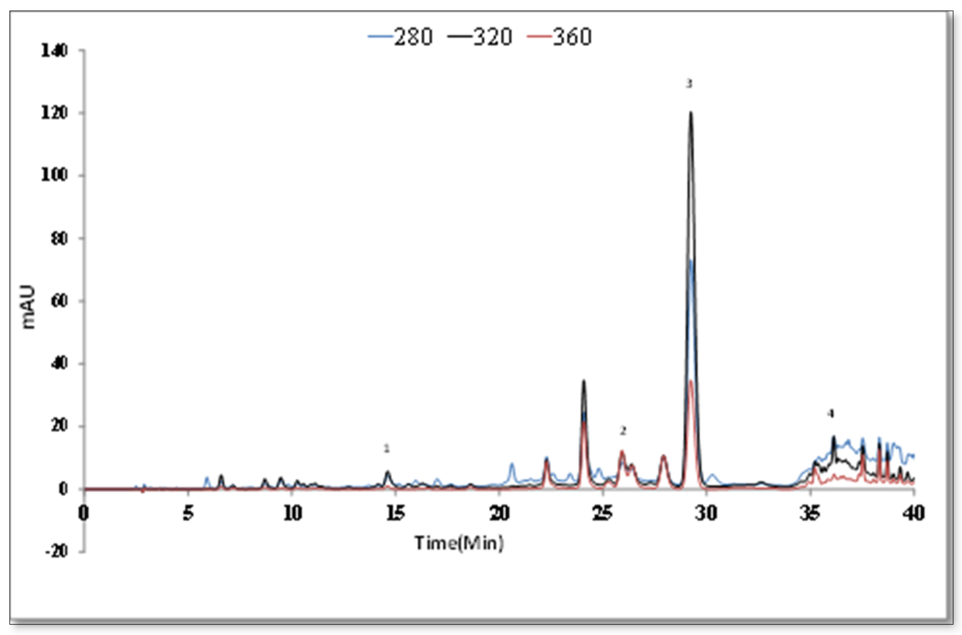
The detection limit (LOD) and the quantitation limit (LOQ) were found as presented in Table 5 for the HPLC method when 10 μL was used as the injection sample volume. Comparing the retention times and UV spectra of the standards, caffeic acid, luteolin-7-O-glycoside, rosmarinic acid, and luteolin were determined (Figure 3). The peak increase of rosmarinic acid at 320 nm was linked to the maximum UV absorption at 329 nm of rosmarinic acid, a phenolic acid. The calibration curves of the standards are used to calculate the quantitative data. For S. aramiensis, the data obtained by both UV spectrophotometry and HPLC analysis showed that the 70% MeOH extract had a high phenolic content. Rosmarinic acid was the main compound, and its concentration was found as 122.32 ± 0.21 mg/g. The luteolin concentration of the extract was 4.06 ± 0.03 mg/g.
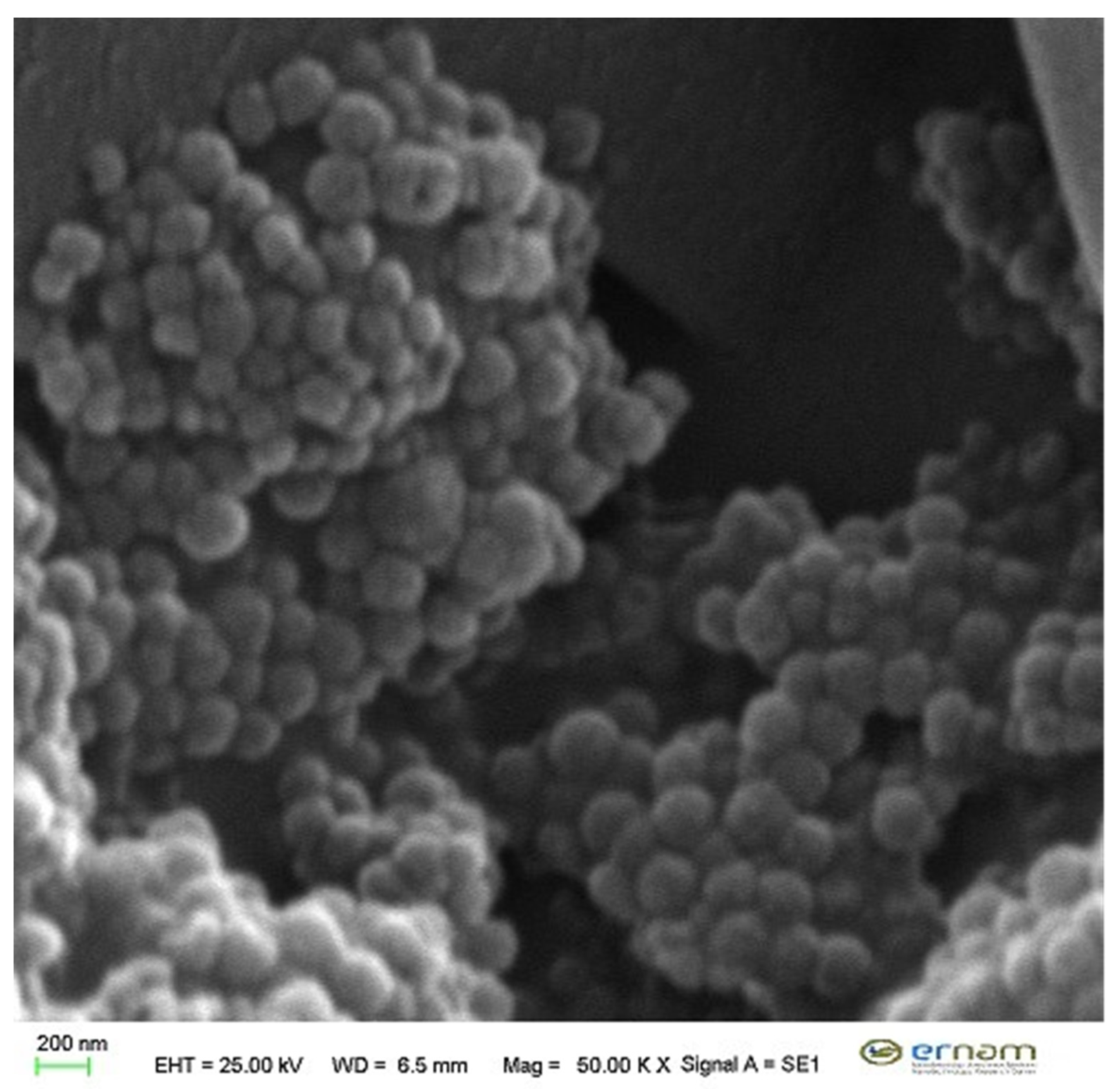
S. aramiensis 70% MeOH extract-loaded LPs were prepared using dry film hydration. Nano drug delivery systems are systems that transmit the low-stability active substance to the lower layers of the skin; moreover, it offers the opportunity to work with a lower dose for a longer time. Therefore, liposomes were preferred. In the characterization studies, PS, ZP, and PDI of liposomes were found as 0.395 ± 0.004 µm, −35.2 ± 2.1 mV, and mono dispersed due to low PDI values (below 0.2) (Table 6). The encapsulating efficiency of the LPs was found as 13.83% ± 1.25%. In the field of pharmaceutical nanosystems, researchers have collected some information about size, stability, and morphology through the microscopic techniques. Of them, SEM was used in our study and the type and size of nanoliposomes that we developed in our study were determined by SEM. The liposomes, which are developed as seen in Figure 4, have a narrow particle size range and a smooth surface. In vitro release experiment from LPs was carried out for 24 h, and cumulative released amount of (RA) was found to be 60% ± 0.85% (83.7 µg/mL extract) with HPLC analyses (Figure 5).
In literature, some studies with nanocarrier systems such as liposome, nanoparticles, etc. were developed with Salvia extracts. In the previous study, anti-neurotoxic efficacy of nanoparticles derived from Salvia officinalis extract against methylmercury-induced rats was evaluated. Sixty % ethanol extract of Salvia officinalis was associated with poly(lactic-co-glycolic acid) (PLGA) nanoparticles to enhance the antioxidant activity against mercury-induced neurotoxicity [47]. In another study, liposome oral liquid containing Cordyceps sinensis, Astragali L., and Salvia miltiorrhiza, which improved liver function, was prepared for the treatment of Hepatitis B and its efficacy was evaluated [48]. Aisha et al. [49] studied LPs which were prepared to contain Orthosiphon stamineus Benth. (Lamiaceae) (OS) ethanolic extract. Liposomes were prepared by the conventional film method with particle size and zeta potential of 152.5 ± 1.1 nm and −49.8 ± 1.0 mV, respectively. Release studies showed 94% ± 0.1% cumulative release in non-formulated extract and 62.4% ± 0.1% after 24 h at 37 °C in OS liposomes [49]. For these reasons, our characterization parameters seem to be compatible with the literature and successful when compared with OS-loaded LPs. In that study, the released extract showed improvement in DPPH scavenging effect. The results showed strong cleavage of DPPH both on formulated and unformulated extracts. However, a stronger effect with unformulated extract (EC50 = 32.4 ± 0.5 μg/mL) in connection with the high rate of release was found compared to OS LPs (EC50 = 23.5 ± 1.1 μg/mL) [49].
The amount of released extract compared to the activity experiments in the present study seems to be sufficient in all the experiments; 83.7 µg/mL of extract released from the LPs is well above the DPPH radical scavenging EC50 value. When we examine the results of the concentrations that we have tested in enzyme inhibition assays, it is clear that the extract released from the LPs will inhibit collagenase and elastase enzymes by more than 66.64% and 76.68%, respectively.
Fibroblast cells are commonly found in tendon, ligament, and skin tissues. They are generally described as collagen-producing cells and are known as the primary source of many extracellular matrix components [50]. L929 mouse fibroblast cells are frequently used to determine cytotoxic concentrations of many samples. It is also shown by international standards (ISO 10993 part 5, 1999; ISO 7405, 1997) as a reference cell for cytotoxicity testing [51]. The data acquired by the SRB cell viability method are given in Figure 6. Significance according to Levene statistics, homogeneity of variance, was found at p < 0.05 and Games–Howell test was used for statistical analysis. Between concentrations of 7.81–250 μg/mL, the 70% MeOH extract did not reduce the viability compared to the control group. The effect of 70% MeOH extract on cell viability at a concentration of 1000 μg/mL was 69%, and the influence of nanoformulation on cell viability was found to be 99.66%. Extract amounts obtained as a result of both encapsulation efficiency and release study are between 82.98 and 138.3 μg/mL. In the toxicity test, both concentrations were not toxic. The lack of toxicity of the LPs on the cell line is the first proof that it is reliable; also, it will shed light on future studies. This study is important in terms of proving the effectiveness of the nanoformulation containing extract rather than classical activity studies and proves the necessity of handling traditional plants with a modern understanding.

4. Conclusions

All data obtained from the study revealed the strong antioxidant effect of S. aramiensis as well as its potential wound-healing effect. The formulation study showed innovations in many respects as well as the potential of the extracts from plants to turn into products. Although the antioxidant activity of plant extracts is frequently studied, it is very important to show antioxidant capacities of formulated extracts. Therefore, in this study, S. aramiensis extract was first formulated with liposome, which is a nano-carrier system that attracts attention and is at the forefront with its advantages. With the successful demonstration of in vivo activity in future studies, a pharmaceutical product obtained from S. aramiensis will be safely functionalized.

Author Contributions

Conceptualization: E.K.A. and G.Ş.K.; methodology: G.Ş.K., Ç.Y., and E.K.A.; validation: G.Ş.K.; formal analysis: G.Ş.K., Ç.Y., and F.G.; investigation: G.Ş.K. and Ç.Y.; data curation: G.Ş.K. and E.K.A.; writing—original draft preparation: G.Ş.K., E.K.A., and E.S.-S.; writing—review and editing: E.K.A. All authors have read and agreed to the published version of the manuscript.

Funding

This research was supported with Project no: TSA-2019-8514 from Erciyes University Research Council.

Conflicts of Interest

The authors declare that they do not have any conflict of interest.

References

  1. Ramasubramania Raja, R. Medicinally potential plants of Labiatae (Lamiaceae) Family: An owerview. Res. J. Med. Plant 2012, 6, 203–213. [Google Scholar]
  2. Dweck, A.C. Introduction the folklore and cosmetic use of various Salvia species. In Sage, the Genus Salvia; Kintzios, S.E., Ed.; Harwood Academic: Amsterdam, The Netherlands, 2000; pp. 1–25. [Google Scholar]
  3. Demirci, B.; Başer, K.H.C.; Tumen, G. Composition of the essential oil of Salvia aramiensis Rech. Fil. growing in Turkey. Flavor Frag. J. 2002, 17, 23–25. [Google Scholar] [CrossRef]
  4. Yeşilada, E.; Honda, G.; Sezik, E.; Tabata, M.; Goto, K.; Ikeshire, Y. Traditional medicine in Turkey IV, folk medicine in Mediterranean Subdivision. J. Ethnopharmacol. 1993, 39, 31–38. [Google Scholar] [CrossRef]
  5. Yeşilada, E.; Honda, G.; Sezik, E.; Tabata, M.; Fujita, T.; Tanaka, T.; Takeda, Y.; Takaishi, H. Traditional medicine in Turkey V. Folk medicine in the inner Taurus Mountain. J. Ethnopharmacol. 1995, 46, 133–152. [Google Scholar] [CrossRef]
  6. Aşkun, T.; Başer, K.H.C.; Tümen, G.; Kürkçüoğlu, M. Characterization of essential oils of some Salvia species and their antimycobacterial activities. Turk J. Biol. 2010, 34, 89–95. [Google Scholar]
  7. Esquivel, B.; Sanchez, A.A.; Aranda, E. Natural Products of Agricultural Interest from Mexican Labiatae. In Phytochemicals and Phytopharmaceuticals; Shahidi, F., Ho, C.H., Eds.; AOCS Press: Champaign, IL, USA, 2009; pp. 371–385. [Google Scholar]
  8. Ulubelen, A.; Topçu, G. Flavonoids and terpenoids from Salvia verticillata and Salvia pinnata. J. Nat. Prod. 1984, 47, 1068. [Google Scholar] [CrossRef]
  9. Ulubelen, A.; Topçu, G.; Bozok-Johansson, C. Norditerpenoids and diterpenoids from Salvia multicaulis with antituberculous activity. J. Nat. Prod. 1997, 60, 1275–1280. [Google Scholar] [CrossRef]
  10. Kiselova, Y.; Ivanova, D.; Chervenkov, T.; Gerova, D.; Galunska, B.; Yankova, T. Correlation between the In Vitro antioxidant activity and polyphenol content of aqueous extracts from Bulgarian herbs. Phytother. Res. 2006, 20, 961–965. [Google Scholar] [CrossRef]
  11. Neagu, E.; Paun, G.; Albu, C.; Radu, G.L. Assessment of acetylcholinesterase and tyrosinase inhibitory and antioxidant activity of Alchemilla vulgaris and Filipendula ulmaria extracts. J. Taiwan Inst. Chem. Eng. 2015, 52, 1–6. [Google Scholar] [CrossRef]
  12. Arslan, M. Cultivation potential of Salvia tomentosa and S. aramiensis under the Eastern Mediterranean conditions. Sci. Pap. Ser. A Agron. 2016, 59, 174–177. [Google Scholar]
  13. Baytop, T. Türkiye’de Bitkilerle Tedavi. In Geçmişte ve Bugün, 2nd ed.; Nobel Tıp Kitapevleri Ltd. Şti: İstanbul, Turkey, 1999; pp. 143–144. [Google Scholar]
  14. Kelen, M.; Tepe, B. Chemical composition, antioxidant and antimicrobial properties of the essential oils of three Salvia species from Turkish flora. Bioresour. Technol. 2008, 99, 4096–4104. [Google Scholar] [CrossRef] [PubMed]
  15. Karaman, Ş.; Ilcim, A.; Çömlekçioğlu, N. Composition of the essential oils of Salvia aramiensis Rech. Fil. and Salvia cyanescens Boiss & Bal. Pak. J. Bot. 2007, 39, 169–172. [Google Scholar]
  16. Genc, Y.; Dereli, F.T.G.; Saracoglu, I.; Akkol, E.K. The inhibitory effects of isolated constituents from Plantago major subsp. major L. on collagenase, elastase and hyaluronidase enzymes: Potential wound healer. Saudi Pharm. J. 2020, 28, 101–106. [Google Scholar]
  17. Thring, T.S.; Hili, P.; Naughton, D.P. Anti-collagenase, anti-elastase and anti-oxidant activities of extracts from 21 plants. BMC Complement. Altern. Med. 2009, 9, 27. [Google Scholar] [CrossRef] [PubMed] [Green Version]
  18. Ndlovu, G.; Fouche, G.; Tselanyane, M.; Cordier, W.; Steenkamp, V. In vitro determination of the anti-aging potential of four southern African medicinal plants. BMC Complement. Altern. Med. 2013, 13, 304. [Google Scholar] [CrossRef] [PubMed] [Green Version]
  19. Barros, S.C.; Martins, J.A.; Marcos, J.C.; Cavaco-Paulo, A. Characterization of potential elastase inhibitor-peptides regulated by a molecular switch for wound dressings applications. Enzym. Microb. Technol. 2012, 50, 107–114. [Google Scholar] [CrossRef] [Green Version]
  20. Shukla, A.; Rasik, A.M.; Dhawan, B.N. Asiaticoside-induced elevation of antioxidant levels in healing wounds. Phytother. Res. 1999, 13, 50–54. [Google Scholar] [CrossRef]
  21. Aksoy, H.; Bingöl Özakpinar, Ö. Wound healing and oxidative stress. Marmara Pharm. J. 2014, 18, 153–158. [Google Scholar] [CrossRef]
  22. Kim, H.J.; Kim, T.H.; Kang, K.C.; Pyo, H.B.; Jeong, H.H. Microencapsulation of rosmarinic acid using polycaprolactone and various surfactants. Int. J. Cosmet. Sci. 2010, 32, 185–191. [Google Scholar] [CrossRef]
  23. Perugini, P.; Genta, I.; Pavanetto, F.; Conti, B.; Scalia, S.; Baruffini, A. Study on glycolic acid delivery by liposomes and microspheres. Int. J. Pharm. 2000, 196, 51–61. [Google Scholar] [CrossRef]
  24. Barenholz, Y. Liposome application: Problems and prospects. Curr. Opin. Colloid Interface Sci. 2001, 6, 66–77. [Google Scholar] [CrossRef]
  25. Chung, T.W.; Yang, M.C.; Tsai, W.J. A fibrin encapsulated liposomes-in-chitosan matrix (FLCM) for delivering water-soluble drugs influences of the surface properties of liposomes and the crosslinked fibrin network. Int. J. Pharm. 2006, 311, 122–129. [Google Scholar] [CrossRef] [PubMed]
  26. Ribeiro, A.S.; Estanqueiro, M.; Beatriz Oliveira, M.; Sousa Lobo, J.M. Main benefits and applicability of plant extracts in skin care products. Cosmetics 2015, 2, 48–65. [Google Scholar] [CrossRef] [Green Version]
  27. Singleton, V.L.; Orthofer, R.; Lamuela-Raventos, R.M. Analysis of total phenols and other oxidation substrates and antioxidants by means of Folin-ciocalteu reagent. Method Enzymol. 1999, 299, 152–178. [Google Scholar]
  28. Zhishen, J.; Mengcheng, T.; Jianming, W. The determination of flavonoid contents in mulberry and their scavenging effects on superoxide radicals. Food Chem. 1999, 64, 555–559. [Google Scholar] [CrossRef]
  29. Braca, A.; De Tommasi, N.; Di Bari, L.; Pizza, C.; Politi, M.; Morelli, I. Antioxidant principles from Bauhinia terapotensis. J. Nat. Prod. 2001, 64, 892–895. [Google Scholar] [CrossRef]
  30. Huang, M.H.; Huang, S.S.; Wang, B.S.; Sheu, M.J.; Hou, W.C. Antioxidant and anti-inflammatory properties of Cardiospermum halicacabum and its reference compounds ex vivo and in vivo. J. Ethnopharmacol. 2011, 133, 743–750. [Google Scholar] [CrossRef]
  31. Velioğlu, Y.S.; Mazza, G.; Oomah, B.D. Antioxidant activity and total phenolics in selected fruits, vegetables, and grain products. J. Agric. Food Chem. 1998, 46, 4113–4117. [Google Scholar] [CrossRef]
  32. Bangham, A.D.; Standish, M.M.; Watkins, J.C. Diffusion of univalent ions across the lamellae of swollen phospholipids. J. Mol. Biol. 1965, 13, 238–252. [Google Scholar] [CrossRef]
  33. Seker Karatoprak, G.; Ilgün, S.; Koşar, M. Phenolic composition, anti-inflammatory, antioxidant, and antimicrobial activities of Alchemilla mollis (Buser) Rothm. Chem. Biodivers. 2017, 14, e1700150. [Google Scholar] [CrossRef]
  34. Koşar, M.; Göger, F.; Başer, K.H.C. In vitro antioxidant properties and phenolic composition of Salvia halophila Hedge from Turkey. Food Chem. 2011, 129, 374–379. [Google Scholar] [CrossRef] [PubMed]
  35. Şeker Karatoprak, G.; İlgün, S.; Koşar, M. Antioxidant properties and phenolic composition of Salvia virgata Jacq. Turk. J. Pharm. Sci. 2016, 13, 201–212. [Google Scholar] [CrossRef]
  36. Akkol, E.K.; Göger, F.; Koşar, M.; Başer, K.H.C. Phenolic composition and biological activities of Salvia halophila and Salvia virgata from Turkey. Food Chem. 2008, 108, 942–949. [Google Scholar] [CrossRef]
  37. Süntar, İ.; Küpeli Akkol, E.; Şenol, F.S.; Keleş, H.; Erdoğan Orhan, İ. Investigating wound healing, tyrosinase inhibitory and antioxidant activities of the ethanol extracts of Salvia cryptantha and Salvia cyanescens using in vivo and in vitro experimental models. J. Ethnopharmacol. 2011, 135, 71–77. [Google Scholar] [CrossRef]
  38. Hatamia, T.; Emamic, S.A.; Miraghaeea, S.S.; Mojarrab, M. Total phenolic contents and antioxidant activities of different extracts and fractions from the aerial parts of Artemisia biennis Willd. Iran. J. Pharm. Res. 2014, 13, 551–558. [Google Scholar]
  39. Hnarayan, S.; Sasmal, D.; Mazumder, P.M. Evaluation of the wound healing effect of herbal ointment formulated with Salvia splendens (Scarlet sage). Int. J. Pharm. Pharm. Sci. 2011, 3, 195–199. [Google Scholar]
  40. Karimzadeh, S.; Farahpour, M.R. Topical application of Salvia officinalis hydroethanolic leaf extract improves wound healing process. Indian J. Exp. Biol. 2017, 55, 98–106. [Google Scholar]
  41. Guaouguaou, F.; Taghzouti, K.; Oukabli, M.; Es-Safi, N.E. The effect of Salvia verbenaca extracts for healing of second-degree burn wounds in rats current bioactive compounds. Curr. Bioact. Compd. 2018, 14, 419–427. [Google Scholar] [CrossRef]
  42. Kapp, K.; Hakala, E.; Orav, A.; Pohjala, L.; Vuorela, P.; Püssa, T.; Vuorela, H.; Raal, A. Commercial peppermint (Mentha × piperita L.) teas: Antichlamydial effect and polyphenolic composition. Food Res. Int. 2013, 53, 758–766. [Google Scholar] [CrossRef]
  43. Cvetkovikj, I.; Stefkov, G.; Acevska, J.; Stanoeva, J.P.; Karapandzova, M.; Stefova, M.; Dimitrovska, A.; Kulevanova, S. Polyphenolic characterization and chromatographic methods for fast assessment of culinary Salvia species from South East Europe. J. Chromatogr. A 2013, 1282, 38–45. [Google Scholar] [CrossRef]
  44. Lu, Y.; Yeap Foo, L. Polyphenolics of Salvia—A review. Phytochemistry 2002, 59, 117–140. [Google Scholar] [CrossRef]
  45. Hossain, M.B.; Rai, D.K.; Brunton, N.P.; Martin-Diana, A.B.; Barry-Ryan, C. Characterization of phenolic composition in Lamiaceae spices by LC-ESI-MS/MS. J. Agric. Food Chem. 2010, 58, 10576–10581. [Google Scholar] [CrossRef] [PubMed]
  46. Marzouk, M.M.; Hussein, S.R.; Elkhateeb, A.; El-shabrawy, M.; Abdel-Hameed, E.S.; Kawashty, S.A. Comparative study of Mentha species growing wild in Egypt: LC-ESI-MS analysis and chemosystematic significance. J. Appl. Pharm. 2018, 8, 116–122. [Google Scholar]
  47. Kahil, W.K.D.; Salvia, K. Effects of Salvia officinalis extract and its nano-encapsulated form on methylmercury induced neurotoxic-stress in male rats. World Appl. Sci. J. 2013, 24, 826–837. [Google Scholar]
  48. Wanfeng, S.; Rongling, S.; Weiling, J. Compound Cordyceps sinensis liposome oral liquid in treatment of patients with Chronic Hepatitis B. China Pharm. 2003, 6, 438–439. [Google Scholar]
  49. Aisha, A.F.; Majid, A.M.; Ismail, Z. Preparation and characterization of nano liposomes of Orthosiphon stamineus ethanolic extract in soybean phospholipids. BMC Biotechnol. 2014, 14, 23–33. [Google Scholar] [CrossRef] [Green Version]
  50. Theerakittayakorn, K.; Bunprasert, T. Differentiation capacity of mouse L929 fibroblastic cell line compare with human dermal fibroblast. World Acad. Sci. Eng. Technol. 2011, 50, 373–376. [Google Scholar]
  51. Karamustafa, F.; Çelebi, N.; Değim, Z.; Yılmaz, Ş. Evaluation of the viability of L-929 cells in the presence of alendronate and absorption enhancers. FABAD J. Pharm. Sci. 2006, 31, 1–5. [Google Scholar]
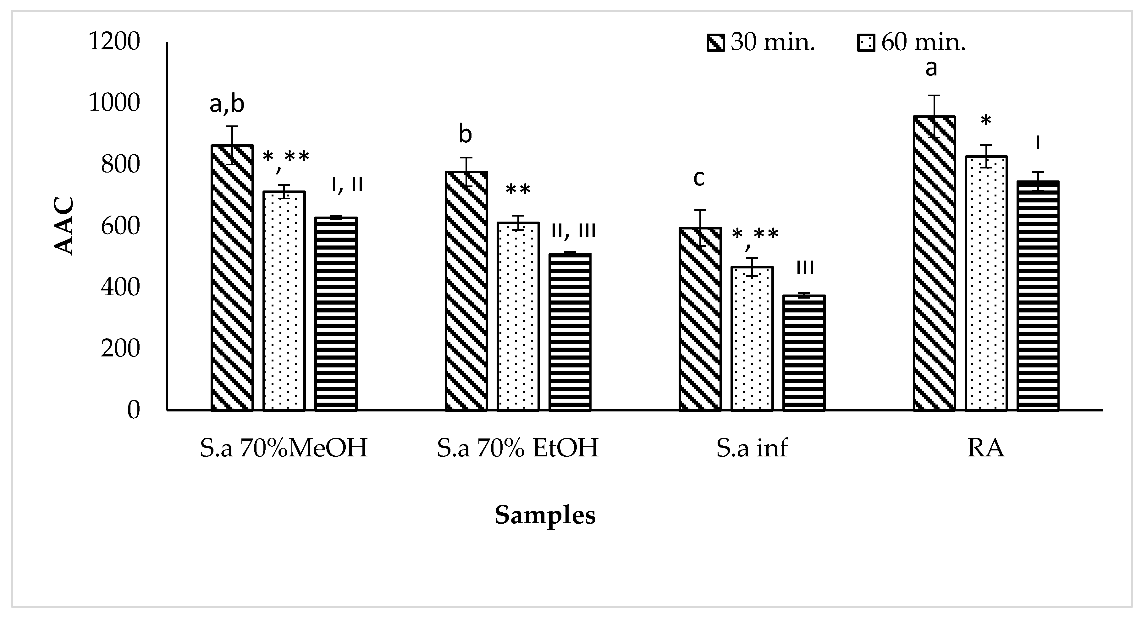
Figure 1. β carotene linoleic acid peroxidation activity of S. aramiensis extracts: Values = mean ± SD, statistical analyses by Games–Howell comparison test; bars with the same lowercase letters (a–d), same symbols (*, **) and Roman numerals (I, III) are not significantly (p > 0.05) different, n = 3. S.a 70% MeOH; S. aramiensis 70% methanol extract, S.a 70% EtOH; S. aramiensis 70% ethanol extract, S.a inf; S. aramiensis 2% infusion.
Antioxidants 09 00293 g001
Figure 2. LC/MS/MS chromatogram of 70% MeOH extract of S. aramiensis.
Antioxidants 09 00293 g002
Figure 3. HPLC chromatogram of 70% MeOH extract of S. aramiensis.
Antioxidants 09 00293 g003
Figure 4. SEM image of liposome.
Antioxidants 09 00293 g004
Figure 5. Chromatogram of released extract from liposome formulation.
Antioxidants 09 00293 g005
Figure 6. Toxicity effects of 70% MeOH extract and liposome formulation: Values given as mean ± SD were specified in the ±95% confidence interval (n = 3). Statistical analyses were by Games–Howell comparison test, and significant differences were reported as * p < 0.01; ** p < 0.05.
Antioxidants 09 00293 g006
Table 1. Total phenol and flavonoid content of S. aramiensis extracts.
ExtractsTotal Phenol
(mgGAE/gextract) a
Total Flavonoids
(mgCA/gextract) b
S.a 70% MeOH198.07 ± 2.77141.76 ± 1.64
S.a 70% EtOH153.24 ± 0.9493.28 ± 9.53
S.a inf124.75 ± 1.58118.85 ± 4.47
a mgGAE/gextract: Total phenols expressed as gallic acid equivs milligrams of gallic acid per gram (dry weight) of extract. b mgCA/gextract: Total flavonoids expressed as rutin per gram (dry weight) of extract. Values are expressed as means ± standard error (n = 3). S.a 70% MeOH; S. aramiensis 70% methanol extract, S.a 70% EtOH; S. aramiensis 70% ethanol extract, S.a inf; S. aramiensis 2% infusion.
Table 2. DPPH and ABTS●+ radical scavenging activities of S. aramiensis extracts.
Extracts/StandardDPPH● EC50 (µg/mL)ABTS●+ (mmol/ L /Trolox)
50 µg/mL100 µg/mL
S.a 70% MeOH28.4 ± 0.002 b1.15 ± 0.101 a1.77 ± 0.09 b
S.a 70% EtOH36.6 ± 0.004 c1.59 ± 0.091 c1.65 ± 0.05 b,c
S.a inf32.6 ± 0.001 b1.24 ± 0.113 a1.37 ± 0.071 a,d
Rosmarinic acid4.4 ± 0.00 a2.16 ± 0.044 b,e2.56 ± 0.001 e
Values = mean ± SD, statistical analyses by Tukey comparison test; values with the same lowercase letters (a–d) are not significantly (p > 0.05) different, n = 3. S.a 70% MeOH; S. aramiensis 70% methanol extract, S.a 70% EtOH; S. aramiensis 70% ethanol extract, S.a inf; S. aramiensis 2% infusion.
Table 3. Collagenase and elastase enzyme inhibition activities of S. aramiensis 70% MeOH extract and rosmarinic acid.
S.a 70% MeOH/StandardCollagenase Enzyme Inhibition (%)Elastase Enzyme Inhibition (%)
50 µg/mL66.64 ± 1.53 b76.68 ± 1.21 a
100 µg/mL69.54 ± 1.49 a,b83.70 ± 1.72 b
200 µg/mL72.66 ± 0.70 a86.91 ± 0.81 c
RA 25 µg/mL72.89 ± 0.99 a80.58 ± 0.89 a,b
Values = mean ± SD, statistical analyses by Tukey comparison test; values with the same lowercase letters (a–c) are not significantly (p > 0.05) different, n = 3. S.a 70% MeOH; S. aramiensis 70% methanol extract, RA: rosmarinic acid.
Table 4. LC-MS/MS results of 70% MeOH extract of S. aramiensis.
Rt[M − H]FragmentsIdentified CompoundsRef
7.2387207, 163Icariside b5/medioresinol[42]
8.7179135Caffeic acid[43]
8.9463300, 271Quercetin glucoside/hydroxyluteolin glucoside[43]
10.7447285Luteolin glucoside[43]
11.2477315, 300, 285Isorhamnetin hexoside[44]
12.3717537, 519, 493, 321Salvianolic acid E[45]
13.3359197, 179, 161,Rosmarinic acid[43]
14.9563387, 207Icariside b5/Medioresinol O-glucoronide[46]
17.3285151, 133Luteolin[43]
Rt: Retention time; [M − H] molecular ion.
Table 5. HPLC results of S. aramiensis extract.
CompoundsExtract *Standards
S.a 70% MeOHEquation and r2LODLOQ
Caffeic acid1.84 ± 0.09y = 44.283x + 40.553 r2 = 0.9980.00670.0204
Luteolin-7-o-glucoside3.70 ± 0.22y = 39.422x + 78.434 r2 = 0.9970.00850.0257
Rosmarinic acid122.32 ± 0.21y = 23.777x − 11.797 r2 = 0.9970.25950.7865
Luteolin4.06 ± 0.03y = 41.981x − 70.752 r2 = 0.9970.14070.4263
* Sa 70% MeOH: 70% MeOH extract. & mg/gextract, mean ± SD.
Table 6. Characterization of liposomes (n = 3).
FormulationPS ± SD (µm)ZP ± SD (mV)PDI ± SDEE ± SD (%)RA ± SD (%)
Liposome0.395 ± 0.004−35.2 ± 2.10.107 ± 0.01713.83 ± 1.2560 ± 0.85
PS: particle size; ZP: zeta potential; PDI: polydispersity index; EE: encapsulation efficiency; RA: released amount of extract. Values are expressed as mean ± SD (n = 3).