
Clinical Applications of Artificial Intelligence—An Updated Overview

 ,
,  ,
,  ,
,  , , ,
, , ,  and
and
Abstract
:1. Introduction
2. Applications of AI in Clinical Care
2.1. Cardiology
2.2. Neurology
2.3. Oncology
2.4. Hematology
2.5. Nephrology
2.6. Gastroenterology and Hepatology
2.7. Orthopedics and Rheumatology
2.8. Other Applications
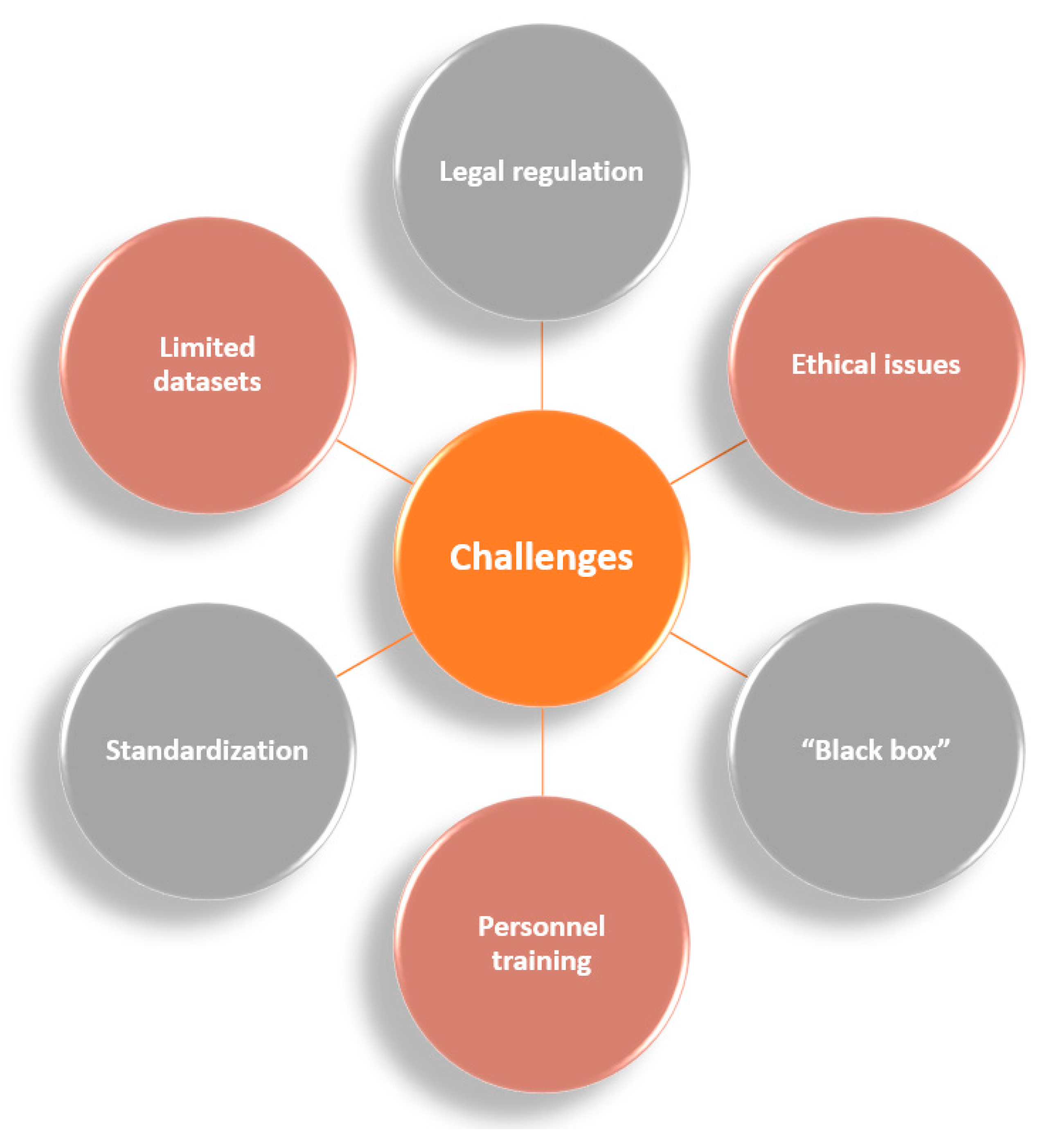
3. Challenges in AI Clinical Integration
4. Conclusions
Author Contributions
Funding
Institutional Review Board Statement
Informed Consent Statement
Data Availability Statement
Conflicts of Interest
Abbreviations
| 1D | one-dimensional |
| 2D | two-dimensional |
| aCLL | accelerated chronic lymphocytic leukemia |
| ADNI | Alzheimer’s Disease Neuroimaging Initiative |
| AI | Artificial Intelligence |
| AIBL | Australian Imaging, Biomarker and Lifestyle Flagship Study of Ageing |
| AIS | acute ischemic stroke |
| AF | atrial fibrillation |
| ANN | artificial neural network |
| AS | aortic stenosis |
| ASAP | atypical small acinar proliferation |
| AVB | atrioventricular block |
| AZA | azacitidine |
| BIDMC | Beth Israel Deaconess Medical Center |
| CAE | convolutional autoencoder |
| CDA | congenital dyserythropoietic anemia |
| CLL | chronic lymphocytic leukemia |
| CNN | convolutional neural network |
| CPCT | chest-pain computed tomography |
| CRISP-DM | cross-industry standard process for data mining |
| CWT | continuous wavelet transform |
| DBN | discrete Bayesian network |
| DCNN | deep convolutional neural network |
| DHS | dehydrated hereditary stomatocytosis |
| DL | deep learning |
| DNN | deep neural network |
| EAR | ectopic atrial rhythm |
| ECG | electrocardiogram |
| EEG | electroencephalogram |
| eGFR | estimated glomerular filtration rate |
| EHR | electronic health record |
| EmA | endomysial autoantibody |
| FCD | focal cortical dysplasia |
| FCN | fully convolutional network |
| FFRCT | computed tomography-derived fractional flow reserve |
| FHS | Framingham Heart Study |
| FM | Formal Methods |
| GENFI | Genetic Frontotemporal dementia Initiative |
| GFR | glomerular filtration rate |
| H&E | hematoxylin and eosin |
| HS | hereditary spherocytosis |
| IC | intensive chemotherapy |
| IDEA | Intraoperative Data Embedded Analytics |
| IED | interictal epileptiform discharges |
| IRIDA | iron-refractory iron-deficiency anemia |
| IVR | idioventricular rhythm |
| KNN | K-nearest neighbors |
| LightGBM | light gradient boosted machine |
| LSTM | long short-term memory |
| LV | left ventricle |
| MFCCs | Mel-frequency cepstral coefficients |
| ML | machine learning |
| MLR | multivariable logistic regression |
| MRI | magnetic resonance imaging |
| NACC | National Alzheimer’s Coordinating Center |
| NAFLD | nonalcoholic fatty liver disease |
| NB | Naïve Bayes |
| NLP | multilayer perceptron |
| NN | neural network |
| MR | magnetic resonance |
| PAS | periodic acid-Schiff |
| PCs | proliferation centers |
| PsA | psoriatic arthritis |
| PsC | cutaneous-only psoriasis |
| RCNN | region convolutional neural network |
| RNN | recurrent neural network |
| RT | Richter transformation |
| RV | right ventricle |
| SAPS | simplified acute physiology score |
| SCA | sudden cardiac arrest |
| STFT | short-time Fourier transform |
| SuStaIn | Subtype and Stage Interference |
| SVM | support vector machine |
| SVT | supraventricular tachycardia |
| THA | total hip arthroplasty |
| TNM | tumor, nodes, and metastases |
| tTG-DGP | tissue transglutaminase-deamidated gliadin peptides |
| VF | ventricular fibrillation |
| XGBoost | extreme gradient boosting |
References
- Muthalaly, R.G.; Evans, R.M. Applications of Machine Learning in Cardiac Electrophysiology. Arrhythm Electrophysiol. Rev. 2020, 9, 71–77. [Google Scholar] [CrossRef] [PubMed]
- Asha, P.; Srivani, P.; Ahmed, A.A.A.; Kolhe, A.; Nomani, M.Z.M. Artificial intelligence in medical Imaging: An analysis of innovative technique and its future promise. Mater. Today Proc. 2021, 56, 2236–2239. [Google Scholar] [CrossRef]
- Yao, L.; Zhang, H.; Zhang, M.; Chen, X.; Zhang, J.; Huang, J.; Zhang, L. Application of artificial intelligence in renal disease. Clin. Ehealth 2021, 4, 54–61. [Google Scholar] [CrossRef]
- Van den Eynde, J.; Lachmann, M.; Laugwitz, K.-L.; Manlhiot, C.; Kutty, S. Successfully Implemented Artificial Intelligence and Machine Learning Applications In Cardiology: State-of-the-Art Review. Trends Cardiovasc. Med. 2022. [Google Scholar] [CrossRef] [PubMed]
- Laptev, V.A.; Ershova, I.V.; Feyzrakhmanova, D.R. Medical Applications of Artificial Intelligence (Legal Aspects and Future Prospects). Laws 2022, 11, 3. [Google Scholar] [CrossRef]
- Williams, S.; Layard Horsfall, H.; Funnell, J.P.; Hanrahan, J.G.; Khan, D.Z.; Muirhead, W.; Stoyanov, D.; Marcus, H.J. Artificial Intelligence in Brain Tumour Surgery—An Emerging Paradigm. Cancers 2021, 13, 5010. [Google Scholar] [CrossRef]
- Hügle, M.; Omoumi, P.; van Laar, J.M.; Boedecker, J.; Hügle, T. Applied machine learning and artificial intelligence in rheumatology. Rheumatol. Adv. Pract. 2020, 4, rkaa005. [Google Scholar] [CrossRef]
- Akkus, Z.; Aly, Y.H.; Attia, I.Z.; Lopez-Jimenez, F.; Arruda-Olson, A.M.; Pellikka, P.A.; Pislaru, S.V.; Kane, G.C.; Friedman, P.A.; Oh, J.K. Artificial Intelligence (AI)-Empowered Echocardiography Interpretation: A State-of-the-Art Review. J. Clin. Med. 2021, 10, 1391. [Google Scholar] [CrossRef]
- Khorsandi, S.E.; Hardgrave, H.J.; Osborn, T.; Klutts, G.; Nigh, J.; Spencer-Cole, R.T.; Kakos, C.D.; Anastasiou, I.; Mavros, M.N.; Giorgakis, E. Artificial Intelligence in Liver Transplantation. Transplant. Proc. 2021, 53, 2939–2944. [Google Scholar] [CrossRef]
- Sana, M.K.; Hussain, Z.M.; Shah, P.A.; Maqsood, M.H. Artificial intelligence in celiac disease. Comput. Biol. Med. 2020, 125, 103996. [Google Scholar] [CrossRef]
- Vinny, P.W.; Vishnu, V.Y.; Padma Srivastava, M.V. Artificial Intelligence shaping the future of neurology practice. Med. J. Armed Forces India 2021, 77, 276–282. [Google Scholar] [CrossRef] [PubMed]
- Mahesh, B. Machine learning algorithms-a review. Int. J. Sci. Res. 2020, 9, 381–386. [Google Scholar]
- Sarker, I.H. Machine Learning: Algorithms, Real-World Applications and Research Directions. SN Comput. Sci. 2021, 2, 160. [Google Scholar] [CrossRef] [PubMed]
- Patel, V.; Shah, M. A comprehensive study on artificial intelligence and machine learning in drug discovery and drug development. Intell. Med. 2021. [Google Scholar] [CrossRef]
- Nakamura, T.; Sasano, T. Artificial intelligence and cardiology: Current status and perspective. J. Cardiol. 2022, 79, 326–333. [Google Scholar] [CrossRef]
- Bahardoust, M.; Baghoi-Hosseinabadi, Z. Role of Adipose-Derived Mesenchymal Stem Cells in the Regeneration of Cardiac Tissue and Improvement of Cardiac Function: A Narrative Review. Biointerface Res. Appl. Chem. 2021, 11, 8446–8456. [Google Scholar] [CrossRef]
- Haleem, A.; Javaid, M.; Singh, R.P.; Suman, R. Applications of Artificial Intelligence (AI) for cardiology during COVID-19 pandemic. Sustain. Oper. Comput. 2021, 2, 71–78. [Google Scholar] [CrossRef]
- Ye, C.; Fu, T.; Hao, S.; Zhang, Y.; Wang, O.; Jin, B.; Xia, M.; Liu, M.; Zhou, X.; Wu, Q. Prediction of incident hypertension within the next year: Prospective study using statewide electronic health records and machine learning. J. Med. Internet Res. 2018, 20, e22. [Google Scholar] [CrossRef]
- Tison, G.H.; Sanchez, J.M.; Ballinger, B.; Singh, A.; Olgin, J.E.; Pletcher, M.J.; Vittinghoff, E.; Lee, E.S.; Fan, S.M.; Gladstone, R.A.; et al. Passive Detection of Atrial Fibrillation Using a Commercially Available Smartwatch. JAMA Cardiol. 2018, 3, 409–416. [Google Scholar] [CrossRef] [Green Version]
- Picon, A.; Irusta, U.; Álvarez-Gila, A.; Aramendi, E.; Alonso-Atienza, F.; Figuera, C.; Ayala, U.; Garrote, E.; Wik, L.; Kramer-Johansen, J.; et al. Mixed convolutional and long short-term memory network for the detection of lethal ventricular arrhythmia. PLoS ONE 2019, 14, e0216756. [Google Scholar] [CrossRef]
- Yang, C.; Ojha, B.D.; Aranoff, N.D.; Green, P.; Tavassolian, N. Classification of aortic stenosis using conventional machine learning and deep learning methods based on multi-dimensional cardio-mechanical signals. Sci. Rep. 2020, 10, 17521. [Google Scholar] [CrossRef] [PubMed]
- Eberhard, M.; Nadarevic, T.; Cousin, A.; von Spiczak, J.; Hinzpeter, R.; Euler, A.; Morsbach, F.; Manka, R.; Keller, D.I.; Alkadhi, H. Machine learning-based CT fractional flow reserve assessment in acute chest pain: First experience. Cardiovasc. Diagn. Ther. 2020, 10, 820–830. [Google Scholar] [CrossRef] [PubMed]
- Nguyen, M.T.; Nguyen, B.V.; Kim, K. Deep Feature Learning for Sudden Cardiac Arrest Detection in Automated External Defibrillators. Sci. Rep. 2018, 8, 17196. [Google Scholar] [CrossRef] [PubMed] [Green Version]
- Retson, T.A.; Masutani, E.M.; Golden, D.; Hsiao, A. Clinical Performance and Role of Expert Supervision of Deep Learning for Cardiac Ventricular Volumetry: A Validation Study. Radiol. Artif. Intell. 2020, 2, e190064. [Google Scholar] [CrossRef]
- Hannun, A.Y.; Rajpurkar, P.; Haghpanahi, M.; Tison, G.H.; Bourn, C.; Turakhia, M.P.; Ng, A.Y. Cardiologist-level arrhythmia detection and classification in ambulatory electrocardiograms using a deep neural network. Nat. Med. 2019, 25, 65–69. [Google Scholar] [CrossRef]
- Ranti, D.; Valliani, A.A.-A.; Costa, A.; Oermann, E.K. Chapter 20—Artificial intelligence as applied to clinical neurological conditions. In Artificial Intelligence in Medicine; Xing, L., Giger, M.L., Min, J.K., Eds.; Academic Press: Cambridge, MA, USA, 2021; pp. 395–413. [Google Scholar]
- Abedi, V.; Avula, V.; Chaudhary, D.; Shahjouei, S.; Khan, A.; Griessenauer, C.J.; Li, J.; Zand, R. Prediction of Long-Term Stroke Recurrence Using Machine Learning Models. J. Clin. Med. 2021, 10, 1286. [Google Scholar] [CrossRef]
- Rava, R.A.; Seymour, S.E.; Snyder, K.V.; Waqas, M.; Davies, J.M.; Levy, E.I.; Siddiqui, A.H.; Ionita, C.N. Automated Collateral Flow Assessment in Patients with Acute Ischemic Stroke Using Computed Tomography with Artificial Intelligence Algorithms. World Neurosurg. 2021, 155, e748–e760. [Google Scholar] [CrossRef]
- Young, A.L.; Marinescu, R.V.; Oxtoby, N.P.; Bocchetta, M.; Yong, K.; Firth, N.C.; Cash, D.M.; Thomas, D.L.; Dick, K.M.; Cardoso, J.; et al. Uncovering the heterogeneity and temporal complexity of neurodegenerative diseases with Subtype and Stage Inference. Nat. Commun. 2018, 9, 4273. [Google Scholar] [CrossRef] [Green Version]
- Eshaghi, A.; Young, A.L.; Wijeratne, P.A.; Prados, F.; Arnold, D.L.; Narayanan, S.; Guttmann, C.R.G.; Barkhof, F.; Alexander, D.C.; Thompson, A.J.; et al. Identifying multiple sclerosis subtypes using unsupervised machine learning and MRI data. Nat. Commun. 2021, 12, 2078. [Google Scholar] [CrossRef]
- Jin, B.; Krishnan, B.; Adler, S.; Wagstyl, K.; Hu, W.; Jones, S.; Najm, I.; Alexopoulos, A.; Zhang, K.; Zhang, J.; et al. Automated detection of focal cortical dysplasia type II with surface-based magnetic resonance imaging postprocessing and machine learning. Epilepsia 2018, 59, 982–992. [Google Scholar] [CrossRef] [Green Version]
- Gleichgerrcht, E.; Munsell, B.C.; Alhusaini, S.; Alvim, M.K.M.; Bargalló, N.; Bender, B.; Bernasconi, A.; Bernasconi, N.; Bernhardt, B.; Blackmon, K.; et al. Artificial intelligence for classification of temporal lobe epilepsy with ROI-level MRI data: A worldwide ENIGMA-Epilepsy study. NeuroImage Clin. 2021, 31, 102765. [Google Scholar] [CrossRef]
- Daoud, H.; Bayoumi, M.A. Efficient Epileptic Seizure Prediction Based on Deep Learning. IEEE Trans. Biomed. Circuits Syst. 2019, 13, 804–813. [Google Scholar] [CrossRef]
- Quon, R.J.; Meisenhelter, S.; Camp, E.J.; Testorf, M.E.; Song, Y.; Song, Q.; Culler, G.W.; Moein, P.; Jobst, B.C. AiED: Artificial intelligence for the detection of intracranial interictal epileptiform discharges. Clin. Neurophysiol. 2022, 133, 1–8. [Google Scholar] [CrossRef]
- Qiu, S.; Joshi, P.S.; Miller, M.I.; Xue, C.; Zhou, X.; Karjadi, C.; Chang, G.H.; Joshi, A.S.; Dwyer, B.; Zhu, S.; et al. Development and validation of an interpretable deep learning framework for Alzheimer’s disease classification. Brain 2020, 143, 1920–1933. [Google Scholar] [CrossRef]
- Shinde, S.; Prasad, S.; Saboo, Y.; Kaushick, R.; Saini, J.; Pal, P.K.; Ingalhalikar, M. Predictive markers for Parkinson’s disease using deep neural nets on neuromelanin sensitive MRI. NeuroImage Clin. 2019, 22, 101748. [Google Scholar] [CrossRef]
- Claassen, J.; Doyle, K.; Matory, A.; Couch, C.; Burger, K.M.; Velazquez, A.; Okonkwo, J.U.; King, J.-R.; Park, S.; Agarwal, S. Detection of brain activation in unresponsive patients with acute brain injury. N. Engl. J. Med. 2019, 380, 2497–2505. [Google Scholar] [CrossRef]
- Shahjouei, S.; Ghodsi, S.M.; Zangeneh Soroush, M.; Ansari, S.; Kamali-Ardakani, S. Artificial Neural Network for Predicting the Safe Temporary Artery Occlusion Time in Intracranial Aneurysmal Surgery. J. Clin. Med. 2021, 10, 1464. [Google Scholar] [CrossRef]
- Brzezicki, M.A.; Bridger, N.E.; Kobetić, M.D.; Ostrowski, M.; Grabowski, W.; Gill, S.S.; Neumann, S. Artificial intelligence outperforms human students in conducting neurosurgical audits. Clin. Neurol. Neurosurg. 2020, 192, 105732. [Google Scholar] [CrossRef]
- Kaur, C.; Garg, U. Artificial intelligence techniques for cancer detection in medical image processing: A review. Mater. Today Proc. 2022, 56, 2236–2239. [Google Scholar] [CrossRef]
- Mansouri, F. Role of Telemedicine and Telegenetics Framework for the Management of Cancer Patients During the COVID-19 Pandemic. Biointerface Res. Appl. Chem. 2021, 11, 8773–8779. [Google Scholar] [CrossRef]
- Puiu, A.G.; Grigoras, O.; Preda, M.I.; Constantin, M.; Vasilescu, A.M. Comparative Analysis Between Cellular Oncogenes and Viral Oncogenes. Biointerface Res. Appl. Chem. 2021, 11, 9939–9951. [Google Scholar] [CrossRef]
- Zaheer, U.; Hassain, N.A.; Banu, S.; Mathew, S. Oncolytic Viruses as Nanomedicines against the Tumor Microenvironment. Biointerface Res. Appl. Chem. 2021, 11, 14825–14852. [Google Scholar] [CrossRef]
- Agarwal, S.; Yadav, A.S.; Dinesh, V.; Vatsav, K.S.S.; Prakash, K.S.S.; Jaiswal, S. By artificial intelligence algorithms and machine learning models to diagnosis cancer. Mater. Today Proc. 2021. [Google Scholar] [CrossRef]
- Rocca, A.; Brunese, M.C.; Santone, A.; Avella, P.; Bianco, P.; Scacchi, A.; Scaglione, M.; Bellifemine, F.; Danzi, R.; Varriano, G.; et al. Early Diagnosis of Liver Metastases from Colorectal Cancer through CT Radiomics and Formal Methods: A Pilot Study. J. Clin. Med. 2022, 11, 31. [Google Scholar] [CrossRef]
- Lu, J.; Liu, R.; Zhang, Y.; Zhang, X.; Zheng, L.; Zhang, C.; Zhang, K.; Li, S.; Lu, Y. Research on the development and application of a detection platform for colorectal cancer tumor sprouting pathological characteristics based on artificial intelligence. Intell. Med. 2021. [Google Scholar] [CrossRef]
- Lee, K.-S.; Jang, J.-Y.; Yu, Y.-D.; Heo, J.S.; Han, H.-S.; Yoon, Y.-S.; Kang, C.M.; Hwang, H.K.; Kang, S. Usefulness of artificial intelligence for predicting recurrence following surgery for pancreatic cancer: Retrospective cohort study. Int. J. Surg. 2021, 93, 106050. [Google Scholar] [CrossRef]
- Pantanowitz, L.; Quiroga-Garza, G.M.; Bien, L.; Heled, R.; Laifenfeld, D.; Linhart, C.; Sandbank, J.; Albrecht Shach, A.; Shalev, V.; Vecsler, M.; et al. An artificial intelligence algorithm for prostate cancer diagnosis in whole slide images of core needle biopsies: A blinded clinical validation and deployment study. Lancet Digit. Health 2020, 2, e407–e416. [Google Scholar] [CrossRef]
- Faron, A.; Opheys, N.S.; Nowak, S.; Sprinkart, A.M.; Isaak, A.; Theis, M.; Mesropyan, N.; Endler, C.; Sirokay, J.; Pieper, C.C.; et al. Deep Learning-Based Body Composition Analysis Predicts Outcome in Melanoma Patients Treated with Immune Checkpoint Inhibitors. Diagnostics 2021, 11, 2314. [Google Scholar] [CrossRef]
- Kim, H.; Jeon, J.; Han, Y.J.; Joo, Y.; Lee, J.; Lee, S.; Im, S. Convolutional Neural Network Classifies Pathological Voice Change in Laryngeal Cancer with High Accuracy. J. Clin. Med. 2020, 9, 3415. [Google Scholar] [CrossRef]
- Chan, J.W.; Hohenstein, N.; Carpenter, C.; Pattison, A.J.; Morin, O.; Valdes, G.; Thompson, M.; Perkins, J.; Solberg, T.D.; Yom, S.S. Artificial Intelligence-Guided Prediction of Dental Doses Before Planning of Radiation Therapy for Oropharyngeal Cancer: Technical Development and Initial Feasibility of Implementation. Adv. Radiat. Oncol. 2022, 7, 100886. [Google Scholar] [CrossRef]
- Houy, N.; Le Grand, F. Personalized oncology with artificial intelligence: The case of temozolomide. Artif. Intell. Med. 2019, 99, 101693. [Google Scholar] [CrossRef] [PubMed]
- Radakovich, N.; Nagy, M.; Nazha, A. Artificial Intelligence in Hematology: Current Challenges and Opportunities. Curr. Hematol. Malig. Rep. 2020, 15, 203–210. [Google Scholar] [CrossRef] [PubMed]
- Walter, W.; Haferlach, C.; Nadarajah, N.; Schmidts, I.; Kühn, C.; Kern, W.; Haferlach, T. How artificial intelligence might disrupt diagnostics in hematology in the near future. Oncogene 2021, 40, 4271–4280. [Google Scholar] [CrossRef] [PubMed]
- Carreras, J.; Nakamura, N.; Hamoudi, R. Artificial Intelligence Analysis of Gene Expression Predicted the Overall Survival of Mantle Cell Lymphoma and a Large Pan-Cancer Series. Healthcare 2022, 10, 155. [Google Scholar] [CrossRef] [PubMed]
- El Hussein, S.; Chen, P.; Medeiros, L.J.; Wu, J.; Khoury, J.D. Artificial Intelligence-Assisted Mapping of Proliferation Centers in Chronic Lymphocytic Leukemia/Small Lymphocytic Lymphoma Identifies Patterns That Reliably Distinguish Accelerated Phase and Large Cell Transformation. Blood 2021, 138, 1558. [Google Scholar] [CrossRef]
- Boldú, L.; Merino, A.; Acevedo, A.; Molina, A.; Rodellar, J. A deep learning model (ALNet) for the diagnosis of acute leukaemia lineage using peripheral blood cell images. Comput. Methods Programs Biomed. 2021, 202, 105999. [Google Scholar] [CrossRef]
- Didi, I.; Simoncini, D.; Vergez, F.; Dumas, P.-Y.; Tavitian, S.; Largeaud, L.; Bidet, A.; Rieu, J.-B.; Luquet, I.; Lechevalier, N.; et al. Artificial Intelligence-Based Predictive Models for Acute Myeloid Leukemia. Blood 2021, 138, 3389. [Google Scholar] [CrossRef]
- AlAgha, A.S.; Faris, H.; Hammo, B.H.; Al-Zoubi, A.M. Identifying β-thalassemia carriers using a data mining approach: The case of the Gaza Strip, Palestine. Artif. Intell. Med. 2018, 88, 70–83. [Google Scholar] [CrossRef]
- Memmolo, P.; Aprea, G.; Bianco, V.; Russo, R.; Andolfo, I.; Mugnano, M.; Merola, F.; Miccio, L.; Iolascon, A.; Ferraro, P. Differential diagnosis of hereditary anemias from a fraction of blood drop by digital holography and hierarchical machine learning. Biosens. Bioelectron. 2022, 201, 113945. [Google Scholar] [CrossRef]
- Dutra, V.d.F.; Biassi, T.P.; Figueiredo, M.S. Sickle cell anemia: Hierarchical cluster analysis and clinical profile in a cohort in Brazil. Hematol. Transfus. Cell Ther. 2021. [Google Scholar] [CrossRef]
- Barbieri, C.; Molina, M.; Ponce, P.; Tothova, M.; Cattinelli, I.; Ion Titapiccolo, J.; Mari, F.; Amato, C.; Leipold, F.; Wehmeyer, W.; et al. An international observational study suggests that artificial intelligence for clinical decision support optimizes anemia management in hemodialysis patients. Kidney Int. 2016, 90, 422–429. [Google Scholar] [CrossRef] [PubMed]
- Haferlach, T.; Pohlkamp, C.; Heo, I.; Drescher, R.; Hänselmann, S.; Lörch, T.; Kern, W.; Haferlach, C.; Nadarajah, N. Automated Peripheral Blood Cell Differentiation Using Artificial Intelligence—A Study with More Than 10,000 Routine Samples in a Specialized Leukemia Laboratory. Blood 2021, 138, 103. [Google Scholar] [CrossRef]
- Osman, M.; Akkus, Z.; Jevremovic, D.; Nguyen, P.L.; Roh, D.; Al-Kali, A.; Patnaik, M.M.; Nanaa, A.; Rizk, S.; Salama, M.E. Classification of Monocytes, Promonocytes and Monoblasts Using Deep Neural Network Models: An Area of Unmet Need in Diagnostic Hematopathology. J. Clin. Med. 2021, 10, 2264. [Google Scholar] [CrossRef] [PubMed]
- Mohamadlou, H.; Lynn-Palevsky, A.; Barton, C.; Chettipally, U.; Shieh, L.; Calvert, J.; Saber, N.R.; Das, R. Prediction of Acute Kidney Injury With a Machine Learning Algorithm Using Electronic Health Record Data. Can. J. Kidney Health Dis. 2018, 5, 2054358118776326. [Google Scholar] [CrossRef] [PubMed] [Green Version]
- Adhikari, L.; Ozrazgat-Baslanti, T.; Ruppert, M.; Madushani, R.W.M.A.; Paliwal, S.; Hashemighouchani, H.; Zheng, F.; Tao, M.; Lopes, J.M.; Li, X.; et al. Improved predictive models for acute kidney injury with IDEA: Intraoperative Data Embedded Analytics. PLoS ONE 2019, 14, e0214904. [Google Scholar] [CrossRef] [PubMed] [Green Version]
- Tomašev, N.; Glorot, X.; Rae, J.W.; Zielinski, M.; Askham, H.; Saraiva, A.; Mottram, A.; Meyer, C.; Ravuri, S.; Protsyuk, I.; et al. A clinically applicable approach to continuous prediction of future acute kidney injury. Nature 2019, 572, 116–119. [Google Scholar] [CrossRef] [PubMed]
- Tran, N.K.; Sen, S.; Palmieri, T.L.; Lima, K.; Falwell, S.; Wajda, J.; Rashidi, H.H. Artificial intelligence and machine learning for predicting acute kidney injury in severely burned patients: A proof of concept. Burns 2019, 45, 1350–1358. [Google Scholar] [CrossRef]
- Lin, K.; Hu, Y.; Kong, G. Predicting in-hospital mortality of patients with acute kidney injury in the ICU using random forest model. Int. J. Med. Inform. 2019, 125, 55–61. [Google Scholar] [CrossRef]
- Chen, T.; Li, X.; Li, Y.; Xia, E.; Qin, Y.; Liang, S.; Xu, F.; Liang, D.; Zeng, C.; Liu, Z. Prediction and Risk Stratification of Kidney Outcomes in IgA Nephropathy. Am. J. Kidney Dis. 2019, 74, 300–309. [Google Scholar] [CrossRef]
- Schena, F.P.; Anelli, V.W.; Trotta, J.; Di Noia, T.; Manno, C.; Tripepi, G.; D’Arrigo, G.; Chesnaye, N.C.; Russo, M.L.; Stangou, M.; et al. Development and testing of an artificial intelligence tool for predicting end-stage kidney disease in patients with immunoglobulin A nephropathy. Kidney Int. 2021, 99, 1179–1188. [Google Scholar] [CrossRef]
- Makino, M.; Yoshimoto, R.; Ono, M.; Itoko, T.; Katsuki, T.; Koseki, A.; Kudo, M.; Haida, K.; Kuroda, J.; Yanagiya, R.; et al. Artificial intelligence predicts the progression of diabetic kidney disease using big data machine learning. Sci. Rep. 2019, 9, 11862. [Google Scholar] [CrossRef] [PubMed] [Green Version]
- Kuo, C.-C.; Chang, C.-M.; Liu, K.-T.; Lin, W.-K.; Chiang, H.-Y.; Chung, C.-W.; Ho, M.-R.; Sun, P.-R.; Yang, R.-L.; Chen, K.-T. Automation of the kidney function prediction and classification through ultrasound-based kidney imaging using deep learning. Npj Digit. Med. 2019, 2, 29. [Google Scholar] [CrossRef] [PubMed]
- Li, N.; Huang, H.; Qian, H.-Z.; Liu, P.; Lu, H.; Liu, X. Improving accuracy of estimating glomerular filtration rate using artificial neural network: Model development and validation. J. Transl. Med. 2020, 18, 120. [Google Scholar] [CrossRef] [PubMed]
- Niel, O.; Bastard, P.; Boussard, C.; Hogan, J.; Kwon, T.; Deschênes, G. Artificial intelligence outperforms experienced nephrologists to assess dry weight in pediatric patients on chronic hemodialysis. Pediatric Nephrol. 2018, 33, 1799–1803. [Google Scholar] [CrossRef] [PubMed]
- Kleiman, R.S.; LaRose, E.R.; Badger, J.C.; Page, D.; Caldwell, M.D.; Clay, J.A.; Peissig, P.L. Using Machine Learning Algorithms to Predict Risk for Development of Calciphylaxis in Patients with Chronic Kidney Disease. AMIA Summits Transl. Sci. Proc. 2018, 2017, 139–146. [Google Scholar] [PubMed]
- Hermsen, M.; de Bel, T.; den Boer, M.; Steenbergen, E.J.; Kers, J.; Florquin, S.; Roelofs, J.J.T.H.; Stegall, M.D.; Alexander, M.P.; Smith, B.H.; et al. Deep Learning–Based Histopathologic Assessment of Kidney Tissue. J. Am. Soc. Nephrol. 2019, 30, 1968. [Google Scholar] [CrossRef]
- Uchino, E.; Suzuki, K.; Sato, N.; Kojima, R.; Tamada, Y.; Hiragi, S.; Yokoi, H.; Yugami, N.; Minamiguchi, S.; Haga, H.; et al. Classification of glomerular pathological findings using deep learning and nephrologist–AI collective intelligence approach. Int. J. Med. Inform. 2020, 141, 104231. [Google Scholar] [CrossRef]
- Raynaud, M.; Aubert, O.; Divard, G.; Reese, P.P.; Kamar, N.; Yoo, D.; Chin, C.-S.; Bailly, É.; Buchler, M.; Ladrière, M.; et al. Dynamic prediction of renal survival among deeply phenotyped kidney transplant recipients using artificial intelligence: An observational, international, multicohort study. Lancet Digit. Health 2021, 3, e795–e805. [Google Scholar] [CrossRef]
- Abdeltawab, H.; Shehata, M.; Shalaby, A.; Khalifa, F.; Mahmoud, A.; El-Ghar, M.A.; Dwyer, A.C.; Ghazal, M.; Hajjdiab, H.; Keynton, R.; et al. A Novel CNN-Based CAD System for Early Assessment of Transplanted Kidney Dysfunction. Sci. Rep. 2019, 9, 5948. [Google Scholar] [CrossRef]
- Cao, Y.; Raoof, M.; Szabo, E.; Ottosson, J.; Näslund, I. Using Bayesian Networks to Predict Long-Term Health-Related Quality of Life and Comorbidity after Bariatric Surgery: A Study Based on the Scandinavian Obesity Surgery Registry. J. Clin. Med. 2020, 9, 1895. [Google Scholar] [CrossRef]
- Caetano dos Santos, F.L.; Michalek, I.M.; Laurila, K.; Kaukinen, K.; Hyttinen, J.; Lindfors, K. Automatic classification of IgA endomysial antibody test for celiac disease: A new method deploying machine learning. Sci. Rep. 2019, 9, 9217. [Google Scholar] [CrossRef]
- Syed, S.; Al-Boni, M.; Khan, M.N.; Sadiq, K.; Iqbal, N.T.; Moskaluk, C.A.; Kelly, P.; Amadi, B.; Ali, S.A.; Moore, S.R.; et al. Assessment of Machine Learning Detection of Environmental Enteropathy and Celiac Disease in Children. JAMA Netw. Open 2019, 2, e195822. [Google Scholar] [CrossRef] [PubMed] [Green Version]
- Choung, R.S.; Khaleghi Rostamkolaei, S.; Ju, J.M.; Marietta, E.V.; Van Dyke, C.T.; Rajasekaran, J.J.; Jayaraman, V.; Wang, T.; Bei, K.; Rajasekaran, K.E.; et al. Synthetic Neoepitopes of the Transglutaminase–Deamidated Gliadin Complex as Biomarkers for Diagnosing and Monitoring Celiac Disease. Gastroenterology 2019, 156, 582–591.e1. [Google Scholar] [CrossRef] [PubMed]
- Yang, L.; Li, Z.; Ma, S.; Yang, X. Artificial intelligence image recognition based on 5G deep learning edge algorithm of Digestive endoscopy on medical construction. Alex. Eng. J. 2022, 61, 1852–1863. [Google Scholar] [CrossRef]
- Zhou, J.; Wu, L.; Wan, X.; Shen, L.; Liu, J.; Zhang, J.; Jiang, X.; Wang, Z.; Yu, S.; Kang, J.; et al. A novel artificial intelligence system for the assessment of bowel preparation (with video). Gastrointest. Endosc. 2020, 91, 428–435.e2. [Google Scholar] [CrossRef]
- Mostafa, F.; Hasan, E.; Williamson, M.; Khan, H. Statistical Machine Learning Approaches to Liver Disease Prediction. Livers 2021, 1, 23. [Google Scholar] [CrossRef]
- Taylor-Weiner, A.; Pokkalla, H.; Han, L.; Jia, C.; Huss, R.; Chung, C.; Elliott, H.; Glass, B.; Pethia, K.; Carrasco-Zevallos, O.; et al. A Machine Learning Approach Enables Quantitative Measurement of Liver Histology and Disease Monitoring in NASH. Hepatology 2021, 74, 133–147. [Google Scholar] [CrossRef]
- Roy, M.; Wang, F.; Vo, H.; Teng, D.; Teodoro, G.; Farris, A.B.; Castillo-Leon, E.; Vos, M.B.; Kong, J. Deep-learning-based accurate hepatic steatosis quantification for histological assessment of liver biopsies. Lab. Investig. 2020, 100, 1367–1383. [Google Scholar] [CrossRef]
- Gawrieh, S.; Sethunath, D.; Cummings, O.W.; Kleiner, D.E.; Vuppalanchi, R.; Chalasani, N.; Tuceryan, M. Automated quantification and architectural pattern detection of hepatic fibrosis in NAFLD. Ann. Diagn. Pathol. 2020, 47, 151518. [Google Scholar] [CrossRef]
- Pérez-Sanz, F.; Riquelme-Pérez, M.; Martínez-Barba, E.; de la Peña-Moral, J.; Salazar Nicolás, A.; Carpes-Ruiz, M.; Esteban-Gil, A.; Legaz-García, M.D.; Parreño-González, M.A.; Ramírez, P.; et al. Efficiency of Machine Learning Algorithms for the Determination of Macrovesicular Steatosis in Frozen Sections Stained with Sudan to Evaluate the Quality of the Graft in Liver Transplantation. Sensors 2021, 21, 1993. [Google Scholar] [CrossRef]
- Narayan, R.R.; Abadilla, N.; Yang, L.; Chen, S.B.; Klinkachorn, M.; Eddington, H.S.; Trickey, A.W.; Higgins, J.P.; Melcher, M.L. Artificial intelligence for prediction of donor liver allograft steatosis and early post-transplantation graft failure. HPB 2021. [Google Scholar] [CrossRef] [PubMed]
- Forlano, R.; Mullish, B.H.; Giannakeas, N.; Maurice, J.B.; Angkathunyakul, N.; Lloyd, J.; Tzallas, A.T.; Tsipouras, M.; Yee, M.; Thursz, M.R.; et al. High-Throughput, Machine Learning–Based Quantification of Steatosis, Inflammation, Ballooning, and Fibrosis in Biopsies From Patients With Nonalcoholic Fatty Liver Disease. Clin. Gastroenterol. Hepatol. 2020, 18, 2081–2090.e9. [Google Scholar] [CrossRef] [PubMed] [Green Version]
- Diaz-Rodriguez, P.; Mariño, C.; Vázquez, J.A.; Caeiro-Rey, J.R.; Landin, M. Targeting joint inflammation for osteoarthritis management through stimulus-sensitive hyaluronic acid based intra-articular hydrogels. Mater. Sci. Eng. C 2021, 128, 112254. [Google Scholar] [CrossRef] [PubMed]
- Bayramoglu, N.; Nieminen, M.T.; Saarakkala, S. Machine learning based texture analysis of patella from X-rays for detecting patellofemoral osteoarthritis. Int. J. Med. Inform. 2022, 157, 104627. [Google Scholar] [CrossRef] [PubMed]
- Roblot, V.; Giret, Y.; Bou Antoun, M.; Morillot, C.; Chassin, X.; Cotten, A.; Zerbib, J.; Fournier, L. Artificial intelligence to diagnose meniscus tears on MRI. Diagn. Interv. Imaging 2019, 100, 243–249. [Google Scholar] [CrossRef] [PubMed]
- Couteaux, V.; Si-Mohamed, S.; Nempont, O.; Lefevre, T.; Popoff, A.; Pizaine, G.; Villain, N.; Bloch, I.; Cotten, A.; Boussel, L. Automatic knee meniscus tear detection and orientation classification with Mask-RCNN. Diagn. Interv. Imaging 2019, 100, 235–242. [Google Scholar] [CrossRef] [PubMed]
- Rouzrokh, P.; Ramazanian, T.; Wyles, C.C.; Philbrick, K.A.; Cai, J.C.; Taunton, M.J.; Maradit Kremers, H.; Lewallen, D.G.; Erickson, B.J. Deep Learning Artificial Intelligence Model for Assessment of Hip Dislocation Risk Following Primary Total Hip Arthroplasty From Postoperative Radiographs. J. Arthroplast. 2021, 36, 2197–2203.e3. [Google Scholar] [CrossRef]
- Patrick, M.T.; Stuart, P.E.; Raja, K.; Gudjonsson, J.E.; Tejasvi, T.; Yang, J.; Chandran, V.; Das, S.; Callis-Duffin, K.; Ellinghaus, E.; et al. Genetic signature to provide robust risk assessment of psoriatic arthritis development in psoriasis patients. Nat. Commun. 2018, 9, 4178. [Google Scholar] [CrossRef]
- Long, N.P.; Park, S.; Anh, N.H.; Min, J.E.; Yoon, S.J.; Kim, H.M.; Nghi, T.D.; Lim, D.K.; Park, J.H.; Lim, J.; et al. Efficacy of Integrating a Novel 16-Gene Biomarker Panel and Intelligence Classifiers for Differential Diagnosis of Rheumatoid Arthritis and Osteoarthritis. J. Clin. Med. 2019, 8, 50. [Google Scholar] [CrossRef] [Green Version]
- Gumbs, A.A.; Frigerio, I.; Spolverato, G.; Croner, R.; Illanes, A.; Chouillard, E.; Elyan, E. Artificial Intelligence Surgery: How Do We Get to Autonomous Actions in Surgery? Sensors 2021, 21, 5526. [Google Scholar] [CrossRef]
- Giannone, F.; Felli, E.; Cherkaoui, Z.; Mascagni, P.; Pessaux, P. Augmented Reality and Image-Guided Robotic Liver Surgery. Cancers 2021, 13, 6268. [Google Scholar] [CrossRef] [PubMed]
- Kirubarajan, A.; Young, D.; Khan, S.; Crasto, N.; Sobel, M.; Sussman, D. Artificial Intelligence and Surgical Education: A Systematic Scoping Review of Interventions. J. Surg. Educ. 2021, 79, 500–515. [Google Scholar] [CrossRef] [PubMed]
- Scafa Udriște, A.; Niculescu, A.-G.; Grumezescu, A.M.; Bădilă, E. Cardiovascular Stents: A Review of Past, Current, and Emerging Devices. Materials 2021, 14, 2498. [Google Scholar] [CrossRef]
- Redaelli, A.; Votta, E. Cardiovascular patient-specific modeling: Where are we now and what does the future look like? APL Bioeng. 2020, 4, 040401. [Google Scholar] [CrossRef] [PubMed]
- Balu, A.; Nallagonda, S.; Xu, F.; Krishnamurthy, A.; Hsu, M.-C.; Sarkar, S. A Deep Learning Framework for Design and Analysis of Surgical Bioprosthetic Heart Valves. Sci. Rep. 2019, 9, 18560. [Google Scholar] [CrossRef] [Green Version]
- Lee, Y.; Veerubhotla, K.; Jeong, M.H.; Lee, C.H. Deep Learning in Personalization of Cardiovascular Stents. J. Cardiovasc. Pharmacol. Ther. 2019, 25, 110–120. [Google Scholar] [CrossRef]
- Liu, X.; Aslan, S.; Hess, R.; Mass, P.; Olivieri, L.; Loke, Y.H.; Hibino, N.; Fuge, M.; Krieger, A. Automatic Shape Optimization of Patient-Specific Tissue Engineered Vascular Grafts for Aortic Coarctation. In Proceedings of the 2020 42nd Annual International Conference of the IEEE Engineering in Medicine & Biology Society (EMBC), Montreal, QC, Canada, 20–24 July 2020; pp. 2319–2323. [Google Scholar]
- Tilton, M.; Lewis, G.S.; Hast, M.W.; Fox, E.; Manogharan, G. Additively manufactured patient-specific prosthesis for tumor reconstruction: Design, process, and properties. PLoS ONE 2021, 16, e0253786. [Google Scholar] [CrossRef]
- Li, J.; Gsaxner, C.; Pepe, A.; Morais, A.; Alves, V.; von Campe, G.; Wallner, J.; Egger, J. Synthetic skull bone defects for automatic patient-specific craniofacial implant design. Sci. Data 2021, 8, 36. [Google Scholar] [CrossRef]
- Li, J. Deep Learning for Cranial Defect Reconstruction. Master’s Thesis, Graz Uviversity of Technology, Graz, Austria, 2020. [Google Scholar]
- Jianning, L.; Antonio, P.; Christina, G.; Jan, E. An online platform for automatic skull defect restoration and cranial implant design. arXiv 2020, arXiv:2006.00980. [Google Scholar]
- Roy, S.; Dey, S.; Khutia, N.; Roy Chowdhury, A.; Datta, S. Design of patient specific dental implant using FE analysis and computational intelligence techniques. Appl. Soft Comput. 2018, 65, 272–279. [Google Scholar] [CrossRef]
- Feehan, M.; Owen, L.A.; McKinnon, I.M.; DeAngelis, M.M. Artificial Intelligence, Heuristic Biases, and the Optimization of Health Outcomes: Cautionary Optimism. J. Clin. Med. 2021, 10, 5284. [Google Scholar] [CrossRef] [PubMed]
- Harrer, S.; Shah, P.; Antony, B.; Hu, J. Artificial intelligence for clinical trial design. Trends Pharmacol. Sci. 2019, 40, 577–591. [Google Scholar] [CrossRef] [PubMed] [Green Version]
- Al Masud, F.; Hosen, M.S.; Ahmed, A.; Ibn Bashar, M.; Muyeed, A.; Jahan, S.; Paul, B.K.; Ahmed, K. Development of Score Based Smart Risk Prediction Tool for Detection of Type-1 Diabetes: A Bioinformatics and Machine Learning Approach. Biointerface Res. Appl. Chem. 2021, 11, 9007–9016. [Google Scholar] [CrossRef]
- Ahmed, M.R.; Rehana, H.; Asaduzzaman, S. Ovarian Cancer Substantial Risk Factor Analysis by Machine Learning: A Low Incoming Country Perspective. Biointerface Res. Appl. Chem. 2021, 11, 8457–8466. [Google Scholar] [CrossRef]
- Mehta, A.; Mazumdar, H. Recent Trends in Machine Learning-based Protein Fold Recognition Methods. Biointerface Res. Appl. Chem. 2021, 11, 11233–11243. [Google Scholar] [CrossRef]
- Al Masud, F.; Royel, M.R.I.; Sajal, M.; Jahan, S.; Paul, B.K.; Ahmed, K. Smart Risk Prediction Tools of Appendicitis Patients: A Machine Learning Approach. Biointerface Res. Appl. Chem. 2021, 11, 7804–7813. [Google Scholar] [CrossRef]
- Mohanty, S.; Harun Ai Rashid, M.; Mridul, M.; Mohanty, C.; Swayamsiddha, S. Application of Artificial Intelligence in COVID-19 drug repurposing. Diabetes Metab. Syndr. Clin. Res. Rev. 2020, 14, 1027–1031. [Google Scholar] [CrossRef]
- Martinho, A.; Kroesen, M.; Chorus, C. A healthy debate: Exploring the views of medical doctors on the ethics of artificial intelligence. Artif. Intell. Med. 2021, 121, 102190. [Google Scholar] [CrossRef]
- Raju, B.; Jumah, F.; Ashraf, O.; Narayan, V.; Gupta, G.; Sun, H.; Hilden, P.; Nanda, A. Big data, machine learning, and artificial intelligence: A field guide for neurosurgeons. J. Neurosurg. 2020, 1, 1–11. [Google Scholar] [CrossRef]
- Nagarajan, V.D.; Lee, S.-L.; Robertus, J.-L.; Nienaber, C.A.; Trayanova, N.A.; Ernst, S. Artificial intelligence in the diagnosis and management of arrhythmias. Eur. Heart J. 2021, 42, 3904–3916. [Google Scholar] [CrossRef]
- Rundle, C.W.; Hollingsworth, P.; Dellavalle, R.P. Artificial intelligence in dermatology. Clin. Dermatol. 2021, 39, 657–666. [Google Scholar] [CrossRef] [PubMed]
- Curchoe, C.L.; Bormann, C.L. Artificial intelligence and machine learning for human reproduction and embryology presented at ASRM and ESHRE 2018. J. Assist. Reprod. Genet. 2019, 36, 591–600. [Google Scholar] [CrossRef] [PubMed]
- Beleites, C.; Neugebauer, U.; Bocklitz, T.; Krafft, C.; Popp, J. Sample size planning for classification models. Anal. Chim. Acta 2013, 760, 25–33. [Google Scholar] [CrossRef] [PubMed] [Green Version]
- Carter, S.M.; Rogers, W.; Win, K.T.; Frazer, H.; Richards, B.; Houssami, N. The ethical, legal and social implications of using artificial intelligence systems in breast cancer care. Breast 2020, 49, 25–32. [Google Scholar] [CrossRef] [Green Version]
- Hendrix, N.; Veenstra, D.L.; Cheng, M.; Anderson, N.C.; Verguet, S. Assessing the Economic Value of Clinical Artificial Intelligence: Challenges and Opportunities. Value Health 2021, 25, 331–339. [Google Scholar] [CrossRef]


| Term | Description | References |
|---|---|---|
| Machine learning (ML) | Process by which an algorithm encodes statistical regularities from a database of examples into parameter weights for future predictions | [11] |
| Deep learning (DL) | Multilayered complex ML platform comprised of numerous computational layers able to make accurate predictions | [6] |
| Supervised learning | Training an ML algorithm using previously labeled training examples, consisting of inputs and desired outputs provided by an expert | [7,11] |
| Unsupervised learning | When an ML algorithm discovers hidden patterns or data groupings without the need for human intervention | [11] |
| Reinforcement learning | Learning strategies towards acting optimally in certain situations with respect to a given criterion; such an algorithm obtains feedback on its performance by comparison with this criterion through reward values during training | [7] |
| Model | A trained ML algorithm that can make predictions from unseen data | [11] |
| Training | Feeding an ML algorithm with examples from a training dataset towards deriving useful parameters for future predictions | [11] |
| Features | Components of a dataset describing the characteristics of the studied observations | [1] |
| Decision tree | Nonparametric supervised learning method visualized as a graph representing the choices and their outcomes in the form of a tree; each tree consists of nodes (attributes in the group to be classified) and branches (values that a node can take) | [12,13] |
| Random forest | Ensemble classification technique that uses “parallel ensembling”, fitting several decision tree classifiers in parallel on dataset subsamples | [13] |
| Naïve Bayes (NB) | Classification technique assuming independence among predictors (i.e., assumes that the presence of a feature in the class is unrelated to the presence of any other feature) | [12] |
| Logistic regression | Algorithm using a logistic function to estimate probabilities that can overfit high-dimensional datasets, being suitable for datasets that can be linearly separated | [13] |
| K-nearest neighbors (KNN) | “Instance-based learning” or a non-generalizing learning algorithm that does not focus on constructing a general internal model but, rather, stores all instances corresponding to the training data in an n-dimensional space and classifies new data points based on similarity measures | [13] |
| Support vector machine (SVM) | Supervised learning model that can efficiently perform linear and nonlinear classifications, implicitly mapping their inputs into high-dimensional feature spaces | [12] |
| Boosting | Family of algorithms converting weak learners (i.e., classifiers) to strong learners (i.e., classifiers that are arbitrarily well-correlated with the true classification) towards decreasing the bias and variance | [12] |
| Artificial neural network (ANN) | An ML technique that processes information in an architecture comprising many layers (“neurons”), each inter-neuronal connection extracting the desired parameters incrementally from the training data | [6,11] |
| Deep neural network (DNN) | A DL architecture with multiple layers between the input and output layers | [11] |
| Convolutional neural network (CNN) | A class of DNN displaying connectivity patterns similar to the connectivity patterns and image processing in the visual cortex | [11] |
| Task/Objective | AI Tool(s) | Data/Validation | Performance | Ref. |
|---|---|---|---|---|
| Prediction of incident essential hypertension within the following year | XGBoost | EHR from Maine Health Information Exchange network: Retrospective—n = 823,627, calendar year 2013 Prospective—n = 680,810, calendar year 2014 | Predictive accuracy: Retrospective—91.7% Prospective—87.0% | [18] |
| Detection of AF using smartwatch data | DNN | 9750 participants from Health eHeart Study and 51 patients undergoing cardioversion at the University of California, San Francisco (Enrollment period: February 2016—March 2017) | External validation: Sensitivity—98.0% Specificity—90.2% Exploratory analysis based on self-report of persistent AF in ambulatory patients: Sensitivity—67.7% Specificity—67.6% | [19] |
| Detection of VF | DL based on 1D-CNN and LSTM network | Public repositories of arrhythmia (n = 87,919) OHCA data recorded by monitor defibrillators during treatment in Akershus (Norway), Stockholm (Sweden), and London (UK) between 2002 and 2004 (n = 10,857) | For 4-s ECG segments Public data: Balanced accuracy—99.3% Sensitivity—99.7% Specificity—98.9% OHCA data: Balanced accuracy—98.0% Sensitivity—99.2% Specificity—96.7% | [20] |
| Classification of AS based on cardio-mechanical signals from noninvasive wearable inertial sensors | Elastic Net (for reducing the features generated by CWT) Several ML algorithms 2D-CNN | 21 AS patients and 13 non-AS subjects | After the reduction of features by 95.47%, the following accuracies were reported: Decision tree: 87% Random forest: 96% Simple neural network: 91% XGBoost: 95% 2D-CNN: 91% Custom-constructed classifier: 89% | [21] |
| Feasibility and potential clinical role of FFRCT in patients presenting to the emergency department with acute chest pain who underwent CPCT | ML-based software | 56 patients with acute chest pain who underwent CPCT and who had at least a mild (≥25% diameter) coronary artery stenosis | Feasibility—68% | [22] |
| SCA detection on ECG signal | CNN (for feature extraction) Boosting classifier | 57 records from Creighton University Ventricular Tachyarrhythmia Database and MIT-BIH Malignant Ventricular Arrhythmia Database, where each record corresponds to an individual patient The data were divided into 70% of training and 30% of evaluation sets corresponding to 40 and 17 records, respectively | Validated accuracy—99.26% Sensitivity—97.07% Specificity—99.44% | [23] |
| Clinical measurement of RV and LV volume and function across cardiac MR images obtained for various clinical indications and pathologies | DL algorithm | First 200 noncongenitally clinical cardiac MRI examinations from June 2015 to June 2017 for which volumetry was available | Correlation between automated measurements and manual measurements RV: End systolic volume: r = 0.93 (p < 0.001) End diastolic volume: r = 0.92 (p < 0.001) Ejection fraction: r = 0.73 (p < 0.001) LV: End systolic volume: r = 0.99 (p < 0.001) End diastolic volume: r = 0.97 (p < 0.001) Ejection fraction: r = 0.94 (p < 0.001) | [24] |
| Classification of various arrythmias from single-lead ECGs | DNN | 91,232 single-lead ECGs from 53,549 patients who used a single-lead ambulatory ECG monitoring device | Specificity | Sensitivity for: Atrial fibrillation and flutter—94.1% | 86.1% AVB—98.1% | 85.8% Bigeminy—99.6% | 92.1% EAR—99.3% | 44.5% IVR—99.1% | 86.7% Junctional rhythm—98.4% | 72.9% Noise—98.3% | 80.3% Sinus rhythm—85.9% | 95.0% SVT—98.3% | 48.7% Ventricular tachycardia—99.6% | 70.2% Wenckebach—98.6% | 65.1% | [25] |
| Task/Objective | AI Tool(s) | Data/Validation | Performance | Ref. |
|---|---|---|---|---|
| Prediction of ischemic stroke recurrence | Several ML algorithms | Geisinger EHR of 2091 ischemic stroke patients | Recurrence within 1-year prediction using random forest with up-sampling the training dataset: Accuracy—88% Positive predictive value—42% Specificity—96% | [27] |
| Automated lesion detection | ANN classifier | 61 patients with pharmacoresistant epilepsy and histologically proven FCD type II from three different epilepsy centers Normal database with 120 healthy controls Additional 35 healthy test controls and 15 disease test controls with histologically confirmed hippocampal sclerosis | Sensitivity—73.7% Specificity—90.0% (91.4% specificity in healthy test group; 86.7% specificity in disease test group) | [31] |
| Early prediction of epileptic seizures | DCNN Several DL-based classifiers | Long-term scalp EEG data for 22 pediatric subjects with intractable seizures from Children’s Hospital Boston | Accuracy—99.66% Sensitivity—99.72% Specificity—99.60% False alarm rate—0.004 h−1 | [33] |
| Prediction of Alzheimer disease status | DL framework linking an FCN to a traditional MLP | Four distinct datasets: ADNI: 229 normal cognition; 188 Alzheimer’s disease AIBL: 320 normal cognition; 62 Alzheimer’s disease FHS: 73 normal cognition; 29 Alzheimer’s disease NACC: 356 normal cognition; 209 Alzheimer’s disease | ADNI test: Accuracy—96.8 ± 1.4% Sensitivity—95.7 ± 1.4% Specificity—97.7 ± 3.1% AIBL: Accuracy—93.2 ± 3.1% Sensitivity—87.7 ± 3.2% Specificity—94.3 ± 4.2% FHS: Accuracy—79.2 ± 3.9% Sensitivity—74.2 ± 18.5% Specificity—80.8 ± 8.2% NACC: Accuracy—85.2 ± 3.7% Sensitivity—92.4 ± 2.5% Specificity—81.0 ± 6.8% | [35] |
| Diagnosis of Parkinson disease | CNN | 45 patients with Parkinson’s disease, 20 patients with atypical parkinsonian syndromes, 35 healthy controls from the general outpatient clinic and movement disorder services at the Department of Neurology, National Institute of Mental Health and Neurosciences, Bangalore, India | Parkinson’s disease vs. healthy controls: Accuracy—80.0% Sensitivity—86% Specificity—70% Parkinson’s disease vs. atypical parkinsonian syndromes: Accuracy—85.7% Sensitivity—100% Specificity—50% | [36] |
| Assessment of collateral flow of patients with AIS | CNN | 200 patients with AIS who presented at the comprehensive stroke center with stroke-like symptoms between March 2019 and January 2020 | Dichotomized classification: Accuracy—85 ± 1% Sensitivity—88 ± 1% Specificity—82 ± 3% Multiclass classification: Accuracy—80 ± 1% Sensitivity—64 ± 1% Specificity—85 ± 1% | [28] |
| Detection of intracranial IED | Template-matching algorithm CNN | 1000 intracranial EEG epochs extracted randomly from 307 subjects with refractory epilepsy enrolled in the Defense Advanced Research Projects Agency (DARPA) Restoring Active Memory (RAM) collaborative agreement | Accuracy for classifying an IED—91% Accuracy for classifying a non-IED—96% Sensitivity—91–100% Specificity—82–97% | [34] |
| Prediction of the safe clipping time of temporary artery occlusion (TAO) during intracranial aneurysm surgery | ANN | 125 patients: 105 patients from a retrospective cohort for training the model and 20 patients from a prospective cohort for validating the model | Accuracy—88% | [38] |
| Task/Objective | AI Tool(s) | Data/Validation | Performance | Ref. |
|---|---|---|---|---|
| Prediction of liver metastasis presence when still undetectable using the standard protocols | FM | CT scan data of 30 patients collected between January 2013 and June 2021 at the Pineta Grande Hospital Castel Volturno, Caserta, Italy | Precision rate—100% Global accuracy—93.3% Recall rate—77.8% | [45] |
| Recognition of colorectal cancer tumor sprouting | Faster RCNN | Retrospectively collected 100 surgical pathological sections of colorectal cancer from January 2019 to October 2019; 1000 images were screened, and the total number of tumor buds was approximately 3226 | Precision rate—85.5% Image diagnosis accuracy—89% Sensitivity—94% Specificity—83% | [46] |
| Detection, grading, and evaluation of clinically relevant findings in digitized slides of prostate core needle biopsies | Multilayered CNN | 1,357,480 image patches from 549 H&E-stained slides for training; 2501 H&E-stained slides for internal test; external dataset of 100 consecutive cases (1627 H&E-stained slides) | Correlation between cancer percentages calculated by the algorithm and pathologists: r = 0.882 (p < 0.0001) Internal test—Sensitivity | Specificity for: Benign vs. cancer—99.59% | 90.14% External validation—Sensitivity | Specificity for: Benign vs. cancer—98.46% | 97.33% Gleason score 6 or ASAP vs. Gleason score 7–10—85.9% | 90.41% ASAP or Gleason pattern 3 or 4 vs. Gleason pattern 5—85% | 90.84% Cancer without vs. with perineural invasion—86.96% | 90.74% | [48] |
| Automated voice signals analysis for differentiating subjects with laryngeal cancer from healthy individuals | Several ML algorithms | Preoperative medical records from a single university center from July 2015 to June 2019 of patients who underwent voice assessments at the time of laryngeal cancer diagnosis; normal voice samples acquired from otherwise healthy subjects who underwent voice assessments prior to general anesthesia for surgical procedures involving sites other than the head and neck region | Accuracy | Sensitivity | Specificity of: SVM—70.5% | 78.0% | 62.2% XGBoost—70.5% | 62.0% | 80.0% LightGBM—71.5% | 70.0% | 73.3% ANN—69.4% | 62.0% | 77.7% 1D-CNN—85.2% | 78.0% | 93.3% 2D-CNN (MFCCs)—73.3% | 69.6% | 77.5% 2D-CNN (STFT)—67.1% | 58.6% | 76.6% | [50] |
| Prediction of radiation doses to subsites of the mandible before planning of radiation therapy for oropharyngeal cancer | ML-based clinical decision support | 86 previously delivered RT treatment plans (for the training set) and 20 patients whose cases were chronologically subsequent to the training dataset (for the test dataset) | Positive predictive value—95% Negative predictive value—88% Correlation between the prediction of the AI algorithm vs. the physician: r = 0.72 (p < 0.001) | [51] |
| Task/Objective | AI Tool(s) | Data/Validation | Performance | Ref. |
|---|---|---|---|---|
| Automation and enhancement of PCs delineation | CNN | Manually annotated 25, 28, and 21 regions of interest encompassing small round PCs and confluent/expanded PCs of 10 CLL, 12 aCLL, and 8 RT digitized H&E-stained slides, respectively | Accuracy using data from: Nuclear size—65.8 ± 11.5% Mean nuclear intensity—67.9 ± 9.4% Heat value frequencies (integrating nuclear size and mean nuclear intensity)—81.3 ± 6.3% | [56] |
| Prediction of overall survival and best treatment for acute myeloid leukemia | Several ML algorithms | 3687 consecutive adult AML patients included in the DATAML registry between 2000 and 2019 (3030 receiving IC, 657 receiving AZA) | Overall survival prediction accuracy for: Patients receiving IC, at the 18-month mark—68.5% Patients receiving AZA, at the 9-month mark—62.1% Best treatment prediction accuracy—88.5% | [58] |
| Prediction of diagnosis of acute leukemia using blood cell images | ALNet (a DL model) | A set of 731 blood smears containing 16,450 single-cell images from 100 healthy controls, 191 patients with viral infections and 148 with acute leukemia | Overall accuracy—94.2% Acute promyelocytic leukemia Sensitivity—100% Specificity—100% Precision—100% Acute myeloid leukemia Sensitivity—100% Specificity—92.3% Precision—93.7% Acute lymphoid leukemia Sensitivity—89% Specificity—100% Precision—100% | [57] |
| Automatic detection of β-thalassemia carriers | CRISP-DM SMOTE (oversampling technique) Several classifiers | Blood parameters of apparently healthy 45,498 individuals who were referred to the Thalassemia and Hemophilia center, Palestine Avenir Foundation in from 2012 to 2016 to be screened for the premarital tests; 44,360 of the study samples were classified as normal while 1138 were confirmed as β-thalassemia carriers | Sensitivity—98.81% Specificity—99.47% | [59] |
| Differential screening of hereditary anemias from a fraction of blood drop | Hierarchical ML decider Several classifiers | 8 patients with clinical and molecular diagnosis of CDA type I, CDA type II, HS, DHS1, IRIDA, and α-thalassemia and 7 healthy donors; for each donor, up to ten independent digital holograms of RBCs were recorded | Overall accuracy of cubic SVM for: Binary classification—84.3% Differential classification—69.5% | [60] |
| Task/Objective | AI Tool(s) | Data/Validation | Performance | Ref. |
|---|---|---|---|---|
| Prediction of future acute kidney injury | DL model | Dataset consisting of all eligible patients during a five-year period across the entire Veterans Affairs healthcare system in the USA (703,782 adult patients across 172 inpatient and 1062 outpatient sites) The test population was a random selection of 10% of these, counting 70,681 individual patients and 252,492 unique admissions | Prediction with a lead time of up to 48 h and a ratio of 2 false alerts for every true alert: 55.8% of all inpatient episodes of acute kidney disease 90.2% of all acute kidney injuries that required subsequent administration of dialysis | [67] |
| Early detection and prediction of acute kidney injury | XGBoost | Patients whose hospital stays lasted between 5 and 1000 h and who had at least one documented measurement of heart rate, respiratory rate, temperature, serum creatinine (SCr), and Glasgow Coma Scale (GCS) (48,582 patients from BIDMC and 19,737 patients from Stanford Medical Center) | Accuracy | Sensitivity | Specificity of prediction for stage 2 or stage 3 acute kidney injury in the BIDMC dataset: Onset—81% | 81% | 75% 12 h before onset—76% | 77% | 62% 24 h before onset—82% | 83% | 56% 48 h before onset—82% | 83% | 48% 72 h before onset—80% | 82% | 45% Accuracy | Sensitivity | Specificity of prediction for stage 2 or stage 3 acute kidney injury in the Stanford dataset: Onset—78% | 77% | 82% 12 h before onset—75% | 75% | 73% 24 h before onset—79% | 79% | 64% 48 h before onset—84% | 85% | 51% 72 h before onset—79% | 78% | 53% | [65] |
| Postoperative acute kidney injury prediction | IDEA (ML algorithm) | Retrospective single-center cohort of 2911 adults who underwent surgery at the University of Florida Health between 2000 and 2010 | Preoperative model: Accuracy—76% Sensitivity—68% Postoperative stacked model Accuracy—78% Sensitivity—80% Postoperative full model Accuracy—80% Sensitivity—81% | [66] |
| Mortality prediction for acute kidney injury patients in the intensive care unit | Several ML algorithms | Medical information mart for intensive care (MIMIC) III database from 19,044 patients with acute kidney injury among which 2586 died | With the prediction sensitivity fixed at 85%, the following accuracies were reported: Random forest—72.8% SVM—72.9% ANN—66.6% Customized SAPS II—58.0% | [69] |
| Prediction of diabetic kidney disease progression | CAE | EHR of 64,059 type II diabetes patients | Accuracy—71% | [72] |
| Automatic determination of the eGFR and chronic kidney disease status | ResNet (CNN) | 4505 kidney ultrasound images labeled using eGFRs derived from serum creatinine concentrations | Accuracy—85.6% Precision—91.3% Correlation between AI and creatinine-based GFR estimations—0.741 | [73] |
| GFR estimation | ANN | 1959 chronic kidney disease patients (development dataset: 1075 participants from January 2012 to December 2014; validation dataset: 877 participants from January 2015 to June 2016) | Accuracy—75.8% | [74] |
| Early detection of acute renal transplant rejection | CNN | Diffusion-weighted MRI dataset of 56 individuals (with associated clinical biomarkers), who had renal transplantation | Accuracy—92.9% Sensitivity—93.3% Specificity—92.3% | [80] |
| Multiclass segmentation of kidney tissue in sections stained by PAS | CNN | Blouin-fixed, paraffin-embedded needle-core biopsies from 101 patients who underwent a kidney transplantation between 2008 and 2012 in the Radboud University Medical Center, Nijmegen, The Netherlands (Radboudumc); 132 PAS-stained slides from Radboudumc pathology archives | Correlation between glomerular counting performed by pathologists vs. AI—0.94 | [77] |
| Task/Objective | AI Tool(s) | Data/Validation | Performance | Ref. |
|---|---|---|---|---|
| Prediction of long-term health-related quality of life and comorbidity after bariatric surgery | DBN MLR | 6542 patients registered in the Scandinavian Obesity Surgery Registry between 2008 and 2012 operated on with primary Roux-en-Y gastric bypass | Accuracy | Sensitivity | Specificity of DBN for predicting 5-year comorbidities: Sleep apnea syndrome—91% | 64% | 92% Hypertension—84% | 83% | 83% Type 2 diabetes—90% | 96% | 89% Depression—87% | 51% | 95% Dyslipidemia—90% | 78% | 91% Accuracy | Sensitivity | Specificity of MLR for predicting 5-year comorbidities: Sleep apnea syndrome—73% | 90% | 73% Hypertension—68% | 73% | 67% Type 2 diabetes—69% | 78% | 68% Depression—57% | 66% | 55% Dyslipidemia—68% | 76% | 67% | [81] |
| Assessment of bowel preparation | ENDOANGEL (DCNN) | 5583 clear and unambiguous colonoscopy images retrospectively collected from over 2000 patients (for training dataset) 20 retrospectively and randomly collected colonoscopy videos, independent of the images (for testing dataset) | Accuracy: Human-machine contest with 120 images—93.33% 100 images with bubbles—80.00% 20 colonoscopy videos—89.04% | [86] |
| Automatic assessment and classification of the EmA test for celiac disease | SVM | 2597 high-quality IgA class EmA images collected in 2017–2018 in the celiac disease service laboratory at the Tampere University, Tampere, Finland | Accuracy—96.80% Sensitivity—82.84% Specificity—99.40% | [82] |
| Identification of immunogenic epitopes of the tTG-DGP complex for use in detection and monitoring patients with celiac disease | SVM | Serum samples from 90 patients with biopsy-proven celiac disease and 79 healthy individuals for the training dataset and 82 patients with newly diagnosed CeD and 217 controls for the validation dataset | Identification of patients with celiac disease: Sensitivity—99% Specificity—100% Identification of patients with mucosa healing status: Sensitivity—84% Specificity—95% | [84] |
| Detection of pathologic morphological features in diseased vs. healthy duodenal tissue | CNN | 3118 segmented images from 121 H&E-stained duodenal biopsy glass slides from 102 patients collected between November 2017 and February 2018 | Accuracy—93.4% | [83] |
| Prediction of liver disease | Several ML algorithms | 615 patients (blood donors and non-blood donors with Hepatitis C) data collected from the University of California Irvine Machine Learning Repository | ANN Accuracy—88.89% Precision—94.84% Sensitivity—95.23% Specificity—82.88% Random forest Accuracy—98.14% Precision—99.08% Sensitivity—99.04% Specificity—97.29% SVM Accuracy—96.75% Precision—96.42% Sensitivity—96.19% Specificity –97.29% | [87] |
| Quantification of steatosis, inflammation, ballooning, and fibrosis in biopsies from patients with NAFLD | ML algorithm | Data from 246 consecutive patients with biopsy-proven NAFLD and followed up in London from January 2010 to December 2016; biopsy specimens from the first 100 patients were used for training, while the other 146 were used for validation | Correlation between manual annotation and software results: Steatosis: r = 0.97 (p < 0.001) Inflammation: r = 0.96 (p < 0.001) Ballooning: r = 0.94 (p < 0.001) Fibrosis: r = 0.92 (p = 0.001) | [93] |
| Detection and quantification of hepatic fibrosis and assessment of its architectural pattern in NAFDL biopsies | Supervised ML models | A set of digital images of trichrome stained slides of 18 unique liver biopsies | Precision of fibrosis patterns: Normal—85.6% Pericellular—76.6% Periportal—72.1% Portal—77% Bridging—84.9% Nodule—89.8% | [90] |
| Automatic objective quantification of macrovesicular steatosis in histopathological liver section slides stained with Sudan | Several ML and DL algorithms | Eight micrometer-thick sections obtained from 20 donor liver samples | Accuracy | Sensitivity | Specificity of KNN—99.6% | 84.4% | 99.9% SVM- 99.6% | 96.2% | 99.7% Random forest 99.6% | 95.6% | 99.7% NB—99.7% | 91.0% | 99.9% Simple NN—99.7% | 96.3% | 99.8% Keras—99.5% | 97.2% | 99.6% | [91] |
| Task/Objective | AI Tool(s) | Data/Validation | Performance | Ref. |
|---|---|---|---|---|
| Automatic knee meniscus tear detection and orientation classification | RCNN | A total of 1128 images, with an imbalanced number of horizontal posterior tears, vertical posterior tears, horizontal anterior tears, and vertical anterior tears | Accuracy—83% Precision—86% | [97] |
| Assessment of the risk of hip dislocation based on postoperative anteroposterior pelvis radiographs | YOLO-V3 ResNet18 (CNNs) | Retrospective radiographs of 13,970 primary THAs with 374 dislocations after 5 years of follow-up, accounting for 1490 radiographs from dislocated and 91,094 from non-dislocated THAs | Accuracy—49.55% Sensitivity—89.02% Specificity—48.77% | [98] |
| Prediction of PsA among psoriasis patients | Several ML algorithms | Data from six cohorts with more than 7000 genotyped PsA and PsC patients | For the top 5% of patients predicted as having PsA: Precision—>90% Specificity—100% | [99] |
| Differential diagnosis of rheumatoid arthritis and osteoarthritis | Several ML algorithms | Affymetrix and Illumina microarrays on gene expression in rheumatoid arthritis and osteoarthritis healthy control synovial tissues curated from Gene Expression Omnibus | Rheumatoid arthritis: Accuracy—86% Sensitivity—100% Specificity—77% Osteoarthritis: Accuracy—85% Sensitivity—90% Specificity—80% | [100] |
Publisher’s Note: MDPI stays neutral with regard to jurisdictional claims in published maps and institutional affiliations. |
© 2022 by the authors. Licensee MDPI, Basel, Switzerland. This article is an open access article distributed under the terms and conditions of the Creative Commons Attribution (CC BY) license (https://creativecommons.org/licenses/by/4.0/).
Share and Cite
Busnatu, Ș.; Niculescu, A.-G.; Bolocan, A.; Petrescu, G.E.D.; Păduraru, D.N.; Năstasă, I.; Lupușoru, M.; Geantă, M.; Andronic, O.; Grumezescu, A.M.; et al. Clinical Applications of Artificial Intelligence—An Updated Overview. J. Clin. Med. 2022, 11, 2265. https://doi.org/10.3390/jcm11082265
Busnatu Ș, Niculescu A-G, Bolocan A, Petrescu GED, Păduraru DN, Năstasă I, Lupușoru M, Geantă M, Andronic O, Grumezescu AM, et al. Clinical Applications of Artificial Intelligence—An Updated Overview. Journal of Clinical Medicine. 2022; 11(8):2265. https://doi.org/10.3390/jcm11082265
Chicago/Turabian StyleBusnatu, Ștefan, Adelina-Gabriela Niculescu, Alexandra Bolocan, George E. D. Petrescu, Dan Nicolae Păduraru, Iulian Năstasă, Mircea Lupușoru, Marius Geantă, Octavian Andronic, Alexandru Mihai Grumezescu, and et al. 2022. "Clinical Applications of Artificial Intelligence—An Updated Overview" Journal of Clinical Medicine 11, no. 8: 2265. https://doi.org/10.3390/jcm11082265