
MRI-Based Radiomics as a Promising Noninvasive Diagnostic Technique for Adenomyosis
 , ,
, ,  , and
, and
Abstract
:1. Introduction
2. Materials and Methods
2.1. Patient Selection
2.2. MRI
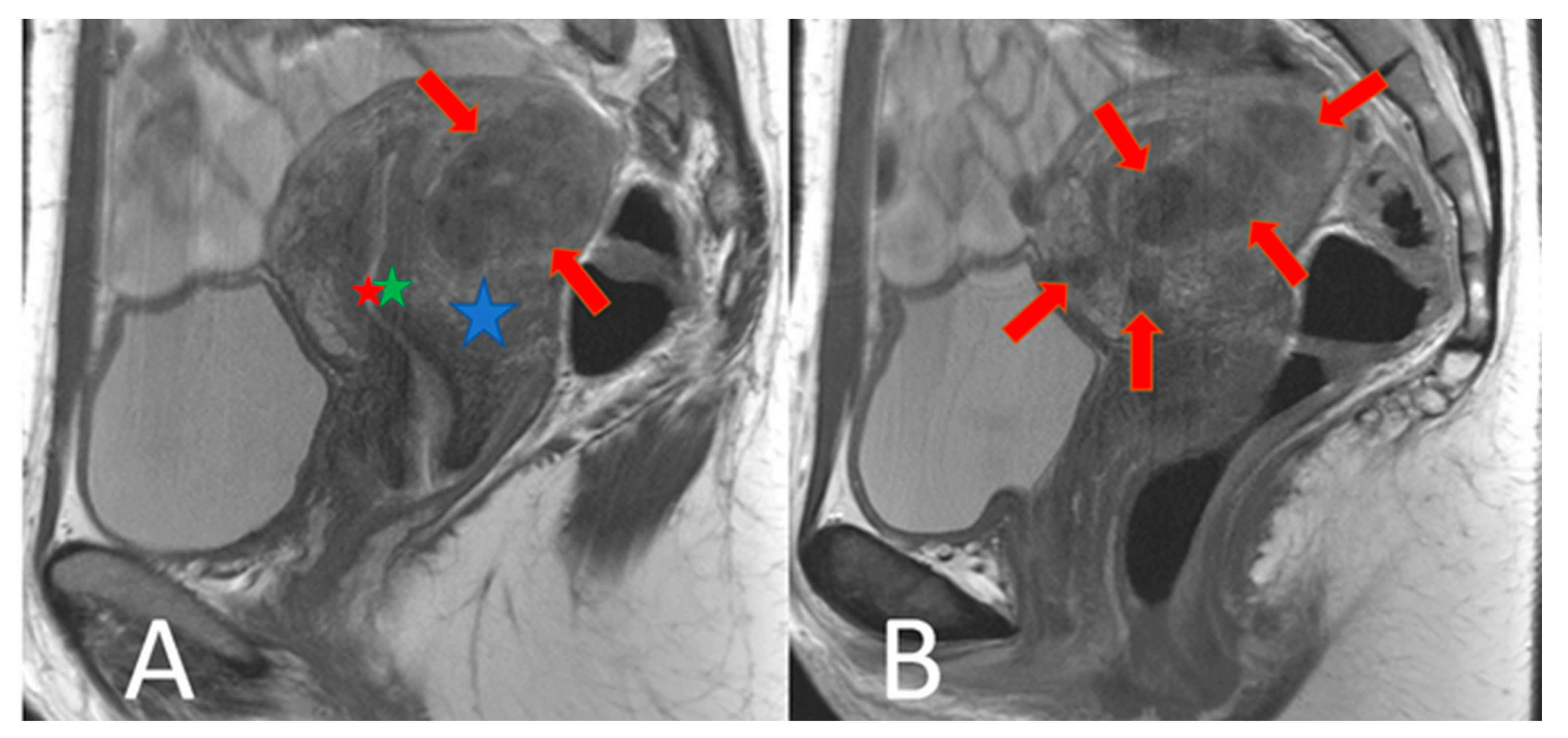
2.3. Image Segmentation and Radiomic Feature Extraction
2.4. Dimension Reduction and Feature Selection
2.5. Data Analysis
3. Results
3.1. Patients
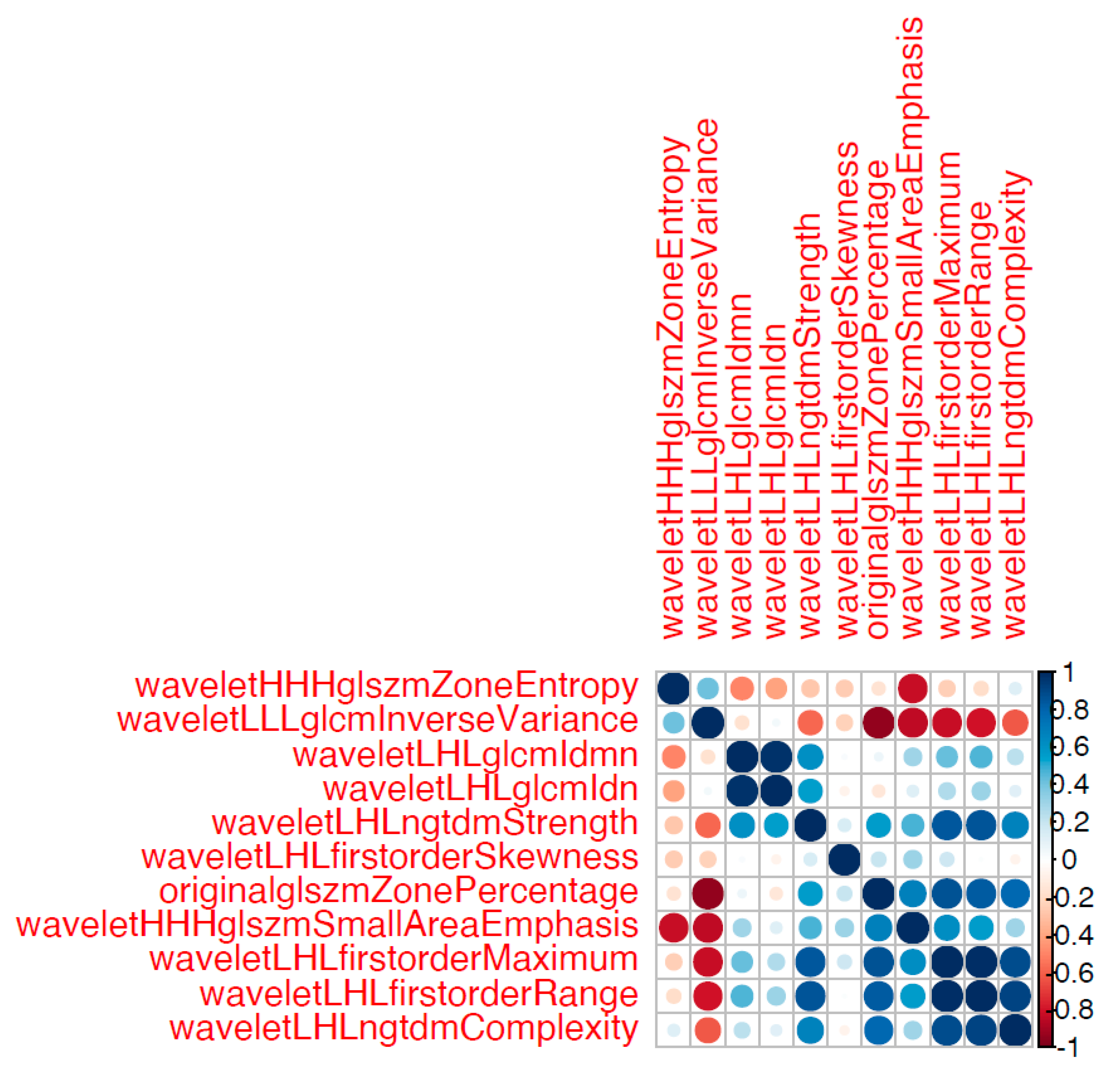
3.2. Radiomics
4. Discussion
Author Contributions
Funding
Institutional Review Board Statement
Informed Consent Statement
Data Availability Statement
Conflicts of Interest
Abbreviations
| MRI | Magnetic Resonance Imaging |
| T2w | T2-weighted |
| SD | Standard Deviation |
| TVUS | Transvaginal Ultrasound |
| TSE | Turbo Spin Echo |
| TR | Repetition Time |
| TE | Echo Time |
| FOV | Field of View |
| NSA | Number of Signal Averages |
| ML | Machine Learning |
| ROC | Receiver Operating Curves |
| AUC | Area under Curve |
| AUPR | Area under the Precision- Recall curve |
| DICOM | Digital Imaging and Communications in Medicine |
| GLCM | Gray-level Co-occurrence Matrix |
| GLRLM | Gray-level Run Length Matrix |
| GLSZM | Gray-level Size Zone Matrix |
| NGTDM | Neighboring Gray Tone Difference Matrix |
| GLDM | Gray-level Dependence Matrix |
| RF | Random Forest |
| CI | Confidence Interval |
| JZ | Junctional Zone |
References
- Vannuccini, S.; Tosti, C.; Carmona, F.; Huang, S.J.; Chapron, C.; Guo, S.W.; Petraglia, F. Pathogenesis of adenomyosis: An update on molecular mechanisms. Reprod. Biomed. Online 2017, 35, 592–601. [Google Scholar] [CrossRef] [PubMed]
- Loring, M.; Chen, T.Y.; Isaacson, K.B. A Systematic Review of Adenomyosis: It Is Time to Reassess What We Thought We Knew about the Disease. J. Minim. Invasive Gynecol. 2021, 28, 644–655. [Google Scholar] [CrossRef] [PubMed]
- Guo, S.W. The Pathogenesis of Adenomyosis vis-à-vis Endometriosis. J. Clin. Med. 2020, 9, 485. [Google Scholar] [CrossRef] [PubMed]
- Horton, J.; Sterrenburg, M.; Lane, S.; Maheshwari, A.; Li, T.C.; Cheong, Y. Reproductive, obstetric, and perinatal outcomes of women with adenomyosis and endometriosis: A systematic review and meta-analysis. Hum. Reprod. Update 2019, 25, 592–632. [Google Scholar] [CrossRef]
- Garcia, L.; Isaacson, K. Adenomyosis: Review of the literature. J. Minim. Invasive Gynecol. 2011, 18, 428–437. [Google Scholar] [CrossRef]
- Komatsu, H.; Taniguchi, F.; Harada, T. Impact of adenomyosis on perinatal outcomes: A large cohort study (JSOG database). BMC Pregnancy Childbirth 2023, 23, 579. [Google Scholar] [CrossRef] [PubMed]
- Yu, O.; Schulze-Rath, R.; Grafton, J.; Hansen, K.; Scholes, D.; Reed, S.D. Adenomyosis incidence, prevalence and treatment: United States population-based study 2006–2015. Am. J. Obstet. Gynecol. 2020, 223, 94.e1–94.e10. [Google Scholar] [CrossRef] [PubMed]
- Harada, T.; Taniguchi, F.; Guo, S.W.; Choi, Y.M.; Biberoglu, K.O.; Tsai, S.S.; Alborzi, S.; Al-Jefout, M.; Chalermchokcharoenkit, A.; Sison-Aguilar, A.G.; et al. The Asian Society of Endometriosis and Adenomyosis guidelines for managing adenomyosis. Reprod. Med. Biol. 2023, 22, e12535. [Google Scholar] [CrossRef]
- Dason, E.S.; Maxim, M.; Sanders, A.; Papillon-Smith, J.; Ng, D.; Chan, C.; Sobel, M. Guideline No. 437: Diagnosis and Management of Adenomyosis. J. Obstet. Gynaecol. Can. 2023, 45, 417–429.e1. [Google Scholar] [CrossRef]
- Harmsen, M.J.; Van den Bosch, T.; de Leeuw, R.A.; Dueholm, M.; Exacoustos, C.; Valentin, L.; Hehenkamp, W.J.K.; Groenman, F.; De Bruyn, C.; Rasmussen, C.; et al. Consensus on revised definitions of Morphological Uterus Sonographic Assessment (MUSA) features of adenomyosis: Results of modified Delphi procedure. Ultrasound Obstet. Gynecol. 2022, 60, 118–131. [Google Scholar] [CrossRef]
- Krentel, H.; Keckstein, J.; Füger, T.; Hornung, D.; Theben, J.; Salehin, D.; Buchweitz, O.; Mueller, A.; Schäfer, S.D.; Sillem, M.; et al. Accuracy of ultrasound signs on two-dimensional transvaginal ultrasound in prediction of adenomyosis: Prospective multicenter study. Ultrasound Obstet. Gynecol. 2023, 62, 739–746. [Google Scholar] [CrossRef] [PubMed]
- Chapron, C.; Vannuccini, S.; Santulli, P.; Abrão, M.S.; Carmona, F.; Fraser, I.S.; Gordts, S.; Guo, S.W.; Just, P.A.; Noël, J.C.; et al. Diagnosing adenomyosis: An integrated clinical and imaging approach. Hum. Reprod. Update 2020, 26, 392–411. [Google Scholar] [CrossRef] [PubMed]
- Rees, C.O.; Nederend, J.; Mischi, M.; van Vliet, H.A.A.M.; Schoot, B.C. Objective measures of adenomyosis on MRI and their diagnostic accuracy—A systematic review & meta-analysis. Acta Obstet. Gynecol. Scand. 2021, 100, 1377–1391. [Google Scholar] [CrossRef] [PubMed]
- Bazot, M.; Daraï, E. Role of transvaginal sonography and magnetic resonance imaging in the diagnosis of uterine adenomyosis. Fertil. Steril. 2018, 109, 389–397. [Google Scholar] [CrossRef]
- Tellum, T.; Nygaard, S.; Lieng, M. Noninvasive Diagnosis of Adenomyosis: A Structured Review and Meta-analysis of Diagnostic Accuracy in Imaging. J. Minim. Invasive Gynecol. 2020, 27, 408–418.e3. [Google Scholar] [CrossRef]
- Zhang, M.; Bazot, M.; Tsatoumas, M.; Munro, M.G.; Reinhold, C. MRI of Adenomyosis: Where Are We Today? Can. Assoc. Radiol. J. 2023, 74, 58–68. [Google Scholar] [CrossRef] [PubMed]
- Aerts, H.J.; Velazquez, E.R.; Leijenaar, R.T.; Parmar, C.; Grossmann, P.; Carvalho, S.; Bussink, J.; Monshouwer, R.; Haibe-Kains, B.; Rietveld, D.; et al. Decoding tumour phenotype by noninvasive imaging using a quantitative radiomics approach. Nat. Commun. 2014, 5, 4006. [Google Scholar] [CrossRef]
- Lambin, P.; Leijenaar, R.T.H.; Deist, T.M.; Peerlings, J.; de Jong, E.E.C.; van Timmeren, J.; Sanduleanu, S.; Larue, R.T.H.M.; Even, A.J.G.; Jochems, A.; et al. Radiomics: The bridge between medical imaging and personalized medicine. Nat. Rev. Clin. Oncol. 2017, 14, 749–762. [Google Scholar] [CrossRef]
- Li, Z.; Zhang, J.; Song, Y.; Yin, X.; Chen, A.; Tang, N.; Prince, M.R.; Yang, G.; Wang, H. Utilization of radiomics to predict long-term outcome of magnetic resonance-guided focused ultrasound ablation therapy in adenomyosis. Eur. Radiol. 2021, 31, 392–402. [Google Scholar] [CrossRef]
- Bazot, M.; Bharwani, N.; Huchon, C.; Kinkel, K.; Cunha, T.M.; Guerra, A.; Manganaro, L.; Buñesch, L.; Kido, A.; Togashi, K.; et al. European society of urogenital radiology (ESUR) guidelines: MR imaging of pelvic endometriosis. Eur. Radiol. 2017, 27, 2765–2775. [Google Scholar] [CrossRef]
- Baessler, B.; Nestler, T.; Pinto Dos Santos, D.; Paffenholz, P.; Zeuch, V.; Pfister, D.; Maintz, D.; Heidenreich, A. Radiomics allows for detection of benign and malignant histopathology in patients with metastatic testicular germ cell tumors prior to post-chemotherapy retroperitoneal lymph node dissection. Eur. Radiol. 2020, 30, 2334–2345. [Google Scholar] [CrossRef] [PubMed]
- Jing, R.; Wang, J.; Li, J.; Wang, X.; Li, B.; Xue, F.; Shao, G.; Xue, H. A wavelet features derived radiomics nomogram for prediction of malignant and benign early-stage lung nodules. Sci. Rep. 2021, 11, 22330. [Google Scholar] [CrossRef] [PubMed]
- van Griethuysen, J.J.M.; Fedorov, A.; Parmar, C.; Hosny, A.; Aucoin, N.; Narayan, V.; Beets-Tan, R.G.H.; Fillion-Robin, J.C.; Pieper, S.; Aerts, H.J.W.L. Computational Radiomics System to Decode the Radiographic Phenotype. Cancer Res. 2017, 77, e104–e107. [Google Scholar] [CrossRef] [PubMed]
- Skawran, S.M.; Kambakamba, P.; Baessler, B.; von Spiczak, J.; Kupka, M.; Müller, P.C.; Moeckli, B.; Linecker, M.; Petrowsky, H.; Reiner, C.S. Can magnetic resonance imaging radiomics of the pancreas predict postoperative pancreatic fistula? Eur. J. Radiol. 2021, 140, 109733. [Google Scholar] [CrossRef] [PubMed]
- Lin, Q.; Ji, Y.F.; Chen, Y.; Sun, H.; Yang, D.D.; Chen, A.L.; Chen, T.W.; Zhang, X.M. Radiomics model of contrast-enhanced MRI for early prediction of acute pancreatitis severity. J. Magn. Reson. Imaging 2020, 51, 397–406. [Google Scholar] [CrossRef] [PubMed]
- Nougaret, S.; McCague, C.; Tibermacine, H.; Vargas, H.A.; Rizzo, S.; Sala, E. Radiomics and radiogenomics in ovarian cancer: A literature review. Abdom. Radiol. 2021, 46, 2308–2322. [Google Scholar] [CrossRef] [PubMed]
- Panico, C.; Avesani, G.; Zormpas-Petridis, K.; Rundo, L.; Nero, C.; Sala, E. Radiomics and Radiogenomics of Ovarian Cancer: Implications for Treatment Monitoring and Clinical Management. Radiol. Clin. N. Am. 2023, 61, 749–760. [Google Scholar] [CrossRef] [PubMed]
- Prayer, F.; Watzenböck, M.L.; Heidinger, B.H.; Rainer, J.; Schmidbauer, V.; Prosch, H.; Ulm, B.; Rubesova, E.; Prayer, D.; Kasprian, G. Fetal MRI radiomics: Non-invasive and reproducible quantification of human lung maturity. Eur. Radiol. 2023, 33, 4205–4213. [Google Scholar] [CrossRef] [PubMed]
- Xiao, M.L.; Fu, L.; Wei, Y.; Liu, A.E.; Cheng, J.J.; Ma, F.H.; Li, H.M.; Li, Y.A.; Lin, Z.J.; Zhang, G.F.; et al. Intratumoral and peritumoral MRI radiomics nomogram for predicting parametrial invasion in patients with early-stage cervical adenocarcinoma and adenosquamous carcinoma. Eur. Radiol. 2024, 34, 852–862. [Google Scholar] [CrossRef]
- Zhou, Y.; Zhang, J.; Chen, J.; Yang, C.; Gong, C.; Li, C.; Li, F. Prediction using T2-weighted magnetic resonance imaging-based radiomics of residual uterine myoma regrowth after high-intensity focused ultrasound ablation. Ultrasound Obstet. Gynecol. 2022, 60, 681–692. [Google Scholar] [CrossRef]
- Tellum, T.; Matic, G.V.; Dormagen, J.B.; Nygaard, S.; Viktil, E.; Qvigstad, E.; Lieng, M. Diagnosing adenomyosis with MRI: A prospective study revisiting the junctional zone thickness cutoff of 12 mm as a diagnostic marker. Eur. Radiol. 2019, 29, 6971–6981. [Google Scholar] [CrossRef]
- Harmsen, M.J.; Trommelen, L.M.; de Leeuw, R.A.; Tellum, T.; Juffermans, L.J.M.; Griffioen, A.W.; Thomassin-Naggara, I.; Van den Bosch, T.; Huirne, J.A.F. Uterine junctional zone and adenomyosis: Comparison of MRI, transvaginal ultrasound and histology. Ultrasound Obstet. Gynecol. 2023, 62, 42–60. [Google Scholar] [CrossRef]
- Munro, M.G. Adenomyosis: A riddle, wrapped in mystery, inside an enigma. Fertil. Steril. 2021, 116, 89–90. [Google Scholar] [CrossRef] [PubMed]
- Zheng, G.; Hou, J.; Shu, Z.; Peng, J.; Han, L.; Yuan, Z.; He, X.; Gong, X. Prediction of neoadjuvant chemotherapy pathological complete response for breast cancer based on radiomics nomogram of intratumoral and derived tissue. BMC Med. Imaging 2024, 24, 22. [Google Scholar] [CrossRef]
- Dashottar, S.; Singh, A.K.; Debnath, J.; Muralidharan, C.G.; Singh, R.K.; Kumar, S. Comparative analysis of changes in MR imaging of pre and post intrauterine progesterone implants in adenomyosis cases. Med. J. Armed Forces India 2015, 71, 145–151. [Google Scholar] [CrossRef]
- Raimondo, D.; Raffone, A.; Aru, A.C.; Giorgi, M.; Giaquinto, I.; Spagnolo, E.; Travaglino, A.; Galatolo, F.A.; Cimino, M.G.C.A.; Lenzi, J.; et al. Application of Deep Learning Model in the Sonographic Diagnosis of Uterine Adenomyosis. Int. J. Environ. Res. Public Health 2023, 20, 1724. [Google Scholar] [CrossRef]
- Guerriero, S.; Pascual, M.; Ajossa, S.; Neri, M.; Musa, E.; Graupera, B.; Rodriguez, I.; Alcazar, J.L. Artificial intelligence (AI) in the detection of rectosigmoid deep endometriosis. Eur. J. Obstet. Gynecol. Reprod. Biol. 2021, 261, 29–33. [Google Scholar] [CrossRef] [PubMed]
- Sone, K.; Toyohara, Y.; Taguchi, A.; Miyamoto, Y.; Tanikawa, M.; Uchino-Mori, M.; Iriyama, T.; Tsuruga, T.; Osuga, Y. Application of artificial intelligence in gynecologic malignancies: A review. J. Obstet. Gynaecol. Res. 2021, 47, 2577–2585. [Google Scholar] [CrossRef]
- Christiansen, F.; Epstein, E.L.; Smedberg, E.; Åkerlund, M.; Smith, K.; Epstein, E. Ultrasound image analysis using deep neural networks for discriminating between benign and malignant ovarian tumors: Comparison with expert subjective assessment. Ultrasound Obstet. Gynecol. 2021, 57, 155–163. [Google Scholar] [CrossRef] [PubMed]
- Taddese, A.A.; Tilahun, B.C.; Awoke, T.; Atnafu, A.; Mamuye, A.; Mengiste, S.A. Deep-learning models for image-based gynecological cancer diagnosis: A systematic review and meta- analysis. Front. Oncol. 2023, 13, 1216326. [Google Scholar] [CrossRef]
- Greener, J.G.; Kandathil, S.M.; Moffat, L.; Jones, D.T. A guide to machine learning for biologists. Nat. Rev. Mol. Cell Biol. 2022, 23, 40–55. [Google Scholar] [CrossRef] [PubMed]
- Martire, F.G.; Russo, C.; Selntigia, A.; Nocita, E.; Soreca, G.; Lazzeri, L.; Zupi, E.; Exacoustos, C. Early noninvasive diagnosis of endometriosis: Dysmenorrhea and specific ultrasound findings are important indicators in young women. Fertil. Steril. 2023, 119, 455–464. [Google Scholar] [CrossRef] [PubMed]
- Millischer, A.E.; Santulli, P.; Da Costa, S.; Bordonne, C.; Cazaubon, E.; Marcellin, L.; Chapron, C. Adolescent endometriosis: Prevalence increases with age on magnetic resonance imaging scan. Fertil. Steril. 2023, 119, 626–633. [Google Scholar] [CrossRef] [PubMed]
- Mayerhoefer, M.E.; Materka, A.; Langs, G.; Häggström, I.; Szczypiński, P.; Gibbs, P.; Cook, G. Introduction to Radiomics. J. Nucl. Med. 2020, 61, 488–495. [Google Scholar] [CrossRef]
- Sartoretti, E.; Sartoretti, T.; Wyss, M.; Reischauer, C.; van Smoorenburg, L.; Binkert, C.A.; Sartoretti-Schefer, S.; Mannil, M. Amide proton transfer weighted (APTw) imaging based radiomics allows for the differentiation of gliomas from metastases. Sci. Rep. 2021, 11, 5506. [Google Scholar] [CrossRef]


| Patients | Age | G/P | Endocrine Therapy | Endocrine Therapy Duration before MRI (Months) | Adenomyosis in TVUS | Adenomyosis in Conventional MRI | Duration MRI—Surgery (Months) | Endometriosis (Intraoperative #Enzian) |
|---|---|---|---|---|---|---|---|---|
| AG | ||||||||
| 1 | 33 | 0/0 | 2 | 1 | No | Yes | 4 | P2, T3/3, A2, B2/1, C1, FA |
| 2 | 42 | 0/0 | 1 | 36 | Yes | Yes | 5 | P1, FA |
| 3 | 44 | II/I | 0 | 0 | Yes | Yes | 3 | P1, O1/1, T1/1, A3, B2/1, FA, FB |
| 4 | 35 | I/I | 2 | 2 | Yes | No | 1 | P2, O1/1, T1,1, A2, FA, FB |
| 5 | 47 | II/II | 3 | 3 | Yes | Yes | 1 | P2, O2/2, T3/3, A2, B1/1, C1, FA, FB |
| 6 | 42 | 0/0 | 4 | 2 | Yes | Yes | 5 | P2, O1/0, T3/1, B1/0, FA |
| 7 | 41 | I/I | 0 | 0 | Yes | Yes | 2 | P2, O1/1, T1/1, A3, B1/1, C3, FA, FB |
| 8 | 39 | 0/0 | 0 | 0 | Yes | Yes | 1 | P3, T2/0, A3, B2/1, C1, FA, FU |
| 9 | 44 | III/II | 2 | 2 | Yes | Yes | 1 | P3, O3/3, T3/3, A3, B3/3, C2, FA, FU, FI (Sigma) |
| CG | ||||||||
| 1 | 38 | II/I | 0 | 0 | No | No | 2 | P1, O1/1, T0/1, B1/1, FB |
| 2 | 48 | I/I | 2 | 1 | No | Yes | 1 | P2, O2/0, B1/1 |
| 3 | 37 | IV/III | 2 | 3 | No | Yes | 1 | P1, O2/1, T1/1, A1 |
| 4 | 40 | II/II | 3 | 5 | No | No | 2 | P1, O1/2, T1/1, A2, B1/0, C1 |
| 5 | 39 | II/II | 0 | 0 | Yes | Yes | 4 | FB |
| 6 | 40 | 0/0 | 0 | 0 | No | Yes | 2 | FO (umbilicus) |
| Feature | AUC (95% Confidence Interval) |
|---|---|
| Original_glszm_ZonePercentage | 0.78 (0.49–1) |
| Wavelet_LHL_glcm_Idmn | 0.98 (0.93–1) |
| Wavelet_LHL_glcm_Idn | 0.93 (0.79–1) |
| Wavelet_LHL_firstorder_Skewness | 0.83 (0.51–1) |
| Wavelet_LHL_firstorder_Maximum | 0.89 (0.67–1) |
| Wavelet_LHL_firstorder_Range | 0.89 (0.67–1) |
| Wavelet_LHL_ngtdm_Complexity | 0.83 (0.6–1) |
| Wavelet_LHL_ngtdm_Strength | 0.91 (0.76–1) |
| Wavelet_HHH_glszm_SmallAreaEmphasis | 0.83 (0.6–1) |
| Wavelet_HHH_glszm_ZoneEntropy | 0.83 (0.6–1) |
| Wavelet_LLL_glcm_InverseVariance | 0.78 (0.49–1) |
Disclaimer/Publisher’s Note: The statements, opinions and data contained in all publications are solely those of the individual author(s) and contributor(s) and not of MDPI and/or the editor(s). MDPI and/or the editor(s) disclaim responsibility for any injury to people or property resulting from any ideas, methods, instructions or products referred to in the content. |
© 2024 by the authors. Licensee MDPI, Basel, Switzerland. This article is an open access article distributed under the terms and conditions of the Creative Commons Attribution (CC BY) license (https://creativecommons.org/licenses/by/4.0/).
Share and Cite
Burla, L.; Sartoretti, E.; Mannil, M.; Seidel, S.; Sartoretti, T.; Krentel, H.; De Wilde, R.L.; Imesch, P. MRI-Based Radiomics as a Promising Noninvasive Diagnostic Technique for Adenomyosis. J. Clin. Med. 2024, 13, 2344. https://doi.org/10.3390/jcm13082344
Burla L, Sartoretti E, Mannil M, Seidel S, Sartoretti T, Krentel H, De Wilde RL, Imesch P. MRI-Based Radiomics as a Promising Noninvasive Diagnostic Technique for Adenomyosis. Journal of Clinical Medicine. 2024; 13(8):2344. https://doi.org/10.3390/jcm13082344
Chicago/Turabian StyleBurla, Laurin, Elisabeth Sartoretti, Manoj Mannil, Stefan Seidel, Thomas Sartoretti, Harald Krentel, Rudy Leon De Wilde, and Patrick Imesch. 2024. "MRI-Based Radiomics as a Promising Noninvasive Diagnostic Technique for Adenomyosis" Journal of Clinical Medicine 13, no. 8: 2344. https://doi.org/10.3390/jcm13082344