Article

Purification and Phytotoxic Analysis of Botrytis cinerea Virulence Factors: New Avenues for Crop Protection

1
Department of Biological Sciences, University of Alabama in Huntsville, 301 Sparkman Drive, Huntsville, AL 35899, USA
2
Department of Chemistry, University of Alabama in Huntsville, 301 Sparkman Drive, Huntsville, AL 35899, USA
*
Author to whom correspondence should be addressed.
Deceased on 12 February 2010.
Agriculture 2012, 2(3), 154-164; https://doi.org/10.3390/agriculture2030154
Submission received: 29 April 2012 / Revised: 18 June 2012 / Accepted: 4 July 2012 / Published: 10 July 2012
(This article belongs to the Special Issue The Impact of Plant Disease on Food Security)

Abstract

:
Botrytis cinerea is a necrotrophic fungus infecting over 230 plant species worldwide. This highly adaptable pathogen can afflict agricultural products from seed to storage, causing significant economic losses and instability in the food supply. Small protein virulence factors secreted by B. cinerea during infection play an important role in initiation and spread of disease. BcSnod1 was found to be abundantly expressed upon exposure to media containing strawberry extract. From sequence similarity, BcSnod2 was also identified and both were recognized as members of the Ceratoplatanin family of small phytotoxic proteins. Recombinant BcSnod1 was shown to have a phytotoxic effect and play an important role in pathogenicity while the role of BcSnod2 remains less clear. Both bacterial and yeast production systems are reported, though the bacterial protein is less toxic and mostly unfolded relative to that made in yeast. Compared to BcSnod1, recombinant bacterial BcSnod2 shows similar, but delayed phytotoxicity on tomato leaves. Further studies of these critical virulence factors and their inhibition promise to provide new avenues for crop protection.

1. Introduction

Botrytis cinerea can infect many agriculturally important food crops with devastating consequences [1,2]. It is a significant concern for agribusiness and food supply continuity with a weighty portion of the fungicide industry focusing on its inhibition. This necrotrophic fungus can attack many plant organs and is a serious threat for post-harvest losses. The invasion strategy of B. cinerea, including the hypersensitive response, has been proposed [3,4,5]. Involvement of the extensively studied secretome has been shown [6,7]. These proteomic studies have shown a number of proteins are secreted, several of which contribute to cell death in plant tissues. One of the most abundant is BcSnod1 (also referred to as BcSpl1 [8]). Closely related though not as ubiquitously expressed is BcSnod2. Both are members of the Ceratoplatanin-family of Snod-like proteins.
Ceratoplatanins, named for the founding member cerato-platanin produced by Ceratocystis fimbriata [9], are extracellular disease related proteins [7,9]. In the case of C. fimbriata, cerato-platanin was detected in the fungal cell wall as well [10]. Ceratoplatanins are moderately hydrophobic with four conserved cysteines that form two intramolecular disulfide bonds. This family induces plant defenses and, in some cases, localized necrosis in the exposed area. Ceratoplatanin knock-outs in different fungi have various effects. In the rice blast fungus, Magnaporthe grisea, deletion of msp1 caused a significant reduction in virulence for young rice and barley [11]. In Leptosphaeria maculans, the cause of blackleg disease of canola, Ceratoplatanin knock-out mutants were as virulent as the wild-type [12]. For B. cinerea, BcSnod1 is required for virulence against tomato and tobacco [13].
Herein we report recombinant production of both BcSnod1 and BcSnod2. Further we demonstrate that these virulence factors by themselves are able to elicit necrosis in tomato and tobacco. With the ability to make large amounts of functional protein recombinantly, the possibilities for better understanding B. cinerea infection and the roles of Ceratoplatanins are increased. Continued structure function studies are forthcoming, all of which contribute to new possibilities for antifungal intervention to help prevent agricultural losses.

2. Results and Discussion

2.1. Cloning and Expression in Bacteria

The BcSnod1 and BcSnod2 genes (the coding region downstream of the signal sequence) were successfully amplified from B. cinerea B05.10 genomic DNA. Fragments of approximately 370 and 410 bp correlated with the expected sizes from genomic sequencing indicating the absence of introns. BcSnod1 and BcSnod2 expressed well in bacteria as fusion proteins with N-terminal hexahistidine tags. Both were insoluble in Luria broth and minimal M9 media, even at reduced growth temperature of 16 °C, see Figure 1. Insoluble protein was denatured using 8 M urea and refolded by dialysis reduction of the urea concentration. Both BcSnod fusion proteins were soluble in the final dialysis buffer of 20 mM Tris, pH 6.8. Approximate molecular weights were as expected, with measured values of 15 kDa for BcSnod1 and 15.5 kDa for BcSnod2 correlating well with calculated weights of 14.70 kDa and 15.20 kDa. Refolded yields for both were approximately 2 milligrams per liter of culture.
Figure 1. Bacterial expressed and purified BcSnods. A. Lane 0, Molecular Weight Marker; Lane 1, induced BcSnod1; Lane 2, uninduced BcSnod1; Lane 3, total soluble lysate; Lane 4, total insoluble pellet. B. Lane 0, Molecular Weight Marker; Lanes 1, 2 and 3 contain eluted fractions from metal chelation chromatography of refolded BcSnod1, BcSnod2, and vector-only control, respectively.
Agriculture 02 00154 g001

2.2. Plant Assays with Recombinant BcSnods from Bacteria

In planta transient assays were conducted by injecting leaves with refolded, bacterially produced BcSnods. Young tomato leaves were injected with 100 μL of BcSnod protein (2 mg/ml in 20 mM Tris, pH 6.8). The area injected with either BcSnod started to discolor 3 days post inoculation (dpi), see Figure 2. Six attempts were made for each protein in which four BcSnod1 injections produced discoloration while three BcSnod2 injections produced discoloration. In this case, BcSnod induced discoloration did not turn into necrosis. Due to poor solubility of the bacterially expressed protein, injections of more concentrated BcSnod protein were not pursued. Controls of empty vector transformed bacteria, treated identically as the BcSnod samples in the same final buffer of 20 mM Tris, pH 6.8, did not cause discoloration.

2.3. Yeast Expressed BcSnods

Pichia pastoris transformation of BcSnod1 and BcSnod2 in the 10 kb pPic9k vector construct yielded approximately 3.0 × 10⁴ colonies/μg of DNA. Five transformants for each were screened using PCR amplification. Only a single band of expected size was observed, indicating transformation by both BcSnod genes. BcSnod1 expressed well as a secreted protein, yielding greater than ten of milligrams per liter of starting culture in minimal glycerol media induced in 100 mL of minimal methanol media. After HPLC purification, the total amount of BcSnod1 obtained was 6 mg (see Figure 3). In contrast, BcSnod2 did not express in P. pastoris in any of the 5 clones tested. PCR amplification re-confirmed the presence of BcSnod2 gene in the genome but no protein was ever detected in the culture media. No sign of BcSnod2 overexpression was found intracellularly either. As similar as the sequences are, BcSnod1 and BcSnod2 certainly behave differently in yeast overexpression. A control strain of Pichia pastoris transformed with an empty pPic9k was also constructed. Treated, induced, and prepared identically, the same HPLC fractions from the yeast vector only served as a background control.
Figure 2. In Planta transient assay with bacterially expressed BcSnod proteins. A. Tomato leaves injected with empty vector control showed minimal discoloration. B and C. Leaves injected with 200 μg of bacterial produced, renatured BcSnod1 and BcSnod2, respectively. Signs of discoloration were readily apparent after 3 days, even more pronounced at 5 dpi (shown here). Arrows designate site of injection.
Agriculture 02 00154 g002
Figure 3. Chromatogram from HPLC purification of Pichia pastoris expressed BcSnod1 (red) and empty vector control (gray). BcSnod1 was present in fractions 41–47, indicated by the gray box. In the inset, SDS-PAGE of the concentrated combined fractions for BcSnod1 (B) and Pichia only control (P) are shown next to a molecular weight marker (M).
Agriculture 02 00154 g003

2.4. Pichia Expressed BcSnod1 Induces Necrosis

When injected in tomato (Solanum lycopersicum) leaves, BcSnod1 obtained from P. pastoris initiated discoloration in the area of infiltration within 3 days. Discoloration turned into necrosis around 10 days after infiltration and spread to adjoining areas within a few days. Neither the leaf veins nor midvein prevented the propagation of necrosis, which continued through the duration of the experiment (i.e., to Day 30), see Figure 4. Control extracts from P. pastoris with the empty pPic9k vector induced yellowing in the region of injection after 10 dpi, but discoloration did not spread to adjoining regions as seen for BcSnod1. Even after 30 days minimal necrosis is observed, all centered at the site of infiltration. The same infiltration assay was performed in tobacco (Nicotiana tabacum var. Xanthi) leaves. For tobacco, the area of injection showed discoloration (an observation identical to the hypersensitive response as in Frías et al. [8]) within a few days after infiltration only against BcSnod1. Likewise, necrosis was readily observable at day 15 again only for BcSnod1 infiltrated leaves. Unlike tomato, necrosis in tobacco was limited to the area of injection and did not cross leaf veins and spread to other areas, see Figure 5.
Figure 4. Tomato leaves injected with HPLC purified P. pastoris produced BcSnod1 and the same volume of vector only control. When 15 µg of BcSnod1 were injected (white arrow indicates site of injection), discoloration started within 3 days post infection and continued up to day 10. The entire injected area was necrotized within 15 days and discoloration was observed in adjacent areas. The lower panels indicate leaves injected with pPic9k (control), where slight discoloration is observed after 15 days with minimal necrosis 30 days post-infiltration (yellow arrow indicates site of injection). No spread of discoloration or necrosis in the adjoining regions was observed.
Agriculture 02 00154 g004
Figure 5. Tobacco leaves infiltrated with HPLC purified P. pastoris produced BcSnod1 and the same volume of vector only control. In tobacco, hypersensitive response was observed from day 5 onwards, turning into necrosis after 15 dpi for BcSnod1. Necrosis did not spread to adjoining areas. No discoloration was observed for the control.
Agriculture 02 00154 g005

2.5. Pichia Expressed BcSnod1 Detached Leaf Assays

Two types of detached leaf assays were performed, one with infiltration of protein into the leaves while the other tested surface exposure by pipetting a drop of protein solution onto the leaves. Healthy tomato leaves were cut off 4 week old plants and placed in a moist box in environmental chamber. For infiltrated leaves, 130 μg of protein solution was injected. Hypersensitive response was observed starting the first day after infiltration for BcSnod1 but not the control, see Figure 6. For BcSnod1, discoloration of the area of infiltration was observed after 3 days with maximum discoloration around day 10. Discoloration did spread to other areas. For the control, some discoloration was observed after 5 days but of very low intensity compared to BcSnod1. Discoloration for the control did not spread.
For surface exposure in detached tomato leaves, a 10 μL drop of solution containing 7 µg of BcSnod1 was placed on the upper side of the leaf. Signs of hypersensitive response were observed within 3 days, and necrosis identified 7 days post-infiltration, see Figure 7. No discoloration, much less necrosis, was observed for an equivalent volume of vector only control pipetted onto the leaf surface. The study was terminated at 8 days due to demise of the detached leaves. These findings show that BcSnod1 is able to elicit necrosis simply by exposure to the plant cell surface. Infiltration significantly expedited the process, but it appears that BcSnod1 (or at least some part of it) is able to enter plant cells and initiate cell death.

2.6. Summary of Plant Assays

Both recombinant BcSnod1 and BcSnod2 displayed phytotoxic activity. However, the activity of bacterially produced BcSnod1 protein was more variable than Pichia produced protein. Noticeable differences were observed for the plant hosts tomato and tobacco in response to BcSnod1. In tomato, the area where the protein was infiltrated initially got discolored and then necrotized. Necrosis started spreading to regions adjoining the region of infiltration. This was different in tobacco plant in which the necrosis could only be observed in the area of infiltration and did not spread to adjoining regions.
Figure 6. Detached tomato leaves were infiltrated with BcSnod1. One hundred and thirty micrograms of Pichia produced HPLC purified BcSnod1 was injected into detached tomato leaves. Discoloration in the area of injection was observed after 1 day. Discoloration increased with time and showed a maximum at 10 dpi. Necrosis was not observed presumably because the detached leaves are not viable after ~10 days. Infiltration with the vector only control (bottom row) showed minor discoloration after day 5, though no signs of spreading were observed. Dotted lines are for reference, showing the area of initial infiltration.
Agriculture 02 00154 g006
Figure 7. Tomato detached leaf assay of BcSnod1 surface exposure. Seven micrograms of Pichia produced HPLC purified BcSnod1 were pipetted on to the upper surface of a detached tomato leaf. Hypersensitive response started 3 days post infiltration with discoloration observed after day 5. No response was observed at day 3 with minimal discoloration after 10 days for the control application.
Agriculture 02 00154 g007

3. Experimental Section

3.1. Molecular Biology

BcSnod1 and BcSnod2 genes were amplified from genomic B. cinerea B05.10 DNA. The primers used for BcSnod1 were 5'-GTGCATATGATCACCGTCTCCTACG and 5'-GTGCTCGAGTTACAATCCACAAGCACTCTTGTCG. For BcSnod2, the primers used were 5'-GTGCATATGATCCAAGTAACCTACG and 5'-GTGCTCGAGCTAAGCCTTACACGGTGATCC. PCR products for both genes were first cloned into pGemTEasy (Promega Corporation, Madison, WI). For subsequent recombinant bacterial expression, BcSnod genes were excised using Nde1 and Xho1 restriction sites and ligated into pET28b (Novagen/EMD/Merck KGaA, Darmstadt, Germany). For yeast expression in Pichia pastoris, excised genes were inserted into pPic9k (Invitrogen/Life Technologies, Grand Island, NY) using EcoR1 and Not1 restriction sites. DNA sequencing confirmed the integrity of all clones.

3.2. Bacterial Protein Production

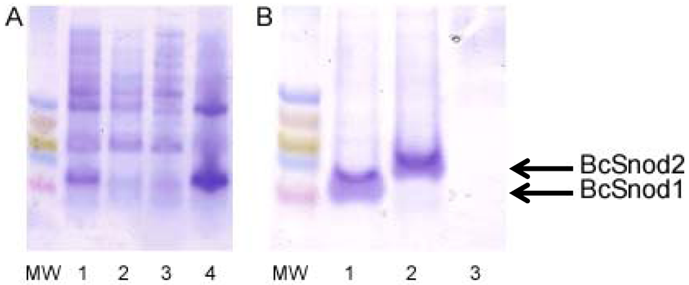
For bacterial expression of both BcSnod1 and BcSnod2, chemically competent Rosetta E. coli were transformed with kanamycin resistant pET28b expression vectors containing the BcSnod protein of interest. Protein expression in Luria broth was induced at an OD600 of 0.8 by adding 1.0 mM isopropyl β-D-thiogalactopyranoside and continued incubation at 37 °C for 4 h. Cells were harvested by centrifugation and stored at −80 °C. After lysis by sonication, insoluble BcSnod protein was collected by centrifugation at 20 kg for 20 min at 0 °C. The pellet, containing BcSnod, was then denatured in 8 M urea with 20 mM Tris pH 7.0, 250 mM NaCl and 3 mM DTT for refolding. Using step-wise dialysis, the protein was refolded by urea removal. Soluble BcSnod was further purified by metal chelation chromatography, eluted with 150 mM imidazole. The final dialysis buffer was 20 mM Tris pH 7.0 and 3 mM DTT.

3.3. Pichia Production

For expression in Pichia pastoris (GS115 His, ATCC-20864), the BcSnod vector of interest was linearized with BglII and Pichia transformed via electroporation. For electroporation, 80 µL of elctrocompetent cells were mixed with 3 ng of DNA and a 1.5 kV, 25 μF, 200 Ω electroporating pulse was applied. Screening for presence of BcSnod insert was carried out by PCR using the AOX primers. MutS mutants of transformed Pichia were used for recombinant expression. For screening, single colonies were inoculated in minimal glycerol media and grown until the OD600 reached 2.0. The cells were pelleted and resuspended in minimal methanol media for protein expression. Every 12 h, 0.5% methanol was added to the media to maintain expression of protein.
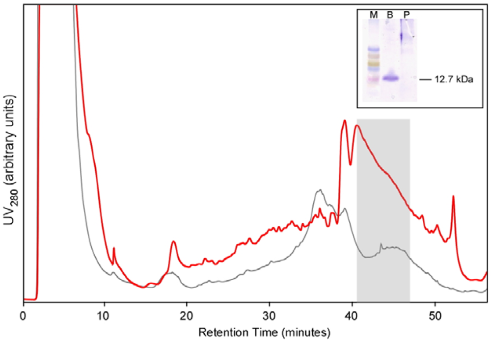
For large scale production, a colony of Pichia expressing BcSnod1 was grown overnight in 1 liter of minimal glycerol media, spun down, and then resuspended in 100 mL of minimal methanol media. Every 12 h, 0.5% methanol was added for protein expression. After 10 days, the supernatant was collected and concentrated to a final volume of 5 mL. The concentrated solution was applied to a C18 HPLC column for purification. A gradient of acetonitrile in 0.05% trifluoroacetic acid was used to purify BcSnod, which eluted at approximately 55% acetonitrile.

3.4. Plant Assays

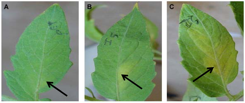
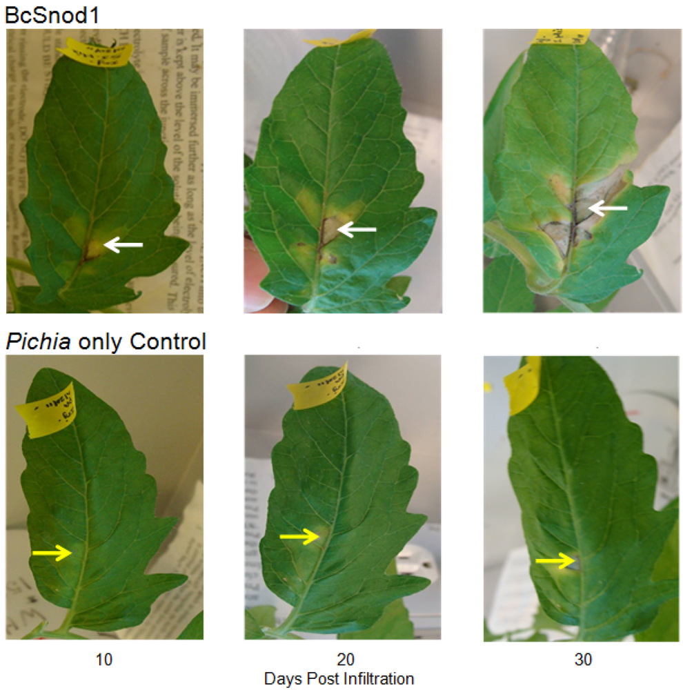
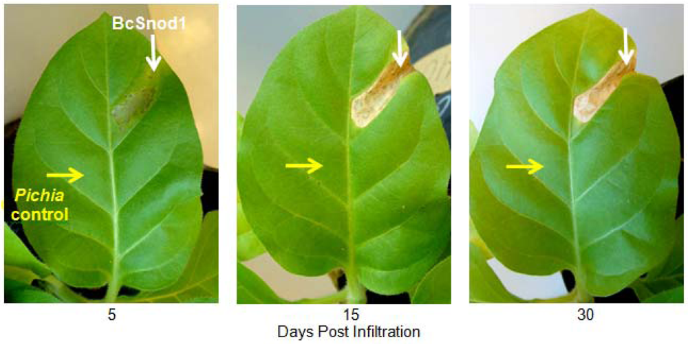
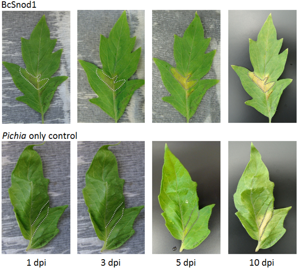
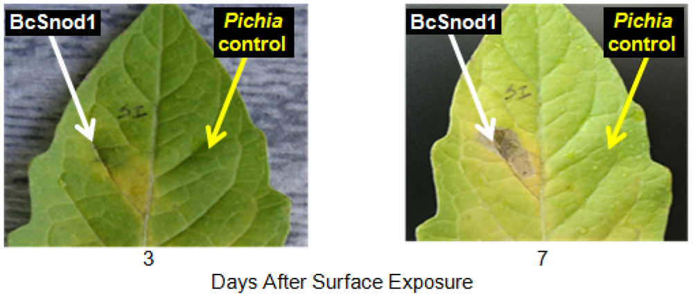
Two month old tomato (Solanum lycopersicum) and four month old tobacco (Nicotiana tabacum var. Xanthi) plants were used for the transient activity assays. All plants were grown in an environmental chamber (24 °C, 14 h, 40W/m2 light intensity, 10 h dark). For detached leaf assays, leaves were cut from the plant and kept in a wet box in the environmental chamber. For the transient assay with bacterially expressed protein, 200 μg of BcSnod1 or BcSnod2 in 100 μL were infiltrated in the midvein of tomato leaves using a syringe and 30.5 gauge needle. For the transient assay with Pichia expressed protein, 15 μg of BcSnod1 protein in 100 μL was infiltrated in the midvein of tomato and tobacco leaves using a syringe and 30.5 gauge needle. In the detached leaf assays, leaves were infiltrated with 100 μL containing 130 μg of protein after plugging out of the plant. In the surface exposed detached leaf assay, 10 µL of 7 μg protein was pipetted on he leaves and kept in wet box in environmental chamber. As a control, extracts from Pichia with no insert were infiltrated or pipetted on the leaves.

4. Conclusions

Understanding plant: pathogen interactions is key in being able to prevent infection and protecting food stuffs. This is especially true for the phytophathogen B. cinerea, which has multiple means to threaten agriculturally important crops. Like other necrotrophic pathogens, B. cinerea secretes an array of proteins with functions ranging from cell wall degradation to initiation of host cell death. Early interaction between the host and pathogen leads to secretion of defense molecules and expression of the genes related to the pathogenesis by the host [14]. The pathogen in return secretes molecules or compounds which disable the plant defense response and help the pathogen to establish an infection. In this melee, BcSnod1 is a potent phytotoxin that causes necrosis simply by surface exposure at sufficient levels. Therefore, understanding BcSnod function and mechanisms of action provides considerable insight into B. cinerea infectivity, creating possibilities for new antifungal development. Also, insight learned from the BcSnods helps define the functional realm of the Ceratoplatanin family.
Recombinant BcSnod1 obtained from E. coli and Pichia had varied intensities of activity against tomato. The delayed hypersensitive response from bacterially produced BcSnod1 and ill-defined NMR spectra (15N-HSQC data not shown; S.R., H.M., and R.M.) indicate a heterogeneous protein sample with multiple conformations (i.e., a poorly folded protein). In contrast, BcSnod1 expressed in Pichia pastoris was much more effective at inducing necrosis in the area infiltrated, demonstrating BcSnod1 is phytotoxic and that recombinant expression in yeast is more efficient at producing properly folded protein. Some Ceratoplatanins have been shown to have post-translational modifications, others do not. The fact that the bacterially expressed protein could still elicit a response suggests that if there is post-translational modification of BcSnods, it is not absolutely necessary for function though it might facilitate or stabilize protein folding.
BcSnod2 was only obtained from overexpression in E. coli and had the same activity as E. coli produced BcSnod1. BcSnod2 caused discoloration in the area of protein infiltration indicating BcSnod2, like BcSnod1, is also phytotoxic. It thus appears that the BcSnods are another example of redundant secreted proteins with overlapping function. However, their differences in yeast expression and preliminary indication of differential temporal expression in the course of infection [15]suggest greater distinction. Phytotoxicity for BcSnod2 has been demonstrated for the first time, but uncertainty still remains.
Compared to other Ceratoplatanin family members, the activities of BcSnod1 and BcSnod2 from E. coli were slower at similar concentrations. However the activity of Pichia produced BcSnod1 displayed a similar time course to what was previously reported. This further supports the conclusion that the bacterially produced protein obtained after renaturation was not homogeneously folded in a fully functional state. The differences in propagation of discoloration or necrosis in tomato versus tobacco may be related to susceptibility of infection. Cell death may help B. cinerea in spreading infection [16]. Being a necrotrophic fungus, host cell death leads to utilization of nutrients/macromolecules present in those cells. Secreting Ceratoplatanins might be the mode of action to kill the plants cells and initiate pathogenesis. Further, once pathogenesis is initiated, BcSnods may be involved in expanding B. cinerea infection causing spread of grey mould. The likening of BcSnods to other Ceratoplatanins also contributes to new antifungal avenues. Transgenic expression of MSP1, the Ceratoplatanin from M. grisea, conferred increased antibiotic resistance to the host [11]. Like Ceratoplatanin homolog in M.grisea, transgenic overexpression of BcSnods could lead to more resistant crops. The differences in BcSnod1 versus BcSnod2 could provide options for expression resulting in even more potent inhibition. Transgenic expression of a single BcSnod may provide different inhibition than when both are expressed simultaneously. Also, determining pathway that lead to necrosis may provide new options to attenuate B. cinerea infection, possibly extending to other Ceratoplatanin producing fungi.

Acknowledgments

The authors thank Gopi Podila and Lynn Boyd for all their help. The authors also thank Hind El Mubarek and Sanchali Ghosh for their previous work with the secretome and cloning.

References

  1. Elad, Y. Responses of plants to infection by Botrytis cinerea and novel means involved in reducing their susceptibility to infection. Biol. Rev. 1997, 72, 381–422. [Google Scholar] [CrossRef]
  2. Elad, Y.; Williamson, B.; Tudzynski, P.; Delen, N. Botrytis: Biology, Pathology and Control; Klewer Academic Publishers: Dordrecht, The Netherlands, 2004. [Google Scholar]
  3. Williamson, B.; Tudzynski, B.; Tudzynski, P.; van Kan, J.A. Botrytis cinerea: The cause of grey mould disease. Mol. Plant Pathol. 2007, 8, 561–580. [Google Scholar] [CrossRef]
  4. Choquer, M.; Fournier, E.; Kunz, C.; Levis, C.; Pradier, J.M.; Simon, A.; Viaud, M. Botrytis cinerea virulence factors: New insights into a necrotrophic and polyphageous pathogen. FEMS Microbiol. Lett. 2007, 277, 1–10. [Google Scholar] [CrossRef]
  5. Van Kan, J.A. Licensed to kill: The lifestyle of a necrotrophic plant pathogen. Trends Plant Sci. 2006, 11, 247–253. [Google Scholar] [CrossRef]
  6. Shah, P.; Atwood, J.A.; Orlando, R.; El Mubarek, H.; Podila, G.K.; Davis, M.R. Comparative proteomic analysis of Botrytis cinerea secretome. J. Proteome Res. 2009, 8, 1123–1130. [Google Scholar] [CrossRef]
  7. Espino, J.J.; Gutierrez-Sanchez, G.; Brito, N.; Shah, P.; Orlando, R.; Gonzalez, C. The Botrytis cinerea early secretome. Proteomics 2010, 10, 3020–3034. [Google Scholar] [CrossRef]
  8. Frias, M.; Gonzalez, C.; Brito, N. BcSpl1, a cerato-platanin family protein, contributes to Botrytis cinerea virulence and elicits the hypersensitive response in the host. New Phytol. 2011, 192, 483–495. [Google Scholar] [CrossRef]
  9. Pazzagli, L.; Cappugi, G.; Manao, G.; Camici, G.; Santini, A.; Scala, A. Purification, characterization, and amino acid sequence of cerato-platanin, a new phytotoxic protein from Ceratocystis fimbriata f. sp. platani. J. Biol. Chem. 1999, 274, 24959–24964. [Google Scholar]
  10. Boddi, S.; Comparini, C.; Calamassi, R.; Pazzagli, L.; Cappugi, G.; Scala, A. Cerato-platanin protein is located in the cell walls of ascospores, conidia and hyphae of Ceratocystis fimbriata f. sp. platani. FEMS Microbiol. Lett. 2004, 233, 341–346. [Google Scholar] [CrossRef]
  11. Jeong, J.S.; Mitchell, T.K.; Dean, R.A. The Magnaporthe grisea snodprot1 homolog, MSP1, is required for virulence. FEMS Microbiol. Lett. 2007, 273, 157–165. [Google Scholar] [CrossRef]
  12. Wilson, L.M.; Idnurm, A.; Howlett, B.J. Characterization of a gene (sp1) encoding a secreted protein from Leptosphaeria maculans, the blackleg pathogen of Brassica napus. Mo. Plant Pathol. 2002, 3, 487–493. [Google Scholar] [CrossRef]
  13. Rathi, S. University of Alabama: Huntsville, AL, USA, 2012; Unpublished work.
  14. Alfano, J.R.; Collmer, A. Type III secretion system effector proteins: Double agents in bacterial disease and plant defense. Annu. Rev. Phytopathol. 2004, 42, 385–414. [Google Scholar] [CrossRef]
  15. El Mubarek, H.; Davis, M. Personal communication. University of Georgia: Athens, GA, USA, 2012. [Google Scholar]
  16. Greenberg, J.T.; Yao, N. The role and regulation of programmed cell death in plant-pathogen interactions. Cell Microbiol. 2004, 6, 201–211. [Google Scholar] [CrossRef]

