Article

Reproductive Effects of Medicinal Plant (Azadirachta indica) Used as Forage and for Ethnoveterinary Practices: New Insights from Animal Models

1
Department of Veterinary Obstetrics and Reproductive Diseases, Faculty of Veterinary Medicine, University of Nigeria, Nsukka 410001, Nigeria
2
Department of Agriculture and Animal Health, Florida Campus, University of South Africa, Johannesburg 1709, South Africa
3
Department of Livestock and Pasture Science, University of Fort Hare, P/Bag X1314, Alice 5700, South Africa
4
Department of Animal Science, Faculty of Agriculture, University of Nigeria, Nsukka 410001, Nigeria
5
Department of Veterinary Physiology and Pharmacology, Faculty of Veterinary Medicine, University of Nigeria, Nsukka 410001, Nigeria
6
Department of Reproduction and Clinic of Farm Animals, Wroclaw University of Environmental and Life Sciences, 55 Grunwaldzka St., 50-357 Wroclaw, Poland
*
Authors to whom correspondence should be addressed.
Challenges 2022, 13(2), 40; https://doi.org/10.3390/challe13020040
Submission received: 6 May 2022 / Revised: 9 August 2022 / Accepted: 10 August 2022 / Published: 13 August 2022

Abstract

:
In some African and Asian countries, Azadirachta indica (AI) has been fed to livestock for decades and traditionally used to treat certain animal and human diseases. Recently, there are suspicions that the plant may possess anti-reproductive properties and concerns that the continued use of AI as forage or for folkloric medicine may detrimentally affect reproduction in the subjects. To address these challenges, this work determined the reproductive and fertility effects of a methanolic seed extract of AI (MSEAI) using adult female albino rats (AFARs) as an experimental model. Sixty-four AFARs were randomly assigned into four groups (A–D) of sixteen rats each. Group A was the control while groups B, C and D were treated daily with 50, 100 and 200 mg/kg of MSEAI respectively, for 28 consecutive days via oral gavage. Blood samples were collected for hormonal and biochemistry assays. Ovarian samples from the experimental rats were harvested for histopathological studies. Thereafter, the remaining experimental rats were bred, and certain fertility indices determined. The mean serum FSH and LH levels were significantly decreased (p ≤ 0.05) in the 100 and 200 mg/kg groups. The histopathological studies revealed massive follicular degeneration in the 100 and 200 mg/kg treatment groups. The fertility indices indicated that the post-implantation survival index was 100% in the control and 0% in the 200 mg/kg treatment group. No abortion occurred in the control and 50 mg/kg groups, but 25% and 100% of the pregnant does aborted in the 100 and 200 mg/kg treatment groups, respectively. Considering that high doses (100 mg/kg and 200 mg/kg) of MSEAI had significant anti-reproductive and antifertility properties, the use of AI as forage or for ethnoveterinary medicine in breeding females may adversely affect their reproductive potentials. However, the anti-reproductive and antifertility effects could be utilized in rodent depopulation programs in animal agriculture and as a contraceptive to limit the proliferation of stray dogs, known to be reservoirs of the rabies virus in developing countries. Moreover, the MSEAI could be further refined for human use as an effective, cheap, eco-friendly and acceptable alternative to synthetic/modern contraceptives, the use of which is limited in developing nations due to superstitious beliefs and their multiple side effects.

1. Introduction

Medicinal plants are very useful, being able to prevent and cure various diseases, in both humans and animals [1,2]. Consequently, the use of Azadirachta indica (AI) as an alternative to synthetic drugs has recently gained popularity in developing countries due to its availability, affordability and reduced side/toxicity effects [3,4,5]. AI, also known as Neem, Nimtree or Indian lilac, is a fast-growing tropical evergreen tree and a well-known plant in the family of mahogany called Meliaceace.
Although not a conventional forage plant, the evergreen and drought-tolerant capabilities of AI make the plant readily available as livestock feed during dry seasons, especially in northern Nigeria and other part of the Sub-Saharan Africa (SSA) [6]. In these regions, livestock (ruminant) farming is mostly organic and their feeding is largely dependent on the availability of pasture (forage/fodder) [7,8]. Pasture is usually scarce during the dry season in SSA as a result of prolonged droughts and desertification occasioned by climate change [7]. Additionally, AI is believed to possess various medicinal values, which has led to its use in the traditional treatment of animal and human ailments, particularly as an antibacterial and anti-parasitic (anthelmintic and anti-malaria) agent [9,10,11,12,13,14,15].
However, recent emerging evidence suggests that various parts and fractions of AI may possess both anti-reproductive and antifertility properties in both male and female animals [16,17,18]. These acclaimed antifertility and anti-reproductive properties of AI could adversely impact on the reproduction of food-producing animals or even humans, when the plant is used as forage or for the traditional treatment of diseases [6,19]. Since the productivity (meat, milk, hides and skin), reproducibility (conception and parturition rates) and the overall profitability of the livestock industries largely depend on the nutritional and health statuses of the animals, the use of AI as a feed or for treatment may be counterproductive, due to the possible devastating reproductive effects [20]. Given that livestock farmers in SSA are compelled to salvage their animals by feeding them AI during droughts, and considering that the therapeutic use of ethnoveterinary drugs has been gaining momentum in Africa in recent times [1], there is need to ascertain the anti-reproductive and antifertility properties of AI in order to provide evidence-based advice on the use of the plant as forage or for the treatment of breeding animals. The anti-reproductive and antifertility effects of a methanolic seed extract of AI (MSEAI) could be utilized in rodent depopulation programs especially in poultry and livestock farms. Rats and other rodents contribute to the increased cost of production in animal agriculture as they consume animal feed and transmit diseases in the process [21]. Most rodenticides currently used for depopulating rodents in farms are toxic, non-biodegradable and/or environmental pollutants [22].
Beside the provision of fact-based reproductive advice to farmers, the need for an effective birth control regimen that could be readily acceptable in developing countries has warranted research efforts to find locally made contraceptive drugs for birth control, particularly in resource-limited settings [23,24,25]. Such a birth control remedy should be effective, safe, cheap and eco-friendly, so that it can offer an acceptable alternative to synthetic/modern contraceptives, which have limited acceptance in Africa and some other developing countries due to their side effects and drug idiosyncrasy [23,24,25,26,27]. These undesirable health problems/side effects include menstrual changes (heavier bleeding, amenorrhea or oligomenorrhea), excessive weight gain or obesity, headaches, dizziness, nausea, abdominal cramps, cardiovascular abnormalities, cholelithiasis, gastric disorders, childbirth complications, contraception failures and carcinoma of the breast/cervix [27,28,29]. Additionally, these contraceptives could be used in non-breeding females, where conception and parturition are not desired, particularly bitches domesticated as pets or security guards. Most importantly, the contraceptives could be utilized to control the proliferation of stray and feral dog populations, known to be major reservoirs of the rabies virus in most developing countries [30,31,32].
Though there have been few reports on the reproductive effects of AI; neither the anti-reproductive/fertility properties of MSEAI nor the mechanisms of action of this extract have been fully elucidated. In view of this, this study evaluated the effects of MSEAI on the reproductive indices, reproductive hormones, serum biochemistry and ovarian histology of treated female albino rats.

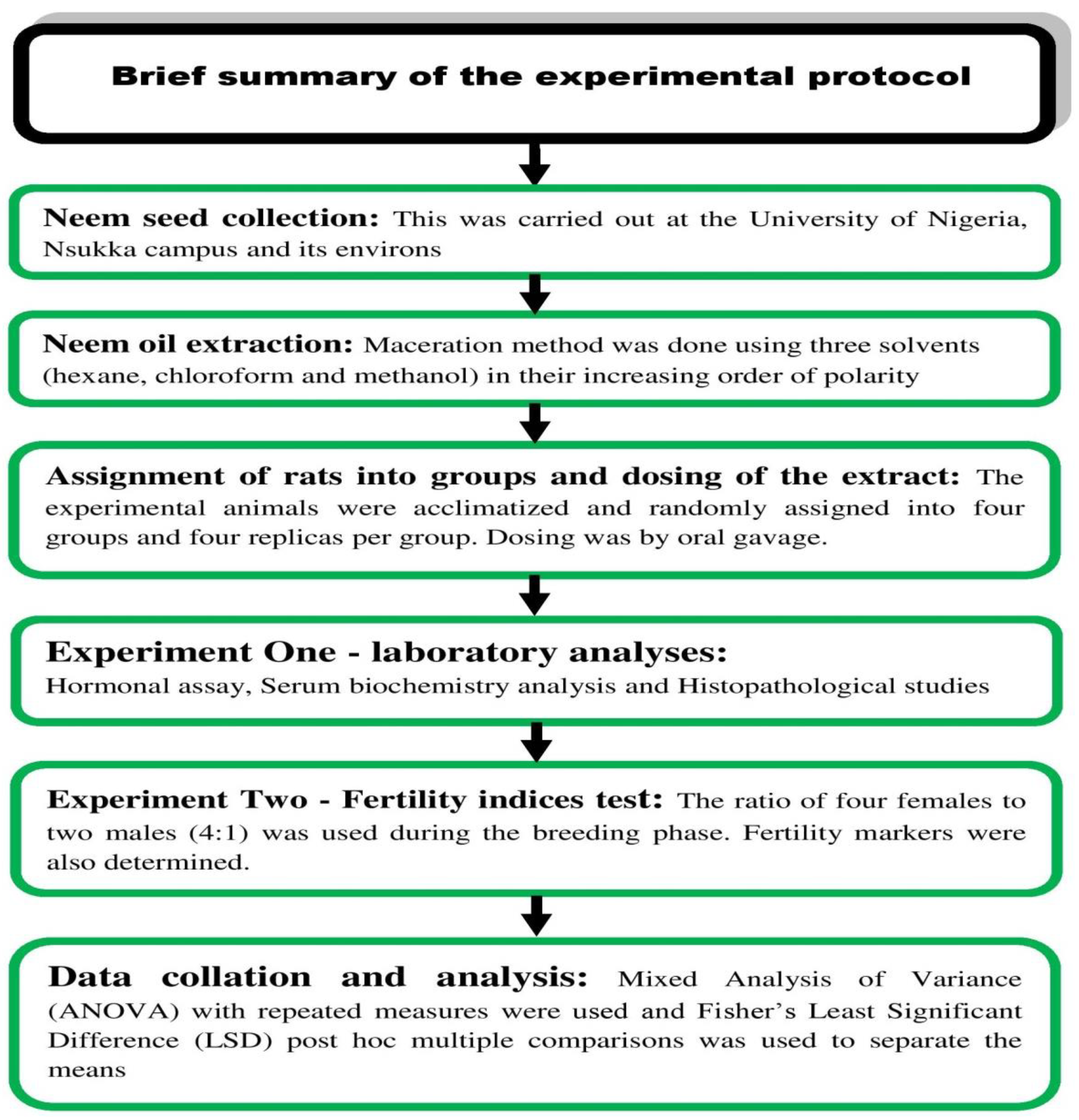
2. Materials and Methods

2.1. Ethical Approval

The Institutional Animal Care and Use Committee (IACUC) of the University of Nigeria, Nsukka granted the ethical clearance for this research (FVM-UNN-IAUCC-2020-0344). The experimental animals were humanely handled in line with guidelines provided by IAUCUC and the National Research Council for the care and use of experimental animals in research [33].

2.2. Neem-Seed Oil Extraction

After identification of the neem tree by a plant taxonomist, the fruits/seeds were harvested, before being authenticated by the same taxonomist. A voucher specimen (PSB/2018/4506/22) was then deposited at the herbarium section in the Department of Plant Science and Biotechnology, University of Nigeria, Nsukka. The harvested AI seeds were sundried to a constant weight and 300 g were weighed out and pulverized. The cold maceration method of neem seed oil extraction, using different solvents (hexane, chloroform and methanol), as described by Attia et al. [34], was carried out. The pulverized seeds were immersed in one liter of each solvent (100%), first with the non-polar solvent “hexane”. The suspension was left on the bench for 72 h at 25 °C but was vigorous agitated for five minutes every six hours. Thereafter, the supernatant was recovered by filtration. The supernatant was concentrated by allowing it to stand for 72 h at 37 °C. The process was repeated for chloroform and methanol using the residues obtained from each step. A concise summary of the experimental protocol of this study is schematically presented in Figure 1.

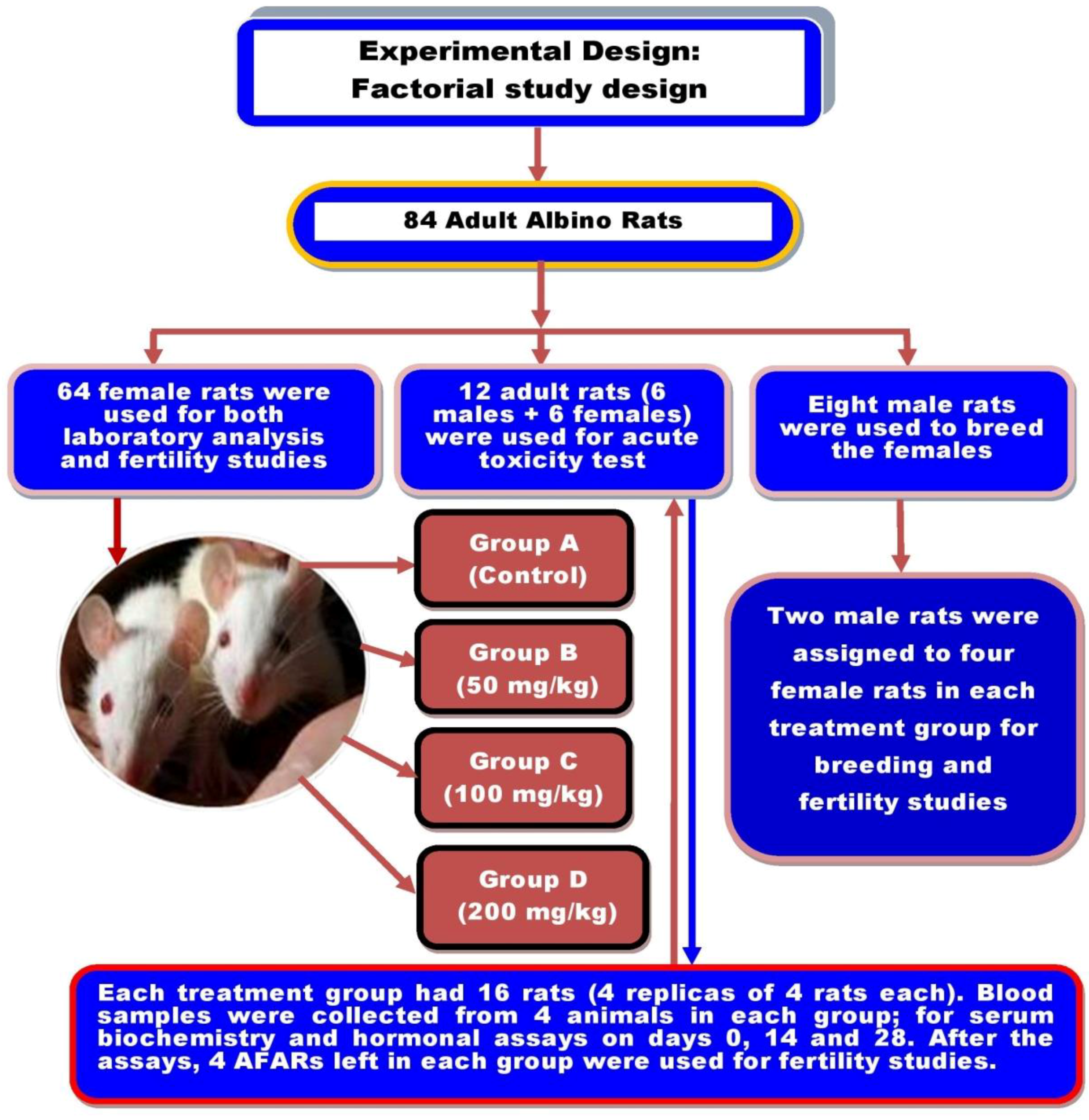
2.3. Experimental Animals and Study Design

In this study, 64 adult female and 8 male rats weighing 183 ± 2.8 g on average were used for the research. The rats were housed in meshed stainless cages at the Animal House Unit of the Department of Veterinary Obstetrics and Reproductive Diseases, University of Nigeria, Nsukka. The rats were acclimatized for 14 days under room temperature (19–24 °C at night and 25–35 °C during the daytime). The rats had unlimited access to commercial feed (TOP® feed broiler starter, 19% crude protein, 3.3 metabolizable energy/Kg) and clean drinking water, which were provided daily. A factorial study design comprised of four treatment groups (A–D) in which each group was replicated four times was used in the study as presented in Figure 2. Rats in group A served as the control while 50, 100 and 200 mg/kg of MSEAI were daily administered to rats via oral gavage in groups B, C and D, respectively. The adult male albino rats (AMARs) were housed in a separate cage and were used only during the fertility study.

2.4. Toxicity Test

The acute toxicity test was performed to determine the LD50 (dose of the extract that can cause mortality in 50% of the rats and hence the toxicological safety of MSEAI) as described by Lorke [35]. The experiment comprised 2 stages using 12 rats. The first phase involved nine rats grouped into three groups of three rats each. Groups 1, 2 and 3 received 10 mg/kg, 100 mg/kg and 1000 mg/kg of the MSEAI, respectively. They were monitored for signs of toxicity or mortality for 24 h. Since there was neither mortality nor any observable sign of toxicity, a second phase of the study involving only three rats was performed. During this second phase, rats 1, 2 and 3 were dosed 1600 mg/kg, 2900 mg/kg and 5000 mg/kg, respectively. Again, the animals were monitored for mortality and signs of toxicity for 24 h to determine the LD50.

2.5. Blood Sample Collection

Blood samples for baseline data were collected from 4 randomly selected rats from each treatment groups on day 0 without replacement, for serum hormonal and biochemical assays. The blood sample collection was done through the left medial canthus by capillary action into a glass capillary tube coated with Ethylene-diamine-tetraacetic acid (EDTA), for haematology. On the other hand, 2 mL of blood samples were collected and transferred into a sterile container for serum biochemical assays. The daily dosing of the animals commenced the same day, immediately after the bleeding. On days 14 and 28 post-treatment, sample collections were repeated for the above-stated assays.

2.6. Hormonal Assays

Serum estradiol concentrations were determined using the microplate enzyme immunoassay (EIA) test kit (Monobind, Lake Forest, CA, USA). The estradiol EIA is based on the principle of competitive binding between estradiol in the test specimen and estradiol-HRP enzyme conjugate for a constant amount of rabbit anti-estradiol antibody [36]. The lower limit of detection for estradiol was 8.2 pg/mL. Likewise, Serum FSH and LH concentrations were determined using the microplate immunoenzymetric assay test kit (Monobind, Lake Forest, CA, USA). The FSH and LH Quantitative Tests were based on a solid phase enzyme-linked immunosorbent assay (ELISA). The assay system utilizes a mouse monoclonal anti-α-FSH (LH) antibody for solid phase 2 (microtitre wells) immobilization and another mouse monoclonal anti-β-FSH (LH) antibody in the antibody-enzyme (HRP) conjugate solution [36]. For these quantitative hormonal assays, the lower limit of detection for FSH and LH were 0.134 mIU/mL and 0.054 mIU/mL, respectively.

2.7. Serum Biochemistry Assays

Serum total protein (TP) was determined by photometric test according to Biuret method [37]. Total cholesterol (TC) was determined based on the principle of enzymatic hydrolysis and oxidation [38]. Similarly, Serum triglyceride concentrations were determined on the basis of enzymatic splitting with lipoprotein lipase as described by Cole et al. [39]. These biochemical analyses were performed using a commercial kit (Dialab®, Wiener Neudorf, Austria) according to the manufacturer’s instructions. The HDL and LDL concentrations were determined using the total cholesterol enzymatic precipitating method and cholesterol LDL precipitating method, respectively [40,41]. The HDL and LDL Cholesterol Precipitating commercial kits (Labkit, Chemelex, Barcelona, Spain; BioSystems, Barcelona, Spain) were used to determine serum HDL and LDL, respectively.

2.8. Histopathological Examination

The ovarian histopathological study was performed as described by Slaoui and Fiette, 2011 [42] with slight modifications. Forty-eight ovarian tissue samples, totaling 12 ovaries per group (four each at day 0, 14 and 28), were fixed in buffered normal saline and dehydrated by passing them through graded alcohol (70%, 80%, 90% and 100%) for two hours in each alcohol percentage. The dehydration in absolute (100%) alcohol was repeated before the tissues were cleared in chloroform overnight. Thereafter, the tissues were infiltrated with paraffin wax before they were embedded (paraffin wax) using an embedding machine. Upon solidification, the embedded tissues were trimmed, mounted on wooden blocks and labelled accordingly. The embedded tissues were then placed on a microtome and sections of five-micron thickness were made and floated on a warm water bath (45 °C) to remove the wrinkles on the cut sections. A clean glass slide was used to pick up each of the sections and oven-dried at 70 °C to remove the water and obtain the sections to adhere to the slides. The slides were then de-waxed with xylene. The xylene was removed with alcohol and the alcohol removed by rinsing the slides in water. Finally, the slides were stained with hematoxylin for 30 min and counterstained with eosin for 30 min. The stained slides were taken through a series of alcohol solutions to remove the water and then through xylene before DPX (Dibutylphthalate Polystyrene Xylene) mountant was applied. Finally, cover slips were placed on the stained tissues on the slides. The slides were allowed to air-dry, after which every fifth section was viewed and analyzed under a light microscope and then photographed with a Motic digital camera.

2.9. Fertility Studies

After sample collection on day 28, two males were assigned to the 4 remaining female rats in each group, for mating. The presence of copulatory plugs, yellowish protein coagulates, which are evidence of mating in rats, was determined daily for 21 days according to the method reported by Ochiogu et al. [43]. Additionally, the body weight (in grams) of each female rat was determined every two days during the fertility study to detect speedy weight gain indicative of pregnancy. The number of fetuses born alive (NFBA) was determined for females who conceived and had parturition successfully. Thereafter, the dams were humanely sacrificed (chloroform gas inhalation in an enclosure) for determination of other reproductive and fertility indices such as numbers of fetal attachment sites, resorption sites, implantation sites and corpus luteum. The percentage resorption, percentage abortion and post implantation survival index (PISI) were computed from these indices, as shown in Figure 3.

2.10. Statistical Analysis

Data analysis was performed using mixed Analysis of Variance (ANOVA) with repeated measures via a general linear model. Treatment doses and duration as well as the interaction between treatment and time were compared for each of the parameters of interest. Where there were significant differences, the differences between the means were compared using Fisher’s Least Significant Difference (LSD) post hoc multiple comparisons. Similarly, data obtained from group fertility indices were analyzed descriptively. Statistical significance was accepted at p ≤ 0.05. All the analyses were performed using IBM® SPSS statistical package version 23 (SPSS Inc., Chicago, IL, USA) at a 5% probability level.

3. Results

3.1. Neem Seed Oil Extraction and Toxicity Testing

The 300 g of the pulverized AI seeds yielded 33.79% crude oil extract. The hexane, chloroform and methanol fractions yielded 18.37%, 4.85% and 10.57% crude oil extracts, respectively. Thus, hexane gave the highest yield, followed by methanol and then chloroform, as shown in Table 1. The rats showed no observable signs of toxicity or adverse health effects even up to three days post-treatment with MSEAI at 5000 mg/kg.

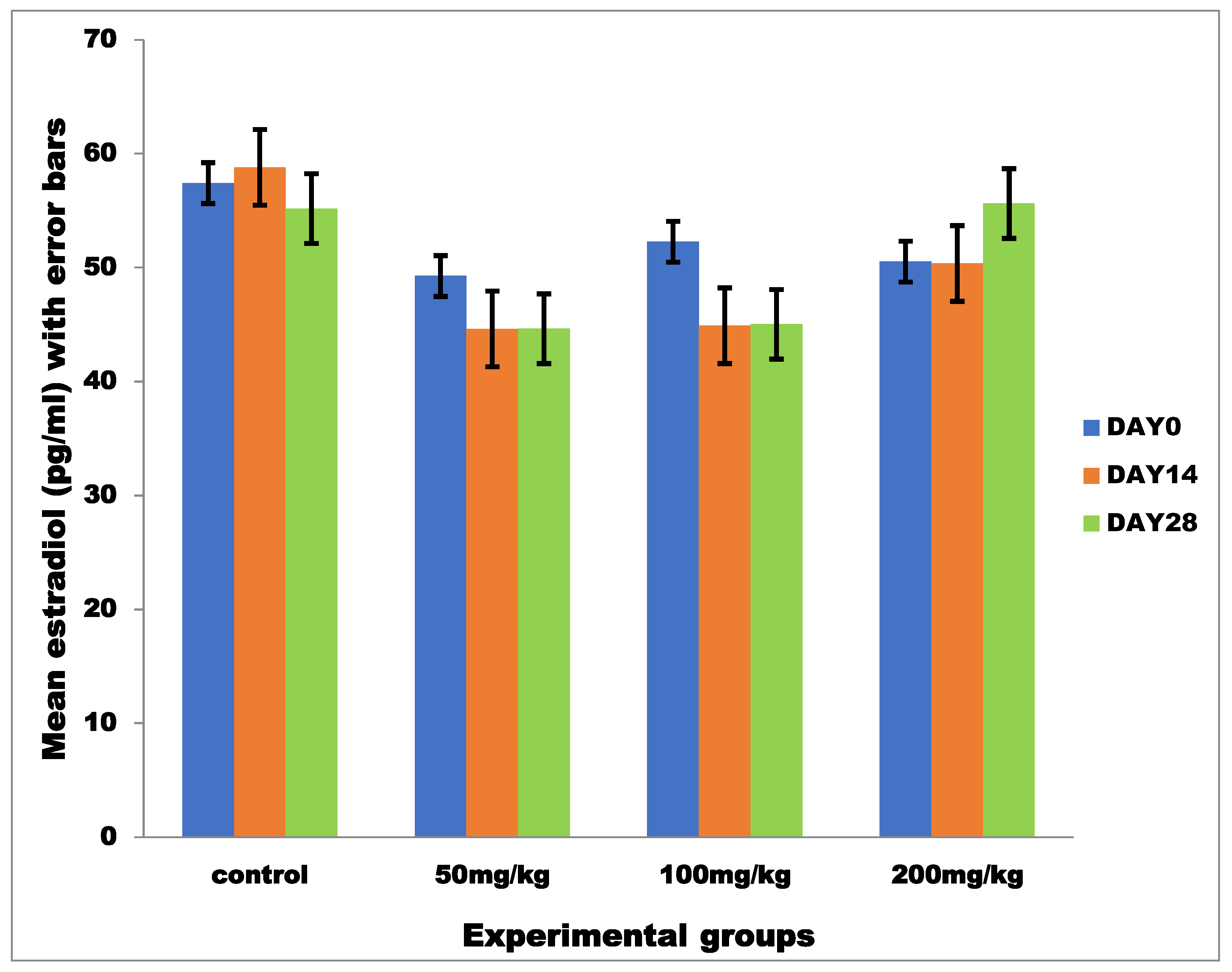
3.2. Hormonal Profiles

3.2.1. Mean Serum Estradiol

The effects of MSEAI on the mean estradiol values of the experimental rats are shown in Figure 4. Analysis of the hormonal profiles of female albino rats showed that there was no significant interaction between the treatment concentration (group) and duration of treatment (time) on the mean serum estradiol concentration, F (6, 24) = 0.105, p = 0.996, partial n2 = 0.026. No significant differences were observed across the groups.

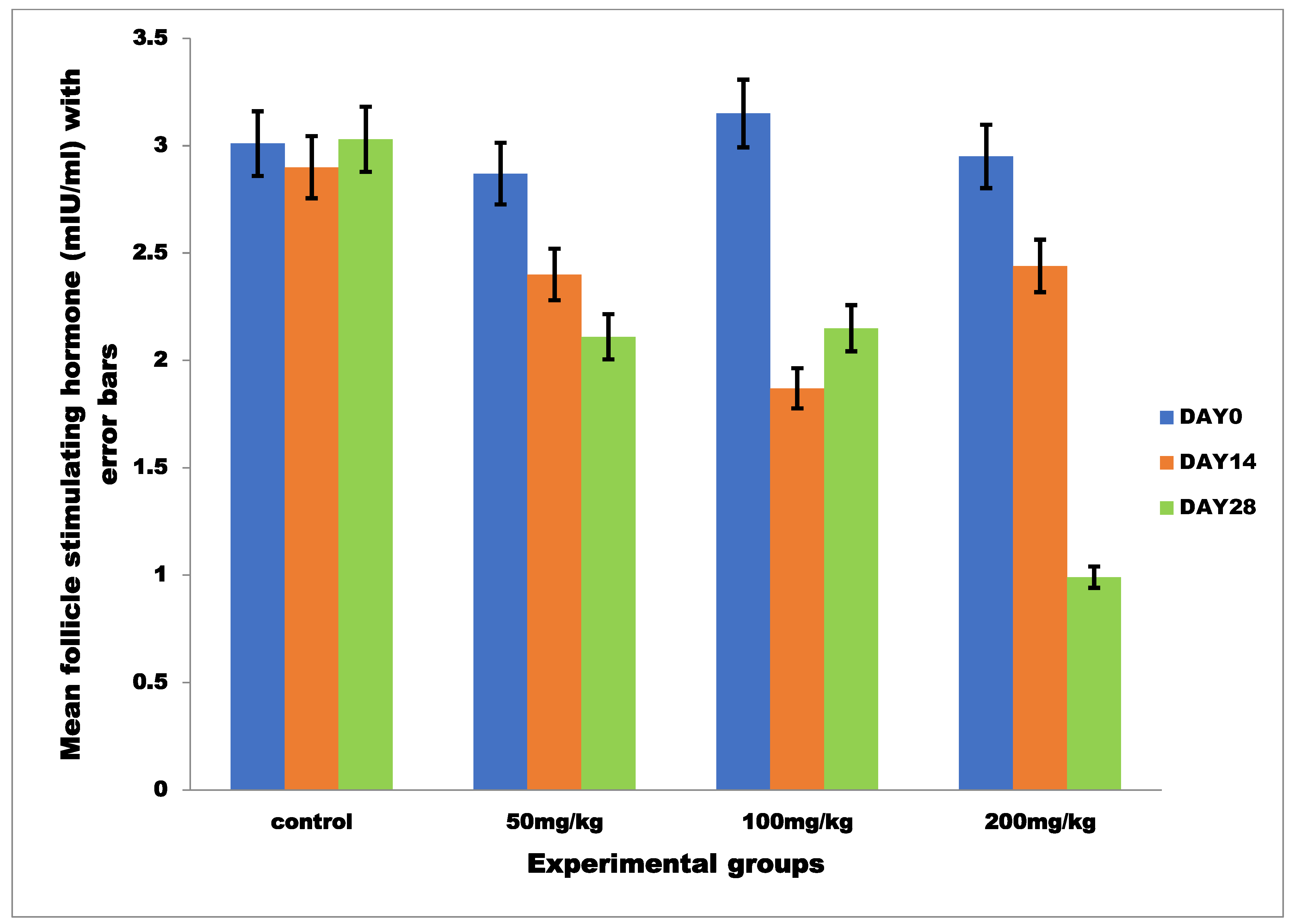
3.2.2. Mean Serum Follicle Stimulating Hormone

The effects of MSEAI on the FSH values of the experimental rats are shown in Figure 5. Statistically, there was no significant interaction between the treatment concentration (group) and duration of treatment (time) on the mean FSH concentration, F (6, 24) = 1.095, p = 0.394, partial n2 = 0.215. However, there was a significant main effect of group on the mean FSH, F (3, 12) = 1.238, p = 0.050, partial n2 = 0.236, and pairwise comparisons between groups showed a significant decrease in the mean FSH in group D when compared to group A on day 28 at p = 0.006. The simple main effect for time on the mean FSH was significant, F (2, 24) = 3.770, p = 0.038, partial n2 = 0.239, and pairwise comparisons for time showed a significant decrease in the mean FSH within group D on day 28 when compared to day 0 at p = 0.006.

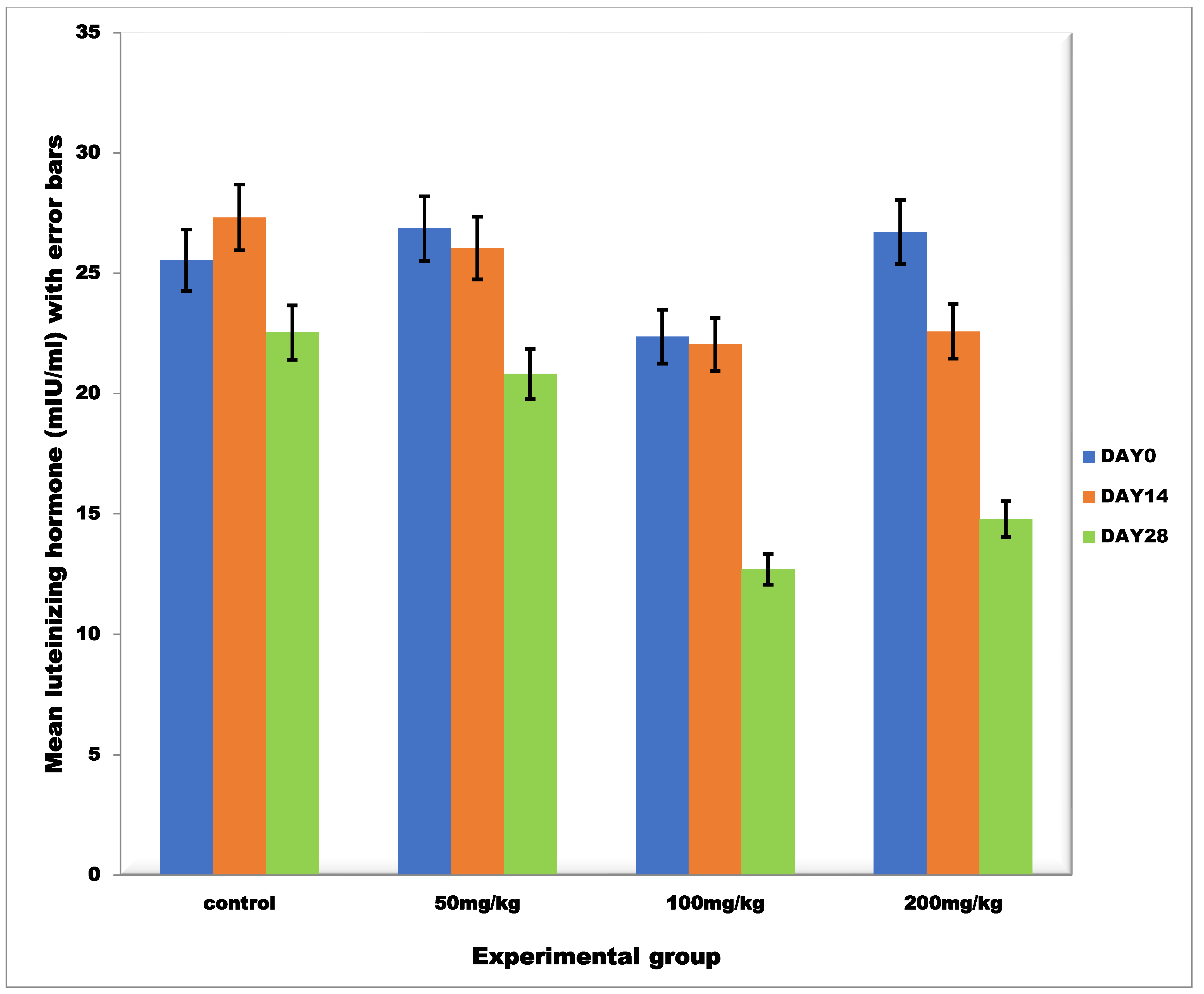
3.2.3. Mean Serum Luteinizing Hormone

The effects of MSEAI on the mean LH values of the experimental rats are shown in Figure 6. Statistical analysis showed that there was no significant interaction between the treatment concentration (group) and duration of treatment (time) on the mean LH concentration, F (6, 24) = 0.301, p = 0.930, partial n2 = 0.070. Nevertheless, there was a significant main effect for group, F (3, 12) = 3.159, p = 0.050, partial n2 = 0.441 and pairwise comparisons for groups showed that there was a significant decrease in the values of LH in groups C and D when compared to group A on day 28 at p = 0.012 and 0.039, respectively. Similarly, the simple main effect for time was significant, F (2, 24) = 4.849, p = 0.017, partial n2 = 0.288 and pairwise comparisons for time showed that, within group D, there was a significant decrease in the mean LH values on day 28 when compared to day 0 at p = 0.037.

3.3. Serum Biochemistry

3.3.1. Mean Serum Total Protein

The effects of MSEAI on the mean serum TP values of the experimental rats are shown in Table 2. Statistical analysis showed a significant interaction between treatment concentration (group) and duration of treatment (time) on serum total protein, F (6, 24) = 3.394, p = 0.015, partial n2 = 0.456. Analysis of simple main effects for groups showed that there was a significant main effect for treatment concentration (group) on total protein, F (3, 12) =1.849, p = 0.05, partial n2 = 0.316 and pairwise comparisons for group showed that, on day 14, there was a significant decrease in the mean serum total protein values in groups B, C and D when compared to the control group at p = 0.049, 0.033 and 0.01, respectively. Similarly, analysis of the simple main effect for time showed a significant main effect for time on serum total protein, F (2, 24) =21.341, p = 0.000, partial n2 = 0.640 and pairwise comparisons for time showed that, within groups B and C, there was a significant decrease in the mean serum total protein levels on day 14 when compared to days 0 and 28 at p = 0.036 and 0.018, respectively. For group D, there was a significant decrease in total protein levels on day 14 compared to days 0 and 28 at p = 0.000.

3.3.2. Lipid Profile

The effects of MSEAI on the mean serum total cholesterol, triglyceride, low-density lipoprotein (LDL) and high-density lipoprotein (HDL) of the treated rats are presented in Table 3. There was a dose-dependent increase in the serum level of HDL across the treatment groups on day 28 (Table 3). However, statistical analysis showed no significant variations (p ≥ 0.05) in the mean serum total cholesterol, triglyceride, LDL and HDL across the experimental groups at days 0, 14 and 28.

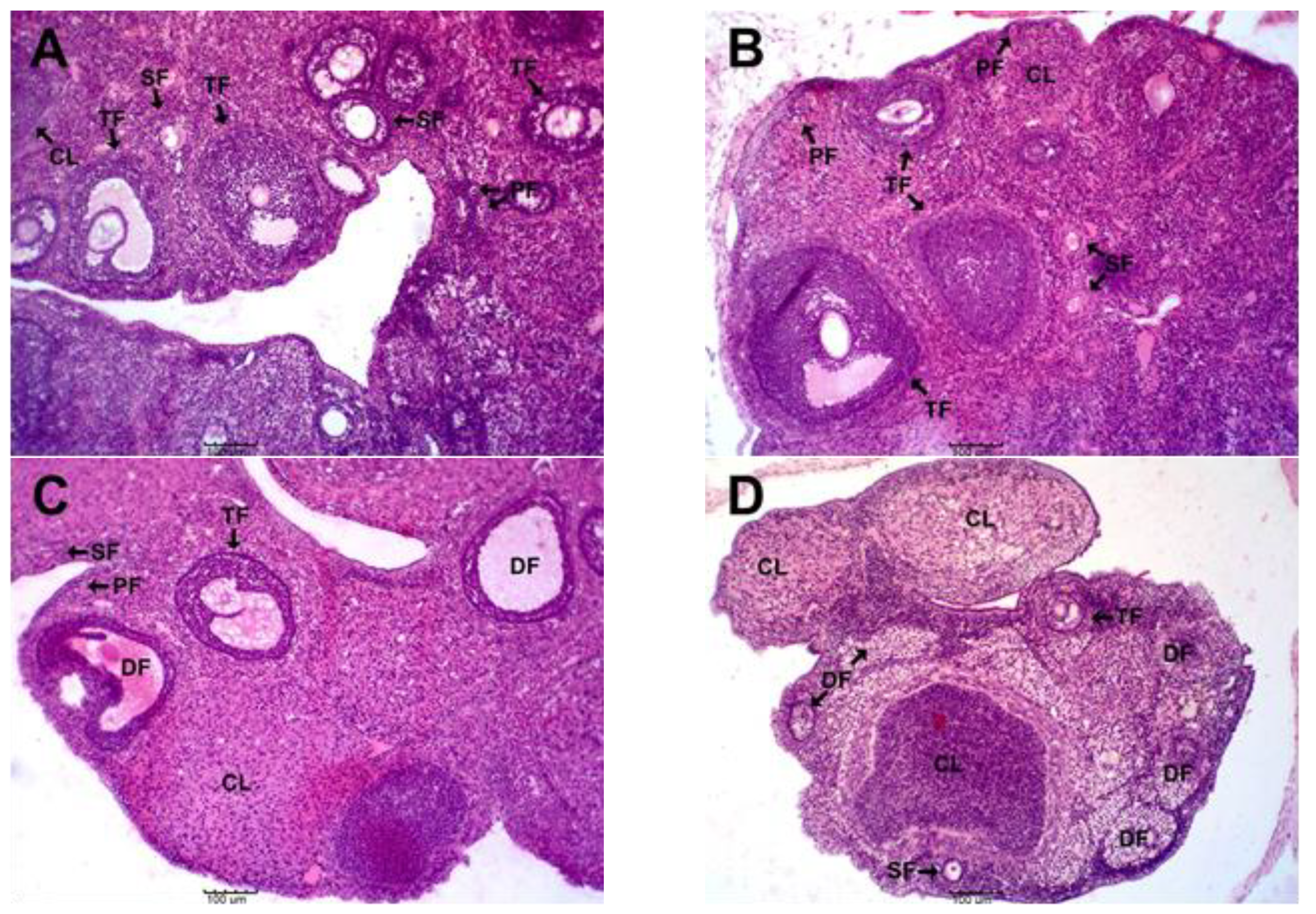
3.4. Effects of MSEAI on Ovarian Histology

The results of the ovarian histology are presented in Figure 7. The normal presence of corpora lutea (CL) and follicles at various stages of development, including primary follicles (PF), secondary follicles (SF) and tertiary follicles (TF) were observed in groups A (control) and B (50 mg/kg) when compared to groups C and D. This was indicated by the abundance of developing follicles in groups A and B; however, groups C (100 mg/kg) and D (200 mg/kg) had a decreased number of developing follicles and an increase in the presence of degenerating follicles (DF). In fact, group D (200 mg/kg) had an increased abundance of degenerating follicles (DF) compared to all the other groups.

3.5. Effects of the MSEAI on Reproductive and Fertility Parameters

The results of the effect of MSEAI on the fertility indices are presented in (Table 4). The post-implantation survival index (PISI) was 100% in the control and 0% in the 200 mg/kg treatment group. No fetal resorption occurred in the control, but 3.1% and 28.9% resorption were recorded in the 100 mg/kg and 50 mg/kg groups, respectively. Abortion was not recorded in the control and 50 mg/kg groups; however, 25% and 100% of the pregnant does aborted in the 100 mg/kg and 200 mg/kg treatment groups on day 5 post evidence of mating. Abortion was evident by the presence of frank blood in the vagina of the female rats.

4. Discussion

The significant decrease in FSH and LH values among the treated groups as well as the time- and dose-dependent negative effects observed on day 28 of treatment in this study showed that AI possesses anti-reproductive properties in the female. These findings are in tandem with those of some researchers who reported a dose-dependent decrease in the levels of these hormones at various doses. Amabe et al. [44] reported that Wister rats treated with 200 mg/kg and 400 mg/kg body weight of leaf extract of AI exhibited a decrease in LH and FSH levels. Ekaluo et al. [45] also reported a significant reduction in FSH and LH values in male rats treated with 50, 100 and 150 mg/kg body weight of aqueous leaf extract. Similarly, Roop et al. [46,47] reported that 3 and 6 mg/kg of non-polar and polar fractions of AI adversely affected folliculogenesis by significantly reducing the total number of normal follicles in the ovary. These are consistent with our result because there cannot be a marked reduction in the number of normal follicles without a commensurate decrease in FSH production and hence the level in the body. In the reproductive physiology of the female, FSH is responsible for the stimulation of follicular growth, maturation of ovarian follicles, production of estradiol and biosynthensis of estrogen from granulosa cells [48,49]. On the other hand, ovulation or the release of matured oocytes into the oviduct occurs as a result of a huge burst of LH secretion, known as the pre ovulatory LH surge [50]. Therefore, the significant reduction in the serum levels of FSH and LH found in this study implies that MSEAI may have impeded these aforementioned reproductive processes and this lends credence to the anti-reproductive and antifertility effects of AI.
The time- and dose-dependent anti-reproductive and antifertility properties of MSEAI found in this study underscore the need to discontinue prolonged (≥28 days) use of AI as forage or for the folkloric treatment of diseases, particularly at high doses (200 mg/kg and 100 mg/kg) in breeding animals. Rather than feeding AI during feed scarcity, large amounts of lush pasture, which are abundant and freely available in SSA during the rainy season, could be stock pilled, preserved as silage or converted to hay. These can then be fed to animals during the dry season to ameliorate their nutritional and welfare statuses during drought [51,52,53]. Furthermore, stalks from harvested crops such as maize and guinea corn can be fed to ruminants during feed scarcity [54,55].
The anti-reproductive and antifertility effects of MSEAI, particularly at 200 mg/kg, could be leveraged as an eco-friendly rodenticide in rodent depopulation programs in animal agriculture. This is particularly important considering the economic and public health importance of rodents in food-animal production and processing facilities in Africa. The antifertility effects of MSEAI could also be exploited as a contraceptive, which could be very useful for stray/feral dog population control in developing countries. Moreover, when further developed/refined for human use, MSEAI could be an effective, safe, cheap and eco-friendly alternative to synthetic/modern contraceptives (not readily used due to their adverse side effects), offering an acceptable form of birth control in resource-limited settings challenged with high population growth and fecundity rates [56,57,58].
Furthermore, the low rate of PISI recorded among the treated groups when compared to the control group may be due to the decreased serum levels of LH caused by MSEAI in the treated groups. The formation of corpora lutea and the synthesis of progesterone (necessary for implantation and maintenance of pregnancy) by luteal cells are dependent on the production and release of LH [50]. Progesterone, in synergy with estrogen, plays an important role in preparing the endometrium for blastocyst receptivity [46,59]. Therefore, when this hormonal pathway is disrupted, embryo implantation could be inhibited and already implanted embryos could be lost, either by resorption or by abortion, as seen in this study. Therefore, it suffices to say that the low rate of PISI reported, particularly in the high-dose treatment groups, may have been caused by the decreased production and/or release of LH due to the MSEAI treatment.
Additionally, the overall increase in degenerated follicles in the 200 mg/kg group could also be attributed to a decrease in serum FSH and LH. The FSH and LH are gonadotrophins, which are synthesized in the anterior pituitary gland when stimulated by the cells of the hypothalamus. The reproductive significance of these hormones on the ovary is that they play vital roles in stimulating the cohort follicular growth per cycle up to the point of maturation and ovulation [60]. Previous reports have indicated that FSH, LH, estrogen and progesterone are anti-apoptotic hormones, meaning a decrease in the serum levels of these hormones could lead to premature atresia/degeneration of the oocytes/follicles [61,62,63]. Early apoptosis or degeneration of oocyte resulting from a decrease in FSH and LH had been reported to have a direct association with reduced oocyte quality, fertilization, pregnancy and live birth rate [61]. The sequela of these disruptions could be the ovulation of abnormal or apoptotic oocytes, thus, an inhibition of implantation. Tiwari et al. [16] reported that neem leaf extract and its bioactive agents induced oocyte apoptosis, hence reduced reproductive outcomes. Therefore, it is possible that the anti-implantation effects observed could be due to a decline in FSH and LH, as each hormone might not have efficiently played its reproductive role, thus constituting a threat to the normal reproductive physiology of the MSEAI-treated female rats. This was established during the course of this work as frank blood due to implantation failure or abortion was observed on day five post-confirmation of mating. This also agrees with the findings of Rodney et al. [64], who reported that implantation in rats takes place on day five post mating.
The inverse proportionality found between the values of mean serum TP and the doses of the extract is similar to the findings of Adamu et al. [65], who reported that treatment with 0.048, 0.096 and 0.192 mg/L concentration of AI leaf powder produced a dose-dependent decrease in the serum TP of treated rats. The decrease in mean serum TP might be due to the high hydrolytic activities of proteins (proteolysis) as a consequence of the elevation of protease activity (proteolysis) [66,67]. The elevated hydrolytic activities might have led to an excessive production of the end products of protein catabolism—urea nitrogen/ammonia, which may affect reproduction, especially when produced and released in excess [68]. In addition, AI has been reported to contain anti-nutritional principles, including azadirachtin and nimbidin [6,69,70]. These anti-nutritional factors may have inhibited the availability and absorption of amino acids, hence the significant decrease (p < 0.05) in serum TP found. Hypoproteinemia and hypoalbuminemia have been reported to have detrimental consequences on fertility, conception and fetal development in mammals [71,72]. Albumin constitutes approximately half of the total protein content in circulation [73]. Albumin plays a vital role in the body, in that it transports hormones, drugs, vitamins and enzymes to different parts of the body for normal physiological processes to take place. It would not be farfetched to extrapolate that the decrease in total protein reported in this study may have also decreased the concentration of albumin in the blood, which is a major component of the total protein. Therefore, a decrease in total protein may have affected the transport of FSH and LH (glycoproteins) from their sites of production to their target organ (the ovary), where folliculogenesis and ovulation take place.
Furthermore, serum albumin is also important in maintaining the proper pH balance in the body, as even a slight alteration in the physiological pH (7.35 to 7.45) can affect many body functions, including reproduction [74]. Adjustment of the serum albumin concentration is one of the body’s means of maintaining an optimal pH. Albumin is slightly acidic. Being negatively charged, it balances/buffers the many positively charged molecules, such as protons (H+), calcium, potassium and magnesium, which are also circulating in the body [75]. The body pH also impacts the pH of semen and that of the uterine environment, which are critically important for fertility and reproduction in females, particularly during early pregnancy [76,77,78]. The reduced TP (hypoproteinemia and hypoalbuminemia) found in the high-dose treatment groups may have caused acidification (reduction in pH) of the uterine environment, which may have in turn caused a hostile uterine environment for implantation and development of the blastocyst and hence the adverse reproductive and fertility effects reported in the 200 mg/kg and 100 mg/kg treatment groups. Therefore, the anti-reproductive properties of MSEAI that were observed may also be due to hypoproteinemia, particularly hypoalbuminemia, which may have contributed to a decrease in body pH and hence the denaturation/malfunction of reproductive and pregnancy-maintenance hormones.
Cholesterol and its transport system, LDL and HDL, are the precursors of steroid hormones (estrogen, testosterone and progesterone), glucocorticoids and mineralocorticoids. The non-significant variation in the biochemical parameters seen among the groups suggests that the anti-reproductive activity of the extract may not be due to a decrease in steroid hormone (estrogen and progesterone) precursors but is probably due to a direct inhibition of these hormones. This finding is contrary to the results of Vijeyata and Ashok [79], who reported a significant increase in cholesterol in rabbits treated with neem seed oil alone and its fractions. This divergence in findings may be species or dose related.
It is most likely that azadirachtin, an active ingredient in neem seed, was responsible for most of the untoward reproductive (contraceptive and antifertility) effects being reported. Praneem®, a purified neem seed extract containing azadirachtin, caused abrogation of pregnancy in rodents via a significant reduction in serum progesterone levels and cytokines (TNFalpha and gamma-interferon), as well as increased immune cell (CD4 and CD8) levels, within the first trimester of gestation [80,81].

5. Conclusions and Recommendations

A methanolic seed extract of AI had anti-reproductive effects in female albino rats. The effects appeared to be dose dependent. Its mechanism of action appeared to be hormonal, particularly via the inhibition of FSH and LH. The ability of MSEAI to adversely affect reproductive parameters in the treated groups suggests that the use of AI as forage or as a folkloric treatment for breeding animals may impair their reproductive potential. However, the antifertility and anti-reproductive properties of MSEAI could be exploited as a contraceptive in non-breeding animals (pets and bitches kept for security reasons). The anti-reproductive properties could also be useful in the control of stray and feral dogs, known to be reservoirs of the rabies virus in most developing countries. Additionally, since high doses (100 mg/kg and 200 mg/kg) of the extract caused detrimental pregnancy outcomes (abortion) in the experimental rats, MSEAI at these doses could therefore be used in rodent depopulation programs, especially in poultry farms, where rodents increase the cost of production by eating up poultry feed and transmitting diseases in the process. Additionally, the anti-reproductive and antifertility properties of MSEAI as found in this study could be further refined for human use, as an effective, cheap, eco-friendly and acceptable alternative to synthetic/modern contraceptives, the use of which is limited in developing nations due to superstitious beliefs and their multiple side effects.

Author Contributions

Conceptualization, U.J.N., C.F.O. and I.S.O.; methodology, U.J.N., O.S.O., I.G.E., S.U.I. and K.O.A.; formal analysis, U.J.N. and C.F.O.; investigation, U.J.N., O.S.O., S.U.I. and C.F.O.; resources, U.J.N., I.S.O. and I.F.J.; data curation, U.J.N.; writing—original draft preparation, U.J.N.; writing—review and editing, I.F.J., O.S.O., S.U.I., O.S.O., I.G.E., K.O.A., C.F.O. and I.S.O.; visualization, U.J.N.; supervision, I.S.O. and C.F.O.; All authors have read and agreed to the published version of the manuscript.

Funding

This research received no external funding.

Institutional Review Board Statement

The study was conducted in accordance with the Declaration of Helsinki, and approved by the Institutional Animal Care and Use Committee (IACUC) of the University of Nigeria, Nsukka (FVM-UNN-IAUCC-2020-0344).

Informed Consent Statement

Not applicable.

Data Availability Statement

All relevant data supporting the findings of this study are contained in this article.

Acknowledgments

The authors are grateful to Emmanuel Njoga, of the Department of Veterinary Public Health and Preventive Medicine, University of Nigeria, Nsukka, for his roles during the experimental studies and manuscript review.

Conflicts of Interest

The authors declare that they have no conflict of interest associated with this work.

References

  1. Aziz, M.A.; Khan, A.H.; Adnan, M.; Ullah, H. Traditional uses of medicinal plants used by Indigenous communities for veterinary practices at Bajaur Agency, Pakistan. J. Ethnobiol. Ethnomed. 2018, 14, 11. [Google Scholar] [CrossRef] [PubMed]
  2. Fatemeh, J.; Zahra, L.; Hossein, A. Medicinal plants: Past history and future perspective. J. HerbMed Pharmacol. 2018, 7, 1–7. [Google Scholar]
  3. Oyedemi, B.O.; Oyedemi, S.O.; Chibuzor, J.V.; Ijeh, I.I.; Coopoosamy, R.M.; Aiyegoro, A.O. Pharmacological Evaluation of Selected Medicinal Plants Used in the Management of Oral and Skin Infections in Ebem-Ohafia District, Abia State, Nigeria. Sci. World J. 2018, 2018, 1–16. [Google Scholar] [CrossRef] [PubMed]
  4. Susana, O.M.; Asafo-Agyei, T.; Archer, M.A.; Junior, P.A.A.; Boamah, D.; Kumadoh, D.; Appiah, A.; Ocloo, A.; Boakye, Y.D.; Agyare, C. Medicinal Plants for Treatment of Prevalent Diseases. In Pharmacognosy-Medicinal Plants; IntechOpen: Rijeka, Croatia, 2019. [Google Scholar] [CrossRef]
  5. Pratik, D.; Nisha, N.; Ahmed, I.A.Y.; Katalin, K.; Rita, B.; György, T. Preliminary Investigation of Effect of Neem-Derived Pesticides on Plasmopara halstedii Pathotype 704 in Sunflower under In Vitro and In Vivo Conditions. Plants 2020, 9, 535. [Google Scholar] [CrossRef]
  6. Adjorlolo, L.K.; Timpong-Jones, E.C.; Boadu, S.; Adogla-Bessa, T. Potential contribution of neem (Azadirachta indica) leaves to dry season feeding of ruminants in West Africa. Livest. Res. Rural. Dev. 2016, 28, 75. [Google Scholar]
  7. Lawal-Adebowale, O.A. Dynamics of Ruminant Livestock Management in the Context of the Nigerian Agricultural System. In Livestock Production; Javed, K., Ed.; IntechOpen: Rijeka, Croatia, 2012. [Google Scholar] [CrossRef]
  8. Distel, R.A.; Arroquy, J.I.; Lagrange, S.; Villalba, J.J. Designing Diverse Agricultural Pastures for Improving Ruminant Production Systems. Front. Sustain. Food Syst. 2020, 4, 596869. [Google Scholar] [CrossRef]
  9. Farahna, M.; Bedri, S.; Khalid, S.; Idris, M.; Pillai, C.R.; Khalil, E.A. Anti-plasmodial effects of Azadirachta indica in experimental cerebral malaria: Apoptosis of cerebellar Purkinje cells of mice as a marker. N. Am. J. Med. Sci. 2010, 2, 518–525. [Google Scholar] [CrossRef]
  10. Bedri, S.; Khalil, E.A.; Khalid, S.A.; Alzohairy, M.A.; Mohieldein, A.; Aldebasi, Y.H.; Etet, P.F.S.; Farahna, M. Azadirachta indica ethanolic extract protects neurons from apoptosis and mitigates brain swelling in experimental cerebral malaria. Malar. J. 2013, 12, 298. [Google Scholar] [CrossRef]
  11. Bharat, P.; Sagar, R.; Sulav, R.; Ankit, P. Investigations of antioxidant and bacterial activity of leaf extracts of Azadirachta indica. Afr. J. Biotechnol. 2015, 14, 3159–3163. [Google Scholar] [CrossRef]
  12. Brahim, M.; Ismahan, L.; Mohamed, L.O. Larvicidal Activity and Influence of Azadirachtin (Neem Tree Extract) on the Longevity and Fecundity of Mosquito Species. Acta Zool. Bulg. 2017, 69, 429–435. [Google Scholar]
  13. Sirsat, S.D.; Visha, P.; Nanjappan, K. Effects of dietary chitosan and Neem leaf meal supplementation on intestinal bacterial count in broiler chickens. Int. J. Microbiol. Appl. Sci. 2018, 7, 3016–3022. [Google Scholar] [CrossRef]
  14. Habluetzel, A.; Pinto, B.; Tapanelli, S.; Nkouangang, J.; Saviozzi, M.; Chianese, G.; Lopatriello, A.; Tenoh, A.R.; Yerbanga, R.S.; Taglialatela-Scafati, O.; et al. Effects of Azadirachta indica seed kernel extracts on early erythrocytic schizogony of Plasmodium berghei and pro-inflammatory response in inbred mice. Malar. J. 2019, 18, 1–9. [Google Scholar] [CrossRef] [PubMed]
  15. Afolabi, O.J.; Simon-Oke, I.A.; Oladokun, O.I. Antiplasmodial Activity of Ethanolic Extract of Neem Leaf (Azadirachta indica) in Albino Mice Infected with Plasmodium berghei. Int. Arch. Clin. Pharmacol. 2020, 7, 24. [Google Scholar] [CrossRef]
  16. Tiwari, M.; Gupta, A.; Prasad, S.; Tripathi, A.; Yadav, P.K.; Pandey, A.N.; Premkumar, K.V.; Pandey, A.K.; Shrivastav, T.G.; Chaube, S.K. Neem (Azadirachta indica L.) and Oocyte Quality. Glob. J. Reprod. Med. 2017, 1, 555553. [Google Scholar]
  17. Seriana, I.; Akmal, M.; Darusman, X.; Wahyumi, S. Neem leaves extract (Azadirachta indica A. Juss) on male reproductive system: A mini-review. In IOP Conference Series: Earth and Environmental Science, Proceedings of the 1st International Seminar on Natural Resources and Environmental Management 2019; Bogor, Indonesia, 15 August 2019, IOP Publishing: Bristol, UK, 2019; Volume 399, p. 399. [Google Scholar] [CrossRef]
  18. Oguejiofor, C.F.; Eke, I.G.; Anya, K.O. Antifertility effects of Azadirachta indica methanol seed extract on canine spermatozoa in vitro. Asian Pac. J. Reprod. 2020, 9, 135–141. [Google Scholar]
  19. Chaube, S.K.; Shrivastav, T.G.; Tiwari, M.; Prasad, S.; Tripathi, A.; Pandey, A.K. Neem (Azadirachta indica L.) leaf extract deteriorates oocyte quality by inducing ROS-mediated apoptosis in mammals. SpringerPlus 2014, 3, 464. [Google Scholar] [CrossRef]
  20. Kgari, R.D.; Muller, J.C.; Dzama, K.; Makgahlela, M.L. Evaluation of female fertility in dairy cattle enterprises—A review. S. Afr. J. Anim. Sci. 2020, 50. [Google Scholar] [CrossRef]
  21. Surgeone, G.A. Rodent Control in Livestock and Poultry Facilities. Available online: https://www.thepoultrysite.com/articles/rodent-control-in-livestock-and-poultry-facilities (accessed on 2 May 2022).
  22. Fisher, P.; Campbell, K.J.; Howald, G.R.; Warburton, B. Anticoagulant Rodenticides, Islands and Animal Welfare Accountancy. Animals 2019, 9, 919. [Google Scholar] [CrossRef] [PubMed]
  23. Williamson, L.M.; Parkes, A.; Wight, D.; Petticrew, M.; Hart, G.J. Limits to modern contraceptive use among young women in developing countries: A systematic review of qualitative research. Reprod. Health 2009, 6, 3. [Google Scholar] [CrossRef] [PubMed]
  24. Jain, R.; Muralidhar, S. Contraceptive methods: Needs, options and utilization. J. Obstet. Gynaecol. India 2011, 61, 626–634. [Google Scholar] [CrossRef]
  25. Tsui, A.O.; Brown, W.; Li, Q. Contraceptive Practice in Sub-Saharan Africa. Popul. Dev. Rev. 2017, 43, 166–191. [Google Scholar] [CrossRef]
  26. Ajayi, A.I.; Adeniyi, O.V.; Akpan, W. Use of traditional and modern contraceptives among childbearing women: Findings from a mixed methods study in two southwestern Nigerian states. BMC Public Health 2018, 18, 604. [Google Scholar] [CrossRef] [PubMed]
  27. Schrumpf, L.A.; Stephens, M.J.; Nsarko, N.E.; Akosah, E.; Baumgartner, J.N.; Ohemeng-Dapaah, S.; Watt, M.H. Side effect concerns and their impact on women’s uptake of modern family planning methods in rural Ghana: A mixed methods study. BMC Women’s Health 2020, 20, 57. [Google Scholar] [CrossRef]
  28. Lee, M. The barriers to using modern contraceptive methods among rural young married women in Moshi Rural District, the Kilimanjaro region, Tanzania. Afr. J. Reprod. Health 2021, 25, 99–107. [Google Scholar]
  29. Varshney, S.; Verma, S.; Arya, R. A review on scientific validity on medicinal plants used as female contraceptives. Int. J. Ayurveda Res. 2016, 7, 277–285. [Google Scholar] [CrossRef]
  30. Rinzin, K.; Population Dynamics and Health Status of Free-Roaming Dogs in Bhutan. Ph.D thesis, Murdoch University, Perth, Australia. Available online: https://researchrepository.murdoch.edu.au/id/eprint/27867/ (accessed on 10 December 2021).
  31. Njoga, U.J.; Ajibo, F.E.; Njoga, E.O. The use of contraceptives for control of stray dog population and spread of rabies virus in Nigeria. Anim. Res. Int. 2020, 17, 3809–3820. [Google Scholar]
  32. Mshelbwala, P.P.; Weese, J.S.; Sanni-Adeniyi, O.A.; Chakma, S.; Okeme, S.S.; Mamun, A.A.; Rupprecht, C.E.; Magalhaes, R.J.S. Rabies epidemiology, prevention and control in Nigeria: Scoping progress towards elimination. PLoS Negl. Trop. Dis. 2021, 15, e0009617. [Google Scholar] [CrossRef]
  33. Institute for Laboratory Animal Research; National Research Council (NRC). The Guide for the Care and Use of Laboratory Animals, 8th ed.; The National Academies Press: Washington, DC, USA, 2011; pp. 56–82. [Google Scholar]
  34. Attia, I.B.; Paolo, Z.; Flaminia, C.M.; Piras, A.; Rosa, A.; Chaieb, M.; Rescigno, A. Chemical Composition and Antioxidant Potential Differences between Cynomorium coccineum L. Growing in Italy and in Tunisia: Effect of Environmental Stress. Diversity 2018, 10, 53. [Google Scholar] [CrossRef]
  35. Lorke, D. A new approach to practical acute toxicity testing. Arch. Toxicol. 1983, 54, 275–871. [Google Scholar] [CrossRef]
  36. Tietz, N.W. Clinical Guide to Laboratory Tests, 3rd ed.; Saunders Co.: Philadelphia, WA, USA, 1995; p. 38. [Google Scholar]
  37. Johnson, A.M.; Rohlfs, E.M.; Silverman, L.M. Proteins. In Tietz Textbook of Clinical Chemistry, 3rd ed.; Burtis, C.A., Ashwood, E.R., Eds.; Saunders Company: Philadelphia, PA, USA, 1999; pp. 477–540. [Google Scholar]
  38. Deeg, R.; Ziegenhorn, J. Kinetic enzymatic method for automated determination of total cholesterol in serum. Clin Chem. 1983, 29, 1798–1802. [Google Scholar] [CrossRef]
  39. Cole, T.G.; Klotzsch, S.G.; McNamara, J. Measurement of triglyceride concentration. In Handbook of Lipoprotein Testing; Rifai, N., Warnick, G.R., Dominiczak, M.H., Eds.; AACC Press: Washington, DC, USA, 1997; pp. 115–126. [Google Scholar]
  40. Naito, H.K. Cholesterol: Clinical Chemistry; The CV Mosby Co.: St Louis, MI, USA; Toronto Princeton: Toronto, ON, Canada, 1984; pp. 1207–1213. [Google Scholar]
  41. Assmann, G.; Jabs, H.U.; Kohnert, U.; Nolte, W.; Schriewer, H. LDL-cholesterol determination in blood serum following precipitation of LDL with polyvinylsulfate. Clin. Chim. Acta 1984, 140, 77–83. [Google Scholar] [CrossRef]
  42. Slaoui, M.; Fiette, L. Histopathology procedures: From tissue sampling to histopathological evaluation. In Drug Safety Evaluation: Methods in Molecular Biology (Methods and Protocols); Gautier, J.C., Ed.; Humana Press: Totowa, NJ, USA, 2011; pp. 69–82. [Google Scholar]
  43. Ochiogu, I.S.; Uchendu, C.N.; Ihedioha, J.I. A new and simple method of confirmatory detection of mating in albino rats (Rattus norvegicus). Anim. Res. Int. 2006, 3, 527–530. [Google Scholar] [CrossRef]
  44. Amabe, O.A.; Moses, B.E.; Kebe, E.O.; Mfon, I.A.; Theresa, B.E. Hormonal and histomorohological effects sof Azadirachta indica leaf extract on the pars anterior of Wister rats. Int. J. Morphol. 2011, 29, 441–445. [Google Scholar]
  45. Ekaluo, U.B.; Ikpeme, E.V.; Udensi, O.; Markson, A.A.; Madunagu, B.E.; Omosun, G.; Umana, E.J. Effects of aqueous leaf extracts of neem (Azadirachta indica) on the hormonal melieu of male rats. Int. J. Cur. Res. 2010, 4, 1–3. [Google Scholar]
  46. Roop, J.K. Effect of fractions of Azadirachta indica A. Juss seed extract on reproduction in female albino rats. Indo Am. J. Pharm. Res. 2015, 5, 2143–2150. [Google Scholar]
  47. Roop, J.K.; Dhaliwal, P.K.; Guraya, S.S. Extracts of Azadirachta Indica and Melia Azadarach seeds inhibit folliculogenesis in albino rats. Braz. J. Med. Biol. Res. 2005, 38, 943–947. [Google Scholar] [CrossRef] [PubMed]
  48. François, C.; Petit, F.; Giton, F.; Gougeon, A.; Ravel, C.; Magre, S.; Cohen-Tannoudji, J.; Guigon, C.J. A novel action of follicle-stimulating hormone in the ovary promotes estradiol production without inducing excessive follicular growth before puberty. Sci. Rep. 2017, 7, 46222. [Google Scholar] [CrossRef] [PubMed]
  49. Serge, A.J. Follicle Stimulating Hormone Abnormalities. Available online: https://emedicinemedscapecom/article/118810-overview (accessed on 14 March 2021).
  50. Richard, B. Gonadotropins: Luteinizing Hormone and Follicle Stimulating Hormone. Available online: www.vivocolostateedu/hbooks/pathphys/endocrine/hypopit/ihfshhtml (accessed on 12 January 2021).
  51. Lamidi, A.A.; Ologbose, F.I. Dry season feeds and feeding: A threat to sustainable ruminant animal production in Nigeria. J. Agric. Soc. Sci. 2014, 14, 17–30. [Google Scholar]
  52. Avornyo, F.K.; Zougmore, R.; Partey, S.; Tengan, K. Candidate fodder trees and shrubs for sustainable ruminant production in northern Ghana. Available online: http://www.lrrd.org/lrrd30/9/favor30154.html (accessed on 30 September 2018).
  53. Ouédraogo, K.; Zaré, A.; Korbéogo, G.; Ouédraogo, O.; Linstädter, A. Resilience strategies of West African pastoralists in response to scarce forage resources. Pastoralism 2021, 11, 16. [Google Scholar] [CrossRef]
  54. Thorne, P.J.; Thornton, P.K.; Kruska, R.L.; Reynolds, L.; Waddington, S.R.; Rutherford, A.S.; Odero, A.N. Maize as Food, Feed and Fertiliser in Intensifying Crop-Livestock Systems in East and Southern Africa: An Exante Impact Assessment of Technology Interventions to Improve Smallholder Welfare; International Livestock Research Institute Impact Assessment Series 11; International Livestock Research Institute: Nairobi, Kenya, 2002; p. 123. [Google Scholar]
  55. Naah, J.B.S.N. Investigating criteria for valuation of forage resources by local agro-pastoralists in West Africa: Using quantitative ethnoecological approach. J. Ethnobiol. Ethnomed. 2018, 14, 62. [Google Scholar] [CrossRef] [PubMed]
  56. Bongaarts, J. Development: Slow down population growth. Nature 2016, 530, 409–412. [Google Scholar] [CrossRef] [PubMed]
  57. Vanzo, J.D.; Adamson, D.M. Family Planning in Developing Countries: An Unfinished Success Story. Available online: https://www.rand.org/pubs/issue_papers/IP176.html (accessed on 3 May 2022).
  58. Ndayizigiye, M.; Fawzi, M.C.S.; Lively, C.T.; Ware, N. Understanding low uptake of contraceptives in resource-limited settings: A mixed-methods study in rural Burundi. BMC Health Serv. Res. 2017, 17, 209. [Google Scholar] [CrossRef] [PubMed]
  59. Alicia, A.G.; Ricardo, P.D.; Geula, G.; Carlos, M.T. Progesterone Promotes Survival of the Rat Corpus Luteum in the Absence of Cognate Receptors. Biol. Reprod. 2003, 68, 151–158. [Google Scholar]
  60. Prasad, B.; Parmar, D.; Sharma, N.C. A study on serum FSH, LH and prolactin levels among infertile women. Int. J. Med. Res. Health Sci. 2015, 4, 876–878. [Google Scholar] [CrossRef]
  61. Sheena, L.P.R.; Phil, G.K.; John, L.Y.; Yee, L.; Frank, A.; Arun, D. Granulosa cell apoptosis in the ovarian follicule—A changing view. Front. Endocrimiol. 2018, 9, 61. [Google Scholar]
  62. Casarini, L.; Riccetti, L.; De-Pascali, F.; Nicoli, A.; Tagliavini, S.; Trenti, T.; La Sala, G.B.; Simoni, M. Follicle-stimulating hormone potentiates the steroidogenic activity of chorionic gonadotropin and the anti-apoptotic activity of luteinizing hormone in human granulosa-lutein cells in vitro. Mol. Cell. Endocrinol. 2016, 15, 103–114. [Google Scholar] [CrossRef]
  63. Casarini, L.; Riccetti, L.; De Pascali, F.; Gilioli, L.; Marino, M.; Vecchi, E.; Morini, D.; Nicoli, A.; La Sala, G.B.; Simoni, M. Estrogen Modulates Specific Life and Death Signals Induced by LH and hCG in Human Primary Granulosa Cells In Vitro. Int. J. Mol. Sci. 2017, 18, 926. [Google Scholar] [CrossRef]
  64. Geisert, R.D.; Smith, M.F.; Schmelzle, A.L.; Green, J.A. Utilizing a rat delayed implantation model to teach integrative endocrinology and reproductive biology. Adv. Physiol. Educ. 2018, 42, 56–63. [Google Scholar] [CrossRef]
  65. Adamu, K.M.; Aliyu-Paiko, M.; Abdullahi, F.; Mustapha, A.Y. Effects of Azadirachta indica leaf powder on some haematological parameters of the African catfish (Clarias gariepinus). Niger. J. Basic Appl. Sci. 2017, 25, 41–50. [Google Scholar] [CrossRef]
  66. Igwenyi, I.O.; Eze, A.C.; Aja, P.M.; Elom, S.O.; Uraku, A.J.; Awoke, J.N.; Obasi, N.A. Cholesterol-Lowering and Hepatoprotective Effect of Fruit Juice Extract of Azadirachta indica on Plasmodium berghei Infected Mice. Int. J. Curr. Microbiol. Appl. Sci. 2017, 6, 3367–3375. [Google Scholar]
  67. Razzaq, A.; Shamsi, S.; Ali, A.; Ali, Q.; Sajjad, M.; Malik, A.; Ashraf, M. Microbial Proteases Applications. Front. Bioeng. Biotechnol. 2019, 7, 110. [Google Scholar] [CrossRef] [PubMed]
  68. Widayati, D.T.; Bintara, S.; Natawihardja, I.; Maharani, D. Blood biochemical profile infertile and repeat breeder Ongole cross breed cows. Pak. J. Biol. Sci 2018, 21, 166–170. [Google Scholar] [CrossRef] [PubMed]
  69. Alzohairy, M.A. Therapeutics Role of Azadirachta indica (Neem) and Their Active Constituents in Diseases Prevention and Treatment. Evid.-Based Complement. Altern. Med. 2016, 2016, 7382506. [Google Scholar] [CrossRef] [PubMed]
  70. Ubua, J.A.; Ozung, P.O.; Inagu, P.G. Dietary Inclusion of Neem (Azadirachta indica) Leaf Meal Can Influence Growth Performance and Carcass Characteristics of Broiler Chickens. Asian J. Biol. Sci. 2019, 12, 180–186. [Google Scholar] [CrossRef]
  71. Tóthová, C.; Mihajlovičová, X.; Nagy, O. The Use of Serum Proteins in the Laboratory Diagnosis of Health Disorders in Ruminants. In Ruminants—The Husbandry, Economic and Health Aspects; IntechOpen: Rijeka, Croatia, 2017. [Google Scholar] [CrossRef]
  72. Gómez-Cantarino, S.; Agulló-Ortuño, M.T.; de Dios-Aguado, M.; Ugarte-Gurrutxaga, M.I.; Bouzas-Mosquera, C. Prevalence of Hypoproteinemia and Hypoalbuminemia in Pregnant Women from Three Different Socioeconomic Populations. Int. J. Environ. Res. Public Health 2020, 17, 6275. [Google Scholar] [CrossRef]
  73. Moman, R.N.; Gupta, N.; Varacallo, M. Physiology, Albumin. In StatPearls; [Internet]; StatPearls Publishing: Treasure Island, FL, USA, 2022. Available online: https://www.ncbi.nlm.nih.gov/books/NBK459198/ (accessed on 8 February 2022).
  74. Aoi, W.; Marunaka, Y. Importance of pH homeostasis in metabolic health and diseases: Crucial role of membrane proton transport. Biomed Res. Int. 2014, 2014, 598986. [Google Scholar] [CrossRef]
  75. Anonymous. Protein Fuctions in the Body. Available online: https://pressbooks-dev.oer.hawaii.edu/humannutrition/chapter/proteins-functions-in-the-body/ (accessed on 7 August 2022).
  76. Vierafertility. Why pH Regulations is So Important for a Health Pregnancy. Available online: https://www.vierafertility.com/blog/why-ph-regulation-is-so-important-for-a-healthy-pregnancy/ (accessed on 7 August 2022).
  77. Mishra, A.K.; Kumar, A.; Swain, D.K.; Yadav, S.; Nigam, R. Insights into pH regulatory mechanisms in mediating spermatozoa functions. Vet. World 2018, 11, 852–858. [Google Scholar] [CrossRef]
  78. Zhou, J.; Chen, L.; Li, J.; Li, H.; Hong, Z.; Xie, M.; Chen, S.; Yao, B. The Semen pH Affects Sperm Motility and Capacitation. PLoS ONE 2015, 10, e0132974. [Google Scholar] [CrossRef]
  79. Vijeyata, V.; Ashok, P. Contraceptive effects of neem seed oil and its active fractions on female albino rabbits. Asian J. Pharm. Clin. Res. 2018, 11, 421–424. [Google Scholar]
  80. Mukherjee, S.; Talwar, G.P. Termination of pregnancy in rodents by oral administration of Praneem, a purified neem seed extract. Am. J. Reprod. Immunol. 1996, 35, 51–56. [Google Scholar] [CrossRef]
  81. Talwar, G.P.; Shah, S.; Mukherjee, S.; Chabra, R. Induced termination of pregnancy by purified extracts of Azadirachta indica (neem): Mechanisms involved. Am. J. Reprod. Immunol. 1977, 37, 485–491. [Google Scholar] [CrossRef] [PubMed]
Figure 1. A schematic flow chart showing an overview of the experimental design.
Challenges 13 00040 g001
Figure 2. A Schematic representation of the groupings of experimental animals.
Challenges 13 00040 g002
Figure 3. A schematic representation of various fertility indices determined in the MSEAI-treated female rats and the description of various methods used during the determination.
Challenges 13 00040 g003
Figure 4. Mean serum estradiol levels in female albino rats exposed to graded doses of MSEAI at different time points.
Challenges 13 00040 g004
Figure 5. Mean serum FSH levels in female albino rats exposed to graded doses of MSEAI at different time points.
Challenges 13 00040 g005
Figure 6. Mean serum LH levels in female albino rats exposed to graded doses of MSEAI at different time points.
Challenges 13 00040 g006
Figure 7. Micrographs of ovarian histology in female albino rats exposed to various doses of methanolic seed extract of AI. Groups (A,B) had more developing follicles, while (C,D) had an abundance of degenerating follicles in the ovary. H and E staining; scale bar (100 µm). PF = Primordial follicle; SF = Secondary follicle; TF = Tertiary follicle; CL = Corpus luteum; DF = Degenerating follicles.
Challenges 13 00040 g007
Table 1. Percentage (%) yield of the different crude fractions extracts from Azadirachta indica seed.
Crude FractionWeight (g)Yield (%)
Hexan55.1118.37
Chloroform14.544.85
Methanol31.7210.57
Total Oil Yield101.3733.79
Table 2. Mean serum total protein values of female albino rats exposed to different doses of AI on day 0, 14 and 28.
Days Experimental Groups
Control50 mg/kg100 mg/kg200 mg/kg
Day 07.4 ± 0.147.3 ± 0.13 x7.3 ± 0.13 x7.5 ± 1.26 x
Day 147.5 ± 0.17 a6.95 ± 0.09 by6.9 ± 0.21 by6.5 ± 0.15 by
Day 287.6 ± 0.087.5 ± 0.17 x7.5 ± 0.19 x7.5 ± 0.21 x
Means ± SEM with different superscripts on the same row and column indicate a significant difference at p ≤ 0.05; Means with the same superscript showed no significant difference between the groups at three different time points at p > 0.05; Superscript “a” and “b” indicate group significance while “x and y” indicate time significance.
Table 3. Mean serum biochemical parameters of female albino rats exposed to different doses of methanolic seed extract of Azadirachta indica.
Biochemical ParametersTreatment DaysExperimental Groups
Control
(Gourp-A)
50 mg/kg
(Group-B)
100 mg/kg
(Group-C)
200 mg/kg
(Group-D)
TC(mg/dL)Day 070.1 ± 1.571.7 ± 4.472.8 ± 3.670.6 ± 4.8
Day 1472.0 ± 4.974.4 ± 5.772.0 ± 5.472.9 ± 4.2
Day 2873.9 ± 2.778.2 ± 4.473.4 ± 1.873.9 ± 2.5
HDL (mg/dL)Day 040.6 ± 5.546.9 ± 5.244.9 ± 6.843.4 ± 4.9
Day 1441.4 ± 7.545.7 ± 7.042.6 ± 5.944.6 ± 2.9
Day 2843.7 ± 6.346.6 ± 6.048.1 ± 5.849.7 ± 4.1
LDL (mg/dL)Day 029.5 ± 5.424.8 ± 1.127.9 ± 3.827.3 ± 2.3
Day 1430.6 ± 5.428.6 ±3.729.4 ± 0.928.2 ± 1.6
Day 2830.2 ± 4.831.6 ± 5.325.3 ± 5.124.2 ± 5.0
TG (mg/dL)Day 01.28 ± 1.391.23 ± 1.321.19 ± 1.541.29 ± 1.65
Day 141.34 ± 1.491.19 ± 1.411.22 ± 2.111.26 ± 2.03
Day 281.3 ± 3.861.27 ± 2.151.14 ± 1.291.34 ± 2.51
TC = Total cholesterol; HDL = High-density lipoprotein; LDL = Low-density lipoprotein; TG = Triglycerides. The values are presented as means ± SEM.
Table 4. Group fertility parameters of adult female albino rats fed graded doses of methanolic extract of Azadirachta indica.
Fertility ParametersTreatment Groups
Control50 mg/kg100 mg/kg200 mg/kg
Percentage abortion0025100
Percentage resorption028.93.10
LFB (%)1001001000
PISI (%)10072730
GI93444100
LFB = Fetuses born alive; PISI = Post implantations survival index; GI = Gestation index.
Publisher’s Note: MDPI stays neutral with regard to jurisdictional claims in published maps and institutional affiliations.

