Nanomaterials and Oxidative Stress


Materials Measurement Laboratory, National Institute of Standards and Technology, 100 Bureau Dr., Gaithersburg, MD 20899, USA
*
Author to whom correspondence should be addressed.
Challenges 2018, 9(1), 17; https://doi.org/10.3390/challe9010017
Submission received: 1 March 2018
/
Revised: 30 March 2018
/
Accepted: 1 April 2018
/
Published: 3 April 2018
Abstract
:With the rapid development of new nanomaterials, it was recognized early that together with their beneficial properties, nanomaterials may pose a risk to human health and the environment. Evidence has accumulated over the last twenty years in support of oxidative stress as a broad mechanistic concept to explain the interaction of engineered nanoparticles with biological substances. As oxidative stress as a physiological response was recognized in redox biology, its wide-ranging use in nanotoxicology has exposed new challenges and limitations. In this commentary, we review certain oxidative stress concepts and their relevance to nanotoxicology.
Nanotechnology has firmly established itself in the list of modern-day high technologies. With the rapid development of new nanomaterials, it was recognized early on that along with their beneficial properties, nanomaterials may pose a hazard risk to human health and the environment [1]. Such awareness led to the separate discipline of nanotoxicology that emerged as an essential part of nanotechnology advancement. Compelling evidence was accumulated over the last twenty years in support of oxidative stress (OS) as a broad mechanistic paradigm behind the interaction of ultra-small engineered particles with biological objects [2,3]. Long before the advent of nanotechnology, OS as a physiological response concept was recognized in redox biology [4,5]. As a result, its wide-ranging use in nanotoxicology assays exposed new challenges and limitations. Here, we will highlight certain OS concepts and some of the experimental problems specific to nanotoxicology.
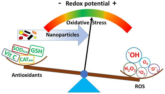
Aerobic organisms depend on multiple electron transfers from high to low energy entities with oxygen as the terminal electron acceptor and strong oxidant with a formal potential E = 0.82 V vs. normal hydrogen electrode (NHE). Fortunately, due to the quantum mechanical restrictions in the electronic ground state, dioxygen reactivity is quite low without additional activation, preventing the spontaneous combustion of organic matter. Thus, multiple redox reactions can occur along the downhill electronic energy slope with rates controlled by dedicated enzymatic structures. Each cellular compartment maintains its redox potential within a “healthy” physiological range by redox homeostasis [6], and OS can be pronounced as a supra-physiological move to higher potential values. Hence, the representation of the OS in quantitative terms would require the establishment of physiological redox potential margins, within which the oxidative outbreaks can still be reversed by internal antioxidants. Similarly, OS is used to describe the buildup of so-called reactive oxygen species (ROS), the umbrella term for partially reduced oxygen and nitrogen derivatives, primarily electrophilic radical species (Figure 1). A major source of radicals in a biological environment is hydrogen peroxide, which accumulates during the respiratory burst. ROS can arise outside cell boundaries or be produced internally, largely within the mitochondria as by-products of oxygen metabolism. ROS reactivity (which is inverse to their lifetime) correlates with the oxidizing strength, best expressed by their formal redox potential and reaction kinetics with a specific biological partner.
Depending on its magnitude, OS is compensated by natural homeostasis, which leads to the activation of the internal antioxidant responses and inflammatory signaling. In extreme cases, OS may lead to cell death by apoptosis, necrosis, or ferroptosis. On a molecular level, ROS react with biomacromolecules, leading to specific pathological outcomes, such as atherosclerosis and cancer. Naturally produced ROS play essential physiological roles in activation of certain cellular signaling pathways, nuclear transcription factors, and detoxification of pathogens.
OS is a principal mechanistic concept in nanotoxicology; however, multiple biological pathways are at its origin due to the vast variety of nanoparticle (NP) physicochemical properties. Typically, ROS measurement data is presented in qualitative or relative terms, which complicates comparability between laboratories. Still, in several nanomaterial categories, characteristic OS initiating descriptors have emerged and can provide a physical basis to model the NP hazard. Burello et al. have proposed a rationale that ties metal oxide electronic structure to potential toxicity, namely to a position of their electronic energy bands relative to the intracellular redox potential range [7]. A favorable position of the NP conduction or valence band promotes electron transfers between a metal oxide NP and a biomolecule in contact, triggering OS. Later, this framework was validated in a systematic study using a series of metal oxide NP tests with zebra fish [8]. Metallic NPs can be reduced, oxidized, and release metal ions in a biological environment [9], largely depending on their formal redox potential value relative to the prevalent biological redox potential range, spanning from −0.38 V to +0.34 V [10]. Certain metal ions (Fe, Cu, Cr, Co), if not complexed, promote redox cycling and enable Fenton type reactions with hydrogen peroxide, producing ˙OH, an extremely reactive (E = 2.8 V) and short-lived (τ = 1 ns) radical. Depending on their location, NPs can cause OS by interacting with the extracellular medium, with transmembrane proteins by inducing intracellular signaling or after their internalization through multiple established pathways [11]. When taken up by the cell, NPs encounter various biomolecules, potentially perturbing redox homeostasis in the cytosol and organelles. NPs also can play an antioxidant role by preventing or slowing oxidative reactions in a biological setting, also as radical scavengers, and these beneficial capabilities are actively sought for potential therapeutic applications in nanomedicine [12,13]. From a chemical standpoint, it is imperative to analyze oxidants and antioxidants in the context of its electron transfer partner(s) formal potential, as the same compound can have an opposite effect in a different cellular compartment [14].
Basically, redox potential seems to be the most accurate descriptor of the system redox balance; therefore, determining real time values in each cellular compartment, together with the corresponding redox reaction kinetic constants, expresses the in vitro redox state. Techniques to measure the intra-cellular redox potential non-invasively and in real time are being developed, ensuing developments in instrumentation and genetic engineering [15,16,17,18].
Perhaps the most widespread experimental in vitro approach in nanomaterial-induced OS measurement embraces incubation with NPs, followed by the assessment of the oxidative damage to biomacromolecules [19,20,21] such as DNA, lipids, and proteins, or an appraisal of cellular antioxidant defenses [14]. Comparison with the non-exposed control group (aka “negative control”) allows one to assess the relative magnitude of the NP-caused OS. In vitro experiments with a known toxic substance (positive control) offers an estimate of the upper OS limit. Having both positive and negative controls allows the dynamic range for the assay output to be determined. To be relevant, the positive control must represent a similar material class, for example metal oxides, metals, etc. A direct ROS concentration measurement using electron paramagnetic resonance spectroscopy (EPR) with spin-trapping or dedicated fluorophores may offer a real time OS representation [22,23,24].
Many assays intended to sense OS were previously developed for soluble chemicals and drugs, and typically require modification for use in the presence of NPs to avoid improper results in the presence of particular matter. Most extra complications could be traced to unidentified particle reactivity with assay reagents [22], test biomolecules and nutrients [23], or interferences with the assay optical readout [24]. Optical issues occur due to inherent light scattering, absorption, and emission by the NPs. Furthermore, NP optical properties and biological activity depend on their chemical composition, aggregation state, and surface coating, all of which could be altered during the assay procedure, such as being added to the cell culture medium. Hence, it is vital to evaluate the NP physico-chemical properties in a biological test setting.
Photocatalytic NPs, such as TiO2, may generate ROS in acellular environments and enhance the macromolecular damage during the assay procedure unless precautions are taken to avoid exposure to ambient light [19]. When analyzing DNA strand breaks with the single cell gel electrophoresis (aka comet) assay, NPs have been shown to slow DNA migration during electrophoresis, thus producing false negative results [25]. Variation of the reduced glutathione (GSH) level is a rather sensitive early OS indicator and can be detected using absorbance with Ellmans reagent or via the rhodamine-based organo-selenium probe [26]. Presence of metallic NPs, however, will significantly quench the probe light emission [27]. The assays that measure the macromolecule damage and antioxidant production typically consume the sample and preclude a repeated collection of data from the same cell [14]. This may hinder the assessment of the OS at low dose levels over extended time periods that simulate the chronic NP exposure. When ROS and antioxidant concentration is monitored in live cells using microscopic imaging with fluorogenic sensors [15], it allows the observation of OS evolution in real time and avoids artifacts due to sample extraction. Application of gene expression analysis to identify underlying OS mechanisms caused by exposure to NPs is expanding following advances in high-content analytical technologies [28,29]. Techniques such as gene chips and reverse transcription polymerase chain reaction allow the recognition of early molecular markers of the redox imbalance by following the expression of genes, responsible for the antioxidant proteins [20].
As NPs are an essential element in these OS tests, several experimental strategies have been reported in order to accommodate particular matter in standard toxicity assays developed for soluble chemicals/drugs. First, it is the comprehensive NP characterization, preferably in the biological test media. Multiple techniques and dedicated instruments are now available for this purpose [30]. Removal of NPs using separation methods such as precipitation, filtering, centrifugation, or careful washing prior to the biological assay is very effective in minimizing artifacts. Another common approach is the validation of the assay output with an orthogonal method. Still, a significant variability in the NP test results between laboratories using the equivalent method and similar materials often hamper data comparison [31]. Fortunately, the development of standardized protocols and reference nanomaterials by standards organizations is addressing this critical need [32]. Finally, cell-free control experiments often allow quantification of the amount of NP interference that could be used to correct the assay endpoint value [22].
Ultimately, in vitro acquired data should give information to predict in vivo and human nanomaterial hazards. Given the efforts to minimize animal use for NP testing, complex in vitro models and those that mimic target organs are being developed [33]. Instead of using relevant cell monocultures, the current trend is to pursue 3D and microfluidics-based in vitro models, to better represent nanomaterial uptake and bio-distribution by the target organ [34]. Along with developments in experimental techniques, computational approaches to model toxicity in the whole body aim to predict the nanomaterial-induced OS and avoid ethical and economic burden associated with animal testing. However, an experimental validation of in silico approaches is still necessary, given the wide toxicological profile of the nanomaterials [35]. Although numerous causal relationships between exposures to toxicant and biological redox responses have been already developed, it is still not clear how the short-term OS level increase relates to the long-term organism adaptability or damage. This problem is heightened in nanotoxicology by the pervasiveness of acute exposure experimental setups and by the acceptance of multiple, different dosing units, whether it is mass-, particle-number-, or surface-area-based.
Acknowledgments
We thank Elijah Petersen for his careful reading of the manuscript and helpful discussions.
Declaration
Official contribution of the National Institute of Standards and Technology; not subject to copyright in the United States.
Author Contributions
Vytas Reipa wrote the original draft, edited the paper, and prepared for publication; Donald Atha co-wrote the original draft and edited the paper.
Conflicts of Interest
The authors declare no conflict of interest.
References
- Donaldson, K.; Stone, V.; Tran, C.L.; Kreyling, W.; Borm, P.J.A. Nanotoxicology. Occup. Environ. Med. 2004, 61, 727–728. [Google Scholar] [CrossRef] [PubMed]
- Li, N.; Xia, T.; Nel, A.E. The role of oxidative stress in ambient particulate matter-induced lung diseases and its implications in the toxicity of engineered nanoparticles. Free Radic. Biol. Med. 2008, 44, 1689–1699. [Google Scholar] [CrossRef] [PubMed]
- Sarkar, A.; Ghosh, M.; Sil, P.C. Nanotoxicity: Oxidative stress mediated toxicity of metal and metal oxide nanoparticles. J. Nanosci. Nanotechnol. 2014, 14, 730–743. [Google Scholar] [CrossRef] [PubMed]
- Blake, D.R.; Hall, N.D.; Bacon, P.A.; Dieppe, P.A.; Halliwell, B.; Gutteridge, J.M.C. The importance of iron in rheumatoid disease. Lancet 1981, 2, 1142–1144. [Google Scholar] [CrossRef]
- Gutteridge, J.M.C.; Richmond, R.; Halliwell, B. Oxygen free-radicals and lipid-peroxidation—Inhibition by the protein ceruloplasmin. FEBS Lett. 1980, 112, 269–272. [Google Scholar] [CrossRef]
- Halliwell, B. Biochemistry of oxidative stress. Biochem. Soc. Trans. 2007, 35, 1147–1150. [Google Scholar] [CrossRef] [PubMed]
- Burello, E.; Worth, A.P. A theoretical framework for predicting the oxidative stress potential of oxide nanoparticles. Nanotoxicology 2011, 5, 228–235. [Google Scholar] [CrossRef] [PubMed]
- Lin, S.J.; Zhao, Y.; Xia, T.; Meng, H.; Ji, Z.X.; Liu, R.; George, S.; Xiong, S.J.; Wang, X.; Zhang, H.Y.; et al. High content screening in zebrafish speeds up hazard ranking of transition metal oxide nanoparticles. ACS Nano 2011, 5, 7284–7295. [Google Scholar] [CrossRef] [PubMed]
- Auffan, M.; Rose, J.; Wiesner, M.R.; Bottero, J.Y. Chemical stability of metallic nanoparticles: A parameter controlling their potential cellular toxicity in vitro. Environ. Pollut. 2009, 157, 1127–1133. [Google Scholar] [CrossRef] [PubMed]
- Plumlee, G.S.; Morman, S.A.; Ziegler, T.L. The toxicological geochemistry of earth materials: An overview of processes and the interdisciplinary methods used to understand them. Rev. Mineral. Geochem. 2006, 64, 5–57. [Google Scholar] [CrossRef]
- Lujan, H.; Sayes, C.M. Cytotoxicological pathways induced after nanoparticle exposure: Studies of oxidative stress at the ‘nano-bio’ interface. Toxicol. Res. 2017, 6, 580–594. [Google Scholar] [CrossRef]
- Sims, C.M.; Hanna, S.K.; Heller, D.A.; Horoszko, C.P.; Johnson, M.E.; Bustos, A.R.M.; Reipa, V.; Riley, K.R.; Nelson, B.C. Redox-active nanomaterials for nanomedicine applications. Nanoscale 2017, 9, 15226–15251. [Google Scholar] [CrossRef] [PubMed]
- Petersen, E.J.; Tu, X.; Dizdaroglu, M.; Zheng, M.; Nelson, B.C. Protective roles of single-wall carbon nanotubes in ultrasonication-induced DNA base damage. Small 2013, 9, 205–208. [Google Scholar] [CrossRef] [PubMed]
- Wages, P.A.; Cheng, W.Y.; Gibbs-Flournoy, E.; Samet, J.M. Live-cell imaging approaches for the investigation of xenobiotic-induced oxidant stress. BBA Gen. Subj. 2016, 1860, 2802–2815. [Google Scholar] [CrossRef] [PubMed]
- Gutscher, M.; Pauleau, A.L.; Marty, L.; Brach, T.; Wabnitz, G.H.; Samstag, Y.; Meyer, A.J.; Dick, T.P. Real-time imaging of the intracellular glutathione redox potential. Nat. Methods 2008, 5, 553–559. [Google Scholar] [CrossRef] [PubMed]
- Rhieu, S.Y.; Urbas, A.A.; Bearden, D.W.; Marino, J.P.; Lippa, K.A.; Reipa, V. Probing the intracellular glutathione redox potential by in-cell NMR spectroscopy. Angew. Chem. Int. Ed. 2014, 53, 447–450. [Google Scholar] [CrossRef] [PubMed]
- Mercatelli, E.; Barbieri, L.; Luchinat, E.; Banci, L. Direct structural evidence of protein redox regulation obtained by in-cell NMR. BBA Mol. Cell Res. 2016, 1863, 198–204. [Google Scholar] [CrossRef] [PubMed]
- Papayan, G.; Petrishchev, N.; Galagudza, M. Autofluorescence spectroscopy for nadh and flavoproteins redox state monitoring in the isolated rat heart subjected to ischemia-reperfusion. Photodiagn. Photodyn. Ther. 2014, 11, 400–408. [Google Scholar] [CrossRef] [PubMed]
- Petersen, E.J.; Reipa, V.; Watson, S.S.; Stanley, D.L.; Rabb, S.A.; Nelson, B.C. DNA damaging potential of photoactivated p25 titanium dioxide nanoparticles. Chem. Res. Toxicol. 2014, 27, 1877–1884. [Google Scholar] [CrossRef] [PubMed]
- Nagy, A.; Hollingsworth, J.A.; Hu, B.; Steinbruck, A.; Stark, P.C.; Rios Valdez, C.; Vuyisich, M.; Stewart, M.H.; Atha, D.H.; Nelson, B.C.; et al. Functionalization-dependent induction of cellular survival pathways by cdse quantum dots in primary normal human bronchial epithelial cells. ACS Nano 2013, 7, 8397–8411. [Google Scholar] [CrossRef] [PubMed]
- Atha, D.H.; Wang, H.; Petersen, E.J.; Cleveland, D.; Holbrook, R.D.; Jaruga, P.; Dizdaroglu, M.; Xing, B.; Nelson, B.C. Copper oxide nanoparticle mediated DNA damage in terrestrial plant models. Environ. Sci. Technol. 2012, 46, 1819–1827. [Google Scholar] [CrossRef] [PubMed]
- Tournebize, J.; Sapin-Minet, A.; Bartosz, G.; Leroy, P.; Boudier, A. Pitfalls of assays devoted to evaluation of oxidative stress induced by inorganic nanoparticles. Talanta 2013, 116, 753–763. [Google Scholar] [CrossRef] [PubMed]
- Guo, L.; Von Dem Bussche, A.; Buechner, M.; Yan, A.H.; Kane, A.B.; Hurt, R.H. Adsorption of essential micronutrients by carbon nanotubes and the implications for nanotoxicity testing. Small 2008, 4, 721–727. [Google Scholar] [CrossRef] [PubMed]
- Guadagnini, R.; Kenzaoui, B.H.; Walker, L.; Pojana, G.; Magdolenova, Z.; Bilanicova, D.; Saunders, M.; Juillerat-Jeanneret, L.; Marcomini, A.; Huk, A.; et al. Toxicity screenings of nanomaterials: Challenges due to interference with assay processes and components of classic in vitro tests. Nanotoxicology 2015, 9, 13–24. [Google Scholar] [CrossRef] [PubMed]
- Karlsson, H.L.; Di Bucchianico, S.; Collins, A.R.; Dusinska, M. Can the comet assay be used reliably to detect nanoparticle-induced genotoxicity? Environ. Mol. Mutagen. 2015, 56, 82–96. [Google Scholar] [CrossRef] [PubMed]
- Gao, W.; Xu, K.H.; Ji, L.F.; Tang, B. Effect of gold nanoparticles on glutathione depletion-induced hydrogen peroxide generation and apoptosis in hl7702 cells. Toxicol. Lett. 2011, 205, 86–95. [Google Scholar] [CrossRef] [PubMed]
- Tournebize, J.; Boudier, A.; Joubert, O.; Eidi, H.; Bartosz, G.; Maincent, P.; Leroy, P.; Sapin-Minet, A. Impact of gold nanoparticle coating on redox homeostasis. Int. J. Pharm. 2012, 438, 107–116. [Google Scholar] [CrossRef] [PubMed]
- Wang, Y.L.; Li, C.C.; Yao, C.J.; Ding, L.; Lei, Z.D.; Wu, M.H. Techniques for investigating molecular toxicology of nanomaterials. J. Biomed. Nanotechnol. 2016, 12, 1115–1135. [Google Scholar] [CrossRef] [PubMed]
- Atha, D.H.; Nagy, A.; Steinbruck, A.; Dennis, A.M.; Hollingsworth, J.A.; Dua, V.; Iyer, R.; Nelson, B.C. Quantifying engineered nanomaterial toxicity: Comparison of common cytotoxicity and gene expression measurements. J. Nanobiotechnol. 2017, 15, 79. [Google Scholar] [CrossRef] [PubMed]
- Nelson, B.C.; Reipa, V. Analytical measurements of nanoparticles in challenging and complex environments. In Metrology and Standatdization for Nanotechnology; Mansfield, E., Ed.; Wiley-VCH: Weinheim, Germany, 2017; pp. 175–196. [Google Scholar]
- Elliott, J.T.; Rosslein, M.; Song, N.W.; Toman, B.; Kinsner-Ovaskainen, A.; Maniratanachote, R.; Salit, M.L.; Petersen, E.J.; Sequeira, F.; Romsos, E.L.; et al. Toward achieving harmonization in a nanocytotoxicity assay measurement through an interlaboratory comparison study. ALTEX Altern. Anim. Exp. 2017, 34, 201–218. [Google Scholar]
- Material Measurement Laboratory. Available online: https://www.nist.gov/mml/nano-measurement-protocols (accessed on 27 February 2018).
- Johnston, H.J.; Verdon, R.; Gillies, S.; Brown, D.M.; Fernandes, T.F.; Henry, T.B.; Rossi, A.G.; Tran, L.; Tucker, C.; Tyler, C.R.; et al. Adoption of in vitro systems and zebrafish embryos as alternative models for reducing rodent use in assessments of immunological and oxidative stress responses to nanomaterials. Crit. Rev. Toxicol. 2018, 48, 252–271. [Google Scholar] [CrossRef] [PubMed]
- Klein, S.G.; Serchi, T.; Hoffmann, L.; Blomeke, B.; Gutleb, A.C. An improved 3D tetraculture system mimicking the cellular organisation at the alveolar barrier to study the potential toxic effects of particles on the lung. Part. Fibre Toxicol. 2013, 10, 31. [Google Scholar] [CrossRef] [PubMed]
- Winkler, D.A.; Mombelli, E.; Pietroiusti, A.; Tran, L.; Worth, A.; Fadeel, B.; McCall, M.J. Applying quantitative structure-activity relationship approaches to nanotoxicology: Current status and future potential. Toxicology 2013, 313, 15–23. [Google Scholar] [CrossRef] [PubMed]
Figure 1.
A significant shift in a biological system redox potential generates oxidative stress.

© 2018 by the authors. Licensee MDPI, Basel, Switzerland. This article is an open access article distributed under the terms and conditions of the Creative Commons Attribution (CC BY) license (http://creativecommons.org/licenses/by/4.0/).
Share and Cite
MDPI and ACS Style
Reipa, V.; Atha, D.H. Nanomaterials and Oxidative Stress. Challenges 2018, 9, 17. https://doi.org/10.3390/challe9010017
AMA Style
Reipa V, Atha DH. Nanomaterials and Oxidative Stress. Challenges. 2018; 9(1):17. https://doi.org/10.3390/challe9010017
Chicago/Turabian StyleReipa, Vytas, and Donald H. Atha. 2018. "Nanomaterials and Oxidative Stress" Challenges 9, no. 1: 17. https://doi.org/10.3390/challe9010017
Note that from the first issue of 2016, this journal uses article numbers instead of page numbers. See further details here.