
Progress of Electrospun Nanofibrous Carriers for Modifications to Drug Release Profiles

1
School of Materials and Chemistry, University of Shanghai for Science and Technology, Shanghai 200093, China
2
Shanghai Engineering Technology Research Center for High-Performance Medical Device Materials, Shanghai 200093, China
3
School of Chemistry and Chemical Engineering, Shanghai University of Engineering Science, 333 Long Teng Road, Shanghai 201620, China
*
Authors to whom correspondence should be addressed.
J. Funct. Biomater. 2022, 13(4), 289; https://doi.org/10.3390/jfb13040289
Submission received: 18 October 2022
/
Revised: 15 November 2022
/
Accepted: 7 December 2022
/
Published: 9 December 2022
(This article belongs to the Special Issue Functional Biomaterials and Biomaterial Composites with Antimicrobial Properties)
Abstract
:Electrospinning is an advanced technology for the preparation of drug-carrying nanofibers that has demonstrated great advantages in the biomedical field. Electrospun nanofiber membranes are widely used in the field of drug administration due to their advantages such as their large specific surface area and similarity to the extracellular matrix. Different electrospinning technologies can be used to prepare nanofibers of different structures, such as those with a monolithic structure, a core–shell structure, a Janus structure, or a porous structure. It is also possible to prepare nanofibers with different controlled-release functions, such as sustained release, delayed release, biphasic release, and targeted release. This paper elaborates on the preparation of drug-loaded nanofibers using various electrospinning technologies and concludes the mechanisms behind the controlled release of drugs.
1. Introduction
Because the body’s self-healing function is limited, when the human body is injured, it is often necessary to use drugs to speed up the repair process. Drugs can inhibit internal and external factors that are not conducive to human health, thereby promoting self-repair. The most common modes of administration are injectable and oral administration. Injectable drug delivery includes intravenous injection, arterial injection, intramuscular injection, and subcutaneous injection; although it has the advantages of being applied to a large number of agents, high bioavailability, and low toxicity, there are shortcomings such as the necessity of frequent administration, difficulties in administering the drugs, and large-dose administration easily causing adverse reactions. Oral drug delivery includes tablets, capsules, and granules, which have the advantages of ease of use, high patient compliance, and easy storage and transportation, but also have the disadvantages of low utilization, slow absorption rates, and the concentration of drugs reaching the target area being low, resulting in poor treatment effectiveness [1,2,3,4,5]. Compared with this systematic system of systemic administration, topical administration shows huge advantages. Topical therapy has the advantages of high local drug concentration, low systemic blood drug concentration, few adverse reactions, and few toxic side effects [6,7]. The most ideal mode of administration is to modify the drug to attain a controlled release function of the drug delivery mode, that is, the drug delivery system (DDS).
The DDS is a technical system that comprehensively regulates the distribution of drugs in organisms in terms of space, time, and dosage, and its main goal is to deliver the appropriate amount of drugs at the appropriate time according to medical conditions so as to maximize the effectiveness of the drugs, improve their efficacy, reduce costs, and reduce toxic side effects [8,9]. The research objects include the drug itself, the carrier materials and devices carrying the drug, and the related technologies for physicochemical modification and modification of drugs or carriers [10]. Modified drug release (modulation release) is compared to regular release, i.e., the rate or timing of drug release, including sustained release, delayed release, controlled release, targeted release, and biphasic release [11,12,13]. Constant release refers to the release of the drug without deliberate modification. Sustained release means that the absorption rate of drugs entering the body is reduced, and the release time is relatively long, which is a non-constant rate of release [14]. Late release means that the drug is not released immediately after administration. Biphasic release refers to the phased release of the drug, the rapid release of the drug in the early stage, and the slow release of the drug in the later stage. Targeted release refers to the targeted delivery of active ingredients to the identified sites for precise drug release, which can control the time, place, and rate of drug release [15].
With the discovery and development of nanotechnology, nano DDSs have attracted increasing attention because of their huge potential. The overall goal of drug delivery using nanocarriers is to effectively treat the disease with minimal side effects, controlling drug release through passive or active targeting in a time-dependent manner at a specific location level [16,17]. At present, electrohydrodynamic jet printing technology, electrospraying technology, and electrospinning technology are all advanced technologies for the preparation of nano-sized to micron-sized materials, which have received widespread attention in the biomedical field [18,19,20,21,22]. Among these, electrospinning technology is the most popular technology. Electrospinning technology is well suited to preparing polymer fibers from the nano to the micron range [23]. It is a technology that uses natural and synthetic polymers to produce nanofiber membranes with diameters ranging from 2 nm to several micrometers [24].
2. Electrospinning Technology
The basic principle of electrospinning is to spray the electrospinnable liquid into a strong electric field to form a continuous fiber that involves an electrohydrodynamic process. The basic device of electrospinning consists of four parts [25]: (a) a high-voltage source, (b) a syringe pump, (c) a spinneret, and (d) a collector. In solution electrospinning, in the case of a strong electric field, first due to the surface tension of the solution, droplets are formed on the nozzle of a spinneret, and the surface of the droplets carries an inductive charge [26]. When the electrostatic force is the same as the surface tension of the solution, the shape of the droplet changes from a hemispherical shape to a cone, which is called a Taylor cone [27]. When the electrostatic force is larger than the surface tension, the solution overcomes the surface tension and forms a jet [28,29]. The jet initially extends in a straight line and then undergoes a violent whiplash movement due to bending instability. During the process of reaching the collection unit, only solid fibers are left due to jet stretching and solvent evaporation [30,31,32].
In general, the electrospinning process can be divided into four consecutive steps: (i) charging of droplets and formation of a Taylor-conical jet, (ii) a charged jet extending in a straight line, (iii) thinning of the jet and growth of electrical bending instability (also known as whipping instability) in the presence of an electric field, and (iv) solidification and collection of the jet as a solid fiber on a ground collector [33,34]. According to the principle of electrospinning, the electrospinning process can be adjusted by system parameters, process parameters, and environmental parameters to change the morphology and size of the nanofibers [35,36,37]. The system parameters include the type of polymer in the spinning fluid, molecular weight, viscosity, solution conductivity, and surface tension. The process parameters include power supply type (direct current (DC) or alternating current (AC)), receiving distance, collector type [38,39,40], voltage, spinneret type, and flow rate. The environmental parameters include humidity and temperature. Figure 1 shows the basic equipment diagram of electrospinning and the influencing factors of its process parameters [41].
Electrospinning technology can be exploited to treat a wide variety of polymer-based working fluids, including polymeric solutions, emulsions, suspensions, and also polymeric melts [42,43]. Different with a homogeneous solution, an emulsion is a heterogeneous fluid containing two or more immiscible solutions. One exists in the form of a continuous phase, and the other as tiny or ultramicro-sized droplets distributed throughout the continuous phase. Suspensions are mixtures of liquids and suspended solid particles [44]. The treated solutions, emulsions and suspensions can be solidified into fibers through solvent evaporation during the electrospinning processes. However, the polymer melt during a melt electrospinning is non-conductive, and the viscosity is much greater than that of the solution. Unlike the solution electrospinning jet, in-evaporative solidification plays its role and the melt jet is cured by cooling [45,46].
Electrospinning technology can be divided into the following types: single-fluid electrospinning, bi-fluid electrospinning, and multi-fluid electrospinning according to the simultaneously treated number of different kinds of working fluids [47]. Among them, most blended electrospinning and emulsion electrospinning belong to single-fluid electrospinning. Coaxial electrospinning and side-by-side electrospinning are typical bi-fluid electrospinning. Multiple-layer coaxial electrospinnings are often referred to the tri-fluid coaxial electrospinning and four-fluid coaxial process [48,49].
Electrospinning technology can also be divided into needleless electrospinning, single-needle electrospinning, and multi-needle electrospinning according to the different spinnerets. Among these, coaxial electrospinning, parallel electrospinning, and triaxial electrospinning are multi-needle electrostatic spinning modes [50].
Electrospun fibers have different types of structures, such as a uniaxial structure, a core shell structure, a Janus structure, a multi-layer structure, or a porous structure. They have the advantages of a large specific surface area and porosity naturally. Meanwhile, the surface functionalization and performance can be facilely tailored [24,51,52,53,54], thus, it is no strange that electrospinning has been widely explored in the fields of biomedicine [55,56], filtration [57,58,59], catalysis [60,61], energy [62], sensing [63,64], and optoelectronics [65], and particularly in the biomedical field [66,67]. The frequently reported medical devices include DDS [68,69,70], antimicrobial systems [71,72,73,74], therapeutic systems [75], and tissue engineering [76,77]. Additionally, because of their typical high biocompatibility and the fact that their morphology resembles the extracellular matrix of the human body [24,51,52,53,78,79,80], this means that they may be well suited for use in wound dressings [81,82,83,84], tissue engineering [85], and, especially, in vivo administration [86]. Most recently, Ding et al. reviewed the series of interesting applications of electrospun fibrous architectures for drug delivery, tissue engineering cancer therapy and pointed out that many electrospun medical fibers are approaching their real clinic applications as commercial products [86]. For drug delivery applications, electrospun nanofibers are typical intermediate dosage forms [87,88,89,90]. Electrospinning technology has almost no limitations in the selection of polymeric matrices and the encapsulated active pharmaceutical ingredients [91,92,93]. The reported drugs include but not limited to water-insoluble, water-soluble, anticancer, and antibacterial drugs; growth factors; and genetic material [94,95,96,97,98,99,100]. In addition, multi-drug-loaded electrospinning nanofiber membranes can also reduce the number of dosing times, thereby improving patient compliance [101,102,103,104]. Figure 2 shows the advantages, structure, and applications of electrospun fibers.
3. Preparation of Drug Nanofibers
Due to the certain particularity of nanofibers in drug transportation, most research in the field of drug carriers gives preference to nanofibers. The most important thing in the preparation of drug-carrying nanofibers is the choice of drug carriers. At present, the carrier materials used for drug delivery in the biomedical field include lipid materials, polymers, inorganic materials, nanoemulsions, and nanocrystals [17,105,106,107,108,109,110,111]. Table 1 shows the size of the drug carrier and its advantages. Among all the nanoscale materials, nanofibers produced from biodegradable and biocompatible polymers have received widespread attention for their flexibility, effectiveness, and unique physicochemical properties [87]. Other popular nanocarriers are mainly nanoparticles and liposomes. Among them, solid lipid nanoparticles are further divided into seven types: monoglycerides, diglycerides, triglycerides, free fatty acids, free fatty alcohols, waxy, and steroids [112].
Currently, a large amount of polymers are successfully being used in electrospinning for the preparation of drug carriers [129,130,131,132]. Polymers can be divided into natural polymers and synthetic polymers according to the source, among which natural polymers are mainly derived from animals and plants [133,134]. According to the different characteristics of the polymers, they can be converted into nanofibers for providing controlled release, sustained release, delayed release, and other kinds of modified release profiles. Table 2 shows the different types of polymers.
Within the nanofibers, a drug can co-exist with a polymer in several manners and in different physical state [135,136,137,138,139]. These cases mainly include: (a) the drug is homogeneously distributed all over the polymeric nanofibers, and most frequently in an amorphous state; (b) the drug distributes on the surface of polymeric nanofibers in a crystal format; and (c) the drug can be firstly encapsulated into the nanoparticles, and then the nanoparticles are loaded into the fiber. They are shown in Figure 3.
4. Drug Carrier Technology and Its Controlled Release
In the literature, several drug controlled release mechanisms have been reported, which are manly based on the properties of the drug carriers. These mechanisms include a diffusion mechanism, a swelling mechanism, an erosion mechanism, an erosion mechanism combined with a diffusion and swelling mechanism, an osmotic pressure mechanism, and an ion exchange mechanism. Among them, Fickian diffusion belongs to simple diffusion; swelling control is a physical trigger, caused by factors such as temperature and light, causing the fibers loaded with the drug to swell or degrade, thereby releasing the drug; dissolution control is a chemical trigger, which is highlighted by factors such as degradation and hydrolysis, resulting in the surface erosion of the drug-carrying fibers to achieve drug controlled release [140]. The osmotic pressure mechanism is the use of osmotic pressure so that the drug evenly penetrates outward. The ion exchange mechanism operates by water-insoluble cross-linked polymer resin; when the drug-carrying resin containing the appropriate charge ions comes into contact with the solution, the drug molecules are exchanged and diffused into the solution. The most important drug release mechanisms are the diffusion and erosion mechanisms. Three other major mechanism for controlling drug release are the following: networking, matrix embedding, and coating [9]. Different drug-carrier fibers prepared by different drug-loading techniques have different release mechanisms. In the following, controlled drug release and its controlling factors are discussed in detail in relation to electrospinning using a number of fluids.
4.1. Uniaxial Electrospinning Drug Carrier
4.1.1. Solution Electrospinning
Solution electrospinning requires a solvent or a solvent mixture, in which both the filament-forming polymer and the drug can reach a certain concentration for an electrospinnable property of the solution and the therapeutic effect of the drug after administration [141]. In addition, because various types of materials and drugs can be processed by electrospinning in solutions, they are not limited to the use of polymers and drugs and require only simple equipment. Solution electrospinning is the most researched and applied technology for loading drugs in electrospinning fibers [142]. Solutions in electrospinning are usually mixed solutions.
Blend-solution electrospinning was developed on the basis of traditional electrospinning; a variety of drugs can be encapsulated into electrospinning nanofiber membranes, and it is the simplest method, belonging to uniaxial electrospinning. According to relevant reports, the electrospinning process has no effect on the biological activity of antibiotics [143] and polyphenols [144]. However, due to high permeability and shear stress, the structure of the liposomes is destroyed during the electrospinning of the mixed solution [145]. In general, there is an initial explosion phenomenon in the process of the electrospinning of blends, which is mainly due to the distribution of the drugs on the surface of the fibers during the electrospinning process, and the surface volume of the electrospinning nanofiber membrane is relatively high [146].
Electrospinning mixed solutions can load drugs with different properties into the same membrane, and a nanofiber membrane with biphasic drug release is prepared by the properties of the drug itself to achieve controlled release of the drug. Biphasic drug release is manifested by rapid release first and sustained release later, reducing the time required to administer the drug [147,148]. The initial dissolution of the drug is due to the appearance of the drug on the surface in an amorphous form, determined by the solubilizing effect and leaching action. Stable releases are due to longer diffusion paths [149]. In the solvent system of acetone and DMF, Li et al. [150] mixed the hydrophilic drug GTP and the hydrophobic drug DEX with PLGA to prepare an electrospun nanofiber membrane with biphasic release. This experiment confirmed that DEX molecules can successfully be released from channels formed by early-release GTP molecules. The release mechanism is shown in Figure 4A, and the two mechanisms involved in GTP and DEX release are the diffusion of drug molecules and the degradation of polymers. The study used the pro-hydrophobicity of the drug to achieve biphasic release of the drug.
Solution electrospinning can also be used to prepare nano drug-loaded fiber membranes with a local drug delivery function, which can control drug release due to the properties of the polymer. Topical administration is an accurate means of administration that can better reduce the toxic side effects to the whole body and also has a more convenient frequency of administration, which is conducive to the sustained release of drugs and improving patient compliance. Andreadis et al. [151] prepared an in situ gelatinous nanofiber film containing PVA and Poloxamer 407 by electrospinning for ocular delivery of TM. This experiment studied the in vitro release curve of TM from electrospun nanofibers, as shown in Figure 4B. Due to the hydrophilic nature of PVA nanofibers, the fibers belong to Fickian diffusion control, allowing the drug to be released quickly. To this end, the controlled release of the drug was achieved due to the hydrophilic and hydrophobic properties of the polymer.
Different drugs can change the internal structure of polymeric substrates by solution electrospinning. The electrospun nanofiber membrane prepared by the same solution will have different drug release effects by loading different drugs and using different encapsulation forms. Hall Barrientos et al. [152] used chloroform and DMF as solvents to prepare PCL electrospun fiber membranes loaded with irgasan and levofloxacin by electrospinning. In vitro drug release studies have shown that PCL-IRG fiber-released irgasan exhibits sustained release behavior, indicating that the drug is released through molecular diffusion, possibly due to the hydrophobicity of the drug, so its membrane also has some degree of hydrophobicity. The release of levofloxacin by PCL-LEVO fibers exhibited explosive release behavior, possibly due to the hydrophilicity of levofloxacin and the way the drug adsorbs on the surface of the polymer. The AFM image and release curve are shown in Figure 4C.
Blended electrospinning can also be used to produce other novel “smart” drug-loaded nanofiber membranes that vary in temperature, allowing pulsatile drug release on demand. Amarjargal et al. [149] used RhB as a drug model, added different proportions of PMMA and ERS to the blend polymer, changed its glass transition temperature (Tg), and successfully prepared a new type of easily processed heat-sensitive ERS/PMMA membrane. Thus, the best drug release effect can be obtained under physiological temperature- and heat-triggered drug release conditions. The drug release mechanism of the thermally triggered electrospinning membrane with a switching function is shown in Figure 4D(a). By studying the pulsating drug release behavior of composite nanofiber F3 under the action of a reversible glass–rubber transition mechanism, the on/off switching release of this drug was preserved in four repeated temperature-driven cycles, confirming the realization of on-demand pulsation release; the in vitro release curve is shown in Figure 4D(b). All the above-mentioned electrospinning processes were conducted by treating only a single solution as the working fluid. The chemical and physical properties of filament-forming polymeric matrices were exploited to modify the loaded drugs’ release behaviors.
4.1.2. Emulsion Electrospinning
Emulsion electrospinning is a uniaxial electrospinning technique, but nanofibers with a core–shell structure can be prepared [153]. The electrospun nanofiber membrane prepared by emulsion electrospinning is released faster in the shell than in the core layer. It plays a role in protecting bioactive substances and allows the sustained release of multiple drugs [154,155]. When two drugs are simultaneously located in the core layer of nanofibers, the release of one drug is affected by the release of another drug [156]. Emulsion electrospinning fibers are less toxic and contribute to the adhesion and proliferation of fibroblasts [157]. In the electrospinning process for emulsions, the drug is usually dissolved in the aqueous phase and then dispersed in an organic polymer solution containing a suitable surfactant, i.e., the oil phase, because the evaporation rate of the oil phase is faster than that of the aqueous phase, resulting in a higher viscosity of the oil phase, and then the aqueous phase moves inside, while the oil phase moves to the edge of the nanofibers due to the viscosity gradient, i.e., stretching and evaporation-induced demulsification occurs, nanofibers with a core–shell structure are prepared, and most of the drugs are encapsulated in the fibers [158,159]. In the emulsification process, hydrophilic drugs are dissolved in water (aqueous phase), while hydrophobic polymers are dissolved in solvents (oil phase), so this process can effectively alleviate or even avoid the phenomenon of drug explosion. In addition, the process can produce good-quality nanofibers from diluted polymer solutions [160,161]. A schematic diagram of the mechanism of the formation of nanofibers in the core–shell structure using the electrospinning process for emulsions is shown in Figure 5A.
Emulsion electrospinning provides a green method that can be used to prepare drugs using simple equipment to control the release of nucleus–shell nanofibers, which is a kind of “green electrospinning”. The so-called “green electrospinning” process involves electrospinning polymers in an aqueous solution to avoid harmful organic solvents [162,163]. Since the interactions between polymer chains affect drug release to a certain extent, different polymers exhibit different controlled-release effects. Hameed et al. [164] blended PVA with CS, CMC, CMS, HPC, and other biopolymers, respectively, and prepared cephalexin-loaded nanofibers with a core–shell structure using emulsion electrospinning. Due to the differences in the interactions between the PVA chains, the release of cephalexin was in the following order: PCS < PCMS < PHPC < PCMC, and they were all controlled by Fickian diffusion, as shown in Figure 5B.
Solution electrospinning has some defects in the burst release of loaded drugs. Emulsion electrospinning can be used to prepare electrospun fibers with continuous and sustained release functions to alleviate the burst release of drugs. By adding emulsion droplets, various lipophilic components can be easily loaded into the hydrophilic polymer matrix of the fibers to avoid bursting phenomena [165]. Basar et al. [166] prepared KET-loaded PCL and PCL/gelatin electrospun fibers using solution electrospinning and emulsion electrospinning. After crosslinking, the drug release property was compared. In vitro drug release studies showed that binary PCL/gelatin electrospun fibers completely inhibited the burst release of KET and exhibited continuous and sustained drug release for up to 4 days, as shown in Figure 5C. This is due to the fact that the drug release from the PCL fibers belongs to Fickian diffusion, while the drug release from the PCL/gelatin fibers belongs to non-Fickian diffusion. Therefore, the drug controlled-release effect can be achieved by adding polymers. The role of gelatin, for example, is to provide a function through cross-linking and then act as an effective barrier against the diffusion of drugs. Similarly, Koosha et al. [167] showed that the sustained release of drugs can be effectively achieved through crosslinking.
Emulsions can be converted into a nanofiber-based solid dispersion through electrospinning. The solid dispersion is a dispersed mixture of one or more APIs in a solid inert carrier [105,168]. Solid dispersions are a versatile strategy that can be used to develop oral solid dosage forms of poorly soluble drugs and also to increase the rate of in vitro dissolution of poorly water-soluble drugs [169]. Shibata et al. [170] used an oil/water (O/W) emulsion consisting of PBC dissolved in ethyl acetate and PVP aqueous solution for electrospinning to improve the solubility of the PBC. In this study, the surface PVA grade, PBC content, O/W emulsifying solvent, and added surfactant content were found to affect solubility and other properties, and the release curve is shown in Figure 5D. The fiber was controlled release by swelling, and the drug was encapsulated in the fiber in an amorphous form, which improved the solubility of the drug to a certain extent. Gelb et al. [171] also investigated the effects of polymer type, polymer molecular weight, solution concentration, and incorporation of the model drug Cip HCl on the delivery properties of electrospun nanofibers. Therefore, the controlled-release effect and drug dissolution enhancement can be achieved through polymer properties (such as degree of polymerization and degree of hydrolysis), drug content, and by adding different concentrations of surfactant.
Figure 5.
Emulsion electrospinning. (A) Schematic diagram of the mechanism of core–shell fiber formation during emulsion electrospinning (reprinted with permission from [158]. Copyright©2006, WILEY-VCH Verlag GmbH & Co., KGaA, Weinheim). (B) (a) In vitro drug release of PVA/biopolymer mixtures and (b) intermolecular interactions between PVA and different biopolymers (reprinted with permission from [164]. Rights managed by Taylor & Francis). (C) In vitro drug release curve of PCL fibers containing KET and PCL/gelatin crosslinked fibers (reprinted with permission from [166]. Copyright©2017, Elsevier B.V. All rights reserved.). (D) (a–d) Release curves of PVA grade, PBC content, use of different solvents, effect of P80 concentration on PBC dissolution of PPA nanofibers (reprinted with permission from [170]. Copyright©2021, Elsevier B.V. All rights reserved).

4.1.3. Melt Electrospinning
Melt electrospinning is a solvent-free method that involves the direct mixing of drugs in polymer melts [172]. However, most biomolecules such as proteins, polysaccharides, nucleic acids, and thermal polymers cannot be electrospun by melt electrospinning. Fibers prepared from melt electrospinning are typically micron sized or larger in diameter and require no downstream processes such as screening [173,174]. This process has the advantages of no solvent residue or release, good biomedical safety, and a large drug load [142]. In general, during the uniaxial elongation of the molten polymer, the jet is unstable with a decrease in the viscosity of the polymer, so the heating time during the drawing process should be shorter because when the heating time is longer, the polymer temperature is higher, that is, the polymer viscosity is lower, and the long-term drawing makes the jet unstable [175].
Unlike solution electrospinning devices, melt spinners require more complex equipment, often with higher rotational voltages and heating systems including temperature sensors. In the melt electrospinning process, one electrode is connected to the spinning melt and the other electrode is connected to a metal collector. When electrostatic power overcomes the surface tension of the polymer melt, the discharge jet erupts and elongates in the electrostatic field, where it then cools and is deposited on the collector [176].
The drug loading of electrospun nanofibers is often limited by drug solubility and solvent concentration in mixed-solvent systems, while melt electrospinning is a solvent-free method, so new drug delivery systems can be developed through the melt electrospinning method to solve the solvent limitation problem. Melt electrospinning can use its filamentation principle to cause polymer molecular chains to rearrange themselves and thus carry out the controlled release of drugs. Lian et al. [177] dispersed the hydrophilic drug daunorubicin hydrochloride in a PCL fused electrospun fiber membrane in a clustered state. In the process of melt electrospinning, the PCL molecular chain is rearranged to form crystals, which inhibit the penetration of water and the diffusion of drugs due to the high crystallization and hydrophobicity of its fiber membrane, thus avoiding the sudden release of drugs. After a period of time, the molecular chains are loosely arranged, and the water is able to diffuse within the fibers through amorphous areas, releasing a large amount of the drug. For this reason, the fiber has a slow release rate and a long-term release period. The release curve is shown in Figure 6A.
Melt electrospinning can carry the drug into polymer fibers with biodegradability, and the drug is controlled by hydrolysis reactions. Gao et al. [178] prepared PLLA/PHB fibers with different concentrations of the DPD drug using fused electrospinning. Figure 6B shows the release curve. During hydrolysis, encapsulated drugs enter the aqueous environment through diffusion mechanisms and surface degradation. The 9:1 PLLA/PHB system was more resistant to polymer hydrolysis than the 7:3 system. The diffusion transfer rate of 7:3 PLLA/PHB fibers was about two times higher than that of the 9:1 system, which was related to its crystallinity. Therefore, the release of the drug can be controlled by the content component ratio of the two polymers.
Melt electrospinning can load poorly water-soluble drugs onto the fibers, thereby increasing the dissolution rate of the drug and the delivery rate of the drug. The higher the viscosity of the melt, the greater the adhesion of the produced fibers and the greater the diameter. However, when the fiber diameter is too large, it is not conducive to the dissolution of drugs with poor water solubility. Controlled release of the drug can also be performed by obtaining the diameter of the fiber according to the viscosity of the melt. Xu et al. [179] prepared different proportions of ultrafine black gypsum fibers and black gypsum/PEG composite fibers using a novel upward melting electrospinning technique. The in vitro drug dissolution results showed that the solubility of Draconis Sanguis, which has poor water solubility, was significantly improved, and the structure of the microfibers and the incorporation of PEG promoted the in vitro release of insoluble drugs in black plaster. The drug release curve is shown in Figure 6C. The mechanism involved in the release of this fiber drug is the degradation of polymers and the diffusion of drug molecules. Therefore, the controlled-release effect of the drug was achieved by changing the polymer concentration.
4.2. Coaxial Electrospinning Drug Carrier
In simple electrospun fiber membranes, the distribution of drugs on the surface of nanofibers, the large surface area of the nanofibers, and the amorphous state of the drugs in the nanofibers all lead to an initial explosive release, which is not ideal for the continuous release of drugs [180]. In order to eliminate the explosive release, post-processing such as cross-linking or chemical modifications of nanofiber membranes is needed. However, these types of post-treatments may lead to a decrease in toxicity and biocompatibility [181]. Coaxial electrospinning is another method by which to eliminate explosive release that is superior to post-processing [182]. Core–shell nanofibers can be formed by the simultaneous delivery of two different fluids through coaxial electrospinning, where one polymer is coated by another polymer, thereby improving the performance of both polymers at the same time [50,183,184]. Hollow fibers with different internal and external components and functional fibers with possible coatings can also be obtained [185,186]. In addition, a small amount of non-spinnable material can be electrospun into core–shell fibers [187].
Coaxial electrospinning has a controlled-release effect to a certain extent due to the nanofibers produced by the nucleus–shell structure. Its use in controlled drug release can also be extended to polymers and drugs with low compatibility. Controlled release is achieved by changing the thickness of the shell because the shell acts as a barrier to diffusion. Zahida et al. [188] prepared PCL nanofibers loaded with the hydrophilic drug ampicillin with shell fluid that could not be fully electrospun and core liquid that was fully electrospinnable. By changing the flow rate of the shell liquid, it was found that samples with a larger shell flow rate showed a slower initial release, possibly due to the thicker fibrous shell layer produced by the larger flow rate. PCL is non-expandable and has a very slow degradation rate within PBS, so the fiber has a Fickian diffusion mechanism, and its release curve is shown in Figure 7A. In addition, this process has the advantage of smaller fiber diameters and less clogging of the needle tip. Therefore, the fiber thickness can be adjusted by changing the housing flow rate, which can be exploited to modify the the drug release profiles. Liu et al. [189] also confirmed this case using a modified tri-axial electrospinning.
As previously mentioned, coaxial electrospinning can alleviate the explosive release of drugs, but coaxial porous nanofibers and uniaxial electrospun nanofibers also exhibit certain phenomena of the explosive release of drugs. Coaxial porous nanofibers can effectively alleviate the problem of drugs’ explosive release and achieve a sustained release of the drugs [190]. The drug release performance of porous microfibers was found to be better than that of non-porous microfibers [8,191], and the release of the drugs was regulated by porosity, so the porous structure increased the dissolution of drugs. Chen et al. [192] prepared ROX-loaded PCL/PLA porous core–shell nanofibers using coaxial electrospinning technology and non-solvent-induced phase separation and compared them with porous core–shell nanofibers and uniaxial porous nanofibers. The results showed that the porous core–shell nanofibers not only slowed down the burst of the drug, but also increased the dissolution of hydrophobic drugs, as shown in the release curve in Figure 7B.
Coaxial electrospinning can also be used to achieve drug-loaded nanofiber membranes with a duplex drug release function [193]. Unlike the previously mentioned solution electrospinning that uses the pro-hydrophobicity of the drug to achieve biphasic release, coaxial electrospinning is achieved using its special structure. He et al. [194] used an improved coaxial electrospinning method to prepare a hybrid material with a PEG small-molecule solution as the sheath solution and an EC solution as the core liquid, and both contained an IBU nanofiber structure hybrid material, that is, engineering spindles-on-a-string (SOS) products. SOS products have a typical biphasic drug release curve, and their formation principle and biphasic release profile of typical drugs are shown in Figure 7C; additionally, the main mechanism of drug release is the Fickian diffusion mechanism. An in vitro dissolution test confirmed that SOS products can provide a typical two-stage release mode; the first stage is due to the solubility of PEG, immediately releasing 40% of the load of IBU within 1 hour, and, in the second stage, the drug is continuously released by the remaining loads encapsulated in the insoluble EC.
Figure 7.
Coaxial electrospinning. (A) CS, CS1, and CS2 fiber membrane in vitro drug release curve (full range and first 4 h) (reprinted with permission from [188]. Copyright©2016, Elsevier B.V. All rights reserved). (B) ROX drug release curve: (a) 14 days and (b) 10 h (reprinted with permission from [192]). (C) (a) Formation mechanism of engineering SOS, (b) overview of biphasic release of typical drugs, (c) percentage of drug release curve vs. time, (d) time required to release a certain amount of the loaded drug from an EHDA product (reprinted with permission from [194]. Copyright©2021, Donghua University, Shanghai, China).

4.3. Side-by-Side Electrospinning Drug Carrier
Janus nanofibers can be prepared using side-by-side electrospinning. They can be prepared by electrospinning using side-by-side needles, but this method is not easy to carry out. Because the two working fluids in side-by-side electrospinning are simultaneously located under an electric field with the same negative or positive charge, this makes it easy for them to repel and separate from each other [195]. Side-by-side electrospinning has traditionally been performed under conditions where the bifurcate has a certain viscosity and conductivity [196,197,198].
Side-by-side electrospinning is a useful tool to combine different polymeric matrices for providing biphasic drug release. For example, the drug can be encapsulated into both sides of the fibers with one side of a water-soluble polymer and the other side of a water-insoluble polymer. The side of soluble polymer can release the drug in an erosion mechanism, and the side of insoluble polymer can manipulate the drug release behavior in an extended manner, and thereby a typical biphasic release can be ensured. Along this way, many novel combinations can be conceived for furnishing multiple-phase release profiles of one drug.
Wang et al. [199] prepared a biphasic advanced drug delivery fiber that released FA by loading the drug FA on both sides of the Janus nanofiber formed by the combination of food polymer corn protein and PVP using side-by-side electrospinning. The drug release curve and biphasic drug release mechanism are shown in Figure 8A. The first stage is the rapid release of FA from the PVP side through the erosion mechanism. The amount of release from the PVP side can be adjusted by adjusting the FA concentration in the working fluid prior to manufacturing. The second stage is the continuous release of FA from the zein side through the diffusion mechanism. Since zein is an insoluble macromolecule, the water molecule penetrates the skeleton of the zeatin, causing the FA molecule to be released from the hydrogen bond with the zeatin molecular group. Then, the free FA molecules penetrate outward into the bulk solution in the fibers.
Li et al. [200] used side-by-side electrospinning to combine PVP K90 with EC to produce Janus bead string products using KET and MB as model drugs. In vitro dissolution tests showed that Janus beaded products also offer a typical dual-drug controlled-release curve as compared to Janus nanofibers, providing the immediate release of MB and sustained release of KET. Figure 8B is a schematic diagram of the drug release mechanism. As with the drug release mechanism described above, the hydrophilic side is a typical erosion mechanism that releases all drugs. The hydrophobic side provides a sustained release of the drug for a typical diffusion mechanism. Additionally, the study also demonstrated that by varying the polymer concentration, Janus beads with different particle distributions and diameters can be obtained. Similarly, Yang et al. [201] combined PVP K90 and EC into Janus fibers using side-by-side electrospinning. The difference was that what they prepared was not biphasic release fibers, but fibers with the immediate release of the drugs. Figure 8C shows the in vitro release curve of the Janus fibers co-loaded with CIP and AgNPs.
Yu et al. [202] prepared a nanofiber membrane with a biphasic drug release function and an adjustable release rate in the second stage using Teflon-coated parallel spinnerets. This is the same as the combination of PVP K60 and EC, but the difference is that PVP K10 was added on the EC side to accelerate the release of the second stage. Studies have shown that doped hydrophilic PVP K10 can easily manipulate the rate of drug release of the EC matrix. The drug release curve and fiber release mechanism of the second stage are shown in Figure 8D. Later, Yu et al. [203] developed a structured spinneret for the three-fluid electrospinning process, but still no application reports about the electrospun complicated nanostructures.
4.4. Triaxial Electrospinning Drug Carrier
The three-layer coaxial electrospinning (triaxial electrospinning) system mainly includes a high-voltage power supply, a spinneret, a collector, and a fluid drive pump [204,205]. The products prepared by triaxial electrospinning technology are three-layer nanofibers. Triaxial electrospinning uses three concentric needles of the spinneret, and the three fluids driven by the different drive pumps are external liquid, intermediate liquid, and core liquid, which meet at the tip of the spinneret; the working principle of this process is the same as that of other electrohydrodynamic atomization methods such as coaxial electrospinning [206,207]. Triaxial electrospinning technology has been developed in various forms, with core polymers surrounded by two different layers of polymers and core polymers surrounded by two layers of the same polymer; there are also core polymers surrounded by an outer layer and a layer of void areas. Liu et al. [208] prepared biodegradable nanofibers with a multilayer structure of gelatin/PEG/gelatin using triaxial electrospinning technology. Growth factors or drugs are incorporated into the core and are released by diffusion and degradation. The three layers of nanofibers can control drug release by containing different types of drugs in each layer.
Triaxial electrospinning technology is a simple method that enables the preparation of functional nanofibers with complex structural features [209]. The use of triaxial electrospinning can be prepared for the linear release or zero-level release of drug-carrying nanofibers, thereby eliminating the phenomenon of drug burst release; additionally, for a long time after administration, the drug can be maintained within a specific therapeutic concentration so as to improve patient compliance and achieve treatment effects with minimal side effects.
Yu et al. [122,210] used triaxial electrospinning to fabricate three layers of nanofibers containing EC in each layer, but the KET concentration content in each layer was different, and it gradually increased from the outer layer to the inner layer, showing a drug gradient distribution. Since EC is insoluble in water, its release mechanism is diffusion release. Figure 9A shows the drug release principle and release curve. As the surface area of the fiber from the nucleus layer to the shell gradually increases (Si < Sm < So), the distance of the drug molecules from the dissolving medium through diffusion also increases gradually (Ri > Rm > Ro). These two factors tend to lead to an initial burst, but with the gradual increase in drug concentration, the effects on the sustained release of the nanofibers can be counteracted, thus achieving the desired release effect. Therefore, controlled release can be achieved by changing the content of the drug.
Liu et al. [211] prepared high-quality three-layer nanofibers using two non-spinnable solutions, i.e., acetone and acetic acid, as the external solution; CA as the intermediate solution; and FA and gliadin as the core solution. The thickness of the CA coating can be precisely adjusted by the flow rate of the intermediate working fluid. Figure 9B shows that the thickness of the CA coating itself is closely related to the flow rate of the intermediate fluid. The CA coating eliminates the initial burst release of the single FA-gliadin fibers and also results in a near-zero-order release curve that can be progressively adjusted by changing the thickness of the coating. In addition, Yang et al. [212] also prepared a three-layer core–shell nanofiber with a CA intermediate solution and FA as a model drug through triaxial electrospinning. The fiber was able to provide near-zero-order release within 36 h without an initial burst release.
In addition to the above, triaxial electrospinning can also be used to prepare drug-loaded nanofiber membranes with targeted drug release functions. Targeted drug release involves targeting the drug to the lesion site, with little impact on non-target organs, tissues, and cells, which not only improves the efficacy but also reduces the toxicity and side effects of the drug. Ding et al. [213] used a new and improved triaxial electrospinning technology to prepare a core–shell nanofiber (CSF) based on ES100, which was loaded with aspirin. It was compared with the monolithic composite nanofiber (MCF) prepared using traditional single-fluid electrospinning. The aspirin release curves of the MCF and CSF nanofibers are shown in Figure 9C. In vitro dissolution assays showed that CSF prolonged the release time of aspirin, protected the first 2 h of gastric membrane under acidic conditions, and prolonged the sustained release time of aspirin in a posterior neutral environment, thereby avoiding excessive cytotoxicity to the circulatory system. Both nanofibers are part of an erosion mechanism that releases drugs. Compared with the double-layer core–shell nanofibers, the tri-layer core–shell nanofibers can support more possibilities for developing novel functional nanoproducts.
Figure 9.
Triaxial electrospinning. (A) (a) Drug gradient distribution yield linear release curve and (b) in vitro dissolution test results (reprinted with permission from [210]. Copyright©2015, American Chemical Society). (B) Flow rate, coating thickness, and release time of the intermediate fluid (reprinted with permission from [211]. Copyright©2018, Elsevier B.V. All rights reserved). (C) Drug release performance in vitro: (a) cumulative aspirin release as a function of drug release time points and (b) time required to achieve a certain percentage of aspirin loading in the prepared nanofibers (reprinted with permission from [213]).

4.5. Other Drug-Carrier Technologies
In addition to the above preparation methods of electrospun nanofibers, more ideal controlled drug release systems can be achieved using a combination of electrospinning and other fabrication techniques. Table 3 shows these combinations, in which drug release mechanisms and their highlights are explained in detail. Among these combined techniques, physical methods such as electrospraying and 3D printing are popular [214,215,216,217,218,219,220,221,222,223,224].
Liu et al. [224] prepared a drug-loaded double-layer mixed film with biphasic controlled release by combining the hybrid electrospinning and flow film method. The membrane releases the drug in a pulsating manner in the first stage, which belongs to the control of the erosion mechanism. In the second stage, it releases residual drugs in an extended manner through a typical Fickian diffusion mechanism. There are no limitations for these combinations [225]. As an interdisciplinary field, pharmaceutics shows a strong trend in that new excipients and advanced techniques are continuously being drawn upon for the creation of functional medicated materials to modify drug release profiles [226,227]. All the introduced techniques and excipients may have the potential to be combined with electrospinning for the development of brand-new functional nanomaterials. Particularly, some new materials such as hydrogels [228,229,230] and even inorganic nanoparticles may enrich the electrospun nanofiber-based functional biomaterials, and thereby provide novel strategies for resolving the challenges of treating difficult miscellaneous diseases [231,232,233].
5. Conclusions and Prospective
In recent decades, with the development of electrospinning technologies such as solution electrospinning, emulsion electrospinning, melt electrospinning, coaxial electrospinning, triaxial electrospinning, and side-by-side electrospinning, electrospun nanofibers have shown great advantages as drug carriers and are widely used in the field of drug delivery systems. By choosing different electrospinning methods, nanofibers of different structures can be prepared, drugs of similar properties can also be loaded, and drugs of different properties can also be loaded at the same time. By combining electrospinning with other preparation techniques, nanofibers with different properties can be prepared. In addition, by combining the properties of the drug itself, the post-treatment process, the polymer properties, the drug-carrying technology, and changes in the process parameters, the release of the drug can be quick, continuous, or continuously adjusted to achieve targeted release, bidirectional release, controlled release, pulse release, sustained release, and other types of drug release; the preparation of nanofibers with linear release or zero-level release functions is also possible.
At present, the research on drug release and drug delivery systems in relation to electrospinning methods almost all concerns the release of drugs in vitro; the release of drugs in a complex in vivo micro-environment is not clear but can be explored in combination with animal experiments. Electrospun nanofibers have good development prospects in amplification technology. Most electrospun nanofibers exist only in laboratories; how to produce electrospun nanofiber products on a large scale, and whether there are good prospects for their development on the industrial scale remain questions to be answered. In addition, degradable electrospun nanofiber materials have relatively few practical clinical applications. How to match the degradation characteristics of electrospun nanofiber membranes with tissue repair remains a huge challenge, so there is great potential in clinical applications. However, with the rapid development of science and technology, all of these problems are expected to be solved in the future.
Author Contributions
Methodology, Y.W.; investigation, Y.W.; resources, D.-G.Y.; writing—original draft preparation, Y.W.; writing—review and editing, Y.W., Y.-N.L., Y.L. and D.-G.Y.; visualization, D.-G.Y.; supervision, Y.-N.L. and D.-G.Y.; project administration, D.-G.Y.; funding acquisition, D.-G.Y. and Y.L. All authors have read and agreed to the published version of the manuscript.
Funding
This investigation was financially supported by the Natural Science Foundation of Shanghai (20ZR1439000).
Institutional Review Board Statement
Not applicable.
Informed Consent Statement
Not applicable.
Data Availability Statement
The data supporting the findings of this manuscript are available from the corresponding authors upon reasonable.
Conflicts of Interest
The authors declare no conflict of interest.
Abbreviations
| AgNPs | Ag nano-particles |
| AMOX | amoxicillin sodium |
| AMPs | antimicrobial peptides |
| bFGF | basic fibroblast growth factor |
| CA | cellulose acetate |
| CD | cyclodextrin |
| CIP | ciprofloxacin |
| Cip HCL | ciprofloxacin hydrochloride |
| CMC | carboxymethyl cellulose |
| CMS | carboxymethyl starch |
| CPGs | CpG motifs |
| CS | chitosan |
| CTGF | connective tissue growth factor |
| DEX | dexamethasone |
| Dex | dexpanthenol |
| DMSN | dendritic mesoporous silica nanoparticles |
| Dox | doxorubicin |
| DPD | dipyridamole |
| EC | ethyecellulose |
| ERS100 | Eudragit RS100 |
| ES100 | Eudragit S100 |
| ETHMC | mannosylated-chitosan (MC) modified Ethosome |
| EVA | ethyl vinyl acetate |
| FA | ferulic acid |
| fBSA | FITC-labeled BSA |
| FLU | fluorescein |
| GTP | green tea polyphenol |
| HA | hyaluronic acid |
| HPC | hydroxy propyl cellulose |
| hpDNA | plasmid DNA |
| HPMC | hydroxypropyl methyl cellulose |
| IBU | ibuprofen |
| KET | ketoprofen |
| LDH | layered double hydroxides |
| MB | methylene blue |
| MC | methyl cellulose |
| MTZ | metronidazole |
| NGF | nerve growth factor |
| NR | nature rubber |
| PAM | polyacrylamide |
| PAN | polyacrylonitrile |
| PASP | polyaspartic acid |
| PBC | probucol |
| PBS | poly butylenes succinate |
| PEG | polyethylene glycol |
| PEI | polyethyleneimine |
| PEO | polyethylene oxide |
| PE | polyethylene |
| PGA | polyglycolic acid |
| PGlu | polyglutamic acid |
| PHAS | Poly-hydroxyalkanoates |
| PHB | polyhydroxybutyrate |
| PLA | polylactic acid |
| PLGA | polylacticcoglycollic acid |
| PLLA | poly-l-lactic acid |
| PMMA | polymethyl methacrylate |
| PPy | polypyrrole |
| PS | polystyrene |
| PVA | polyvinyl alcohol |
| PVC | polyvinyl chloride |
| PVDF | polyvinylidene fluoride |
| PVP | polyvinylpyrrolidone |
| RhB | rhodamine B |
| ROX | roxithromycin |
| SA | sodium alginate |
| SF | silk fibroin |
| SOS | spindles-on-a-string |
| SPL | spironolactone |
| TM | timolol maleate |
| TRP2 | tyrosinase-related protein-2 |
| Trp2 | tyrosinase-related protein 2 |
References
- Kong, X.; Houzong, R.; Fu, J.; Shao, K.; Wang, L.; Ma, Y.; Shi, J. Application of a Novel Thermo-sensitive Injectable Hydrogel in Therapy in Situ for Drug Accurate Controlled Release. J. Biomed. Mater. Res. B Appl. Biomater. 2020, 108, 3200–3216. [Google Scholar] [CrossRef] [PubMed]
- Fenton, O.S.; Olafson, K.N.; Pillai, P.S.; Mitchell, M.J.; Langer, R. Advances in Biomaterials for Drug Delivery. Adv. Mater. 2018, 30, 1705328. [Google Scholar] [CrossRef] [PubMed]
- Vrettos, N.-N.; Roberts, C.J.; Zhu, Z. Gastroretentive Technologies in Tandem with Controlled-Release Strategies: A Potent Answer to Oral Drug Bioavailability and Patient Compliance Implications. Pharmaceutics 2021, 13, 1591. [Google Scholar] [CrossRef] [PubMed]
- Singh, B.; Kim, K.; Park, M.-H. On-Demand Drug Delivery Systems Using Nanofibers. Nanomaterials 2021, 11, 3411. [Google Scholar] [CrossRef]
- Tizu, M.; Mărunțelu, I.; Cristea, B.M.; Nistor, C.; Ishkitiev, N.; Mihaylova, Z.; Tsikandelova, R.; Miteva, M.; Caruntu, A.; Sabliov, C.; et al. PLGA Nanoparticles Uptake in Stem Cells from Human Exfoliated Deciduous Teeth and Oral Keratinocyte Stem Cells. J. Funct. Biomater. 2022, 13, 109. [Google Scholar] [CrossRef] [PubMed]
- Wang, Y.; Sun, L.; Mei, Z.; Zhang, F.; He, M.; Fletcher, C.; Wang, F.; Yang, J.; Bi, D.; Jiang, Y.; et al. 3D Printed Biodegradable Implants as an Individualized Drug Delivery System for Local Chemotherapy of Osteosarcoma. Mater. Des. 2020, 186, 108336. [Google Scholar] [CrossRef]
- Asgari, S.; Pourjavadi, A.; Setayeshmehr, M.; Boisen, A.; Ajalloueian, F. Encapsulation of Drug-Loaded Graphene Oxide-Based Nanocarrier into Electrospun Pullulan Nanofibers for Potential Local Chemotherapy of Breast Cancer. Macromol. Chem. Phys. 2021, 222, 2100096. [Google Scholar] [CrossRef]
- Sharifi, F.; Sooriyarachchi, A.C.; Altural, H.; Montazami, R.; Rylander, M.N.; Hashemi, N. Fiber Based Approaches as Medicine Delivery Systems. ACS Biomater. Sci. Eng. 2016, 2, 1411–1431. [Google Scholar] [CrossRef] [Green Version]
- Heng, P.W.S. Controlled Release Drug Delivery Systems. Pharm. Dev. Technol. 2018, 23, 833. [Google Scholar] [CrossRef]
- Yu, D.-G.; Wang, M.; Ge, R. Strategies for Sustained Drug Release from Electrospun Multi-Layer Nanostructures. WIREs Nanomed. Nanobiotech. 2022, 14, e1772. [Google Scholar] [CrossRef]
- Afshar, S.K.; Abdorashidi, M.; Dorkoosh, F.A.; Javar, H.A. Electrospun Fibers: Versatile Approaches for Controlled Release Applications. Int. J. Polym. Sci. 2022, 2022, e9116168. [Google Scholar] [CrossRef]
- Lv, H.; Guo, S.; Zhang, G.; He, W.; Wu, Y.; Yu, D.-G. Electrospun Structural Hybrids of Acyclovir-Polyacrylonitrile at Acyclovir for Modifying Drug Release. Polymers 2021, 13, 4286. [Google Scholar] [CrossRef] [PubMed]
- Boddu, S.H.S.; Bhagav, P.; Karla, P.K.; Jacob, S.; Adatiya, M.D.; Dhameliya, T.M.; Ranch, K.M.; Tiwari, A.K. Polyamide/Poly (Amino Acid) Polymers for Drug Delivery. J. Funct. Biomater. 2021, 12, 58. [Google Scholar] [CrossRef] [PubMed]
- Garkal, A.; Kulkarni, D.; Musale, S.; Mehta, T.; Giram, P. Electrospinning Nanofiber Technology: A Multifaceted Paradigm in Biomedical Applications. New J. Chem. 2021, 45, 21508–21533. [Google Scholar] [CrossRef]
- Zhang, Y.; Kim, I.; Lu, Y.; Xu, Y.; Yu, D.-G.; Song, W. Intelligent Poly(l-Histidine)-Based Nanovehicles for Controlled Drug Delivery. J. Control. Release 2022, 349, 963–982. [Google Scholar] [CrossRef] [PubMed]
- ud Din, F.; Aman, W.; Ullah, I.; Qureshi, O.S.; Mustapha, O.; Shafique, S.; Zeb, A. Effective Use of Nanocarriers as Drug Delivery Systems for the Treatment of Selected Tumors. Int. J. Nanomed. 2017, 12, 7291–7309. [Google Scholar] [CrossRef] [PubMed] [Green Version]
- Vega-Vásquez, P.; Mosier, N.S.; Irudayaraj, J. Nanoscale Drug Delivery Systems: From Medicine to Agriculture. Front. Bioeng. Biotechnol. 2020, 8, 79. [Google Scholar] [CrossRef] [Green Version]
- Kang, S.; Hou, S.; Chen, X.; Yu, D.-G.; Wang, L.; Li, X.; Williams, G.R. Energy-Saving Electrospinning with a Concentric Teflon-Core Rod Spinneret to Create Medicated Nanofibers. Polymers 2020, 12, 2421. [Google Scholar] [CrossRef]
- Butreddy, A.; Nyavanandi, D.; Narala, S.; Austin, F.; Bandari, S. Application of Hot Melt Extrusion Technology in the Development of Abuse-Deterrent Formulations: An Overview. Curr. Drug Deliv. 2021, 18, 4–18. [Google Scholar] [CrossRef]
- Shen, Y.; Yu, X.; Cui, J.; Yu, F.; Liu, M.; Chen, Y.; Wu, J.; Sun, B.; Mo, X. Development of Biodegradable Polymeric Stents for the Treatment of Cardiovascular Diseases. Biomolecules 2022, 12, 1245. [Google Scholar] [CrossRef]
- Azimi, B.; Thomas, L.; Fusco, A.; Kalaoglu-Altan, O.I.; Basnett, P.; Cinelli, P.; De Clerck, K.; Roy, I.; Donnarumma, G.; Coltelli, M.-B.; et al. Electrosprayed Chitin Nanofibril/Electrospun Polyhydroxyalkanoate Fiber Mesh as Functional Nonwoven for Skin Application. J. Funct. Biomater. 2020, 11, 62. [Google Scholar] [CrossRef] [PubMed]
- Zhao, J.; Tian, H.; Shang, F.; Lv, T.; Chen, D.; Feng, J. Injectable, Anti-Cancer Drug-Eluted Chitosan Microspheres against Osteosarcoma. J. Funct. Biomater. 2022, 13, 91. [Google Scholar] [CrossRef] [PubMed]
- Mei, L.; Wang, Y.; Tong, A.; Guo, G. Facile Electrospinning of an Efficient Drug Delivery System. Expert Opin. Drug Deliv. 2016, 13, 741–753. [Google Scholar] [CrossRef] [PubMed]
- Mishra, R.K.; Mishra, P.; Verma, K.; Mondal, A.; Chaudhary, R.G.; Abolhasani, M.M.; Loganathan, S. Electrospinning Production of Nanofibrous Membranes. Environ. Chem. Lett. 2019, 17, 767–800. [Google Scholar] [CrossRef]
- Sivan, M.; Madheswaran, D.; Hauzerova, S.; Novotny, V.; Hedvicakova, V.; Jencova, V.; Kostakova, E.K.; Schindler, M.; Lukas, D. AC electrospinning: Impact of high voltage and solvent on the electrospinnability and productivity of polycaprolactone electrospun nanofibrous scaffolds. Mater. Today Chem. 2022, 26, 101025. [Google Scholar] [CrossRef]
- Cleeton, C.; Keirouz, A.; Chen, X.; Radacsi, N. Electrospun Nanofibers for Drug Delivery and Biosensing. ACS Biomater. Sci. Eng. 2019, 5, 4183–4205. [Google Scholar] [CrossRef]
- Horning, D.W.; Hendricks, C.D. Study of an Electrically Driven Jet. J. Appl. Phys. 1979, 50, 2614–2617. [Google Scholar] [CrossRef]
- Chen, L.; Wang, S.; Yu, Q.; Topham, P.D.; Chen, C.; Wang, L. A Comprehensive Review of Electrospinning Block Copolymers. Soft Matter 2019, 15, 2490–2510. [Google Scholar] [CrossRef]
- Sultana, N.; Zainal, A. Cellulose Acetate Electrospun Nanofibrous Membrane: Fabrication, Characterization, Drug Loading and Antibacterial Properties. Bull. Mater. Sci. 2016, 39, 337–343. [Google Scholar] [CrossRef] [Green Version]
- Zhan, L.; Deng, J.; Ke, Q.; Li, X.; Ouyang, Y.; Huang, C.; Liu, X.; Qian, Y. Grooved Fibers: Preparation Principles Through Electrospinning and Potential Applications. Adv. Fiber Mater. 2022, 4, 203–213. [Google Scholar] [CrossRef]
- Zhang, X.; Guo, S.; Qin, Y.; Li, C. Functional Electrospun Nanocomposites for Efficient Oxygen Reduction Reaction. Chem. Res. Chin. Univ. 2021, 37, 379–393. [Google Scholar] [CrossRef]
- Hameed, A.; Rehman, T.U.; Rehan, Z.A.; Noreen, R.; Iqbal, S.; Batool, S.; Qayyum, M.A.; Ahmed, T.; Farooq, T. Development of Polymeric Nanofibers Blended with Extract of Neem (Azadirachta indica), for Potential Biomedical Applications. Front. Mater. 2022, 9, 1042304. [Google Scholar] [CrossRef]
- Xue, J.; Wu, T.; Dai, Y.; Xia, Y. Electrospinning and Electrospun Nanofibers: Methods, Materials, and Applications. Chem. Rev. 2019, 119, 5298–5415. [Google Scholar] [CrossRef] [PubMed]
- Liao, Y.; Loh, C.-H.; Tian, M.; Wang, R.; Fane, A.G. Progress in Electrospun Polymeric Nanofibrous Membranes for Water Treatment: Fabrication, Modification and Applications. Prog. Polym. Sci. 2018, 77, 69–94. [Google Scholar] [CrossRef]
- Xue, J.; Xie, J.; Liu, W.; Xia, Y. Electrospun Nanofibers: New Concepts, Materials, and Applications. Acc. Chem. Res. 2017, 50, 1976–1987. [Google Scholar] [CrossRef]
- Ziyadi, H.; Baghali, M.; Bagherianfar, M.; Mehrali, F.; Faridi-Majidi, R. An Investigation of Factors Affecting the Electrospinning of Poly (Vinyl Alcohol)/Kefiran Composite Nanofibers. Adv. Compos. Hybrid Mater. 2021, 4, 768–779. [Google Scholar] [CrossRef]
- Azimi, B.; Maleki, H.; Zavagna, L.; De la Ossa, J.G.; Linari, S.; Lazzeri, A.; Danti, S. Bio-Based Electrospun Fibers for Wound Healing. J. Funct. Biomater. 2020, 11, 67. [Google Scholar] [CrossRef]
- Li, D.; Xia, Y. Electrospinning of Nanofibers: Reinventing the Wheel? Adv. Mater. 2004, 16, 1151–1170. [Google Scholar] [CrossRef]
- Sivan, M.; Madheswaran, D.; Valtera, J.; Kostakova, E.K.; Lukas, D. Alternating Current Electrospinning: The Impacts of Various High-Voltage Signal Shapes and Frequencies on the Spinnability and Productivity of Polycaprolactone Nanofibers. Mater. Des. 2022, 213, 110308. [Google Scholar] [CrossRef]
- Huang, X.; Jiang, W.; Zhou, J.; Yu, D.-G.; Liu, H. The Applications of Ferulic-Acid-Loaded Fibrous Films for Fruit Preservation. Polymers 2022, 14, 4947. [Google Scholar] [CrossRef]
- Cao, X.; Chen, W.; Zhao, P.; Yang, Y.; Yu, D.-G. Electrospun Porous Nanofibers: Pore−Forming Mechanisms and Applications for Photocatalytic Degradation of Organic Pollutants in Wastewater. Polymers 2022, 14, 3990. [Google Scholar] [CrossRef] [PubMed]
- Tan, R.; Yang, X.; Shen, Y. Robot-Aided Electrospinning toward Intelligent Biomedical Engineering. Robot. Biomim. 2017, 4, 17. [Google Scholar] [CrossRef] [PubMed] [Green Version]
- Ren, X.; Han, Y.; Wang, J.; Jiang, Y.; Yi, Z.; Xu, H.; Ke, Q. An Aligned Porous Electrospun Fibrous Membrane with Controlled Drug Delivery—An Efficient Strategy to Accelerate Diabetic Wound Healing with Improved Angiogenesis. Acta Biomater. 2018, 70, 140–153. [Google Scholar] [CrossRef] [PubMed]
- Agarwal, S.; Greiner, A. On the Way to Clean and Safe Electrospinning-Green Electrospinning: Emulsion and Suspension Electrospinning: On the Way to Clean and Safe Electrospinning. Polym. Adv. Technol. 2011, 22, 372–378. [Google Scholar] [CrossRef]
- Hutmacher, D.W.; Dalton, P.D. Melt Electrospinning. Chem. Asian J. 2011, 6, 44–56. [Google Scholar] [CrossRef]
- Khorshidi, S.; Solouk, A.; Mirzadeh, H.; Mazinani, S.; Lagaron, J.M.; Sharifi, S.; Ramakrishna, S. A Review of Key Challenges of Electrospun Scaffolds for Tissue-Engineering Applications. J. Tissue Eng. Regen. Med. 2016, 10, 715–738. [Google Scholar] [CrossRef]
- Zhao, P.; Chen, W.; Feng, Z.; Liu, Y.; Liu, P.; Xie, Y.; Yu, D.-G. Electrospun Nanofibers for Periodontal Treatment: A Recent Progress. Int. J. Nanomed. 2022, 17, 4137–4162. [Google Scholar] [CrossRef]
- Tabakoglu, S.; Kołbuk, D.; Sajkiewicz, P. Multifluid Electrospinning for Multi-Drug Delivery Systems: Pro and Con, Challenges, and Future Directions. Biomater. Sci. 2022, 24, 6853–7164. [Google Scholar] [CrossRef]
- Yu, D.-G.; Wang, M.; Li, X.; Liu, X.; Zhu, L.M.; Annie Bligh, S.W. Multifluid Electrospinning for the Generation of Complex Nanostructures. Wiley Interdiscip. Rev. Nanomed. Nanobiotechnol. 2020, 12, e1601. [Google Scholar] [CrossRef]
- Pant, B.; Park, M.; Park, S.-J. Drug Delivery Applications of Core-Sheath Nanofibers Prepared by Coaxial Electrospinning: A Review. Pharmaceutics 2019, 11, 305. [Google Scholar] [CrossRef]
- Shahriar, S.; Mondal, J.; Hasan, M.; Revuri, V.; Lee, D.; Lee, Y.-K. Electrospinning Nanofibers for Therapeutics Delivery. Nanomaterials 2019, 9, 532. [Google Scholar] [CrossRef] [PubMed] [Green Version]
- Martínez-Pérez, C.A. Electrospinning: A Promising Technique for Drug Delivery Systems. Rev. Adv. Mater. Sci. 2020, 59, 441–454. [Google Scholar] [CrossRef]
- Sun, Y.; Cheng, S.; Lu, W.; Wang, Y.; Zhang, P.; Yao, Q. Electrospun Fibers and Their Application in Drug Controlled Release, Biological Dressings, Tissue Repair, and Enzyme Immobilization. RSC Adv. 2019, 9, 25712–25729. [Google Scholar] [CrossRef] [PubMed] [Green Version]
- Li, W.; Hua, G.; Cai, J.; Zhou, Y.; Zhou, X.; Wang, M.; Wang, X.; Fu, B.; Ren, L. Multi-Stimulus Responsive Multilayer Coating for Treatment of Device-Associated Infections. J. Funct. Biomater. 2022, 13, 24. [Google Scholar] [CrossRef] [PubMed]
- Dziemidowicz, K.; Sang, Q.; Wu, J.; Zhang, Z.; Zhou, F.; Lagaron, J.M.; Mo, X.; Parker, G.J.M.; Yu, D.-G.; Zhu, L.-M.; et al. Electrospinning for Healthcare: Recent Advancements. J. Mater. Chem. B 2021, 9, 939–951. [Google Scholar] [CrossRef]
- Murugesan, R.; Raman, S. Recent Trends in Carbon Nanotubes Based Prostate Cancer Therapy: A Biomedical Hybrid for Diagnosis and Treatment. Curr. Drug Deliv. 2022, 19, 229–237. [Google Scholar] [CrossRef]
- Zhou, Y.; Liu, Y.; Zhang, M.; Feng, Z.; Yu, D.-G.; Wang, K. Electrospun Nanofiber Membranes for Air Filtration: A Review. Nanomaterials 2022, 12, 1077. [Google Scholar] [CrossRef]
- Kang, S.; Zhao, K.; Yu, D.-G.; Zheng, X.; Huang, C. Advances in Biosensing and Environmental Monitoring Based on Electrospun Nanofibers. Adv. Fiber Mater. 2022, 4, 404–435. [Google Scholar] [CrossRef]
- Jiang, S.; Schmalz, H.; Agarwal, S.; Greiner, A. Electrospinning of ABS Nanofibers and Their High Filtration Performance. Adv. Fiber Mater. 2020, 2, 34–43. [Google Scholar] [CrossRef] [Green Version]
- Lu, X.; Li, M.; Wang, H.; Wang, C. Advanced Electrospun Nanomaterials for Highly Efficient Electrocatalysis. Inorg. Chem. Front. 2019, 6, 3012–3040. [Google Scholar] [CrossRef]
- Lv, H.; Zhang, M.; Wang, P.; Xu, X.; Liu, Y.; Yu, D.-G. Ingenious Construction of Ni (DMG)2/TiO2-Decorated Porous Nanofibers for the Highly Efficient Photodegradation of Pollutants in Water. Colloids Surf. Physicochem. Eng. Asp. 2022, 650, 129561. [Google Scholar] [CrossRef]
- Pang, Y.; Pan, J.Y.; Yang, J.H.; Zheng, S.Y.; Wang, C.S. Electrolyte/Electrode Interfaces in All-Solid-State Lithium Batteries: A Review. Electrochem. Energy Rev. 2021, 4, 169–193. [Google Scholar] [CrossRef]
- Du, Y.; Zhang, X.; Liu, P.; Yu, D.-G.; Ge, R. Electrospun Nanofiber-Based Glucose Sensors for Glucose Detection. Front. Chem. 2022, 10, 944428. [Google Scholar] [CrossRef] [PubMed]
- Luong, J.H.T.; Narayan, T.; Solanki, S.; Malhotra, B.D. Recent Advances of Conducting Polymers and Their Composites for Electrochemical Biosensing Applications. J. Funct. Biomater. 2020, 11, 71. [Google Scholar] [CrossRef] [PubMed]
- Soltanmoradi, A.; Bakhtiargonbadi, F.; Esfahani, H.; Moakhar, R.S. Effects of Microstructure and Stoichiometry of ZnO Electrospun Seed Layer on the Photoelectrical and Photoelectrochemical Properties of ZnO Nanorods Grown on the FTO Substrate. Mater. Chem. Phys. 2021, 267, 124718. [Google Scholar] [CrossRef]
- Brimo, N.; Serdaroğlu, D.Ç.; Uysal, B. Comparing Antibiotic Pastes with Electrospun Nanofibers as Modern Drug Delivery Systems for Regenerative Endodontics. Curr. Drug Deliv. 2021, 19, 904–917. [Google Scholar] [CrossRef]
- Yu, D.-G.; Zhao, P. The Key Elements for Biomolecules to Biomaterials and to Bioapplications. Biomolecules 2022, 12, 1234. [Google Scholar] [CrossRef]
- Kajdič, S.; Planinšek, O.; Gašperlin, M.; Kocbek, P. Electrospun Nanofibers for Customized Drug-Delivery Systems. J. Drug Deliv. Sci. Technol. 2019, 51, 672–681. [Google Scholar] [CrossRef]
- Liu, M.; Wang, X.; Wang, Y.; Jiang, Z. Controlled Stimulation-Burst Targeted Release by PH-Sensitive HPMCAS/Theophylline Composite Nanofibers Fabricated through Electrospinning. J. Appl. Polym. Sci. 2020, 137, 48383. [Google Scholar] [CrossRef]
- Chen, W.; Zhao, P.; Yang, Y.; Yu, D.-G. Electrospun Beads-on-the-String Nanoproducts: Preparation and Drug Delivery Application. Curr. Drug Deliv. 2022, 19, 1–17. [Google Scholar] [CrossRef]
- Li, H.; Chen, X.; Lu, W.; Wang, J.; Xu, Y.; Guo, Y. Application of Electrospinning in Antibacterial Field. Nanomaterials 2021, 11, 1822. [Google Scholar] [CrossRef]
- Cervantes, M.Y.G.; Han, L.; Kim, J.; Chitara, B.; Wymer, N.; Yan, F. N-Halamine-Decorated Electrospun Polyacrylonitrile Nanofibrous Membranes: Characterization and Antimicrobial Properties. React. Funct. Polym. 2021, 168, 105058. [Google Scholar] [CrossRef]
- Jiang, W.; Zhao, P.; Song, W.; Wang, M.; Yu, D.-G. Electrospun Zein/Polyoxyethylene Core-Sheath Ultrathin Fibers and Their Antibacterial Food Packaging Applications. Biomolecules 2022, 12, 1110. [Google Scholar] [CrossRef] [PubMed]
- Gupta, A.; Maruthapandi, M.; Das, P.; Saravanan, A.; Jacobi, G.; Natan, M.; Banin, E.; Luong, J.H.T.; Gedanken, A. Cuprous Oxide Nanoparticles Decorated Fabric Materials with Anti-Biofilm Properties. ACS Appl. Bio Mater. 2022, 5, 4310–4320. [Google Scholar] [CrossRef]
- Santocildes-Romero, M.E.; Hadley, L.; Clitherow, K.H.; Hansen, J.; Murdoch, C.; Colley, H.E.; Thornhill, M.H.; Hatton, P.V. Fabrication of Electrospun Mucoadhesive Membranes for Therapeutic Applications in Oral Medicine. ACS Appl. Mater. Interfaces 2017, 9, 11557–11567. [Google Scholar] [CrossRef]
- Rahmati, M.; Mills, D.K.; Urbanska, A.M.; Saeb, M.R.; Venugopal, J.R.; Ramakrishna, S.; Mozafari, M. Electrospinning for Tissue Engineering Applications. Prog. Mater. Sci. 2021, 117, 100721. [Google Scholar] [CrossRef]
- Huang, C.; Dong, J.; Zhang, Y.; Chai, S.; Wang, X.; Kang, S.; Yu, D.; Wang, P.; Jiang, Q. Gold Nanoparticles-Loaded Polyvinylpyrrolidone/Ethylcellulose Coaxial Electrospun Nanofibers with Enhanced Osteogenic Capability for Bone Tissue Regeneration. Mater. Des. 2021, 212, 110240. [Google Scholar] [CrossRef]
- Chen, J.; Zhang, G.; Zhao, Y.; Zhou, M.; Zhong, A.; Sun, J. Promotion of Skin Regeneration through Co-Axial Electrospun Fibers Loaded with Basic Fibroblast Growth Factor. Adv. Compos. Hybrid Mater. 2022, 5, 1111–1125. [Google Scholar] [CrossRef]
- Wang, M.; Tan, Y.; Li, D.; Xu, G.; Yin, D.; Xiao, Y.; Xu, T.; Chen, X.; Zhu, X.; Shi, X. Negative Isolation of Circulating Tumor Cells Using a Microfluidic Platform Integrated with Streptavidin-Functionalized PLGA Nanofibers. Adv. Fiber Mater. 2021, 3, 192–202. [Google Scholar] [CrossRef]
- Wang, Z.; Wang, H.; Xiong, J.; Li, J.; Miao, X.; Lan, X.; Liu, X.; Wang, W.; Cai, N.; Tang, Y. Fabrication and in Vitro Evaluation of PCL/Gelatin Hierarchical Scaffolds Based on Melt Electrospinning Writing and Solution Electrospinning for Bone Regeneration. Mater. Sci. Eng. C 2021, 128, 112287. [Google Scholar] [CrossRef]
- El-Shanshory, A.A.; Agwa, M.M.; Abd-Elhamid, A.I.; Soliman, H.M.A.; Mo, X.; Kenawy, E.-R. Metronidazole Topically Immobilized Electrospun Nanofibrous Scaffold: Novel Secondary Intention Wound Healing Accelerator. Polymers 2022, 14, 454. [Google Scholar] [CrossRef] [PubMed]
- Feng, X.; Hao, J. Identifying New Pathways and Targets for Wound Healing and Therapeutics from Natural Sources. Curr. Drug Deliv. 2021, 18, 1064–1084. [Google Scholar] [CrossRef] [PubMed]
- Zhou, Y.; Wang, M.; Yan, C.; Liu, H.; Yu, D.-G. Advances in the Application of Electrospun Drug-Loaded Nanofibers in the Treatment of Oral Ulcers. Biomolecules 2022, 12, 1254. [Google Scholar] [CrossRef] [PubMed]
- Xu, X.; Wang, S.; Wu, H.; Liu, Y.; Xu, F.; Zhao, J. A Multimodal Antimicrobial Platform Based on MXene for Treatment of Wound Infection. Colloids Surf. B Biointerfaces 2021, 207, 111979. [Google Scholar] [CrossRef]
- Zhang, L.; He, G.; Yu, Y.; Zhang, Y.; Li, X.; Wang, S. Design of Biocompatible Chitosan/Polyaniline/Laponite Hydrogel with Photothermal Conversion Capability. Biomolecules 2022, 12, 1089. [Google Scholar] [CrossRef]
- Ding, Y.; Li, W.; Zhang, F.; Liu, Z.; Ezazi, N.Z.; Liu, D.; Santos, H.A. Electrospun Fibrous Architectures for Drug Delivery, Tissue Engineering and Cancer Therapy. Adv. Funct. Mater. 2019, 29, 1802852. [Google Scholar] [CrossRef]
- Luraghi, A.; Peri, F.; Moroni, L. Electrospinning for Drug Delivery Applications: A Review. J. Control. Release 2021, 334, 463–484. [Google Scholar] [CrossRef]
- Islam, M.S.; Ang, B.C.; Andriyana, A.; Afifi, A.M. A Review on Fabrication of Nanofibers via Electrospinning and Their Applications. SN Appl. Sci. 2019, 1, 1248. [Google Scholar] [CrossRef] [Green Version]
- Guo, Y.; Wang, X.; Shen, Y.; Dong, K.; Shen, L.; Alzalab, A.A.A. Research Progress, Models and Simulation of Electrospinning Technology: A Review. J. Mater. Sci. 2022, 57, 58–104. [Google Scholar] [CrossRef]
- Li, J.; Guan, S.; Su, J.; Liang, J.; Cui, L.; Zhang, K. The Development of Hyaluronic Acids Used for Skin Tissue Regeneration. Curr. Drug Deliv. 2021, 18, 836–846. [Google Scholar] [CrossRef]
- Torres-Martinez, E.J.; Cornejo-Bravo, J.M.; Serrano-Medina, A.; Pérez-González, G.L.; Villarreal-Gómez, L.J. A Summary of Electrospun Nanofibers as Drug Delivery System: Drugs Loaded and Biopolymers Used as Matrices. Curr. Drug Deliv. 2018, 15, 1360–1374. [Google Scholar] [CrossRef] [PubMed]
- Guo, S.; Jiang, W.; Shen, L.; Zhang, G.; Gao, Y.; Yang, Y.; Yu, D.-G. Electrospun Hybrid Films for Fast and Convenient Delivery of Active Herb Extracts. Membranes 2022, 12, 398. [Google Scholar] [CrossRef] [PubMed]
- Obeidat, W.M.; Al-Natour, M.A. Assessment of Once Daily Controlled-Release Ibuprofen Matrix Tablets Prepared Using Eudragit ®E100/Carbopol® 971P NF Polymers and Their Salt Combinations. Curr. Drug Deliv. 2022, 19, 74–85. [Google Scholar] [CrossRef] [PubMed]
- Li, X.; Niu, X.; Chen, Y.; Yuan, K.; He, W.; Yang, S.; Tang, T.; Yu, D.-G. Electrospraying micro-nano structures on chitosan composite coatings for enhanced antibacterial effect. Prog. Org. Coat. 2023, 174, 107310. [Google Scholar] [CrossRef]
- Yuan, Z.; Sheng, D.; Jiang, L.; Shafiq, M.; Khan, A.u.R.; Hashim, R.; Chen, Y.; Li, B.; Xie, X.; Chen, J.; et al. Vascular Endothelial Growth Factor-Capturing Aligned Electrospun Polycaprolactone/Gelatin Nanofibers Promote Patellar Ligament Regeneration. Acta Biomater. 2022, 140, 233–246. [Google Scholar] [CrossRef] [PubMed]
- Chen, Y.; Abdalkarim, S.Y.H.; Yu, H.-Y.; Li, Y.; Xu, J.; Marek, J.; Yao, J.; Tam, K.C. Double Stimuli-Responsive Cellulose Nanocrystals Reinforced Electrospun PHBV Composites Membrane for Intelligent Drug Release. Int. J. Biol. Macromol. 2020, 155, 330–339. [Google Scholar] [CrossRef]
- Köse, M.D.; Ungun, N.; Bayraktar, O. Eggshell Membrane Based Turmeric Extract Loaded Orally Disintegrating Films. Curr. Drug Deliv. 2021, 19, 547–559. [Google Scholar] [CrossRef]
- Sultana, M.; Sultana, S.; Hussain, K.; Saeed, T.; Butt, M.A.; Raza, S.A.; Mahmood, R.; Hassan, S.; Anwer, U.U.; Bukhari, N.I. Enhanced Mefenamic Acid Release from Poloxamer-Silicon Dioxide Gel Filled in Hard Gelatin Capsules—An Application of Liquid Semisolid Matrix Technology for Insoluble Drug. Curr. Drug Deliv. 2021, 19, 801–811. [Google Scholar] [CrossRef]
- Ning, T.; Zhou, Y.; Xu, H.; Guo, S.; Wang, K.; Yu, D.-G. Orodispersible Membranes from a Modified Coaxial Electrospinning for Fast Dissolution of Diclofenac Sodium. Membranes 2021, 11, 802. [Google Scholar] [CrossRef]
- Liu, M.; Zhang, Y.; Sun, S.; Khan, A.R.; Ji, J.; Yang, M.; Zhai, G. Recent Advances in Electrospun for Drug Delivery Purpose. J. Drug Target. 2019, 27, 270–282. [Google Scholar] [CrossRef]
- Lan, X.; Wang, H.; Bai, J.; Miao, X.; Lin, Q.; Zheng, J.; Ding, S.; Li, X.; Tang, Y. Multidrug-Loaded Electrospun Micro/Nanofibrous Membranes: Fabrication Strategies, Release Behaviors and Applications in Regenerative Medicine. J. Control. Release 2020, 330, 1264–1287. [Google Scholar] [CrossRef] [PubMed]
- Ejeta, F.; Gabriel, T.; Joseph, N.M.; Belete, A. Formulation, Optimization and In Vitro Evaluation of Fast Disintegrating Tablets of Salbutamol Sulphate Using a Combination of Superdisintegrant and Subliming Agent. Curr. Drug Deliv. 2022, 19, 129–141. [Google Scholar] [CrossRef] [PubMed]
- Khalid, G.M.; Selmin, F.; Musazzi, U.M.; Gennari, C.G.M.; Minghetti, P.; Cilurzo, F. Trends in the Characterization Methods of Orodispersible Films. Curr. Drug Deliv. 2021, 18, 935–946. [Google Scholar] [CrossRef]
- Ortega, C.A.; Favier, L.S.; Cianchino, V.A.; Cifuente, D.A. New Orodispersible Mini Tablets of Enalapril Maleate by Direct Compression for Pediatric Patients. Curr. Drug Deliv. 2020, 17, 505–510. [Google Scholar] [CrossRef] [PubMed]
- Zare, M.; Dziemidowicz, K.; Williams, G.R.; Ramakrishna, S. Encapsulation of Pharmaceutical and Nutraceutical Active Ingredients Using Electrospinning Processes. Nanomaterials 2021, 11, 1968. [Google Scholar] [CrossRef]
- Esim, O.; Hascicek, C. Lipid-Coated Nanosized Drug Delivery Systems for an Effective Cancer Therapy. Curr. Drug Deliv. 2021, 18, 147–161. [Google Scholar] [CrossRef]
- Al-Ghamdi, A.A.; Galhoum, A.A.; Alshahrie, A.; Al-Turki, Y.A.; Al-Amri, A.M.; Wageh, S. Mesoporous Magnetic Cysteine Functionalized Chitosan Nanocomposite for Selective Uranyl Ions Sorption: Experimental, Structural Characterization, and Mechanistic Studies. Polymers 2022, 14, 2568. [Google Scholar] [CrossRef]
- Yang, X.; Wang, S.; Zhang, X.; Ye, C.; Wang, S.; An, X. Development of PVA-Based Microsphere as a Potential Embolization Agent. Biomater. Adv. 2022, 135, 112677. [Google Scholar] [CrossRef]
- Xie, D.; Zhou, X.; Xiao, B.; Duan, L.; Zhu, Z. Mucus-Penetrating Silk Fibroin-Based Nanotherapeutics for Efficient Treatment of Ulcerative Colitis. Biomolecules 2022, 12, 1263. [Google Scholar] [CrossRef]
- Liu, Y.; Lv, H.; Liu, Y.; Gao, Y.; Kim, H.Y.; Ouyang, Y.; Yu, D.-G. Progresses on Electrospun Metal–Organic Frameworks Nanofibers and Their Wastewater Treatment Applications. Mater. Today Chem. 2022, 25, 100974. [Google Scholar] [CrossRef]
- Negrescu, A.-M.; Mitran, V.; Draghicescu, W.; Popescu, S.; Pirvu, C.; Ionascu, I.; Soare, T.; Uzun, S.; Croitoru, S.M.; Cimpean, A. TiO2 Nanotubes Functionalized with Icariin for an Attenuated In Vitro Immune Response and Improved In Vivo Osseointegration. J. Funct. Biomater. 2022, 13, 43. [Google Scholar] [CrossRef] [PubMed]
- Mendoza-Muñoz, N.; Urbán-Morlán, Z.; Leyva-Gómez, G.; Zambrano-Zaragoza, M.d.l.L.; Piñón-Segundo, E.; Quintanar-Guerrero, D. Solid Lipid Nanoparticles: An Approach to Improve Oral Drug Delivery. J. Pharm. Pharm. Sci. 2021, 24, 509–532. [Google Scholar] [CrossRef]
- Kumar, B.; Jalodia, K.; Kumar, P.; Gautam, H.K. Recent Advances in Nanoparticle-Mediated Drug Delivery. J. Drug Deliv. Sci. Technol. 2017, 41, 260–268. [Google Scholar] [CrossRef]
- Elsherbini, A.M.; Sabra, S.A. Nanoparticles-in-Nanofibers Composites: Emphasis on Some Recent Biomedical Applications. J. Control. Release 2022, 348, 57–83. [Google Scholar] [CrossRef] [PubMed]
- Wang, J.; Gong, J.; Wei, Z. Strategies for Liposome Drug Delivery Systems to Improve Tumor Treatment Efficacy. AAPS PharmSciTech 2021, 23, 27. [Google Scholar] [CrossRef] [PubMed]
- Mandal, A.K. Dendrimers in Targeted Drug Delivery Applications: A Review of Diseases and Cancer. Int. J. Polym. Mater. Polym. Biomater. 2021, 70, 287–297. [Google Scholar] [CrossRef]
- Xiao, Y.; Tan, A.; Jackson, A.W.; Boyd, B.J. Nonspherical Nanocapsules as Long-Circulating Drug Delivery Systems. Chem. Mater. 2022, 34, 2503–2530. [Google Scholar] [CrossRef]
- Pham, D.T.; Chokamonsirikun, A.; Phattaravorakarn, V.; Tiyaboonchai, W. Polymeric Micelles for Pulmonary Drug Delivery: A Comprehensive Review. J. Mater. Sci. 2021, 56, 2016–2036. [Google Scholar] [CrossRef]
- Sábio, R.M.; Meneguin, A.B.; dos Santos, A.M.; Monteiro, A.S.; Chorilli, M. Exploiting Mesoporous Silica Nanoparticles as Versatile Drug Carriers for Several Routes of Administration. Microporous Mesoporous Mater. 2021, 312, 110774. [Google Scholar] [CrossRef]
- Wang, S.; Yang, Y.; Wu, H.; Li, J.; Xie, P.; Xu, F.; Zhou, L.; Zhao, J.; Chen, H. Thermosensitive and Tum or Microenvironment Activated Nanotheranostics for the Chemodynamic/Photothermal Therapy of Colorectal Tumor. J. Colloid Interface Sci. 2022, 612, 223–234. [Google Scholar] [CrossRef]
- Hata, K.; Ikeda, H.; Nagamatsu, Y.; Masaki, C.; Hosokawa, R.; Shimizu, H. Dental Poly (Methyl Methacrylate)-Based Resin Containing a Nanoporous Silica Filler. J. Funct. Biomater. 2022, 13, 32. [Google Scholar] [CrossRef] [PubMed]
- Zhao, Z.; Ma, R.; Guo, Z.-R.; Zhang, C.; Xiong, Y.; Wang, G.-X.; Zhu, B. Single-Walled Carbon Nanotubes as Drug Carrier Loaded with Moroxydine Hydrochloride against Infectious Spleen and Kidney Necrosis Virus in Mandarin Fish. Aquaculture 2021, 536, 736469. [Google Scholar] [CrossRef]
- Heister, E.; Neves, V.; Lamprecht, C.; Silva, S.R.P.; Coley, H.M.; McFadden, J. Drug Loading, Dispersion Stability, and Therapeutic Efficacy in Targeted Drug Delivery with Carbon Nanotubes. Carbon 2012, 50, 622–632. [Google Scholar] [CrossRef] [Green Version]
- Butwong, N.; Kunawong, T.; Luong, J.H.T. Simultaneous Analysis of Hydroquinone, Arbutin, and Ascorbyl Glucoside Using a Nanocomposite of Ag@AgCl Nanoparticles, Ag2S Nanoparticles, Multiwall Carbon Nanotubes, and Chitosan. Nanomaterials 2020, 10, 1583. [Google Scholar] [CrossRef]
- Ghasemiyeh, P. Novel Topical Drug Delivery Systems in Acne Management: Molecular Mechanisms and Role of Targeted Delivery Systems for Better Therapeutic Outcomes. J. Drug Deliv. Sci. Technol. 2022, 16, 103595. [Google Scholar] [CrossRef]
- Parmar, P.K.; Wadhawan, J.; Bansal, A.K. Pharmaceutical Nanocrystals: A Promising Approach for Improved Topical Drug Delivery. Drug Discov. Today 2021, 26, 2329–2349. [Google Scholar] [CrossRef]
- Yue, P.; Zhou, W.; Huang, G.; Lei, F.; Chen, Y.; Ma, Z.; Chen, L.; Yang, M. Nanocrystals Based Pulmonary Inhalation Delivery System: Advance and Challenge. Drug Deliv. 2022, 29, 637–651. [Google Scholar] [CrossRef]
- Sun, L.; Xiang, H.; Ge, C.; Chen, X.; Zhang, Q.; Zhang, Y.; Miao, X. A Nanocrystals-Based Topical Drug Delivery System with Improved Dermal Penetration and Enhanced Treatment of Skin Diseases. J. Biomed. Nanotechnol. 2021, 17, 2319–2337. [Google Scholar] [CrossRef]
- Zhang, Y.; Song, W.; Lu, Y.; Xu, Y.; Wang, C.; Yu, D.-G.; Kim, I. Recent Advances in Poly (α-L-Glutamic Acid)-Based Nanomaterials for Drug Delivery. Biomolecules 2022, 12, 636. [Google Scholar] [CrossRef]
- Han, W.; Wang, L.; Li, Q.; Ma, B.; He, C.; Guo, X.; Nie, J.; Ma, G. A Review: Current Status and Emerging Developments on Natural Polymer-Based Electrospun Fibers. Macromol. Rapid Commun. 2022, 43, 2200456. [Google Scholar] [CrossRef]
- Zdiri, K.; Cayla, A.; Elamri, A.; Erard, A.; Salaun, F. Alginate-Based Bio-Composites and Their Potential Applications. J. Funct. Biomater. 2022, 13, 117. [Google Scholar] [CrossRef] [PubMed]
- Maruthapandi, M.; Saravanan, A.; Gupta, A.; Luong, J.H.T.; Gedanken, A. Antimicrobial Activities of Conducting Polymers and Their Composites. Macromol 2022, 2, 78–99. [Google Scholar] [CrossRef]
- Montegiove, N.; Calzoni, E.; Emiliani, C.; Cesaretti, A. Biopolymer Nanoparticles for Nose-to-Brain Drug Delivery: A New Promising Approach for the Treatment of Neurological Diseases. J. Funct. Biomater. 2022, 13, 125. [Google Scholar] [CrossRef] [PubMed]
- Luong, A.D.; Buzid, A.; Luong, J.H.T. Important Roles and Potential Uses of Natural and Synthetic Antimicrobial Peptides (AMPs) in Oral Diseases: Cavity, Periodontal Disease, and Thrush. J. Funct. Biomater. 2022, 13, 175. [Google Scholar] [CrossRef] [PubMed]
- Pattnaik, S.; Swain, K.; Ramakrishna, S. Optimal Delivery of Poorly Soluble Drugs Using Electrospun Nanofiber Technology: Challenges, State of the Art, and Future Directions. WIREs Nanomed. Nanobiotechnol. 2022, 14, e1859. [Google Scholar] [CrossRef] [PubMed]
- Bukhary, H.; Williams, G.R.; Orlu, M. Fabrication of Electrospun Levodopa-Carbidopa Fixed-Dose Combinations. Adv. Fiber Mater. 2020, 2, 194–203. [Google Scholar] [CrossRef] [Green Version]
- Zhao, K.; Lu, Z.-H.; Zhao, P.; Kang, S.-X.; Yang, Y.-Y.; Yu, D.-G. Modified Tri–Axial Electrospun Functional Core–Shell Nanofibrous Membranes for Natural Photodegradation of Antibiotics. Chem. Eng. J. 2021, 425, 131455. [Google Scholar] [CrossRef]
- Zhou, K.; Wang, M.; Zhou, Y.; Sun, M.; Xie, Y.; Yu, D.-G. Comparisons of Antibacterial Performances between Electrospun Polymer@drug Nanohybrids with Drug-Polymer Nanocomposites. Adv. Compos. Hybrid Mater. 2022, 5, 907–919. [Google Scholar] [CrossRef]
- Li, J.; Tang, R.; Zhang, P.; Yuan, M.; Li, H.; Yuan, M. The Preparation and Characterization of Chitooligosaccharide–Polylactide Polymers, and In Vitro Release of Microspheres Loaded with Vancomycin. J. Funct. Biomater. 2022, 13, 113. [Google Scholar] [CrossRef]
- Ahmed, S.; Alhareth, K.; Mignet, N. Advancement in Nanogel Formulations Provides Controlled Drug Release. Int. J. Pharm. 2020, 584, 119435. [Google Scholar] [CrossRef]
- Painuly, D.; Nisha, U.; Arya, S.; Sangeeth Krishnan, J.B. Effect on In-Vitro Release of Individual and Dual Contraceptive Drug Loading from Gelatin Electrospun Fibers. J. Drug Deliv. Sci. Technol. 2019, 51, 454–463. [Google Scholar] [CrossRef]
- Lian, H.; Meng, Z. Melt Electrospinning vs. Solution Electrospinning: A Comparative Study of Drug-Loaded Poly (ε-Caprolactone) Fibres. Mater. Sci. Eng. C 2017, 74, 117–123. [Google Scholar] [CrossRef] [PubMed]
- Kim, K.; Luu, Y.K.; Chang, C.; Fang, D.; Hsiao, B.S.; Chu, B.; Hadjiargyrou, M. Incorporation and Controlled Release of a Hydrophilic Antibiotic Using Poly (Lactide-Co-Glycolide)-Based Electrospun Nanofibrous Scaffolds. J. Control. Release 2004, 98, 47–56. [Google Scholar] [CrossRef]
- Ghitescu, R.-E.; Popa, A.-M.; Popa, V.I.; Rossi, R.M.; Fortunato, G. Encapsulation of Polyphenols into PHEMA E-Spun Fibers and Determination of Their Antioxidant Activities. Int. J. Pharm. 2015, 494, 278–287. [Google Scholar] [CrossRef] [PubMed]
- Mickova, A.; Buzgo, M.; Benada, O.; Rampichova, M.; Fisar, Z.; Filova, E.; Tesarova, M.; Lukas, D.; Amler, E. Core/Shell Nanofibers with Embedded Liposomes as a Drug Delivery System. Biomacromolecules 2012, 13, 952–962. [Google Scholar] [CrossRef] [PubMed]
- Yu, H.; Chen, X.; Cai, J.; Ye, D.; Wu, Y.; Liu, P. Dual Controlled Release Nanomicelle-in-Nanofiber System for Long-Term Antibacterial Medical Dressings. J. Biomater. Sci. Polym. Ed. 2019, 30, 64–76. [Google Scholar] [CrossRef]
- Adala, I.; Ramis, J.; Moussinga, C.N.; Janowski, I.; Amer, M.H.; Bennett, A.J.; Alexander, C.; Rose, F.R.A.J. Mixed Polymer and Bioconjugate Core/Shell Electrospun Fibres for Biphasic Protein Release. J. Mater. Chem. B 2021, 9, 4120–4133. [Google Scholar] [CrossRef]
- Jankovic, S.; O’Dwyer, P.J.; Box, K.J.; Imanidis, G.; Reppas, C.; Kuentz, M. Biphasic Drug Release Testing Coupled with Diffusing Wave Spectroscopy for Mechanistic Understanding of Solid Dispersion Performance. Eur. J. Pharm. Sci. 2019, 137, 105001. [Google Scholar] [CrossRef]
- Amarjargal, A.; Brunelli, M.; Fortunato, G.; Spano, F.; Kim, C.S.; Rossi, R.M. On-Demand Drug Release from Tailored Blended Electrospun Nanofibers. J. Drug Deliv. Sci. Technol. 2019, 52, 8–14. [Google Scholar] [CrossRef]
- Li, J.; Fu, R.; Li, L.; Yang, G.; Ding, S.; Zhong, Z.; Zhou, S. Co-Delivery of Dexamethasone and Green Tea Polyphenols Using Electrospun Ultrafine Fibers for Effective Treatment of Keloid. Pharm. Res. 2014, 31, 1632–1643. [Google Scholar] [CrossRef]
- Andreadis, I.I.; Karavasili, C.; Thomas, A.; Komnenou, A.; Tzimtzimis, M.; Tzetzis, D.; Andreadis, D.; Bouropoulos, N.; Fatouros, D.G. In Situ Gelling Electrospun Ocular Films Sustain the Intraocular Pressure-Lowering Effect of Timolol Maleate: In Vitro, Ex Vivo, and Pharmacodynamic Assessment. Mol. Pharm. 2022, 19, 274–286. [Google Scholar] [CrossRef] [PubMed]
- Barrientos, I.J.H.; Paladino, E.; Brozio, S.; Passarelli, M.K.; Moug, S.; Black, R.A.; Wilson, C.G.; Lamprou, D.A. Fabrication and Characterisation of Drug-Loaded Electrospun Polymeric Nanofibers for Controlled Release in Hernia Repair. Int. J. Pharm. 2017, 517, 329–337. [Google Scholar] [CrossRef] [PubMed] [Green Version]
- Huang, Z.-X.; Wu, J.-W.; Wong, S.-C.; Qu, J.-P.; Srivatsan, T.S. The Technique of Electrospinning for Manufacturing Core-Shell Nanofibers. Mater. Manuf. Process. 2018, 33, 202–219. [Google Scholar] [CrossRef]
- Garcia-Orue, I.; Gainza, G.; Gutierrez, F.B.; Aguirre, J.J.; Evora, C.; Pedraz, J.L.; Hernandez, R.M.; Delgado, A.; Igartua, M. Novel Nanofibrous Dressings Containing RhEGF and Aloe Vera for Wound Healing Applications. Int. J. Pharm. 2017, 523, 556–566. [Google Scholar] [CrossRef]
- Bruni, G.P.; de Oliveira, J.P.; Gómez-Mascaraque, L.G.; Fabra, M.J.; Martins, V.G.; Zavareze, E.d.R.; López-Rubio, A. Electrospun β-Carotene–Loaded SPI: PVA Fiber Mats Produced by Emulsion-Electrospinning as Bioactive Coatings for Food Packaging. Food Packag. Shelf Life 2020, 23, 100426. [Google Scholar] [CrossRef]
- Chen, X.; Wang, J.; An, Q.; Li, D.; Liu, P.; Zhu, W.; Mo, X. Electrospun Poly (l-Lactic Acid-Co-ɛ-Caprolactone) Fibers Loaded with Heparin and Vascular Endothelial Growth Factor to Improve Blood Compatibility and Endothelial Progenitor Cell Proliferation. Colloids Surf. B Biointerfaces 2015, 128, 106–114. [Google Scholar] [CrossRef]
- Hu, J.; Wei, J.; Liu, W.; Chen, Y. Preparation and Characterization of Electrospun PLGA/Gelatin Nanofibers as a Drug Delivery System by Emulsion Electrospinning. J. Biomater. Sci. Polym. Ed. 2013, 24, 972–985. [Google Scholar] [CrossRef]
- Xu, X.; Zhuang, X.; Chen, X.; Wang, X.; Yang, L.; Jing, X. Preparation of Core-Sheath Composite Nanofibers by Emulsion Electrospinning. Macromol. Rapid Commun. 2006, 27, 1637–1642. [Google Scholar] [CrossRef]
- Coimbra, P.; Freitas, J.P.; Gonçalves, T.; Gil, M.H.; Figueiredo, M. Preparation of Gentamicin Sulfate Eluting Fiber Mats by Emulsion and by Suspension Electrospinning. Mater. Sci. Eng. C 2019, 94, 86–93. [Google Scholar] [CrossRef]
- Hu, J.; Prabhakaran, M.P.; Tian, L.; Ding, X.; Ramakrishna, S. Drug-Loaded Emulsion Electrospun Nanofibers: Characterization, Drug Release and in Vitro Biocompatibility. RSC Adv. 2015, 5, 100256–100267. [Google Scholar] [CrossRef]
- Hu, J.; Prabhakaran, M.P.; Ding, X.; Ramakrishna, S. Emulsion Electrospinning of Polycaprolactone: Influence of Surfactant Type towards the Scaffold Properties. J. Biomater. Sci. Polym. Ed. 2015, 26, 57–75. [Google Scholar] [CrossRef] [PubMed]
- Chen, W.; Li, D.; EI-Shanshory, A.; El-Newehy, M.; EI-Hamshary, H.A.; Al-Deyab, S.S.; He, C.; Mo, X. Dexamethasone Loaded Core–Shell SF/PEO Nanofibers via Green Electrospinning Reduced Endothelial Cells Inflammatory Damage. Colloids Surf. B Biointerfaces 2015, 126, 561–568. [Google Scholar] [CrossRef] [PubMed]
- Sun, J.; Bubel, K.; Chen, F.; Kissel, T.; Agarwal, S.; Greiner, A. Nanofibers by Green Electrospinning of Aqueous Suspensions of Biodegradable Block Copolyesters for Applications in Medicine, Pharmacy and Agriculture. Macromol. Rapid Commun. 2010, 31, 2077–2083. [Google Scholar] [CrossRef] [PubMed]
- Hameed, M.M.A.; Khan, S.A.P.M.; Thamer, B.M.; Al-Enizi, A.; Aldalbahi, A.; El-Hamshary, H.; El-Newehy, M.H. Core-Shell Nanofibers from Poly (Vinyl Alcohol) Based Biopolymers Using Emulsion Electrospinning as Drug Delivery System for Cephalexin Drug. J. Macromol. Sci. Part A 2021, 58, 130–144. [Google Scholar] [CrossRef]
- Arecchi, A.; Mannino, S.; Weiss, J. Electrospinning of Poly (Vinyl Alcohol) Nanofibers Loaded with Hexadecane Nanodroplets. J. Food Sci. 2010, 75, N80–N88. [Google Scholar] [CrossRef]
- Basar, A.O.; Castro, S.; Torres-Giner, S.; Lagaron, J.M.; Sasmazel, H.T. Novel Poly(ε-Caprolactone)/Gelatin Wound Dressings Prepared by Emulsion Electrospinning with Controlled Release Capacity of Ketoprofen Anti-Inflammatory Drug. Mater. Sci. Eng. C 2017, 81, 459–468. [Google Scholar] [CrossRef]
- Koosha, M.; Aalipour, H.; Shirazi, M.J.S.; Jebali, A.; Chi, H.; Hamedi, S.; Wang, N.; Li, T.; Moravvej, H. Physically Crosslinked Chitosan/PVA Hydrogels Containing Honey and Allantoin with Long-Term Biocompatibility for Skin Wound Repair: An In Vitro and In Vivo Study. J. Funct. Biomater. 2021, 12, 61. [Google Scholar] [CrossRef]
- Bigogno, E.R.; Soares, L.; Mews, M.H.R.; Zétola, M.; Bazzo, G.C.; Stulzer, H.K.; Pezzini, B.R. It Is Possible to Achieve Tablets with Good Tabletability from Solid Dispersions—The Case of the High Dose Drug Gemfibrozil. Curr. Drug Deliv. 2021, 18, 460–470. [Google Scholar] [CrossRef]
- Craig, D.Q.M. The Mechanisms of Drug Release from Solid Dispersions in Water-Soluble Polymers. Int. J. Pharm. 2002, 231, 131–144. [Google Scholar] [CrossRef]
- Shibata, T.; Yoshimura, N.; Kobayashi, A.; Ito, T.; Hara, K.; Tahara, K. Emulsion-Electrospun Polyvinyl Alcohol Nanofibers as a Solid Dispersion System to Improve Solubility and Control the Release of Probucol, a Poorly Water-Soluble Drug. J. Drug Deliv. Sci. Technol. 2022, 67, 102953. [Google Scholar] [CrossRef]
- Gelb, M.B.; Punia, A.; Sellers, S.; Kadakia, P.; Ormes, J.D.; Khawaja, N.N.; Wylie, J.; Lamm, M.S. Effect of Drug Incorporation and Polymer Properties on the Characteristics of Electrospun Nanofibers for Drug Delivery. J. Drug Deliv. Sci. Technol. 2022, 68, 103112. [Google Scholar] [CrossRef]
- An, Y.; Yu, S.; Li, S.; Wang, X.; Yang, W.; Yousefzadeh, M.; Bubakir, M.M.; Li, H. Melt-Electrospinning of Polyphenylene Sulfide. Fibers Polym. 2018, 19, 2507–2513. [Google Scholar] [CrossRef]
- Sill, T.J.; von Recum, H.A. Electrospinning: Applications in Drug Delivery and Tissue Engineering. Biomaterials 2008, 29, 1989–2006. [Google Scholar] [CrossRef] [PubMed]
- Yu, D.-G.; Li, J.-J.; Williams, G.R.; Zhao, M. Electrospun Amorphous Solid Dispersions of Poorly Water-Soluble Drugs: A Review. J. Control. Release 2018, 292, 91–110. [Google Scholar] [CrossRef] [PubMed] [Green Version]
- Takasaki, M.; Nakashima, K.; Tsuruda, R.; Tokuda, T.; Tanaka, K.; Kobayashi, H. Drug Release Behavior of a Drug-Loaded Polylactide Nanofiber Web Prepared via Laser-Electrospinning. J. Macromol. Sci. Part B 2019, 58, 592–602. [Google Scholar] [CrossRef]
- Zhang, L.-H.; Duan, X.-P.; Yan, X.; Yu, M.; Ning, X.; Zhao, Y.; Long, Y.-Z. Recent Advances in Melt Electrospinning. RSC Adv. 2016, 6, 53400–53414. [Google Scholar] [CrossRef]
- Lian, H.; Meng, Z. Melt Electrospinning of Daunorubicin Hydrochloride-Loaded Poly (ε-Caprolactone) Fibrous Membrane for Tumor Therapy. Bioact. Mater. 2017, 2, 96–100. [Google Scholar] [CrossRef]
- Cao, K.; Liu, Y.; Olkhov, A.A.; Siracusa, V.; Iordanskii, A.L. PLLA-PHB Fiber Membranes Obtained by Solvent-Free Electrospinning for Short-Time Drug Delivery. Drug Deliv. Transl. Res. 2018, 8, 291–302. [Google Scholar] [CrossRef]
- Xu, Y.; Li, K.; Liu, Y.; An, Y.; Xing, C. Black Plaster Composite Fiber Prepared by Upward Electrospinning. J. Appl. Polym. Sci. 2019, 136, 47662. [Google Scholar] [CrossRef]
- Chunder, A.; Sarkar, S.; Yu, Y.; Zhai, L. Fabrication of Ultrathin Polyelectrolyte Fibers and Their Controlled Release Properties. Colloids Surf. B Biointerfaces 2007, 58, 172–179. [Google Scholar] [CrossRef]
- Han, Y.; Xu, Y.; Zhang, S.; Li, T.; Ramakrishna, S.; Liu, Y. Progress of Improving Mechanical Strength of Electrospun Nanofibrous Membranes. Macromol. Mater. Eng. 2020, 305, 2000230. [Google Scholar] [CrossRef]
- Xu, X.; Zhang, M.; Lv, H.; Zhou, Y.; Yang, Y.; Yu, D.-G. Electrospun Polyacrylonitrile-Based Lace Nanostructures and Their Cu(II) Adsorption. Sep. Purif. Technol. 2022, 288, 120643. [Google Scholar] [CrossRef]
- Liu, X.; Zhang, M.; Song, W.; Zhang, Y.; Yu, D.-G.; Liu, Y. Electrospun Core (HPMC–Acetaminophen)–Shell (PVP–Sucralose) Nanohybrids for Rapid Drug Delivery. Gels 2022, 8, 357. [Google Scholar] [CrossRef] [PubMed]
- Jiang, W.; Zhang, X.; Liu, P.; Zhang, Y.; Song, W.; Yu, D.-G.; Lu, X. Electrospun Healthcare Nanofibers from Medicinal Liquor of Phellinus Igniarius. Adv. Compos. Hybrid Mater. 2022, 5, 3045–3056. [Google Scholar] [CrossRef]
- Khalf, A.; Madihally, S.V. Recent Advances in Multiaxial Electrospinning for Drug Delivery. Eur. J. Pharm. Biopharm. 2017, 112, 1–17. [Google Scholar] [CrossRef] [PubMed]
- Yang, X.; Li, L.; Yang, D.; Nie, J.; Ma, G. Electrospun Core–Shell Fibrous 2D Scaffold with Biocompatible Poly (Glycerol Sebacate) and Poly-l-Lactic Acid for Wound Healing. Adv. Fiber Mater. 2020, 2, 105–117. [Google Scholar] [CrossRef] [Green Version]
- Sun, Z.; Zussman, E.; Yarin, A.L.; Wendorff, J.H.; Greiner, A. Compound Core–Shell Polymer Nanofibers by Co-Electrospinning. Adv. Mater. 2003, 15, 1929–1932. [Google Scholar] [CrossRef]
- Sultanova, Z.; Kaleli, G.; Kabay, G.; Mutlu, M. Controlled Release of a Hydrophilic Drug from Coaxially Electrospun Polycaprolactone Nanofibers. Int. J. Pharm. 2016, 505, 133–138. [Google Scholar] [CrossRef]
- Liu, Y.; Chen, X.; Gao, Y.; Liu, Y.; Yu, D.; Liu, P. Electrospun Core–Sheath Nanofibers with Variable Shell Thickness for Modifying Curcumin Release to Achieve a Better Antibacterial Performance. Biomolecules 2022, 12, 1057. [Google Scholar] [CrossRef]
- Liu, R.; Hou, L.; Yue, G.; Li, H.; Zhang, J.; Liu, J.; Miao, B.; Wang, N.; Bai, J.; Cui, Z.; et al. Progress of Fabrication and Applications of Electrospun Hierarchically Porous Nanofibers. Adv. Fiber Mater. 2022, 4, 604–630. [Google Scholar] [CrossRef]
- Tang, Y.; Varyambath, A.; Ding, Y.; Chen, B.; Huang, X.; Zhang, Y.; Yu, D.; Kim, I.; Song, W. Porous Organic Polymers for Drug Delivery: Hierarchical Pore Structures, Variable Morphologies, and Biological Properties. Biomater. Sci. 2022, 10, 5369–5390. [Google Scholar] [CrossRef] [PubMed]
- Chen, X.; Li, H.; Lu, W.; Guo, Y. Antibacterial Porous Coaxial Drug-Carrying Nanofibers for Sustained Drug-Releasing Applications. Nanomaterials 2021, 11, 1316. [Google Scholar] [CrossRef] [PubMed]
- Ghazalian, M.; Afshar, S.; Rostami, A.; Rashedi, S.; Bahrami, S.H. Fabrication and Characterization of Chitosan-Polycaprolactone Core-Shell Nanofibers Containing Tetracycline Hydrochloride. Colloids Surf. Physicochem. Eng. Asp. 2022, 636, 128163. [Google Scholar] [CrossRef]
- He, H.; Wu, M.; Zhu, J.; Yang, Y.; Ge, R.; Yu, D.-G. Engineered Spindles of Little Molecules Around Electrospun Nanofibers for Biphasic Drug Release. Adv. Fiber Mater. 2021, 4, 305–317. [Google Scholar] [CrossRef]
- Li, R.; Cheng, Z.; Yu, X.; Wang, S.; Han, Z.; Kang, L. Preparation of Antibacterial PCL/PVP-AgNP Janus Nanofibers by Uniaxial Electrospinning. Mater. Lett. 2019, 254, 206–209. [Google Scholar] [CrossRef]
- Castillo-Henríquez, L.; Vargas-Zúñiga, R.; Pacheco-Molina, J.; Vega-Baudrit, J. Electrospun Nanofibers: A Nanotechnological Approach for Drug Delivery and Dissolution Optimization in Poorly Water-Soluble Drugs. Electrospun Nanofibers 2020, 8, 325–353. [Google Scholar] [CrossRef]
- Wang, M.; Yu, D.-G.; Williams, G.R.; Bligh, S.W.A. Co-Loading of Inorganic Nanoparticles and Natural Oil in the Electrospun Janus Nanofibers for a Synergetic Antibacterial Effect. Pharmaceutics 2022, 14, 1208. [Google Scholar] [CrossRef]
- Xu, H.; Zhang, F.; Wang, M.; Lv, H.; Yu, D.-G.; Liu, X.; Shen, H. Electrospun Hierarchical Structural Films for Effective Wound Healing. Biomater. Adv. 2022, 136, 212795. [Google Scholar] [CrossRef]
- Wang, M.; Li, D.; Li, J.; Li, S.; Chen, Z.; Yu, D.-G.; Liu, Z.; Guo, J.Z. Electrospun Janus Zein–PVP Nanofibers Provide a Two-Stage Controlled Release of Poorly Water-Soluble Drugs. Mater. Des. 2020, 196, 109075. [Google Scholar] [CrossRef]
- Li, D.; Wang, M.; Song, W.-L.; Yu, D.-G.; Bligh, S.W.A. Electrospun Janus Beads-On-A-String Structures for Different Types of Controlled Release Profiles of Double Drugs. Biomolecules 2021, 11, 635. [Google Scholar] [CrossRef]
- Yang, J.; Wang, K.; Yu, D.-G.; Yang, Y.; Bligh, S.W.A.; Williams, G.R. Electrospun Janus Nanofibers Loaded with a Drug and Inorganic Nanoparticles as an Effective Antibacterial Wound Dressing. Mater. Sci. Eng. C 2020, 111, 110805. [Google Scholar] [CrossRef] [PubMed]
- Yu, D.-G.; Yang, C.; Jin, M.; Williams, G.R.; Zou, H.; Wang, X.; Bligh, S.W.A. Medicated Janus Fibers Fabricated Using a Teflon-Coated Side-by-Side Spinneret. Colloids Surf. B Biointerfaces 2016, 138, 110–116. [Google Scholar] [CrossRef] [PubMed] [Green Version]
- Yu, D.-G.; Li, J.-J.; Zhang, M.; Williams, G.R. High-Quality Janus Nanofibers Prepared Using Three-Fluid Electrospinning. Chem. Commun. 2017, 53, 4542–4545. [Google Scholar] [CrossRef] [Green Version]
- Ghosal, K.; Augustine, R.; Zaszczynska, A.; Barman, M.; Jain, A.; Hasan, A.; Kalarikkal, N.; Sajkiewicz, P.; Thomas, S. Novel Drug Delivery Systems Based on Triaxial Electrospinning Based Nanofibers. React. Funct. Polym. 2021, 163, 104895. [Google Scholar] [CrossRef]
- Liu, Y.; Chen, X.; Gao, Y.; Yu, D.-G.; Liu, P. Elaborate Design of Shell Component for Manipulating the Sustained Release Behavior from Core–Shell Nanofibres. J. Nanobiotechnology 2022, 20, 244. [Google Scholar] [CrossRef] [PubMed]
- Chang, S.; Wang, M.; Zhang, F.; Liu, Y.; Liu, X.; Yu, D.-G.; Shen, H. Sheath-Separate-Core Nanocomposites Fabricated Using a Trifluid Electrospinning. Mater. Des. 2020, 192, 108782. [Google Scholar] [CrossRef]
- Liu, H.; Wang, H.; Lu, X.; Murugadoss, V.; Huang, M.; Yang, H.; Wan, F.; Yu, D.-G.; Guo, Z. Electrospun Structural Nanohybrids Combining Three Composites for Fast Helicide Delivery. Adv. Compos. Hybrid Mater. 2022, 5, 1017–1029. [Google Scholar] [CrossRef]
- Liu, W.; Ni, C.; Chase, D.B.; Rabolt, J.F. Preparation of Multilayer Biodegradable Nanofibers by Triaxial Electrospinning. ACS Macro Lett. 2013, 2, 466–468. [Google Scholar] [CrossRef]
- Wang, M.; Hou, J.; Yu, D.-G.; Li, S.; Zhu, J.; Chen, Z. Electrospun Tri-Layer Nanodepots for Sustained Release of Acyclovir. J. Alloys Compd. 2020, 846, 156471. [Google Scholar] [CrossRef]
- Yu, D.-G.; Li, X.-Y.; Wang, X.; Yang, J.-H.; Bligh, S.W.A.; Williams, G.R. Nanofibers Fabricated Using Triaxial Electrospinning as Zero Order Drug Delivery Systems. ACS Appl. Mater. Interfaces 2015, 7, 18891–18897. [Google Scholar] [CrossRef]
- Liu, X.; Yang, Y.; Yu, D.-G.; Zhu, M.-J.; Zhao, M.; Williams, G.R. Tunable Zero-Order Drug Delivery Systems Created by Modified Triaxial Electrospinning. Chem. Eng. J. 2019, 356, 886–894. [Google Scholar] [CrossRef] [Green Version]
- Yang, G.-Z. Nanosized Sustained-Release Drug Depots Fabricated Using Modified Tri-Axial Electrospinning. Acta Biomater. 2017, 53, 233–241. [Google Scholar] [CrossRef] [PubMed]
- Ding, Y.; Dou, C.; Chang, S.; Xie, Z.; Yu, D.-G.; Liu, Y.; Shao, J. Core–Shell Eudragit S100 Nanofibers Prepared via Triaxial Electrospinning to Provide a Colon-Targeted Extended Drug Release. Polymers 2020, 12, 2034. [Google Scholar] [CrossRef] [PubMed]
- Song, B.; Xu, S.; Shi, S.; Jia, P.; Xu, Q.; Hu, G.; Zhang, H.; Wang, C. Superhydrophobic Coating to Delay Drug Release from Drug-Loaded Electrospun Fibrous Materials. Appl. Surf. Sci. 2015, 359, 245–251. [Google Scholar] [CrossRef]
- Chen, J.; Xu, F.; Wei, Y.; Qi, J. Dual-Carrier Drug-Loaded Composite Membrane Dressings of Mesoporous Silica and Layered Double Hydroxides. J. Drug Deliv. Sci. Technol. 2022, 75, 103634. [Google Scholar] [CrossRef]
- Li, W.; Yu, Q.; Yao, H.; Zhu, Y.; Topham, P.D.; Yue, K.; Ren, L.; Wang, L. Superhydrophobic Hierarchical Fiber/Bead Composite Membranes for Efficient Treatment of Burns. Acta Biomater. 2019, 92, 60–70. [Google Scholar] [CrossRef]
- Chiu, Y.-J.; Zhang, Z.; Dziemidowicz, K.; Nikoletopoulos, C.-G.; Angkawinitwong, U.; Chen, J.-T.; Williams, G.R. The Effect of Solvent Vapor Annealing on Drug-Loaded Electrospun Polymer Fibers. Pharmaceutics 2020, 12, 139. [Google Scholar] [CrossRef] [Green Version]
- Miar, S.; Ong, J.L.; Bizios, R.; Guda, T. Electrically Stimulated Tunable Drug Delivery from Polypyrrole-Coated Polyvinylidene Fluoride. Front. Chem. 2021, 9, 599631. [Google Scholar] [CrossRef]
- Chen, Y.-P.; Lo, T.-S.; Lin, Y.-T.; Chien, Y.-H.; Lu, C.-J.; Liu, S.-J. Fabrication of Drug-Eluting Polycaprolactone/Poly (Lactic-Co-Glycolic Acid) Prolapse Mats Using Solution-Extrusion 3D Printing and Coaxial Electrospinning Techniques. Polymers 2021, 13, 2295. [Google Scholar] [CrossRef]
- Song, X.; Jiang, Y.; Zhang, W.; Elfawal, G.; Wang, K.; Jiang, D.; Hong, H.; Wu, J.; He, C.; Mo, X.; et al. Transcutaneous Tumor Vaccination Combined with Anti-Programmed Death-1 Monoclonal Antibody Treatment Produces a Synergistic Antitumor Effect. Acta Biomater. 2022, 140, 247–260. [Google Scholar] [CrossRef]
- Wang, K. Delivery of MRNA Vaccines and Anti-PDL1 SiRNA through Non-Invasive Transcutaneous Route Effectively Inhibits Tumor Growth. Compos. Part B 2022, 233, 109648. [Google Scholar] [CrossRef]
- Chen, X.; Zhu, Q.; Wen, Y.; Li, Z.; Cao, S.; Yan, H.; Lin, Q. Chemical Modification of Alginate via the Oxidation-Reductive Amination Reaction for the Development of Alginate Derivative Electrospun Composite Nanofibers. J. Drug Deliv. Sci. Technol. 2022, 68, 103113. [Google Scholar] [CrossRef]
- Najafiasl, M.; Osfouri, S.; Azin, R.; Zaeri, S. Alginate-Based Electrospun Core/Shell Nanofibers Containing Dexpanthenol: A Good Candidate for Wound Dressing. J. Drug Deliv. Sci. Technol. 2020, 57, 101708. [Google Scholar] [CrossRef]
- Liu, H.; Jiang, W.; Yang, Z.; Chen, X.; Yu, D.-G.; Shao, J. Hybrid Films Prepared from a Combination of Electrospinning and Casting for Offering a Dual-Phase Drug Release. Polymers 2022, 14, 2132. [Google Scholar] [CrossRef]
- Xu, X.; Lv, H.; Zhang, M.; Wang, M.; Yu, D.-G. Recent progress in electrospun nanofibers and their applications in heavy metal wastewater treatment. Front. Chem. Sci. Eng. 2022, 17, 1–27. [Google Scholar] [CrossRef]
- Yu, D.-G. Preface—Bettering Drug Delivery Knowledge from Pharmaceutical Techniques and Excipients. Curr. Drug Deliv. 2021, 18, 2–3. [Google Scholar] [CrossRef]
- Yu, D.-G.; Lv, H. Preface—Striding into Nano Drug Delivery. Curr. Drug Deliv. 2022, 19, 1–3. [Google Scholar] [CrossRef]
- Wang, X.; Wu, B.; Zhang, Y.; Dou, X.; Zhao, C.; Feng, C. Polydopamine-Doped Supramolecular Chiral Hydrogels for Postoperative Tumor Recurrence Inhibition and Simultaneously Enhanced Wound Repair. Acta Biomater. 2022, 153, 204–215. [Google Scholar] [CrossRef]
- Zhu, H.; Xing, C.; Dou, X.; Zhao, Y.; Peng, Y.; Feng, C.; Fang, Y. Chiral Hydrogel Accelerates Re-Epithelization in Chronic Wounds via Mechanoregulation. Adv. Healthcare Mater. 2022, 11, 2201032. [Google Scholar] [CrossRef]
- Wang, X.; Feng, C. Chiral Fiber Supramolecular Hydrogels for Tissue Engineering. Wiley Interdiscip. Rev. Nanomed. Nanobiotechnol. 2022, 15, e1847. [Google Scholar] [CrossRef]
- Wu, Y.L.; Li, Y.L.; Lv, G.L.; Bu, W.B. Redox Dyshomeostasis Strategy for Tumor Therapy Based on Nanomaterials Chemistry. Chem. Sci. 2022, 13, 2202–2217. [Google Scholar] [CrossRef] [PubMed]
- Chen, L.J.; Jiang, X.W.; Lv, M.; Wang, X.L.; Zhao, P.R.; Zhang, M.; Lv, G.L.; Wu, J.Y.; Liu, Y.Y.; Yang, Y.; et al. Reductive-Damage-Induced Intracellular Maladaptation for Cancer Electronic Interference Therapy. Chem 2022, 8, 866–879. [Google Scholar] [CrossRef]
- Tang, Z.M.; Wu, S.M.; Zhao, P.R.; Wang, H.; Ni, D.L.; Li, H.Y.; Jiang, X.W.; Wu, Y.L.; Meng, Y.; Yao, Z.W.; et al. Chemical Factory-Guaranteed Enhanced Chemodynamic Therapy for Orthotopic Liver Cancer. Adv. Sci. 2022, 9, 2201232. [Google Scholar] [CrossRef] [PubMed]
Figure 1.
Basic equipment diagram of electrospinning and the influencing factors of process parameters.
Figure 1.
Basic equipment diagram of electrospinning and the influencing factors of process parameters.

Figure 2.
Applications of electrospinning.

Figure 3.
The drug is in the encapsulated form of nanofibers.

Figure 4.
Solution electrospinning. (A) Release mechanism of GTP and DEX wrapped in phosphate-buffered aqueous solution at 37 °C (reprinted with permission from [150]. Copyright©2014, Springer Science Business Media New York). (B) At 35 °C (n = 3; AFM images of PCL-IRG fibers and PCL-LEVO fibers at STF pH 7.4 at mean ± SD) (reprinted with permission from [151]. Copyright©2022, American Chemical Society). (C) (a–c) PCL-IRG fibers and PCL-LEVO fibers at STF pH 7.4; percentage of cumulative drug release released by IRG and LEVO in phosphate-buffered saline media (reprinted with permission from [152]. Copyright©2019 Elsevier B.V. All rights reserved). (D) (a) Release mechanism of thermally triggered electrospinning membranes with switching function. (b) In vitro pulsating release curve of RhB of F3 composite nanofibers during heating (45 °C)/off (37 °C) cycle while taking the release curve at 37 °C for comparison (reprinted with permission from [149]. Copyright©2019 Elsevier B.V. All rights reserved.).

Figure 6.
Melt electrospinning. (A) Drug release curve of PCL melt electrospun fibers containing daunorubicin hydrochloride: (a–c) 1wt%, 5wt%, and 10 wt% (reprinted with permission from [177]. Copyright©2017, the authors. Production and hosting by Elsevier B.V. on behalf of KeAi Communications Co., Ltd.). (B) (a) Kinetic characteristics of drug release of PLLA/PHB pads and (b) effect of diffusion on controlled drug release (reprinted with permission from [178]. Copyright©2017, Controlled Release Society). (C) Dissolution rate of Draconis Sanguis in vitro (reprinted with permission from [179]. Copyright©2019, Wiley Periodicals, Inc.).

Figure 8.
Side-by-side electrospinning. (A) (a) In vitro drug release curve and (b) mechanism by which Janus fibers provide a biphasic release curve (reprinted with permission from [199]. Copyright©2020, Elsevier). (B) Drug release mechanism diagram (reprinted with permission from [200]). (C) In vitro release curve of CIP from drug−-loaded fibers and pure drug granules (reprinted with permission from [201]. Copyright©2020, Elsevier B.V. All rights reserved.). (D) (a) Phase II drug release curve and (b) Janus fiber release drug mechanism (reprinted with permission from [202]. Copyright©2015, Elsevier B.V. All rights reserved.).


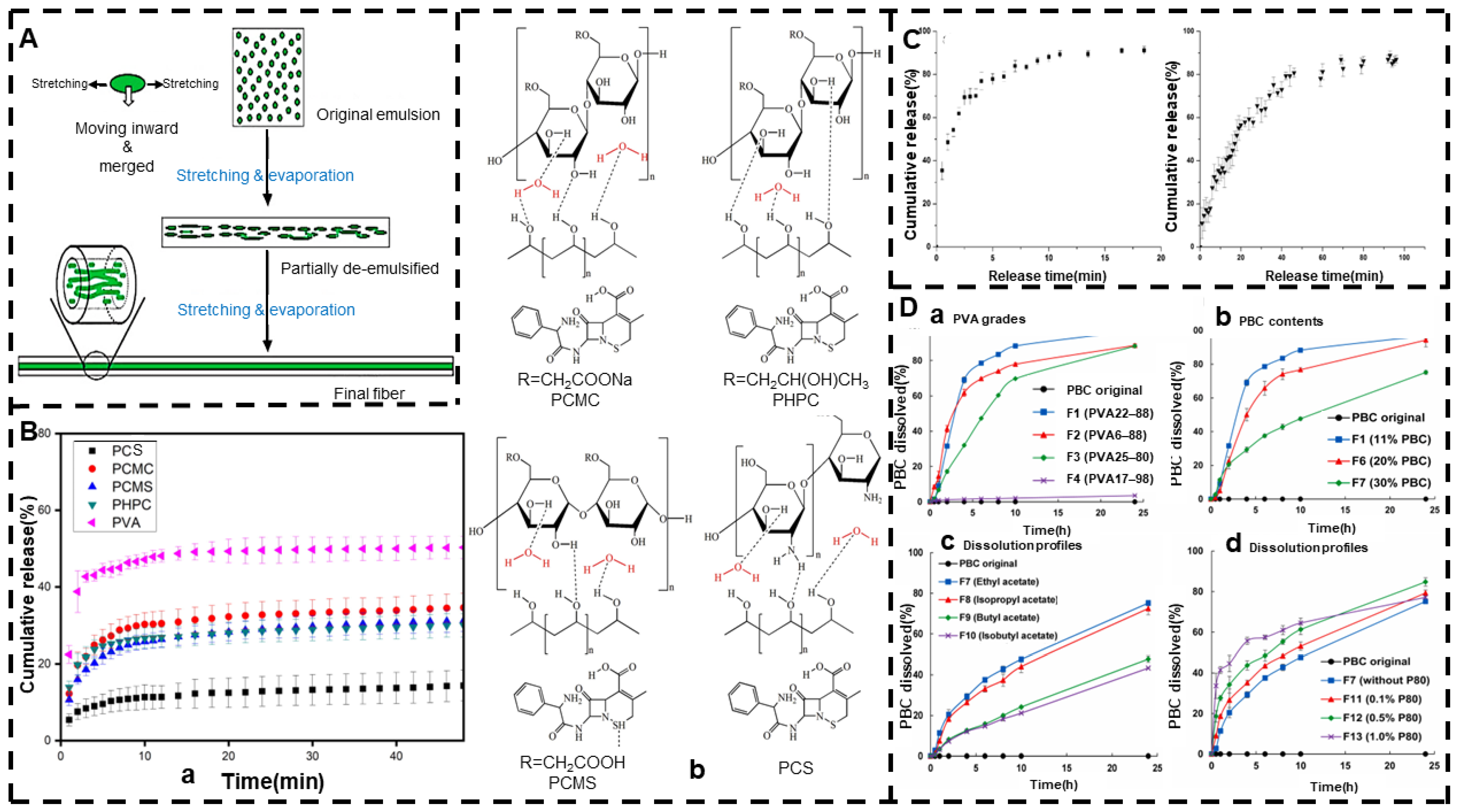
Table 1.
Size and advantages of drug carrier materials.
| Name | Diameter/Aperture | Advantage | References |
|---|---|---|---|
| Solid lipid nanoparticles | 100–1000 nm | Low toxicity, highly effective drug targeting, controlled-release drugs, high drug loads (especially lipophilic drugs), prevents degradation and has versatility | [112,113,114] |
| Lipidosome | 50–200 nm | Biocompatible, biodegradable, non-toxic and non-immunogenic | [115] |
| Dendritic polymer | 1.5–14.5 nm | Spherical homogeneous structure, high biocompatibility, lipophilic, variable composition | [116] |
| Nanocapsule | 10–1000 nm | Improves efficacy and bioavailability, prevents drug degradation, and provides controlled-release delivery | [117] |
| Polymeric micelle | 10–100 nm | Improves bioavailability, alters drug release curves, and improves patient compliance | [118] |
| Mesoporous silica material | 2–50 nm | Good biocompatibility, large specific surface area, large porosity, high drug carrying capacity, good thermal and chemical stability, can carry hydrophilic and lipophilic drugs | [119,120,121] |
| Carbon nano tube | 0.4–2 nm | Water solubility, biocompatibility, low toxicity, high drug load, intrinsic stability, high specific surface area | [122,123,124] |
| 2–100 nm | |||
| Nano-emulsion | Submicron order | High stability, high load capacity, improved solubility and bioavailability | [125] |
| Nanocrystal | 1–1000 nm | Stabilized by surfactants or stabilizers, no need for carrier materials, drug nanocrystals can enhance the adhesion to biofilms, increases the saturation solubility of drugs, large specific surface area, high bioavailability, high stability, high drug loading capacity, stable dissolution, sustained release drugs and safety | [126,127,128] |
Table 2.
Different types of polymer carriers.
| Natural Polymers | Water-Soluble | Water-Insoluble | Degradable | Small Molecule |
|---|---|---|---|---|
| Botany | Alginate | Cellulose | Glucan | CD |
| Zein | NR | |||
| Pectin | ||||
| Animal | Gelatin (soluble in hot water) | Collagen | PASP | |
| CS | ||||
| Microorganism | PGlu | |||
| Synthetic polymers | Water-soluble | Water-insoluble | Degradable | Small molecule |
| HPMC | PVC | Polyanhydride | PE | |
| Polyacrylate | EVA | PGA | PS | |
| PEG | PMMA | Polynitrile alkyl acrylate | PEI | |
| PVA | PE | polyorthoester | PEG | |
| MC | CA | PHB | ||
| PAM | PAN | PLLA | ||
| PEO | PS | PLGA | ||
| PVP | PPy | PHAS | ||
| PVDF | PBS | |||
| EC | PLA | |||
| PA | PCL |
Table 3.
Electrospinning combined with other techniques.
| Technologies | Drugs | Carrier | Release Mechanism | Highlights | Literature |
|---|---|---|---|---|---|
| Hybrid electrospinning + electrospray | FLU/RHB | PLGA | Drug diffusion mechanism Polymer degradation mechanism | The superhydrophobic layer can inhibit the release of FLU and RHB. After 720 h, FLU was released at a rate of about 16.5%, 25.9%, and 37.5%, and RHB was released at a rate of about 21.7%, 29.2%, and 34.6%, respectively, and the deposition times were 5, 10, and 15 min, respectively. It controls the rate of drug release by adjusting the thickness of the superhydrophobic coating. | [214] |
| Hydrothermal treatment co-precipitation + electrospinning | AMOX | LDH/DMSN/PCL | Diffusion mechanism | The drug release rate of complex membrane A was 87.81%. The drug release rate of complex membrane B was 94.65%. | [215] |
| Coaxial electrospinning + electrospray | AMPs/Curcumin | PLA/PVP/PEG | Diffusion mechanism | Shell-controlled-release AMPs reached about 70% within 24 h and more than 90% within 72 h for pre-treatment; in the middle and late stages of treatment, the sustained release of curcumin from the core layer can be extended to about 5 days. | [216] |
| Hybrid electrospinning + solvent steam annealing | SPL | PCL | Diffusion mechanism | Slower SPL release (more than 360 h) can be observed from annealed fibers and a decrease in the final percentage of SPL release (~50–60%). | [217] |
| Side-by-side electrospinning + electrodeposition | bFGF/NGF | PPy/PVDF | Ion exchange mechanism | Release curves of different growth factors (NGF and bFGF) showed electrically sensitive release behavior, which remained biologically active after release. | [218] |
| Solution extrusion 3D printing + coaxial electrospinning | Lidocaine/Estradiol/MTZ/CTGF | PCL/PLGA | Drug diffusion mechanism Polymer degradation mechanism | The duration of sustainable release of metronidazole, lidocaine, and estradiol was 4, 25, and 30 days, respectively. | [219] |
| Hybrid electrospinning + electrospray | CPGs/TRP2/Dox/hpDNA/fBSA | PVA/PEI/PVP/SF | Diffusion mechanism | fBSA and hpDNA were effectively released into the skin, and the cumulative release percentage of DNA was higher than that of BSA. | [220] |
| Electrospinning + electrospray | CPGs/TRP2/DNATrp2@ETHMC/DNATrp2 | MC/PVP/HA | Diffusion mechanism | Cumulative transdermal drug release DNATrp2@ETHMC loaded patch within 36 h was 35.4%, significantly higher than the release of free DNATrp2. | [221] |
| Redox amination + electrospinning | IBU | SA/PVA | Mechanisms of polymer swelling and degradation Diffusion mechanism | Adjusts the drug release rate by adjusting the RAOA/PVA volume ratio. RAOA can effectively encapsulate hydrophobic ibuprofen, thereby slowing the spread rate of the drug. | [222] |
| Electrospinning + crosslinking post-processing | Dex | SA/PVA | Diffusion mechanism | The release of Dex from the nanofibers was controlled by the chemical potential gradient and expansion penetration. Coaxial nanofibers protected the drug molecule in the core and also supported its sustained release curve. | [223] |
| Blended electrospinning + casting | IBU | EC/PVP K60 | Diffusion mechanism | The first stage exhibited a biphasic controlled release for the pulsating mode, and the residue was released in an extended manner in the second stage. | [224] |
Publisher’s Note: MDPI stays neutral with regard to jurisdictional claims in published maps and institutional affiliations. |
© 2022 by the authors. Licensee MDPI, Basel, Switzerland. This article is an open access article distributed under the terms and conditions of the Creative Commons Attribution (CC BY) license (https://creativecommons.org/licenses/by/4.0/).
Share and Cite
MDPI and ACS Style
Wang, Y.; Yu, D.-G.; Liu, Y.; Liu, Y.-N. Progress of Electrospun Nanofibrous Carriers for Modifications to Drug Release Profiles. J. Funct. Biomater. 2022, 13, 289. https://doi.org/10.3390/jfb13040289
AMA Style
Wang Y, Yu D-G, Liu Y, Liu Y-N. Progress of Electrospun Nanofibrous Carriers for Modifications to Drug Release Profiles. Journal of Functional Biomaterials. 2022; 13(4):289. https://doi.org/10.3390/jfb13040289
Chicago/Turabian StyleWang, Ying, Deng-Guang Yu, Yang Liu, and Ya-Nan Liu. 2022. "Progress of Electrospun Nanofibrous Carriers for Modifications to Drug Release Profiles" Journal of Functional Biomaterials 13, no. 4: 289. https://doi.org/10.3390/jfb13040289
Note that from the first issue of 2016, this journal uses article numbers instead of page numbers. See further details here.