


The Influence of Hydroxyapatite Nanoparticle Morphology on Embryonic Development in a Zebrafish Exposure Model
Abstract
1. Introduction
2. Results and Discussion
3. Conclusions
4. Materials and Methods
4.1. Particle Preparation and Characterization
4.2. Zebrafish Embryo Exposure to HANPs
Acknowledgments
Author Contributions
Conflicts of Interest
References
- Donaldson, K.; Stone, V.; Tran, C.L.; Kreyling, W.; Borm, P.J. Nanotoxicology. Occup. Environ. Med. 2004, 61, 727–728. [Google Scholar] [CrossRef] [Scilit] [PubMed]
- Nel, A.; Xia, T.; Madler, L.; Li, N. Toxic potential of materials at the nanolevel. Science 2006, 311, 622–627. [Google Scholar] [CrossRef] [Scilit] [PubMed]
- Sharifi, S.; Behzadi, S.; Laurent, S.; Forrest, M.L.; Stroeve, P.; Mahmoudi, M. Toxicity of nanomaterials. Chem. Soc. Rev. 2012, 41, 2323–2343. [Google Scholar] [CrossRef] [Scilit] [PubMed]
- De Groot, K. Bioceramics consisting of calcium phosphate salts. Biomaterials 1980, 1, 47–50. [Google Scholar] [CrossRef] [Scilit]
- Dorozhkin, S.V.; Epple, M. Biological and medical significance of calcium phosphates. Angew. Chem. 2002, 41, 3130–3146. [Google Scholar] [CrossRef] [Scilit]
- Hak, D.J. The use of osteoconductive bone graft substitutes in orthopaedic trauma. J. Am. Acad. Orthop. Surg. 2007, 15, 525–536. [Google Scholar] [CrossRef] [Scilit] [PubMed]
- Huracek, J.; Spirig, P. The effect of hydroxyapatite coating on the fixation of hip prostheses. Arch. Orthop. Trauma Surg. 1994, 113, 72–77. [Google Scholar] [CrossRef] [Scilit] [PubMed]
- Jaffe, W.L.; Scott, D.F. Total hip arthroplasty with hydroxyapatite-coated prostheses. J. Bone Jt. Surg. Am. Vol. 1996, 78, 1918–1934. [Google Scholar] [CrossRef] [Scilit]
- Bloebaum, R.D.; Zou, L.; Bachus, K.N.; Shea, K.G.; Hofmann, A.A.; Dunn, H.K. Analysis of particles in acetabular components from patients with osteolysis. Clin. Orthop. Relat. Res. 1997, 338, 109–118. [Google Scholar] [CrossRef] [Scilit]
- Morscher, E.W.; Hefti, A.; Aebi, U. Severe osteolysis after third-body wear due to hydroxyapatite particles from acetabular cup coating. J. Bone Jt. Surg. Br. Vol. 1998, 80, 267–272. [Google Scholar] [CrossRef] [Scilit]
- Yamashita, K.; Yoshioka, Y.; Higashisaka, K.; Mimura, K.; Morishita, Y.; Nozaki, M.; Yoshida, T.; Ogura, T.; Nabeshi, H.; Nagano, K.; et al. Silica and titanium dioxide nanoparticles cause pregnancy complications in mice. Nat. Nanotechnol. 2011, 6, 321–328. [Google Scholar] [CrossRef] [Scilit] [PubMed]
- Browning, L.M.; Huang, T.; Xu, X.H. Real-time in vivo imaging of size-dependent transport and toxicity of gold nanoparticles in zebrafish embryos using single nanoparticle plasmonic spectroscopy. Interface Focus 2013, 3, 20120098. [Google Scholar] [CrossRef] [Scilit] [PubMed]
- Di Bona, K.R.; Xu, Y.; Ramirez, P.A.; DeLaine, J.; Parker, C.; Bao, Y.; Rasco, J.F. Surface charge and dosage dependent potential developmental toxicity and biodistribution of iron oxide nanoparticles in pregnant CD-1 mice. Reprod. Toxicol. 2014, 50, 36–42. [Google Scholar] [CrossRef] [Scilit] [PubMed]
- Hong, J.S.; Park, M.K.; Kim, M.S.; Lim, J.H.; Park, G.J.; Maeng, E.H.; Shin, J.H.; Kim, Y.R.; Kim, M.K.; Lee, J.K.; et al. Effect of zinc oxide nanoparticles on dams and embryo-fetal development in rats. Int. J. Nanomed. 2014, 9, 145–157. [Google Scholar]
- Chakraborty, C.; Sharma, A.R.; Sharma, G.; Lee, S.S. Zebrafish: A complete animal model to enumerate the nanoparticle toxicity. J. Nanobiotechnol. 2016, 14, 65. [Google Scholar] [CrossRef] [Scilit] [PubMed]
- Beliaeva, N.F.; Kashirtseva, V.N.; Medvedeva, N.V.; Khudoklinova, I.; Ipatova, O.M.; Archakov, A.I. Zebrafish as a model organism for biomedical studies. Biomed. Khimiia 2010, 56, 120–131. [Google Scholar] [CrossRef] [Scilit]
- Varshney, G.K.; Lu, J.; Gildea, D.E.; Huang, H.; Pei, W.; Yang, Z.; Huang, S.C.; Schoenfeld, D.; Pho, N.H.; Casero, D.; et al. A large-scale zebrafish gene knockout resource for the genome-wide study of gene function. Genome Res. 2013, 23, 727–735. [Google Scholar] [CrossRef] [Scilit] [PubMed]
- Pujari-Palmer, S.; Chen, S.; Rubino, S.; Weng, H.; Xia, W.; Engqvist, H.; Tang, L.; Ott, M.K. In vivo and in vitro evaluation of hydroxyapatite nanoparticle morphology on the acute inflammatory response. Biomaterials 2016, 90, 1–11. [Google Scholar] [CrossRef] [Scilit] [PubMed]
- King-Heiden, T.C.; Wiecinski, P.N.; Mangham, A.N.; Metz, K.M.; Nesbit, D.; Pedersen, J.A.; Hamers, R.J.; Heideman, W.; Peterson, R.E. Quantum dot nanotoxicity assessment using the zebrafish embryo. Environ. Sci. Technol. 2009, 43, 1605–1611. [Google Scholar] [CrossRef] [Scilit] [PubMed]
- Boix, N.P.E.; Gomez-Catalan, J.; Teixido, E.; Llobet, J.M. The zebrafish embryo as a model for studying oxidative stress effect during embryonic development. Reprod. Toxicol. 2013, 41, 21–34. [Google Scholar] [CrossRef] [Scilit]
- Brown, D.M.; Bangert, U.; Windle, A.H.; Walter, D.M.; Walker, G.S.; Scotchford, C.A.; Donaldson, K.; Sone, V. An in vitro study of the potential of carbon nanotubes and nanofibres to induce inflammatory mediators and frustrated phagocytosis. Carbon 2007, 45, 1743–1756. [Google Scholar] [CrossRef] [Scilit]
- Duffin, R.; Tran, L.; Brown, D.; Stone, V.; Donaldson, K. Proinflammogenic effects of low-toxicity and metal nanoparticles in vivo and in vitro: Highlighting the role of particle surface area and surface reactivity. Inhal. Toxicol. 2007, 19, 849–856. [Google Scholar] [CrossRef] [Scilit] [PubMed]
- Oberdorster, G.; Ferin, J.; Lehnert, B.E. Correlation between particle size, in vivo particle persistence, and lung injury. Environ. Health Perspect. 1994, 102, 173–179. [Google Scholar] [CrossRef] [Scilit] [PubMed]
- Tran, C.L.; Buchanan, D.; Cullen, R.T.; Searl, A.; Jones, A.D.; Donaldson, K. Inhalation of poorly soluble particles. II. Influence of particle surface area on inflammation and clearance. Inhal. Toxicol. 2000, 12, 1113–1126. [Google Scholar] [PubMed]
- Lee, K.J.; Nallathamby, P.D.; Browning, L.M.; Osgood, C.J.; Xu, X.H. In vivo imaging of transport and biocompatibility of single silver nanoparticles in early development of zebrafish embryos. ACS Nano 2007, 1, 133–143. [Google Scholar] [CrossRef] [Scilit] [PubMed]
- Xia, T.; Zhao, Y.; Sager, T.; George, S.; Pokhrel, S.; Li, N.; Schoenfeld, D.; Meng, H.; Lin, S.; Wang, X.; et al. Decreased dissolution of ZnO by iron doping yields nanoparticles with reduced toxicity in the rodent lung and zebrafish embryos. ACS Nano 2011, 5, 1223–1235. [Google Scholar] [CrossRef] [Scilit] [PubMed]
- Zhao, X.; Ong, K.J.; Ede, J.D.; Stafford, J.L.; Ng, K.W.; Goss, G.G.; Loo, S.C. Evaluating the toxicity of hydroxyapatite nanoparticles in catfish cells and zebrafish embryos. Small 2013, 9, 1734–1741. [Google Scholar] [CrossRef] [Scilit] [PubMed]
- Xu, Z.; Zhang, Y.L.; Song, C.; Wu, L.L.; Gao, H.W. Interactions of hydroxyapatite with proteins and its toxicological effect to zebrafish embryos development. PLoS ONE 2012, 7, e32818. [Google Scholar] [CrossRef] [Scilit] [PubMed]
- Maccormack, T.J.; Clark, R.J.; Dang, M.K.; Ma, G.; Kelly, J.A.; Veinot, J.G.C.; Goss, G.G. Inhibition of enzyme activity by nanomaterials: Potential mechanisms and implications for nanotoxicity testing. Nanotoxicology 2012, 6, 514–525. [Google Scholar] [CrossRef] [Scilit] [PubMed]
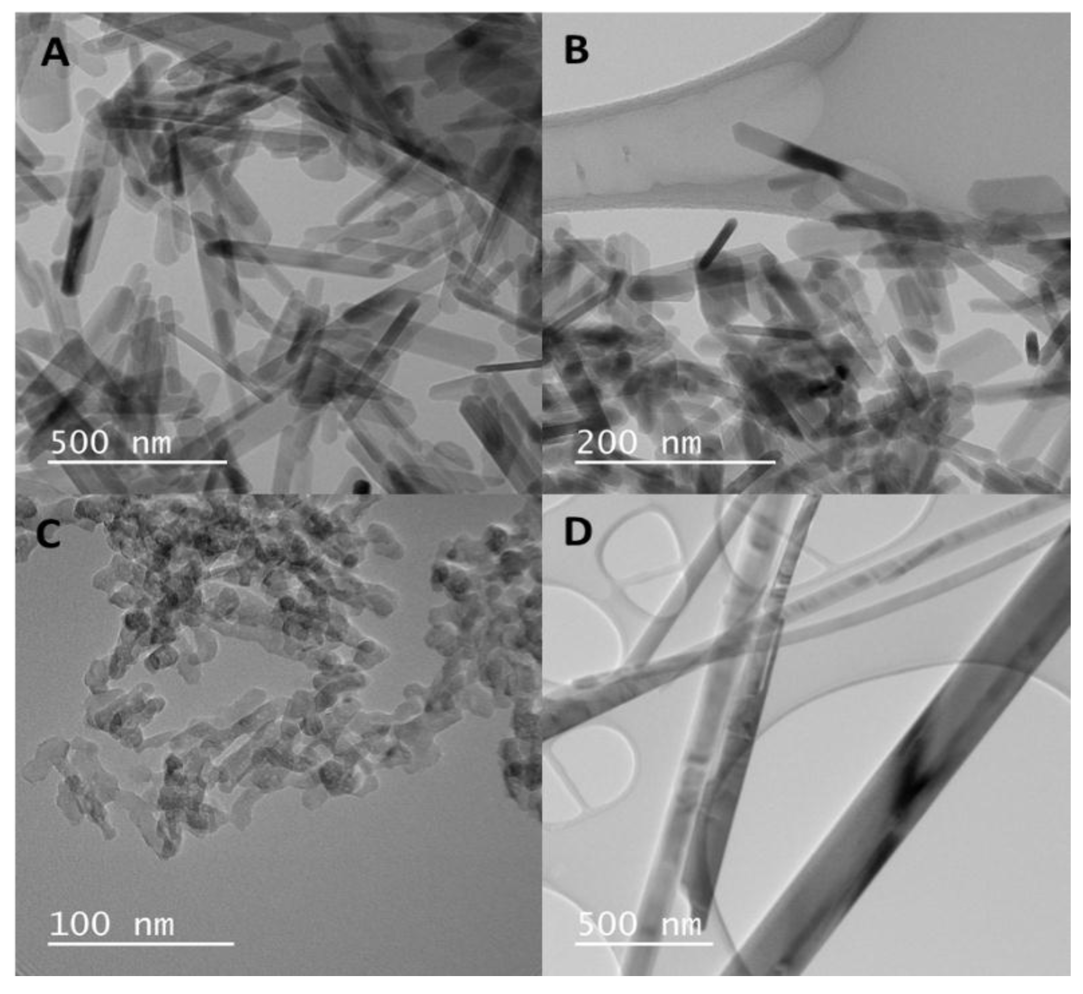

| Particle Morphology | Dimensions | Surface Area |
|---|---|---|
| Long Rods | 20 nm × 200 nm | 71.6 |
| Dots | 15 nm × 15 nm | 91.5 |
| Sheets | 75 nm × 30 nm | 58.6 |
| Fibers | 60 nm × 1–4 μm | 52.7 |
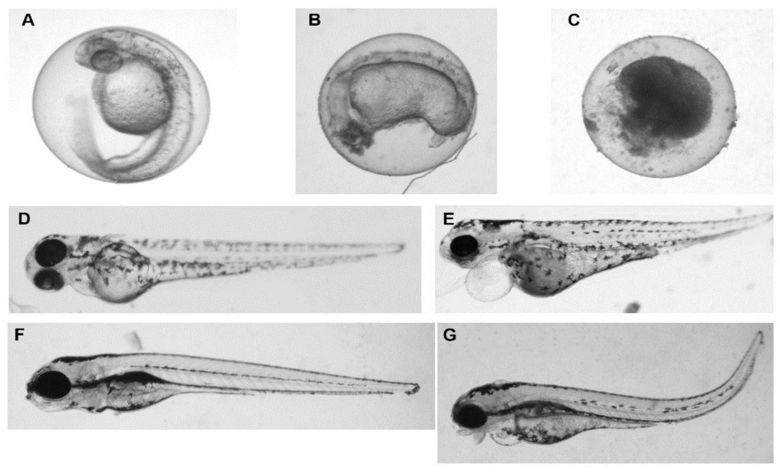
| Sheets | Dots | Long Rods | Fibers | Control | ||||||
|---|---|---|---|---|---|---|---|---|---|---|
| 40 mg/L | Malformed | Dead | Malformed | Dead | Malformed | Dead | Malformed | Dead | Malformed | Dead |
| Day 1 | 0% | 14% | 20% | 20% | 0% | 25% | 33% | 67% | 0% | 29% |
| Day 3 | 27% | 0% | 25% | 0% | 17% | 0% | 0% | 100% | 0% | 0% |
| Day 5 | 33% | 0% | 0% | 17% | 0% | 0% | 0% | 100% | 0% | 0% |
| Sheets | Dots | Long Rods | Fibers | Control | ||||||
|---|---|---|---|---|---|---|---|---|---|---|
| 100 mg/L | Malformed | Dead | Malformed | Dead | Malformed | Dead | Malformed | Dead | Malformed | Dead |
| Day 1 | 0% | 25% | 0% | 18% | 0% | 18% | 0% | 100% | 0% | 29% |
| Day 3 | 67% | 33% | 88% | 38% | 67% | 22% | 0% | 100% | 0% | 0% |
| Day 5 | 0% | 100% | 0% | 100% | 0% | 100% | 0% | 100% | 0% | 0% |
© 2017 by the authors. Licensee MDPI, Basel, Switzerland. This article is an open access article distributed under the terms and conditions of the Creative Commons Attribution (CC BY) license (http://creativecommons.org/licenses/by/4.0/).
Share and Cite
Pujari-Palmer, S.; Lu, X.; Karlsson Ott, M. The Influence of Hydroxyapatite Nanoparticle Morphology on Embryonic Development in a Zebrafish Exposure Model. Nanomaterials 2017, 7, 89. https://doi.org/10.3390/nano7040089
Pujari-Palmer S, Lu X, Karlsson Ott M. The Influence of Hydroxyapatite Nanoparticle Morphology on Embryonic Development in a Zebrafish Exposure Model. Nanomaterials. 2017; 7(4):89. https://doi.org/10.3390/nano7040089
Chicago/Turabian StylePujari-Palmer, Shiuli, Xi Lu, and Marjam Karlsson Ott. 2017. "The Influence of Hydroxyapatite Nanoparticle Morphology on Embryonic Development in a Zebrafish Exposure Model" Nanomaterials 7, no. 4: 89. https://doi.org/10.3390/nano7040089
APA StylePujari-Palmer, S., Lu, X., & Karlsson Ott, M. (2017). The Influence of Hydroxyapatite Nanoparticle Morphology on Embryonic Development in a Zebrafish Exposure Model. Nanomaterials, 7(4), 89. https://doi.org/10.3390/nano7040089

