Next Issue
Volume 7, May
Previous Issue
Volume 7, March


Nanomaterials, Volume 7, Issue 4 (April 2017) – 21 articles

Cover Story (view full-size image): 
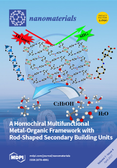
Functional organic ligands play an important role in constructing multifunctional Metal-Organic Framework (MOF) materials. Most of these ligands contain benzene rings and specific functional groups such as –NH2, –COOH, –CONH; however, ligands composed of inherent homochiral moieties are relative rare. By incorporating the homochiral moieties and other functional groups into the ligands for MOF construction, the obtained materials may aid homochirality and other functionalities, which may broaden their range of applications. Herein, we have designed a chiral ligand by successfully combining the chiral camphoric moiety and acylamide group with synthesized homochiral MOF material. The chiral camphoric group imparts homochirality to the MOF and the acylamide group enables interactions between the framework and guest molecules. Finally, the obtained product has the capacity to separate water from alcohol [...] Read more.
  • Issues are regarded as officially published after their release is announced to the table of contents alert mailing list.
  • You may sign up for e-mail alerts to receive table of contents of newly released issues.
  • PDF is the official format for papers published in both, html and pdf forms. To view the papers in pdf format, click on the "PDF Full-text" link, and use the free Adobe Reader to open them.
Order results
Result details
Section
Select all
Export citation of selected articles as:
13284 KiB  
Review
Gold Nanoparticles for Modulating Neuronal Behavior
by Chiara Paviolo and Paul R. Stoddart
Nanomaterials 2017, 7(4), 92; https://doi.org/10.3390/nano7040092 - 24 Apr 2017
Cited by 67 | Viewed by 7756
Abstract
Understanding the detailed functioning and pathophysiology of the brain and the nervous system continues to challenge the scientific community, particularly in terms of scaling up techniques for monitoring and interfacing with complex 3D networks. Nanotechnology has the potential to support this scaling up, [...] Read more.
Understanding the detailed functioning and pathophysiology of the brain and the nervous system continues to challenge the scientific community, particularly in terms of scaling up techniques for monitoring and interfacing with complex 3D networks. Nanotechnology has the potential to support this scaling up, where the eventual goal would be to address individual nerve cells within functional units of both the central and peripheral nervous system. Gold nanoparticles provide a variety of physical and chemical properties that have attracted attention as a light-activated nanoscale neuronal interface. This review provides a critical overview of the photothermal and photomechanical properties of chemically functionalized gold nanoparticles that have been exploited to trigger a range of biological responses in neuronal tissues, including modulation of electrical activity and nerve regeneration. The prospects and challenges for further development are also discussed. Full article
(This article belongs to the Special Issue Nanoparticle-Mediated Cell and Tissue Stimulation)
Show Figures

Graphical abstract

9663 KiB  
Article
The Interactions between ZnO Nanoparticles (NPs) and α-Linolenic Acid (LNA) Complexed to BSA Did Not Influence the Toxicity of ZnO NPs on HepG2 Cells
by Yiwei Zhou, Xin Fang, Yu Gong, Aiping Xiao, Yixi Xie, Liangliang Liu and Yi Cao
Nanomaterials 2017, 7(4), 91; https://doi.org/10.3390/nano7040091 - 24 Apr 2017
Cited by 35 | Viewed by 5947
Abstract
Background: Nanoparticles (NPs) entering the biological environment could interact with biomolecules, but little is known about the interaction between unsaturated fatty acids (UFA) and NPs. Methods: This study used α-linolenic acid (LNA) complexed to bovine serum albumin (BSA) for UFA and HepG2 cells [...] Read more.
Background: Nanoparticles (NPs) entering the biological environment could interact with biomolecules, but little is known about the interaction between unsaturated fatty acids (UFA) and NPs. Methods: This study used α-linolenic acid (LNA) complexed to bovine serum albumin (BSA) for UFA and HepG2 cells for hepatocytes. The interactions between BSA or LNA and ZnO NPs were studied. Results: The presence of BSA or LNA affected the hydrodynamic size, zeta potential, UV-Vis, fluorescence, and synchronous fluorescence spectra of ZnO NPs, which indicated an interaction between BSA or LNA and NPs. Exposure to ZnO NPs with the presence of BSA significantly induced the damage to mitochondria and lysosomes in HepG2 cells, associated with an increase of intracellular Zn ions, but not intracellular superoxide. Paradoxically, the release of inflammatory cytokine interleukin-6 (IL-6) was decreased, which indicated the anti-inflammatory effects of ZnO NPs when BSA was present. The presence of LNA did not significantly affect all of these endpoints in HepG2 cells exposed to ZnO NPs and BSA. Conclusions: the results from the present study indicated that BSA-complexed LNA might modestly interact with ZnO NPs, but did not significantly affect ZnO NPs and BSA-induced biological effects in HepG2 cells. Full article
Show Figures

Figure 1

3224 KiB  
Article
Bioactivity Studies on Titania Coatings and the Estimation of Their Usefulness in the Modification of Implant Surfaces
by Aleksandra Radtke, Adrian Topolski, Tomasz Jędrzejewski, Wiesław Kozak, Beata Sadowska, Marzena Więckowska-Szakiel and Piotr Piszczek
Nanomaterials 2017, 7(4), 90; https://doi.org/10.3390/nano7040090 - 22 Apr 2017
Cited by 19 | Viewed by 4751
Abstract
Morphologically different titania coatings (nanofibers (TNFs), nanoneedles (TNNs), and nanowires (TNWs)) were studied as potential biomedical materials. The abovementioned systems were produced in situ on Ti6Al4V substrates via direct oxidation processes using H2O2 and H2O2/CaCl2 [...] Read more.
Morphologically different titania coatings (nanofibers (TNFs), nanoneedles (TNNs), and nanowires (TNWs)) were studied as potential biomedical materials. The abovementioned systems were produced in situ on Ti6Al4V substrates via direct oxidation processes using H2O2 and H2O2/CaCl2 agents, and via thermal oxidation in the presence of Ar and Ar/H2O2. X-ray diffraction and Raman spectroscopy have been used to structurally characterize the produced materials. The morphology changes on the titanium alloy surface were investigated using scanning electron microscopy. The bioactivity of the samples has been estimated by the analysis of the produced titania coatings’ biocompatibility, and by the determination of their ability to reduce bacterial biofilm formation. The photoactivity of the produced nanocoatings was also analyzed, in order to determine the possibility of using titania coated implant surfaces in the sterilization process of implants. Photocatalytic activity was estimated using the methylene blue photodegradation kinetics, in the presence of UV light. Full article
(This article belongs to the Special Issue Nanofibrous Scaffolds for Biomedical Application)
Show Figures

Graphical abstract

1060 KiB  
Communication
The Influence of Hydroxyapatite Nanoparticle Morphology on Embryonic Development in a Zebrafish Exposure Model
by Shiuli Pujari-Palmer, Xi Lu and Marjam Karlsson Ott
Nanomaterials 2017, 7(4), 89; https://doi.org/10.3390/nano7040089 - 22 Apr 2017
Cited by 12 | Viewed by 5296
Abstract
Nanomaterials are used in many different industries such as cosmetics, food, clothing, and electronics. There is increasing concern that exposure to nanoparticles (NPs) during pregnancy can adversely affect fetal development. It is well known that the size, charge, and chemistry of a nanoparticle [...] Read more.
Nanomaterials are used in many different industries such as cosmetics, food, clothing, and electronics. There is increasing concern that exposure to nanoparticles (NPs) during pregnancy can adversely affect fetal development. It is well known that the size, charge, and chemistry of a nanoparticle can modulate embryological development. The role that particle morphology plays on early development, however, is still widely unknown. The present study aims to investigate the effect of hydroxyapatite nanoparticle (HANP) morphology on embryological development in a zebrafish exposure model. Four distinct HANP morphologies (dots, long rods, sheets, and fibers) were fabricated and characterized. Zebrafish embryos were exposed to HANPs (0–100 mg/L), and viability and developmental deformities were evaluated for up to 5 days post-fertilization (dpf). Malformations such as pericardial edema and axial curvature were apparent in embryos as early as 1 dpf, following exposure to the dot and fiber particles, and developed in embryos by 3 dpf in the sheet and long rod particle groups. Minimal death was observed in response to dot, long rod, and sheet particles (≤25%), while fiber particles induced overwhelming toxicity (≤60%) after 1 dpf, and complete toxicity during all subsequent time points. Collectively, these results suggest that nanoparticle morphology can significantly impact embryological development and should be a required consideration when designing nanomaterials for commercial use. Full article
Show Figures

Figure 1

2710 KiB  
Article
A Homochiral Multifunctional Metal-Organic Framework with Rod-Shaped Secondary Building Units
by Kun Cai, Nian Zhao, Ning Zhang, Fu-Xing Sun, Qing Zhao and Guang-Shan Zhu
Nanomaterials 2017, 7(4), 88; https://doi.org/10.3390/nano7040088 - 21 Apr 2017
Cited by 12 | Viewed by 5525
Abstract
A new homochiral multifunctional metal-organic framework, [Zn2(CTBA)2·H2O] (JUC-112), was synthesized under solvothermal conditions, through the design of chiral ligand 4-(3-carboxy-2,2,3-trimethylcyclopentanecarboxamido) benzoic acid (H2CTBA) based on camphoric acid as building block. The crystal structure of the [...] Read more.
A new homochiral multifunctional metal-organic framework, [Zn2(CTBA)2·H2O] (JUC-112), was synthesized under solvothermal conditions, through the design of chiral ligand 4-(3-carboxy-2,2,3-trimethylcyclopentanecarboxamido) benzoic acid (H2CTBA) based on camphoric acid as building block. The crystal structure of the new material is a 2-dimensional (2D) chiral layer packed with infinite rod-shaped secondary building units (SBUs). The homochiral framework was identified by circular dichroism (CD) spectrum. Thermogravimetric measurement indicates its high thermal stability up to 450 °C. In addition, JUC-112 exhibits the capability of separating water from alcohols, second-order nonlinear optical effect, and photoluminescence. Full article
(This article belongs to the Special Issue Frontiers in Chiral Nanomaterials)
Show Figures

Graphical abstract

2460 KiB  
Review
Nanomaterials as Assisted Matrix of Laser Desorption/Ionization Time-of-Flight Mass Spectrometry for the Analysis of Small Molecules
by Minghua Lu, Xueqing Yang, Yixin Yang, Peige Qin, Xiuru Wu and Zongwei Cai
Nanomaterials 2017, 7(4), 87; https://doi.org/10.3390/nano7040087 - 21 Apr 2017
Cited by 78 | Viewed by 10613
Abstract
Matrix-assisted laser desorption/ionization (MALDI), a soft ionization method, coupling with time-of-flight mass spectrometry (TOF MS) has become an indispensible tool for analyzing macromolecules, such as peptides, proteins, nucleic acids and polymers. However, the application of MALDI for the analysis of small molecules (<700 [...] Read more.
Matrix-assisted laser desorption/ionization (MALDI), a soft ionization method, coupling with time-of-flight mass spectrometry (TOF MS) has become an indispensible tool for analyzing macromolecules, such as peptides, proteins, nucleic acids and polymers. However, the application of MALDI for the analysis of small molecules (<700 Da) has become the great challenge because of the interference from the conventional matrix in low mass region. To overcome this drawback, more attention has been paid to explore interference-free methods in the past decade. The technique of applying nanomaterials as matrix of laser desorption/ionization (LDI), also called nanomaterial-assisted laser desorption/ionization (nanomaterial-assisted LDI), has attracted considerable attention in the analysis of low-molecular weight compounds in TOF MS. This review mainly summarized the applications of different types of nanomaterials including carbon-based, metal-based and metal-organic frameworks as assisted matrices for LDI in the analysis of small biological molecules, environmental pollutants and other low-molecular weight compounds. Full article
(This article belongs to the Special Issue Nanomaterials for Mass Spectrometry Applications)
Show Figures

Figure 1

153 KiB  
Editorial
Nanoscale in Photocatalysis
by Zhaohui Wang and Hongqi Sun
Nanomaterials 2017, 7(4), 86; https://doi.org/10.3390/nano7040086 - 17 Apr 2017
Cited by 3 | Viewed by 3425
Abstract
Rationally harvesting sunlight to carry out chemical reactions, for example, photochemistry and photocatalysis, has appeared as a beautiful episode within the long history of solar-energy utilization by human beings.[...] Full article
3441 KiB  
Article
Release of Doxorubicin by a Folate-Grafted, Chitosan-Coated Magnetic Nanoparticle
by Chung-Lin Yang, Jyh-Ping Chen, Kuo-Chen Wei, Ju-Yu Chen, Chia-Wen Huang and Zi-Xian Liao
Nanomaterials 2017, 7(4), 85; https://doi.org/10.3390/nano7040085 - 13 Apr 2017
Cited by 40 | Viewed by 6724
Abstract
In clinical tumor therapy, chemotherapeutic routes have caused severe side effects; current delivery methods are unsatisfactory. Successful design of a remotely folate (FA)-grafted chitosan (CS)-coated magnetic nanoparticle (MNP) with low toxicity, has been achieved. A chemotherapeutic drug such as doxorubicin (DOX), is loaded [...] Read more.
In clinical tumor therapy, chemotherapeutic routes have caused severe side effects; current delivery methods are unsatisfactory. Successful design of a remotely folate (FA)-grafted chitosan (CS)-coated magnetic nanoparticle (MNP) with low toxicity, has been achieved. A chemotherapeutic drug such as doxorubicin (DOX), is loaded in the MNP-based matrix (FA-grafted CS-DOX-TPP-MNP), which is coated by an activated target tumor molecule of FA-grafted CS biopolymer with the inclusion of tripolyphosphate (TPP) as a linker. The resultant nano-complexes exhibited random aggregates (~240 nm) and zeta potential (−24.9 mV). In vivo experiments using athymic BALB/c nude mice with human glioblastoma U87 cells in a subcutaneous tumor model revealed that magnetic guidance of FA-grafted CS-DOX-TPP-MNP, injected via the tail vein, significantly decreased tumor growth. This manuscript demonstrates the feasibility of magnetizing control of FA-grafted CS-DOX-TPP-MNP to enhance the localization of drug release. Full article
Show Figures

Graphical abstract

4537 KiB  
Article
Electrospun Fiber Pads of Cellulose Acetate and Essential Oils with Antimicrobial Activity
by Ioannis L. Liakos, Alina Maria Holban, Riccardo Carzino, Simone Lauciello and Alexandru Mihai Grumezescu
Nanomaterials 2017, 7(4), 84; https://doi.org/10.3390/nano7040084 - 12 Apr 2017
Cited by 76 | Viewed by 6989
Abstract
The method of electrospinning was used to create nanofibers made of cellulose acetate (CA) and essential oils (EOs). CA polymer at 15% w/v was dissolved in acetone and then 1% or 5% v/v of EOs was added to the [...] Read more.
The method of electrospinning was used to create nanofibers made of cellulose acetate (CA) and essential oils (EOs). CA polymer at 15% w/v was dissolved in acetone and then 1% or 5% v/v of EOs was added to the polymer solution. The utilized essential oils were rosemary and oregano oils. Then, the CA/EOs in acetone solution were electrospun, creating micro/nanofibers, approximately 700–1500 nm in diameter. Raman spectroscopy was used to detect the attachment of the EOs in the CA electrospun fibers (ESFs). Scanning electron microscopy was used to study the morphology, topography and dimensions of the ESFs. The formed CA/EOs ESFs are found to have good antimicrobial properties against three common microbial species, frequently found in difficult to treat infections: Bacteria species Staphylococcus aureus, Escherichia coli and the yeast Candida albicans. ESFs with 5% v/v oregano oil with respect to the initial solution, showed the best antimicrobial and anti-biofilm effects due to the potency of this EO against bacteria and fungi, especially for Escherichia coli and Candida albicans. This work describes an effective and simple method to prepare CA/EOs ESFs and opens up many new applications of micro/nanofibers such as improved antimicrobial wound dressings, anti-biofilm surfaces, sensors and packaging alternatives. Full article
(This article belongs to the Special Issue Nanoarchitectonics: A Novel Approach for Drug Delivery and Targeting)
Show Figures

Graphical abstract

3768 KiB  
Article
An Assessment of the Potential Use of BNNTs for Boron Neutron Capture Therapy
by Tiago H. Ferreira, Marcelo C. Miranda, Zildete Rocha, Alexandre S. Leal, Dawidson A. Gomes and Edesia M. B. Sousa
Nanomaterials 2017, 7(4), 82; https://doi.org/10.3390/nano7040082 - 12 Apr 2017
Cited by 36 | Viewed by 5602
Abstract
Currently, nanostructured compounds have been standing out for their optical, mechanical, and chemical features and for the possibilities of manipulation and regulation of complex biological processes. One of these compounds is boron nitride nanotubes (BNNTs), which are a nanostructured material analog to carbon [...] Read more.
Currently, nanostructured compounds have been standing out for their optical, mechanical, and chemical features and for the possibilities of manipulation and regulation of complex biological processes. One of these compounds is boron nitride nanotubes (BNNTs), which are a nanostructured material analog to carbon nanotubes, but formed of nitrogen and boron atoms. BNNTs present high thermal stability along with high chemical inertia. Among biological applications, its biocompatibility, cellular uptake, and functionalization potential can be highlighted, in addition to its eased utilization due to its nanometric size and tumor cell internalization. When it comes to new forms of therapy, we can draw attention to boron neutron capture therapy (BNCT), an experimental radiotherapy characterized by a boron-10 isotope carrier inside the target and a thermal neutron beam focused on it. The activation of the boron-10 atom by a neutron generates a lithium atom, a gamma ray, and an alpha particle, which can be used to destroy tumor tissues. The aim of this work was to use BNNTs as a boron-10 carrier for BNCT and to demonstrate its potential. The nanomaterial was characterized through XRD, FTIR, and SEM. The WST-8 assay was performed to confirm the cell viability of BNNTs. The cells treated with BNNTs were irradiated with the neutron beam of a Triga reactor, and the apoptosis caused by the activation of the BNNTs was measured with a calcein AM/propidium iodide test. The results demonstrate that this nanomaterial is a promising candidate for cancer therapy through BNCT. Full article
(This article belongs to the Special Issue Nanoparticle-Mediated Cell and Tissue Stimulation)
Show Figures

Figure 1

3914 KiB  
Article
Investigating the Origins of Toxic Response in TiO2 Nanoparticle-Treated Cells
by Gamze Kuku and Mustafa Culha
Nanomaterials 2017, 7(4), 83; https://doi.org/10.3390/nano7040083 - 11 Apr 2017
Cited by 19 | Viewed by 6557
Abstract
Titanium dioxide nanoparticles (TiO2 NPs) are widely used in sunscreens, cosmetics and body implants, and this raises toxicity concerns. Although a large number of reports claim that they are safe to use, others claim that they induce reactive oxygen species formation and [...] Read more.
Titanium dioxide nanoparticles (TiO2 NPs) are widely used in sunscreens, cosmetics and body implants, and this raises toxicity concerns. Although a large number of reports claim that they are safe to use, others claim that they induce reactive oxygen species formation and can be carcinogenic. In this study, the origins of toxic response to TiO2 NPs were investigated by using Surface-enhanced Raman spectroscopy (SERS) which provides multidimensional information on the cellular dynamics at single cell level without any requirement for cell fixation. Three cell lines of vein (HUVEC), lung carcinoma (A549) and skin (L929) origin were tested for their toxic response upon exposure to 20, 40, 80 and 160 µg/mL anatase-TiO2 NPs for 24 h. It was demonstrated that the level of toxic response is both cell line and dose-dependent. L929 fibroblasts were the most resistant cell line to oxidative stress whereas in HUVEC and A549, cell lines collagen and lipid deformation were observed, respectively. Full article
(This article belongs to the Special Issue Nanoparticle-Mediated Cell and Tissue Stimulation)
Show Figures

Figure 1

27750 KiB  
Article
Stepwise Splitting Growth and Pseudocapacitive Properties of Hierarchical Three-Dimensional Co3O4 Nanobooks
by Huilong Chen, Shuang Lu, Feilong Gong, Huanzhen Liu and Feng Li
Nanomaterials 2017, 7(4), 81; https://doi.org/10.3390/nano7040081 - 10 Apr 2017
Cited by 12 | Viewed by 4697
Abstract
Three-dimensional hierarchical Co3O4 nanobooks have been synthesized successfully on a large scale by calcining orthorhombic Co(CO3)0.5(OH)·0.11H2O precursors with identical morphologies. Based on the influence of reaction time and urea concentration on the nanostructures of [...] Read more.
Three-dimensional hierarchical Co3O4 nanobooks have been synthesized successfully on a large scale by calcining orthorhombic Co(CO3)0.5(OH)·0.11H2O precursors with identical morphologies. Based on the influence of reaction time and urea concentration on the nanostructures of the precursors, a stepwise splitting growth mechanism can be proposed to understand the formation of the 3D nanobooks. The 3D Co3O4 nanobooks exhibit excellent pseudocapacitive performances with specific capacitances of 590, 539, 476, 453, and 421 F/g at current densities of 0.5, 1, 2, 4, and 8 A/g, respectively. The devices can retain ca. 97.4% of the original specific capacitances after undergoing charge–discharge cycle tests 1000 times continuously at 4 A/g. Full article
(This article belongs to the Special Issue Nanomaterials Based Fuel Cells and Supercapacitors)
Show Figures

Graphical abstract

3242 KiB  
Article
Cytotoxicity of ZnO Nanowire Arrays on Excitable Cells
by Yongchen Wang, Yu Wu, Farhan Quadri, Jordan D. Prox and Liang Guo
Nanomaterials 2017, 7(4), 80; https://doi.org/10.3390/nano7040080 - 07 Apr 2017
Cited by 21 | Viewed by 6617
Abstract
Zinc oxide (ZnO) nanowires have been widely studied for their applications in electronics, optics, and catalysts. Their semiconducting, piezoelectric, fluorescent, and antibacterial properties have also attracted broad interest in their biomedical applications. Thus, it is imperative to evaluate the biosafety of ZnO nanowires [...] Read more.
Zinc oxide (ZnO) nanowires have been widely studied for their applications in electronics, optics, and catalysts. Their semiconducting, piezoelectric, fluorescent, and antibacterial properties have also attracted broad interest in their biomedical applications. Thus, it is imperative to evaluate the biosafety of ZnO nanowires and their biological effects. In this study, the cellular level biological effects of ZnO nanowire arrays are specifically tested on three types of excitable cells, including NG108-15 neuronal cell line, HL-1 cardiac muscle cell line, and neonatal rat cardiomyocytes. Vertically aligned and densely packed ZnO nanowire arrays are synthesized using a solution-based method and used as a substrate for cell culture. The metabolism levels of all three types of cells cultured on ZnO nanowire arrays are studied using the 3-(4,5-dimethyl-2-thiazolyl)-2,5-diphenyl-2H-tetrazolium bromide (MTT) assays of a full factorial design. Under the studied settings, the results show statistically significant inhibitory effects of ZnO nanowire arrays on the metabolism of NG108-15 and HL-1 cells in comparison to gold, glass, and polystyrene substrates, and on the metabolism of cardiomyocytes in comparison to gold substrate. Full article
(This article belongs to the Special Issue Nanoparticle-Mediated Cell and Tissue Stimulation)
Show Figures

Figure 1

4109 KiB  
Article
Cellular Response to Titanium Dioxide Nanoparticles in Intestinal Epithelial Caco-2 Cells is Dependent on Endocytosis-Associated Structures and Mediated by EGFR
by Kristin Krüger, Katrin Schrader and Martin Klempt
Nanomaterials 2017, 7(4), 79; https://doi.org/10.3390/nano7040079 - 07 Apr 2017
Cited by 20 | Viewed by 7154
Abstract
Titanium dioxide (TiO2) is one of the most applied nanomaterials and widely used in food and non-food industries as an additive or coating material (E171). It has been shown that E171 contains up to 37% particles which are smaller than 100 [...] Read more.
Titanium dioxide (TiO2) is one of the most applied nanomaterials and widely used in food and non-food industries as an additive or coating material (E171). It has been shown that E171 contains up to 37% particles which are smaller than 100 nm and that TiO2 nanoparticles (NPs) induce cytotoxicity and inflammation. Using a nuclear factor Kappa-light-chain enhancer of activated B cells (NF-κB) reporter cell line (Caco-2nfkb-RE), Real time polymerase chain reaction (PCR), and inhibition of dynamin and clathrin, it was shown that cellular responses induced by 5 nm and 10 nm TiO2 NPs (nominal size) depends on endocytic processes. As endocytosis is often dependent on the epithelial growth factor receptor (EGFR), further investigations focused on the involvement of EGFR in the uptake of TiO2 NPs: (1) inhibition of EGFR reduced inflammatory markers of the cell (i.e., nuclear factor (NF)-κB activity, mRNA of IL8, CCL20, and CXCL10); and (2) exposure of Caco-2 cells to TiO2 NPs activated the intracellular EGFR cascade beginning with EGFR-mediated extracellular signal-regulated kinases (ERK)1/2, and including transcription factor ELK1. This was followed by the expression of ERK1/2 target genes CCL2 and CXCL3. We concluded that TiO2 NPs enter the cell via EGFR-associated endocytosis, followed by activation of the EGFR/ERK/ELK signaling pathway, which finally induces NF-κB. No changes in inflammatory response are observed in Caco-2 cells exposed to 32 nm and 490 nm TiO2 particles. Full article
(This article belongs to the Special Issue Frontiers in Toxicity and Functionalization of Nanomaterials)
Show Figures

Figure 1

2825 KiB  
Article
Breakthrough to Non-Vacuum Deposition of Single-Crystal, Ultra-Thin, Homogeneous Nanoparticle Layers: A Better Alternative to Chemical Bath Deposition and Atomic Layer Deposition
by Yu-Kuang Liao, Yung-Tsung Liu, Dan-Hua Hsieh, Tien-Lin Shen, Ming-Yang Hsieh, An-Jye Tzou, Shih-Chen Chen, Yu-Lin Tsai, Wei-Sheng Lin, Sheng-Wen Chan, Yen-Ping Shen, Shun-Jen Cheng, Chyong-Hua Chen, Kaung-Hsiung Wu, Hao-Ming Chen, Shou-Yi Kuo, Martin D. B. Charlton, Tung-Po Hsieh and Hao-Chung Kuo
Nanomaterials 2017, 7(4), 78; https://doi.org/10.3390/nano7040078 - 06 Apr 2017
Cited by 6 | Viewed by 5430
Abstract
Most thin-film techniques require a multiple vacuum process, and cannot produce high-coverage continuous thin films with the thickness of a few nanometers on rough surfaces. We present a new ”paradigm shift” non-vacuum process to deposit high-quality, ultra-thin, single-crystal layers of coalesced sulfide nanoparticles [...] Read more.
Most thin-film techniques require a multiple vacuum process, and cannot produce high-coverage continuous thin films with the thickness of a few nanometers on rough surfaces. We present a new ”paradigm shift” non-vacuum process to deposit high-quality, ultra-thin, single-crystal layers of coalesced sulfide nanoparticles (NPs) with controllable thickness down to a few nanometers, based on thermal decomposition. This provides high-coverage, homogeneous thickness, and large-area deposition over a rough surface, with little material loss or liquid chemical waste, and deposition rates of 10 nm/min. This technique can potentially replace conventional thin-film deposition methods, such as atomic layer deposition (ALD) and chemical bath deposition (CBD) as used by the Cu(In,Ga)Se2 (CIGS) thin-film solar cell industry for decades. We demonstrate 32% improvement of CIGS thin-film solar cell efficiency in comparison to reference devices prepared by conventional CBD deposition method by depositing the ZnS NPs buffer layer using the new process. The new ZnS NPs layer allows reduction of an intrinsic ZnO layer, which can lead to severe shunt leakage in case of a CBD buffer layer. This leads to a 65% relative efficiency increase. Full article
Show Figures

Graphical abstract

4808 KiB  
Review
siRNA Delivery Strategies: A Comprehensive Review of Recent Developments
by Katyayani Tatiparti, Samaresh Sau, Sushil Kumar Kashaw and Arun K. Iyer
Nanomaterials 2017, 7(4), 77; https://doi.org/10.3390/nano7040077 - 05 Apr 2017
Cited by 303 | Viewed by 18375
Abstract
siRNA is a promising therapeutic solution to address gene overexpression or mutations as a post-transcriptional gene regulation process for several pathological conditions such as viral infections, cancer, genetic disorders, and autoimmune disorders like arthritis. This therapeutic method is currently being actively pursued in [...] Read more.
siRNA is a promising therapeutic solution to address gene overexpression or mutations as a post-transcriptional gene regulation process for several pathological conditions such as viral infections, cancer, genetic disorders, and autoimmune disorders like arthritis. This therapeutic method is currently being actively pursued in cancer therapy because siRNA has been found to suppress the oncogenes and address mutations in tumor suppressor genes and elucidate the key molecules in cellular pathways in cancer. It is also effective in personalized gene therapy for several diseases due to its specificity, adaptability, and broad targeting capability. However, naked siRNA is unstable in the bloodstream and cannot efficiently cross cell membranes besides being immunogenic. Therefore, careful design of the delivery systems is essential to fully utilize the potential of this therapeutic solution. This review presents a comprehensive update on the challenges of siRNA delivery and the current strategies used to develop nanoparticulate delivery systems. Full article
(This article belongs to the Special Issue Nanomaterials in Gene Therapy)
Show Figures

Figure 1

3975 KiB  
Article
Highly Enhanced Photoreductive Degradation of Polybromodiphenyl Ethers with g-C3N4/TiO2 under Visible Light Irradiation
by Weidong Ye, Yingying Shao, Xuefeng Hu, Chulin Liu and Chunyan Sun
Nanomaterials 2017, 7(4), 76; https://doi.org/10.3390/nano7040076 - 03 Apr 2017
Cited by 15 | Viewed by 5730
Abstract
A series of high activity photocatalysts g-C3N4-TiO2 were synthesized by simple one-pot thermal transformation method and characterized by transmission electron microscopy (TEM), scanning electron microscopy (SEM), X-ray diffraction (XRD), X-ray photoelectron spectroscopy, Brunauer–Emmett–Teller (BET) surface area, and ultraviolet–visible diffuse reflectance spectroscopy (UV-Vis-DRS). The [...] Read more.
A series of high activity photocatalysts g-C3N4-TiO2 were synthesized by simple one-pot thermal transformation method and characterized by transmission electron microscopy (TEM), scanning electron microscopy (SEM), X-ray diffraction (XRD), X-ray photoelectron spectroscopy, Brunauer–Emmett–Teller (BET) surface area, and ultraviolet–visible diffuse reflectance spectroscopy (UV-Vis-DRS). The g-C3N4-TiO2 samples show highly improved photoreductive capability for the degradation of polybromodiphenyl ethers compared with g-C3N4 under visible light irradiation. Among all the hybrids, 0.02-C3N4-TiO2 with 2 wt % g-C3N4 loaded shows the highest reaction rate, which is 15 times as high as that in bare g-C3N4. The well-matched band gaps in heterojunction g-C3N4-TiO2 not only strengthen the absorption intensity, but also show more effective charge carrier separation, which results in the highly enhanced photoreductive performance under visible light irradiation. The trapping experiments show that holetrapping agents largely affect the reaction rate. The rate of electron accumulation in the conductive band is the rate-determining step in the degradation reaction. A possible photoreductive mechanism has been proposed. Full article
(This article belongs to the Special Issue Nanoscale in Photocatalysis)
Show Figures

Graphical abstract

2325 KiB  
Review
Mechanisms of Nanophase-Induced Desorption in LDI-MS. A Short Review
by Rosaria Anna Picca, Cosima Damiana Calvano, Nicola Cioffi and Francesco Palmisano
Nanomaterials 2017, 7(4), 75; https://doi.org/10.3390/nano7040075 - 02 Apr 2017
Cited by 69 | Viewed by 6762
Abstract
Nanomaterials are frequently used in laser desorption ionization mass spectrometry (LDI-MS) as DI enhancers, providing excellent figures of merit for the analysis of low molecular weight organic molecules. In recent years, literature on this topic has benefited from several studies assessing the fundamental [...] Read more.
Nanomaterials are frequently used in laser desorption ionization mass spectrometry (LDI-MS) as DI enhancers, providing excellent figures of merit for the analysis of low molecular weight organic molecules. In recent years, literature on this topic has benefited from several studies assessing the fundamental aspects of the ion desorption efficiency and the internal energy transfer, in the case of model analytes. Several different parameters have been investigated, including the intrinsic chemical and physical properties of the nanophase (chemical composition, thermal conductivity, photo-absorption efficiency, specific heat capacity, phase transition point, explosion threshold, etc.), along with morphological parameters such as the nanophase size, shape, and interparticle distance. Other aspects, such as the composition, roughness and defects of the substrate supporting the LDI-active nanophases, the nanophase binding affinity towards the target analyte, the role of water molecules, have been taken into account as well. Readers interested in nanoparticle based LDI-MS sub-techniques (SALDI-, SELDI-, NALDI- MS) will find here a concise overview of the recent findings in the specialized field of fundamental and mechanistic studies, shading light on the desorption ionization phenomena responsible of the outperforming MS data offered by these techniques. Full article
(This article belongs to the Special Issue Nanomaterials for Mass Spectrometry Applications)
Show Figures

Figure 1

2897 KiB  
Review
Review on the Processing and Properties of Polymer Nanocomposites and Nanocoatings and Their Applications in the Packaging, Automotive and Solar Energy Fields
by Kerstin Müller, Elodie Bugnicourt, Marcos Latorre, Maria Jorda, Yolanda Echegoyen Sanz, José M. Lagaron, Oliver Miesbauer, Alvise Bianchin, Steve Hankin, Uwe Bölz, Germán Pérez, Marius Jesdinszki, Martina Lindner, Zuzana Scheuerer, Sara Castelló and Markus Schmid
Nanomaterials 2017, 7(4), 74; https://doi.org/10.3390/nano7040074 - 31 Mar 2017
Cited by 527 | Viewed by 28319
Abstract
For the last decades, nanocomposites materials have been widely studied in the scientific literature as they provide substantial properties enhancements, even at low nanoparticles content. Their performance depends on a number of parameters but the nanoparticles dispersion and distribution state remains the key [...] Read more.
For the last decades, nanocomposites materials have been widely studied in the scientific literature as they provide substantial properties enhancements, even at low nanoparticles content. Their performance depends on a number of parameters but the nanoparticles dispersion and distribution state remains the key challenge in order to obtain the full nanocomposites’ potential in terms of, e.g., flame retardance, mechanical, barrier and thermal properties, etc., that would allow extending their use in the industry. While the amount of existing research and indeed review papers regarding the formulation of nanocomposites is already significant, after listing the most common applications, this review focuses more in-depth on the properties and materials of relevance in three target sectors: packaging, solar energy and automotive. In terms of advances in the processing of nanocomposites, this review discusses various enhancement technologies such as the use of ultrasounds for in-process nanoparticles dispersion. In the case of nanocoatings, it describes the different conventionally used processes as well as nanoparticles deposition by electro-hydrodynamic processing. All in all, this review gives the basics both in terms of composition and of processing aspects to reach optimal properties for using nanocomposites in the selected applications. As an outlook, up-to-date nanosafety issues are discussed. Full article
Show Figures

Figure 1

145 KiB  
Retraction
Retraction: Khan et al. Structural and Magnetic Response in Bimetallic Core/Shell Magnetic Nanoparticles. Nanomaterials 2016, 6, 72
by Nanomaterials Editorial Office
Nanomaterials 2017, 7(4), 73; https://doi.org/10.3390/nano7040073 - 29 Mar 2017
Cited by 13 | Viewed by 6031
Abstract
It has come to our attention that Figure 3 of the title paper [1] contains unacceptable levels of image manipulation and thus does not provide firm evidence of the particles reported [...] Full article
2850 KiB  
Article
Assembly of Iron Oxide Nanocubes for Enhanced Cancer Hyperthermia and Magnetic Resonance Imaging
by Minjung Cho, Antonio Cervadoro, Maricela R. Ramirez, Cinzia Stigliano, Audrius Brazdeikis, Vicki L. Colvin, Pierluigi Civera, Jaehong Key and Paolo Decuzzi
Nanomaterials 2017, 7(4), 72; https://doi.org/10.3390/nano7040072 - 28 Mar 2017
Cited by 52 | Viewed by 6854
Abstract
Multiple formulations of iron oxide nanoparticles (IONPs) have been proposed for enhancing contrast in magnetic resonance imaging (MRI) and for increasing efficacy in thermal ablation therapies. However, insufficient accumulation at the disease site and low magnetic performance hamper the clinical application of IONPs. [...] Read more.
Multiple formulations of iron oxide nanoparticles (IONPs) have been proposed for enhancing contrast in magnetic resonance imaging (MRI) and for increasing efficacy in thermal ablation therapies. However, insufficient accumulation at the disease site and low magnetic performance hamper the clinical application of IONPs. Here, 20 nm iron oxide nanocubes were assembled into larger nanoconstructs externally stabilized by a serum albumin coating. The resulting assemblies of nanocubes (ANCs) had an average diameter of 100 nm and exhibited transverse relaxivity (r2 = 678.9 ± 29.0 mM‒1·s‒1 at 1.41 T) and heating efficiency (specific absorption rate of 109.8 ± 12.8 W·g‒1 at 512 kHz and 10 kA·m‒1). In mice bearing glioblastoma multiforme tumors, Cy5.5-labeled ANCs allowed visualization of malignant masses via both near infrared fluorescent and magnetic resonance imaging. Also, upon systemic administration of ANCs (5 mgFe·kg‒1), 30 min of daily exposure to alternating magnetic fields for three consecutive days was sufficient to halt tumor progression. This study demonstrates that intravascular administration of ANCs can effectively visualize and treat neoplastic masses. Full article
(This article belongs to the Special Issue Functional Magnetic Nanoparticles in Nanomedicine)
Show Figures

Figure 1

Previous Issue
Next Issue
Back to TopTop