Optimization of Iron Oxide Tracer Synthesis for Magnetic Particle Imaging

 , , and
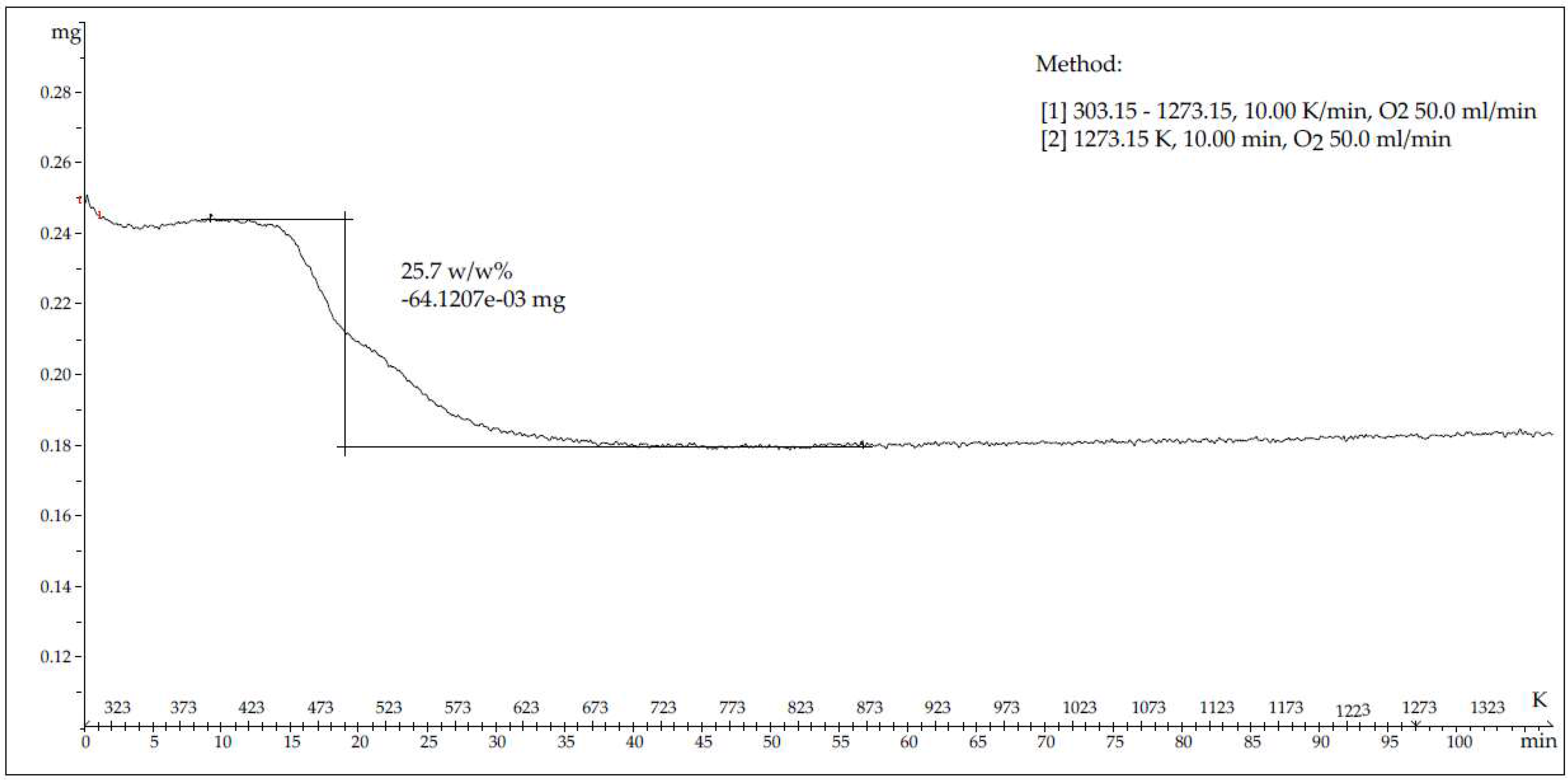
Abstract
:1. Introduction
2. Materials and Methods
2.1. Materials
2.2. Characterization Techniques
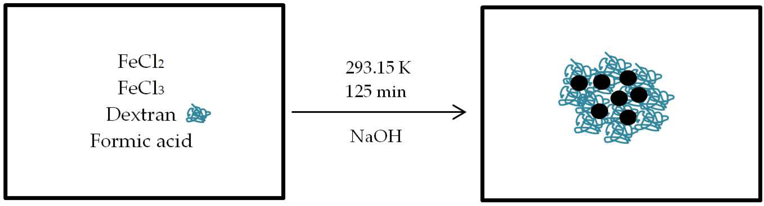
2.3. Water-Based Sythesis of Multicore Superparamagnetic Iron Oxide Nanoparticles (MC-SPIONs)
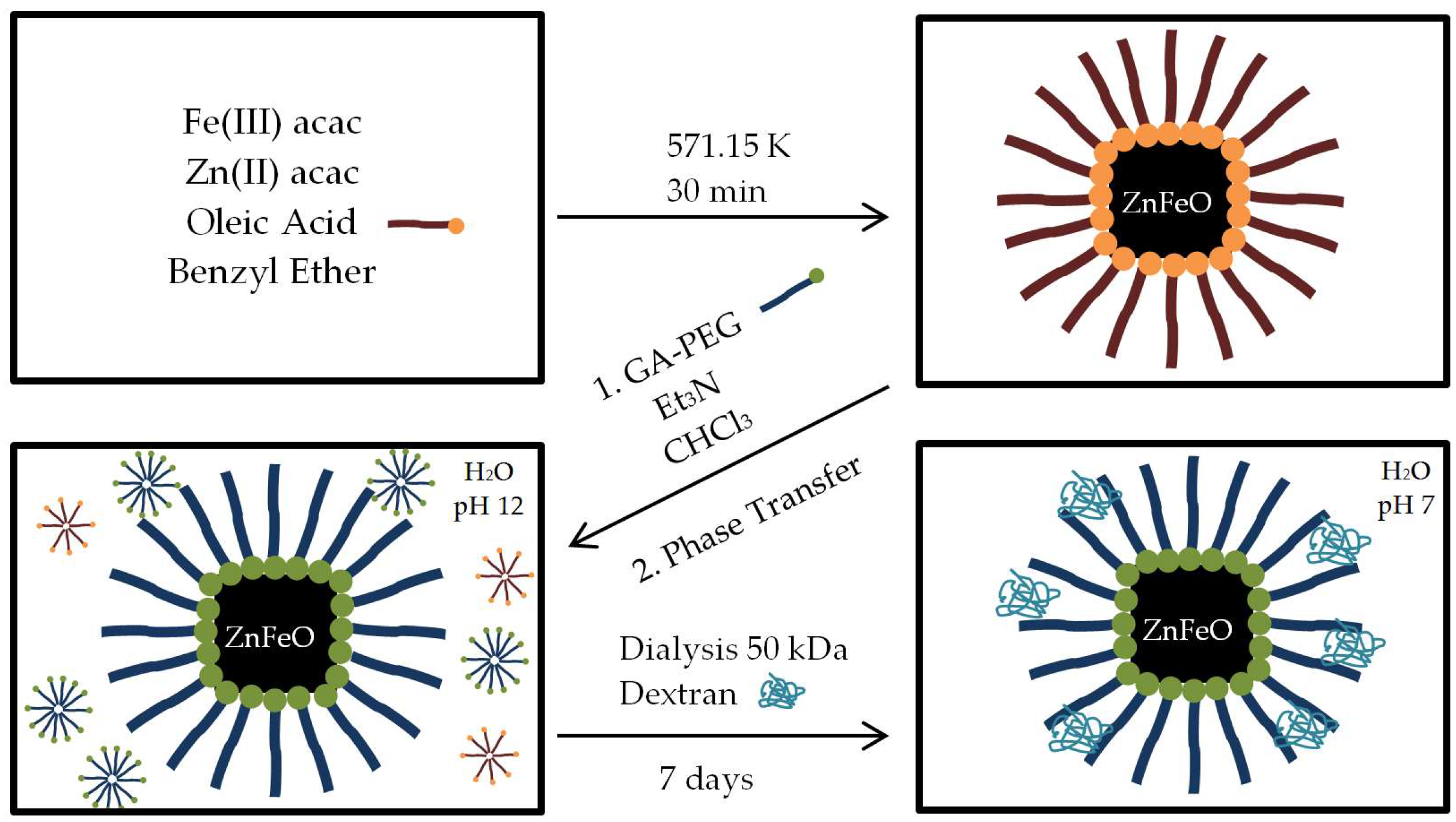
2.4. Organic Synthesis of Coated Single-Core Superparamagnetic Iron Oxide Nanoparticles Doped with Zinc (SC-SPIONs)
2.4.1. Organic Synthesis of Nanoparticles
2.4.2. Gallic Acid–PEG Ligand Synthesis
2.4.3. Phase Transfer and Purification of Particles
3. Results and Discussion
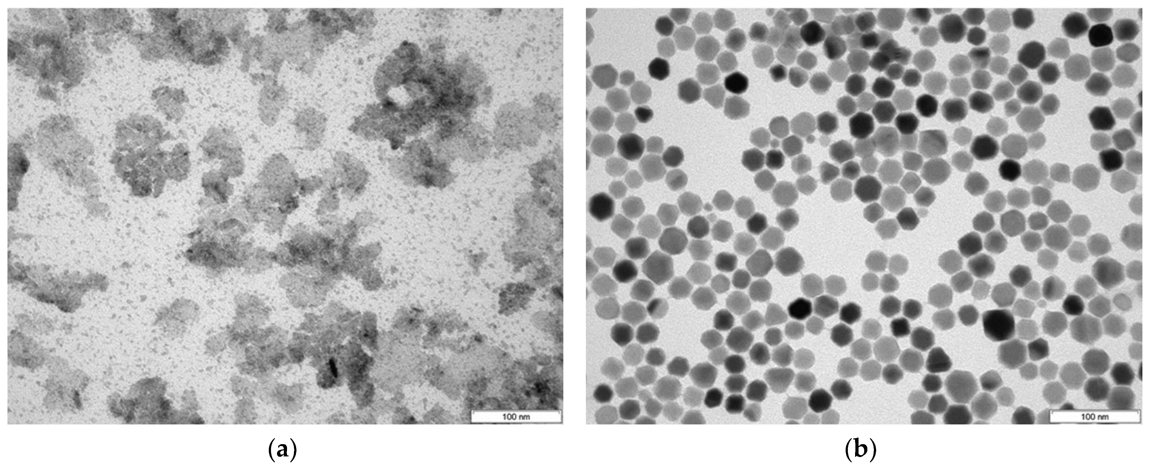
3.1. Nanoparticles Synthesis
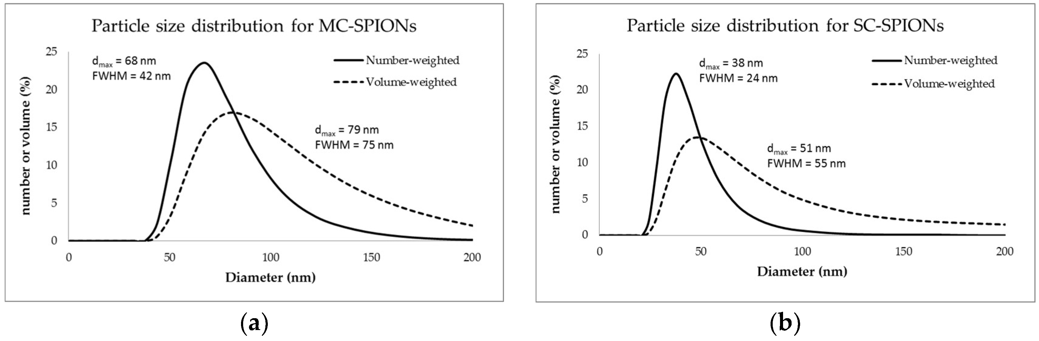
3.2. Particle Size
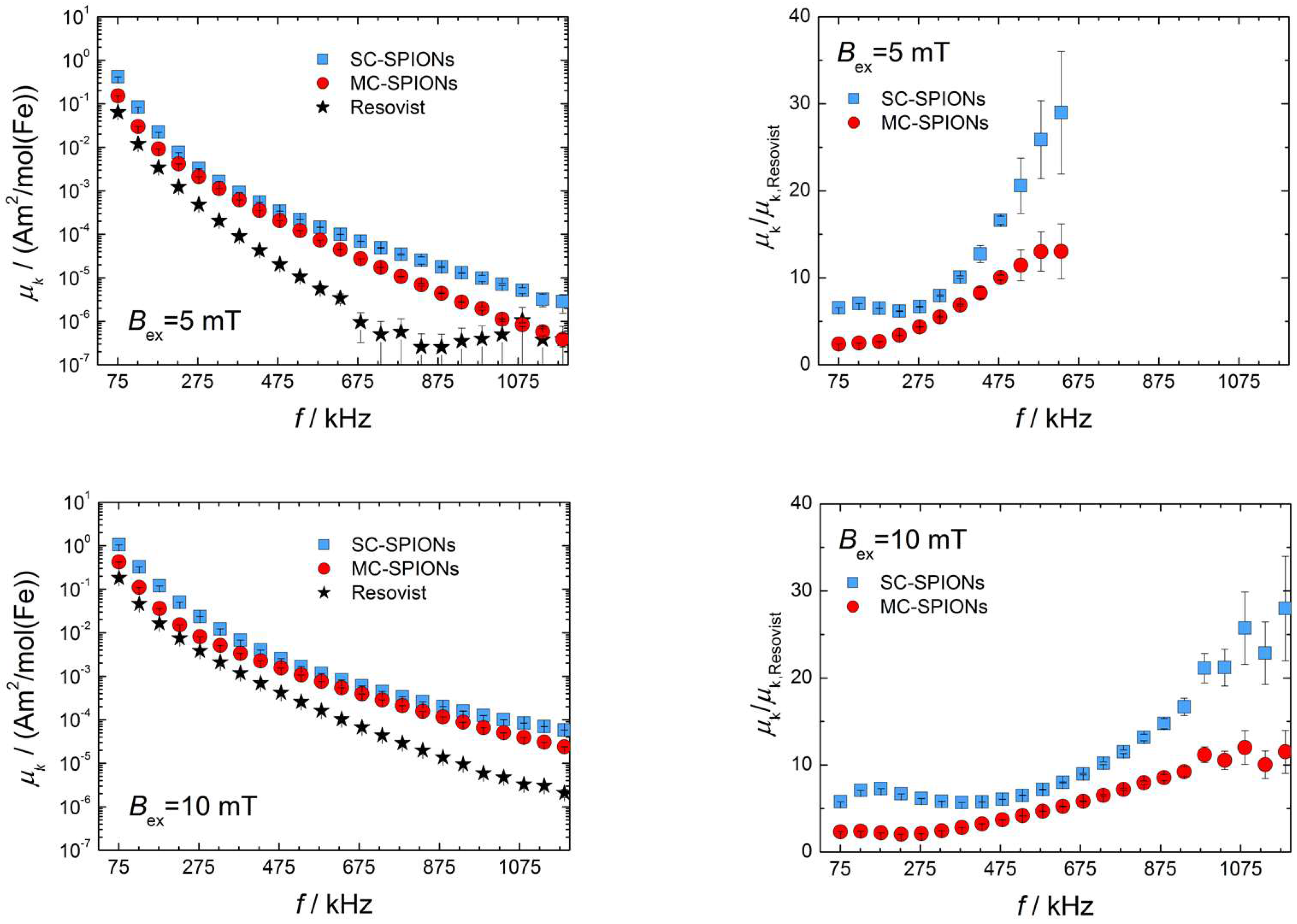
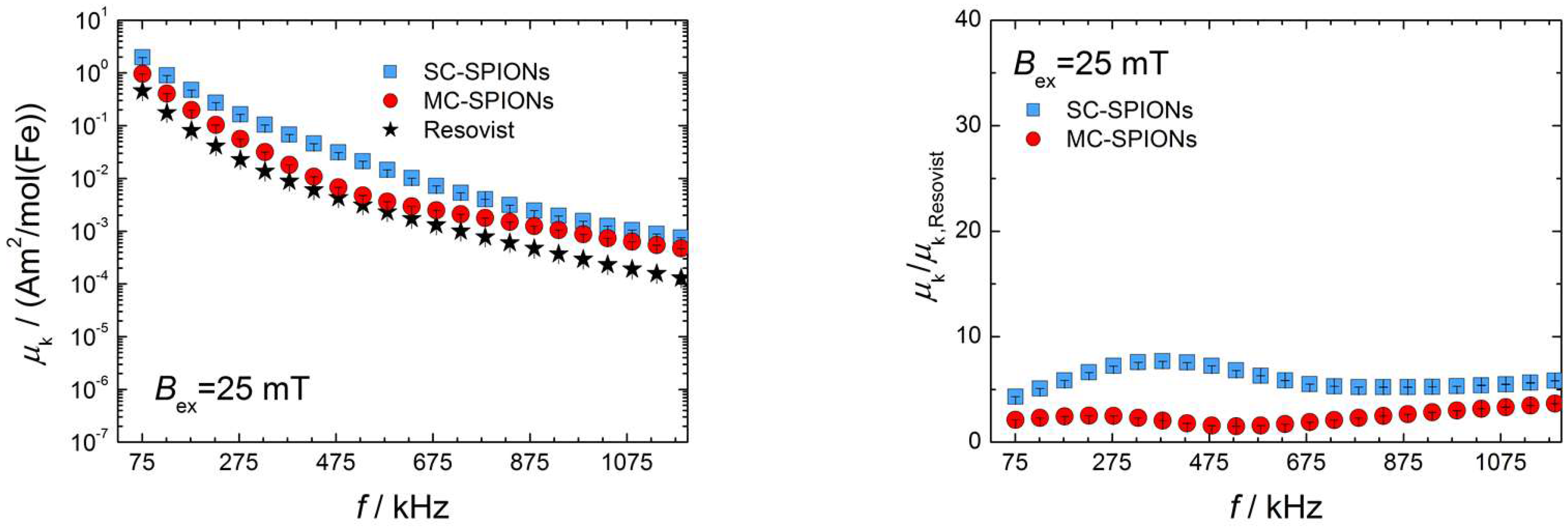
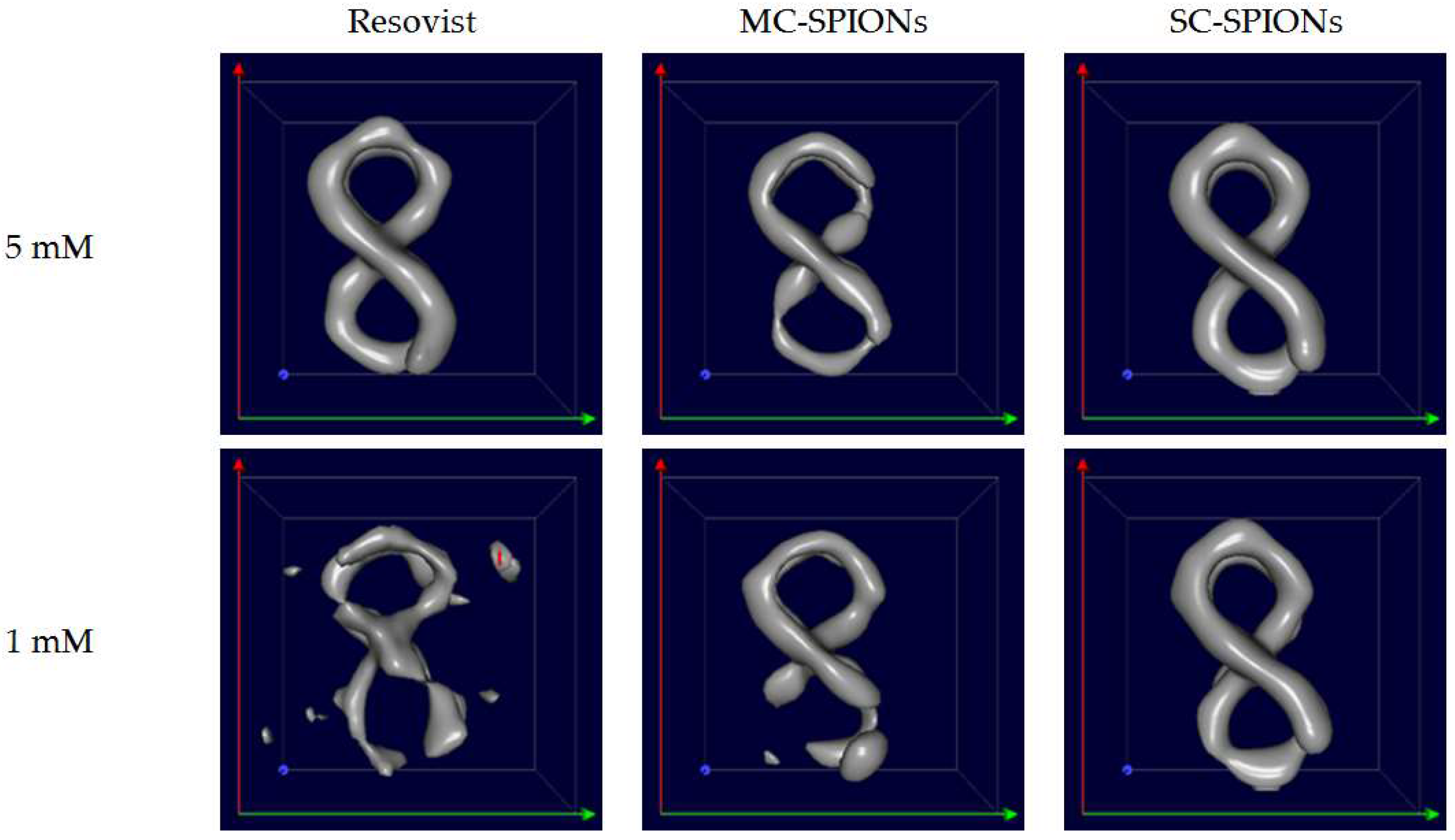
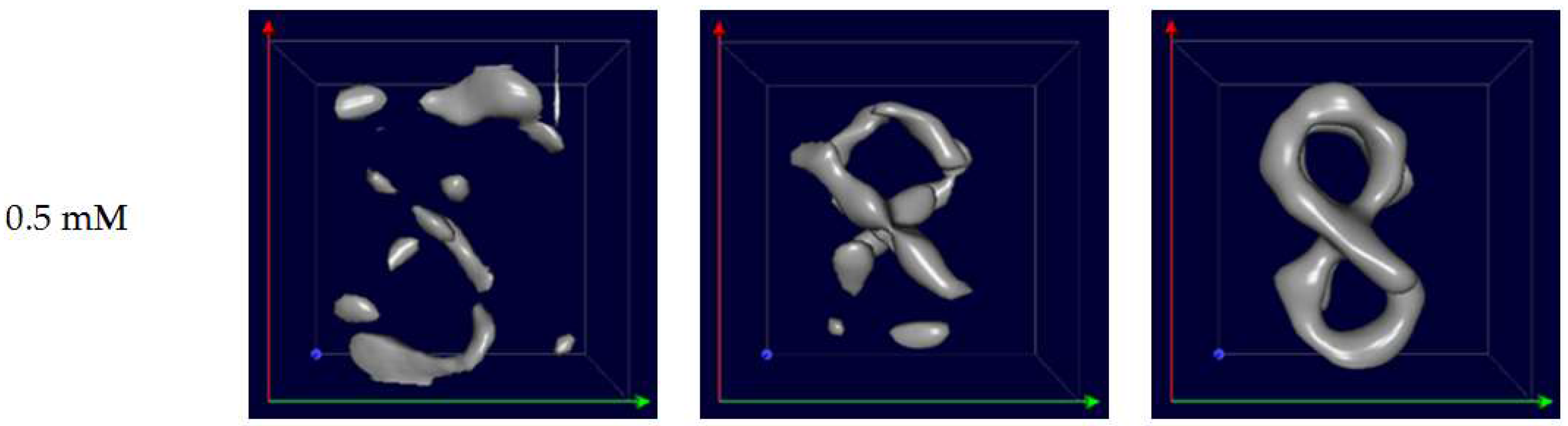
3.3. MPS Analysis and MPI Imaging
4. Conclusions
Acknowledgments
Author Contributions
Conflicts of Interest
Appendix A

References
- Gleich, B.; Weizenecker, J. Tomographic imaging using the nonlinear response of magnetic particles. Nature 2005, 435, 1214–1217. [Google Scholar] [CrossRef] [PubMed]
- Wells, J.; Paysen, H.; Kosch, O.; Loewa, N.; Schmitzberger, F.; Makowski, M.; Franke, J.; Trahms, L.; Wiekhorst, F. Characterizing a preclinical magnetic particle imaging system with separate pick-up coil. IEEE Trans. Magn. 2017, 53, 1–5. [Google Scholar] [CrossRef]
- Panagiotopoulos, N.; Duschka, R.L.; Ahlborg, M.; Bringout, G.; Debbeler, C.; Graeser, M.; Kaethner, C.; Ludtke-Buzug, K.; Medimagh, H.; Stelzner, J.; et al. Magnetic particle imaging: Current developments and future directions. Int. J. Nanomed. 2015, 10, 3097–3114. [Google Scholar] [CrossRef] [PubMed]
- Arami, H.; Teeman, E.; Troksa, A.; Bradshaw, H.; Saatchi, K.; Tomitaka, A.; Gambhir, S.S.; Häfeli, U.O.; Liggitt, D.; Krishnan, K.M. Tomographic magnetic particle imaging of cancer targeted nanoparticles. Nanoscale 2017, 9, 18723–18730. [Google Scholar] [CrossRef] [PubMed]
- Them, K.; Salamon, J.; Szwargulski, P.; Sequeira, S.; Kaul, M.; Lange, C.; Ittrich, H.; Knopp, T. Increasing the sensitivity for stem cell monitoring in system-function based magnetic particle imaging. Phys. Med. Biol. 2016, 61, 3279–3290. [Google Scholar] [CrossRef] [PubMed]
- Rahmer, J.; Halkola, A.; Gleich, B.; Schmale, I.; Borgert, J. First experimental evidence of the feasibility of multi-color magnetic particle imaging. Phys. Med. Biol. 2015, 60, 1775–1791. [Google Scholar] [CrossRef] [PubMed]
- Haegele, J.; Vaalma, S.; Panagiotopoulos, N.; Barkhausen, J.; Vogt, F.M.; Borgert, J.; Rahmer, J. Multi-color magnetic particle imaging for cardiovascular interventions. Phys. Med. Biol. 2016, 61, N415–N426. [Google Scholar] [CrossRef] [PubMed]
- Khandhar, A.P.; Keselman, P.; Kemp, S.J.; Ferguson, R.M.; Goodwill, P.W.; Conolly, S.M.; Krishnan, K.M. Evaluation of PEG-coated iron oxide nanoparticles as blood pool tracers for preclinical magnetic particle imaging. Nanoscale 2017, 9, 1299–1306. [Google Scholar] [CrossRef] [PubMed]
- Borgert, J.; Schmidt, J.D.; Schmale, I.; Bontus, C.; Gleich, B.; David, B.; Weizenecker, J.; Jockram, J.; Lauruschkat, C.; Mende, O.; et al. Perspectives on clinical magnetic particle imaging. Biomed. Tech. 2013, 58, 551–556. [Google Scholar] [CrossRef] [PubMed]
- Eberbeck, D.; Wiekhorst, F.; Wagner, S.; Trahms, L. How the size distribution of magnetic nanoparticles determines their magnetic particle imaging performance. Appl. Phys. Lett. 2011, 98, 182502-1-3. [Google Scholar] [CrossRef]
- Ludwig, F.; Eberbeck, D.; Lowa, N.; Steinhoff, U.; Wawrzik, T.; Schilling, M.; Trahms, L. Characterization of magnetic nanoparticle systems with respect to their magnetic particle imaging performance. Biomed. Tech. 2013, 58, 535–545. [Google Scholar] [CrossRef] [PubMed]
- Löwa, N.; Knappe, P.; Wiekhorst, F.; Eberbeck, D.; Thünemann, A.F.; Trahms, L. Hydrodynamic and magnetic fractionation of superparamagnetic nanoparticles for magnetic particle imaging. J. Magn. Magn. Mater. 2015, 380, 266–270. [Google Scholar] [CrossRef]
- Yoshida, T.; Othman, N.B.; Enpuku, K. Characterization of magnetically fractionated magnetic nanoparticles for magnetic particle imaging. J. Appl. Phys. 2013, 114, 173908-1-7. [Google Scholar] [CrossRef]
- Arami, H.; Ferguson, R.M.; Khandhar, A.P.; Krishnan, K.M. Size-dependent ferrohydrodynamic relaxometry of magnetic particle imaging tracers in different environments. Med. Phys. 2013, 40, 071904-1-14. [Google Scholar] [CrossRef] [PubMed]
- Deissler, R.J.; Wu, Y.; Martens, M.A. Dependence of Brownian and Neel relaxation times on magnetic field strength. Med. Phys. 2014, 41, 012301-1-12. [Google Scholar] [CrossRef] [PubMed]
- Ferguson, R.M.; Minard, K.R.; Khandhar, A.P.; Krishnan, K.M. Optimizing magnetite nanoparticles for mass sensitivity in magnetic particle imaging. Med. Phys. 2011, 38, 1619–1626. [Google Scholar] [CrossRef] [PubMed]
- Ferguson, R.M.; Khandhar, A.P.; Krishnan, K.M. Tracer design for magnetic particle imaging (invited). J. Appl. Phys. 2012, 111, 07B318-1-5. [Google Scholar] [CrossRef] [PubMed]
- Weizenecker, J.; Gleich, B.; Rahmer, J.; Borgert, J. Micro-magnetic simulation study on the magnetic particle imaging performance of anisotropic mono-domain particles. Phys. Med. Biol. 2012, 57, 7317–7327. [Google Scholar] [CrossRef] [PubMed]
- Bauer, L.M.; Situ, S.F.; Griswold, M.A.; Samia, A.C.S. Magnetic Particle Imaging Tracers: State-of-the-Art and Future Directions. J. Phys. Chem. Lett. 2015, 6, 2509–2517. [Google Scholar] [CrossRef] [PubMed]
- Eberbeck, D.; Dennis, C.L.; Huls, N.F.; Krycka, K.L.; Gruttner, C.; Westphal, F. Multicore Magnetic Nanoparticles for Magnetic Particle Imaging. IEEE Trans. Magn. 2013, 49, 269–274. [Google Scholar] [CrossRef]
- Gutiérrez, L.; Costo, R.; Grüttner, C.; Westphal, F.; Gehrke, N.; Heinke, D.; Fornara, A.; Pankhurst, Q.; Johansson, C.; Veintemillas-Verdaguer, S. Synthesis methods to prepare single-and multi-core iron oxide nanoparticles for biomedical applications. Dalton Trans. 2015, 44, 2943–2952. [Google Scholar] [CrossRef] [PubMed]
- Ferguson, R.M.; Khandhar, A.P.; Kemp, S.J.; Arami, H.; Saritas, E.U.; Croft, L.R.; Konkle, J.; Goodwill, P.W.; Halkola, A.; Rahmer, J.; et al. Magnetic particle imaging with tailored iron oxide nanoparticle tracers. IEEE Trans. Med. Imaging 2015, 34, 1077–1084. [Google Scholar] [CrossRef] [PubMed]
- Biederer, S.; Knopp, T.; Sattel, T.F.; Lüdtke-Buzug, K.; Gleich, B.; Weizenecker, J.; Borgert, J.; Buzug, T.M. Magnetization response spectroscopy of superparamagnetic nanoparticles for magnetic particle imaging. J. Phys. D Appl. Phys. 2009, 42, 205007. [Google Scholar] [CrossRef]
- Starmans, L.W.; Burdinski, D.; Haex, N.P.; Moonen, R.P.; Strijkers, G.J.; Nicolay, K.; Grull, H. Iron oxide nanoparticle-micelles (ION-micelles) for sensitive (molecular) magnetic particle imaging and magnetic resonance imaging. PLoS ONE 2013, 8, e57335-1-9. [Google Scholar] [CrossRef] [PubMed]
- Kraupner, A.; Eberbeck, D.; Heinke, D.; Uebe, R.; Schuler, D.; Briel, A. Bacterial magnetosomes—Nature’s powerful contribution to MPI tracer research. Nanoscale 2017, 9, 5788–5793. [Google Scholar] [CrossRef] [PubMed]
- Tay, Z.W.; Hensley, D.W.; Vreeland, E.C.; Zheng, B.; Conolly, S.M. The relaxation wall: Experimental limits to improving MPI spatial resolution by increasing nanoparticle core size. Biomed. Phys. Eng. Express 2017, 3, 035003. [Google Scholar] [CrossRef] [PubMed]
- Rahmer, J.; Weizenecker, J.; Gleich, B.; Borgert, J. Signal encoding in magnetic particle imaging: Properties of the system function. BMC Med. Imaging 2009, 9, 4. [Google Scholar] [CrossRef] [PubMed]
- Kaczmarz, S. Angenaherte auflosung von systemen linearer gleichungen. Bull. Int. Acad. Sci. Pol. A 1937, 35, 355–357. [Google Scholar]
- Noh, S.H.; Na, W.; Jang, J.T.; Lee, J.H.; Lee, E.J.; Moon, S.H.; Lim, Y.; Shin, J.S.; Cheon, J. Nanoscale magnetism control via surface and exchange anisotropy for optimized ferrimagnetic hysteresis. Nano Lett. 2012, 12, 3716–3721. [Google Scholar] [CrossRef] [PubMed]
- Zalipsky, S.; Gilon, C.; Zilkha, A. Esterification of Polyethylene Glycols. J. Macromol. Sci. Part A Chem. 1984, 21, 839–845. [Google Scholar] [CrossRef]
- Guardia, P.; Riedinger, A.; Nitti, S.; Pugliese, G.; Marras, S.; Genovese, A.; Materia, M.E.; Lefevre, C.; Manna, L.; Pellegrino, T. One pot synthesis of monodisperse water soluble iron oxide nanocrystals with high values of the specific absorption rate. J. Mater. Chem. B 2014, 2, 4426–4434. [Google Scholar] [CrossRef]
- Löwa, N.; Radon, P.; Kosch, O.; Wiekhorst, F. Concentration dependent MPI tracer performance. Int. J. Magn. Part. Imaging 2016, 2, 1601001-1-5. [Google Scholar]
- Kim, D.; Lee, N.; Park, M.; Kim, B.H.; An, K.; Hyeon, T. Synthesis of uniform ferrimagnetic magnetite nanocubes. J. Am. Chem. Soc. 2008, 131, 454–455. [Google Scholar] [CrossRef] [PubMed]
- Lee, N.; Hyeon, T. Designed synthesis of uniformly sized iron oxide nanoparticles for efficient magnetic resonance imaging contrast agents. Chem. Soc. Rev. 2012, 41, 2575–2589. [Google Scholar] [CrossRef] [PubMed]
- Otsuka, H.; Nagasaki, Y.; Kataoka, K. PEGylated nanoparticles for biological and pharmaceutical applications. Adv. Drug Deliv. Rev. 2012, 64, 246–255. [Google Scholar] [CrossRef]
- Araujo, P.Z.; Morando, P.J.; Blesa, M.A. Interaction of catechol and gallic acid with titanium dioxide in aqueous suspensions. 1. Equilibrium studies. Langmuir 2005, 21, 3470–3474. [Google Scholar] [CrossRef] [PubMed]
- Davis, K.; Qi, B.; Witmer, M.; Kitchens, C.L.; Powell, B.A.; Mefford, O.T. Quantitative measurement of ligand exchange on iron oxides via radiolabeled oleic acid. Langmuir 2014, 30, 10918–10925. [Google Scholar] [CrossRef] [PubMed]
- Chen, D.-X.; Sun, N.; Gu, H.-C. Size analysis of carboxydextran coated superparamagnetic iron oxide particles used as contrast agents of magnetic resonance imaging. J. Appl. Phys. 2009, 106, 063906-1-9. [Google Scholar] [CrossRef]
- Dhavalikar, R.; Maldonado-Camargo, L.; Garraud, N.; Rinaldi, C. Ferrohydrodynamic modeling of magnetic nanoparticle harmonic spectra for magnetic particle imaging. J. Appl. Phys. 2015, 118, 173906-1-8. [Google Scholar] [CrossRef] [PubMed]


© 2018 by the authors. Licensee MDPI, Basel, Switzerland. This article is an open access article distributed under the terms and conditions of the Creative Commons Attribution (CC BY) license (http://creativecommons.org/licenses/by/4.0/).
Share and Cite
Ziemian, S.; Löwa, N.; Kosch, O.; Bajj, D.; Wiekhorst, F.; Schütz, G. Optimization of Iron Oxide Tracer Synthesis for Magnetic Particle Imaging. Nanomaterials 2018, 8, 180. https://doi.org/10.3390/nano8040180
Ziemian S, Löwa N, Kosch O, Bajj D, Wiekhorst F, Schütz G. Optimization of Iron Oxide Tracer Synthesis for Magnetic Particle Imaging. Nanomaterials. 2018; 8(4):180. https://doi.org/10.3390/nano8040180
Chicago/Turabian StyleZiemian, Sabina, Norbert Löwa, Olaf Kosch, Daniel Bajj, Frank Wiekhorst, and Gunnar Schütz. 2018. "Optimization of Iron Oxide Tracer Synthesis for Magnetic Particle Imaging" Nanomaterials 8, no. 4: 180. https://doi.org/10.3390/nano8040180