Applications of Nanosheets in Frontier Cellular Research


1
Micro/Nano Technology Center, Tokai University, 4-1-1 Kitakaname, Hiratsuka-city, Kanagawa 259-1292, Japan
2
Department of Bioengineering, School of Engineering, The University of Tokyo, 7-3-1 Hongo, Bunkyo-ku, Tokyo 113-8656, Japan
3
Department of Mechanical Engineering, Tokai University, 4-1-1 Kitakaname, Hiratsuka-city, Kanagawa 259-1292, Japan
4
Division of Biomedical Engineering, Renal Division, Department of Medicine, Brigham and Women’s Hospital, Harvard Medical School, Cambridge, MA 02139, USA
*
Authors to whom correspondence should be addressed.
†
These authors contributed equally to this work.
Nanomaterials 2018, 8(7), 519; https://doi.org/10.3390/nano8070519
Submission received: 28 May 2018
/
Revised: 5 July 2018
/
Accepted: 10 July 2018
/
Published: 12 July 2018
Abstract
:Several types of nanosheets, such as graphene oxide (GO) nanosheet, molybdenum disulfide (MoS2) and poly(l-lactic acid) (PLLA) nanosheets, have been developed and applied in vitro in cellular research over the past decade. Scientists have used nanosheet properties, such as ease of modification and flexibility, to develop new cell/protein sensing/imaging techniques and achieve regulation of specific cell functions. This review is divided into three main parts based on the application being examined: nanosheets as a substrate, nanosheets as a sensitive surface, and nanosheets in regenerative medicine. Furthermore, the applications of nanosheets are discussed, with two subsections in each section, based on their effects on cells and molecules. Finally, the application prospects of nanosheets in cellular research are summarized.
1. Introduction
To date, many fabrication technologies have been developed for different types of nanosheets, such as graphene oxide (GO) nanosheet, molybdenum disulfide (MoS2), and poly(l-lactic acid) (PLLA) nanosheets [1,2,3,4]. Shortly after the development of the fabrication technology, researchers applied nanosheets to cellular research. Monocrystalline graphitic films a few atoms thick were first reported in 2004, and MoS2 nanosheets were fabricated successfully in 2011 (Liu et al., 2008) [2]. Furthermore, NO nanosheets were applied as carriers of SN38 in 2008—a drug with high cancer cell killing potency—and MoS2 nanosheets were used for the detection of DNA and small molecules due to their fluorescence-quenching ability and DNA affinity [5,6,7]. Fujie et al. developed fabrication techniques for PLLA nanosheets in 2007, and they investigated the cell adhesion properties of the sheets in 2011 [3,8]. As is known, most nanosheets are transparent and have excellent electrical and thermal properties with a large size-aspect ratio (>106). Therefore, the application of nanosheets in relation to cellular research is due to the following properties. (1) The nanosheet surface can be modified (biofunctionalized) relatively easy for applications in cellular research [9,10,11]. (2) Cells or molecules show functional responses after the interaction with materials of nanosheets [12,13,14]. (3) Nanosheets are flexible, and cells adhered to the sheets are transplanted without cell detachment [15]. Further, fragments of nanosheets can be endocytosed by cells [16,17].
Besides GO, MoS2, and PLLA, many other nanosheets or two-dimensional (2D) materials have been newly developed. For example, in 2017, π-conjugated 2D porous organic nanosheets called NUS-24 were developed, and they showed high sensitivity toward Fe3+ ions and nitro-containing compounds, although their application in cellular research has not been realized [18]. On the other hand, many of the 2D graphene analogues, including π-conjugated 2D porous organic nanosheets, 2D graphene analogues (such as hexagonal boron nitride (h-BN), carbon nitride, and transition metal di-chalcogenides), have been developed, and some of them were applied in cellular research [19,20,21]. Analogues, such as h-BN and monolayer 2D graphitic carbon nitride (g-C3N4) nanosheets, were fabricated and used for cell imaging [19,22]. Further, there are techniques to prepare nanosheets on titanium surfaces for applications in biomedical research [13,23,24]. Therefore, nanosheets made from various types of materials have been used in cellular research, which makes their application extensive.
Diverse research on the application of nanosheets promotes the development of new technologies for disease diagnosis/therapy and regenerative medicine at cellular levels [25,26,27,28,29]. This review focuses on the research of cell capturing, regulation of cell adhesion and function, protein delivery into cells/transfection, cellular-level sensing/imaging, and regeneration regulation at cell levels using nanosheets.
2. Nanosheets as a Substrate
2.1. Effects of Nanosheets on Cell Adhesion and Function
Cell adhesion to nanosheets with various functions was investigated for further applications. Cells adhere to the extracellular matrix (ECM) through transmembrane proteins called integrins, which are a type of focal adhesion protein that serve as the mechanical linkages to the ECM. Substrate conditions, such as coating of extracellular matrix, substrate stiffness, and substrate topology, are important for cell adhesion and growth. A study by Okada et al. suggested that hydroxyapatite (HAp) nanosheet substrates with contact domains larger than 100 nm were required for the stable adhesion of rat bone marrow-derived mesenchymal stem cells [30]. Da Silva et al. developed an ultrathin flat sheet with controlled wettability using the spontaneous assembly of a peptide bolaamphiphile called RFL4FR (R, arginine; F, phenylalanine; L, leucine). Growth of human corneal stromal fibroblast (hCSF) cells was successful on this type of nanosheet, due to the wettability of the bolaamphiphile peptides resulting from hydrophilic peptide groups at both ends of a long hydrophobic hydrocarbon chain or peptide sequence [31]. The adhesion properties of nanosheets can be modified by the coating of ECM, and the coating method may influence the cell adhesion properties. Poly-l-lactic acid (PLLA) nanosheets possess anti-adhesive properties and can prevent unwanted wound adhesion, therefore, the nanosheets can be used as wound dressing. Niwa et al. showed that spin-coating collagen on the PLLA nanosheet promotes the adhesion of murine fibroblast cell line NIH3T3 [32]. Cell adhesion is also dependent on the stiffness of the underlying substrate: when rat cardiomyocytes were cultured on PLLA nanosheets placed on flat SiO2 substrate and metal meshes, cells showed almost equal distribution on the substrate but adhered specifically to the parts along the metal meshes (Figure 1) [8]. In addition, some novel types of nanosheets are developed specifically for cell culture and tissue engineering. Laurenti et al. developed a magnesium phosphate nanosheet-based thixotropic gel, and osteoblast cells could adhere onto the 2D structures and form colonies [33]. Functionalized nanosheets were also synthesized onto traditional medical materials for improving cell culture. For example, when calcium-containing nanosheets were synthesized onto a sandblasted and acid-etched (SLA) titanium surface, a sustained release of Ca2+ ions was measured, and adhesion/spreading of MC3T3-E1 cells was upregulated [34]. Coating of flower-like calcium phosphate nanosheets onto a titanium surface can also promote cell adhesion and spreading due to its wettability [35].
The adhesion of HaCaT cells—a spontaneously transformed aneuploidy immortal keratinocyte cell line—was realized on the surface of low-viscosity liquids (for example, a fluorinated oil (Novec 7500, 0.77 cSt) containing the surfactant) by the self-assembly of mechanically strong protein nanosheets on a liquid surface (Figure 2) [36]. Cell adhesion and spreading are critical for various cell functions, and enhancing the adhesion of rat bone marrow cells by culturing on nanosheets of titanium alloys resulted in increased osteogenic differentiation [13]. Nanosheets, such as graphene oxide (GO) nanosheets, are used extensively in living systems. Even so, we cannot ignore some toxic effects of nanosheets on cells. Although this type of nanosheet is not cytotoxic, RBL-2H3 cells cultured on the nanosheet may “feel” environment stress, and shedding of plasma membranes (e.g., detection of membrane fragments) was confirmed [37]. Further, disruption of the cell cytoskeleton was observed in A549 lung carcinoma cells cultured on GO nanosheets [38].
Pan et al. fabricated positively or negatively charged palladium nanosheets by treating the sheets with short chain thiolated carboxylic acids or amines, and they reported that cytotoxic effects may depend on the surface charge. 80% of HepG2 cells died on the positively charged nanosheets, however, only 30% died on the negatively charge nanosheets [39]. Molybdenum disulfide (MoS2) nanosheets were also reported to disrupt structures of α–helical peptides, a common motif in the secondary structure of proteins [40]. On the other hand, scientists are making great efforts to reduce the cytotoxicity. Sasidharan et al. reported that cell apoptosis was observed on as-synthesized graphene because of high oxidative stress, but no toxicity was observed on carboxyl hydrophilic graphene [41]. Likewise, graphene modified with biocompatible polyethylene glycol (PEG) did not show acute toxicity [11,42]. PEGylation decreased the affinity of graphene-based nanomaterials, so, coating nanomaterials with PEG chains was reported as a method to reduce toxicity [43,44]. However, we note that Luo et al. demonstrated that PEGylation could not bypass cytotoxicity, because interactions between PEGylated GO and the membranes of macrophage cells promoted high levels of cytokine secretion through integrin-mediated signaling pathways [45]. As an alternative, GO was functionalized with poly(acrylic acid), and the in vitro and in vivo biocompatibility was found to be superior to PEGylation. The differences in biocompatibility may result from differential compositions of protein corona formed on the surface of GO, because the composition may influence their interaction with cell membrane and cellular uptake [46].
2.2. Delivery Functions Acted on by Nanosheets
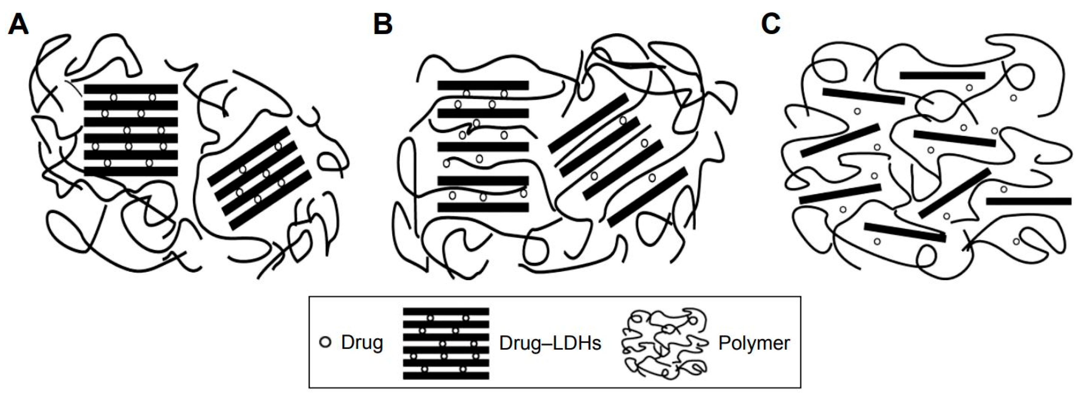
Various types of nanosheets provide flexible platforms for cell culturing, and the surface-modified nanosheets exhibit excellent drug- or DNA-binding properties. Therefore, nanosheets are naturally used as tools in the delivery of drugs, cell monolayers, and genes. Fujie et al. developed a micropatterned and biodegradable poly(lactic-co-glycolic acid) (PLGA) nanosheet for the delivery of engineered epithelial monolayers into the narrow subretinal space by a simple injection through a syringe needle [25]. Nanosheets with adhered retinal pigmented epithelial cells can withstand deformation in a syringe needle without heavily damaging the cells [25]. This method is impossible for conventional delivery methods, which have used collagen, poly(ethylene terephthalate) (PET), or poly(methyl methacrylate) (PMMA) with sizes of several micrometers to several millimeters as delivery substrates [47,48,49]. Various structures developed from nanosheets were applied as drug carriers. For example, chitosan-glutathione-valine-valine layered double hydroxides (LDHs) were used to deliver drug to human corneal epithelial primary cells and retinal pigment epithelial cells, and nanosheets with drug (pirenoxine sodium) were uptaken through a cell membrane transporter, peptide transporter-1 (Figure 3) [50]. GO nanosheets were modified by natural peptide protamine sulfate and sodium alginate, and self-assembly of the nanosheets was developed as a drug delivery system. The system possessed properties such as a pH-sensitive release of anticancer drugs and suppression of protein adhesion. Further, the modified GO nanosheets could be uptaken by MCF-7 cells (breast cancer cells) [51]. Delaminated Mg-Al-lactate layered double hydroxide nanosheets were used as vectors to deliver salmon DNA to 293T cells, which was facilitated by the sheets’ high DNA adsorption capacity and ability to be uptaken by cells [10]. Black phosphorus (BP) nanosheets were modified with polyethylene glycol amine to improve the loading capacity of theranostic agents such as doxorubicin (DOX) for chemotherapy and cyanine7 (Cy7) for in vivo near-infrared (NIR) imaging [52]. Greek Testudo-like structures were developed from two-dimensional MoS2 nanosheets with DNA oligonucleotides, and the structures responded to heightened ATP metabolism in cancer cells and to released drugs [53].
Conventional transfection processes for cell transfection or gene therapy are complicated, toxic, and less effective, therefore, nanosheets were used as advanced tools for nonviral-based gene delivery substrates. When GO nanosheets formed a complex with cell-penetrating peptides (CPPs), the cytotoxicity of CPPs was mitigated and the transfection efficiency of CPPs with oligonucleotides was highly improved [54]. Positively charged MoS2-polyethylene glycol-polyethylenimine nanosheets were loaded with small interfering RNA and added to the cell (HepG2 cell line) culture medium for cell transfection [16]. Unlike the method of adding nanosheets to the cell culture medium (solution-based transfection), Ji et al. developed a silica nanosheet to immobilize the DNA complex, and transfected the cells attached to the nanosheet. The transfection efficiency was approximately 20% higher than that of solution-based transfection [55]. Further, naked DNA on silica glass without any vectors was transferred into human mesenchymal stem cells, a difficult-to-transfect cell type [56].
3. Nanosheets as a Sensitive Surface
3.1. Cell Capturing Using Surface-Modified Nanosheets
Several technologies have been developed for the capturing of circulating tumor cells, such as microfluidic chips and functionalized and structured medical wire [57,58,59,60,61]. Even so, nanosheets together with other nanomaterials have emerged as effective tools for the isolation of tumor cells [62]. Surface modification of graphene oxide (GO) nanosheets can be easily realized, and GO nanosheets coated with specific antibodies were used as tools for cell capturing. GO nanosheets are carbon-based materials with a benzene basal structure [1]. The structure can interact with the aromatic functional groups of most biomolecules of interest [63]. Since a cell capturing system using three-dimensional microfluidic devices limits cell spreading and further observation on the chip after capturing, Yoon et al. developed GO nanosheet platforms absorbed onto arrayed flat (two-dimensional) gold patterns. The nanosheets were coated with NeutrAvidin, and two human breast cancer cell lines (MCF-7 and Hs-578T) and a human prostate cancer cell line (PC-3) were captured by the interactions between NeutrAvidin and biotinylated EpCAM antibody [64]. In the study by Chen et al., GO nanosheets were coated with single-domain antibody fragments for two different types of white blood cells [65]. Instead of using as-synthesized GO nanosheets, Bardhan et al. improved the nanosheets’ function through changing the distribution of oxygen functional groups before coating with an antibody fragment (VHH7), and Class-II MHC-positive cells from murine whole blood were captured quickly and effectively (Figure 4) [63].
3.2. Cell Sensing and Imaging
Nanosheets are used extensively in the fields of sensing and imaging related to cellular research [9,10,22,26,66,67,68,69,70,71]. Cell-signaling sensing using nanosheets will shed new light on essential cell/protein functions. For example, hydrogen peroxide (H2O2) signaling in cells is important, because it is involved in diseases such as Alzheimer’s, Parkinson’s, and cancer [72,73,74,75]. MoS2 nanosheets enable the sensing of cell H2O2 signaling due to their edge sites displaying electrochemical activities [76]. Based on this understanding, Tang et al. developed a three-dimensional nanosheet for the effective sensing of H2O2 in living RAW 264.7 macrophage cells and neurons on tissue-like three-dimensional structures by exposing abundant nanosheet edge sites [77]. Cytokine tumor necrosis factor (TNF)-α in live cells can be detected by a reduced GO decorated with gold nanoparticles and aryldiazonium salts [78]. A manganese dioxide (MnO2) nanosheet-aptamer nanoprobe was fabricated for tumor cell imaging based on a dual-activatable fluorescence/MRI strategy, and the advantages are as follows: (1) Fluorescence signals (aptamers, fluorescein amidate-labeled 7-base DNA) was quenched on the MnO2 nanosheets. After the nanosheets were uptaken by tumor cells, the adsorption of aptamers became weak, and partial fluorescence was recovered; (2) The MnO2 nanosheet outside the tumor cells has low T1- or T2-weighted contrast, and after the nanosheets were uptaken by tumor cells, large amounts of Mn2+ ions were generated for MRI imaging [17]. Similarly, using the fluorescence-quenching ability of GO nanosheets, a platform for the detection of multiple nucleotides was created: the fluorescence was turned “off” by ATP aptamer–FAM and GTP aptamer–Cy5 binding to the GO nanosheet; aptamers were released into cells after the detection of target nucleotides, leading to fluorescence being turned on (fluorescence off/on switch) (Figure 5) [79].
4. Nanosheets in Regenerative Medicine
4.1. Nanosheets as a Scaffold Element
As a scaffold element for tissue regeneration, nanosheets show excellent application features.
Firstly, nanosheets were used to construct three-dimensional structures and endowed the structure with tissue-like anisotropic mechanical properties. A three-dimensional mechanical microenvironment is important for cell differentiation and growth. Liu et al. built up a hydrogel structure with a titanate(IV) nanosheet, and the structure possessed the same anisotropic mechanical properties as articular cartilage. Titanate nanosheets in aqueous colloidal dispersions were exposed to a strong magnetic field, and the nanosheets aligned cofacially. The anisotropic hydrogel structure can withstand compressive forces in the vertical direction, but it is deformable in the horizontal direction (Figure 6) [80]. Nanosheets were used to improve the mechanical properties of scaffolds for cell culturing [81]. For example, Shuai et al. reported that induction of boron nitride nanosheets enhanced the compressive stress of an akermanite scaffold, a promising bioactive material in the field of tissue engineering. In addition, the seeded MG63 osteoblast-like cells and human bone marrow stromal cells adhered and proliferated well [82]. Further, a MoS2-akermanite three-dimensional scaffold for tissue regeneration after the removal of tumor tissues was developed using a three-dimensional printing technique [83].
Secondly, nanosheets are applied as two-dimensional cell sheets. Cell sheets mimic basement membranes. In vivo, the membrane is composed of proteins such as type-IV collagen, laminin, and fibronectin, and has nano-/micropores for cell–cell communication. A porous nanosheets made from a polymer ethyl acetate solution consisting of poly(d,l-lactic acid) and polystyrene. Using the nanosheets, a multilayered structure was created by rolling the nanosheets with adherent C2C12 myoblasts around a cylinder-shaped template [28].
Thirdly, nanosheets are used as a cell carrier for transplantation into a narrow space. As stated by Suzuki et al., nanosheets, such as a self-assembled monolayer (SAM) of thiol molecules, can be used as a cell carrier without changing the cell–cell contacts during the process of transplantation. A problem is that the SAM is adsorbed onto a gold surface, and scientists have to consider the harvesting method. A negative electrical potential was applied to make the nanosheet with cells to disconnect from the underlying gold surface for transplantation [15].
4.2. Nanosheets for Stem Cell Differentiation and Tissue Regeneration
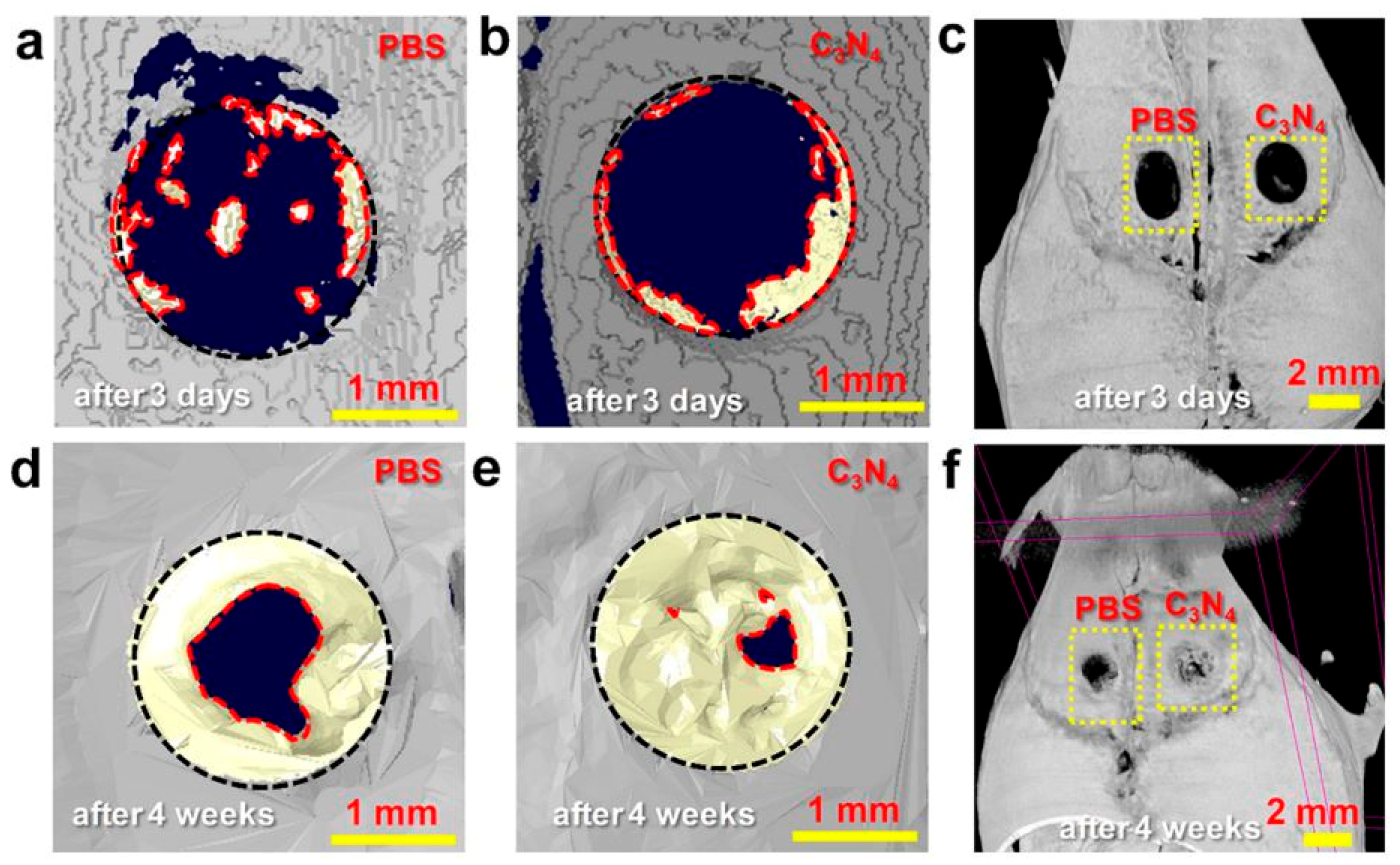
Various types of nanosheets have been used for cell differentiation and tissue regeneration at protein and cellular levels. Nanosheets were used in several studies to control the differentiation of stem cells into the desired cell types (the control of stem cell fate), and these studies were reviewed in an article by Kenry et al. [29]. A specific physical and topographical microenvironment resulting from a nanosheet-based platform may regulate stem cell differentiation [84]. GO nanosheets were reported to sustain the self-renewal of mouse embryonic stem cells via a signaling pathway involving integrin [85]. Graphene can also be used to induce the differentiation of mesenchymal stem cells and neural stem cells [86]. Due to the high affinity of insulin resulting from H-bonding and electrostatic interactions, the culture of bone marrow-derived mesenchymal stem cells (MSCs) on GO nanosheets led to adipogenesis [87]. With regard to cell-based tissue regeneration, carbon nitride (C3N4) nanosheets were used in bone regeneration with the following properties: (1) This carbon nanosheet had a size of less than 200 nm and low toxicity; (2) The nanosheet can disperse in water easily due to structural polarization (polarized C and N atoms) from positive or negative charges. Red light induced a transient photocurrent of C3N4 nanosheets, and the photocurrent led to the translocation of Ca2+ and increase in cytosolic Ca2+. Red light absorption of the sheets resulted in human bone marrow-derived mesenchymal stem cells (Figure 7) [88].
5. Important Issues: Internalization, Distribution, and Cellular Responses
The above-mentioned studies have shown that cells are capable of internalizing nanosheets, and the internalization and cellular distribution is related not only to the cytotoxicity of nanosheets but also to their potential applications. There are several mechanisms of eukaryotic endocytosis (cell transport of exogenous molecules into the cell), such as clathrin-mediated and caveolae-mediated internalization and macropinocytosis, and the endocytotic mechanism “selected” by cells may depend on the size of the exogenous molecules [89]. The internalization mechanism may depend on types of cells and nanosheets, and the distribution of uptaken nanosheets throughout the cell membrane was observed in several organelles. In a study, fluorescein isothiocyanate (FITC)–GO material (PEGylated GO) was added to the culture medium of Sao-2 osteoblasts, MC3T3-E1 preosteoblasts, and RAW 264.7 macrophages seeded on glass coverslips to observe its internalization and distribution. Results showed that internalization of nanosheets depended on the cell type, and FITC–GO molecules were co-localized with F-actin filaments and induced cell-cycle alterations, apoptosis, and oxidative stress [90]. Macropinocytosis was found to be the general mechanism for internalizing FITC-PEG-GOs (ca. 100 nm) for human Saos-2 osteoblasts, human HepG2 hepatocytes, and murine RAW 264.7 macrophages, although there may be additional endocytotic mechanism (e.g., clathrin-dependent mechanisms and phagocytosis) for each cell type [91]. Protein (FITC–BSA)-coated GO nanosheets entered cells after surface adhesion for 30 min and appeared in intracellular vesicles [92]. The internalization processes of few-layer pristine graphene (GR) and monolayer GO flakes by primary cortical neurons were observed in a study by Bramini et al. Exposure to GO, but not GR, reduced the number of excitatory synaptic contacts and altered the Ca2+ dynamics, and a high percentage of GO flakes were found to enter the lysosome [93]. Similarly, a dual-modal stimulated Raman scattering (SRS)/transient absorption (TA) microscopy technique was developed to observe the distribution of MoS2 nanosheets in live HeLa cells, and it was observed that MoS2 localized with lipid-rich structures, such as endosomes and lysosomes [94]. Further, the nanoassembly of GO/poly(sodium-4-styrenesulfonate) (PLL)/PEGylated PLL can cross the cell membrane, after which it localizes in the lysosomal compartment. Moreover, GO and TiO2–GO composites entered A549 cells and localized to the cytoplasm and nucleus [95,96].
As demonstrated by Zhand et al. in a paper discussing the interaction between mammalian cells and graphene materials, the internalization of nanosheets may alter the distribution of intercellular organelles [97]. For example, redistribution of cytoplasmic lactate dehydrogenase (LDH) after exposure to GO and redistribution of mitochondrial cytochrome c in RAW 264.7 cells cultured in a medium with pristine graphene were observed in experiments [98,99]. An interesting observation is that normal and cancer cells may respond differently to nanosheet internalization, and nanosheets have been applied as theranostic materials based on their specific biodistribution and release at the tumor sites and distribution of nanosheets in cancer cells [100,101]. Zhou et al. reported that mitochondrial oxidative phosphorylation (OXPHOS) was impaired in breast cancer cells (MDA-MB-231, MDA-MB-436, and SK-BR-3) by PEG–GO, but OXPHOS in noncancerous cells (MCF-10A) was almost unchanged. A phenomenon like this may be related to the development of a new approach to treating breast cancer [102]. Moreover, compared to normal tissues, tumor tissues have an acidic environment (pH value ranging from 4.5 to 5.0), and MnO2 nanosheets functionalized with hyaluronic acid (HA) were used to deliver cisplatin (cis-diamminedichloroplatinum (CDDP)) to tumor cells in tumor-bearing animals (mice). These nanosheet-based drug carriers were observed to specifically distribute in the cytoplasm of A549 cancer cells after intracellular uptake and enhance apoptosis of tumor cells [103,104]. GO nanosheets may not be effective for the delivery of anticancer drugs, but polyethylene glycol-grafted GO (pGO) combining doxorubicin (DOX) with the photosensitizer chlorin e6 (Ce6) could accumulate in tumor sites over 3 days. This nanosheet-based carrier was distributed in the cytoplasm of SCC7 cells, and disruption of tumor nuclei was observed in tumor sections [105]. αvβ3 integrin was overexpressed on cancer but not normal cells [106]. Using this knowledge, bismuth selenide (Bi2Se3) nanosheets decorated with chitosan and RGD peptide showed the ability to target αvβ3 integrin on cancer cells, and specific biodistribution of nanosheets was observed to be involved in the suppression of cell growth at tumor sites. Uptake of the Bi2Se3-chitosan-RGD nanosheets increased prominently in HeLa (cancer) cells, but not in Etc1/E6E7 (normal) cells, resulting in morphological changes of mitochondria and enhanced radiation-induced activation of the p53 signaling pathway involved in cancer cell apoptosis [107]. In a study by Li et al., RAW 264.7 cells (macrophage) were cultured with Bi2Se3 nanosheets for cellular uptake of the nanosheets, and the macrophages acting as “Trojan horses” (vehicles) were used to transport nanoparticles specifically to tumor tissues [108].
For cell internalization of nanosheets, dimension is an important factor. Nanosheets of different sizes have been applied in cellular research. Small nanosheets were used in experiments of cell transfection or protein sensing in live cells. In a study by Mu et al., GO nanosheets were purchased from Cheaptubes.com (Cheap Tubes Inc., Brattleboro, VT, USA), and nanosheets without coating and protein-coated GO nanosheets were investigated using atomic force microscopy. The averaged diameter and height of GO nanosheets was approximately 0.84 μm and 1.1 nm, respectively [92]. They found that small nanosheets were uptaken by cells through clathrin-mediated endocytosis (CME), and large nanosheets entered cells through CME and phagocytosis [92]. Simulations by Wang et al. suggested that the degree of oxidation, but not the thickness of graphene, is a crucial factor for cell entry [109]. It has been reported that a large size and a higher oxidization degree may be related to stronger cytotoxicity [97,110].
6. Cost, Fabrication, and Limitation
Graphene can be produced from natural graphite (flake graphite and microcrystalline graphite). The recent progress in synthetic strategies of 2D nanosheets were summarized by Yang et al., and approaches to the assembly of 2D nanosheets into 3D architectures were demonstrated by Shen et al. [111,112]. Nowadays, graphene sheets are commercially available [113]. For example, GO can be bought from Aladdin® (Shanghai, China) [85]. According to the price list shown by Cheaptubes.com (Brattleboro, VT, USA), single-layer GO could be purchased at a price of $400.00–$450.00 per gram, and the price of few-layer GO is approximately half that. On the other hand, most of the newly developed nanosheets are still commercially unavailable. Graphene is fabricated by several methods, including chemical vapor deposition, micromechanical exfoliation of graphite, and so on [5,114,115,116]. During the process of chemical vapor deposition, a temperature above 1000 °C was required [117]. Ping et al. developed a cost-effective method to prepare graphene at a relative low temperature (300 °C) [118]. Further, a spin-coating method was developed to fabricate graphene and metal oxide nanosheets with a size of approximately 30 mm in 1 min, which also lowers the cost [76]. However, even when the nanosheets are made from the same material, the fabrication processes are important because of cost and biocompatibility evaluation. Kuilla et al. have pointed out that the quality of graphene is an important factor determining its performance [119]. Biological molecules can be adsorbed more easily onto thin graphene nanosheets with fewer layers compared to thicker ones [120]. Small and sharp nanosheets should be internalized easily into cells, and nanomaterials containing impurities may affect the cell uptake mode-of-action and cytotoxicity [12,120,121]. Microsized GO induced stronger inflammation responses compared to nanosized GO [121]. In a study by Yue et al., GO was prepared using a modified Hummers method, and the sheets were separated by using specific sedimentation rates, e.g., 100–200 g and 10,000–30,000 g centrifugal forces for the separation of the GO nanosheets of 2 μm and 350 nm, respectively [121]. Fabrication methods of other types of nanosheets were reviewed by Kurapati et al., and they pointed out that the methods can be summarized as the top-down approach and the bottom-up approach: the former is based on the direct exfoliation of bulk material, and the latter is the process of generating nanosheets via atomic-level control of their composition and structure [122]. In cellular research, nanosheets are not applied as synthesized; additional processes, such as functionalization, modification, and coating, are always necessary (examples shown in Table 1). Further, as mentioned above, an increasing number of nanosheets types are emerging as new biomaterials using conventional medical materials, and these nanosheets could meet specific requirements for applications in cellular research. For example, a nanosheet structure on titanium alloys (Ti6A14V) was formed by a simple NaOH treatment (alkali etching, 10 M ad. NaOH treatment at 30 °C for 24 h) [13].
Currently, there are still limitations to the development and application of nanosheets in cellular research. First, isolation of graphene nanosheets was just realized in 2004, and nanosheets have been used in cellular research for only about 10 years. The understanding of cell–nanosheet interaction is limited with respect to the mechanical and electrical properties of nanosheets [29]. As mentioned above, cells may respond to physical factors such as size and oxidative degree through specific functional proteins, and a high concentration of nanosheets may be toxic to cells [92,110,123]. Therefore, when a new type of nanosheet is developed, large numbers of pre-experiments on cells may be necessary to optimize the fabrication processes. Without any functional improvement, nanosheets may not only influence cell adhesion and spreading, but also induce previously uncharacterized cellular responses [37], which may result in an inflammatory response. Second, regarding their application as scaffolds in tissue engineering, although there have been many types of nanosheets, few sophisticated techniques have been developed for the induction of nanosheets into a three-dimensional structure as an in vivo-like microenvironment. Cells cultured on a solid flat surface as a monolayer may respond to drugs differently from those in three-dimensional culture models [124]. Interaction of a specific functional group of nanosheet with cells is crucial to the cells’ ability to adhere [97]. Although nanosheets have been applied to strengthen the scaffold in tissue engineering, there are no techniques to control the local distribution and alignment of nanosheets within three-dimensional structures. It is expected that there is little diffusion in a solid scaffold, and without precise control of the local distribution, there may be undefined cell–nanosheet interactions. There are still other reasons for precise control. For example, mechanical properties of articular cartilage are different at the surface, middle, and deep layers, and, to regenerate engineered tissue, precise control of mechanical properties is necessary [125]. Further, Bressan et al. pointed out that, because graphene is in the form of a dry powder at some point, there may be a potential health risk through inhalation [86]. Graphene-family nanomaterials may also lead to adsorptive and quenching artifacts in biological assays [86,126].
7. Conclusions and Perspectives
Application examples of nanosheets in cellular research are listed in Table 1. This review investigates the effects of several types of nanosheets on cells and the development of fabrication techniques. As a substrate, nanosheets may influence the function of cells, depending on parameters such as the sheet thickness and the mechanical properties of supporting substrate. The surface of nanosheets can be modified to reduce cell cytotoxicity and improve biocompatibility. Coating of nanosheets with specific antibodies makes the nanosheets sensitive to targeted cells. Since nanosheets can interact strongly with DNA or drugs and have a fluorescence-quenching ability, these materials have been extensively applied as a delivery carrier and as a substrate for cell sensing and imaging in cellular research. Further, nanosheets are involved in regulation of stem cell differentiation, acting as a scaffold for cellular organization. Based on the above-mentioned applications, it can be concluded that nanosheets have several important advantages as a biomaterial benefiting cellular research: (1) Compared to nanoparticles, nanosheets offer a large surface area for cell adhesion or construction of flat nanoplatforms by the attachment of various functional molecules or even nanoparticles. Further, nanosheets show some excellent properties, such as highly polarized positive and negative charges; (2) Unlike conventional biomaterials, such as Polydimethylsiloxane (PDMS) membranes at the microscale, although the thickness may be several nanometers, nanosheets of different lengths and widths—from several centimeters to several nanometers—are available according to experimental needs. Further, nanosheets of the appropriate size can be internalized by cells, and flexible nanosheets with adhered cells can be transplanted through injection; (3) Because the internalization of nanosheets differs between normal and cancer cells, there is a trend to employ nanosheets in the research of cancer diagnosis and therapy. Layered nanosheets are also capable of enveloping drugs, forming a drug delivery structure. Further, nanosheets may change in form (e.g., curled shape to flat shape) in response to microenvironment conditions, such as pH. Even so, the following will promote the application of nanosheets to cellular research in the future: (1) A deep understanding of interaction of nanosheets with cells through cell membranes is a prerequisite. Although there is also a trend to develop nanosheets with conventional medical materials, it is necessary to improve the biocompatibility of nanosheets by performing sufficient investigations on cytotoxicity on specific cell types and improving fabrication techniques; (2) Technical breakthrough in harvesting of nanosheets is important for the handling and transplanting of cell/nanosheet constructs. Further, delivery and distribution of nanosheets to targeted cells in a three-dimensional structure, or coupling of nanosheets into a three-dimensional structure, is still an important issue; (3) It will be remarkable to develop design theories and the fabrication techniques for the functionalization of large-scale nanosheets with additional functions for cell culture, such as oxygen permeability and porosity.
Author Contributions
S.Z. did most writing for the manuscript and literature studying; W.H. provided the idea and framework; Y.S. and H.K. partially supervised the manuscript process.
Funding
This research was funded by MEXT (Japanese Ministry of Education, Culture, Sports, Science and Technology)-Supported Program for the Strategic Research Foundation at Private Universities, Grant # S1411010.
Conflicts of Interest
The authors declare no conflict of interest.
References
- Li, X.; Zhang, G.; Bai, X.; Sun, X.; Wang, X.; Wang, E.; Dai, H. Highly conducting graphene sheets and Langmuir-Blodgett films. Nat. Nanotechnol. 2008, 3, 538–542. [Google Scholar] [CrossRef] [PubMed]
- Coleman, J.N.; Lotya, M.; O’Neill, A.; Bergin, S.D.; King, P.J.; Khan, U.; Young, K.; Gaucher, A.; De, S.; Smith, R.J.; et al. Two-Dimensional Nanosheets Produced by Liquid Exfoliation of Layered Materials. Science 2011, 331, 568–571. [Google Scholar] [CrossRef] [PubMed] [Green Version]
- Fujie, T.; Okamura, Y.; Takeoka, S. Ubiquitous Transference of a Free—Standing Polysaccharide Nanosheet with the Development of a Nano-Adhesive Plaster. Adv. Mater. 2007, 19, 3549–3553. [Google Scholar] [CrossRef]
- Zhang, S.; Sunami, Y.; Hashimoto, H. Mini Review: Nanosheet Technology towards Biomedical Application. Nanomaterials 2017, 7, 246. [Google Scholar] [CrossRef] [PubMed]
- Novoselov, K.S.; Geim, A.K.; Morozov, S.V.; Jiang, D.; Zhang, Y.; Dubonos, S.V.; Grigorieva, I.V.; Firsov, A.A. Electric Field Effect in Atomically Thin Carbon Films. Science 2004, 306, 666–669. [Google Scholar] [CrossRef] [PubMed] [Green Version]
- Liu, Z.; Robinson, J.T.; Sun, X.; Dai, H. PEGylated Nano-Graphene Oxide for Delivery of Water Insoluble Cancer Drugs. J. Am. Chem. Soc. 2008, 130, 10876–10877. [Google Scholar] [CrossRef] [PubMed]
- Zhu, C.; Zeng, Z.; Li, H.; Li, F.; Fan, C.; Zhang, H. Single-Layer MoS2-Based Nanoprobes for Homogeneous Detection of Biomolecules. J. Am. Chem. Soc. 2013, 135, 5998–6001. [Google Scholar] [CrossRef] [PubMed]
- Fujie, T.; Ricotti, L.; Desii, A.; Menciassi, A.; Dario, P.; Mattoli, V. Evaluation of Substrata Effect on Cell Adhesion Properties Using Freestanding Poly(l-lactic acid) Nanosheets. Langmuir 2011, 27, 13173–13182. [Google Scholar] [CrossRef] [PubMed]
- Oudeng, G.; Au, M.; Shi, J.; Wen, C.; Yang, M. One-Step in Situ Detection of miRNA-21 Expression in Single Cancer Cells Based on Biofunctionalized MoS2 Nanosheets. ACS Appl. Mater. Interfaces 2018, 10, 350–360. [Google Scholar] [CrossRef] [PubMed]
- Wang, J.; Bao, W.; Umar, A.; Wang, Q.; O’Hare, D.; Wan, Y. Delaminated Layered Double Hydroxide Nanosheets as an Efficient Vector for DNA Delivery. J. Biomed. Nanotechnol. 2016, 12, 922–933. [Google Scholar] [CrossRef] [PubMed]
- Berlin, J.M.; Leonard, A.D.; Pham, T.T.; Sano, D.; Marcano, D.C.; Yan, S.; Fiorentino, S.; Milas, Z.L.; Kosynkin, D.V.; Katherine Price, B.; et al. Effective Drug Delivery, in vitro and in vivo, By Carbon-Based Nanovectors Non-Covalently Loaded With Unmodified Paclitaxel. ACS Nano 2010, 4, 4621–4636. [Google Scholar] [CrossRef] [PubMed]
- Li, Y.; Yuan, H.; von dem Bussche, A.; Creighton, M.; Hurt, R.H.; Kane, A.B.; Gao, H. Graphene microsheets enter cells through spontaneous membrane penetration at edge asperities and corner sites. Proc. Natl. Acad. Sci. USA 2013, 110, 12295–12300. [Google Scholar] [CrossRef] [PubMed] [Green Version]
- Komasa, S.; Kusumoto, T.; Taguchi, Y.; Nishizaki, H.; Sekino, T.; Umeda, M.; Okazaki, J.; Kawazoe, T. Effect of Nanosheet Surface Structure of Titanium Alloys on Cell Differentiation. J. Nanomater. 2014, 2014, 170. [Google Scholar] [CrossRef]
- Li, H.; Fierens, K.; Zhang, Z.; Vanparijs, N.; Schuijs, M.J.; Van Steendam, K.; Feiner Gracia, N.; De Rycke, R.; De Beer, T.; De Beuckelaer, A.; et al. Spontaneous Protein Adsorption on Graphene Oxide Nanosheets Allowing Efficient Intracellular Vaccine Protein Delivery. ACS Appl. Mater. Interfaces 2016, 8, 1147–1155. [Google Scholar] [CrossRef] [PubMed]
- Suzuki, J.; Nagai, N.; Nishizawa, M.; Abe, T.; Kaji, H. Electrochemical manipulation of cell populations supported by biodegradable polymeric nanosheets for cell transplantation therapy. Biomater. Sci. 2017, 5, 216–222. [Google Scholar] [CrossRef] [PubMed]
- Kou, Z.; Wang, X.; Yuan, R.; Chen, H.; Zhi, Q.; Gao, L.; Wang, B.; Guo, Z.; Xue, X.; Cao, W.; et al. A promising gene delivery system developed from PEGylated MoS2 nanosheets for gene therapy. Nanoscale Res. Lett. 2014, 9, 587. [Google Scholar] [CrossRef] [PubMed]
- Zhao, Z.; Fan, H.; Zhou, G.; Bai, H.; Liang, H.; Wang, R.; Zhang, X.; Tan, W. Activatable Fluorescence/MRI Bimodal Platform for Tumor Cell Imaging via MnO2 Nanosheet-Aptamer Nanoprobe. J. Am. Chem. Soc. 2014, 136, 11220–11223. [Google Scholar] [CrossRef] [PubMed]
- Dong, J.; Zhang, K.; Li, X.; Qian, Y.; Zhu, H.; Yuan, D.; Xu, Q.-H.; Jiang, J.; Zhao, D. Ultrathin two-dimensional porous organic nanosheets with molecular rotors for chemical sensing. Nat. Commun. 2017, 8, 1142. [Google Scholar] [CrossRef] [PubMed] [Green Version]
- Liang, Q.; Li, Z.; Bai, Y.; Huang, Z.-H.; Kang, F.; Yang, Q.-H. Reduced-sized monolayer carbon nitride nanosheets for highly improved photoresponse for cell imaging and photocatalysis. Sci. China Mater. 2017, 60, 109–118. [Google Scholar] [CrossRef]
- Brent, J.R.; Savjani, N.; O’Brien, P. Synthetic approaches to two-dimensional transition metal dichalcogenide nanosheets. Prog. Mater. Sci. 2017, 89, 411–478. [Google Scholar] [CrossRef]
- Sang, X.; Li, X.; Zhao, W.; Dong, J.; Rouleau, C.M.; Geohegan, D.B.; Ding, F.; Xiao, K.; Unocic, R.R. In situ edge engineering in two-dimensional transition metal dichalcogenides. Nat. Commun. 2018, 9, 2051. [Google Scholar] [CrossRef] [PubMed]
- Nurunnabi, M.; Nafiujjaman, M.; Lee, S.-J.; Park, I.-K.; Huh, K.M.; Lee, Y.-K. Preparation of ultra-thin hexagonal boron nitride nanoplates for cancer cell imaging and neurotransmitter sensing. Chem. Commun. 2016, 52, 6146–6149. [Google Scholar] [CrossRef] [PubMed]
- Xu, K.; Shen, X.; Chen, W.; Mu, C.; Jiang, C.; Zhao, Y.; Cai, K. Nanosheet-pore topographical titanium substrates: A biophysical regulator of the fate of mesenchymal stem cells. J. Mater. Chem. B 2016, 4, 1797–1810. [Google Scholar] [CrossRef]
- Komasa, S.; Yingmin, S.; Taguchi, Y.; Yamawaki, I.; Tsutsumi, Y.; Kusumoto, T.; Nishizaki, H.; Miyake, T.; Umeda, M.; Tanaka, M.; et al. Bioactivity of Titanium Surface Nanostructures Following Chemical Processing and Heat Treatment. J. Hard Tissue Biol. 2015, 24, 257–266. [Google Scholar] [CrossRef] [Green Version]
- Fujie, T.; Mori, Y.; Ito, S.; Nishizawa, M.; Bae, H.; Nagai, N.; Onami, H.; Abe, T.; Khademhosseini, A.; Kaji, H. Micropatterned Polymeric Nanosheets for Local Delivery of an Engineered Epithelial Monolayer. Adv. Mater. 2014, 26, 1699–1705. [Google Scholar] [CrossRef] [PubMed]
- Sharafeldin, M.; Bishop, G.W.; Bhakta, S.; El-Sawy, A.; Suib, S.L.; Rusling, J.F. Fe3O4 nanoparticles on graphene oxide sheets for isolation and ultrasensitive amperometric detection of cancer biomarker proteins. Biosens. Bioelectron. 2017, 91, 359–366. [Google Scholar] [CrossRef] [PubMed]
- Sabbaghi-Nadooshan, R.; Siasar Karbasaki, M. Diagnosis GLY120 Antigen for the Blood and Breast Cancers Using Graphene Nanosheet. Int. J. Nanosci. Nanotechnol. 2017, 13, 327–333. [Google Scholar]
- Suzuki, S.; Nishiwaki, K.; Takeoka, S.; Fujie, T. Large-Scale Fabrication of Porous Polymer Nanosheets for Engineering Hierarchical Cellular Organization. Adv. Mater. Technol. 2016, 1, 1600064. [Google Scholar] [CrossRef]
- Lee, W.C.; Loh, K.P.; Lim, C.T. When stem cells meet graphene: Opportunities and challenges in regenerative medicine. Biomaterials 2018, 155, 236–250. [Google Scholar]
- Okada, S.; Ito, H.; Nagai, A.; Komotori, J.; Imai, H. Adhesion of osteoblast-like cells on nanostructured hydroxyapatite. Acta Biomater. 2010, 6, 591–597. [Google Scholar] [CrossRef] [PubMed]
- Da Silva, E.R.; Walter, M.N.M.; Reza, M.; Castelletto, V.; Ruokolainen, J.; Connon, C.J.; Alves, W.A.; Hamley, I.W. Self-Assembled Arginine-Capped Peptide Bolaamphiphile Nanosheets for Cell Culture and Controlled Wettability Surfaces. Biomacromolecules 2015, 16, 3180–3190. [Google Scholar] [CrossRef] [PubMed]
- Niwa, D.; Fujie, T.; Lang, T.; Goda, N.; Takeoka, S. Heterofunctional nanosheet controlling cell adhesion properties by collagen coating. J. Biomater. Appl. 2011, 27, 131–141. [Google Scholar] [CrossRef] [PubMed]
- Laurenti, M.; Al Subaie, A.; Abdallah, M.-N.; Cortes, A.R.G.; Ackerman, J.L.; Vali, H.; Basu, K.; Zhang, Y.L.; Murshed, M.; Strandman, S.; et al. Two-Dimensional Magnesium Phosphate Nanosheets Form Highly Thixotropic Gels That Up-Regulate Bone Formation. Nano Lett. 2016, 16, 4779–4787. [Google Scholar] [CrossRef] [PubMed]
- Shao, S.-Y.; Ming, P.-P.; Qiu, J.; Yu, Y.-J.; Yang, J.; Chen, J.-X.; Tang, C.-B. Modification of a SLA titanium surface with calcium-containing nanosheets and its effects on osteoblast behavior. RSC Adv. 2017, 7, 6753–6761. [Google Scholar] [CrossRef] [Green Version]
- Ma, Q.; Liao, J.; Tian, T.; Zhang, Q.; Cai, X. A potential flower-like coating consisting of calcium-phosphate nanosheets on titanium surface. Chin. Chem. Lett. 2017, 28, 1893–1896. [Google Scholar] [CrossRef]
- Kong, D.; Megone, W.; Nguyen, K.D.Q.; Di Cio, S.; Ramstedt, M.; Gautrot, J.E. Protein Nanosheet Mechanics Controls Cell Adhesion and Expansion on Low-Viscosity Liquids. Nano Lett. 2018, 18, 1946–1951. [Google Scholar] [CrossRef] [PubMed]
- Sun, C.; Wakefield, D.L.; Han, Y.; Muller, D.A.; Holowka, D.A.; Baird, B.A.; Dichtel, W.R. Graphene Oxide Nanosheets Stimulate Ruffling and Shedding of Mammalian Cell Plasma Membranes. Chem 2016, 1, 273–286. [Google Scholar] [CrossRef] [PubMed] [Green Version]
- Tian, X.; Yang, Z.; Duan, G.; Wu, A.; Gu, Z.; Zhang, L.; Chen, C.; Chai, Z.; Ge, C.; Zhou, R. Graphene Oxide Nanosheets Retard Cellular Migration via Disruption of Actin Cytoskeleton. Small 2016, 13, 1602133. [Google Scholar] [CrossRef] [PubMed]
- Pan, Y.-T.; Smith, C.E.; Kwok, K.S.; Chen, J.; Kong, H.; Yang, H. Functionalized ultrathin palladium nanosheets as patches for HepG2 cancer cells. Chem. Commun. 2015, 51, 14171–14174. [Google Scholar] [CrossRef] [PubMed] [Green Version]
- Gu, Z.; Li, W.; Hong, L.; Zhou, R. Exploring biological effects of MoS2 nanosheets on native structures of α-helical peptides. J. Chem. Phys. 2016, 144, 175103. [Google Scholar] [CrossRef] [PubMed]
- Sasidharan, A.; Panchakarla, L.S.; Chandran, P.; Menon, D.; Nair, S.; Rao, C.N.R.; Koyakutty, M. Differential nano-bio interactions and toxicity effects of pristine versus functionalized graphene. Nanoscale 2011, 3, 2461–2464. [Google Scholar] [CrossRef] [PubMed]
- Xu, Z.; Zhu, S.; Wang, M.; Li, Y.; Shi, P.; Huang, X. Delivery of Paclitaxel Using PEGylated Graphene Oxide as a Nanocarrier. ACS Appl. Mater. Interfaces 2015, 7, 1355–1363. [Google Scholar] [CrossRef] [PubMed]
- Liu, Z.; Davis, C.; Cai, W.; He, L.; Chen, X.; Dai, H. Circulation and long-term fate of functionalized, biocompatible single-walled carbon nanotubes in mice probed by Raman spectroscopy. Proc. Natl. Acad. Sci. USA 2008, 105, 1410–1415. [Google Scholar] [CrossRef] [PubMed] [Green Version]
- Sacchetti, C.; Motamedchaboki, K.; Magrini, A.; Palmieri, G.; Mattei, M.; Bernardini, S.; Rosato, N.; Bottini, N.; Bottini, M. Surface Polyethylene Glycol Conformation Influences the Protein Corona of Polyethylene Glycol-Modified Single-Walled Carbon Nanotubes: Potential Implications on Biological Performance. ACS Nano 2013, 7, 1974–1989. [Google Scholar] [CrossRef] [PubMed]
- Luo, N.; Weber, J.K.; Wang, S.; Luan, B.; Yue, H.; Xi, X.; Du, J.; Yang, Z.; Wei, W.; Zhou, R.; et al. PEGylated graphene oxide elicits strong immunological responses despite surface passivation. Nat. Commun. 2017, 8, 14537. [Google Scholar] [CrossRef] [PubMed] [Green Version]
- Xu, M.; Zhu, J.; Wang, F.; Xiong, Y.; Wu, Y.; Wang, Q.; Weng, J.; Zhang, Z.; Chen, W.; Liu, S. Improved In Vitro and In Vivo Biocompatibility of Graphene Oxide through Surface Modification: Poly(Acrylic Acid)-Functionalization is Superior to PEGylation. ACS Nano 2016, 10, 3267–3281. [Google Scholar] [CrossRef] [PubMed]
- Thumann, G.; Viethen, A.; Gaebler, A.; Walter, P.; Kaempf, S.; Johnen, S.; Salz, A.K. The in vitro and in vivo behaviour of retinal pigment epithelial cells cultured on ultrathin collagen membranes. Biomaterials 2009, 30, 287–294. [Google Scholar] [CrossRef] [PubMed]
- Stanzel, B.V.; Liu, Z.; Brinken, R.; Braun, N.; Holz, F.G.; Eter, N. Subretinal Delivery of Ultrathin Rigid-Elastic Cell Carriers Using a Metallic Shooter Instrument and Biodegradable Hydrogel Encapsulation. Investig. Ophthalmol. Vis. Sci. 2012, 53, 490–500. [Google Scholar] [CrossRef] [PubMed] [Green Version]
- Tao, S.; Young, C.; Redenti, S.; Zhang, Y.; Klassen, H.; Desai, T.; Young, M.J. Survival, migration and differentiation of retinal progenitor cells transplanted on micro-machined poly(methyl methacrylate) scaffolds to the subretinal space. Lab Chip 2007, 7, 695–701. [Google Scholar] [CrossRef] [PubMed]
- Chi, H.; Gu, Y.; Xu, T.; Cao, F. Multifunctional organic-inorganic hybrid nanoparticles and nanosheets based on chitosan derivative and layered double hydroxide: Cellular uptake mechanism and application for topical ocular drug delivery. Int. J. Nanomed. 2017, 12, 1607–1620. [Google Scholar] [CrossRef] [PubMed]
- Xie, M.; Zhang, F.; Liu, L.; Zhang, Y.; Li, Y.; Li, H.; Xie, J. Surface modification of graphene oxide nanosheets by protamine sulfate/sodium alginate for anti-cancer drug delivery application. Appl. Surf. Sci. 2018, 440, 853–860. [Google Scholar] [CrossRef]
- Tao, W.; Zhu, X.; Yu, X.; Zeng, X.; Xiao, Q.; Zhang, X.; Ji, X.; Wang, X.; Shi, J.; Zhang, H.; et al. Black Phosphorus Nanosheets as a Robust Delivery Platform for Cancer Theranostics. Adv. Mater. 2016, 29, 1603276. [Google Scholar] [CrossRef] [PubMed] [Green Version]
- Li, B.L.; Setyawati, M.I.; Chen, L.; Xie, J.; Ariga, K.; Lim, C.-T.; Garaj, S.; Leong, D.T. Directing Assembly and Disassembly of 2D MoS2 Nanosheets with DNA for Drug Delivery. ACS Appl. Mater. Interfaces 2017, 9, 15286–15296. [Google Scholar] [CrossRef] [PubMed]
- Dowaidar, M.; Abdelhamid, H.N.; Hällbrink, M.; Zou, X.; Langel, Ü. Graphene oxide nanosheets in complex with cell penetrating peptides for oligonucleotides delivery. Biochim. Biophys. Acta (BBA) Gen. Subj. 2017, 1861, 2334–2341. [Google Scholar] [CrossRef] [PubMed]
- Ji, Q.; Yamazaki, T.; Hanagata, N.; Lee, M.V.; Hill, J.P.; Ariga, K. Silica-based gene reverse transfection: An upright nanosheet network for promoted DNA delivery to cells. Chem. Commun. 2012, 48, 8496–8498. [Google Scholar] [CrossRef] [PubMed]
- Huang, N.-C.; Ji, Q.; Ariga, K.; Hsu, S.-H. Nanosheet transfection: Effective transfer of naked DNA on silica glass. NPG Asia Mater. 2015, 7, e184. [Google Scholar] [CrossRef]
- Nagrath, S.; Sequist, L.V.; Maheswaran, S.; Bell, D.W.; Irimia, D.; Ulkus, L.; Smith, M.R.; Kwak, E.L.; Digumarthy, S.; Muzikansky, A.; et al. Isolation of rare circulating tumour cells in cancer patients by microchip technology. Nature 2007, 450, 1235–1239. [Google Scholar] [CrossRef] [PubMed] [Green Version]
- Saucedo-Zeni, N.; Mewes, S.; Niestroj, R.; Gasiorowski, L.; Murawa, D.; Nowaczyk, P.; Tomasi, T.; Weber, E.; Dworacki, G.; Morgenthaler, N.G.; et al. A novel method for the in vivo isolation of circulating tumor cells from peripheral blood of cancer patients using a functionalized and structured medical wire. Int. J. Oncol. 2012, 41, 1241–1250. [Google Scholar] [PubMed] [Green Version]
- Asadian-Birjand, M.; Biglione, C.; Bergueiro, J.; Cappelletti, A.; Rahane, C.; Chate, G.; Khandare, J.; Klemke, B.; Strumia Miriam, C.; Calderón, M. Transferrin Decorated Thermoresponsive Nanogels as Magnetic Trap Devices for Circulating Tumor Cells. Macromol. Rapid Commun. 2015, 37, 439–445. [Google Scholar] [CrossRef] [PubMed]
- Zheng, X.; Cheung, L.S.-L.; Schroeder, J.A.; Jiang, L.; Zohar, Y. A high-performance microsystem for isolating circulating tumor cells. Lab Chip 2011, 11, 3269–3276. [Google Scholar] [CrossRef] [PubMed]
- Ozkumur, E.; Shah, A.M.; Ciciliano, J.C.; Emmink, B.L.; Miyamoto, D.T.; Brachtel, E.; Yu, M.; Chen, P.-I.; Morgan, B.; Trautwein, J.; et al. Inertial Focusing for Tumor Antigen–Dependent and –Independent Sorting of Rare Circulating Tumor Cells. Sci. Transl. Med. 2013, 5, 179ra47. [Google Scholar] [CrossRef] [PubMed]
- Yoon, H.J.; Kozminsky, M.; Nagrath, S. Emerging Role of Nanomaterials in Circulating Tumor Cell Isolation and Analysis. ACS Nano 2014, 8, 1995–2017. [Google Scholar] [CrossRef] [PubMed]
- Bardhan, N.M.; Kumar, P.V.; Li, Z.; Ploegh, H.L.; Grossman, J.C.; Belcher, A.M.; Chen, G.-Y. Enhanced Cell Capture on Functionalized Graphene Oxide Nanosheets through Oxygen Clustering. ACS Nano 2017, 11, 1548–1558. [Google Scholar] [CrossRef] [PubMed] [Green Version]
- Yoon, H.J.; Kim, T.H.; Zhang, Z.; Azizi, E.; Pham, T.M.; Paoletti, C.; Lin, J.; Ramnath, N.; Wicha, M.S.; Hayes, D.F.; et al. Sensitive capture of circulating tumour cells by functionalized graphene oxide nanosheets. Nat. Nanotechnol. 2013, 8, 735–741. [Google Scholar] [CrossRef] [PubMed] [Green Version]
- Chen, G.-Y.; Li, Z.; Theile, C.S.; Bardhan, N.M.; Kumar, P.V.; Duarte, J.N.; Maruyama, T.; Rashidfarrokh, A.; Belcher, A.M.; Ploegh, H.L. Graphene Oxide Nanosheets Modified with Single Domain Antibodies for Rapid and Efficient Capture of Cells. Chemistry 2015, 21, 17178–17183. [Google Scholar] [CrossRef] [PubMed]
- Ou, M.; Huang, J.; Yang, X.; Quan, K.; Yang, Y.; Xie, N.; Wang, K. MnO2 nanosheet mediated “DD–A” FRET binary probes for sensitive detection of intracellular mRNA. Chem. Sci. 2017, 8, 668–673. [Google Scholar] [CrossRef] [PubMed]
- Hu, C.; Kong, X.J.; Yu, R.Q.; Chen, T.T.; Chu, X. MnO2Nanosheet-based Fluorescence Sensing Platform for Sensitive Detection of Endonuclease. Anal. Sci. 2017, 33, 783–788. [Google Scholar] [CrossRef] [PubMed]
- Tanveer, A.T.; Md Zahidul, I.P.; Hasan, H.; Alma, A.M.R.; Trefa, M.A.; Jacqueline, L.W.; Shaowei, Z. In vitro toxic effects of reduced graphene oxide nanosheets on lung cancer cells. Nanotechnology 2017, 28, 504001. [Google Scholar] [Green Version]
- Thirumalraj, B.; Dhenadhayalan, N.; Chen, S.-M.; Liu, Y.-J.; Chen, T.-W.; Liang, P.-H.; Lin, K.-C. Highly sensitive fluorogenic sensing of L-Cysteine in live cells using gelatin-stabilized gold nanoparticles decorated graphene nanosheets. Sens. Actuators B Chem. 2018, 259, 339–346. [Google Scholar] [CrossRef]
- Thangasamy, P.; Santhanam, M.; Sathish, M. Supercritical Fluid Facilitated Disintegration of Hexagonal Boron Nitride Nanosheets to Quantum Dots and Its Application in Cells Imaging. ACS Appl. Mater. Interfaces 2016, 8, 18647–18651. [Google Scholar] [CrossRef] [PubMed]
- Olivier, G.K.; Cho, A.; Sanii, B.; Connolly, M.D.; Tran, H.; Zuckermann, R.N. Antibody-Mimetic Peptoid Nanosheets for Molecular Recognition. ACS Nano 2013, 7, 9276–9286. [Google Scholar] [CrossRef] [PubMed]
- Ohshima, H.; Tatemichi, M.; Sawa, T. Chemical basis of inflammation-induced carcinogenesis. Arch. Biochem. Biophys. 2003, 417, 3–11. [Google Scholar] [CrossRef]
- Maruyama, W.; Dostert, P.; Matsubara, K.; Naoi, M. N-methyl(r)salsolinol produces hydroxyl radicals: Involvement to neurotoxicity. Free Radic. Biol. Med. 1995, 19, 67–75. [Google Scholar] [CrossRef]
- Amatore, C.; Arbault, S.; Bruce, D.; de Oliveira, P.; Erard, M.; Vuillaume, M. Characterization of the Electrochemical Oxidation of Peroxynitrite: Relevance to Oxidative Stress Bursts Measured at the Single Cell Level. Chem. Eur. J. 2001, 7, 4171–4179. [Google Scholar] [CrossRef]
- Miller, E.W.; Albers, A.E.; Pralle, A.; Isacoff, E.Y.; Chang, C.J. Boronate-Based Fluorescent Probes for Imaging Cellular Hydrogen Peroxide. J. Am. Chem. Soc. 2005, 127, 16652–16659. [Google Scholar] [CrossRef] [PubMed] [Green Version]
- Bai, J.; Jiang, X. A Facile One-Pot Synthesis of Copper Sulfide-Decorated Reduced Graphene Oxide Composites for Enhanced Detecting of H2O2 in Biological Environments. Anal. Chem. 2013, 85, 8095–8101. [Google Scholar] [CrossRef] [PubMed]
- Tang, J.; Quan, Y.; Zhang, Y.; Jiang, M.; Al-Enizi, A.M.; Kong, B.; An, T.; Wang, W.; Xia, L.; Gong, X.; et al. Three-dimensional WS2 nanosheet networks for H2O2 produced for cell signaling. Nanoscale 2016, 8, 5786–5792. [Google Scholar] [CrossRef] [PubMed]
- Qi, M.; Zhang, Y.; Cao, C.; Zhang, M.; Liu, S.; Liu, G. Decoration of Reduced Graphene Oxide Nanosheets with Aryldiazonium Salts and Gold Nanoparticles toward a Label-Free Amperometric Immunosensor for Detecting Cytokine Tumor Necrosis Factor-α in Live Cells. Anal. Chem. 2016, 88, 9614–9621. [Google Scholar] [CrossRef] [PubMed]
- Wang, Y.; Tang, L.; Li, Z.; Lin, Y.; Li, J. In situ simultaneous monitoring of ATP and GTP using a graphene oxide nanosheet–based sensing platform in living cells. Nat. Protoc. 2014, 9, 1944–1955. [Google Scholar] [CrossRef] [PubMed]
- Liu, M.; Ishida, Y.; Ebina, Y.; Sasaki, T.; Hikima, T.; Takata, M.; Aida, T. An anisotropic hydrogel with electrostatic repulsion between cofacially aligned nanosheets. Nature 2014, 517, 68–72. [Google Scholar] [CrossRef] [PubMed]
- Zhang, W.; Yu, J.; Chang, H. Two dimensional nanosheets as conductive, flexible elements in biomaterials. J. Mater. Chem. B 2015, 3, 4959–4964. [Google Scholar] [CrossRef]
- Shuai, C.; Han, Z.; Feng, P.; Gao, C.; Xiao, T.; Peng, S. Akermanite scaffolds reinforced with boron nitride nanosheets in bone tissue engineering. J. Mater. Sci. Mater. Med. 2015, 26, 188. [Google Scholar] [CrossRef] [PubMed]
- Wang, X.; Li, T.; Ma, H.; Zhai, D.; Jiang, C.; Chang, J.; Wang, J.; Wu, C. A 3D-printed scaffold with MoS2 nanosheets for tumor therapy and tissue regeneration. NPG Asia Mater. 2017, 9, e376. [Google Scholar] [CrossRef]
- Chueng, S.-T.D.; Yang, L.; Zhang, Y.; Lee, K.-B. Multidimensional nanomaterials for the control of stem cell fate. Nano Converg. 2016, 3, 23. [Google Scholar] [CrossRef] [PubMed]
- Jing, G.; Wang, Z.; Zhuang, X.; He, X.; Wu, H.; Wang, Q.; Cheng, L.; Liu, Z.; Wang, S.; Zhu, R. Suspended graphene oxide nanosheets maintain the self-renewal of mouse embryonic stem cells via down-regulating the expression of Vinculin. Biomaterials 2018, 171, 1–11. [Google Scholar] [CrossRef] [PubMed]
- Bressan, E.; Ferroni, L.; Gardin, C.; Sbricoli, L.; Gobbato, L.; Ludovichetti, F.S.; Tocco, I.; Carraro, A.; Piattelli, A.; Zavan, B. Graphene based scaffolds effects on stem cells commitment. J. Transl. Med. 2014, 12, 296. [Google Scholar] [CrossRef] [PubMed] [Green Version]
- Lee, W.C.; Lim, C.H.Y.X.; Shi, H.; Tang, L.A.L.; Wang, Y.; Lim, C.T.; Loh, K.P. Origin of Enhanced Stem Cell Growth and Differentiation on Graphene and Graphene Oxide. ACS Nano 2011, 5, 7334–7341. [Google Scholar] [CrossRef] [PubMed]
- Tiwari, J.N.; Seo, Y.-K.; Yoon, T.; Lee, W.G.; Cho, W.J.; Yousuf, M.; Harzandi, A.M.; Kang, D.-S.; Kim, K.-Y.; Suh, P.-G.; et al. Accelerated Bone Regeneration by Two-Photon Photoactivated Carbon Nitride Nanosheets. ACS Nano 2017, 11, 742–751. [Google Scholar] [CrossRef] [PubMed]
- Mayor, S.; Pagano, R.E. Pathways of clathrin-independent endocytosis. Nat. Rev. Mol. Cell Biol. 2007, 8, 603–612. [Google Scholar] [CrossRef] [PubMed]
- Matesanz, M.-C.; Vila, M.; Feito, M.-J.; Linares, J.; Gonçalves, G.; Vallet-Regi, M.; Marques, P.-A.A.; Portolés, M.-T. The effects of graphene oxide nanosheets localized on F-actin filaments on cell-cycle alterations. Biomaterials 2013, 34, 1562–1569. [Google Scholar] [CrossRef] [PubMed]
- Linares, J.; Matesanz, M.C.; Vila, M.; Feito, M.J.; Gonçalves, G.; Vallet-Regí, M.; Marques, P.-A.A.; Portolés, M.T. Endocytic Mechanisms of Graphene Oxide Nanosheets in Osteoblasts, Hepatocytes and Macrophages. ACS Appl. Mater. Interfaces 2014, 6, 13697–13706. [Google Scholar] [CrossRef] [PubMed]
- Mu, Q.; Su, G.; Li, L.; Gilbertson, B.O.; Yu, L.H.; Zhang, Q.; Sun, Y.-P.; Yan, B. Size-Dependent Cell Uptake of Protein-Coated Graphene Oxide Nanosheets. ACS Appl. Mater. Interfaces 2012, 4, 2259–2266. [Google Scholar] [CrossRef] [PubMed]
- Bramini, M.; Sacchetti, S.; Armirotti, A.; Rocchi, A.; Vázquez, E.; León Castellanos, V.; Bandiera, T.; Cesca, F.; Benfenati, F. Graphene Oxide Nanosheets Disrupt Lipid Composition, Ca2+ Homeostasis, and Synaptic Transmission in Primary Cortical Neurons. ACS Nano 2016, 10, 7154–7171. [Google Scholar] [CrossRef] [PubMed]
- Zhang, L.; Shen, S.; Liu, Z.; Ji, M. Label-Free, Quantitative Imaging of MoS2-Nanosheets in Live Cells with Simultaneous Stimulated Raman Scattering and Transient Absorption Microscopy. Adv. Biosyst. 2017, 1, 1700013. [Google Scholar] [CrossRef]
- Li, Y.; Lu, Z.; Li, Z.; Nie, G.; Fang, Y. Cellular uptake and distribution of graphene oxide coated with layer-by-layer assembled polyelectrolytes. J. Nanopart. Res. 2014, 16, 2384. [Google Scholar] [CrossRef]
- Jin, C.; Wang, F.; Tang, Y.; Zhang, X.; Wang, J.; Yang, Y. Distribution of Graphene Oxide and TiO2-Graphene Oxide Composite in A549 Cells. Biol. Trace Elem. Res. 2014, 159, 393–398. [Google Scholar] [CrossRef] [PubMed]
- Zhang, B.; Wei, P.; Zhou, Z.; Wei, T. Interactions of graphene with mammalian cells: Molecular mechanisms and biomedical insights. Adv. Drug Deliv. Rev. 2016, 105, 145–162. [Google Scholar] [CrossRef] [PubMed]
- Li, Y.; Wu, Q.; Zhao, Y.; Bai, Y.; Chen, P.; Xia, T.; Wang, D. Response of MicroRNAs to In Vitro Treatment with Graphene Oxide. ACS Nano 2014, 8, 2100–2110. [Google Scholar] [CrossRef] [PubMed]
- Li, Y.; Liu, Y.; Fu, Y.; Wei, T.; Le Guyader, L.; Gao, G.; Liu, R.-S.; Chang, Y.-Z.; Chen, C. The triggering of apoptosis in macrophages by pristine graphene through the MAPK and TGF-beta signaling pathways. Biomaterials 2012, 33, 402–411. [Google Scholar] [CrossRef] [PubMed]
- Zhang, X.-D.; Chen, J.; Min, Y.; Park Gyeong, B.; Shen, X.; Song, S.-S.; Sun, Y.-M.; Wang, H.; Long, W.; Xie, J.; et al. Metabolizable Bi2Se3 Nanoplates: Biodistribution, Toxicity, and Uses for Cancer Radiation Therapy and Imaging. Adv. Funct. Mater. 2013, 24, 1718–1729. [Google Scholar] [CrossRef]
- Yin, F.; Hu, K.; Chen, Y.; Yu, M.; Wang, D.; Wang, Q.; Yong, K.-T.; Lu, F.; Liang, Y.; Li, Z. SiRNA Delivery with PEGylated Graphene Oxide Nanosheets for Combined Photothermal and Genetherapy for Pancreatic Cancer. Theranostics 2017, 7, 1133–1148. [Google Scholar] [CrossRef] [PubMed]
- Zhou, T.; Zhang, B.; Wei, P.; Du, Y.; Zhou, H.; Yu, M.; Yan, L.; Zhang, W.; Nie, G.; Chen, C.; et al. Energy metabolism analysis reveals the mechanism of inhibition of breast cancer cell metastasis by PEG-modified graphene oxide nanosheets. Biomaterials 2014, 35, 9833–9843. [Google Scholar] [CrossRef] [PubMed]
- Gerweck, L.E.; Seetharaman, K. Cellular pH Gradient in Tumor versus Normal Tissue: Potential Exploitation for the Treatment of Cancer. Cancer Res. 1996, 56, 1194–1198. [Google Scholar] [PubMed]
- Hao, Y.; Wang, L.; Zhang, B.; Li, D.; Meng, D.; Shi, J.; Zhang, H.; Zhang, Z.; Zhang, Y. Manganese dioxide nanosheets-based redox/pH-responsive drug delivery system for cancer theranostic application. Int. J. Nanomed. 2016, 11, 1759–1778. [Google Scholar]
- Miao, W.; Shim, G.; Lee, S.; Lee, S.; Choe, Y.S.; Oh, Y.-K. Safety and tumor tissue accumulation of pegylated graphene oxide nanosheets for co-delivery of anticancer drug and photosensitizer. Biomaterials 2013, 34, 3402–3410. [Google Scholar] [CrossRef] [PubMed]
- Desgrosellier, J.S.; Cheresh, D.A. Integrins in cancer: Biological implications and therapeutic opportunities. Nat. Rev. Cancer 2010, 10, 9–22. [Google Scholar] [CrossRef] [PubMed]
- Song, Z.; Chang, Y.; Xie, H.; Yu, X.-F.; Chu, P.K.; Chen, T. Decorated ultrathin bismuth selenide nanosheets as targeted theranostic agents for in vivo imaging guided cancer radiation therapy. NPG Asia Mater. 2017, 9, e439. [Google Scholar] [CrossRef]
- Li, Z.; Shao, J.; Luo, Q.; Yu, X.-F.; Xie, H.; Fu, H.; Tang, S.; Wang, H.; Han, G.; Chu, P.K. Cell-borne 2D nanomaterials for efficient cancer targeting and photothermal therapy. Biomaterials 2017, 133, 37–48. [Google Scholar] [CrossRef] [PubMed]
- Wang, J.; Wei, Y.; Shi, X.; Gao, H. Cellular entry of graphene nanosheets: The role of thickness, oxidation and surface adsorption. RSC Adv. 2013, 3, 15776–15782. [Google Scholar] [CrossRef]
- Mao, J.; Guo, R.T.; Yan, L. Simulation and analysis of cellular internalization pathways and membrane perturbation for graphene nanosheets. Biomaterials 2014, 35, 6069–6077. [Google Scholar] [CrossRef] [PubMed]
- Yang, W.; Zhang, X.; Xie, Y. Advances and challenges in chemistry of two-dimensional nanosheets. Nano Today 2016, 11, 793–816. [Google Scholar] [CrossRef]
- Shen, J.; Zhu, Y.; Jiang, H.; Li, C. 2D nanosheets-based novel architectures: Synthesis, assembly, and applications. Nano Today 2016, 11, 483–520. [Google Scholar] [CrossRef]
- Xie, W.; Zhu, X.; Xu, S.; Yi, S.; Guo, Z.; Kuang, J.; Deng, Y. Cost-effective fabrication of graphene-like nanosheets from natural microcrystalline graphite minerals by liquid oxidation-reduction method. RSC Adv. 2017, 7, 32008–32019. [Google Scholar] [CrossRef]
- Kim, K.S.; Zhao, Y.; Jang, H.; Lee, S.Y.; Kim, J.M.; Kim, K.S.; Ahn, J.-H.; Kim, P.; Choi, J.-Y.; Hong, B.H. Large-scale pattern growth of graphene films for stretchable transparent electrodes. Nature 2009, 457, 706–710. [Google Scholar] [CrossRef] [PubMed]
- Tetlow, H.; Posthuma de Boer, J.; Ford, I.J.; Vvedensky, D.D.; Coraux, J.; Kantorovich, L. Growth of epitaxial graphene: Theory and experiment. Phys. Rep. 2014, 542, 195–295. [Google Scholar] [CrossRef] [Green Version]
- McAllister, M.J.; Li, J.-L.; Adamson, D.H.; Schniepp, H.C.; Abdala, A.A.; Liu, J.; Herrera-Alonso, M.; Milius, D.L.; Car, R.; Prud’homme, R.K.; et al. Single Sheet Functionalized Graphene by Oxidation and Thermal Expansion of Graphite. Chem. Mater. 2007, 19, 4396–4404. [Google Scholar] [CrossRef]
- Faggio, G.; Capasso, A.; Messina, G.; Santangelo, S.; Dikonimos, T.; Gagliardi, S.; Giorgi, R.; Morandi, V.; Ortolani, L.; Lisi, N. High-Temperature Growth of Graphene Films on Copper Foils by Ethanol Chemical Vapor Deposition. J. Phys. Chem. C 2013, 117, 21569–21576. [Google Scholar] [CrossRef] [Green Version]
- Ping, G.; Zhang, J.; Cheng, J.; Shi, L. Graphene nanosheets prepared by low-temperature exfoliation and reduction technique toward fabrication of high-performance poly(1-butene)/graphene films. Iran. Polym. J. 2017, 26, 55–69. [Google Scholar] [CrossRef]
- Kuilla, T.; Bhadra, S.; Yao, D.; Kim, N.H.; Bose, S.; Lee, J.H. Recent advances in graphene based polymer composites. Prog. Polym. Sci. 2010, 35, 1350–1375. [Google Scholar] [CrossRef]
- Guo, X.; Mei, N. Assessment of the toxic potential of graphene family nanomaterials. J. Food Drug Anal. 2014, 22, 105–115. [Google Scholar] [CrossRef] [PubMed]
- Yue, H.; Wei, W.; Yue, Z.; Wang, B.; Luo, N.; Gao, Y.; Ma, D.; Ma, G.; Su, Z. The role of the lateral dimension of graphene oxide in the regulation of cellular responses. Biomaterials 2012, 33, 4013–4021. [Google Scholar] [CrossRef] [PubMed]
- Kurapati, R.; Kostarelos, K.; Prato, M.; Bianco, A. Biomedical Uses for 2D Materials Beyond Graphene: Current Advances and Challenges Ahead. Adv. Mater. 2016, 28, 6052–6074. [Google Scholar] [CrossRef] [PubMed]
- Meng, S.; Peng, R. Growth and Follow-Up of Primary Cortical Neuron Cells on Nonfunctionalized Graphene Nanosheet Film. J. Appl. Biomater. Funct. Mater. 2016, 14, 26–34. [Google Scholar] [CrossRef] [PubMed]
- Antoni, D.; Burckel, H.; Josset, E.; Noel, G. Three-Dimensional Cell Culture: A Breakthrough in Vivo. Int. J. Mol. Sci. 2015, 16, 5517–5527. [Google Scholar] [CrossRef] [PubMed] [Green Version]
- Zhang, L.; Hu, J.; Athanasiou, K.A. The Role of Tissue Engineering in Articular Cartilage Repair and Regeneration. Crit. Rev. Biomed. Eng. 2009, 37, 1–57. [Google Scholar] [CrossRef] [PubMed] [Green Version]
- Nel, A.E.; Madler, L.; Velegol, D.; Xia, T.; Hoek, E.M.; Somasundaran, P.; Klaessig, F.; Castranova, V.; Thompson, M. Understanding biophysicochemical interactions at the nano-bio interface. Nat. Mater. 2009, 8, 543–557. [Google Scholar] [CrossRef] [PubMed]
Figure 1.
Substrata effect on nano–bio interface [8]. PLLA nanosheets were placed onto a metal substrate or a mesh to evaluate the effects of substrate stiffness on cell adhesion. Most of the H9c2 cells adhered onto the nanosheet part of the metal wire but not the part of the meshed lattice. On the other hand, cells adhered almost evenly across the substrate without differences in stiffness. Reproduced from [8] with permission from American Chemical Society, 2018.

Figure 2.
Schematic figure of a cell applying forces across an oil–water interface in the normal and tangential directions. Cell spreading and proliferation are enabled by the protein nanosheets [36]. Forces are presumed to be transmitted from the cell cytoskeleton to the protein nanosheets (extracellular environment) through focal adhesion proteins, and the forces exerted by cells are thought to be counterbalanced by the strength of the protein nanosheets. Reproduced from [8] with permission from American Chemical Society, 2018.

Figure 3.
Schematic diagrams of (A) organic-coated inorganic hybrid layered double hydroxide (LDH) nanoparticles; (B) organic-intercalated inorganic hybrid LDH nanoparticles; and (C) organic–inorganic hybrid LDH nanosheets [50]. Reproduced from [50] with permission from Dove Medical Press, 2018.

Figure 4.
Cell capture improvement by using graphene oxide (GO) nanosheets through oxygen clustering [63]. A mild thermal annealing treatment was used to control and tune the distribution of oxygen functional groups on GO. Reproduced from [63] with permission from American Chemical Society, 2018.

Figure 5.
Schematic figure of in vivo and in situ molecular probing in living cells by using the aptamer/GO-nanosheet nanocomplex [79]. When ATP aptamer–FAM and GTP aptamer–Cy5 bind to GO nanosheets, fluorescence is “off” because of the fluorescence-quenching ability of GO. On the other hand, fluorescence is “on” when the aptamers are released into cells. Reproduced from [79] with permission from Nature Publishing Group, 2018.

Figure 6.
Schematic figure of negatively charged (a) unilamellar metal oxide nanosheets of titanate (TiNS) and (b) niobate (NbNS) [80]. The cofacially oriented charged nanosheets embedded in hydrogels made the material deform easily when exposed to shear forces while being resilient to compressive forces in the vertical direction. Reproduced from [80] with permission from Nature Publishing Group, 2018.

Figure 7.
(a–f) Three-dimensional μ-CT images after 3 days or 4 weeks of PBS and C3N4 nanosheet-assisted treatment for the enhanced repair of cranial bone defect under red light in vivo [88]. C3N4 sheets showed the potential for promoting bone formation. Reproduced from [88] with permission from American Chemical Society, 2018.


Table 1.
Application examples of nanosheets in cellular research. PEG: polyethylene glycol; hCSF: human corneal stromal fibroblast; PLGA: poly(lactic-co-glycolic acid); BP: black phosphorus; MSC: marrow-derived mesenchymal stem cell.
Table 1.
Application examples of nanosheets in cellular research. PEG: polyethylene glycol; hCSF: human corneal stromal fibroblast; PLGA: poly(lactic-co-glycolic acid); BP: black phosphorus; MSC: marrow-derived mesenchymal stem cell.
| Materials | Modification/ Functionalization/FABrication | Applications | Effectiveness | Ref. | |
|---|---|---|---|---|---|
| Cell adhesion | RFL4FR | ̶ | Improved adhesion/growth of hCSF cells | Wettability enhancement | [31] |
| PLLA | Collagen coating | Adhesion of NIH3T3 cell line | [32] | ||
| Calcium-phosphate nanosheet + titanium | ̶ | Improved adhesion of osteoblast cell | Wettability enhancement | [35] | |
| Delivery substrate | PLGA | Engineered cell monolayer on surface | Injection into subretinal space together with cells | Small size; high flexibility and biodegradability | [25] |
| BP | Modification with polyethylene glycol-amine | Drug and dye carrier | Targeted cancer therapy | [52] | |
| GO | Cell-penetrating peptides | Plasmid transfection into Hela cells | Cytotoxicity reduction and biocompatibility improvement | [54] | |
| Cell capturing | GO | NeutrAvidin (cancer-related biomarker) coating | Capturing of cancer cells: MCF-7, Hs-578T, and PC-3 | Sensitive, microfluidic-free, and planar | [64] |
| GO | A phase transformation through oxygen clustering | Capturing of Class-II MHC-positive cells | Sensitive, microfluidic-free, and planar | [63] | |
| GO | Coating with VHH7 and VHH DC 13 | Capturing of Class II MHC-eGFP+ and CD11b+ cells | Effective, rapid, and microfluidic-free | [65] | |
| Cell sensing /imaging | WS2 | Three-dimensional reconstruction | Sensing of H2O2 in living RAW 264.7 macrophage cells | Effective in a three-dimensional structure | [77] |
| Reduced GO | Decoration with gold nanoparticles and aryldiazonium salts | Sensing of TNF-α secreted by live BV-2 cells | High sensitivity and stability | [78] | |
| MnO2 | In combination with fluorescent probe | Tumor cell imaging after cell uptake | Fluorescence off/on switch | [17] | |
| Scaffold elements | Akermanite + boron nitride nanosheets (BNNSs) | Fabrication technique: selective laser sintering system | In vivo-like microenvironment for MG63 osteoblast-like cells | Increased compressive strength and fracture toughness | [82] |
| Polymer ethyl acetate solution (poly(d,l-lactic acid) and polystyrene) | Fabrication technique: gravure coating and polymer-based phase separation | Application as a basement membrane for the cell–cell (C2C12 myoblasts) communication | Porous nanosheets | [28] | |
| PLGA | Self-assemble monolayer of L-cysteine | Transplantation of cells on PLGA | PLGA detachment from substrate in response to a negative electrical potential | [15] | |
| Stem cell differentiation and tissue regeneration | GO | ̶ | Sustaining the self-renewal of mouse embryonic stem cells | A signaling pathway involving integrin | [85] |
| GO | ̶ | Differentiation of MSCs to adipogenesis | High affinity of insulin resulting from H-bonding and electrostatic interactions | [87] | |
| C3N4 nanosheets | ̶ | Accelerated bone regeneration | Increase of cytosolic Ca2+ by photoinduced charge transfer | [88] | |
| Internalization and redistribution of nanosheets and cellular organelles | GO | PEGylation + FITC | Investigation of cellular distribution | Co-localization with F-actin filaments | [90] |
| GO or TiO2–GO composite | ̶ | Investigation of cytotoxicity on A549 cells | Internalization and entry into the cytoplasm and nucleus | [96] | |
| Pristine graphene | ̶ | Investigation of the biological effects on murine RAW 264.7 macrophages | Redistribution of pro-apoptotic mitochondrial factors | [99] | |
| GO | PEGylation | A potential anti-metastatic agent | Impairment of mitochondrial oxidative phosphorylation | [102] |
© 2018 by the authors. Licensee MDPI, Basel, Switzerland. This article is an open access article distributed under the terms and conditions of the Creative Commons Attribution (CC BY) license (http://creativecommons.org/licenses/by/4.0/).
Share and Cite
MDPI and ACS Style
Huang, W.; Sunami, Y.; Kimura, H.; Zhang, S. Applications of Nanosheets in Frontier Cellular Research. Nanomaterials 2018, 8, 519. https://doi.org/10.3390/nano8070519
AMA Style
Huang W, Sunami Y, Kimura H, Zhang S. Applications of Nanosheets in Frontier Cellular Research. Nanomaterials. 2018; 8(7):519. https://doi.org/10.3390/nano8070519
Chicago/Turabian StyleHuang, Wenjing, Yuta Sunami, Hiroshi Kimura, and Sheng Zhang. 2018. "Applications of Nanosheets in Frontier Cellular Research" Nanomaterials 8, no. 7: 519. https://doi.org/10.3390/nano8070519
Note that from the first issue of 2016, this journal uses article numbers instead of page numbers. See further details here.