Realization of Lasing Emission from One Step Fabricated WSe2 Quantum Dots
by
 , ,
, ,
Pengpeng Ren
† ,
,
Wenfei Zhang
*,†,
Yiqun Ni
,
Di Xiao
,
Honghao Wan
,
Ya-Pei Peng
,
Ling Li
,
Peiguang Yan
and
Shuangchen Ruan
Shenzhen Key Laboratory of Laser Engineering, College of Optoelectronic Engineering, Shenzhen University, Shenzhen 518060, China
*
Author to whom correspondence should be addressed.
†
These authors contributed equally to this work.
Nanomaterials 2018, 8(7), 538; https://doi.org/10.3390/nano8070538
Submission received: 8 June 2018
/
Revised: 6 July 2018
/
Accepted: 12 July 2018
/
Published: 17 July 2018
Abstract
:Two-dimensional (2D) transition-metal dichalcogenides (TMDCs) quantum dots (QDs) are the vanguard due to their unique properties. In this work, WSe2 QDs were fabricated via one step ultrasonic probe sonication. Excitation wavelength dependent photoluminescence (PL) is observed from WSe2 QDs. Room-temperature lasing emission which benefits from 3.7 times enhancement of PL intensity by thermal treatment at ~470 nm was achieved with an excitation threshold value of ~3.5 kW/cm2 in a Fabry–Perot laser cavity. To the best of our knowledge, this is the first demonstration of lasing emission from TMDCs QDs. This indicates that TMDCs QDs are a superior candidate as a new type of laser gain medium.
1. Introduction
Nanoluminescent materials have always been the hotspot of scientific research [1,2,3]. Among these, pioneering work on layered materials has been done to explore the potential of layered materials in diverse applications [4]. Two-dimensional (2D) transition-metal dichalcogenides (TMDCs), especially, have attracted significant attention because of their unique properties [5,6,7,8,9,10]. TMDCs quantum dots (QDs) have also been studied intensively as future optoelectronic materials owing to their dramatic properties [4,11,12,13] and widespread applications, such as fluorescent imaging, biomedical imaging, biological sensing [14], light emitting devices [15], photocatalytic, and hydrogen generation [16] et al. [17,18]. TMDCs systems have been demonstrated to exhibit distinctive photoluminescence (PL) characteristics [19,20,21,22] because of their direct energy band gap. Increasing PL by decreasing the number of layers of materials was reported by Wang et al. [23]. Sefaattin Tongay et al., successfully exfoliated single-layer MoSe2 for the first time which showed good thermal stability with a 1.55 eV direct bandgap [24]. After that, PL from 2D TMDCs have been widely studied due to their unique photoluminescence properties. Hao et al., realized both near infrared-to- near infrared up-conversion and down-conversion light-emissions by fabricating a novel 2D system of Er-doped MoS2 layered nanosheets [25]. Chen et al. significantly enhanced the Raman signal and PL of various 2D materials by using a two-layer nanocavity which comprised a non-absorbing spacer deposited on a flat metal reflector [26]. TMDCs QDs also exhibit excellent PL properties which profit from quantum confinement effect and some of the inherent merits of the 2D materials. X. Bai et al. fabricated ultrasmall WS2 QDs with catalytic properties and PL characteristics at visible wavelength [27]. Ultraviolet irradiation-induced oxidative etching in an aqueous solution to prepare photoluminescent MoS2 and WS2 QDs was reported by X. Lu et al. [28]. However, all the reported studies were just focused on normal PL from TMDCs QDs. Stimulated emission (i.e., lasing emission) from TMDCs QDs has not yet been reported. The realization of lasing emission is proof of excellent optical properties of QD materials. Perovskite QDs [29,30] and carbon nanodots [31,32] have already achieved lasing emission due to its distinct optical properties.
2. Experiment
WSe2 QDs were fabricated by using a method of probe sonication. Commercial WSe2 powder with an average particle size of 10–20 μm was dispersed in 50 mL Polyethylene Glycol 400 (PEG400). Then, the mixture was sonicated for 12 h in the sonicator. In this process, the WSe2 powder was crushed into QDs. After that, the suspension was centrifuged at the speed of 10,000 rpm for 10 min. After that, WSe2 QDs in PEG400 were collected from the supernatant. Finally, the as-prepared WSe2 QDs in PEG400 was placed inside a temperature regulated oven for thermal treatment at different temperatures.
The morphology of the WSe2 QDs was characterized by a JEOL JEM-2100F high-resolution transmission electron microscope (HR-TEM, Tokyo, Japan). Raman measurements were conducted on a Horiba-JY T64000 system (Horiba, Tokyo, Japan) using backscattering geometry; the incident laser power was 65 mW, and the laser wavelength was 514.5 nm with spot size about 2 μm. PL spectra were measured by a HORIBA iHR320 spectrometer (Minami-ku, Kyoto, Japan).The lasing characteristics of WSe2 QDs were studied by third harmonic generation from a neodymium-doped yttrium aluminum garnet (Nd:YAG) pulsed laser (355 nm, 10 Hz, Continuum Surelite, San Jose, CA, USA) with an optical parameter oscillator (Continuum Horizon, San Jose, CA, USA) to expand the Nd:YAG laser to different excitation wavelengths. The laser beam was focused onto the sample by an optical lens with a focal length of 50 mm and laser spot diameter of 125 μm. All of the measurements were conducted at room temperature.
3. Results and Discussions
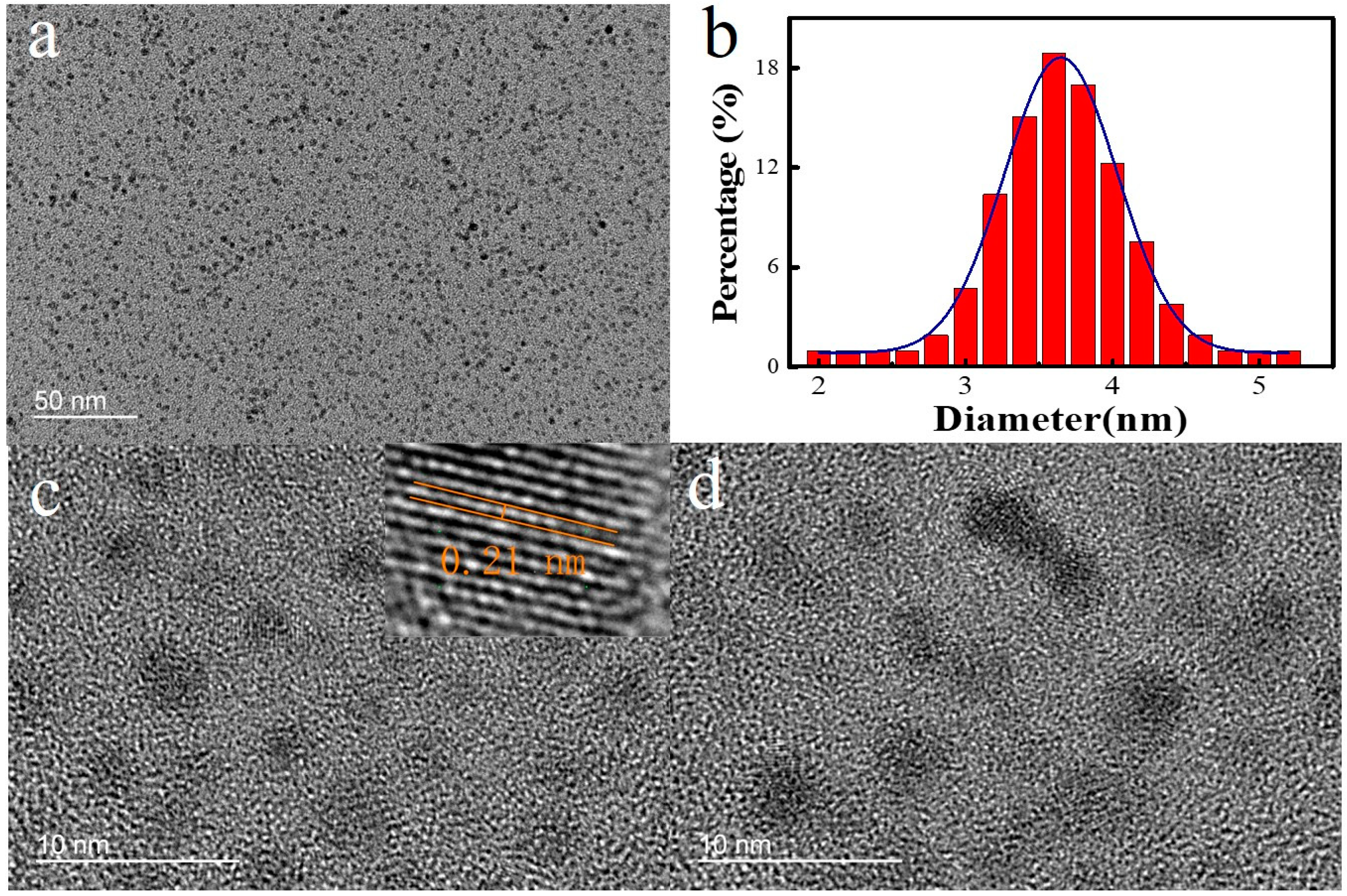
Ultrasonic probe sonication has been proved to be an efficient method to fabricate TMDCs QDs in liquid solvent because of the process of cavitation [11]. The morphology of the as-prepared WSe2 QDs shows a uniform distribution in the transmission electron microscopy (TEM) analysis. Figure 1a and b shows that the size of WSe2 QDs is distributed between 2 nm and 6 nm with an average size of 3.6 nm. The size distributions of WSe2 QDs exhibits a Gaussian profiler. (Figure 1b). Figure 1c,d are the HR-TEM of the WSe2 QDs. The lattice spacing, which is measured from an HR-TEM in the inset of Figure 1c of the WSe2 QDs which correspond to the (104) orientation, is measured to be about 0.21 nm [33]. These measurements indicate that the TMDCs QDs are highly crystalline in nature.
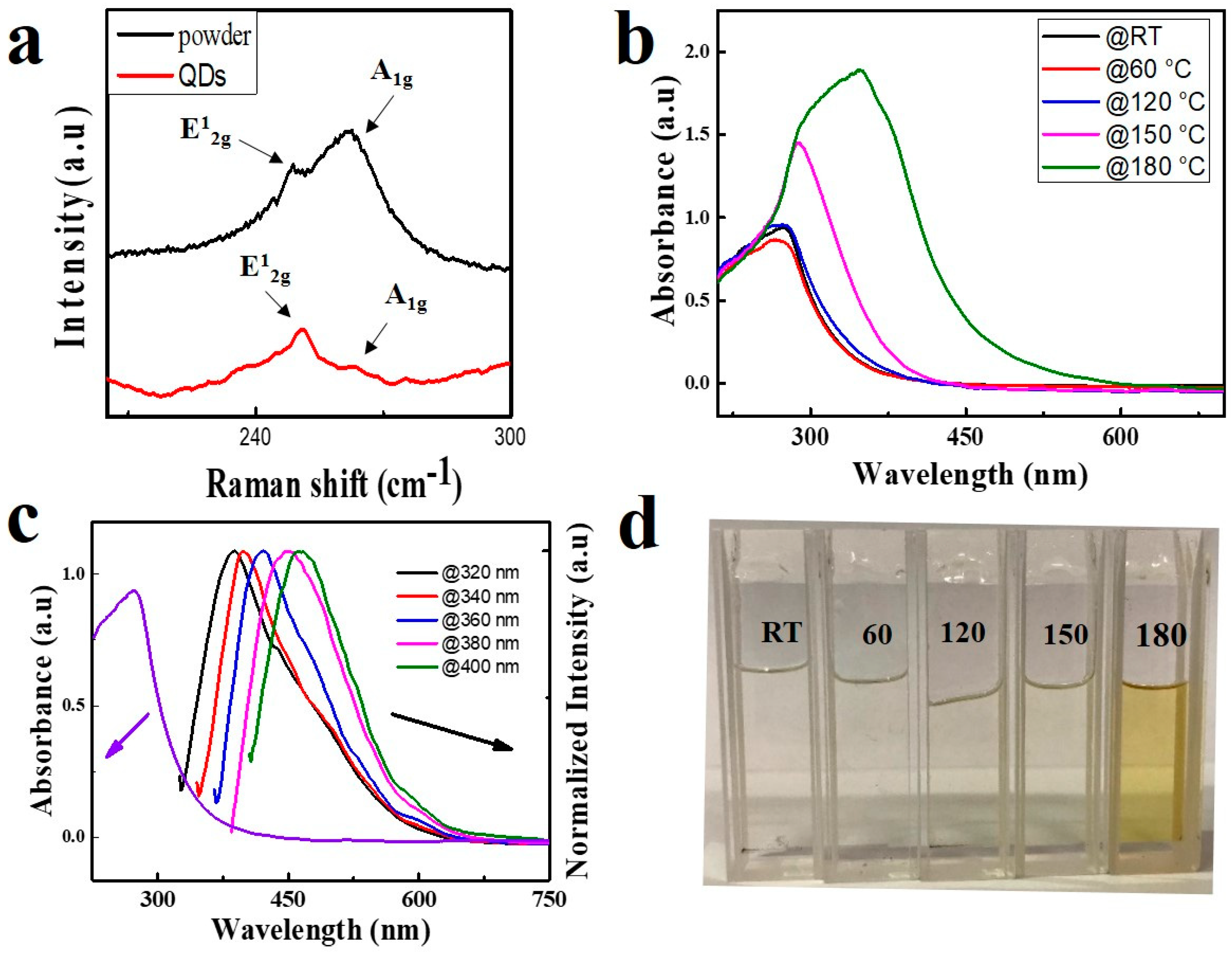
Figure 2a plots the Raman spectra of the WSe2 QDs and the WSe2 powder. A peak located at around 250 cm−1 can be seen clearly which represents E12g and A1g peak of the WSe2 powder. Similarly, the same Raman signal can be found in that of WSe2 QDs [34]. This implies that the structure of the WSe2 QDs is similar to their powder. To further investigate the optical property of WSe2 QDs, ultraviolet-visible light absorption spectra were recorded for the as-prepared WSe2 QDs as shown in Figure 2b. Strong absorption from 250 to 400 nm is observed with an absorption peak located at 270 nm. Therefore, excitation wavelengths shorter than 400 nm are suitable for WSe2 QDs PL studies. Excitation wavelength dependent PL is also observed from WSe2 QDs. As shown in Figure 2c, the emission peak wavelength of WSe2 QDs redshift from 386 to 465 nm when excitation wavelength varies from 320 to 400 nm. The emission peak wavelengths of WSe2 QDs are located at the blue-violet area, which is significantly shorter than that of WSe2 bulk (i.e., located at the near-infrared area) [35]. This is due to the band broadening casing by quantum effects similar to some other QDs materials.
Intensive studies have been done on TMDCs QDs since the first discovery of their PL. However, most of the previous works were focused on normal PL only rather than stimulated emission from TMDCs QDs. This is because lower PL intensity and high optical loss hinder the application of TMDCs QDs in stimulated emission. In this work, the thermal treatment method is used to improve the PL intensity of the WSe2 QDs. Figure 2b plots the absorption spectrum of WSe2 QDs at different thermal treatment temperatures for 16 h. It is shown that the absorption at near ultraviolet increases with the increase of temperatures. Simultaneously, the absorption peak of the sample redshifts from 270 to 350 nm with temperature increases from room temperature to 180 °C. This phenomenon can also be confirmed by a change in color of the WSe2 QDs solutions. Figure 2d shows photographs of the WSe2 QDs solution with different thermal treatment temperatures. It can be seen that the color of WSe2 QDs solutions varies from colorless to pale yellow. This coincides with the ultraviolet-visible spectra.
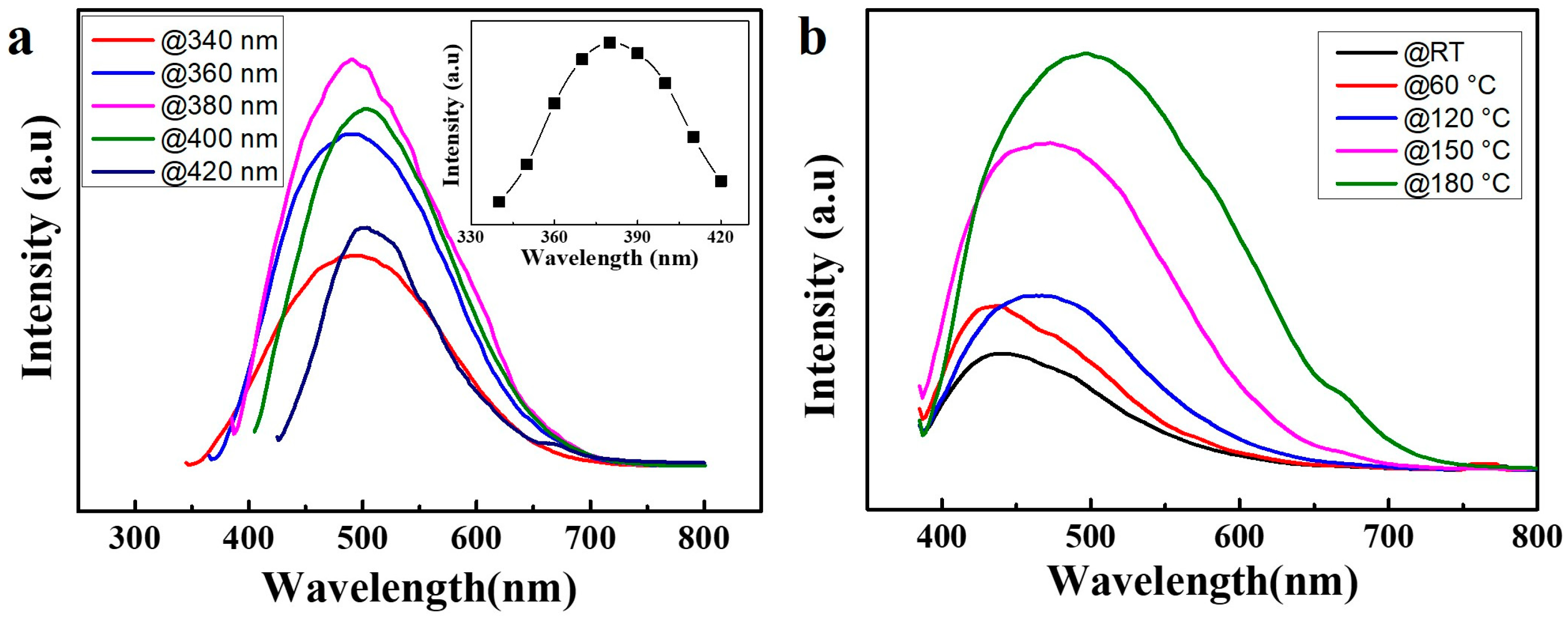
Figure 3a shows that the emission peak wavelength of WSe2 QDs (thermal treatment temperature of 180 °C) redshift from 477 to 505 nm when the excitation wavelength varies from 340 to 420 nm with a maximum emission at 380 nm excitation. The highest PL intensity is obtained at 380 nm excitation as is shown in the inset of Figure 3a. As a result, 380 nm is chosen as the excitation light source in the following studies. The Figure 3b shows the PL spectrum of as-prepared samples at different thermal treatment temperatures. The emission peak of WSe2 QDs redshifts from around 430 nm to 470 nm with increasing temperatures at 380 nm excitation. Most importantly, the intensity of PL can be enhanced by increasing the thermal treatment temperature. PL enhancement of as high as 3.7 times can be achieved by thermal treatment of WSe2 QDs at 180 °C.
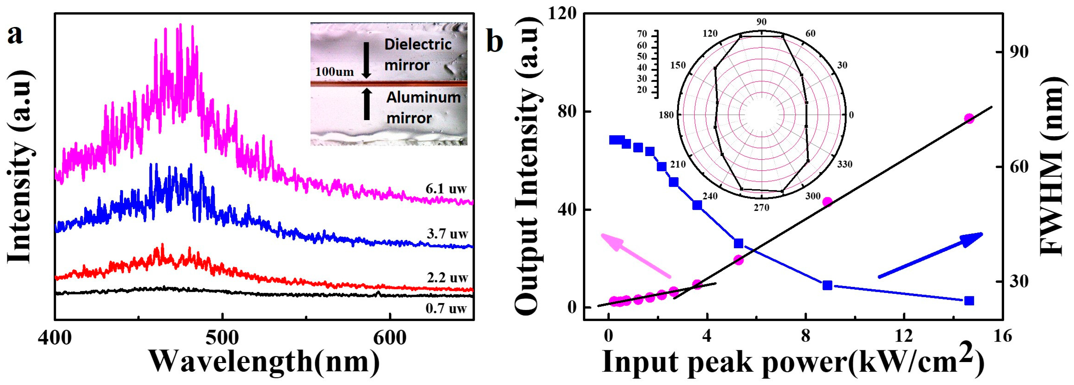
A Fabry-Perot laser cavity, which utilizes WSe2 QDs as laser gain medium, was fabricated by sandwiching a layer of WSe2 QDs in PEG400 (after thermal treatment at 180 °C for 16 h) between an aluminum mirror and a dielectric mirror. A 380 nm laser, which is the optimized excitation wavelength for WSe2 QDs, was chosen as the pump source. The inset of Figure 4a shows a photograph of the designed microcavity. The WSe2 QDs solution film, about 100 μm thick, was formed between an aluminum mirror and dielectric mirror. The mirrors are used to improve the longitudinal confinement of light and achieve optical feedback along the laser microcavity. The laser beam was focused onto a spot of 125 μm in diameter on the WSe2 QDs film through the dielectric mirror. Laser emission is detected from the side of the dielectric mirror perpendicular to the mirror plane. A plot of emission spectra of WSe2 QDs laser at room temperature versus different excitation power is shown in the Figure 4a. A broad spontaneous emission band centered at ~470 nm is observed when the excitation power is below an excitation threshold value of ~3.5 kW/cm2 as shown in Figure 4b. More sharp peaks emerge from the emission spectra with increasing pump power. Meanwhile, the full width at half-maximum (FWHM) of the spectrum also rapidly decreased from 67 nm to 25 nm. Such phenomenon indicates that the stimulated emission has played a leading role. Due to the coherent optical feedback provided by the WSe2 QDs to form the closed light loop path, the sharp peaks represent the realization of lasing. However, it can be observed from Figure 4a that the laser mode is distributed randomly. This is because a certain amount of heat is generated when the cavity is excited by the excitation light. Under the thermal perturbation, the liquid solution produces an uneven distribution of refractive index which leads to light scattering. The polarization character of the laser is also studied. The linear polarizer demonstrates the anisotropic properties of lasing emissions. The degree of polarization, defined as P = (I1 − I2)/(I1 + I2), is estimated, where I1 and I2 are the intensity of lasing emission with polarization at two different directions respectively. As shown in the inset of Figure 4b, the lasing emission of the WSe2 QDs is polarized, with the degree of polarization as P = 27.6%.
4. Conclusions
In this study, WSe2 QDs were fabricated via one step ultrasonic probe sonication in PEG400 [11,36]. Fluorescence enhancement as high as 3.7 times was achieved by thermal treatment of WSe2 QDs in PEG400. A Fabry-Perot laser cavity, which utilize WSe2 QDs as laser gain medium was fabricated by sandwiching a layer of WSe2 QDs in PEG400 between an aluminum mirror and a dielectric mirror. Lasing emission was achieved from the Fabry-Perot laser cavity by optical pumping at 380 nm. A laser threshold of ~3.5 kW/cm2 was recorded. FWHM of the lasing spectrum which narrowed from 67 nm to 25 nm was observed for the first time. This demonstration of lasing emission expands TMDCs QDs into a new application area.
Author Contributions
P.R. collected most of the data; P.R. and W.Z. wrote and revised the paper; Y.N., D.X. H.W. and Y.-P.P. performed the data characterization; and L.L., P.Y. and S.R. offered helpful suggestions and analyzed some data.
Funding
This work was supported by National Nature Science Foundation of China (51502176), Science and Technology Projects of Shenzhen (JCYJ20170818101651195 and JCYJ20150324141711618), Natural Science Foundation of SZU (827-000130).
Acknowledgments
We would like to thank Lu (Wei Lu, Hong Kong Polytechnic University) for his help on transmission electron microscope analysis.
Conflicts of Interest
The authors declare no conflict of interest.
References
- Repp, S.; Erdem, E. Controlling the exciton energy of zinc oxide (ZnO) quantum dots by changing the confinement conditions. Spectrochim. Acta A Mol. Biomol. Spectrosc. 2016, 152, 637–644. [Google Scholar] [CrossRef] [PubMed]
- Repp, S.; Weber, S.; Erdem, E. Defect evolution of nonstoichiometric ZnO quantum dots. J. Phys. Chem. C 2016, 120, 25124–25130. [Google Scholar] [CrossRef]
- Erdem, E. Defect induced p-type conductivity in zinc oxide at high temperature: Electron paramagnetic resonance spectroscopy. Nanoscale 2017, 9, 10983–10986. [Google Scholar] [CrossRef] [PubMed]
- Valappil, M.O.; Anil, A.; Shaijumon, M.; Pillai, V.K.; Alwarappan, S. A single-step electrochemical synthesis of luminescent WS2 quantum dots. Chemistry 2017, 23, 9144–9148. [Google Scholar] [CrossRef] [PubMed]
- Zhao, Y.H.; Yang, F.; Wang, J.; Guo, H.; Ji, W. Continuously tunable electronic structure of transition metal dichalcogenides superlattices. Sci. Rep. 2015, 5, 8356. [Google Scholar] [CrossRef] [PubMed] [Green Version]
- Osada, M.; Sasaki, T. Exfoliated oxide nanosheets: New solution to nanoelectronics. J. Mater. Chem. 2009, 19, 2503. [Google Scholar] [CrossRef]
- Wang, Y.; Ta, V.D.; Gao, Y.; He, T.C.; Chen, R.; Mutlugun, E.; Demir, H.V.; Sun, H.D. Stimulated emission and lasing from CdSe/CdS/ZnS core-multi-shell quantum dots by simultaneous three-photon absorption. Adv. Mater. 2014, 26, 2954–2961. [Google Scholar] [CrossRef] [PubMed] [Green Version]
- Yu, T.; Lim, B.; Xia, Y. Aqueous-phase synthesis of single-crystal ceria nanosheets. Angew. Chem. 2010, 49, 4484–4487. [Google Scholar] [CrossRef] [PubMed]
- Mak, K.F.; Shan, J. Photonics and optoelectronics of 2D semiconductor transition metal dichalcogenides. Nat. Photon. 2016, 10, 216–226. [Google Scholar] [CrossRef]
- Wang, Q.H.; Kalantar-Zadeh, K.; Kis, A.; Coleman, J.N.; Strano, M.S. Electronics and optoelectronics of two-dimensional transition metal dichalcogenides. Nat. Nanotech. 2012, 7, 699–712. [Google Scholar] [CrossRef] [PubMed] [Green Version]
- Bayat, A.; Saievar-Iranizad, E. Synthesis of blue photoluminescent WS2 quantum dots via ultrasonic cavitation. J. Lumin. 2017, 185, 236–240. [Google Scholar] [CrossRef]
- Kapatel, S.; Mania, C.; Sumesh, C.K. Salt assisted sonochemical exfoliation and synthesis of highly stable few-to-monolayer WS2 quantum dots with tunable optical properties. J. Mater. Sci.-Mater. Electron. 2017, 28, 7184–7189. [Google Scholar] [CrossRef]
- Long, H.; Tao, L.; Chiu, C.P.; Tang, C.Y.; Fung, K.H.; Chai, Y.; Tsang, Y.H. The WS2 quantum dot: Preparation, characterization and its optical limiting effect in polymethylmethacrylate. Nanotechnology 2016, 27, 414005. [Google Scholar] [CrossRef] [PubMed]
- Yan, Y.; Zhang, C.; Gu, W.; Ding, C.; Li, X.; Xian, Y. Facile synthesis of water-soluble WS2 quantum dots for turn-on fluorescent measurement of lipoic acid. J. Phys. Chem. C 2016, 120, 12170–12177. [Google Scholar] [CrossRef]
- Ghorai, A.; Bayan, S.; Gogurla, N.; Midya, A.; Ray, S.K. Highly luminescent WS2 quantum dots/ZnO heterojunctions for light emitting devices. ACS Appl. Mater. Interfaces 2017, 9, 558–565. [Google Scholar] [CrossRef] [PubMed]
- Xu, S.; Li, D.; Wu, P. One-pot, facile, and versatile synthesis of monolayer MoS2/WS2 quantum dots as bioimaging probes and efficient electrocatalysts for hydrogen evolution reaction. Adv. Funct. Mater. 2015, 25, 1127–1136. [Google Scholar] [CrossRef]
- Zhao, X.; Ma, X.; Sun, J.; Li, D.; Yang, X. Enhanced catalytic activities of surfactant-assisted exfoliated WS(2) nnanodots for hydrogen evolution. ACS Nano 2016, 10, 2159–2166. [Google Scholar] [CrossRef] [PubMed]
- Zhou, L.; Yan, S.; Wu, H.; Song, H.; Shi, Y. Facile sonication synthesis of WS2 quantum dots for photoelectrochemical performance. Catalysts 2017, 7, 18. [Google Scholar] [CrossRef]
- Tonndorf, P.; Schmidt, R.; Böttger, P.; Zhang, X.; Börner, J.; Liebig, A.; Albrecht, M.; Kloc, C.; Gordan, O.; Zahn, D.R.T.; et al. Photoluminescence emission and raman response of monolayer MoS2, MoSe2, and WSe2. Opt. Express 2013, 21, 4908–4916. [Google Scholar] [CrossRef] [PubMed]
- Park, J.; Lee, W.; Choi, T.; Hwang, S.H.; Myoung, J.M.; Jung, J.H.; Kim, S.H.; Kim, H. Layer-modulated synthesis of uniform tungsten disulfide nanosheet using gas-phase precursors. Nanoscale 2015, 7, 1308–1313. [Google Scholar] [CrossRef] [PubMed]
- Song, Q.; Liu, L.; Xiao, S.; Zhou, X.; Wang, W.; Xu, L. Unidirectional high intensity narrow-linewidth lasing from a planar random microcavity laser. Phys. Rev. Lett. 2006, 96, 033902. [Google Scholar] [CrossRef] [PubMed]
- Ouyang, Q.; Zeng, S.; Jiang, L.; Hong, L.; Xu, G.; Dinh, X.Q.; Qian, J.; He, S.; Qu, J.; Coquet, P.; et al. Sensitivity enhancement of transition metal dichalcogenides/silicon nanostructure-based surface plasmon resonance biosensor. Sci. Rep. 2016, 6, 28190. [Google Scholar] [CrossRef] [PubMed]
- Splendiani, A.; Sun, L.; Zhang, Y.; Li, T.; Kim, J.; Chim, C.Y.; Galli, G.; Wang, F. Emerging photoluminescence in monolayer MoS2. Nano Lett. 2010, 10, 1271–1275. [Google Scholar] [CrossRef] [PubMed]
- Tongay, S.; Zhou, J.; Ataca, C.; Lo, K.; Matthews, T.S.; Li, J.; Grossman, J.C.; Wu, J. Thermally driven crossover from indirect toward direct bandgap in 2D semiconductors: MoSe2 versus MoS2. Nano Lett. 2012, 12, 5576–5580. [Google Scholar] [CrossRef] [PubMed]
- Bai, G.; Yuan, S.; Zhao, Y.; Yang, Z.; Choi, S.Y.; Chai, Y.; Yu, S.F.; Lau, S.P.; Hao, J. 2D layered materials of rare-earth er-doped MoS2 with NIR-to-NIR down- and up-conversion photoluminescence. Adv. Mater. 2016, 28, 7472–7477. [Google Scholar] [CrossRef] [PubMed]
- Lee, Y.C.; Tseng, Y.C.; Chen, H.L. Single type of nanocavity structure enhances light outcouplings from various two-dimensional materials by over 100-fold. ACS Photon. 2016, 4, 93–105. [Google Scholar] [CrossRef]
- Bai, X.; Wang, J.; Mu, X.; Yang, J.; Liu, H.; Xu, F.; Jing, Y.; Liu, L.; Xue, X.; Dai, H.; et al. Ultrasmall WS2 quantum dots with visible fluorescence for protection of cells and animal models from radiation-induced damages. ACS Biomater. Sci. Eng. 2017, 3, 460–470. [Google Scholar] [CrossRef]
- Lu, X.; Wang, R.; Hao, L.; Yang, F.; Jiao, W.; Peng, P.; Yuan, F.; Liu, W. Oxidative etching of MoS2/WS2 nanosheets to their QDs by facile UV irradiation. Phys. Chem. Chem. Phys. 2016, 18, 31211–31216. [Google Scholar] [CrossRef] [PubMed]
- Tang, X.; Hu, Z.; Chen, W.; Xing, X.; Zang, Z.; Hu, W.; Qiu, J.; Du, J.; Leng, Y.; Jiang, X.; et al. Room temperature single-photon emission and lasing for all-inorganic colloidal perovskite quantum dots. Nano Energy 2016, 28, 462–468. [Google Scholar] [CrossRef]
- Wang, Y.; Li, X.; Song, J.; Xiao, L.; Zeng, H.; Sun, H. All-Inorganic colloidal perovskite quantum dots: A new class of lasing materials with favorable characteristics. Adv. Mater. 2015, 27, 7101–7108. [Google Scholar] [CrossRef] [PubMed]
- Zhang, W.F.; Zhu, H.; Yu, S.F.; Yang, H.Y. Observation of lasing emission from carbon nanodots in organic solvents. Adv. Mater. 2012, 24, 2263–2267. [Google Scholar] [CrossRef] [PubMed]
- Qu, S.; Liu, X.; Guo, X.; Chu, M.; Zhang, L.; Shen, D. Amplified spontaneous green emission and lasing emission from carbon nanoparticles. Adv. Funct. Mater. 2014, 24, 2689–2695. [Google Scholar] [CrossRef]
- Zhang, X.; Lai, Z.; Liu, Z.; Tan, C.; Huang, Y.; Li, B.; Zhao, M.; Xie, L.; Huang, W.; Zhang, H. A facile and universal top-down method for preparation of monodisperse transition-metal dichalcogenide nanodots. Angew. Chem. 2015, 54, 5425–5428. [Google Scholar] [CrossRef] [PubMed]
- Li, H.; Wu, J.; Yin, Z.; Zhang, H. Preparation and applications of mechanically exfoliated single-layer and multilayer MoS(2) and WSe(2) nanosheets. Acc. Chem. Res. 2014, 47, 1067–1075. [Google Scholar] [CrossRef] [PubMed]
- Karfa, P.; Madhuri, R.; Sharma, P.K. Multifunctional fluor escent chalcogenide hybrid nanodots (MoSe2:CdS and WSe2:CdS) as electro catalyst (for oxygen reduction/oxygen evolution reactions) and sensing probe for lead. J. Mater. Chem. A 2017, 5, 1495–1508. [Google Scholar] [CrossRef]
- Hernandez, Y.; Nicolosi, V.; Lotya, M.; Blighe, F.M.; Sun, Z.; De, S.; McGovern, I.T.; Holland, B.; Byrne, M.; Gun’ko, Y.K.; et al. High-yield production of graphene by liquid-phase exfoliation of graphite. Nat. Nanotech. 2008, 3, 563–568. [Google Scholar] [CrossRef] [PubMed] [Green Version]
Figure 1.
(a) Transmission electron microscopy (TEM) image, (b) Size distribution, (c,d) High-resolution transmission electron microscope (HR-TEM) image of the WSe2 QDs. Inset of (c): partial enlarged detail.
Figure 1.
(a) Transmission electron microscopy (TEM) image, (b) Size distribution, (c,d) High-resolution transmission electron microscope (HR-TEM) image of the WSe2 QDs. Inset of (c): partial enlarged detail.

Figure 2.
(a) Raman spectra of WSe2 quantum dots (QDs) (b) Ultraviolet-visible absorption spectrum of WSe2 QDs at different thermal treatment temperatures for 16 h. (c) Ultraviolet-visible light absorption spectra of WSe2 QDs and photoluminescence (PL) spectra of WSe2 QDs at different excitation wavelengths. (d) Photographs of the WSe2 QDs solution with different thermal treatment temperatures.

Figure 3.
(a) PL spectra of WSe2 QDs after thermal treatment temperature of 180 °C. The inset is the excitation spectra of WSe2 QDs after thermal treatment temperature of 180 °C for 16 h. (b) PL spectrum of as-prepared samples at different thermal treatment temperatures at 380 nm excitation.
Figure 3.
(a) PL spectra of WSe2 QDs after thermal treatment temperature of 180 °C. The inset is the excitation spectra of WSe2 QDs after thermal treatment temperature of 180 °C for 16 h. (b) PL spectrum of as-prepared samples at different thermal treatment temperatures at 380 nm excitation.

Figure 4.
(a) Emission spectra versus different excitation power. The inset is the photograph of the designed microcavity (b) The output intensity and full width at half-maximum (FWHM) of the emission spectra versus input peak power. The inset polar patterns plot the lasing intensity at each given polarization state.
© 2018 by the authors. Licensee MDPI, Basel, Switzerland. This article is an open access article distributed under the terms and conditions of the Creative Commons Attribution (CC BY) license (http://creativecommons.org/licenses/by/4.0/).
Share and Cite
MDPI and ACS Style
Ren, P.; Zhang, W.; Ni, Y.; Xiao, D.; Wan, H.; Peng, Y.-P.; Li, L.; Yan, P.; Ruan, S. Realization of Lasing Emission from One Step Fabricated WSe2 Quantum Dots. Nanomaterials 2018, 8, 538. https://doi.org/10.3390/nano8070538
AMA Style
Ren P, Zhang W, Ni Y, Xiao D, Wan H, Peng Y-P, Li L, Yan P, Ruan S. Realization of Lasing Emission from One Step Fabricated WSe2 Quantum Dots. Nanomaterials. 2018; 8(7):538. https://doi.org/10.3390/nano8070538
Chicago/Turabian StyleRen, Pengpeng, Wenfei Zhang, Yiqun Ni, Di Xiao, Honghao Wan, Ya-Pei Peng, Ling Li, Peiguang Yan, and Shuangchen Ruan. 2018. "Realization of Lasing Emission from One Step Fabricated WSe2 Quantum Dots" Nanomaterials 8, no. 7: 538. https://doi.org/10.3390/nano8070538
Note that from the first issue of 2016, this journal uses article numbers instead of page numbers. See further details here.