1. Introduction
Studies on rare-earth-doped polymer-based light-emitting materials are of great interest, because they have played a pivotal role in the fields of medical diagnostics, displays, and detection systems [
1]. Although the organic light emitters based on polymer materials show very good prospects, unfortunately, these materials have limited color purity and low luminous efficiency [
2,
3]. Therefore, finding materials with stable, narrow-linewidth, and high luminous efficiency is the purpose of much research. Rare-earth elements have a special electron configuration, imparting uncommon photonic properties. The luminescence characteristics are mainly based on the transitions of their 4f electrons within the f–f configuration or between the f–d configurations. Since the f–f transitions of rare-earth ions belong to forbidden transitions, the absorption cross section of rare-earth ions in the visible or ultraviolet region is very small. However, organic ligands often have large absorption cross sections in the ultraviolet region, and energy can be transferred to rare-earth ions through intramolecular energy transfer, greatly improving the emission intensity of rare-earth ions [
4,
5,
6]. Rino et al. successfully prepared a Tb(acac)
3phen complex and used it as an emissive layer in organic light Emitting diodes (OLED) [
2]. Zheng developed Tb(acac)
3AAP and worked on its photoluminescent and electroluminescent properties [
3]. Bukvetskii studied the atomic structure of crystals of Tb(acac)
3phen, which were characterized by means of the X-ray structural analysis method [
7]. We have selected terbium (Tb
3+) elements with strong luminescent properties (characteristic green fluorescence) as a luminous center. In addition, the matching of energy levels between rare-earth ions and organic ligands is one of the main factors affecting the luminescent efficiency of rare-earth complexes; because of the energy levels of terbium and acetylacetone (acac), they are matched properly. In this section, acetylacetone and phenanthroline are used as ligands to synthesize Tb (acac)
3phen complexes.
Rare-earth ion complexes with organic ligands have many advantages compared to other fluorophore types, such as narrow emission bands, high quantum yields, and nontoxicity. However, rare-earth complexes are unstable at high temperatures and in the presence of acids. In a complex environment, the quantum yield of the rare-earth complexes may decrease. SiO
2 is a versatile material that has exceptional compatibility in many biomedical applications, with its optical transparency, chemical inertness, and easy generation. SiO
2 can protect rare-earth complexes, improving their thermal stability for the preparation of durable, functional materials [
7]. Tagaya et al. successfully prepared Eu(III)-doped nanoporous silica spheres by the sol–gel method and found that Eu
3+ ions were located in a low-symmetry environment [
8]. Mukhametshina studied the effects of silica coating and further silica surface decoration by phospholipid bilayers on the quenching of Tb(III) complexes by adrenochrome [
9]. Binnemans provided detailed information on certain materials, including sol–gel technology-based composites using silica precursors as the host matrix [
10].
Herein, we report a new route for anhydrous preparation of SiO2 nanoparticles containing fluorescent Tb3+ complexes with acetyl acetone (acac) and phenanthroline (phen) (SiO2–Tb3+). We also report the effect of pH on the fluorescence of Tb3+ complexes. To obtain stable fluorescent hybrid nanospheres, an anhydrous preparation was used to prepare spherical SiO2 doped with Tb(acac)3phen complexes, resulting in uniform particle size. Polyvinylpyrrolidone (PVP) ultrafine fibers containing SiO2–Tb3+ nanoparticles were prepared by electrospinning. This new method for the preparation of fluorescent hybrid nanofibers has potential applications in the field of organic light-emitting materials.
2. Materials and Methods
2.1. Materials
Terbium oxide (Tb4O7, 99.99%, A.R.), 1,10-phenanthroline (Phen, 99%, A.R.), acetylacetone (Hacac, 98%, A.R.), hydrochloric acid (HCL, 38%, A.R.), hydrogen peroxide (H2O2, 30%, A.R.), sodium hydroxide (NaOH, A.R.), tetraethyl orthosilicate (TEOS, 99 wt. %, A.R.), ammonium hydroxide (NH3∙H2O, 28%, A.R.), and polyvinylpyrrolidone (PVP, = 1,300,000) were analytically pure and purchased from China National Medicines Group (Beijing, China).
2.2. Characterization
A JEOL JEM-2100F (JEOL Inc., Tokyo, Japan) transmission electron microscope was used for the identification of morphology and size of hybrid nanoparticles. The structure and crystal phases of Tb(acac)3phen and SiO2–Tb3+ hybrid nanofibers were determined by powder X-ray diffraction (XRD, Ultima IV, Rigaku Corporation, Tokyo, Japan). High-resolution transmission electron microscopy (HRTEM) was performed using a FEI Talos F200i microscope (Thermo Fisher Scientific Inc., Waltham, MA, USA) operating at 200 kV. The elemental composition was determined using scanning transmission electron microscopy with energy-dispersive X-ray spectroscopy (STEM-EDS) using a FEI Tecnai G2 F20 S-TWIN (FEI Inc., Hillsboro, OR, USA). Scanning electron microscope (SEM) images of the electrospun fibers were obtained using a SIGMA 500/PV (SIGMA Inc., St. Louis, MO, USA), with the electron microscope operating at 200 kV. The luminescence spectra and lifetime measurements were performed using an Edinburgh FLS1000 (Edinburgh Inc., Livingston, UK) photoluminescence spectrometer.
2.3. Preparation of Terbium Complexes with Different pH
Preparation of Tb(acac)3phen complex: 0.5 g Tb4O7 was added to 10 mL of H2O2 and then stirred at room temperature for 0.5 h. Then, 4 mL of hydrochloric acid (HCL, 38%) was added to the solution and the reaction mixture was stirred until a completely transparent solution had formed. The solution was crystallized at 60 °C and dried at 50 °C in the oven for more than 12 h, to obtain white crystallized powder of terbium chloride hexahydrate. Anhydrous ethanol was added to some of the powder to make a 0.1 mol/L TbCl3 solution, marked solution A. Acetylacetone (0.601 g, 6 mmol) and 1,10-phenanthroline (0.360 g, 2 mmol) were added to a flask, followed by addition of anhydrous ethanol under stirring to obtain 20 mL total volume, marked solution B. Then, 20 mL 0.1 mol/L solution A (2 mmol) was added dropwise to solution B while stirring, and the mixture was allowed to react for 2 h, to obtain Tb(acac)3phen complexes in solution.
The resulting 40 mL Tb(acac)3phen solution was divided into eight conical flasks, and the pH of each of these solutions containing the complexes was adjusted to either 6, 7, 8, 9, 10, 11, 12, or 13 by adding 0.1 mol/L sodium hydroxide (NaOH) solution. These eight Tb(acac)3phen solutions were centrifuged (speed 10,000 r/min, 20 min) and washed with ethanol two times, and then 5 mL of ethanol was added dropwise to form a stable and clear solution. These solutions were marked solutions C1–C8.
2.4. Anhydrous Preparation of SiO2–Tb3+ Nanoparticles
Three milliliters of NH3∙H2O was added to 50 mL of anhydrous ethanol; the mixed solution was vigorously stirred for 20 min to promote hydrolysis and to adjust the pH. Then, 1.5 mL tetraethoxysilane (TEOS) was added dropwise to the above solution. The mixed solution was vigorously stirred for 24 h at room temperature. The resulting solution was marked solution D. Two milliliters of solution D were added separately to solutions C1 through C8, and the mixed solutions were vigorously stirred for 4 h, centrifuged (speed 10,000 r/min, 20 min), washed with ethanol two times, and dried in an oven at 50 °C to form a white crystallized powder, which was the SiO2/Tb(acac)3phen (SiO2–Tb3+) hybrid nanoparticles.
2.5. Preparation of PVP-Based Fluorescence Solution
The SiO2–Tb3+ nanoparticles with a concentration of 0.01 mol/L were added to an anhydrous mixture of ethanol and N,N-Dimethylformamide (DMF) (1:1 w/w) at room temperature under continuous stirring, and subsequently, was stirred for 2 h at room temperature. PVP electrospinning solution was prepared by dissolving 1.739 g PVP into 20 g of the above solution, which was stirred for about 10 h, to ensure complete dissolution of the PVP and to form a uniform SiO2–Tb3+/PVP electrospinning solution.
2.6. Electrospinning of PVP-Based Fluorescence Nanofibers
The resulting stable homogeneous solution was loaded into a 5-mL plastic syringe for electrospinning at the same temperature (20 °C) and with a tip-to-collector distance of 20 cm. The needle (22 G) was connected to a high-voltage power source and the SiO
2–Tb
3+/PVP solution was fed at a constant rate (0.033 mL/min). A piece of flat aluminum foil was placed on the receiving board to serve as the collection electrode. The nanofibers were spun at a voltage of 29 kV.
Figure 1 is a schematic showing the structure of the resulting fibers.
3. Results and Discussion
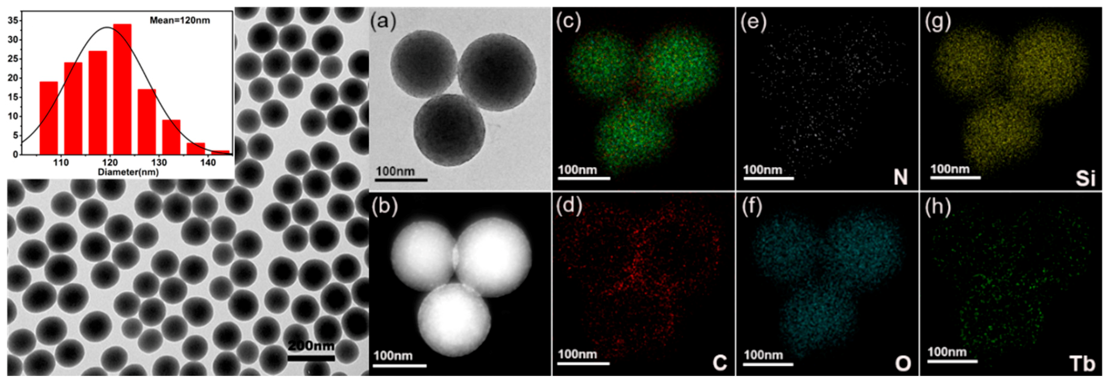
The morphological and structural feature of the resulting samples were examined by TEM; from this, it is clear that SiO
2 nanoparticles are uniform and nonagglomerated with monodisperse spheres with average diameter of about 120 nm, as shown in
Figure 2a. SiO
2 and Tb(acac)
3phen form composite nanospheres, and then SiO
2–Tb
3+ nanospheres still maintain good dispersion and the same size. From
Figure 2a,b, we can see that most of the Tb
3+ complexes are attached to the SiO
2 surfaces and a small amount of them are embedded inside the SiO
2 nanoparticles.
Figure 2d–h shows the dark-field scanning transmission electron microscopy (STEM) images and the elemental mapping of C (red), N (white), O (indigo blue), Si (yellow), and Tb (green). Detection of C, Tb, and N confirms the presence of the Tb(acac)
3phen complexes in the SiO
2 nanoparticles.
Figure 3a shows fluorescence properties of Tb(acac)
3phen complexes at different neutral or alkaline pH (pH = 6, 7, 8, 9 10, 11, 12, or 13). Understanding the influence of pH may also give insight into the influence that the environment has on the overall functionality of the complexes. The fluorescence spectrum of each solution was recorded upon incremental addition of base (NaOH) at an excitation wavelength of 325 nm. A significant increase of Tb(acac)
3phen emission intensity occurs as the pH is raised from pH = 6 to pH = 9, while an obvious reduction in Tb(acac)
3phen emission is observed from pH = 9 to pH = 13. Under the condition of strong acid, the fluorescence intensity of Tb(acac)
3phen drops sharply, due in part to the protonation of the imines, which reduces the complexation with Tb
3+ [
11]. In the strong alkali solution, Tb
3+ and OH
− produce Tb(OH)
3 precipitation, also reducing the concentration of the Tb(acac)
3phen complex. To further investigate the luminescence properties of Tb(acac)
3phen complexes at different pH, the room-temperature luminescence decay curves are presented in
Figure 3b: excited at 325 nm and monitored at the
5D
4–
7F
5 emission line. The decay curves fit a single exponential function:
[
12]. The emission lifetimes of Tb(acac)
3phen complexes are ranged from 0.36 ms to 1.28 ms (
Table 1). In fluorescence spectroscopy, the quantum yield and fluorescence lifetime are determined by the decay from radiative, Γ, and nonradiative,
knr, mechanisms. According to the Jablonski energy level table [
13], for free fluorescent molecules without other special quenching processes, the quantum yield, Q, and fluorescence lifetime,
τ0, are:
It can be seen from Equation (1) that the greater the radiation attenuation, the higher the quantum yield of the fluorescent molecule and the shorter the lifetime. The radiation attenuation for the terbium ion is a fixed constant. On the other hand, NaOH is used to adjust the solution pH in our experiment. Therefore, OH
− ions are the major origin of fluorescence efficiency change. Since samples prepared at different pH have the same apparent Tb(acac)
3phen crystal structure, OH
− ions are apparently behaving as efficient quenchers of the luminescence of Tb
3+ through multiphonon relaxation above pH 9. In samples of Tb(acac)
3phen complexes from pH 6 to 9, the concentration of OH
− ions is orders of magnitude lower, resulting in increased lifetimes compared to the conditions at higher alkalinity. Therefore, remarkably increasing fluorescence efficiency and lifetime are observed in samples of Tb(acac)
3phen complexes from pH 6 to 9. It is seen from
Figure 3b that the fluorescence decay curve of Tb(acac)
3phen complexes is nearly linear on the semilog plot, indicating almost single-exponential fluorescence decay at the lower pH conditions. The nonradiative decay of
5D
4 of Tb
3+ should include the nonradiative transition of
5D
4–
7F
j (j = 3, 4, 5, 6) and the energy transfer from
5D
4 to other Tb
3+ ions. The above results indicate that the nonradiative transition caused by OH
− ions is the leading contribution to the nonradiative decay processes of the
5D
4 of Tb
3+, and that the energy transfer from
5D
4 of a Tb
3+ to other Tb
3+ ions may be negligible [
11,
14,
15]. Therefore,
Figure 3a,b indicates that tunable fluorescent lifetimes with various intensities are achieved by changing the pH of Tb(acac)
3phen complexes, which can be potentially useful for multichannel bioimaging and optoelectronics [
16].
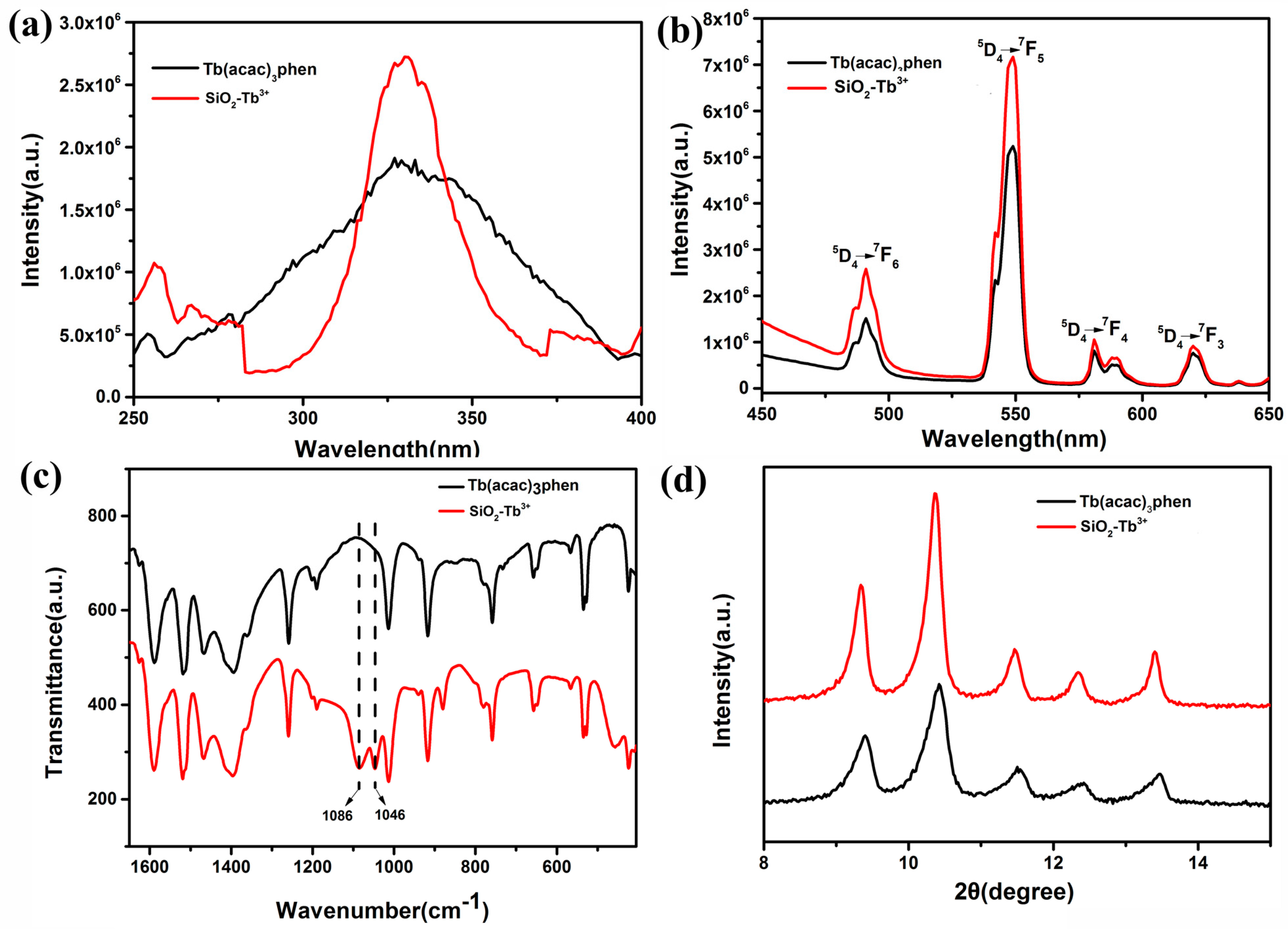
The normalized excitation and emission spectra for the Tb(acac)
3phen complex and SiO
2–Tb
3+ hybrid nanoparticles are shown in
Figure 4a,b, respectively. The excitation spectra of Tb(acac)
3phen shows a wide band, which is mainly attributed to the π–π* electron transfer of ligands. The excitation spectrum for the Tb(acac)
3phen complex is very different from that of the SiO
2–Tb
3+ hybrid nanoparticles. In the excitation spectra of SiO
2–Tb
3+ nanospheres, the corresponding broadband absorption transition of organic ligand b–diketone still exists. The line width of the excitation spectrum is 25 nm, but the absorption band position of organic ligand remains unchanged; the excitation spectrum of SiO
2–Tb
3+ nanospheres is narrower compared with the pure Tb(acac)
3phen complex, because intramolecular energy transfer occurs much more effectively between the ligands and Tb
3+ following silica encapsulation. The excitation peaks are assigned to the π–π* transition of the acetylacetone and phenanthroline [
17]. The absorption at 325 nm in the excitation spectrum of SiO
2–Tb
3+ is stronger than the corresponding absorption in the Tb(acac)
3phen complex, indicating that SiO
2 nanoparticles participate in the coordination of phenanthroline and acetylacetone molecules. An anhydrous preparation of SiO
2–Tb
3+ nanoparticles leads to different symmetry of the environment. Due to the effect of the rigid silica shell, the nanoclusters of rare-earth complexes cannot be agglomerated. The specific surface area of rare-earth complex nanoclusters is larger, and there are more molecules on the surface than those occupying the interior of clusters. We consider that the increase of excitation intensity is due to the increase of molecules on the surface of rare-earth complexes. The strongest fluorescence is obtained by excitation at 325 nm, indicating that the fluorescence intensity of the complex is strongest under this excitation wavelength. Both the emission spectra of Tb(acac)
3phen and SiO
2–Tb
3+ are obtained by monitoring the strongest emission wavelength of the Tb
3+ ions at 548 nm. As can be seen from
Figure 4b, the characteristic emission peaks of Tb
3+ appear at 491 nm, 548 nm, 581 nm, and 620 nm. These bands are attributed to the
5D
4→
7F
6,
5D
4→
7F
5,
5D
4→
7F
4, and
5D
4→
7F
3 transitions of Tb
3+ ions. The
5D
4→
7F
5 transition at the emission wavelength of 548 nm is the hypersensitive transition with strongest fluorescence intensity [
17,
18,
19]. The SiO
2–Tb
3+ hybrid nanoparticles have stronger luminescence than the corresponding pure Tb(acac)
3phen complex; the electric dipole transition and its corresponding intense emission at 548 nm aree significantly increased [
20]. The amorphous silica nanoparticles provide a microenvironment for the terbium molecules that decrease nonradiative transitions by confinement, resulting in the increasing of fluorescence intensity of SiO
2–Tb
3+ [
21].
Figure 4c displays the FT-IR spectra of the Tb(acac)
3phen complex and SiO
2–Tb
3+ hybrid nanoparticles. The vibrations located at the wavenumbers of 1046 cm
−1 and 1086 cm
−1 observed in the SiO
2–Tb
3+ spectrum are evidence of coordination bonds between the Tb(acac)
3phen complex and SiO
2 nanoparticles [
22]. The Tb(acac)
3phen complex powder shows a sharp and strong crystalline peak at the diffraction angle of 2θ = 10.36 (
Figure 4d). Strong crystalline peaks are also observed at 2θ = 9.360, 11.460, 12.420, and 13.460, indicating that both Tb(acac)
3phen and SiO
2–Tb
3+ hybrid nanoparticles are highly crystalline and that the crystals are relatively regular [
18,
23]. The Tb(acac)
3phen complex and SiO
2–Tb
3+ hybrid nanoparticles have the same crystalline peak positions, indicating that the encapsulation of Tb(acac)
3phen complexes in silica nanoparticles does not change the crystal structure of the Tb(acac)
3phen complex.
The SiO
2–Tb
3+/PVP fluorescent hybrid materials were analyzed by HRTEM, SEM, and photoluminescence spectrometry. The SiO
2–Tb
3+/PVP hybrid nanofibers display bright-green light upon radiation with ultraviolet light at 365 nm. To comprehensively reveal the underlying distribution of nano-SiO
2–Tb
3+ in PVP fibers, the elemental distribution of the SiO
2–Tb
3+/PVP hybrid nanofibers was evaluated with scanning transmission electron microscopy (STEM) (
Figure 5a). From
Figure 5b, it can be seen that the SiO
2–Tb
3+ particles were not affected by electrospinning, as the structure of the SiO
2–Tb
3+ nanospheres is well preserved.
Figure 5d–f shows the elemental mapping analysis of C (red), O (violet), and Si (indigo-blue), demonstrating that SiO
2–Tb
3+ hybrid nanoparticles are successfully encapsulated in the PVP nanofibers. From
Figure 5a, it can be seen that SiO
2–Tb
3+ hybrid nanoparticles are well separated and are compatibilized with the polymer [
4].
Figure 6a shows the excitation at the absorption band (325 nm) of the SiO
2–Tb
3+/PVP fibers. The emission spectrum of the SiO
2–Tb
3+/PVP fibers is red-shifted compared with the fluorescence spectrum of the SiO
2–Tb
3+ hybrid nanoparticles. This is evidence that although the Tb
3+ complex is prepared so as to have complete coordination, the complexes are still able to interact with the PVP. SiO
2–Tb
3+/PVP nanofibers show strong green luminescence and exhibit Tb
3+ corresponding to the characteristic transition of
5D
4–
7F
J (J = 6, 5, 4, 3). The green emission at 548 nm from the electronic dipole transition
5D
4–
7F
5 possesses the strongest intensity, indicating that the position of Tb
3+ is in an environment without reverse symmetry [
24]. Strong
5D
4–
7F
5 peak intensity indicates a highly polarized chemical environment around the Tb
3+ ion and is responsible for producing bright-green light upon 325-nm ultraviolet radiation. The emission lifetimes of SiO
2–Tb
3+ hybrid nanoparticles and SiO
2–Tb
3+/PVP nanofibers are 1.54 ms and 1.83 ms, respectively, and are shown in
Figure 6b. SiO
2–Tb
3+/PVP hybrid fibers have longer fluorescence lifetimes compared with SiO
2–Tb
3+ nanoparticles. The main factor affecting the fluorescence lifetimes (τ) are the radiative transition rate (A
rad) and nonradiative transition rate (A
nrad) of Tb
3+ ions. The relationship between A
rad and A
nrad is
[
25]. TEM shows that SiO
2–Tb
3+ nanoparticles are enveloped by PVP polymer, which demonstrates that the site of Tb
3+ ions is located in an environment without inversion symmetry, and effectively increasing the rate of radiative transition (A
rad). The change of radiation transition rate is dependent on the index of refraction on the border molecule/polymer. The increase of radiation transition rate to Tb
3+ in SiO
2–Tb
3+/PVP hybrid fibers is due to the increase of the refractive index of the external medium. At the same time, due to the relatively regular dispersion of SiO
2–Tb
3+ nanoparticles in PVP nanofibers, the rate of nonradiative transition (A
nrad) decreases with the decrease of energy transfer between molecules. Nonradiative transitions between rare-earth ions can transfer energy through phonons. Rare-earth ions emit multiple phonons from the excited state at the same time, then relaxing to the next adjacent level. This process is called multiphonon nonradiative relaxation. The nonradiative decay of
5D
4 of Tb
3+ includes the nonradiative transition of
5D
4–
7F
j (j = 3, 4, 5, 6) and the energy transfer from
5D
4 to other Tb
3+ ions. Given the relatively regular dispersion of SiO
2–Tb
3+ nanoparticles in PVP nanofibers, the energy shift caused by energy transfer of phonons can be neglected. The surface defect states (OH molecules) of SiO
2 nanoparticles decrease sharply after SiO
2–Tb
3+ nanoparticles are doped into PVP fibers; while OH
− ions have larger vibration energy, they can greatly increase the multiphonon nonradiative relaxation between energy levels, and therefore the multiphonon relaxation between energy levels decreases after the reduction of OH
− ions, and then the nonradiative transition probability decreases. Therefore, when the reduction of the nonradiative transition rate (A
nrad) is greater than the increase of the radiative transition rate (A
rad), the sum of the two factors will decrease. As a result, the fluorescence lifetimes of the SiO
2–Tb
3+/PVP hybrid fibers is longer than that of the SiO
2–Tb
3+ nanoparticles, improving the luminescence monochromaticity. However, the longer the fluorescence lifetime is, the greater the energy storage capacity [
26,
27].

 ,
, 


