Article

Investigating the Role of Antibiotics on Induction, Inhibition and Eradication of Biofilms of Poultry Associated Escherichia coli Isolated from Retail Chicken Meat

1
Lab of Microbial Genomics and Epidemiology, Department of Microbiology, Quaid-i-Azam University, Islamabad 45320, Pakistan
2
Department of Microbiology, Hazara University, Mansehra 21120, Pakistan
*
Author to whom correspondence should be addressed.
These authors contributed equally to this work.
Antibiotics 2022, 11(11), 1663; https://doi.org/10.3390/antibiotics11111663
Submission received: 10 October 2022 / Revised: 3 November 2022 / Accepted: 7 November 2022 / Published: 19 November 2022

Abstract

:
Background: Widespread use of antibiotics as growth promoters and prophylactic agents has dramatic consequences for the development of antibiotic resistance. In this study, we investigated effects of selected antibiotics on bacterial biofilms and performed extensive antibiotic and VF profiling of poultry-meat associated E. coli strains. Methods: Antibiotic susceptibility was performed by a disc diffusion method, followed by molecular screening of resistance and virulence determinants. Further biofilm formation assays, MIC-p, MIC-b, MBIC and MBEC, were performed using standard tissue culture plate method. Results: In total, 83 (75%) samples were confirmed as E. coli from poultry sources, 26 different antibiotics were tested, and maximum numbers of the isolates were resistant to lincomycin (100%), while the least resistance was seen against cefotaxime (1%) and polymyxin B (1%). Overall, 48% of the isolates were ESBL producers and 40% showed carbapenemase activity; important virulence genes were detected in following percentages: fimH32 (39%), papC21 (25%), iutA34 (41%), kpsMT-II23 (28%), papEF9 (11%), papGII22 (27%) and fyuA13 (16%). Colistin showed remarkable anti-biofilm activity, while at sub-MIC levels, gentamicin, ceftriaxone and enrofloxin significantly (p < 0.01) inhibited the biofilms. A strong induction of bacterial biofilm, after exposure to sub-minimal levels of colistin clearly indicates risk of bacterial overgrowth in a farm environment, while use of colistin aggravates the risk of emergence of colistin resistant Enterobacteriaceae, a highly undesirable public health scenario.

1. Introduction

Escherichia coli (E. coli), a Gram-negative rod-shaped bacterium belongs to the family Enterobacteriaceae. As a part of human microbiota, it aids in digestion process and synthesis of vitamins. E. coli is one of the first microorganisms that colonize infant gut after birth [1]. Pathogenic E. coli equipped with diverse virulence factors cause different infections, including urinary tract infections, hospital-acquired pneumonia, GI tract infections and meningitis [2]. Based on site of infection, bacteria are categorized into two major sub-groups; intestinal and extra-intestinal pathogenic E. coli. ExPEC include uropathogenic, septicemia-associated, meningitis-associated and avian pathogenic strains. Isolates from different environmental samples such as sewage, food, water, domestic and wild animals show remarkable resemblance to ExPEC, causing human infections [3]. Recently, presence of ExPEC strains was confirmed in various food products [4,5]. Meat has been used for centuries as a source of food, and chicken meat is the second most-consumed meat type in the world [6,7]. Despite COVID-19, in year 2020 there was an estimated increase of 9.1% in poultry meat production in Pakistan [8]. Apart from poultry meat contamination, as a food-bore pathogen, E. coli causes colibacillosis, septicemia, entero-colitis and omphalitis in the poultry [9]. Antibiotics such as penicillin, salinomycin, chlortetracycline, bacitracin, and colistin are widely used for the growth promotion, prophylaxis and treatment of infectious diseases [10]. Particularly in Pakistan, neomycin, amoxicillin, enrofloxacin, colistin, doxycycline, and tylosin are frequently used as prophylactic agents in the poultry industry. Presence of multidrug-resistant strains of E. coli in poultry has been reported in different parts of the world [11]. Earlier, Olsen and co-workers attributed 50% of the layer flock mortalities to antibiotic-resistant E. coli [12]. The most common antibiotic resistance phenomenon among Enterobacteriaceae is the production of extended spectrum β-lactamases (ESBLs). These genes located on plasmids are transferred horizontally from one bacterial cell to another and foster dissemination of antibiotic-resistance at an exponential rate [13]. For the human host, contaminated meat can be a source of E. coli infection and direct transmission occurs via contaminated surfaces. Moreover, avian pathogenic E. coli is able to stabilize itself on meat processing equipment by forming biofilms [14]. Biofilm formation requires set of virulence factors needed for the adherence and colonization of different surfaces [15]. Bacteria as biofilms show remarkable tolerance towards antibiotics [16,17]. Studies have been reported on the effects of sub-minimal inhibitory concentrations (sub-MIC) of antibiotics on E. coli, confirming inhibitory or stimulatory effects on the biofilms [16,17]. Due to their widespread association with human health and food chain, understanding current antibiotic resistance patterns and nature of the prevailing E. coli strains in terms of their virulence potential is crucial. Moreover, use of antibiotics in farm environment, even at sub-minimal concentrations leads to persistence of bacteria as biofilm that serves as an important reservoir for the development of MDR strains. Such strains are closely linked to the food chain; therefore transmissibility to humans is inevitable. In this context, the present study was conducted to investigate effects of ceftriaxone, gentamicin, enrofloxacin and colistin on the biofilm formation of E. coli recovered from poultry meat samples.

2. Results

2.1. Prevalence of Antibiotic Resistance among E. coli

In total, 83 (75%) of the isolates were confirmed as E. coli from poultry sources. The antibiotic susceptibility testing was performed for 26 different antibiotics. Higher resistance was observed against lincomycin (100%), oxytetracycline (90%), ampicillin (96%), amoxicillin (90%), tetracycline (90%), streptomycin (81%), trimethoprim (77%), chloramphenicol (64%), levofloxacin (61%), ciprofloxacin (61%), doxycycline (60%), neomycin (53%), cephalothin (43%) and colistin (polymyxin E, 26%). Lower resistance was observed against cefotaxime (1%), polymyxin B (1%), augmentin (5%), cefepime (5%), imipenem (5%), tobramycin (6%), cefixime (7%), ceftazidime (7%), gentamicin (8%), nitrofurantoin (8%) and meropenem (9%) (p > 0.05). Overall, n = 40 (48%) of the isolates were confirmed as ESBL producers. Detailed comparison of antibiotic resistance traits of MDR, XDR, ESBL and non ESBL strains are shown in Table 1 and Table 2.

2.2. Detection of VF and ESBL Genes

Overall, 41% of the isolates carried iutA, 39% fimH, 28% kpsMTII, 27% papGII and 25% papC. Likewise, fyuA and papEF were detected in 16% and 11% of the isolates, respectively. In ESBL producing isolates, bla-TEM was the most frequently detected ESBL factor (55%), followed by bla-OXA (20%) and bla-SHV (10%). A significant association (p < 0.05) was observed between ESBL phenotypes and occurrence of following VF genes; iutA, kpsMTII and fyuA. Genotypic and phenotypic correlation of ESBL is shown in (Table 2).

2.3. Biofilm Formation Potential

Out of a total n = 83, E. coli isolates, 29% showed strong biofilm formation, while 37% formed biofilms at moderate levels and 34% were weak biofilm formers.

2.4. Measurement of MIC-p and MIC-b

The MIC-p and MIC-b values for ceftriaxone, colistin, enrofloxacin and gentamicin were determined. For ceftriaxone and enrofloxacin, MIC-p was ≤0.5 µg/mL, while for colistin and gentamicin, it was ≥1 µg/mL. MIC-b values of ceftriaxone ranged from 16 µg/mL to 32 µg/mL. Likewise, for enrofloxacin and colistin, it was 4 µg/mL and ≥1 µg/mL, respectively (Table 3).

2.5. Determination of MBIC/MRC and MBEC

MBIC/MRC for the tested strains (n = 2), #7 and #21, were determined as follows: gentamicin (8 µg/mL and 32 µg/mL), colistin (8 µg/mL and 16 µg/mL), ceftriaxone (128 µg/mL and 256 µg/mL) and enrofloxacin (256 µg/mL and 64 µg/mL). Likewise, MBEC values for the gentamicin (32 µg/mL and 128 µg/mL), colistin (128 µg/mL and 64 µg/mL), ceftriaxone (>2048 µg/mL) and enrofloxacin (>2048 µg/mL) were recorded. Although MBEC of ceftriaxone and enrofloxacin (>2048 µg/mL) were higher than colistin and gentamicin (ranging from 32 µg/mL to 128 µg/mL), both antibiotics (gentamicin and colistin) very effectively eradicated biofilms of selected E. coli isolates (Table 4).

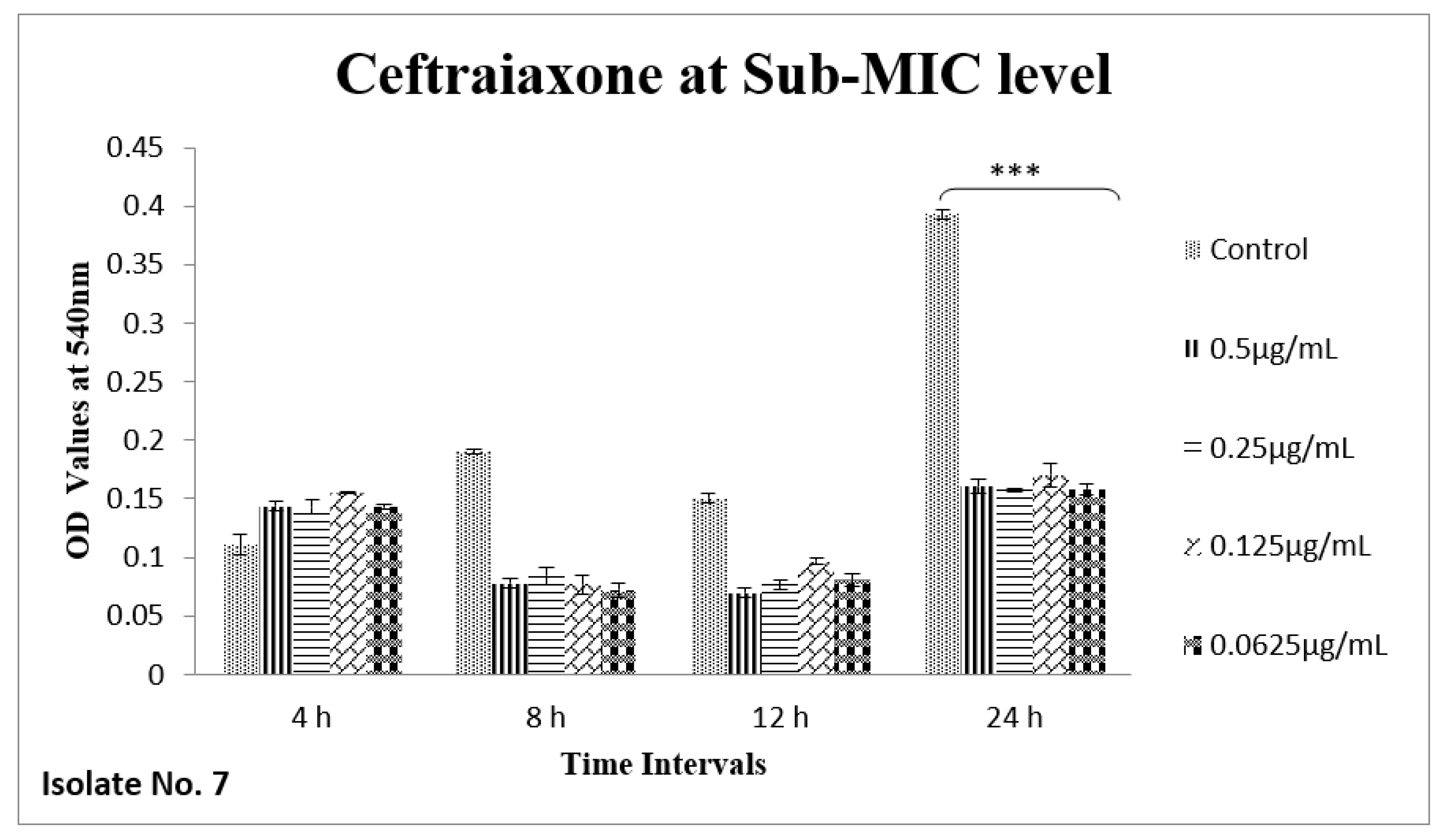
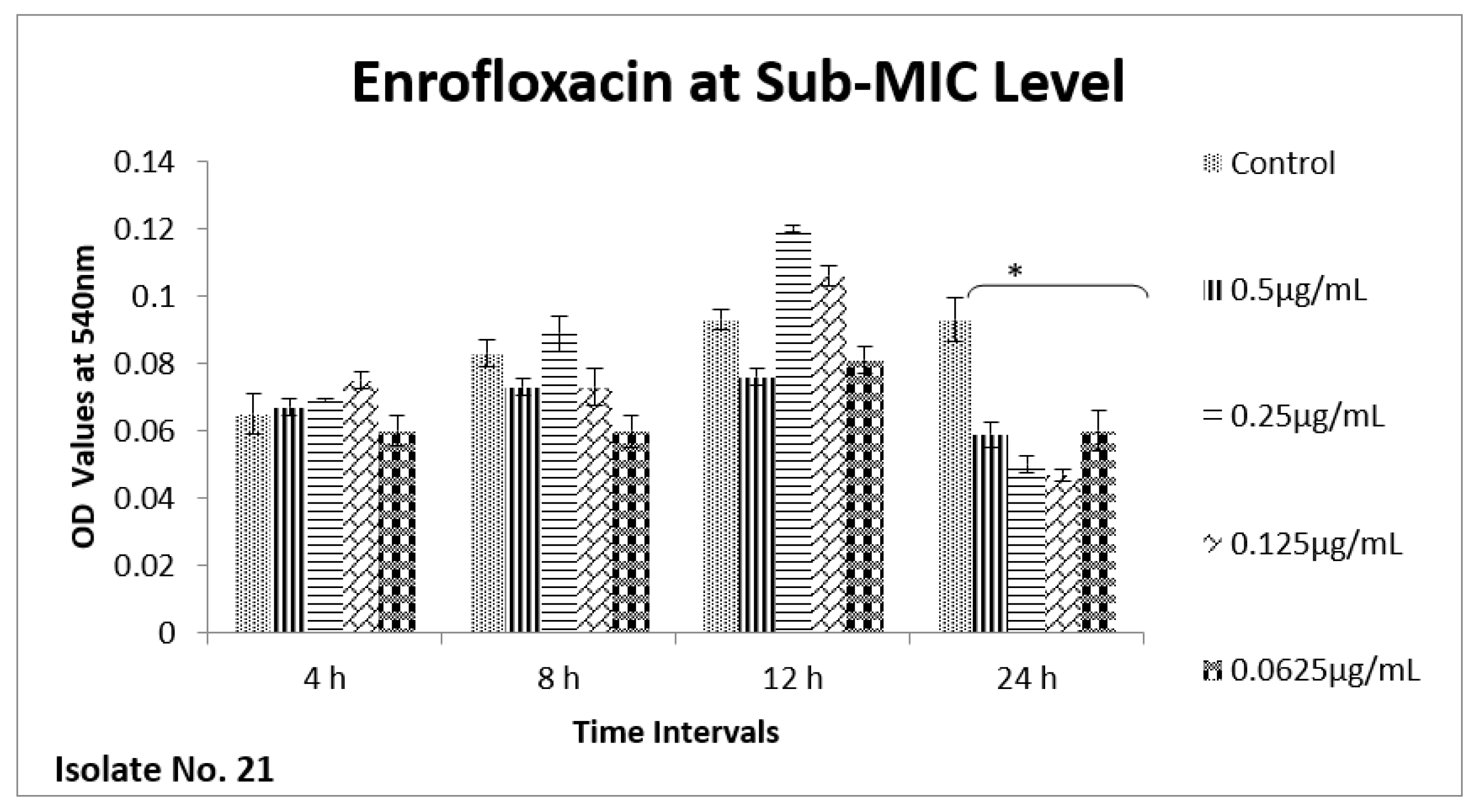
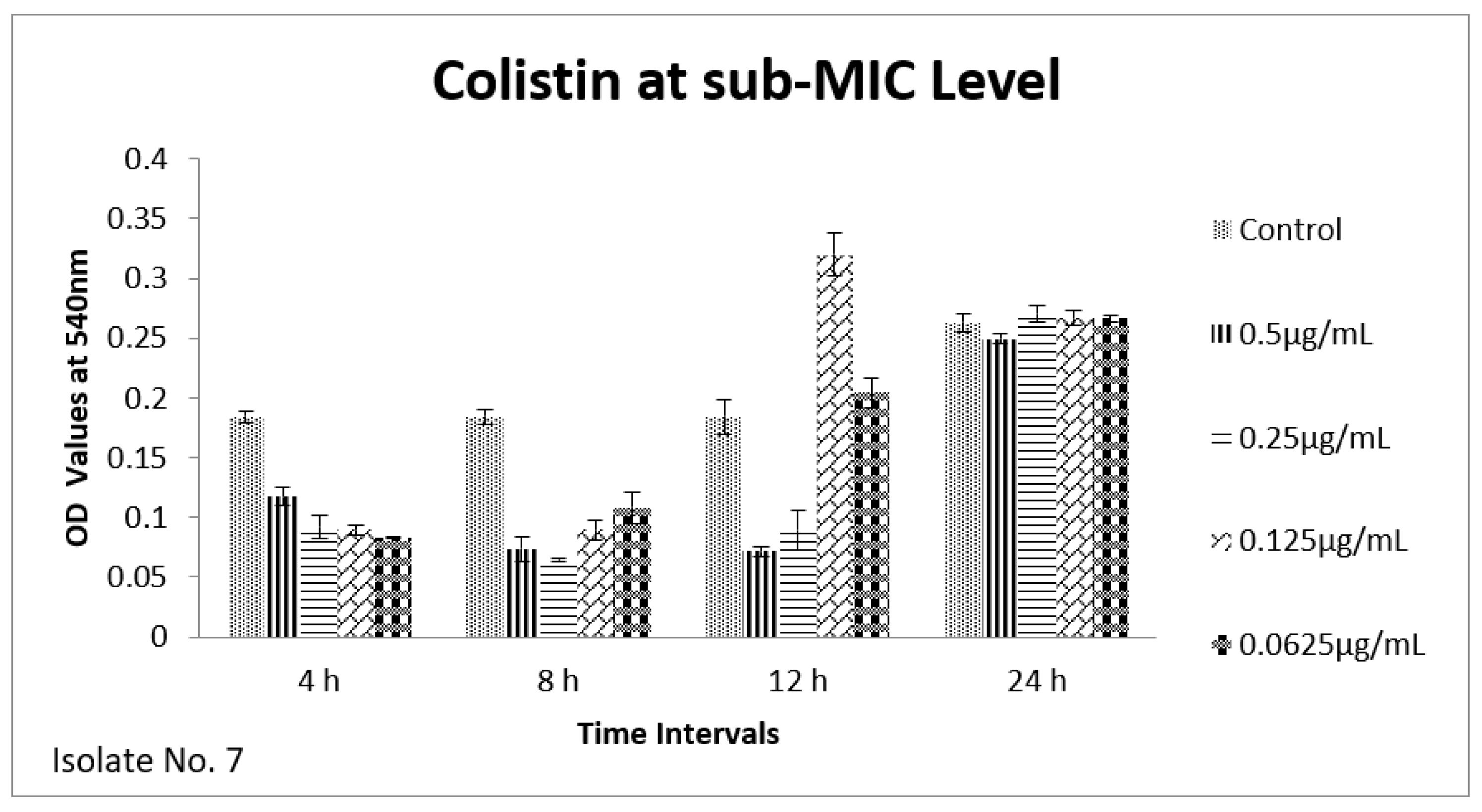
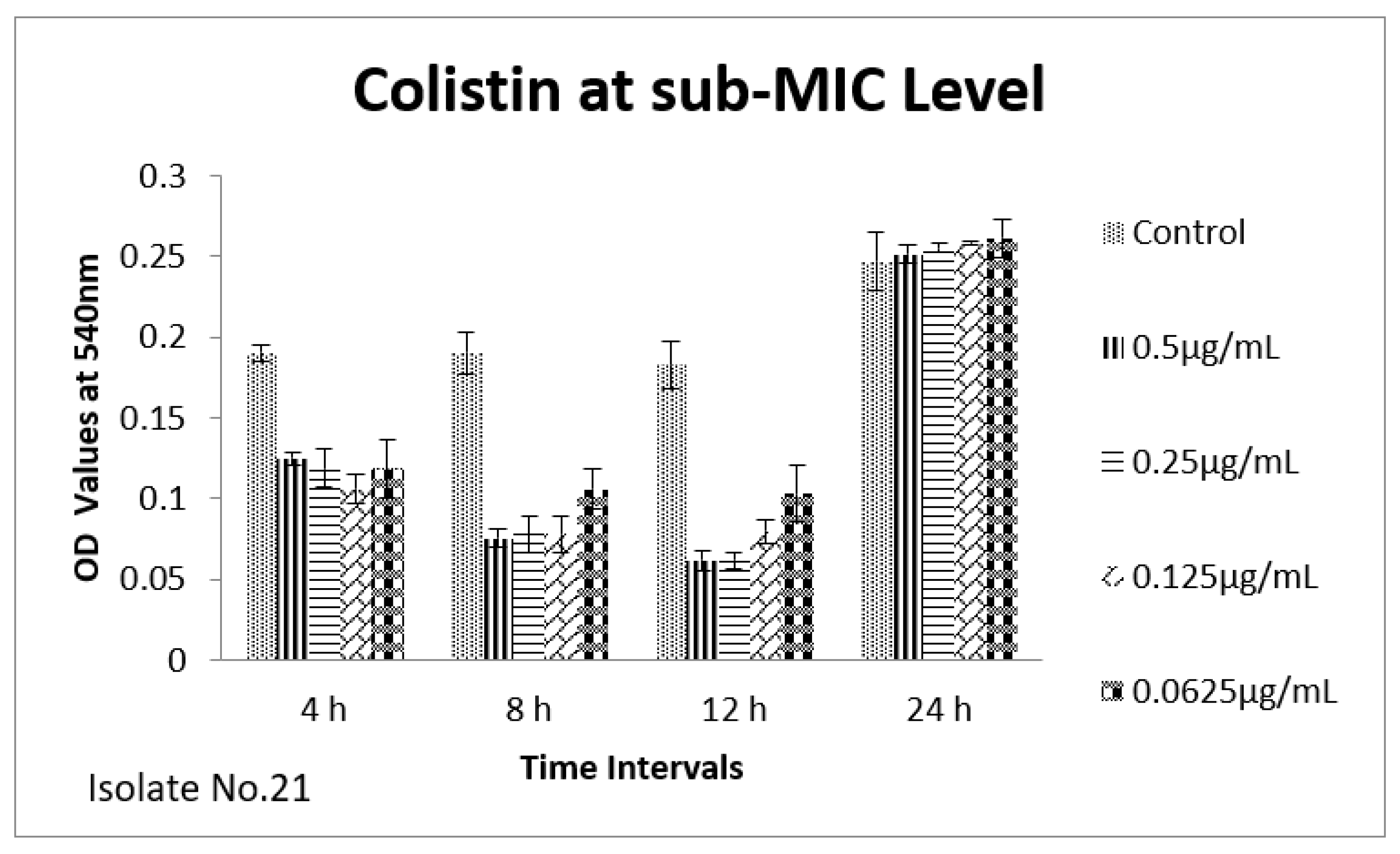
2.6. Effect of Sub-MICs on Biofilm

The effect of sub-MIC of colistin on biofilm was investigated at different time intervals (after, 4 h, 8 h, 12 h and 24 h) and showed variable reduction at different concentrations. Similarly, ceftriaxone, enrofloxacin and gentamicin also showed variable reductions at different concentrations at different time intervals. Overall, colistin showed stimulatory effects on biofilm formation, specifically after 24 h of incubation at sub-MIC level. In contrast, gentamicin, ceftriaxone and enrofloxacin showed inhibitory effects at sub-MIC level (Figure 1, Figure 2, Figure 3, Figure 4, Figure 5, Figure 6, Figure 7 and Figure 8).

3. Discussion

E. coli is one of the frequently encountered bacteria in poultry meat. In this study, isolation rate of E. coli from chicken meat was 75%. Earlier, varying resistance patterns against antibiotics have been reported from this region [18,19]. In different geographical areas different antibiotics have been used as feed additives, and prophylactic agents; consequently frequent use of these agents in farm environment asserts significant selective pressure for the antibiotic resistance development. For example, lincomycin hydrochloride soluble powder has been widely used for treating necrotic enteritis caused by Clostridium perfringens in different geographical areas, and chronic respiratory diseases caused by mycoplasma and respiratory bacterial infections were widely treated with the same antibiotic. On other hand, lincomycin has frequently been used in poultry feed (29.09 mg/fPU) in Pakistan [20,21]. In this study, 100% of the isolates were resistant to Lincomycin. Likewise, ampicillin is used in poultry to treat different bacterial infections caused by E. coli, Salmonella and Clostridia spp. and it has been one of the highly recommended therapeutic treatment options for the infections caused by Clostridia spp. Currently, due to loss of efficacy, it’s not been recommended in this region [21]. In this study, 96% of the E. coli isolates were resistant to ampicillin. Another broad-spectrum antibiotic amoxicillin is used to treat fowl typhoid, colibacillosis, and necrotic enteritis. In this study, resistance against amoxicillin was 90%. For the treatment of urinary tract infections caused by ExPEC (UPEC), fluoroquinolones are frequently prescribed in Asia, compounding 24% of the antibiotic prescription and are enlisted first choice for pyelonephritis [22,23,24]. In this study, 61% of the E. coli isolates were resistant to ciprofloxacin and levofloxacin. Surprisingly, even today, in Pakistan, doxycycline is frequently used in poultry, particularly in combination with tylosin and colistin to treat CRD, while in India, this antibiotic is banned in food producing animals and poultry to curb the AMR challenge [25]. Similarly, China already banned the use of colistin in poultry. In this study, we observed 27% of the isolates were resistant to colistin. Recently, we reported combined resistance to carbapenem and colistin in Klebsiella pneumoniae isolates that was linked to the presence of mcr-1 and mcr-2genes [26]. Here, we reiterate that colistin is one of the highest priority critically important antibiotics (HPCIA) enlisted by WHO; therefore, it is crucial to completely ban its use for the growth promotion in poultry industry, particularly to limit co-selection of carbapenem and colistin resistant Enterobacteriaceae. Cationic polymyxins, cyclic lipodecapeptide antibiotics, interact with lipid A part of LPS that contains anionic phosphate and pyrophosphate groups, which bind with polymyxin, eventually leading to the release of LPS and loss of function for Gram-negative cells [27]. Polymyxins confer rapid bactericidal actions and are postulated in inhibition of biofilm formation of A. baumannii and UPEC indicating its suitability to treat biofilm associated infections [28,29]. In contrast, however, induction of biofilm in different bacterial species upon exposure to sub-MIC levels of other antibiotics has also been reported [17,30,31]. Since in this study, we observed 27% of the E. coli isolates resistant to polymyxin E (colistin), obviously a much higher prevalence rate when compared to UPEC and K. pneumoniae.
We therefore hypothesized, role of polymyxins and other antibiotics in induction, inhibition and eradication of biofilms of poultry associated E. coli. Hence, we tested four frequently used antibiotics in poultry farms, colistin, gentamicin, enrofloxacin and ceftriaxone. Our results confirmed biofilm formation as early as 4 h of incubation and the highest level of biofilm formation was observed after 24 h of incubation. In biofilm form cells otherwise susceptible to all four antibiotics, became tolerant to significant levels of ceftriaxone and enrofloxacin (~32–64 times), when compared to their planktonic forms (MIC-p). Quite interestingly, our results showed lower MIC-b in comparison to MIC-p for other two antibiotics, colistin and gentamicin. Earlier, Klinger et al. (2017) reported inhibition of biofilm formation in colistin resistant isolates [29,32]. In contrast to that study, we used colistin susceptible strong biofilm former isolates of E. coli, definitely more relevant for such assessments. Taken together, our results regarding MBIC/MRC and MBEC confirm higher efficacy of colistin on biofilm inhibition and eradication. We also tested all four antibiotics for their efficacy on biofilm forming ability of selected isolates at sub-MIC level. Though there was variation in biofilm inhibition at 4–12 h, sub-MIC of gentamicin, ceftriaxone and enrofloxin resulted into significant (p < 0.01) reduction of biofilms. However, on contrast a significant induction and increase in biofilm formation was observed upon exposure to sub-MIC level of colistin, after 24 h of incubation. A recent study on Acinetobacter baumannii reported similar results, where a notable increase in the biofilm was observed in the presence of sub-MIC of colistin [33]. Colistin has been used in poultry industry as a growth promoter and to prevent Enterobacteriaceae mediated infections, including APEC. Recently, emergence of plasmid mediated colistin resistance in China was reported [34]. Exposure to sub-optimal concentration usually falls within/below the selection window (MSW) that eventually selects the resistant phenotypes, which, in turn, enhances the biofilm and virulence potential of pathogens. On a molecular level, sub-MIC of colistin apparently induces the expression of biofilm associated genes such as poly-acetyl-glucosamine-porin (pgaA), autoinducer synthase(abaI) and efflux pumps (adeB and adeG) [32,33].
Henceforth, it is very important to stop the irrational use of this antibiotic as prophylactic in farm practice, as even at sub-MIC level of antibiotic may lead to the dramatic surge in colistin resistance Enterobacteriaceae and widespread dissemination of plasmid encoded mcr genes.

4. Materials and Methods

4.1. Isolation and Identification of Bacterial Samples

A total of 110 meat samples were collected randomly from retail chicken meat shops of Peshawar, Pakistan (34°0’28” N, 71°34’24” E). The study was carried out from February 2018 to December 2020 at the Department of Microbiology, Quaid-i-Azam University, Islamabad, Pakistan. Meat samples were collected in sterile falcon tubes and were transported to the laboratory in a cool bank within 24 h of collection. These frozen meat samples were thawed at room temperature and transferred to Brain Heart Infusion (BHI) broth in sterile falcon tubes. The broth was incubated at 37 °C for 24 h to promote growth of E. coli. After 24 h, 500 μL of broth was separately transferred to freshly prepared MacConkey agar (Oxoid Ltd., Hamsphire, UK), and plates were subsequently incubated at 37 °C for 24 h. After the visual inspection for colony morphology, Gram-staining was performed to carry out microscopic examination. In total, 83 isolates were identified as E. coli by using standard biochemical and molecular methods as described elsewhere [35]. Bacterial samples were cultured on MacConkey agar and stored in glycerol stocks at −20 °C.

4.2. Antibiotic Susceptibility Testing

Antibiotic susceptibility testing and phenotypic detection of ESBL production was performed as per CLSI guidelines [36]. MDR and XDR strains were defined as described elsewhere [37]. Bacterial isolates were tested against twenty-six different antibiotics (Oxoid) commonly used in public health and livestock sectors (Table 1). Briefly, 0.5 McFarland bacterial suspensions were prepared and using a sterile cotton swab they were spread on plates containing Muller–Hinton Agar (MHA). Antibiotic discs of known concentrations were placed on plate using sterile forceps. Plates were incubated at 37 °C for 16–18 h and zone diameters were measured.

4.3. PCR Detection of Virulence and ESBL Genes

Detection of eight virulence genes (fimH, papC, iutA, kpsMT-II, papEF, papGII, fyuA and papGIII) and four ESBL genes (bla-TEM, bla-OXA, bla-SHV and bla-PSE) was carried out by polymerase chain reaction as described elsewhere [33]. Bacterial DNA was extracted by using phenol–chloroform method. PCR conditions were as follows: 95 °C for 1 min, followed by 35 cycles of denaturation at 95 °C for 45 s, annealing at 56 °C for 45 s, extension at 72 °C for one minute and a final extension at 72 °C for 10 min. The amplified products were observed on agarose gel and band sizes were compared with DNA ladder of 100 bp (Solis Biodyne).

4.4. Biofilm Formation Assay

A total of 83 E. coli isolates were tested for biofilm formation. Quantitative assessment of biofilms was performed by using microtiter plate method, a gold standard for biofilm quantification. Biofilm quantification was performed as previously described [17]. Briefly, a 200 µL of standardized 0.5 McFarland suspension was added to a 96 well clear, sterile polystyrene microtiter plate, and incubated at 37 °C for 24 h. After the incubation, content was removed, and the cells were washed three times with 0.9% phosphate buffer saline (PBS). Adherent bacteria were stained with crystal violet for 15 min and wells were again washed with PBS. Each sample was processed in triplicate and E. coli strain ATCC25922 was used as an experimental control. Wells without bacterial suspension (sterile MH broth) were taken as negative control.

4.5. Measurement of MIC-p and MBC

Two strong biofilm forming isolates (isolate #7 and #21) sensitive to all four antibiotics colistin, ceftriaxone, enrofloxacin and gentamicin were selected. To confirm sensitivity, minimum inhibitory concentrations (MIC) against colistin, ceftriaxone, enrofloxacin and gentamicin were measured by following standard procedure [36]. After the incubation (24 h at 37 °C), starting from lowest concentration, the first dilution of the tested antibiotic showing no turbidity was taken as the MIC of that particular antibiotic. The clear well next to the MIC well was tested for Minimum Bactericidal Concentration (MBC) by plating the contents of the well on the nutrient agar plate. Absence of growth on plate after 24 h incubation at 37 °C was confirmed as MBC.

4.6. Determination of MIC-b, MBIC/MRC and MBEC

The two selected isolates (isolate #7 and #21) were then tested for Minimum Inhibitory Concentration of biofilms (MIC-b), Minimum Biofilm Inhibition Concentration (MBIC/MRC) and Minimum Biofilm Eradication Concentration (MBEC) of colistin, ceftriaxone, enrofloxacin and gentamicin through procedures described elsewhere [17].

4.7. Sub-MIC Concentration of Ceftriaxone

The effect of sub-MIC concentrations of ceftriaxone, gentamicin, enrofloxacin and colistin on biofilm forming ability of ExPEC was tested at different time intervals, i.e., 4 h, 8 h, 12 h and 24 h. The antibiotics were diluted at sub-MIC levels of 0.5 µg/mL 0.25 µg/mL, 0.125 µg/mL and 0.0625 µg/mL; MH broth was used for the preparation of antibiotic dilutions. For sub-MIC level testing, a 100 µL of each diluted antibiotic was added to each treatment well containing a 100 µL of standardized bacterial inoculum. All micro-titer plates were incubated at 37 °C. Quantification of biofilms was performed by measuring OD at 540 nm after adding crystal violet dye [17]. For all assays, antibiotic free standardized bacterial suspension was added as a positive control while bacteria free antibiotic solution was included as negative control. Biofilm formation was analyzed by comparing the reading with OD cut-off values.
Cut off OD of negative control (NC) was determined by using formula: ODcut off = ODavg of NC + 3 × standard deviation (SD) of ODs of NC. Each isolate was then categorized according to the following criteria: weak biofilm producer, OD = 2 × ODc; moderate biofilm producer, 2 × ODc ≤ 4 × ODc; strong biofilm producer, OD ≥ 4 × ODc. * The cut off OD for NC was 0.055.

4.8. Statistical Analysis

All the biofilm assays were performed in triplicates and repeated at least twice to ensure reproducibility of results. Statistical analysis of antibiotic resistance, virulence factors, resistance factors and that of multi-/extensively drug resistant strains (MDR/XDR) was performed by chi-square test using GraphPad Prism v.7.04. A p-value < 0.05 was considered statistically significant. Differences in biofilm formation upon treatments were concluded by using paired t-test.

5. Conclusions

Conclusively, irrational use of colistin as a prophylactic agent in farm practice results in to dramatic surge of resistant strains of Enterobacteriaceae linked to the human food chain and public health. Henceforth, this vital antibiotic must be restricted for rational use only.

Author Contributions

A.N. and J.Z. prepared the initial draft. H.M., A.F., H.S., J.A., A.H., S.N. and K.D. executed the experimental work. Z.R. reviewed the draft and performed statistical analysis. J.I.D. conceived the idea secured funding, wrote and reviewed the draft. All authors have read and agreed to the published version of the manuscript.

Funding

The research was funded by University Research Fund (URF), Quaid-i-Azam University, Islamabad. URF 2017/18/19/20.

Institutional Review Board Statement

Not applicable.

Informed Consent Statement

Not applicable.

Data Availability Statement

Data and strains used in this study can be made available upon request.

Conflicts of Interest

The authors declare no conflict of interest.

References

  1. Alkeskas, A.; Ogrodzki, P.; Saad, M.; Masood, N.; Rhoma, N.R.; Moore, K.; Farbos, A.; Paszkiewicz, K.; Forsythe, S. The molecular characterisation of Escherichia coli K1 isolated from neonatal nasogastric feeding tubes. BMC Infect. Dis. 2015, 15, 1–14. [Google Scholar] [CrossRef] [Green Version]
  2. Madappa, T.; Go, C.H.U. Escherichia coli(E coli) Infections: Background. Pathophysiology 2019. [Google Scholar]
  3. Barbieri, N.L.; Oliveira, A.L.d.; Tejkowski, T.M.; Pavanelo, D.B.; Matter, L.B.; Pinheiro, S.R.S.; Vaz, T.M.I.; Nolan, L.K.; Logue, C.M.; Brito, B.G.d. Molecular characterization and clonal relationships among Escherichia coli strains isolated from broiler chickens with colisepticemia. Foodborne Pathog. Dis. 2015, 12, 74–83. [Google Scholar] [CrossRef] [PubMed]
  4. Mitchell, N.M.; Johnson, J.R.; Johnston, B.; Curtiss, R.; Mellata, M. Zoonotic Potential of Escherichia coli Isolates from Retail Chicken Meat Products and Eggs. Appl. Environ. Microbiol. 2015, 81, 1177–1187. [Google Scholar] [CrossRef] [PubMed] [Green Version]
  5. Sarowska, J.; Futoma-Koloch, B.; Jama-Kmiecik, A.; Frej-Madrzak, M.; Ksiazczyk, M.; Bugla-Ploskonska, G.; Choroszy-Krol, I. Virulence factors, prevalence and potential transmission of extraintestinal pathogenic Escherichia coli isolated from different sources: Recent reports. Gut Pathog. 2019, 11, 1–16. [Google Scholar] [CrossRef] [PubMed] [Green Version]
  6. Belova, V.A.; Smutka, L.; Rosochatecká, E. World chicken meat market–its development and current status. Acta Univ. Agric. Et Silvic. Mendel. Brun. 2012, 60, 15–30. [Google Scholar] [CrossRef] [Green Version]
  7. Pozdniakova, E.; Pozdniakov, V.; Brench, A. Development trends and risk factors of meat global exports. Ukr. Food J. 2019, 8, 645–658. [Google Scholar] [CrossRef]
  8. Hafez, M.H.; Attia, Y.A. Challenges to the poultry industry: Current perspectives and strategic future after the COVID-19 outbreak. Front. Vet. Sci. 2020, 7, 516. [Google Scholar] [CrossRef]
  9. Swelum, A.A.; Elbestawy, A.R.; El-Saadony, M.T.; Hussein, E.O.; Alhotan, R.; Suliman, G.M.; Taha, A.E.; Ba-Awadh, H.; El-Tarabily, K.A.; El-Hack, M.E.A. Ways to minimize bacterial infections, with special reference to Escherichia coli, to cope with the first-week mortality in chicks: An updated overview. Poult. Sci. 2021, 100, 101039. [Google Scholar] [CrossRef]
  10. Agyare, C.; Boamah, V.E.; Zumbi, C.N.; Osei, F.B. Antibiotic use in poultry production and its effects on bacterial resistance. In Antimicrobial Resistance—A Global Threat; Kumar, Y., Ed.; IntechOpen: Rijeka, Croatia, 2018; pp. 33–51. [Google Scholar]
  11. Valiakos, G.; Kapna, I. Colistin resistant mcr genes prevalence in livestock animals (swine, bovine, poultry) from a multinational perspective. A Systematic Review. Vet. Sci. 2021, 8, 265. [Google Scholar] [CrossRef]
  12. Olsen, R.; Frantzen, C.; Christensen, H.; Bisgaard, M. An investigation on first-week mortality in layers. Avian Dis. 2012, 56, 51–57. [Google Scholar] [CrossRef] [PubMed]
  13. Benz, F.; Huisman, J.S.; Bakkeren, E.; Herter, J.A.; Stadler, T.; Ackermann, M.; Diard, M.; Egli, A.; Hall, A.R.; Hardt, W.-D. Plasmid-and strain-specific factors drive variation in ESBL-plasmid spread in vitro and in vivo. ISME J. 2021, 15, 862–878. [Google Scholar] [CrossRef] [PubMed]
  14. Silva, O.V.; Soares, L.O.; Júnior, A.S.; Mantovani, H.C.; Chang, Y.-F.; Moreira, M.A.S. Biofilm formation on biotic and abiotic surfaces in the presence of antimicrobials by Escherichia coli isolates from cases of bovine mastitis. Appl. Environ. Microbiol. 2014, 80, 6136–6145. [Google Scholar] [CrossRef] [PubMed] [Green Version]
  15. Stępień-Pyśniak, D.; Hauschild, T.; Kosikowska, U.; Dec, M.; Urban-Chmiel, R. Biofilm formation capacity and presence of virulence factors among commensal Enterococcus spp. from wild birds. Sci. Rep. 2019, 9, 1–7. [Google Scholar] [CrossRef] [Green Version]
  16. Dong, G.; Li, J.; Chen, L.; Bi, W.; Zhang, X.; Liu, H.; Zhi, X.; Zhou, T.; Cao, J. Effects of sub-minimum inhibitory concentrations of ciprofloxacin on biofilm formation and virulence factors of Escherichia coli. Braz. J. Infect. Dis. 2019, 23, 15–21. [Google Scholar] [CrossRef] [PubMed]
  17. Rafaque, Z.; Abid, N.; Liaqat, N.; Afridi, P.; Siddique, S.; Masood, S.; Kanwal, S.; Dasti, J.I. In-vitro investigation of antibiotics efficacy against uropathogenic Escherichia coli biofilms and antibiotic induced biofilm formation at sub-minimum inhibitory concentration of ciprofloxacin. Infect. Drug Resist. 2020, 13, 2801. [Google Scholar] [CrossRef] [PubMed]
  18. Ali, I.; Rafaque, Z.; Ahmed, S.; Malik, S.; Dasti, J.I. Prevalence of multi-drug resistant uropathogenic Escherichia coli in Potohar region of Pakistan. Asian Pac. J. Trop. Biomed. 2016, 6, 60–66. [Google Scholar] [CrossRef] [Green Version]
  19. Wanger, A.; Chávez, V. Antibiotic susceptibility Testing. In Practical Handbook of Microbiology; CRC Press: Boca Raton, FL, USA, 2021; pp. 119–128. [Google Scholar]
  20. Ali, I.; Rafaque, Z.; Ahmed, I.; Tariq, F.; Graham, S.E.; Salzman, E.; Foxman, B.; Dasti, J.I. Phylogeny, sequence-typing and virulence profile of uropathogenic Escherichia coli(UPEC) strains from Pakistan. BMC Infect. Dis. 2019, 19, 620. [Google Scholar] [CrossRef] [Green Version]
  21. Abd El Tawab, A.A.; Ammar, A.M.; Nasef, S.A.; Reda, R.M. Antibacterial resistance and resistance gene detriments of E. coli isolated from chicken. Benha Vet. Med. J. 2015, 28, 231–240. [Google Scholar] [CrossRef] [Green Version]
  22. Mohamed, M.A.; Shehata, M.A.; Rafeek, E. Virulence genes content and antimicrobial resistance in Escherichia coli from broiler chickens. Vet. Med. Int. 2014, 2014, 1–6. [Google Scholar] [CrossRef] [Green Version]
  23. Mund, M.D.; Khan, U.H.; Tahir, U.; Mustafa, B.-E.; Fayyaz, A. Antimicrobial drug residues in poultry products and implications on public health: A review. Int. J. Food Prop. 2017, 20, 1433–1446. [Google Scholar] [CrossRef]
  24. Mohsin, M.; van Boeckel, T.P.; Saleemi, M.K.; Umair, M.; Naseem, M.N.; He, C.; Khan, A.; Laxminarayan, R. Excessive use of medically important antimicrobials in food animals in Pakistan: A five-year surveillance survey. Glob. Health Action 2019, 12, 1697541. [Google Scholar] [CrossRef] [PubMed] [Green Version]
  25. Khan, G.J.; Khan, R.A.; Majeed, I.; Siddiqui, F.A.; Khan, S. Ciprofloxacin: The frequent use in poultry and its consequences on human health. Prof. Med. J. 2015, 22, 001–005. [Google Scholar] [CrossRef]
  26. Choe, H.-S.; Lee, S.-J.; Yang, S.S.; Hamasuna, R.; Yamamoto, S.; Cho, Y.-H.; Matsumoto, T. The Committee for Development of the UAA-AAUS Guidelines for UTI and STI Summary of the UAA-AAUS guidelines for urinary tract infections. Int. J. Urol. 2018, 25, 175–185. [Google Scholar] [CrossRef] [PubMed] [Green Version]
  27. Choe, H.-S.; Lee, S.-J.; Cho, Y.-H.; Çek, M.; Tandoğdu, Z.; Wagenlehner, F.; Bjerklund-Johansen, T.E.; Naber, K.; Nikfallah, A.; Kassem, A.M. Aspects of urinary tract infections and antimicrobial resistance in hospitalized urology patients in Asia: 10-Year results of the Global Prevalence Study of Infections in Urology (GPIU). J. Infect.Chemother. 2018, 24, 278–283. [Google Scholar] [CrossRef] [Green Version]
  28. Kranz, J.; Schmidt, S.; Lebert, C.; Schneidewind, L.; Mandraka, F.; Kunze, M.; Helbig, S.; Vahlensieck, W.; Naber, K.; Schmiemann, G. The 2017 update of the German clinical guideline on epidemiology, diagnostics, therapy, prevention, and management of uncomplicated urinary tract infections in adult patients. Part II: Therapy and prevention. Urol. Int. 2018, 100, 271–278. [Google Scholar] [CrossRef]
  29. Mead, A.; Richez, P.; Azzariti, S.; Pelligand, L. Pharmacokinetics of Colistin in the Gastrointestinal Tract of Poultry Following Dosing via Drinking Water and Its Bactericidal Impact on Enteric Escherichia coli. Front. Vet. Sci. 2021, 8, 634. [Google Scholar] [CrossRef]
  30. Imtiaz, W.; Syed, Z.; Rafaque, Z.; Andrews, S.C.; Dasti, J.I. Analysis of antibiotic resistance and virulence traits (genetic and phenotypic) in Klebsiella pneumoniae clinical isolates from Pakistan: Identification of significant levels of carbapenem and colistin resistance. Infect. Drug Resist. 2021, 14, 227. [Google Scholar] [CrossRef]
  31. Vaara, M. Polymyxins and their potential next generation as therapeutic antibiotics. Front. Microbiol. 2019, 10, 1689. [Google Scholar] [CrossRef]
  32. Pour, N.K.; Dusane, D.H.; Dhakephalkar, P.K.; Zamin, F.R.; Zinjarde, S.S.; Chopade, B.A. Biofilm formation by Acinetobacter baumannii strains isolated from urinary tract infection and urinary catheters. FEMS Immunol. Med. Microbiol. 2011, 62, 328–338. [Google Scholar] [CrossRef] [Green Version]
  33. Sato, Y.; Unno, Y.; Ubagai, T.; Ono, Y. Sub-minimum inhibitory concentrations of colistin and polymyxin B promote Acinetobacter baumannii biofilm formation. PLoS ONE 2018, 13, e0194556. [Google Scholar] [CrossRef]
  34. Nucleo, E.; Steffanoni, L.; Fugazza, G.; Migliavacca, R.; Giacobone, E.; Navarra, A.; Pagani, L.; Landini, P. Growth in glucose-based medium and exposure to subinhibitory concentrations of imipenem induce biofilm formation in a multidrug-resistant clinical isolate of Acinetobacter baumannii. BMC Microbiol. 2009, 9, 1–14. [Google Scholar] [CrossRef] [PubMed] [Green Version]
  35. He, X.; Lu, F.; Yuan, F.; Jiang, D.; Zhao, P.; Zhu, J.; Cheng, H.; Cao, J.; Lu, G. Biofilm formation caused by clinical Acinetobacter baumannii isolates is associated with overexpression of the AdeFGH efflux pump. Antimicrob. Agents Chemother. 2015, 59, 4817–4825. [Google Scholar] [CrossRef] [PubMed] [Green Version]
  36. Klinger-Strobel, M.; Stein, C.; Forstner, C.; Makarewicz, O.; Pletz, M.W. Effects of colistin on biofilm matrices of Escherichia coli and Staphylococcus aureus. Int. J. Antimicrob. Agents 2017, 49, 472–479. [Google Scholar] [CrossRef] [PubMed]
  37. Magiorakos, A.P.; Srinivasan, A.; Carey, R.B.; Carmeli, Y.; Falagas, M.E.; Giske, C.G.; Harbarth, S.; Hindler, J.F.; Kahlmeter, G.; Olsson-Liljequist, B.; et al. Multidrug-resistant, extensively drug-resistant and pandrug-resistant bacteria: An international expert proposal for interim standard definitions for acquired resistance. Clin. Microbiol. Infect. 2012, 18, 268–281. [Google Scholar] [CrossRef]
Figure 1. Shown are the effects of gentamicin on E. coli biofilms (OD 540 nm). Bar graphs showing significant reduction (** p value < 0.01) in biofilm formation at different time points when compared with the control samples.
Antibiotics 11 01663 g001
Figure 2. Bar graph shows the effects of gentamicin on E. coli biofilms (OD 540 nm). Overall significant reduction (*** p value < 0.001) in biofilm formation was observed after 24 h in comparison to the control samples.
Antibiotics 11 01663 g002
Figure 3. Shown are the effects of ceftriaxone on biofilm formation of E. coli at different time intervals (OD 540 nm). Bar graph represents significant reduction (*** p value < 0.001) of biofilm formation at different time points compared to the control samples.
Antibiotics 11 01663 g003
Figure 4. Shown are the effects of ceftriaxone on E. coli biofilms at different time intervals (OD 540 nm). Overall significant reduction (*** p value < 0.001) in biofilm formation was observed up to 24 h, when compared to the control samples.
Antibiotics 11 01663 g004
Figure 5. Shown are the effects of enrofloxacin on bacterial biofilm at different time points (OD 540 nm). Bar graph represents significant reduction (* p value < 0.05) in biofilm formation after 24 h of incubation when compared to the control samples.
Antibiotics 11 01663 g005
Figure 6. Shown are the effects of enrofloxacin on E. coli biofilm at different time intervals (OD 540 nm). Bar graph represents significant reduction (* p value < 0.05) in biofilm formation up to 24 h, of incubation when compared to the control samples.
Antibiotics 11 01663 g006
Figure 7. Shown are the effects of colistin on E. coli biofilm at different time points (OD 540 nm). Bar graph represents highest biofilm formation at 24 h of incubation.
Antibiotics 11 01663 g007
Figure 8. Shown are the effects of colistin on E. coli biofilm at different time intervals (OD 540 nm). Bar graph represents highest biofilm formation after 24 h of incubation.
Antibiotics 11 01663 g008
Table 1. Comparison of antibiotic resistant traits of MDR and XDR phenotypes of E. coli isolates (n = 83). p values were calculated by chi-square test.
Resistance TraitsTotal Isolates n (%)MDR (n = 70)XDR (n = 12)p Value
Ampicillin80 (96.38)67120.4650
Amoxicillin75 (90.36)62120.1179
Levofloxacin51 (61.44)40100.0857
Ciprofloxacin51 (61.44)4280.6618
Tobramycin5 (6.02)23 0.0031
Gentamycin7 (8.43)34 0.0009
Neomycin44 (53.01)3212 0.0005
Streptomycin67 (80.72)55110.2902
Tigecycline000-
Tetracycline75 (90.36)6590.0541
Oxytetracycline80 (90.38)68110.3505
Doxycycline50 (60.24)40100.0857
Nitrofurantoin7 (8.43)43 0.0272
Chloramphenicol53 (63.85)4211 0.0340
Cefotaxime1 (1.20)100.6770
Cefixime6 (7.22)24 0.0002
Cephalothin36 (43.37)2610 0.0029
Ceftazidime6 (7.22)420.1783
Cefepime4 (4.81)13 0.0005
Imipenem4 (4.81)22 0.0402
Meropenem8 (9.63)53 0.0541
TMP-SMX64 (77.10)52110.1873
Lincomycin83 (100)70120.1945
Augmentin4 (4.81)210 0.0001
Polymyxin B1 (1.20)100.6770
Colistin (Polymyxin E)22 (26.50)166 0.0499
Virulence Genes
fimH32 (38.55)2560.3457
papC21 (25.30)1920.4424
iutA34 (40.96)2670.1667
kpsMT-II23 (27.711840.5821
papEF9 (10.84)630.0925
papGII22 (26.50)1750.2093
fyuA13 (15.66)1030.3478
papGIII000-
Resistance Genes
blaTEM-122 (26.50)1750.2093
blaOXA8 (9.63)44 0.0029
blaSHV4 (4.81)13 0.0005
blaPSE000-
MDR, Multi-drug Resistant; XDR, Extensively Drug Resistant.
Table 2. Comparison of antibiotic resistant traits of ESBL producing E. coli isolates (n = 83). p values were calculated by chi-square test.
Resistance TraitsTotal Isolates
n = 83 (%)
ESBL Producers n (%)Non-ESBL
n (%)
p Value
Ampicillin80 (96.38)38 (47.50)42 (52.5)0.5142
Amoxicillin75 (90.36)35 (46.66)40 (53.33)0.3942
Levofloxacin51 (61.44)26 (50.98)25 (49.01)0.5211
Ciprofloxacin51 (61.44)31 (60.78)20 (39.2) 0.0038
Tobramycin5 (6.02)3 (60)2 (40)0.5857
Gentamycin7 (8.43)4 (57.14)3 (42.85)0.6204
Neomycin44 (53.01)27 (61.36)17 (38.63) 0.0108
Streptomycin67 (80.72)32 (47.76)35 (52.23)0.8721
Tigecycline000-
Tetracycline75 (90.36)38 (50.66)37 (49.33)0.1673
Oxytetracycline80 (90.38)38 (47.5)42 (52.5)0.5142
Doxycycline50 (60.24)25 (50)25 (50)0.6850
Nitrofurantoin7 (8.43)4 (57.1)3 (42.85)0.6204
Chloramphenicol53 (63.85)28 (52.83)25 (47.16)0.2611
Cefotaxime1 (1.20)1 (100)00.2969
Cefixime6 (7.22)6 (100)0 0.0084
Cephalothin36 (43.37)22 (61.11)14 (38.88) 0.0393
Ceftazidime6 (7.22)4 (60)2 (40)0.5804
Cefepime4 (4.81)4 (100)0 0.0335
Imipenem4 (4.81)3 (75)1 (25)0.2714
Meropenem8 (9.63)6 (75)2 (25)0.1104
TMP-SMX64 (77.10)32 (50)32 (50)0.5453
Lincomycin83 (100)40 (48.19)43 (51.80)>0.9999
Augmentin4 (4.81)3 (75)1 (25)0.2714
Polymyxin B1 (1)01 (100)0.2969
Colistin (Polymyxin E)22 (26.50)13 (59.09)9 (40.90)0.2327
fimH32 (38.55)17 (53.12)15 (46.87)0.4763
papC21 (25.30)8 (38.09)13 (61.90)0.2840
iutA34 (40.96)16 (47.05)18 (52.94) 0.0039
kpsMT-II23 (27.717 (30.43)16 (69.56) 0.0450
papEF9 (10.84)5 (55.55)4 (44.44)0.6397
papGII22 (26.50)11(50)11(50)0.8431
fyuA13 (15.66)10 (76.92)3 (23.07) 0.0240
papGIII000-
blaTEM-122 (26.50)22 (100)0 0.0001
blaOXA8 (9.63)8 (100)0 0.0020
blaSHV4 (4.81)4 (100)0 0.0335
blaPSE000-
ESBLs, Extended Spectrum β-lactamases.
Table 3. MIC-p and MIC-b analysis of selected antibiotics.
Isolate No. MIC-p µg/mLMIC-b µg/mL
GENCOLCEFENRGENCOLCEFENR
7420.50.0611164
21120.50.12511324
MIC-p, Minimum Inhibitory Concentration for planktonic bacteria; MIC-b, Minimum Inhibitory concentration for biofilm; GEN, Gentamicin; COL, Colistin; CEF, Ceftriaxone; ENR, Enrofloxacin.
Table 4. Analysis of * MBIC/MRC and MBEC of selected antibiotics on bacterial biofilms.
Isolate No.GENCOLCEFENR
MBIC µg/mLMBEC µg/mLMBIC µg/mLMBEC µg/mLMBIC µg/mLMBEC µg/mLMBIC µg/mLMBEC µg/mL
78328128128>2048256>2048
21321281664256>204864>2048
MBIC/MRC, Minimum Biofilm Inhibitory Concentration; MBEC, Minimum Biofilm Eradication Concentration; GEN, Gentamicin; COL, Colistin; CEF, Ceftriaxone; ENR, Enrofloxacin. * Term MBIC/MRC is used synonymously. * MBIC/MRC is used synonymously.
Publisher’s Note: MDPI stays neutral with regard to jurisdictional claims in published maps and institutional affiliations.

