Article

Artemisia brevifolia Wall. Ex DC Enhances Cefixime Susceptibility by Reforming Antimicrobial Resistance

1
Department of Pharmacy, Faculty of Biological Sciences, Quaid-i-Azam University, Islamabad 45320, Pakistan
2
Cadson College of Pharmacy, Kharian 50090, Pakistan
3
Faculty of Pharmacy, Hamdard University, Islamabad 45550, Pakistan
4
Shifa College of Pharmaceutical Sciences, Shifa Tameer-e-Millat University, Islamabad 44000, Pakistan
5
Department of Pharmacy, Faculty of Medicine and Pharmacy, University of Oradea, 410028 Oradea, Romania
6
Doctoral School of Biomedical Sciences, University of Oradea, 410087 Oradea, Romania
*
Authors to whom correspondence should be addressed.
Antibiotics 2023, 12(10), 1553; https://doi.org/10.3390/antibiotics12101553
Submission received: 2 September 2023 / Revised: 12 October 2023 / Accepted: 16 October 2023 / Published: 20 October 2023
(This article belongs to the Special Issue Antibacterial Resistance and Novel Therapeutic Strategies)

Abstract

:
(1) Background: A possible solution to antimicrobial resistance (AMR) is synergism with plants like Artemisia brevifolia Wall. ex DC. (2) Methods: Phytochemical quantification of extracts (n-hexane (NH), ethyl acetate (EA), methanol (M), and aqueous (Aq)) was performed using RP-HPLC and chromogenic assays. Extracts were screened against resistant clinical isolates via disc diffusion, broth dilution, the checkerboard method, time–kill, and protein quantification assays. (3) Results: M extract had the maximum phenolic (15.98 ± 0.1 μg GAE/mgE) and flavonoid contents (9.93 ± 0.5 μg QE/mgE). RP-HPLC displayed the maximum polyphenols in the M extract. Secondary metabolite determination showed M extract to have the highest glycosides, alkaloids, and tannins. Preliminary resistance profiling indicated that selected isolates were resistant to cefixime (MIC 20–40 µg/mL). Extracts showed moderate antibacterial activity (MIC 60–100 µg/mL). The checkerboard method revealed a total synergy between EA extract and cefixime with 10-fold reductions in cefixime dose against resistant P. aeruginosa and MRSA. Moreover, A. brevifolia extracts potentiated the antibacterial effect of cefixime after 6 and 9 h. The synergistic combination was non- to slightly hemolytic and could inhibit bacterial protein in addition to cefixime disrupting the cell wall, thus making it difficult for bacteria to survive. (4) Conclusion: A. brevifolia in combination with cefixime has the potential to inhibit AMR.

1. Introduction

Antibiotic treatment is the most significant therapy for bacterial infections and has marvelously enhanced human health. At present, there are more than 14 classes of antibiotics on the market to treat several infections [1]. In the past few decades, antibiotic resistance has emerged, leading to treatment failures. Unraveling the mechanisms of resistance is a priority in order to support work to devise efficacious therapies against life-threatening resistant infections. Resistance can be due to many factors such as antibiotic inactivation by bacteria or reduced bacterial membrane penetration. Furthermore, previously susceptible bacteria can develop resistance through genetic mutations or via generating biofilms to protect bacterial colonies from exogenous damage [2]. This stems from the overuse of antibiotics, promoting evolutionary resistance in bacteria via natural selection. Healthcare professionals have stressed the need for new drugs or therapies to combat emerging resistance among pathogens to antibiotics already available [3]. Therefore, there is ongoing research using both herbal and synthetic compounds in an attempt to find effective therapies against resistant bacterial isolates. Medicinal plants with proven antibacterial activity are good options, considering their wide availability and safety profiles [4].
The traditional medicinal system has established its role as a therapeutic alternative to the allopathic system to treat wounds, abrasions, and infections [5] and has been used and developed through the centuries since ancient times [6]. This natural-compound-based medicine has contributed effective antimalarial (artemisinin and quinine) and anticancer drugs (vinblastine and taxols) to treat the respective diseases [7]. Accordingly, scientists are inclined to investigate medicinal plants as an alternative source of antibiotics. Plants are enriched with several secondary metabolites, which exhibit antimicrobial properties. For example, terpenoids and essential oils have shown antibacterial effects by affecting cell membrane permeability [8].
Essential oils, besides other numerous applications [9], can be used in synergy with antibiotics to alter the bacterial membrane permeability [10]. Furthermore, tannins bind to protein, alkaloids intercalate into DNA, and flavonoids bind in an adhesion complex with the cell and also inhibit the enzymatic activity of bacterial cells [11]. Furthermore, tannins demonstrate dose-dependent antibacterial effects by potentially inhibiting extracellular enzymes, modulating bacterial cell metabolism via oxidative phosphorylation inhibition, limiting the substrate required for bacterial growth, and targeting proteolytic enzymes that inhibit protein synthesis [12].
Pakistan’s flora includes 5521 species representing 1572 genera with ethnomedicinal significance [13]. Notable among them, Matricaria chamomilla combats S. aureus, M. tuberculosis, and Helminthes, while plants like Allium sativum, Piper betel, Malus sylyestris, Pimenta dioica, and Syzygium aromaticum exhibit antibacterial properties, Panax notoginseng targets E. coli, and Aloe barbadensis and Lawsonia inermis are potent against S. aureus. Moreover, Medicago sativa and Erythroxylum coca combat active Gram-positive and Gram-negative bacteria, respectively [11].
Artemisia L. is a common and diverse genus of the Asteraceae family consisting of more than 500 species with significant therapeutic and economic importance [14]. Currently, 38 species of Artemisia have been identified and botanically reported in Pakistan, which are mainly present in dry and semidry areas of Khyber Pakhtunkhwa, Northern Punjab, Baluchistan, and Kashmir [14]. Artemisia brevifolia Wall. ex DC is locally known as “Tarkha” or “Mori”. It is one of the most commonly found species in cold desert areas, the Himalayas, Ladakh, Kashmir, and Afghanistan [15]. It is broadly spread in many areas of Pakistan over 2500 m in altitude, including Chitral, Gilgit, Swat, Baltistan, Khaghan, Astor, and the Deosai plains [16]. Traditionally, A. brevifolia has been used as an antiseptic, anthelmintic, antidiabetic, carminative, blood purifier, stress reliever, diuresis, pain reliever, antitussive, stomachic, febrifuge, and as an antidote for the scorpion sting [15].
Alkaloids, terpenoids, essential oils, and flavonoids have proven antibacterial activity. Previous studies on A. brevifolia validated the presence of alkaloids, flavonoids, terpenoids, glycosides, essential oils, and vitamins. It is hypothesized that A. brevifolia possesses significant antibacterial proficiency [16,17,18] owing to the presence of formerly reported chemical constituents.
Therefore, this research unveils a novel dimension in the scientific exploration of a plant species belonging to the commercially significant genus Artemisia, known for its well-established ethno-medicinal applications. Remarkably, despite its established reputation, the comprehensive exploration of the antimicrobial potential of this particular plant species remains conspicuously underrepresented in the existing scientific literature. An innovative approach to countering the formidable challenge of drug resistance is emerging: the synergistic interplay of phytochemicals with contemporary antibiotics. In this pioneering study, we delve into uncharted territory by thoroughly assessing the hitherto unexplored synergistic efficacy of A. brevifolia. This marks the very first instance in which such an evaluation has been undertaken, thereby adding a novel dimension to the field of antimicrobial research. Furthermore, in an unprecedented stride, our research embarks upon a biocompatibility study of A. brevifolia, a facet that has hitherto remained unreported in scientific investigations. This groundbreaking endeavor underscores our commitment to shedding new light on the properties and applications of this plant species, further emphasizing the unique and pioneering nature of our research.
In sum, this research aimed to study the antibacterial effect of A. brevifolia in resistant clinical isolates as a single therapy and as a combination therapy with an antibiotic. Here, we demonstrate the synergistic activity of A. brevifolia extracts and cefixime in reducing the growth of cefixime-resistant clinical isolates. Our results provide evidence of the antibacterial activity of A. brevifolia and present it as a source of isolating antibacterial compounds.

2. Results

2.1. Percentage Yield

A. brevifolia crude extracts were prepared in four solvents spanning the polar to non-polar range. There was the highest percent extract recovery in aqueous (Aq) extract (6.1% w/w), which gradually declined with decreasing solvent polarity. The M, EA, and NH solvents extracted the phytoconstituents from A. brevifolia with values of 4.65, 2.95, and 1.65%, respectively, of the total weight of dry plant (12 kg) used for extraction.

2.2. Phytochemical Analysis

2.2.1. Total Flavonoid Content Estimation

The total flavonoid content in A. brevifolia extracts is presented in Table 1. It was calculated with the calibration curve, y = 0.0649, x − 0.043, R2 = 0.9927. The results show that the highest TFC content was present in the M extract, followed by the Aq extract and then the EA extract. The lowest number of flavonoids were present in the NH extract.

2.2.2. Total Phenolic Content Estimation

Total phenolic content was expressed as µg GAE/mgE and calculated with the calibration curve, y = 0.0915, x − 0.098, R2 = 0.9939 (Table 1). The highest phenolic content was found in the M extract, followed by the Aq extract and then the EA extract, while the NH extract had the lowest phenolic content.

2.2.3. Secondary Metabolite Estimation

A. brevifolia extracts were qualitatively evaluated for the presence of phytochemicals (Table 2). The results showed that cardiac glycosides, alkaloids, and terpenoids were present in the NH extract. The EA extract had cardiac glycosides, anthraquinone glycosides, and terpenoids. On the contrary, the M and Aq extracts exhibited glycosides (cardiac, anthraquinone, and coumarin), alkaloids, and tannins. Saponin content was observed only in the Aq extract.

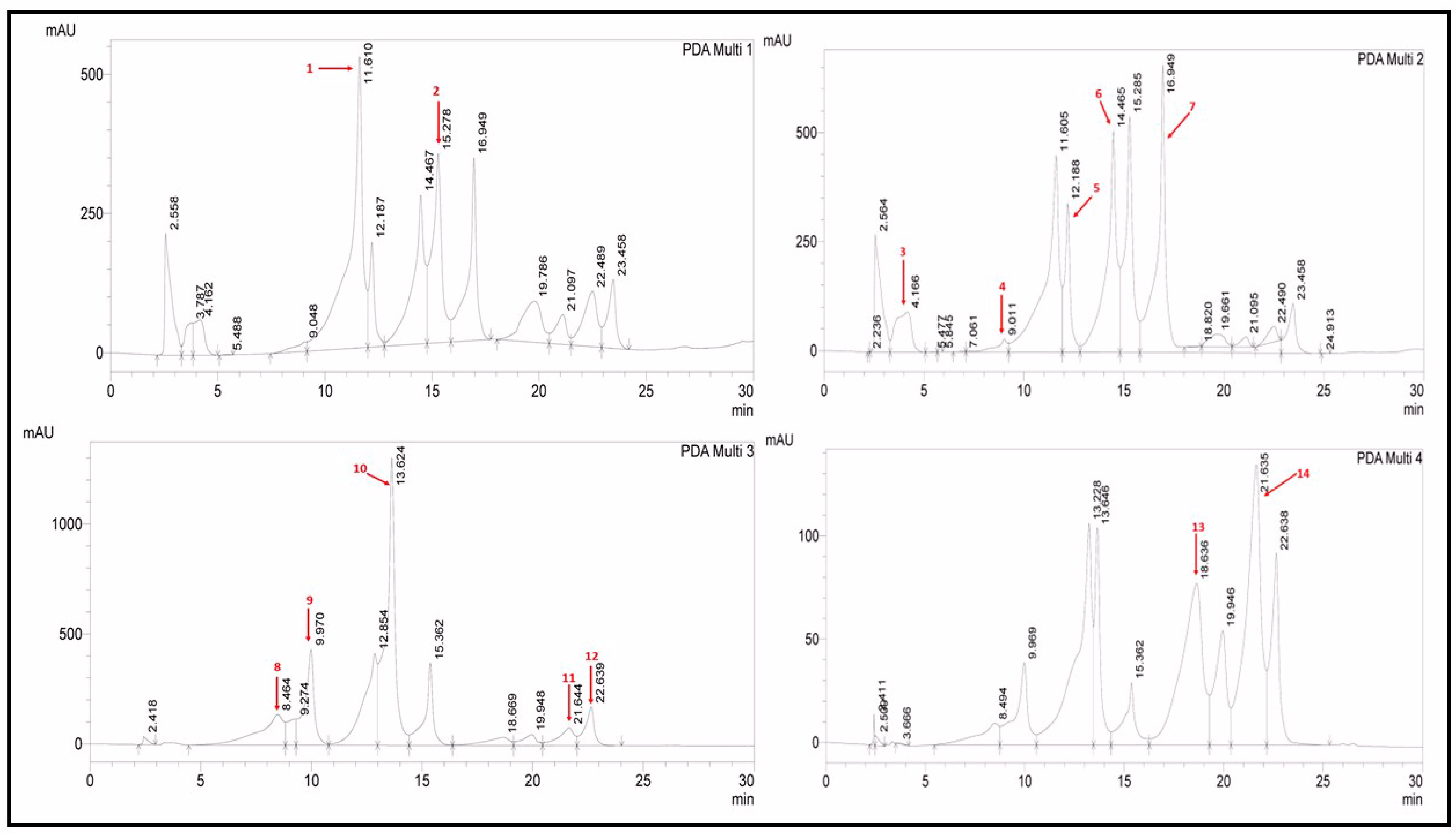
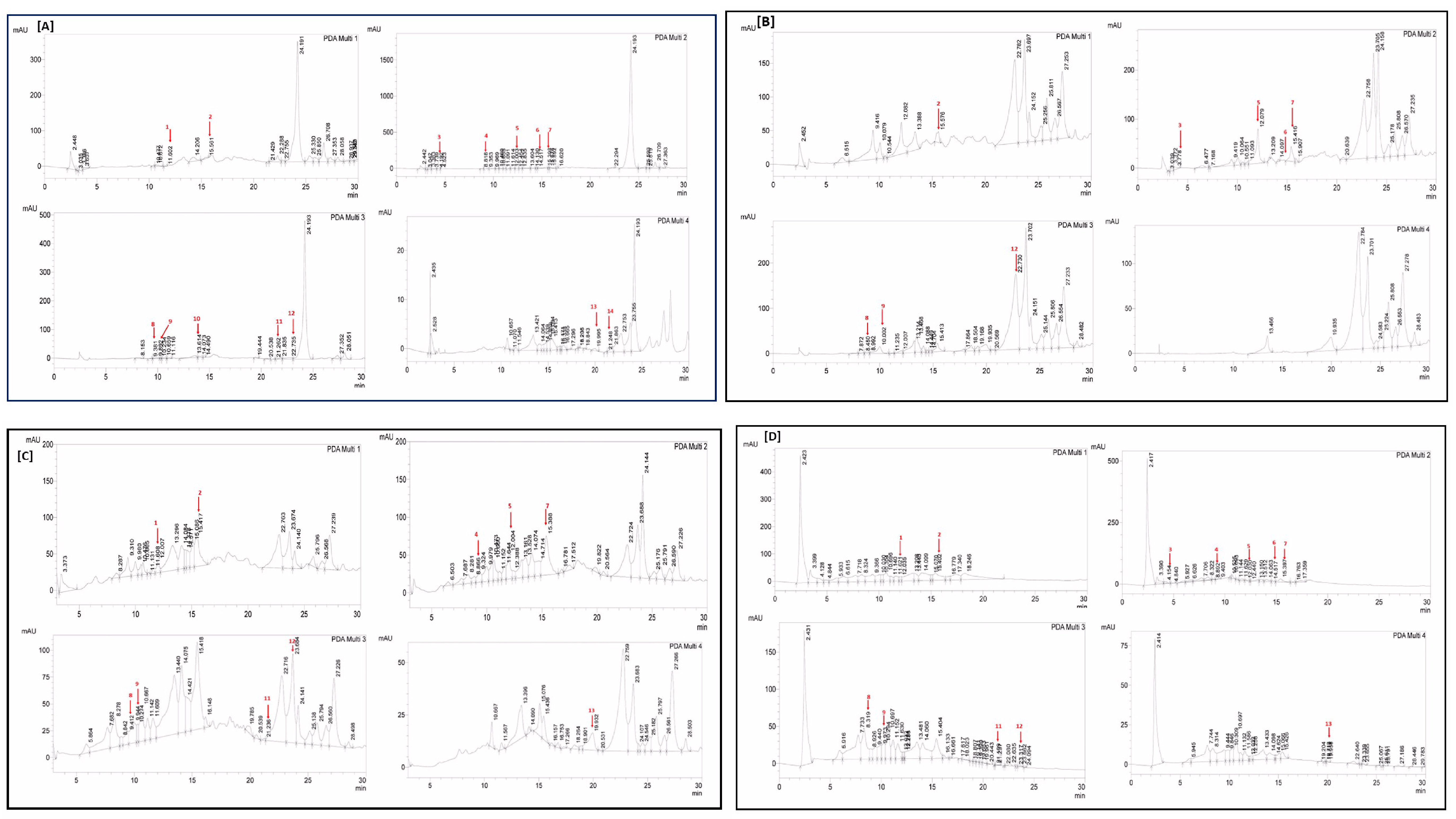
2.2.4. RP-HPLC Analysis: Detection of Polyphenolic Content

The quantification of various polyphenols was performed using the RP-HPLC-DAD method by comparing the UV spectra and retention times of the standard with those of test extracts (Table 3 and Figure 1 and Figure 2). Polyphenols in four extracts of A. brevifolia were quantified using 14 standards. The NH extract showed the maximum concentration of catechin (0.82 ± 0.09 µg/mgE) while all other polyphenols were present in minute quantities. The EA extract was found to have maximum concentrations of apigenin (8.65 ± 0.012 µg/mgE) and syringic acid (2.54 ± 0.04 µg/mgE). Apigenin (3.13 ± 0.015 µg/mgE), rutin (2.85 ± 0.025 µg/mgE), and catechin (2.43 ± 0.08 µg/mgE) were detected at the highest concentrations in M extract among all other extracts, whereas Aq extract showed all polyphenols had rutin (1.11 ± 0.04 µg/mgE) and catechin (1.35 ± 0.08 µg/mgE) in the maximum concentrations, though those were much lower amounts than for the other extracts. As depicted in the results, more polyphenols were quantified in the M extract than in the EA extract, suggesting it was the best candidate for bioactivity evaluation.

2.3. Clinical Bacterial Isolates Were Resistant to Cefixime

Susceptibility of selected clinical isolates of Gram-positive (MRSA and S. hemolyticus) and Gram-negative (E. coli and P. aeruginosa) bacteria to antibiotics was assessed using the disc diffusion method (Table 4). According to the Clinical and Laboratory Standard Institute (CLSI) guidelines, any antibiotic with ZOI ≤ 14 mm is considered resistant at the standard CLSI set dose [19]. The results showed that the growth of selected clinical isolates was inhibited by ciprofloxacin, doxycycline, clarithromycin, and lincomycin. Their ZOI ranged from 17 to 30 mm, 16 to 35 mm, 24 to 37 mm, and 20 to 35 mm for E. coli, P. aeruginosa, S. hemolyticus, and MRSA, respectively. Interestingly, all four clinical isolates were resistant to cefixime (10 µg) with no ZOI values. This corresponds to the CLSI guidelines that set the resistance ZOI value for cefixime at ≤15 mm at 5 µg/disc [19]. Hence, cefixime was used in combination with A. brevifolia extracts for synergistic studies against cefixime-resistant clinical bacterial isolates.
Table 3. RP-HPLC-DAD analysis of A. brevifolia extracts for their polyphenolic composition.
PolyphenolLODLOQRTΛ [20]Polyphenols (µg/mg of Sample)
NHEAMAq
Vanillic Acid0.020.0611.610257--0.21 ± 0.5 h0.13 ± 0.04 f
Rutin0.180.5415.278257-0.55 ± 0.5 c2.85 ± 0.025 b1.11 ± 0.04 b
Gallic Acid0.050.164.1662790.16 ± 0.3 d0.33 ± 0.3 e0.00 ± 0.010.156 ± 0.02 f
Catechin0.040.119.0112790.82 ± 0.09 a-2.43 ± 0.08 c1.35 ± 0.08 a
Syringic Acid0.050.1412.1882790.24 ± 0.08 c2.54 ± 0.04 b1.22 ± 0.09 e0.28 ± 0.09 d
Apocynin0.010.0414.4052790.04 ± 0.04 f0.08 ± 0.05 g-0.07 ± 0.05 g
Coumaric Acid0.080.2316.9492790.26 ± 0.03 c0.42 ± 0.01 d1.19 ± 0.09 e0.23 ± 0.04 e
Gentisic Acid0.060.188.464325-0.00 ± 0.010.43 ± 0.07 g0.90 ± 0.01 c
Caffeic Acid0.030.099.9703250.10 ± 0.05 e0.21 ± 0.01 f0.44 ± 0.02 g0.22 ± 0.015 e
Ferulic Acid0.030.1013.6243250.14 ± 0.05 de---
Luteolin0.250.7621.644325--0.97 ± 0.3 f0.22 ± 0.01 e
Apigenin0.120.3522.6393250.38 ± 0.01 b8.65 ± 0.012 a3.13 ± 0.015 a-
Quercetin0.441.3418.6363680.01 ± 0.02 g-1.54 ± 0.02 d-
Kaempferol0.090.2821.635368----
Cumulative 2.15 ± 0.0212.63 ± 0.0114.41 ± 0.024.45 ± 0.02
NH: n-hexane, EA: ethyl acetate, M: methanol, Aq: aqueous extract, RT: retention time, λ: wavelength, µg/mg of sample: microgram of polyphenols per milligram of the sample, means with different superscript letters in the column are significantly (p < 0.05) different from one another, -: not detected.

A. brevifolia Extracts Possess Mild to Moderate Antibacterial Activity

Next, the antibacterial activity of A. brevifolia extracts (100 µg/disc) was established by using the disc diffusion method. All extracts showed mild to moderate growth inhibition of selected clinical isolates (Table 4) as compared to ciprofloxacin (10 µg/disc). A. brevifolia M extract showed ZOI of 11 mm against cefixime-resistant E. coli, P. aeruginosa, and MRSA. Likewise, A. brevifolia EA extract exhibited a maximum of 12 mm ZOI against cefixime-resistant E. coli and MRSA. Lastly, noteworthy growth inhibition was observed in cefixime-resistant E. coli in the presence of EA extract (ZOI 12 ± 0.7 mm).
Subsequent evaluation of MIC (Table 5) using the broth dilution method endorsed the results for the initial antibacterial activity. A. brevifolia Aq extract was the least active with the highest MIC value of 100.2 µg/mL against cefixime-resistant P. aeruginosa and MRSA. The EA extract demonstrated MIC values of 66.3 µg/mL against cefixime-resistant P. aeruginosa and 67.9 µg/mL against E. coli. Similar MIC values (79 and 82 µg/mL) were obtained in the case of S. hemolyticus and MRSA when treated with A. brevifolia M extract. Further evaluation of MIC for cefixime validated its resistance profile against selected clinical isolates. Cefixime exhibited an MIC of 20–40 µg/mL against selected clinical isolates, which was higher than the CLSI set value of 0.25 µg/mL [19].

2.4. A. brevifolia Ethyl Acetate Extract Showed Total Synergism with Cefixime

The checkerboard method was used to determine the antibacterial efficacy of A. brevifolia extracts in combination with cefixime. For each sample, two-fold serial dilutions starting from the MICs were used, where cefixime was diluted vertically while extracts were diluted horizontally in a 96-well plate. Treatment of all clinical isolates with the combination of cefixime and A. brevifolia EA extract showed a three- to five-fold reduction (Table 6) in MIC values of the extract. Interestingly, the MIC of cefixime declined four- to ten-fold in the presence of A. brevifolia EA extract. This was supported by the fractional inhibitory concentration index (FICI) values, which were ≤0.5, indicating total synergism between the two samples.
Similarly, A. brevifolia M extract enhanced the susceptibility of E. coli to cefixime by two-fold, with a FICI value of 0.66, demonstrating partial synergism between the extract and antibiotic. The rest of the extracts also demonstrated partial synergism with cefixime, except for NH extract, which showed no synergistic effect against E. coli. It appears that the aqueous extract was least effective in mitigating resistance to cefixime, with no synergistic activity (FICI = 1) against S. hemolyticus.

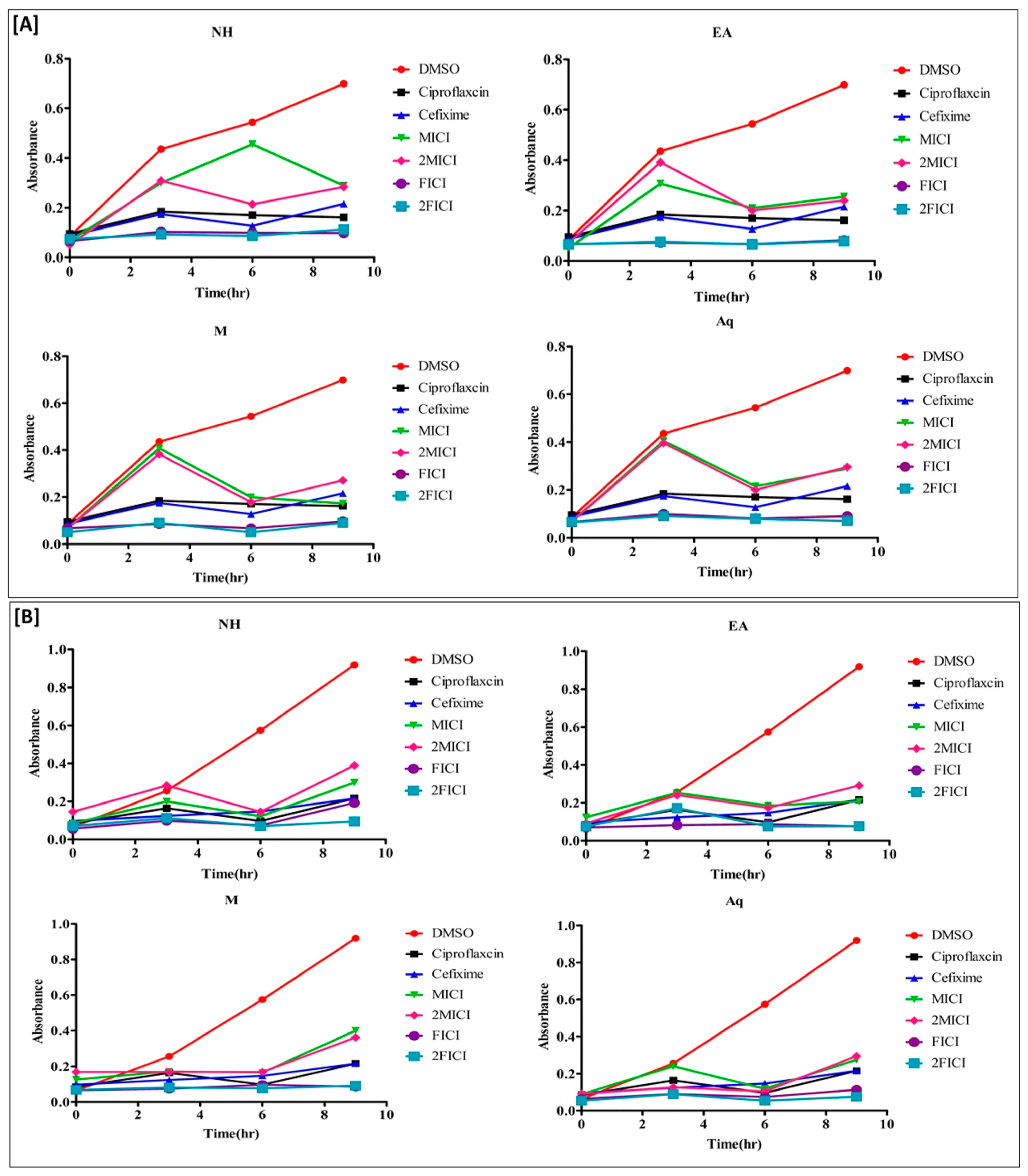
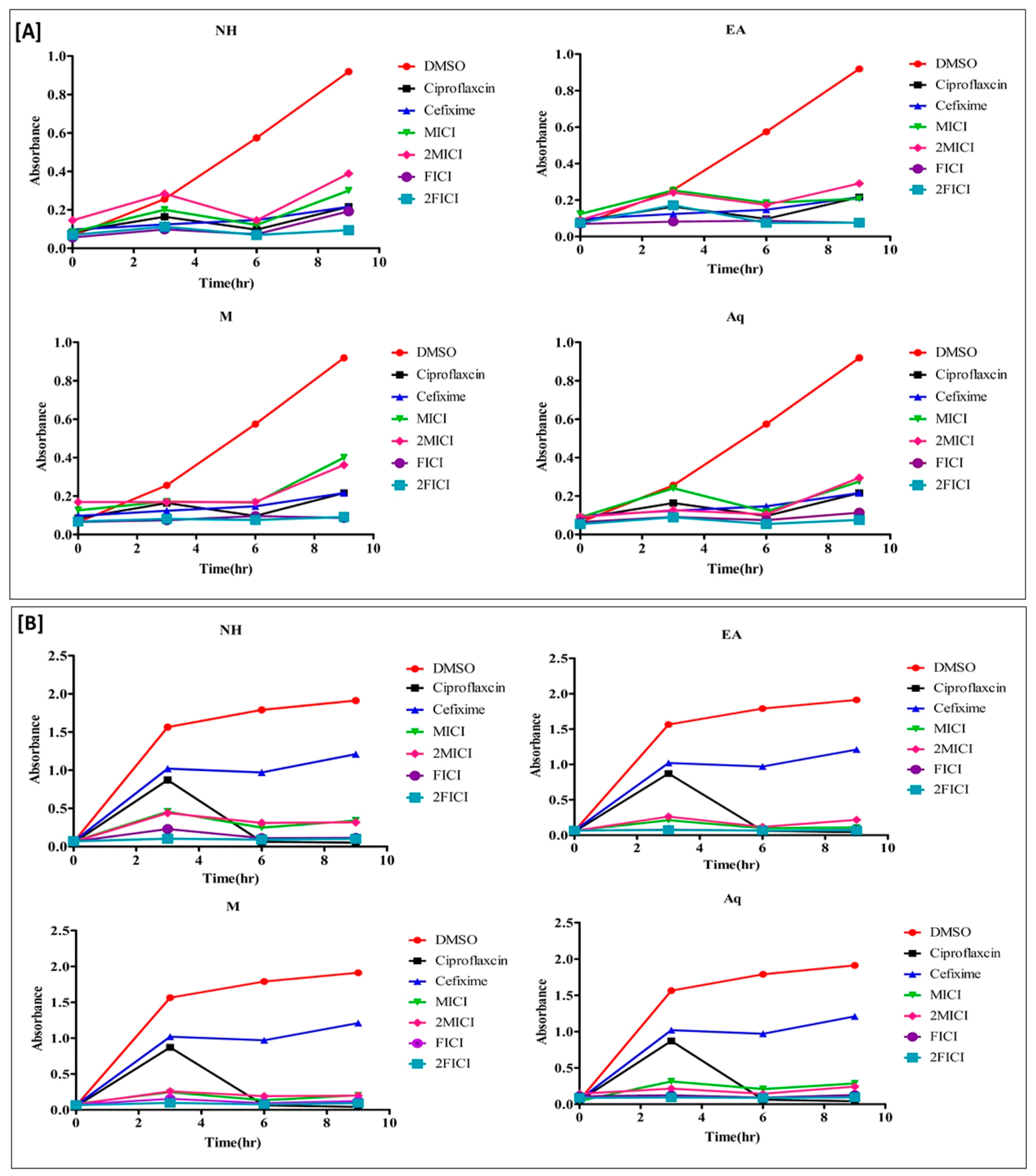
2.5. The Effect of the Extracts Alone and in Combination Is Time Dependent

Time–kill kinetic studies were performed to assess whether the effect of A. brevifolia extracts alone and in combination with cefixime was time-dependent or concentration-dependent. All clinical isolates were tested at MIC, 2MIC, FICI, and 2FICI values. Overall, clinical isolates treated with the combination of cefixime and extracts at FICI and 2FICI values demonstrated significant growth inhibition throughout the treatment duration as compared to individual treatments. The results were comparable with those of ciprofloxacin (positive control), to which the clinical isolates were susceptible. Bacterial growth in samples treated with extracts alone was much lower as compared to cefixime and DMSO (negative control).
Treatment of resistant E. coli with A. brevifolia EA extract (Figure 3A) at FICI values demonstrated maximum growth inhibition of 91.8% and 81.7% at 3 h and 9 h, respectively, as compared to 50% and 40.3% inhibition at the same time points with cefixime alone. Likewise, the Aq, EA, and NH extracts at 2FICI values showed growth inhibition of 93%, 83.5%, and 67.3% after 9 h of treatment. Treatment of cefixime-resistant P. aeruginosa (Figure 3B) with extracts alone (MIC and 2MIC) or cefixime showed inhibition of bacterial growth till 6 h of treatment. Later, there was an exponential increase in bacterial growth as depicted by the increased absorbance of the samples. The combination of cefixime with A. brevifolia EA extract at 2FICI demonstrated 100% and 98.7% inhibition of clinical isolates as compared to 66% and 44.9% inhibition with cefixime alone at 6 h and 9 h of treatment, respectively. Similarly, 2FICI dosing of NH, M, and Aq extracts also showed 100%, 89.5%, and 100% inhibition, respectively, of clinical isolates at 6 h of treatment. Although this declined to 73.4%, 74.7%, and 72.4% inhibition at 9 h for NH, M, and Aq extracts, the values were still higher than for cefixime alone (44.9%), indicating continued synergism between samples.
The succeeding analysis on S. hemolyticus (Figure 4B) demonstrated a similar pattern of growth as that of E. coli but with different periods. The 2FICI dosing for all extracts was most effective in inhibiting the growth of cefixime-resistant S. hemolyticus, which peaked after 6 h of treatment. There was 67.3%, 80.3%, 84.2%, and 76.3% growth inhibition at 6 h with 2FICI of NH, EA, M, and Aq extracts, respectively, as compared to 54.7% inhibition when using cefixime alone.
There was a drastic reduction in MRSA resistance (Figure 4A) to cefixime with FICI and 2FICI dosing. The growth of MRSA clinical isolates was reduced by >10-fold through the synergistic action of cefixime and A. brevifolia extracts. Like all other samples, the effect of FICI and 2FICI peaked at 6 h with percent inhibitions of 79.6%, 100%, 87.2%, and 100% at 2FICI for NH, EA < M, and Aq extracts, respectively. It was much higher than the 6.7% growth inhibition by cefixime alone at 6 h. In short, A. brevifolia extracts potentiated the antibacterial effect of cefixime at FICI and 2FICI values with a stationary growth phase after treatment, irrespective of time duration.

2.6. A. brevifolia Extracts Reduce Bacterial Protein Content

Disintegration of the cell envelope can be quantified using the leakage of cellular protein as a function of cell death. Protein content in the extracellular medium of treated and untreated bacterial strains was analyzed (Table 6) to understand the underlying cause of the antibacterial effect. Bovine serum albumin was used as a positive control. There was little reduction (5.4%) in protein content after treatment of resistant S. hemolyticus with cefixime alone. This increased to 79.8%, 68.4%, 78.4%, and 62.7% reduction in protein content when resistant S. hemolyticus was treated with the combination of cefixime and NH, EA, M, and Aq extracts, respectively. This indicates that the extracts and antibiotic synergy can degrade the bacterial protein, making its survival difficult. Furthermore, there was 73.9%, 74.4%, 82.5%, and 75% inhibition of protein content in cefixime-resistant MRSA isolates when treated with the combination of NH, EA < M, and Aq extract, respectively, as compared to cefixime (35% reduction). Similarly, a percent protein reduction of 58.8–80.2% was observed in resistant P. aeruginosa and E. coli isolates due to the synergistic activity of cefixime and A. brevifolia extracts. It is postulated that the extracts can inhibit a bacterial protein that works together with the cell wall synthesis inhibitor cefixime to inhibit the growth of resistant clinical isolates. This seems to be the case considering the results of the protein content estimation.

2.7. Hemolytic Analysis

Hemolytic analysis was performed to check whether the drug or compound was toxic to red blood cells causing hemolysis. According to ASTM F756-00 protocols for assessment of the hemolytic properties of samples, substances with hemolysis percentages of >5%, <5%, and <2% are considered hemolytic, slightly hemolytic, and non-hemolytic, respectively [22]. To our surprise, the extracts were hemolytic with >5% hemolysis when used alone. However, their hemolytic potential declined when given in combination with cefixime. All combinations except NH/cefixime (6.45% hemolysis) had values ranging between 1.34% and 5.13% for FICI and 0% and 3.78% for 2FICI. This indicated that the majority of combinations were safe to use with either slight or non-hemolytic character. These results were significantly (p < 0.05) lower than for the positive control Triton-X 100 (100% hemolysis).

3. Discussion

Antimicrobial resistance has multiple causes, and it is a great concern in health sciences as it causes treatment failures and poor prognoses of infectious diseases. Researchers are investigating multiple compounds from natural and synthetic sources that can work either alone or in combination with standard antibiotics to eliminate resistant infections [23]. Phytoconstituents of various medicinal plants have proven antibacterial activity against standard antibiotic-susceptible and -resistant bacteria [24,25,26,27]. Plants act as the greatest apothecary and a potential source of treatments for multiple diseases like arthritis, cancer, diabetes mellitus, and oxidative stress disorder [28,29]. Considering the beneficial aspects of medicinal plants, in the current work, we established the antibacterial activity of Artemisia brevifolia extracts in resistant clinical bacterial isolates. We also verified the synergistic interaction between cefixime and A. brevifolia extracts that potentiates the antibacterial activity against cefixime-resistant clinical isolates.
A. brevifolia extracts were prepared in different solvents to yield variable phytoconstituents based on polarity, as follows:
  • Extraction can be a potentially rate-limiting step when preparing samples for screening bioactive compounds of interest. The efficiency of this step is affected by many factors such as solvent polarity, extraction method, physical characteristics size of sample particles, and period of extraction. These contingency factors were addressed first by selecting four solvents for extraction, n-hexane, methanol, ethyl acetate, and water, depicting variable polarity.
  • Second, samples were macerated for 72 h, ensuring sufficient time for solvents to penetrate the fine particles of powdered plant.
  • Third, maceration was combined with periodic sonication, aiding the diffusion of solvent and extraction of phytoconstituents from powdered plant material. A significant extraction yield (6.1%) was obtained with Aq extract, which indicated the presence of more polar content.
The oligosaccharides, sugars, and resins often solubilize better in distilled water compared to other solvents, contributing to overall extraction yield [30]. Although extraction yield depends on the polarity of phytoconstituents and solvent used for extraction, the maximum yield does not dictate the medicinal value since it is directed by the chemical composition and inherent nature of phytochemicals [31].
Preliminary phytochemical analysis of A. brevifolia extracts showed the presence of alkaloids, glycosides, tannins, and terpenoids in different extracts. Alkaloids can inhibit bacterial growth by altering membrane permeability, inhibiting nucleic acid synthesis, and disrupting cell division [32]. In addition, research showed that glycosides such as glycyrrhizin have promising antibacterial effects due to inhibition of RNA synthesis in bacteria [33]. Furthermore, tannins exhibit antidiarrheal, antibacterial, antiviral, antitussive, antitumor, and wound-healing activities [34]. Previously reported studies showed that saponins have detergent-like activity and an antibacterial effect by increasing bacterial cell wall permeability [21]. The presence of these phytoconstituents in A. brevifolia extracts can be responsible for the subsequent antibacterial activity of the plant.
Bacteria have started to develop resistance against commonly used antibiotics. Resistant species of S. aureus, S. hemolyticus, E. coli, etc., have been recognized in various clinical settings as causing frequent infections and prolonged duration of the infectious diseases [35,36,37].
Medicinal plants are being investigated to optimize therapy for resistant infections. In the current study, the susceptibility of bacterial clinical isolates to selected antibiotics and A. brevifolia extracts was assessed to determine the resistance profile and antibacterial capacity of the samples. The disc diffusion method creates zones of inhibition around the sample-impregnated discs, signifying the antibacterial activity. The greater the size of ZOI, the higher the susceptibility of microorganisms to test samples. Results revealed all the extracts demonstrated mild to moderate antibacterial activity, whereby EA extract was more active against MRSA and R. S. hemolyticus. On the contrary, NH and M extracts were active against R. E. coli and R. Pseudomonas aeruginosa, respectively.
Hydroxylated phenols and phenolic compounds are found to be toxic to many microorganisms. The toxicity of phenols to microorganisms depends on the level of hydroxylation, where higher hydroxylation levels are more toxic to the microorganisms [11]. Genus Artemisia is rich in several essential metabolites such as glucosinolates, saponins, cyanogenic glycosides, tannins, unsaturated lactones, phenols, and flavonoids. These phytochemicals are used to treat multiple ailments such as malaria, bacterial infection, cancer, and inflammation [38]. In this study, the antibacterial activity of extracts might have been due to hydroxylated phenols present in this plant, which were quantified through HPLC (emodin, luteolin vanillic acid, syringic acid, gallic acid, coumarins, flavonoids, and flavones) [39].
Furthermore, the susceptibility testing revealed that all clinical isolates were resistant to cefixime. Hence, clinical isolates were treated with cefixime in combination with A. brevifolia extracts, to observe the possibility of synergism. The “one drug, one target, one disease” paradigm has become an orthodox pharmaceutical strategy considering the emergence of resistance to even previously potent antibiotics. Currently, a multi-drug-target approach is utilized to augment the efficacy of antibacterial therapy. This pattern shift is dictated by the limited effectiveness, resistance, and side effects of monotherapy [40]. The prime advantage of plant-based drugs is that they can be safe, easily affordable, have minimal or no side effects, and have multiple biological targets. A combination of standard antibiotics with plant-based drugs can provide better synergism with the least side effects, particularly against resistant infections. Synergism can decrease the MIC of many marketed antibiotics in the presence of plant extracts. Research shows that polyphenols decrease beta-lactam resistance while flavonoids, diterpenes, and triterpenes have resistance-modulating abilities on many contemporary antibiotics [28,41,42,43].
Researchers have believed that generally some mechanisms that can cause this interaction are inhibition of the sequence of biochemical paths, membranotropic agent usage to enhance the diffusion of other antibacterial drugs, inhibition of enzymes that protect microorganisms, and a membrane-active agent used in combination [44].
In the current study, the checkerboard method was used to determine the synergistic interaction between cefixime and A. brevifolia extracts. Previous research outlined that if the MIC of the extract and antibiotic decreases by four-fold, then the combination is known as synergistic, while if MIC of the first test sample decreases by four-fold and the other by two-fold, then the interaction is known as partial synergistic [45,46]. Treatment of all clinical isolates with a combination of cefixime and A. brevifolia EA extract showed a four- to ten-fold reduction in MIC values of cefixime. This was reinforced by the FICI values, which were ≤0.5, indicating total synergism between the two samples. Likewise, A. brevifolia M extract enhanced the susceptibility of E. coli to cefixime by two-fold, with a FICI value of 0.66, demonstrating partial synergism. The limitation of the checkerboard method is that more resources are used to test antibacterial combinations and more than one antimicrobial cannot be checked at a single time [45]. However, it provided evidence that the extract–cefixime synergism successfully inhibited the growth of both Gram-positive and Gram-negative clinical isolates used in this study, showing a broad spectrum of activity. It is possible that A. brevifolia extracts either inhibited the sequence of biochemical paths, enhanced diffusion of cefixime, inhibited protein synthesis of bacteria, or inhibited degradation of antibiotics [44]. Future work is planned to assess the mechanism of synergistic interaction observed in this study.
Next, we determined using time–kill kinetic studies whether the interaction between A. brevifolia extracts and cefixime was bactericidal or bacteriostatic. Jacqueline et al. described how time–kill kinetic studies are used to determine the bactericidal effect, which may be dependent on time in place of concentration [47]. It was observed that there was significant inhibition of E. coli growth when treated with the combination of A. brevifolia extracts and cefixime (FICI, 2FICI) as compared to A. brevifolia extracts alone (MIC, 2MIC). DMSO (negative control) did not interfere with the results, with constant exponential growth for all isolates. Cefixime-resistant clinical isolates showed initial growth from 0 to 3 h after treatment with MIC and 2MIC values of the extracts. Then, it gradually started to decline after 3 h of treatment. On the other hand, the growth of E. coli started to decline after 6 h of treatment with A. brevifolia NH extract. Samples at their FICI and 2FICI values obstructed the clinical isolates in their stationary phase of the growth curve throughout the treatment duration.
The trend of resistant S. hemolytic growth was the log phase, partial death phase, and again log phase for the durations of 0–3, 3–6, and 6–9 h, respectively, when treated at MIC and 2MIC values. On the contrary, clinical isolates treated at 2FICI more or less remained in the stationary phase, with no significant overall growth. Although isolates treated with FICI also displayed greater growth inhibition of cefixime-resistant S. hemolyticus as compared to cefixime or extracts alone, an increase in the bacterial growth was observed after 9 h of treatment. Exposure of cefixime-resistant P. aeruginosa to MIC and 2MIC of extracts or cefixime alone displayed inhibition of clinical isolates till 6 h of treatment. Later, there was an exponential increase in bacterial growth, as represented by the amplified absorbance of the samples. On the contrary, growth inhibition after treatment with the combination of cefixime and extracts (FICI, 2FICI) was more pronounced as compared to lone treatments. Moreover, A. brevifolia M extract imparted better synergism against cefixime-resistant P. aeruginosa as compared to other extracts by keeping the clinical isolates in a stationary phase.
The MRSA clinical isolate that was used in the present study showed noteworthy resistance against cefixime with exponential growth. In contrast, there was exponential bacterial growth in the first 3 h of treatment with ciprofloxacin (positive control) but it drastically declined later than 6 h of treatment. Likewise, all A. brevifolia (MIC; 2MIC) extracts significantly halted MRSA growth. The competition between survival and growth inhibition of bacteria generated slight variability in percent inhibition values at different time points. However, overall, it could be seen that the combination of extract and cefixime augmented the activity of cefixime against clinical isolates.
In the current study, the combination of cefixime with plant extract against all bacterial isolates showed synergistic interaction when calculating the FICI index in the checkerboard method, while time–kill kinetic curves showed additive interaction. This same pattern of interaction was reported previously for Helichrysum pedunculatum plant methanolic extracts when given in synergy with antibiotics against Staphylococcus aureus [41]. Likewise, the interactions of antibiotic drugs with acetone extract of seeds of Garcinia kola [48] and Thymus vulgaris were also reported as synergistic/additive when time–kill kinetic assays were performed [49].
Antibacterial drugs inhibit bacterial growth or kill bacteria by targeting proteins, cell walls, cell membranes, and nucleic acid synthesis [50]. We assessed A. brevifolia and cefixime synergy by analyzing the protein content of the medium in clinical isolates. Cefixime disrupts cell wall integrity [51], and its combination with A. brevifolia extract significantly (p < 0.05) reduced viable proteins. Cefixime’s impact on protein content is limited due to its role in cell wall inhibition. However, combined treatment likely curbed protein synthesis or induced apoptosis, lowering protein content, suggesting A. brevifolia’s influence on bacterial protein synthesis, potentially through tRNA release inhibition, peptide bond synthesis, or initiation of complex formation [50]. Similar effects were observed for Cymbopogon khasianus on resistant clinical isolates [3].
HPLC quantification was performed using 14 polyphenol standards, and it confirmed the flavonoids and polyphenols in A. brevifolia. Some of the phytochemicals present in A. brevifolia were vanillic acid, gallic acid, caffeic acid, and syringic acid [39]. A literature review showed that vanillic acid ruptures the cell membrane and inhibits cell growth [52], while gallic acid inhibits biofilm production and disrupts the bacterial cell membrane [53]. Caffeic acid acts by inhibiting the RNA polymerase enzyme, and syringic acid is an ATP synthesis inhibitor [11]. All these phytochemicals have been quantified in the subject plant. Therefore, it can be hypothesized that when these polyphenols combined with cefixime resulted in an additive mechanism of action, as suggested in the literature, phytochemicals may have acted upon the cell wall integrity [54]. It has been also testified that some plant chemical compounds inhibit bacterial growth or improve the effect of antibacterial drugs by acting in the same site as peptidoglycan [55].
In the present study, we performed a toxicity analysis as a part of the efficacy study. For this purpose, a hemolytic assay was performed that gives information regarding the cytotoxicity of samples on blood. This model is frequently used because of the easy availability and isolation protocols of red blood cells. Moreover, the membrane physiology of red blood cells is similar to the membranes of other cells present in the body [56]. A. brevifolia extracts in combination with cefixime displayed slightly hemolytic or non-hemolytic character; this was different from A. brevifolia NH and M extracts alone, which presented hemolytic character. The EA extracts were found to be safer to use in humans as a component of antibacterial therapy. Yet, in vivo toxicity studies must be conducted to determine detailed toxicity versus efficacy profiles.
The strengths of this study are evident in the significant findings that highlight the potential of A. brevifolia as a potent antibacterial agent. The identification of substantial minimum inhibitory concentrations in both ethyl acetate and methanolic extracts against resistant bacterial strains, including Staphylococcus haemolyticus, methicillin-resistant Staphylococcus aureus, Escherichia coli, and Pseudomonas aeruginosa, underscores the broad-spectrum antibacterial activity of these extracts. Moreover, the observation of total synergistic effects when the ethyl acetate extract was used in combination against all bacterial strains is particularly promising. The study’s assessment of safety, as indicated by negligible hemolysis of red blood cells, adds to its credibility.
However, it is important to acknowledge the limitations of this research. While the in vitro findings are encouraging, the transition to in vivo studies is crucial to determine the real efficacy and potential toxicity effects when these extracts are used in living organisms. Clinical controlled trials are necessary to validate the therapeutic potential of these extracts in treating infectious diseases, and further research should aim to increase the variety of drugs, expand the number of clinical isolates, and identify the specific compounds within the plant extracts responsible for their antibacterial properties. Additionally, a deeper exploration into the mechanism of action and the formulation of pharmacological agents based on these extracts would enhance the practical application of this promising research.

4. Materials and Methods

4.1. Materials

4.1.1. Chemicals and reagents

Methanol (M), ethyl acetate (EA), n-hexane [49], acetonitrile (ACN), acetic acid (AA), sulfuric acid, nutrient agar, nutrient broth, fetal bovine serum, and Giemsa stain were purchased from Merck (Darmstadt, Germany). Dimethyl sulfoxide (DMSO) was purchased from Duksan Pure Chemicals (Ansan-si, Republic of Korea). All other chemicals and drugs were purchased from Sigma-Aldrich (Darmstadt, Germany) [51] unless otherwise stated.

4.1.2. Cultures

Four resistant bacterial clinical isolates that were maintained in the laboratory were used to evaluate the antimicrobial potential of extracts. These were resistant (R.) Staphylococcus (S.) hemolyticus (MIC-101), R. Escherichia coli [57] (MIC-102), R. Pseudomonas (P.) aeruginosa (MIC-103), and methicillin resistant S. aureus (MRSA; MIC-104).

4.2. Methods

4.2.1. Preparation of Extracts

A. brevifolia was collected in August 2018 from Hunza Valley, Baltistan by Dr. Ihsan ul Haq, Associate Professor, Department of Pharmacy, Quaid-i-Azam University, Islamabad. It was identified by Dr Sher Wali Khan, Karakoram International University, Gilgit Baltistan. The specimen was submitted (Herbarium No# PHM 512) to the Herbarium of Medicinal Plant, Quaid-i-Azam University, Islamabad, Pakistan. The collection and investigations of A. brevifolia were supported in part by the Indigenous Fellowship of HEC provided to the first author (520-142973-2MD6-130 (50093185)). About 12 kg of plant material was washed, shade dried (3 weeks), pulverized to a coarse powder, and stored in an airtight container. Successive maceration aided with ultra-sonication was used to extract plant material as previously reported [31]. Dry powder was macerated with four analytical-grade solvents (non-polar to polar) including NH, EA, M, and distilled water (Aq) at a ratio of 1:4 (powder:solvent) for 72 h at 25 °C with 10 min of sonication each day. After 3 days, extracts were filtered and concentrated using a reduced-pressure rotary evaporator (Ribby, UK) at 45 °C. Plant extracts were collected in labeled containers and stored at −80 °C temperature for further testing. The dried extracts were weighed to calculate the percentage extract recovery using the formula:
% age   Extract   Recovery = T o t a l   w e i g h t   o f   e x t r a c t   a f t e r   d r y i n g T o t a l   w e i g h t   o f   p l a n t   p o w d e r × 100 ,

4.2.2. Total Phenolic Content

The protocol used to determine total phenolic content was given by [31] as was subject to few modifications. In our work, 20 µL of each test sample (4 mg/mL) was taken and poured into a 96-well plate followed by the addition of 90 µL Folin–Ciocalteu reagent. The plate was incubated for 5 min, and then sodium carbonate was added to the reaction mixture. After that, the absorbance of the plate was taken at 630 nm using a microplate reader (BioTek; Shoreline, USA). DMSO was used as negative while gallic acid was used as positive control. The assay was carried out in triplicate and results were given as mg gallic acid equivalent per gram dry weight.

4.2.3. Glycoside, Alkaloidal, Tannin, Saponin, and Terpenoid Contents

Three types of glycosides (cardiac, anthraquinone, and coumarin) were determined using the method given by Shaikh et al. [58] with minor modifications. Cardiac glycosides were confirmed through Keller Killani, Salkowski, and Baljet tests. Anthraquinone glycosides were estimated via borax and modified Bontrager’s tests. On the other hand, the sodium-hydroxide-mediated fluorescence method indicated the presence of coumarin glycosides.
The alkaloidal content of extracts was determined using Wagner’s, tannic acid, and Dragendroff’s reagents, as given by [59].
Tannins in A. brevifolia extracts were detected using the protocol given by [59]. Ferric chloride and gelatin solution were used for this purpose.
Saponin content in A. brevifolia extracts was evaluated via foam formation with or without olive oil, as previously described by [60].
Phytochemical analysis for the presence of terpenoids was performed using the method given by [61], which uses chloroform and sulfuric acid to precipitate reddish brown terpenoids.

4.2.4. Antimicrobial Evaluation: Preliminary Resistance Profiling of Antibiotics

Initially, antibiotics were tested against clinical isolates by using the disc diffusion method. Stock solutions (4 mg/mL) of antibiotics (cefixime, ciprofloxacin, doxycycline, lincomycin, and clarithromycin) were prepared in DMSO. Agar media was poured on plates and sterile discs loaded with 5 µL of antibiotics were placed on the plates and incubated for 24 h at 37 °C. The zone of inhibition (ZOI) around each disc was measured using a vernier caliper. The assay was carried out in triplicate. The antibiotic with little or no ZOI was selected for further studies. Minimum inhibitory concentrations [21] of antibiotics were determined by using the micro broth dilution protocol reported by [62] with little modification.

4.2.5. RP-HPLC Analysis

LOD and LOQ Determination

LOD and LOQ for HPLC analysis were determined. LOD stands for the limit of detection, which is the lowest concentration of sample that can be detected in HPLC, while LOQ stands for the limit of quantification, which is the minimum concentration of sample that can be quantified using HPLC. These two parameters are determined using the following formulae:
LOD = 3.3 × S D S
LOQ = 10 × S D S
where SD = standard deviation of regression and S = slope of the calibration curve.

Analysis of Polyphenols

To identify and measure the number of polyphenols present in A. brevifolia crude extracts, RP-HPLC was utilized according to the standard protocol with minor modifications [20,39]. A zorbex-C8 analytical column (5 µm; 4.6 × 250 mm) connected with a diode array detector (DAD) was supplied with an HPLC system (Shimadzu; Kyoto, Japan). A binary gradient system with mobile phase A (methanol: water: acetic acid: acetonitrile in 10:85:1:5) and mobile phase B (acetonitrile: methanol: acetic acid in 40:60:1) was used to accomplish the polyphenol detection. The column was injected with 50 µL of sample solution prepared in methanol, and the flow rate was adjusted at 1.2 mL/min. The gradient volume of mobile phase B was changed from 0% to 75% in the first 0–30 min, to 75–100% in the next 30–31 min, then to 100% in 31 to 35 min and, lastly, to 0% in the final 36 to 40 min. The column was reconditioned before injecting a new sample. The concentration of each standard was 50 µg/mL in methanol, while extracts were prepared at 10 mg/mL in methanol. The quantification of polyphenols was performed by comparing the UV-Vis spectra and retention times of chromatographic peaks to reference standards; polyphenols were identified at 257 nm for vanillic acid and rutin, 279 nm for apocynin, coumaric acid, catechin, syringic acid, and gallic acid, 325 nm for apigenin, caffeic acid, ferulic acid, gentisic acid, and luteolin, and 368 nm for quercetin and kaempferol. The results were quantified in terms of µg/ mg extract (µg/mgE).

4.2.6. Antibacterial Assay

The antibacterial potential of A. brevifolia extracts was determined by using the disc diffusion method [62] as described above. A bacterial culture at a seeding density of 1 × 10⁶ CFU/mL was used to make bacterial spreads on nutrient agar plates. About 5 µL of each test sample (cefixime/ciprofloxacin, 20 µg/disc; extracts, 100 µg/disc) was poured on sterile filter discs, and then placed on a nutrient agar plate and incubated (24 h at 37 °C). Ciprofloxacin was used as a positive control, whereas DMSO served as a negative control. After 24 h, ZOI was calculated by using a vernier caliper. The assay was run in triplicate.
Next, samples that showed ZOI ≥ 12 mm were further tested to determine the MIC by using the micro broth dilution method [62]. A bacterial inoculum was prepared by adjusting the seeding density at 5 × 10⁴ CFU/mL. Three-fold serial dilutions of extracts (100, 33.3, 11.1, and 3.34 µg/mL) and antibiotics (10, 3.33, 1.11, and 0.334 µg/mL) were prepared in nutrient broth. About 5 µL of the test sample and 195 µL of inoculum were mixed in each well of a 96-well plate. The plate was then incubated at 37 °C for 24 h, and absorbance was measured at 600 nm after 30 min (0 h reading) and 24 h of incubation in a microplate reader (BioTek; Shoreline, DC, USA).
The checkerboard method was used to determine that the potential synergistic interaction between antibiotic and A. brevifolia extracts [63]. Two-fold serial dilutions of the sample were prepared such that the antibiotic was diluted vertically while extracts were diluted horizontally in a 96-well plate. An aliquot of 5 µL of sample was poured in each well containing 2.5 µL of extract and 2.5 µL of antibiotic, followed by 195 µL of inoculum (density 4 × 10⁴ colony-forming unit (CFU)/mL) in each well. The plates were then incubated at 37 °C for 24 h, and FICI values were determined. Absorbance was taken at 0 h and after 24 h for further calculations:
FICI = M I C I B A M I C A + M I C I A B M I C B ,
where MICIA/B = MIC of compound A in combination with compound B, MICA = MIC of compound A, MICIB/A = MIC of compound B in combination with compound A, and MICB = MIC of compound B.
The interaction between antibiotic and extract was considered “total synergism” or “partial synergism” at FICI ≤ 0.5 and 0.5 < FICI ≤ 0.75, respectively. If FICI was ≤1 or between 1 and 4, then the interaction was termed “Indifference” or “No effect”. If the FICI value was more than 4, then it was “Antagonism” [63].
Time–kill kinetics was performed using the protocol described previously [3,64] with few modifications. Resistant bacterial strains were grown to the mid-logarithmic phase. Bacterial cells were diluted up to 104 CFU/mL. This bacterial suspension was then incubated with MIC, 2MIC, FICI, and 2FICI concentrations of extracts alone and in combination with selected antibiotics. Readings were taken at time intervals of 0, 3, 6, and 9 h after incubation. Results were measured using a UV spectrophotometer (Sigma Aldrich; Darmstadt, Germany) at 600 nm.
Bacteria were grown up to the mid-logarithm phase, as described previously, and were treated with MIC, 2MIC, FICI, and 2FICI of extract alone and in combination with selected antibiotic [65]. Protein content estimation of clinical isolates was conducted using the Bradford reagent to check the possible mechanism of action of bacterial growth inhibition. After incubation of samples clinically isolated for 24 h, 5 µL of the reaction mixture was mixed with 195 µL of Bradford reagent and incubated for 5 min at room temperature with constant sonication in triplicate. Then, absorbance was measured at 595 nm. The protein content of samples was calculated using the formula:
Absorbance   of   unknown   sample   ( x ) = A b s o r b a n c e b   m ,
Phosphate buffer was used as a diluent in this assay. The negative control, positive control, and blank were constituted by the clinical isolate inoculum, bovine albumin serum (0–50 µg/mL), and distilled water, respectively.

4.2.7. Hemolytic Assay

Hemolytic evaluation was performed using freshly drawn human blood following the guidelines given by the ethical committee of Quaid-i-Azam University, Islamabad, Pakistan. Bioethical approval was given by the Ethical Committee of the university (BEC-FBS-QAU2021-261 dated 2 March 2021). Informed consent was also obtained from the volunteer to draw blood samples. Blood was collected and centrifuged at 13,000 rpm to separate red blood cells (RBCs). After that, RBCs were washed thrice with normal saline and re-suspended in phosphate buffer to form a 5% solution. The RBC suspension was incubated with samples at 37 °C for 30 min in Eppendorf (2 mL) tubes. Afterward, it was centrifuged at 2000 rpm for 20 min and the supernatant was separated. Triton X-100 (0.1%) was used as the positive control, whereas phosphate buffer served as the negative control [3]. The absorbance of the supernatant was measured at 360 nm in a microplate reader (BioTek; Shoreline USA). Hemolysis was calculated using the following formula:
% age   Hemolysis = A b s o r b a n c e   o f   s a m p l e A b s o r b a n c e   o f   N e g a t i v e A b s o r b a n c e   o f   p o s i t i v e A b s o r b a n c e   o f   N e g a t i v e × 100 ,

4.3. Statistical Evaluation

Statistical analysis of the experimental results was conducted utilizing GraphPad Prism 5 (version 5.00 for Windows, San Diego, CA, USA). The presented data included mean values accompanied by either the standard error of the mean (SEM) or the standard deviations (SDs) of individual replicates. Additionally, for each experiment, the analysis software employed and the number of observations were specified.

5. Conclusions

In this research, all A. brevifolia extracts that were examined exhibited noteworthy antibacterial activity. Particularly, the ethyl acetate extract demonstrated significant activity against resistant strains of Staphylococcus hemolyticus and methicillin-resistant Staphylococcus aureus. Additionally, the methanol extract displayed notable inhibition of growth in resistant strains of Escherichia coli and Pseudomonas aeruginosa.
Furthermore, the ethyl acetate extract exhibited complete synergism when combined with cefixime against all cefixime-resistant clinical isolates. This synergistic effect was observed to be time-dependent, with the maximum bactericidal activity occurring at 2FICI concentrations after 6 and 9 h of treatment, varying among different bacterial strains.
Notably, our study revealed a significant reduction in the protein content of bacterial samples, suggesting potential mechanisms of action. This reduction could be attributed to the combined effects of protein synthesis inhibition or protein degradation induced by the A. brevifolia/cefixime combination, along with the disruption of cell wall integrity caused by cefixime.
In light of these findings, our research strongly advocates for further comprehensive investigations into the application of A. brevifolia extracts in synergy with cefixime as a potential strategy for combating bacterial infections that have developed resistance. These promising results warrant in-depth exploration and consideration for the development of novel treatments for drug-resistant bacterial infections.

Author Contributions

Conceptualization, A.Z. and I.u.H.; methodology, A.Z.; software, A.Z. and Y.W.; validation, D.M., I.u.H. and M.M.; formal analysis, D.M.; investigation, A.Z., Y.W. and I.u.H.; resources, I.u.H.; data curation, A.Z., Y.W. and M.M.; writing—original draft preparation, A.Z. and S.G.B.; writing—review and editing, D.M., M.M. and S.G.B.; visualization, I.u.H.; supervision, S.G.B. and I.u.H.; project administration, I.H; funding acquisition, S.G.B. All authors have read and agreed to the published version of the manuscript.

Funding

This research was funded by the Romanian Ministry of Research, Innovation and Digitization through Program 1—Development of the National Research and Development System, Subprogram 1.2—Institutional Performance—Projects for funding the excellence in RDI, Contract No. 29 PFE/30.12.2021 with the University of Oradea.

Institutional Review Board Statement

The plant used in this study is widely available in Pakistan and is not an endangered species. All studies on plants and microorganisms were conducted according to the ethical guidelines of Quaid-i-Azam University, Islamabad (BEC-FBS-QAU2021-261).

Informed Consent Statement

Not applicable.

Data Availability Statement

Data related to the present research are provided throughout the manuscript and/or are supported by the inserted references.

Acknowledgments

The authors express their gratitude to the University of Oradea, Oradea, Romania, for supporting the APC.

Conflicts of Interest

The authors declare no conflict of interest. The funders had no role in the design of the study; in the collection, analyses, or interpretation of data; in the writing of the manuscript; or in the decision to publish the results.

References

  1. Hamad, B. The antibiotics market. Nat. Rev. Drug Discov. 2010, 9, 675–676. [Google Scholar] [CrossRef] [PubMed]
  2. Bower, C.; Daeschel, M. Resistance responses of microorganisms in food environments. Int. J. Food Microbiol. 1999, 50, 33–44. [Google Scholar] [CrossRef]
  3. Singh, G.; Katoch, M. Antimicrobial activities and mechanism of action of Cymbopogon khasianus (Munro ex Hackel) Bor essential oil. BMC Complement. Med. Ther. 2020, 20, 331. [Google Scholar] [CrossRef]
  4. Thomford, N.E.; Senthebane, D.A.; Rowe, A.; Munro, D.; Seele, P.; Maroyi, A.; Dzobo, K. Natural products for drug discovery in the 21st century: Innovations for novel drug discovery. Int. J. Mol. Sci. 2018, 19, 1578. [Google Scholar] [CrossRef] [PubMed]
  5. Newman, D.J.; Cragg, G.M.; Snader, K.M. The influence of natural products upon drug discovery. Nat. Prod. Rep. 2000, 17, 215–234. [Google Scholar] [CrossRef] [PubMed]
  6. Bungãu, S.G.; Popa, V.-C. Between religion and science some aspects concerning illness and healing in antiquity. Transylv. Rev. 2015, 24, 3–18. [Google Scholar]
  7. Koehn, F.E.; Carter, G.T. The evolving role of natural products in drug discovery. Nat. Rev. Drug Discov. 2005, 4, 206–220. [Google Scholar] [CrossRef]
  8. Nogueira, J.O.E.; Campolina, G.A.; Batista, L.R.; Alves, E.; Caetano, A.R.S.; Brandão, R.M.; Nelson, D.L.; Cardoso, M.D.G. Mechanism of action of various terpenes and phenylpropanoids against Escherichia coli and Staphylococcus aureus. FEMS Microbiol. Lett. 2021, 368, fnab052. [Google Scholar] [CrossRef]
  9. Tit, D.M.; Bungau, S.G. Antioxidant activity of essential oils. Antioxidants 2023, 12, 383. [Google Scholar]
  10. Rufai, M.S. Essential Oils and Their Antimicrobial Activity: A Review. UMYU J. Microbiol. Res. 2017, 2, 87–93. [Google Scholar] [CrossRef]
  11. Cowan, M.M. Plant products as antimicrobial agents. Clin. Microbiol. Rev. 1999, 12, 564–582. [Google Scholar] [CrossRef] [PubMed]
  12. Scalbert, A. Antimicrobial properties of tannins. Phytochemistry 1991, 30, 3875–3883. [Google Scholar] [CrossRef]
  13. Khan, N.; Ahmed, M.; Ahmed, A.; Shaukat, S.; Wahab, M.; Ajaib, M.; Siddiqui, M.F.; Nasir, N. Important medicinal plants of chitral gol National park (cgnp) Pakistan. Pak. J. Bot. 2011, 43, 797–809. [Google Scholar]
  14. Ashraf, M.; Hayat, M.Q.; Jabeen, S.; Shaheen, N.; Khan, M.A.; Yasmin, G. Artemisia L. species recognized by the local community of northern areas of Pakistan as folk therapeutic plants. J. Med. Plants Res. 2010, 4, 112–119. [Google Scholar]
  15. Ahsan, H.; Ijaz, M.U.; Ashraf, A.; Ehsan, N.; Noreen, R.; Zafar, S.; Samad, A.; Al-Ghanim, K.A.; Al-Misned, F.; Al-Mulahim, N.; et al. Methanol extract of Artemisia brevifolia as a curative agent against CCl4 induced nephrotoxicity in albino rats. J. King Saud Univ. Sci. 2020, 32, 3072–3078. [Google Scholar] [CrossRef]
  16. Hayat, M.Q.; Khan, M.A.; Ashraf, M.; Jabeen, S. Ethnobotany of the genus Artemisia L. (Asteraceae) in Pakistan. Ethnobot. Res. Appl. 2009, 7, 147–162. [Google Scholar] [CrossRef]
  17. Verma, S.; Singh, S. Current and future status of herbal medicines. Vet. World 2008, 1, 347. [Google Scholar] [CrossRef]
  18. Ashraf, M.; Hayat, M.Q.; Mumtaz, A.S. A study on elemental contents of medicinally important species of Artemisia L. (Asteraceae) found in Pakistan. J. Med. Plants Res. 2010, 4, 2256–2263. [Google Scholar]
  19. Humphries, R.; Bobenchik, A.M.; Hindler, J.A.; Schuetz, A.N. Overview of changes to the clinical and laboratory standards institute performance standards for antimicrobial susceptibility testing, M100. J. Clin. Microbiol. 2021, 59, e00213–e00221. [Google Scholar] [CrossRef]
  20. Sarkar, T.; Bharadwaj, K.K.; Salauddin, M.; Pati, S.; Chakraborty, R. Phytochemical characterization, antioxidant, anti-inflammatory, anti-diabetic properties, molecular docking, pharmacokinetic profiling, and network pharmacology analysis of the major phytoconstituents of raw and differently dried mangifera indica (himsagar cultivar): An in vitro and in silico investigations. Appl. Biochem. Biotechnol. 2021, 194, 950–987. [Google Scholar]
  21. Arabski, M.; Węgierek-Ciuk, A.; Czerwonka, G.; Lankoff, A.; Kaca, W. Effects of Saponins against Clinical E. coli Strains and Eukaryotic Cell Line. J. Biomed. Biotechnol. 2012, 2012, 286216. [Google Scholar] [CrossRef]
  22. Elahi, M.F.; Guan, G.; Wang, L. Hemocompatibility of surface modified silk fibroin materials: A review. Rev. Adv. Mater. Sci. 2014, 38, 148–159. [Google Scholar]
  23. Bungau, S.; Tit, D.M.; Behl, T.; Aleya, L.; Zaha, D.C. Aspects of excessive antibiotic consumption and environmental influences correlated with the occurrence of resistance to antimicrobial agents. Curr. Opin. Environ. Sci. Health 2021, 19, 100224. [Google Scholar] [CrossRef]
  24. Lee, K.-H. Discovery and development of natural product-derived chemotherapeutic agents based on a medicinal chemistry approach. J. Nat. Prod. 2010, 73, 500–516. [Google Scholar] [CrossRef]
  25. Rout, S.P.; Choudary, K.; Kar, D.; Das, L.; Jain, A. Plants in traditional medicinal system -future source of new drugs. Int. J. Pharm. Pharm. Sci. 2009, 1, 1–23. [Google Scholar]
  26. Vică, M.L.; Glevitzky, M.; Tit, D.M.; Behl, T.; Heghedűş-Mîndru, R.C.; Zaha, D.C.; Ursu, F.; Popa, M.; Glevitzky, I.; Bungău, S. The antimicrobial activity of honey and propolis extracts from the central region of Romania. Food Biosci. 2021, 41, 101014. [Google Scholar] [CrossRef]
  27. Bogdan, M.A.; Bungau, S.; Tit, D.M.; Zaha, D.C.; Nechifor, A.C.; Behl, T.; Chambre, D.; Lupitu, A.I.; Copolovici, L.; Copolovici, D.M. Chemical profile, antioxidant capacity, and antimicrobial activity of essential oils extracted from three different varieties (Moldoveanca 4, Vis Magic 10, and Alba 7) of Lavandula angustifolia. Molecules 2021, 26, 4381. [Google Scholar] [CrossRef] [PubMed]
  28. Tabassum, S.; Ahmed, M.; Mirza, B.; Naeem, M.; Zia, M.; Shanwari, Z.K.; Khan, G.M. Appraisal of phytochemical and in vitro biological attributes of an unexplored folklore: Rhus Punjabensis Stewart. BMC Complement. Altern. Med. 2017, 17, 146. [Google Scholar] [CrossRef]
  29. Behl, T.; Sharma, A.; Sharma, L.; Sehgal, A.; Zengin, G.; Brata, R.; Fratila, O.; Bungau, S. Exploring the multifaceted therapeutic potential of withaferin A and its derivatives. Biomedicines 2020, 8, 571. [Google Scholar] [CrossRef]
  30. Souri, E.; Amin, G.; Farsam, H.; Barazandeh, T.M. Screening of antioxidant activity and phenolic content of 24 medicinal plant extracts. DARU J. Pharm. Sci. 2008, 16, 83–87. [Google Scholar]
  31. Fatima, H.; Khan, K.; Zia, M.; Ur-Rehman, T.; Mirza, B.; Haq, I.U. Extraction optimization of medicinally important metabolites from Datura innoxia Mill.: An in vitro biological and phytochemical investigation. BMC Complement. Altern. Med. 2015, 15, 376. [Google Scholar] [CrossRef] [PubMed]
  32. Othman, L.; Sleiman, A.; Abdel-Massih, R.M. Antimicrobial activity of polyphenols and alkaloids in middle eastern plants. Front. Microbiol. 2019, 10, 911. [Google Scholar] [CrossRef]
  33. Soulef, K.; Yahia, A. Anti-Bacterial Effects of Glycosides Extract of Glycyrrhiza Glabra L. from the Region of Djamâa (South-East of Algeria). J. Med. Herbs Ethnomedicine 1970, 3, 22–25. [Google Scholar] [CrossRef]
  34. Edewor, T.; Owa, S.; Ologan, A.; Akinfemi, F. Quantitative determination of the saponin content and GC-MS study of the medicinal plant Cassytha fiiformis (linn.) leaves. J. Coast. Life Med. 2016, 4, 154–156. [Google Scholar] [CrossRef]
  35. Foster, T.J. Antibiotic resistance in Staphylococcus aureus. Current status and future prospects. FEMS Microbiol. Rev. 2017, 41, 430–449. [Google Scholar] [CrossRef] [PubMed]
  36. Kranjec, C.; Kristensen, S.S.; Bartkiewicz, K.T.; Brønner, M.; Cavanagh, J.P.; Srikantam, A.; Mathiesen, G.; Diep, D.B. A bacteriocin-based treatment option for Staphylococcus haemolyticus biofilms. Sci. Rep. 2021, 11, 13909. [Google Scholar] [CrossRef] [PubMed]
  37. Wu, D.; Ding, Y.; Yao, K.; Gao, W.; Wang, Y. Antimicrobial resistance analysis of clinical Escherichia coli isolates in neonatal ward. Front. Pediatr. 2021, 9, 670470. [Google Scholar] [CrossRef]
  38. Hussain, A.; Hayat, M.Q.; Sahreen, S.; ul Ain, Q.; Bokhari, S.A. Pharmacological Promises of Genus Artemisia (Asteraceae): A Review: Pharmacological Promises of Genus Artemisia. Proc. Pak. Acad. Sci. B Life Environ. Sci. 2017, 54, 265–287. [Google Scholar]
  39. Kazmi, S.T.B.; Naz, I.; Saniya Zahra, S.; Nasar, H.; Fatima, H.; Shuja Farooq, A.; Haq, I.U. Phytochemical analysis and comprehensive evaluation of pharmacological potential of Artemisia brevifolia Wall. ex DC. Saudi Pharm. J. 2022, 30, 793–814. [Google Scholar] [CrossRef]
  40. Zhou, X.; Seto, S.W.; Chang, D.; Kiat, H.; Razmovski-Naumovski, V.; Chan, K.; Bensoussan, A. Synergistic effects of Chinese herbal medicine: A comprehensive review of methodology and current research. Front. Pharmacol. 2016, 7, 201. [Google Scholar] [CrossRef]
  41. Aiyegoro, O.A.; Afolayan, A.J.; Okoh, A.I. Synergistic interaction of Helichrysum pedunculatum leaf extracts with antibiotics against wound infection associated bacteria. Biol. Res. 2009, 42, 327–338. [Google Scholar] [CrossRef]
  42. Ahmad, I.; Aqil, F. In vitro efficacy of bioactive extracts of 15 medicinal plants against ESβL-producing multidrug-resistant enteric bacteria. Microbiol. Res. 2007, 162, 264–275. [Google Scholar] [CrossRef]
  43. Al-hebshi, N.; Al-haroni, M.; Skaug, N. In vitro antimicrobial and resistance-modifying activities of aqueous crude khat extracts against oral microorganisms. Arch. Oral Biol. 2006, 51, 183–188. [Google Scholar] [CrossRef] [PubMed]
  44. Haroun, M.F.; Al-Kayali, R.S. Synergistic effect of Thymbra spicata L. extracts with antibiotics against multidrug-resistant Staphylococcus aureus and Klebsiella pneumoniae strains. Iran. J. Basic Med. Sci. 2016, 19, 1193. [Google Scholar] [PubMed]
  45. Gibbons, S.; Ohlendorf, B.; Johnsen, I. The genus Hypericum—A valuable resource of anti-staphylococcal leads. Fitoterapia 2002, 73, 300–304. [Google Scholar] [CrossRef] [PubMed]
  46. Sader, H.S.; Streit, J.M.; Fritsche, T.R.; Jones, R.N. Antimicrobial activity of daptomycin against multidrug-resistant Gram-positive strains collected worldwide. Diagn. Microbiol. Infect. Dis. 2004, 50, 201–204. [Google Scholar] [CrossRef]
  47. Jacqueline, C.; Caillon, J.; Le Mabecque, V.; Miegeville, A.F.; Donnio, P.Y.; Bugnon, D.; Potel, G. In vitro activity of linezolid alone and in combination with gentamicin, vancomycin or rifampicin against methicillin-resistant Staphylococcus aureus by time–kill curve methods. J. Antimicrob. Chemother. 2003, 51, 857–864. [Google Scholar] [CrossRef]
  48. Sib, T.; Olaniran, A.; Okoh, A. In vitro antibacterial activities of crude extracts of Garcinia kola seeds against wound sepsis associated Staphylococcus strains. J. Med. Plants Res. 2010, 4, 710–716. [Google Scholar]
  49. Iten, F.; Saller, R.; Abel, G.; Reichling, J. Additive antmicrobial effects of the active components of the essential oil of Thymus vulgaris–chemotype carvacrol. Planta Medica 2009, 75, 1231–1236. [Google Scholar] [CrossRef]
  50. Mabeku, L.B.K.; Emmanuel, T.; Kouam, J.; Zra, T.; Louis, O.E.J. Synergetic effects of plant extracts and antibiotics on Vibrio cholerae O1 strains isolated from clinical specimens. Int. J. Biol. 2013, 5, 64. [Google Scholar]
  51. Katzung, B.G.; Masters, S.B.; Trevor, A.J. Basic Clinical Pharmacology; McGraw Hill: New York, NY, USA, 2004. [Google Scholar]
  52. Qian, W.; Yang, M.; Wang, T.; Sun, Z.; Liu, M.; Zhang, J.; Zeng, Q.; Cai, C.; Li, Y. Antibacterial mechanism of vanillic acid on physiological, morphological, and biofilm properties of carbapenem-resistant enterobacter hormaechei. J. Food Prot. 2020, 83, 576–583. [Google Scholar] [CrossRef]
  53. Kim, D.-H.; Je, J.-Y. Antimicrobial activity of gallic acid-grafted-chitosan against fish pathogens. J. Carbohydr. Chem. 2015, 34, 163–171. [Google Scholar] [CrossRef]
  54. Jayaraman, P.; Sakharkar, M.K.; Lim, C.S.; Tang, T.H.; Sakharkar, K.R. Activity and interactions of antibiotic and phytochemical combinations against Pseudomonas aeruginosa in vitro. Int. J. Biol. Sci. 2010, 6, 556. [Google Scholar] [CrossRef] [PubMed]
  55. Zhao, W.-H.; Hu, Z.Q.; Okubo, S.; Hara, Y.; Shimamura, T. Mechanism of synergy between epigallocatechin gallate and β-lactams against methicillin-resistant Staphylococcus aureus. Antimicrob. Agents Chemother. 2001, 45, 1737–1742. [Google Scholar] [CrossRef] [PubMed]
  56. Zohra, M.; Fawzia, A. Hemolytic activity of different herbal extracts used in Algeria. Int. J. Pharma Sci. Res. 2014, 5, 495–500. [Google Scholar]
  57. Jasmine, R.; Selvakumar, B.N.; Daisy, P. Investigating the mechanism of action of terpenoids and the effect of interfering substances on an Indian medicinal plant extract demonstrating antibacterial. Int. J. Pharm. Stud. Res. 2011, 5, 1832–1837. [Google Scholar]
  58. Shaikh, J.; Patil, M. Qualitative tests for preliminary phytochemical screening: An overview. Int. J. Chem. Stud. 2020, 8, 603–608. [Google Scholar] [CrossRef]
  59. De, S.; Dey, Y.; Ghosh, A. Phytochemical investigation and chromatographic evaluation of the different extracts of tuber of Amorphaphallus paeoniifolius (Araceae). Int. J. Pharm. Biol. Res. 2010, 1, 150–157. [Google Scholar]
  60. Devmurari, V. Phytochemical screening study and antibacterial evaluation of Symplocos racemosa Roxb. Arch. Appl. Sci. Res. 2010, 2, 354–359. [Google Scholar]
  61. Indumathi, C.; Durgadevi, G.; Nithyavani, S.; Gayathri, P. Estimation of terpenoid content and its antimicrobial property in Enicostemma litorrale. Int. J. ChemTech Res. 2014, 6, 4264–4267. [Google Scholar]
  62. Zahra, S.S.; Ahmed, M.; Qasim, M.; Gul, B.; Zia, M.; Mirza, B.; Haq, I.U. Polarity based characterization of biologically active extracts of Ajuga bracteosa Wall. ex Benth. and RP-HPLC analysis. BMC Complement. Altern. Med. 2017, 17, 443. [Google Scholar] [CrossRef] [PubMed]
  63. Fadli, M.; Saad, A.; Sayadi, S.; Chevalier, J.; Mezrioui, N.E.; Pagès, J.M.; Hassani, L. Antibacterial activity of Thymus maroccanus and Thymus broussonetii essential oils against nosocomial infection–bacteria and their synergistic potential with antibiotics. Phytomedicine 2012, 19, 464–471. [Google Scholar] [CrossRef] [PubMed]
  64. Das, B.; Dash, S.K.; Mandal, D.; Ghosh, T.; Chattopadhyay, S.; Tripathy, S.; Das, S.; Dey, S.K.; Das, D.; Roy, S. Green synthesized silver nanoparticles destroy multidrug resistant bacteria via reactive oxygen species mediated membrane damage. Arab. J. Chem. 2017, 10, 862–876. [Google Scholar] [CrossRef]
  65. Nouroozi, R.V.; Noroozi, M.V.; Ahmadizadeh, M. Determination of protein concentration using bradford microplate protein quantification assay. Dis. Diagn. 2015, 4, 11–17. [Google Scholar] [CrossRef]
Figure 1. RP-HPLC chromatograms of 14 standards showing the presence of polyphenols and derivatives. 1: vanillic acid, 2: rutin, 3: gallic acid, 4: catechin, 5: syringic acid, 6: apocynin, 7: coumaric acid, 8: gentisic acid, 9: caffeic acid, 10: ferulic acid, 11: luteolin, 12: apigenin, 13: quercetin, 14: kaempferol.
Antibiotics 12 01553 g001
Figure 2. RP-HPLC chromatograms of A. brevifolia extracts for polyphenols and derivatives. Chromatograms of (A) n-hexane extract, (B) ethyl acetate extract, (C) methanol extract, and (D) aqueous extract showing the presence of polyphenols and derivatives. 1: vanillic acid, 2: rutin, 3: gallic acid, 4: catechin, 5: syringic acid, 6: apocynin, 7: coumaric acid, 8: gentisic acid, 9: caffeic acid, 10: ferulic acid, 11: luteolin, 12: apigenin, 13: quercetin, 14: kaempferol.
Antibiotics 12 01553 g002
Figure 3. Time–kill kinetics curves of Artemisia brevifolia, cefixime, and their combination against cefixime -resistant Gram-negative bacterial strains (A) E. coli and (B) R. P. aeruginosa. The count of dead cells was monitored for 0, 3, 6, and 9 h. The color of a line indicates the concentration of the treatment used in the experiment: red, untreated control; black, positive control; blue, 1X MIC of cefixime; green, 1X MIC; pink, 2X MIC; purple, 1X FICI; and yellow, 2X FICI.
Antibiotics 12 01553 g003
Figure 4. Time–kill kinetics curves of Artemisia brevifolia, cefixime, and their combination against cefixime-resistant Gram-positive bacterial strains (A) R. S. hemolyticus and (B) MRSA. The count of dead cells was monitored for 0, 3, 6, and 9 h. The color of a line indicates the concentration of the treatment used in the experiment: red, untreated control; black, positive control; blue, 1X MIC of cefixime; green, 1X MIC; pink, 2X MIC; purple, 1X FICI; and yellow, 2X FICI.
Antibiotics 12 01553 g004
Table 1. Phytochemical analysis of A. brevifolia.
ExtractTFC (µgQE/mgE)TPC (µgGAE/mgE)
NH1.72 ± 0.02 d1.52 ± 0.029 d
EA2.82 ± 0.3 c5.2 ± 0.04 c
M9.93 ± 0.5 a15.98 ± 0.1 a
Aq7.65 ± 0.025 b11.25 ± 0.3 b
NH: n-hexane, EA: ethyl acetate, M: methanol, Aq: aqueous extract, TFC: total flavonoid content, TPC: total phenolic content, µgQE/mgE: microgram quercetin equivalent per milligram of extract, µgGAE/mgE: microgram gallic acid equivalent per milligram of extract. Means with different superscript (a–d) letters in the column are significantly (p < 0.05) different from one another.
Table 2. Secondary metabolite screening of A. brevifolia.
ExtractGlycosidesAlkaloidsSaponinsTanninsTerpenoids
CardiacAnthraquinoneCoumarin
NH+++
EA+++
M+++++
Aq++++++
NH: n-hexane, EA: ethyl acetate, M: methanol, Aq: aqueous extract, “+” indicates presence; “−” indicates absence.
Table 4. Antibacterial susceptibility testing of antibiotics from major antibiotic class.
Antibiotic
(20 µg/disc)
Antibacterial Activity (ZOI mm ± SD)
R. E. coliR. P. aeruginosaR. S. haemolyticusMRSA
Ciprofloxacin17 ± 0.6 c16 ± 0.1 c24 ± 0.4 b20 ± 0.12 c
Doxycycline30 ± 0.1 a25 ± 0.76 b24 ± 0.01 b28 ± 0.11 b
Cefixime----
Clarithromycin30 ± 0.3 a35 ± 0.23 a37 ± 0.1 a35 ± 0.10 a
Lincomycin21 ± 0.3 b25 ± 0.5 b24 ± 0.1 b20 ± 0.01 c
ZOI: zone of inhibition, SD: standard deviation, “-” indicates no activity. Means with different superscript (a–c) letters in the column are significantly (p < 0.05) different from one another.
Table 5. Minimum inhibitory concentration of A. brevifolia extracts and cefixime.
PathogenA. brevifolia Extracts (µg/mL)Cefixime (µg/mL)Ciprofloxacin (µg/mL)
NHEAMAq
R. P. aeruginosa80.966.366.6100.2401.11
R. E. coli69.5684.5373.5105.7401.11
R. S. haemolyticus79.1779.379100.4400.37
MRSA86.467.982100.2203.33
NH: n-hexane, EA: ethyl acetate, M: methanol, Aq: aqueous extract, R. P. aeruginosa: resistant Pseudomonas aeruginosa, R. E. coli: resistant Escherichia coli, R. S. haemolyticus: resistant Staphylococcus haemolyticus, MRSA: methicillin-resistant Staphylococcus aureus.
Table 6. Minimum inhibitory concentration [21] of A. brevifolia and cefixime alone and in combination against cefixime-resistant bacterial strains.
Strain MIC (µg/mL) (Alone)MIC (µg/mL)
Combination
Fold ReductionFractional Inhibitory Concentration Index (FICI)Synergism
R. P. aeruginosaNH80.93030.62Partial
Cefixime40104
EA66.32030.36Total
Cefixime402.516
M66.63020.58Partial
Cefixime4058
Aq100.25020.75Partial
Cefixime40104
R. E. coliNH69.564020.83Indifferent
Cefixime40104
EA84.532040.36Total
Cefixime4058
M73.53020.66Partial
Cefixime40104
Aq105.72540.74Partial
Cefixime40202
R. S. haemolyticusNH79.172040.75Partial
Cefixime40202
EA79.31550.44Total
Cefixime40104
M792040.75Partial
Cefixime40202
Aq100.45021.00Indifferent
Cefixime40202
MRSANH86.44020.71Partial
Cefixime2054
EA67.91550.28Total
Cefixime201.2516
M824020.74Partial
Cefixime2054
Aq100.22540.75Partial
Cefixime20102
NH: n-hexane, EA: ethyl acetate, M: methanol, Aq: aqueous, MIC: minimum inhibitory concentration.
Disclaimer/Publisher’s Note: The statements, opinions and data contained in all publications are solely those of the individual author(s) and contributor(s) and not of MDPI and/or the editor(s). MDPI and/or the editor(s) disclaim responsibility for any injury to people or property resulting from any ideas, methods, instructions or products referred to in the content.

