Article

In Vivo Effect of a Nisin–Biogel on the Antimicrobial and Virulence Signatures of Canine Oral Enterococci

1
CIISA—Centre for Interdisciplinary Research in Animal Health, Faculty of Veterinary Medicine, University of Lisbon, Av. da Universidade Técnica, 1300-477 Lisboa, Portugal
2
Associate Laboratory for Animal and Veterinary Sciences (AL4AnimalS), 1300-477 Lisboa, Portugal
3
Casa dos Animais de Lisboa, Estrada da Pimenteira,1300-459 Lisboa, Portugal
4
BioISI—Biosystems and Integrative Sciences Institute, Faculdade de Ciências da Universidade de Lisboa, 1749-016 Lisboa, Portugal
*
Author to whom correspondence should be addressed.
Antibiotics 2023, 12(3), 468; https://doi.org/10.3390/antibiotics12030468
Submission received: 31 January 2023 / Revised: 20 February 2023 / Accepted: 23 February 2023 / Published: 25 February 2023
(This article belongs to the Special Issue Antimicrobial Resistance and Virulence – 3rd Volume)

Abstract

:
Periodontal disease is a relevant oral disease in dogs and nisin–biogel has been previously proposed to be used in its control. Enterococci, as inhabitants of the oral cavity with a high genetic versatility, are a reliable bacterial model for antimicrobial studies. Our goal was to evaluate the in vivo influence of the long-term dental application of the nisin–biogel on the virulence and antimicrobial signatures of canine oral enterococci. Twenty dogs were randomly allocated to one of two groups (treatment group—TG with nisin–biogel dental application, or control group—CG without treatment) and submitted to dental plaque sampling at day 0 and after 90 days (T90). Samples were processed for Enterococcus spp. isolation, quantification, identification, molecular typing and antimicrobial and virulence characterization. From a total of 140 enterococci, molecular typing allowed us to obtain 70 representative isolates, mostly identified as E. faecalis and E. faecium. No significant differences (p > 0.05) were observed in the virulence index of the isolates obtained from samples collected from the TG and CG at T90. At T90, a statistically significant difference (p = 0.0008) was observed in the antimicrobial resistance index between the isolates from the TC and CG. Oral enterococci were revealed to be reservoirs of high resistant and virulent phenotypes.

1. Introduction

Enterococci are commensal inhabitants of the intestinal tract of humans and other mammals [1]. These bacteria have a high genome plasticity, resulting in the ability of acquiring, conserving and disseminating genetic determinants, being an interesting bacterial model for antimicrobial studies [2,3]. Nevertheless, Enterococcus faecium, classified by the World Health Organizaion (WHO) as a high-priority pathogen for the research and development of new antimicrobial compounds, along with other enterococcal species, may become an opportunistic pathogen and be associated with life-threatening infections [2,4,5]. If present in the dental plaque microbiota, enterococci can participate in chronic endodontic lesions and periodontitis, being associated with systemic consequences both in humans and dogs [6,7,8,9]. Among Enterococcus species, E. faecium and E. faecalis are the two most common species isolated from clinical specimens in dogs [9,10,11].
Periodontal disease (PD) is a widespread oral inflammatory disease, presenting high impact in the veterinary field. Studies describe PD prevalences higher than 80% in dogs over 2 years of age, reaching 100% in some breeds [12,13]. Additionally, PD may be associated with several local and systemic consequences, reinforcing the impact of this disease on global animal health [14,15,16,17]. Considering that, new therapeutic and preventative approaches are required to control PD in these animals.
Previous studies have focused on the potential of the antimicrobial peptide nisin incorporated in a guar gum biogel as a promising compound for PD control in dogs [3,18,19,20]. Cunha and collaborators have demonstrated that the nisin–biogel has anti-biofilm activity against different bacterial species from the canine dental plaque, keeping its antimicrobial activity in the presence of canine saliva and over two years of storage at different temperatures [3,19,20]. This anti-biofilm ability may contribute to the prevention of biofilm formation by inhibiting the bacterial dental attachment, but the nisin–biogel can also act on mature biofilms, since nisin can penetrate the biofilm structure without being inactivated by its matrix, leading to bacterial death and biofilm destruction [3,19,21,22]. Additionally, the nisin–biogel showed no toxicity against eukaryotic cells [20]. However, previous in vitro studies have revealed that nisin may induce changes in the antimicrobial resistance profile of enterococci [23].
In order to understand the in vivo impact of the dental application of the nisin–biogel to dogs on the virulence and antimicrobial profile of oral enterococci, we evaluated the influence of the in vivo long-term dental application of the nisin–biogel in the virulence and antimicrobial signatures of oral enterococci, using samples collected during a previous randomized controlled clinical trial with dogs.

2. Results

Supragingival dental plaque samples were collected from twenty dogs at two timepoints (T0—timepoint 0, beginning of the study; T90—timepoint 90, 90 days after). Each animal was allocated to one of two groups: the control group (n = 10), to which no treatment was applied, or the treatment group (n = 10), in which each animal was submitted to a dental, topical application of the nisin–biogel (200 µg/mL), each 48 h. Then, samples were processed for Enterococcus spp. isolation, quantification, identification, molecular typing and antimicrobial and virulence characterization.

2.1. Enterococci Identification and Typing

At T0, 17 animals were positive for oral enterococci, while at T90, 18 animals were positive for this bacterial group. From each positive sample, four colonies with macroscopic morphology compatible with enterococci were selected, allowing us to collect a total of 68 isolates from samples obtained at T0 and 72 isolates from those collected at T90, in a total of 140 isolates. Mean enterococci counts obtained from T0 samples were of 3 × 10⁷ CFU/mL, while in T90 samples a reduction in bacterial quantification was observed, with a mean count of 5.8 × 10⁵ CFU/mL. Considering only the treatment group, T0 samples presented an enterococci concentration of 2.2 × 10⁷ CFU/mL, while in the T90 samples the concentration was 5.9 × 10⁵ CFU/mL. In the control group, a value of 4.1 × 10⁷ CFU/mL was obtained in the T0 samples, while in the T90 samples an enterococci count of 5.6 × 10⁵ CFU/mL was observed.
Isolate’s genotyping allowed us to gather a collection of 70 representative isolates, including 38 enterococci collected from the T0 samples and 32 isolates from the T90 samples (See Supplementary File S1).
Species identification of the 38 enterococci recovered from the T0 samples revealed that 39.47% (n = 15/38) of the isolates belonged to the species E. faecalis, 18.42% (n = 7/38) to E. faecium and 42.11% (n = 16/38) were identified as Enterococcus spp. (Figure 1). Considering the isolates recovered from samples collected at the end of the clinical trial (timepoint 90), species distribution was as follows: 46.88% (n = 15/32) of the isolates were identified as E. faecalis, 31.25% (n = 10/32) as E. faecium, 12.50% (n = 4/32) as E. hirae and 9.34% (n = 3/32) as Enterococcus spp. (Figure 1). E. durans isolates were not detected in any of the oral samples.

2.2. Enterococci Virulence Signatures

The number of isolates that phenotypically expressed the virulence determinants under study is described in Table 1, being organized by timepoint and animal group. The ability of producing biofilm was the most prevalent virulence factor detected in this study, regardless of the sample group or timepoint, followed by lipase and haemolysin production.
The isolates mean virulence index is presented in Table 1. A slight increase in the mean virulence index was observed in the isolates from the TG samples collected at T0 and T90, but without statistical significance (p-value > 0.05). No significant differences (p-value > 0.05) were observed when comparing the virulence index of the isolates obtained from the TG and CG samples collected after nisin–biogel application at T90.

2.3. Enterococci Antimicrobial Resistance Profile

It was possible to observe a mean multiple antimicrobial resistance (MAR) index equal or higher than 0.4 in all the isolates from the bacterial collection, independently of the timepoint or test group of origin.
At the end of the clinical trial (T90), a statistically significant difference (p = 0.0008) was observed between the mean MAR index of the isolates from the treatment and control groups. Additionally, the MAR values increased from timepoint 0 to timepoint 90 in both groups (TG and CG) (Table 2).
Isolates were classified as showing multidrug resistance (MDR) when they were non-susceptible to at least one agent in three or more different antimicrobial classes [24]. A high MDR prevalence was detected in the isolates under study, independently of the timepoint or test group, ranging from 90 to 100% (Table 2). Considering high-level aminoglycoside resistance (HLAR) determination, the control group showed a higher number of positive isolates in comparison to the treatment group (Table 2).

3. Discussion

Antimicrobial peptides (AMPs) are relevant molecules in the fight against antimicrobial resistance dissemination. Until now, several AMPs have been investigated with this aim, with nisin being one of the most well-studied compounds from this antimicrobial class [25]. Recent reports have shown promising results from in vitro and in vivo studies regarding nisin and nisin–biogel use for canine PD control [3,18,19,20,26]. PD onset includes the formation of a dental plaque polymicrobial biofilm composed by several bacterial species [27,28,29,30]. Among this complex biofilm, enterococci can be found, being commensal inhabitants of the oral cavity of dogs, with high genetic plasticity [8].
This study analysed oral samples obtained from animals that participated in a previous clinical trial [31] in which the efficacy of the administration of the nisin–biogel for PD prevention in dogs was evaluated. In the present work, enterococci were used as bacterial models to study the in vivo influence of nisin–biogel on virulence and antimicrobial resistance profiles. It was possible to isolate enterococci from the samples collected from the majority of dogs at the start (n = 17/20) and at the end (n = 18/20) of the clinical study, allowing us to observe a reduction in enterococci counts from T0 to T90 in the animals from both test groups (CG or TG). Considering that, we believe that the dental application of the nisin–biogel had no direct effect on bacterial counts, and the observed reduction may be attributed to the dental plaque scaling procedure performed on all animals at T0, as suggested by other studies [32,33,34]. In fact, the animals from both groups (CG and TC) showed a reduction in their periodontal indices (gingivitis, dental plaque accumulation and periodontal pocket depth) at the end of the clinical trial [31], which was also related to the scaling procedure, in agreement with the results from the present report.
Molecular characterization of isolates allowed us to establish a bacterial collection of 70 representative enterococci. A predominance of E. faecalis and E. faecium was observed in dental plaque samples, agreeing with other studies on enterococci from healthy dogs [8,9,10,35]. In the animals submitted to nisin–biogel treatment (TG), it was possible to observe a reduction of Enterococcus spp.; however, animals from both test groups (TG and CG) showed similar species distribution at the end of the clinical trial (Figure 1).
Several virulence determinants have been identified and studied in enterococci from different origins [36,37,38,39,40]. The production of virulence factors has been associated with host immune system evasion, degradation of substrates, and adhesion to cell surfaces, favouring disease establishment [41]. In our study, biofilm forming ability was the most prevalent virulent determinant, followed by lipase production and haemolytic activity (Table 3). Several studies have reported similar virulence factors prevalences [42,43]. Biofilm forming ability is linked to an increased antimicrobial resistance profile and recalcitrant conditions, impairing bacterial infections resolution via conventional treatments [44,45]. The complex biofilm environment allows bacteria to interact and exchange genetic material, protects them from the host immune system and from antimicrobials inhibitory action and helps bacterial survival and dissemination [46,47]. Along with a high biofilm ability, the isolates from this study presented mean virulence indexes ranging from 0.39 to 0.49 (Table 3), which may indicate that the enterococci under study have the potential to become pathogenic, as stated by Singh at al. [48]. In addition, it was possible to observe an increase in the mean virulence index of the isolates from the animals of the treatment group from T0 to T90, which was not observed in the control group. This may suggest that the nisin–biogel application may have contributed to a slight virulence increase, probably by inducing selective pressure in the oral cavity.
Along with virulence evaluation, to better understand bacterial pathogenic potential it is essential to study their antimicrobial resistance signatures. Antimicrobial resistance (AMR) is a major health problem of our time. Before applying new antimicrobial compounds, as the case of the nisin-biogel, it is essential to study their potential contribution to AMR development. A previous in vitro study has proposed that nisin may lead to an increase in antimicrobial resistance in oral enterocicci [23]. In this study, a MDR prevalence of 90 to 100% was detected among the isolates, independently of the timepoint or test group of the associated samples. Previous studies also have described high resistance profiles in canine enterococci, with Bertelloni et al. [9] observing an MDR prevalence of 61% and Stepien-Pysniak et al. [10] describing a MDR prevalence of 86% in enterococci obtained from healthy dogs. In addition, Cunha et al. (2020) [21] observed that 75% of the enterococci obtained from the oral cavity of dogs with PD had an MDR profile. These results reinforce the relevance of enterococci in AMR dissemination and maintenance.
Considering the AMR topic, the high-level aminoglycoside resistance of the enterococci collection was also evaluated, using gentamycin and streptomycin. It is known that enterococci are intrinsically resistant to aminoglycosides [49]; however, the aminoglycosides gentamycin and streptomycin may be successfully used in combination with β-lactams for the treatment of enterococcal infections if HLAR is not detected [49,50]. In agreement with the MDR profile detected in our study, we observed a parallel increase in HLAR in isolates from both test groups during the clinical trial. Several studies have revealed HLAR prevalences ranging from 21% to 47.1% in enterococci from an animal origin, with isolates showing higher resistance to streptomycin [9,10]. In our study, we observed a higher HLAR, but without a significant difference between gentamycin or streptomycin resistance. Sienko and collaborators [46] also reported high HLAR prevalences in human enterococci isolates from different countries. Unfortunately, the HLAR is now extremely widespread, and the synergistic effect between β-lactams and aminoglycosides is being lost, hindering the treatment of life-threatening enterococci infections [46].
By evaluating the MAR index of the isolates under study, high values were observed in enterococci from both groups, with a similar increase during the 90-days trial. The isolates from the control group showed the highest MAR value, being statistically different from the MAR index of the isolates from the TG at T90. According to Singh and collaborators [48], isolates with MAR index ≥0.3 and virulence index ≥0.5 are highly threatening isolates, with a significant pathogenic potential for humans or animals. Considering that, the isolates from our collection may have the potential to became pathogenic. Comparing the results from the control and treatment groups, nisin seemed to have a low influence on in vivo AMR dissemination, which is a promising result supporting its potential clinical application.
This study presented some limitations, including the number of animals that participated in the trial, which could be higher to allow to evaluate a larger number of samples. Despite that, it was possible to isolate enterococci from the oral cavity of the majority of the animals under trial, allowing the construction of a relevant bacterial collection.
It is important to notice that, to the authors knowledge, this was the first in vivo evaluation of the potential effect of nisin–biogel on the dissemination of antimicrobial resistance and virulence bacterial patterns. When compared with in vitro studies, a low effect on Enterococcus antimicrobial and virulence signatures was observed, which reinforces that nisin–biogel is a valuable compound to be used for PD control.

4. Materials and Methods

4.1. Nisin–Biogel Preparation

Nisin–biogel was prepared as described elsewhere [3,19,20,26,51]. Briefly, a nisin solution of 1000 μg/mL was obtained by dissolving 1 g of nisin powder (2.5% purity, 1000 IU/mg, Sigma-Aldrich, St. Louis, MO, USA) in 25 mL of HCl (0.02 M) (Merck, Alges, Portugal). A 1.5% guar-gum biogel (w/v) solution was obtained by dissolving 0.75 g of guar gum (Sigma-Aldrich, St. Louis, MO, USA) in 50 mL of sterile distilled water, which was then heat sterilized via an autoclave. Afterwards, nisin was incorporated within the biogel in a proportion of 1:1, in order to obtain a gel with a final concentration of 200 µg/mL to be used in the clinical trial [26,31]. The nisin–biogel concentration was selected based on previous reports regarding its cytotoxicity [20]. For posology establishment, standard procedures for PD prevention were considered [52,53].

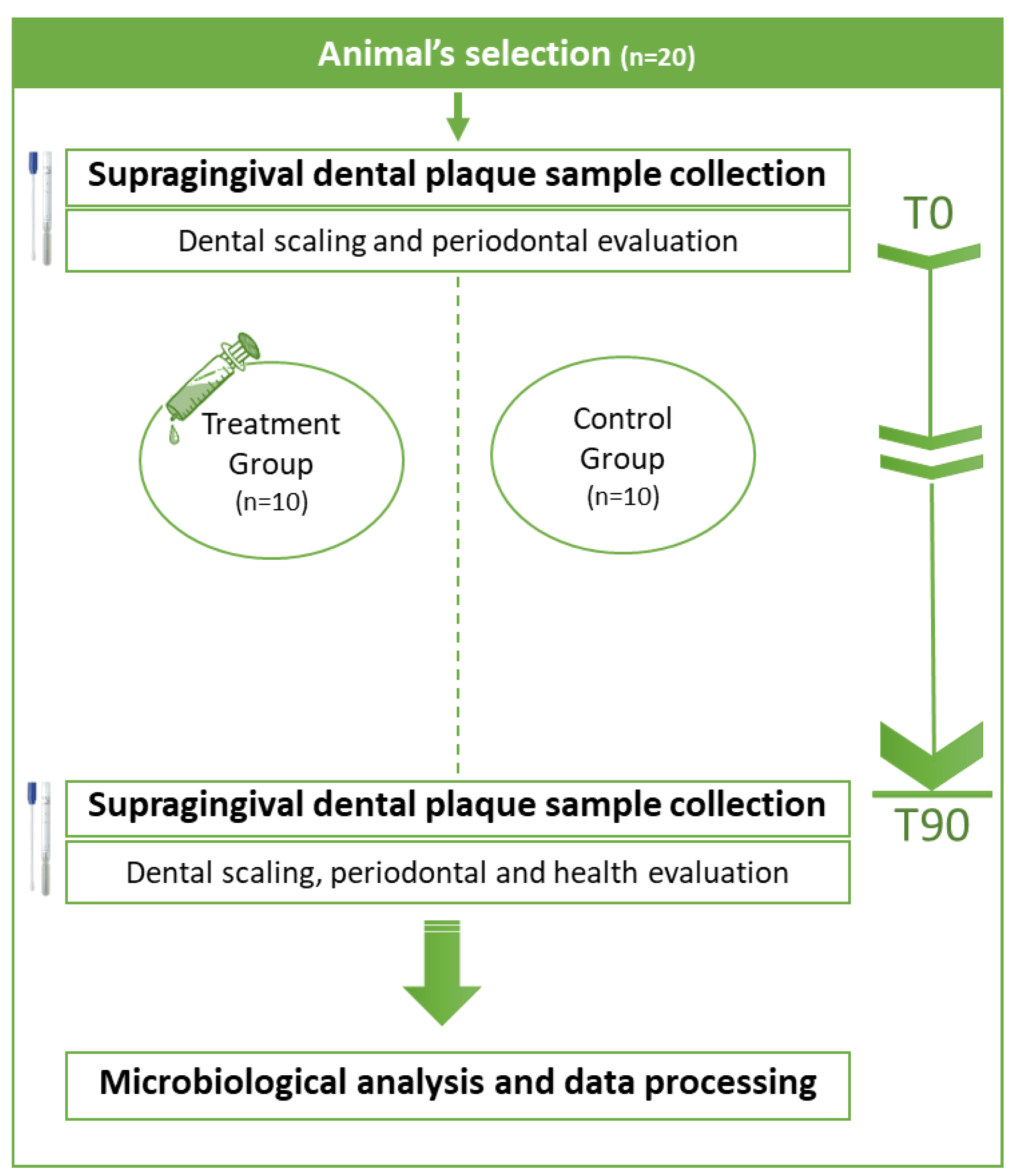
4.2. Clinical Trial and Sample Collection

All samples used in this study were obtained from a clinical trial previously performed by our team (Figure 2) [48]. In this former trial, a total of 20 dogs were selected from an official animal’s rescue institution, according to the Veterinary Oral Health Council (VOHC) instructions for testing compounds aiming at PD prevention. The clinical trial was approved by the Ethical Committee for Research and Teaching (CEIE) of the Faculty of Veterinary Medicine, University of Lisbon, Portugal (N/Ref 014/2020) [31].
Briefly, the dogs that participated in this study were healthy dogs, more than 2 years old, without severe PD and no history of antimicrobial therapy in the previous month. All dogs were housed in the same facilities and had access to the same food and housing routines. Prior to the study, all animals were submitted to a clinical examination and oral handling/cleaning, and a complete blood analysis to detect any deviations that would impair their inclusion in the study was performed. Then, the animals were randomly allocated to one of two groups: the treatment group (n = 10) or control group (n = 10). Animals from the treatment group were submitted to the dental topical application of the nisin–biogel (200 µg/mL) every 48 h; animals from the control group were not submitted to any treatment. Animals were kept in this trial for 90 days [31].
All dogs were submitted to a supragingival dental plaque sample collection using a swab (AMIES®, VWR, Amadora, Portugal), applied to all of the dental surface, at day 0 (Timepoint 0—T0) and at day 90 (Timepoint 90—T90). Swabs were transported to the Laboratory of Microbiology and Immunology, Faculty of Veterinary Medicine, University of Lisbon, and processed for Enterococcus spp. isolation, quantification, identification, and characterization, according to Semedo-Lemsaddek et al. [37], Oliveira et al. [8] and Belo et al. [54].

4.3. Enterococci Identification and Typing

Slanetz and Bartley agar medium (SBA, PanReac AppliChem, Barcelona, Spain) was used for performing a presumptive selection and quantification of enterococci, by using ten-fold serial dilutions, a medium inoculation and determination of the colony forming units [8,37]. From each animal and timepoint (T0 and T90), four typical single colonies presenting distinct morphologies were randomly selected from the SBA plates for further characterization using conventional microbiological procedures [8]. Then, molecular identification at the genus level was performed via conventional PCR, according to Ke et al. [55]. Identification at species level was performed via a multiplex PCR using species-specific primers and conditions previously described by Jackson et al. [51]. Genomic typing was performed using the primers OPC19 and (GTG)5 in independent mixtures, as described by Semedo-Lemsaddek et al. [37] and Oliveira et al. [8]. The profiles obtained were analysed using BioNumerics® 6.6 (Applied Maths, Kortrijk, Belgium), through a hierarchical numerical approach with Pearson correlation coefficient (optimization 1.5%) and the unweighted pair group method with arithmetic average (UPGMA) as the agglomerative clustering. The isolates’ similarity was evaluated using a composite analysis of the fingerprints obtained with the two primers. The cut level applied to select representative isolates from each timepoint (T0 and T90) was 95.9%, since it was the reproducibility value determined as the average similarity value of all replicate pairs. The selected isolates were further evaluated regarding their virulence and antimicrobial resistance signatures. All primers used in this study are presented in Table 3.
Table 3. Description of all primers used for the enterococci genus and species identification and genomic typing.
TargetPrimersSequenceProduct SizeReference
Enterococcus spp.Ent 15′-TACTGACAAACCATTCATGATG-3′112 bp[50]
Ent 25′-AACTTCGTCACCAACGCGAAC-3′
E. faecalisFL15′-ACTTATGTGACTAACTTAACC-3′360 bp[56]
FL25′-TAATGGTGAATCTTGGTTTGG-3′
E. faeciumFM15′-GAAAAAACAATAGAAGAATTAT-3′215 bp
FM25′-TGCTTTTTTGAATTCTTCTTTA-3′
E. hiraeHI15′-CTTTCTGATATGGATGCTGTC-3′187 bp
HI25′-TAAATTCTTCCTTAAATGTTG-3′
E. duransDU15′-CCTACTGATATTAAGACAGCG-3′295 bp
DU25′-TAATCCTAAGATAGGTGTTTG-3′
GenotypingOPC5′-GTTGCCAGCC-3′[200–3000][8,37]
(GTG)55‘-GTGGTGGTGGTGGTG-3′[200–3000]

4.4. Enterococci Virulence Signature

The production of gelatinase, lipase, DNase, lecithinase, proteinase, hemolysin and biofilm were evaluated using plate assays, according to Freeman et al. [57], Semedo-Lemsaddek et al. [37] and Fernandes et al. [58]. Virulence factors were evaluated after streaking the colonies onto the respective medium and incubating at 37 °C for 48 h, except for haemolysis and biofilm evaluation.
Briefly, for gelatinase activity evaluation, the Nutrient Gelatin Medium (Oxoid, Basingstoke, Hampshire, UK) was used, with the presence of a liquid medium after incubation indicating a positive result. Lipase production was evaluated using Spirit blue agar and lipase reagent (BD Life Sciences, VWR, Amadora, Portugal), with lipolytic organisms showing a white clearing beneath and surrounding the colonies after incubation. The ability to produce DNase was evaluated using DNAse test agar (Remel, Termo Scientific, Lenexa, KS, USA) supplemented with blue toluidine, and the presence of pink colonies after incubation was considered a positive result. Lecitinase production was determined using Tryptic Soy agar (VWR, Leuven, Belgium) supplemented with 10% of egg yolk emulsion (VWR, Leuven, Belgium), and a positive activity resulted in the development of a white precipitate around colonies after incubation. Proteinase activity was determined using a skim milk medium (VWR, Leuven, Belgium), and colonies presenting a white halo after incubation being considered positive. Hemolysin production was evaluated after isolates streaking on Columbia agar supplemented with 5% sheep blood (Biomeriux, Marcy-l’Étoile, France), incubated at 37 °C for 72 h in anaerobic conditions. The presence of a clearing halo around the colonies was interpreted as a positive result (β-haemolysis), and the absence of a clearing (ɣ-haemolysis) or the presence of a greenish zone around the colonies (α-haemolysis) were considered negative results. Biofilm forming ability was evaluated using Congo Red agar medium (Sigma-Aldrich, St. Louis, MO, USA), incubated for 72 h at 37 °C, after which the development of black colonies indicated the isolates’ ability to produce biofilm. According to the time required for biofilm production, isolates were considered as strong (24 h), moderate (48 h) or weak biofilm producers (72 h). The evaluation of the isolate’s phenotypic virulence profile included the determination of their virulence index (number of positive virulence factors/number of virulence factors tested).

4.5. Enterococci Antimicrobial Resistance Profile

Isolates’ antimicrobial resistance profile was determined using the disk diffusion method according to the guidelines of the Clinical and Laboratory Standards Institute (CLSI) (M100S and Vet01-02) [59,60]. A total of 12 antimicrobials (Oxoid, Hampshire, UK), presented in Table 4, were used, being selected based on their relevance to veterinary medicine, as well as to public health.
Isolates were categorised according to their multidrug-resistant patterns, as described by Magiorakos et al. [24], and by their multiple antibiotic resistance (MAR) indices (number of antibiotics to which isolates were resistant/number of antibiotics tested) [58,61]. In addition, isolates with non-susceptibility to high doses of gentamicin (120 µg) and/or streptomycin (300 µg) were considered to present high-level aminoglycoside resistance (HLAR) [50,59,60].

4.6. Statistical Analysis

Data statistical analysis was carried out using Microsoft Excel 2016®. Differences in the virulence and MAR index between groups and timepoints were evaluated using Student’s t test.
Quantitative variables are expressed as mean values ± standard deviation. A confidence interval of 95% was considered, with a p-value ≤ 0.05 indicating statistical significance.

5. Conclusions

Enterococci are organisms with an impressive genetic versatility and unparalleled ability to recruit and express antimicrobial resistance and virulence determinants. Their commensal nature allows them to colonize healthy individuals and quickly participate in complicated infections. Present in the oral cavity, they are considered an interesting bacterial model to be used in odontology and pharmaceutical research, since they can act as reservoirs of resistant and virulent phenotypes. The long-term dental application of the nisin–biogel to dogs showed to be an interesting alternative to be used for PD control, with a low effect on Enterococcus antimicrobial and virulence profiles.
Considering all the results obtained in this study regarding the dental application of the nisin–biogel to dogs, in the future, this compound could be commercially developed to be used by clinicians in PD management in dogs.

Supplementary Materials

The following supporting information can be downloaded at: https://www.mdpi.com/article/10.3390/antibiotics12030468/s1, Supplementary File S1: Dendrograms based on the composite analysis of the isolates fingerprints with primers OPC19 and (GTG)5, using Pearson correlation coefficient.

Author Contributions

Conceptualization, M.O., L.M.C. and E.C.; methodology, E.C., A.F.F., S.V. and M.V.; software, E.C., L.C. and A.F.F.; validation, M.O., L.T. and E.C.; formal analysis, E.C. and L.C.; investigation, E.C., S.V., A.F.F. and A.M.; resources, M.O., M.V. and L.M.C.; data curation, M.O.; writing—original draft preparation, E.C., L.C. and A.M.; writing—review and editing, M.O., L.T. and L.C.; visualization, M.O. and L.C.; supervision, M.O.; project administration, M.O.; funding acquisition, M.O., L.T. and E.C. All authors have read and agreed to the published version of the manuscript.

Funding

This work was supported by CIISA—Centre for Interdisciplinary Research in Animal Health, Faculty of Veterinary Medicine University of Lisbon, Project UIDB/00276/2020 (Funded by FCT); and by the Associate Laboratory for Animal and Veterinary Sciences (LA/P/0059/2020-AL4AnimalS).

Institutional Review Board Statement

All experimental procedures were approved by the Ethical Committee for Research and Teaching (CEIE) of the Faculty of Veterinary Medicine, University of Lisbon, Portugal (N/Ref 014/2020). This study complied with relevant institutional, national, and ARRIVE guidelines for the care and use of animals. All experiments were performed in accordance with relevant institutional, national, and ARRIVE guidelines for the care and use of animals. Written informed consent was obtained from the institution responsible for the animals.

Informed Consent Statement

Not applicable.

Data Availability Statement

The datasets used and/or analyzed in the current study are available from the corresponding author upon reasonable request.

Acknowledgments

The authors would like to acknowledge all technical workers at the Casa dos Animais de Lisboa and the Centro de Medicina Veterinária Anjos de Assis for their support in this study.

Conflicts of Interest

The authors declare no conflict of interest.

References

  1. Geraldes, C.; Tavares, L.; Gil, S.; Oliveira, M. Enterococcus Virulence and Resistant Traits Associated with Its Permanence in the Hospital Environment. Antibiotics 2022, 11, 857. [Google Scholar] [CrossRef] [PubMed]
  2. Werner, G.; Coque, T.M.; Franz, C.M.A.P.; Grohmann, E.; Hegstad, K.; Jensen, L.; van Schaik, W.; Weaver, K. Antibiotic Resistant Enterococci—Tales of a Drug Resistance Gene Trafficker. Int. J. Med. Microbiol. 2013, 303, 360–379. [Google Scholar] [CrossRef]
  3. Cunha, E.; Trovão, T.; Pinheiro, A.; Nunes, T.; Santos, R.; Moreira da Silva, J.; São Braz, B.; Tavares, L.; Veiga, A.S.; Oliveira, M. Potential of Two Delivery Systems for Nisin Topical Application to Dental Plaque Biofilms in Dogs. BMC Vet. Res. 2018, 14, 375. [Google Scholar] [CrossRef] [PubMed] [Green Version]
  4. WHO Pubishes List of Bacteria for Which New Antibiotics Are Urgently Needed. Available online: https://www.who.int/news/item/27-02-2017-who-publishes-list-of-bacteria-for-which-new-antibiotics-are-urgently-needed (accessed on 10 May 2019).
  5. Kantele, A.; Mero, S.; Kirveskari, J.; Lääveri, T. Fluoroquinolone Antibiotic Users Select Fluoroquinolone-Resistant ESBL-Producing Enterobacteriaceae (ESBL-PE)—Data of a Prospective Traveller Study. Travel Med. Infect. Dis. 2017, 16, 23–30. [Google Scholar] [CrossRef] [PubMed]
  6. Colombo, A.P.V.; Teles, R.P.; Torres, M.C.; Souto, R.; Rosalém, W.; Mendes, M.C.S.; Uzeda, M. Subgingival Microbiota of Brazilian Subjects With Untreated Chronic Periodontitis. J. Periodontol. 2002, 73, 360–369. [Google Scholar] [CrossRef]
  7. Souto, R.; Colombo, A.P.V. Prevalence of Enterococcus faecalis in Subgingival Biofilm and Saliva of Subjects with Chronic Periodontal Infection. Arch. Oral Biol. 2008, 53, 155–160. [Google Scholar] [CrossRef]
  8. Oliveira, M.; Tavares, M.; Gomes, D.; Touret, T.; São Braz, B.; Tavares, L.; Semedo-Lemsaddek, T. Virulence Traits and Antibiotic Resistance among Enterococci Isolated from Dogs with Periodontal Disease. Comp. Immunol. Microbiol. Infect. Dis. 2016, 46, 27–31. [Google Scholar] [CrossRef]
  9. Bertelloni, F.; Salvadori, C.; Lotti, G.; Cerri, D.; Ebani, V.V. Antimicrobial Resistance in Enterococcus Strains Isolated from Healthy Domestic Dogs. Acta Microbiol. Et Immunol. Hung. 2016, 64, 301–312. [Google Scholar] [CrossRef] [Green Version]
  10. Stępień-Pyśniak, D.; Bertelloni, F.; Dec, M.; Cagnoli, G.; Pietras-Ożga, D.; Urban-Chmiel, R.; Ebani, V.V. Characterization and Comparison of Enterococcus Spp. Isolates from Feces of Healthy Dogs and Urine of Dogs with UTIs. Animals 2021, 11, 2845. [Google Scholar] [CrossRef]
  11. Tumpa, A.; Štritof, Z.; Pintarić, S. Prevalence and antimicrobial susceptibility of Enterococcus spp. from urine of dogs and cats in northwestern Croatia. Res. Vet. Sci. 2022, 151, 42–46. [Google Scholar] [CrossRef]
  12. Stella, J.L.; Bauer, A.E.; Croney, C.C. A cross-sectional study to estimate prevalence of periodontal disease in a population of dogs (Canis familiaris) in commercial breeding facilities in Indiana and Illinois. PLoS ONE 2018, 13, e0191395. [Google Scholar] [CrossRef] [PubMed]
  13. Kačírová, J.; Maďar, M.; Štrkolcová, G.; Maďari, A.; Nemcová, R. Dental Biofilm as Etiological Agent of Canine Periodontal Disease. In Bacterial Biofilms; Dincer, S., Sümengen Özdenefe, M., Arkut, A., Eds.; IntechOpen: London, UK, 2020; ISBN 978-1-78985-899-0. [Google Scholar]
  14. Pavlica, Z.; Petelin, M.; Juntes, P.; Eržen, D.; Crossley, D.A.; Skalerič, U. Periodontal Disease Burden and Pathological Changes in Organs of Dogs. J. Vet. Dent. 2008, 25, 97–105. [Google Scholar] [CrossRef] [PubMed]
  15. Glickman, L.T.; Glickman, N.W.; Moore, G.E.; Goldstein, G.S.; Lewis, H.B. Evaluation of the Risk of Endocarditis and Other Cardiovascular Events on the Basis of the Severity of Periodontal Disease in Dogs. JAVMA 2009, 234, 486–494. [Google Scholar] [CrossRef] [PubMed]
  16. Glickman, L.T.; Glickman, N.W.; Moore, G.E.; Lund, E.M.; Lantz, G.C.; Pressler, B.M. Association between Chronic Azotemic Kidney Disease and the Severity of Periodontal Disease in Dogs. Prev. Vet. Med. 2011, 99, 193–200. [Google Scholar] [CrossRef]
  17. Pereira dos Santos, J.D.; Cunha, E.; Nunes, T.; Tavares, L.; Oliveira, M. Relation between Periodontal Disease and Systemic Diseases in Dogs. Res. Vet. Sci. 2019, 125, 136–140. [Google Scholar] [CrossRef]
  18. Howell, T.H.; Fiorellini, J.P.; Blackburn, P.; Projan, S.J.; Harpe, J.; Williams, R.C. The Effect of a Mouthrinse Based on Nisin, a Bacteriocin, on Developing Plaque and Gingivitis in Beagle Dogs. J. Clin. Periodontol. 1993, 20, 335–339. [Google Scholar] [CrossRef]
  19. Cunha, E.; Rebelo, S.; Carneiro, C.; Tavares, L.; Carreira, L.M.; Oliveira, M. A Polymicrobial Biofilm Model for Testing the Antimicrobial Potential of a Nisin-Biogel for Canine Periodontal Disease Control. BMC Vet. Res. 2020, 16, 469. [Google Scholar] [CrossRef] [PubMed]
  20. Cunha, E.; Freitas, F.B.; São Braz, B.; Moreira da Silva, J.; Tavares, L.; Veiga, A.S.; Oliveira, M. Polyphasic Validation of a Nisin-Biogel to Control Canine Periodontal Disease. Antibiotics 2020, 9, 180. [Google Scholar] [CrossRef] [Green Version]
  21. Davison, W.M.; Pitts, B.; Stewart, P.S. Spatial and Temporal Patterns of Biocide Action against Staphylococcus Epidermidis Biofilms. Antimicrob. Agents Chemother. 2010, 54, 2920–2927. [Google Scholar] [CrossRef] [Green Version]
  22. Zhao, M.; Qu, Y.; Liu, J.; Mai, S.; Gu, L. A Universal Adhesive Incorporating Antimicrobial Peptide Nisin: Effects on Streptococcus mutans and Saliva-Derived Multispecies Biofilms. Odontology 2020, 108, 376–385. [Google Scholar] [CrossRef]
  23. Cunha, E.; Janela, R.; Costa, M.; Tavares, L.; Veiga, A.S.; Oliveira, M. Nisin Influence on the Antimicrobial Resistance Ability of Canine Oral Enterococci. Antibiotics 2020, 9, 890. [Google Scholar] [CrossRef] [PubMed]
  24. Magiorakos, A.-P.; Srinivasan, A.; Carey, R.B.; Carmeli, Y.; Falagas, M.E.; Giske, C.G.; Harbarth, S.; Hindler, J.F.; Kahlmeter, G.; Olsson-Liljequist, B.; et al. Multidrug-Resistant, Extensively Drug-Resistant and Pandrug-Resistant Bacteria: An International Expert Proposal for Interim Standard Definitions for Acquired Resistance. Clin. Microbiol. Infect. 2012, 18, 268–281. [Google Scholar] [CrossRef] [Green Version]
  25. Shin, J.M.; Ateia, I.; Paulus, J.R.; Liu, H.; Fenno, J.C.; Rickard, A.H.; Kapila, Y.L. Antimicrobial Nisin Acts against Saliva Derived Multi-Species Biofilms without Cytotoxicity to Human Oral Cells. Front. Microbiol. 2015, 6, 617. [Google Scholar] [CrossRef] [PubMed] [Green Version]
  26. Cunha, E.; Valente, S.; Nascimento, M.; Pereira, M.; Tavares, L.; Dias, R.; Oliveira, M. Influence of the Dental Topical Application of a Nisin-Biogel in the Oral Microbiome of Dogs: A Pilot Study. PeerJ 2021, 9, e11626. [Google Scholar] [CrossRef] [PubMed]
  27. Riggio, M.P.; Lennon, A.; Taylor, D.J.; Bennett, D. Molecular Identification of Bacteria Associated with Canine Periodontal Disease. Vet. Microbiol. 2011, 150, 394–400. [Google Scholar] [CrossRef] [PubMed] [Green Version]
  28. Holcombe, L.J.; Patel, N.; Colyer, A.; Deusch, O.; O’Flynn, C.; Harris, S. Early Canine Plaque Biofilms: Characterization of Key Bacterial Interactions Involved in Initial Colonization of Enamel. PLoS ONE 2014, 9, e113744. [Google Scholar] [CrossRef] [Green Version]
  29. Wallis, C.; Marshall, M.; Colyer, A.; O’Flynn, C.; Deusch, O.; Harris, S. A Longitudinal Assessment of Changes in Bacterial Community Composition Associated with the Development of Periodontal Disease in Dogs. Vet. Microbiol. 2015, 181, 271–282. [Google Scholar] [CrossRef] [Green Version]
  30. Ruparell, A.; Inui, T.; Staunton, R.; Wallis, C.; Deusch, O.; Holcombe, L.J. The Canine Oral Microbiome: Variation in Bacterial Populations across Different Niches. BMC Microbiol. 2020, 20, 42. [Google Scholar] [CrossRef] [Green Version]
  31. Cunha, E.; Carreira, L.M.; Nunes, T.; Videira, M.; Tavares, L.; Veiga, A.S.; Oliveira, M. In Vivo Evaluation of the Efficacy of a Nisin–Biogel as a New Approach for Canine Periodontal Disease Control. Pharmaceutics 2022, 14, 2716. [Google Scholar] [CrossRef]
  32. Kamran, M.A. Clinical, Microbiological and Immunological Outcomes with Photodynamic Therapy as an Adjunct to Full-Mouth Scaling in Patients Undergoing Fixed Orthodontic Treatment. Photodiagnosis Photodyn. Ther. 2020, 29, 101585. [Google Scholar] [CrossRef]
  33. Elsadek, M.F. Clinical and bacterial outcomes of photodynamic therapy in the treatment of chronic necrotizing ulcerative periodontitis. Photodiagnosis Photodyn. Ther. 2022, 39, 102977. [Google Scholar] [CrossRef] [PubMed]
  34. Flancman, R.; Singh, A.; Weese, J.S. Evaluation of the Impact of Dental Prophylaxis on the Oral Microbiota of Dogs. PLoS ONE 2018, 13, e0199676. [Google Scholar] [CrossRef] [PubMed]
  35. de Andrade Ferreira, F.B.; Campos Rabang, H.R.; Pinheiro, E.T.; Gadê-Neto, C.R.; Zaia, A.A.; Randi Ferraz, C.C.; de Souza-Filho, F.J.; de Almeida Gomes, B.P.F. Root Canal Microbiota of Dogs’ Teeth with Periapical Lesions Induced by Two Different Methods. Oral Surg. Oral Med. Oral Pathol. Oral Radiol. Endodontology 2006, 102, 564–570. [Google Scholar] [CrossRef] [PubMed]
  36. Ribeiro, T.; Oliveira, M.; Fraqueza, M.J.; Lauková, A.; Elias, M.; Tenreiro, R.; Barreto, A.S.; Semedo-Lemsaddek, T. Antibiotic Resistance and Virulence Factors among Enterococci Isolated from Chouriço, a Traditional Portuguese Dry Fermented Sausage. J. Food Prot. 2011, 74, 465–469. [Google Scholar] [CrossRef] [Green Version]
  37. Semedo-Lemsaddek, T.; Nóbrega, C.S.; Ribeiro, T.; Pedroso, N.M.; Sales-Luís, T.; Lemsaddek, A.; Tenreiro, R.; Tavares, L.; Vilela, C.L.; Oliveira, M. Virulence Traits and Antibiotic Resistance among Enterococci Isolated from Eurasian Otter (Lutra Lutra). Vet. Microbiol. 2013, 163, 378–382. [Google Scholar] [CrossRef]
  38. Medeiros, A.W.; Pereira, R.I.; Oliveira, D.V.; Martins, P.D.; d’Azevedo, P.A.; Van der Sand, S.; Frazzon, J.; Frazzon, A.P.G. Molecular Detection of Virulence Factors among Food and Clinical Enterococcus faecalis Strains in South Brazil. Braz. J. Microbiol. 2014, 45, 327–332. [Google Scholar] [CrossRef] [Green Version]
  39. Semedo-Lemsaddek, T.; Mottola, C.; Alves-Barroco, C.; Cavaco-Silva, P.; Tavares, L.; Oliveira, M. Characterization of Multidrug-Resistant Diabetic Foot Ulcer Enterococci. Enferm. Infecc. Y Microbiol. Clínica 2016, 34, 114–116. [Google Scholar] [CrossRef]
  40. Alzahrani, O.M.; Fayez, M.; Alswat, A.S.; Alkafafy, M.; Mahmoud, S.F.; Al-Marri, T.; Almuslem, A.; Ashfaq, H.; Yusuf, S. Antimicrobial Resistance, Biofilm Formation, and Virulence Genes in Enterococcus Species from Small Backyard Chicken Flocks. Antibiotics 2022, 11, 380. [Google Scholar] [CrossRef]
  41. Rotta, I.S.; Rodrigues, W.F.; Dos Santos, C.T.B.; Mantovani, H.C.; De Oliveira, A.G.; Machado, A.B.F.; Paiva, A.D. Clinical isolates of E. faecalis and E. faecium harboring virulence genes show the concomitant presence of CRISPR loci and antibiotic resistance determinants. Microb. Pathog. 2022, 171, 105715. [Google Scholar] [CrossRef]
  42. Woźniak-Biel, A.; Bugla-Płoskońska, G.; Burdzy, J.; Korzekwa, K.; Ploch, S.; Wieliczko, A. Antimicrobial Resistance and Biofilm Formation in Enterococcus Spp. Isolated from Humans and Turkeys in Poland. Microb. Drug Resist. 2019, 25, 277–286. [Google Scholar] [CrossRef] [Green Version]
  43. Kubašová, I.; Strompfová, V.; Lauková, A. Safety Assessment of Commensal Enterococci from Dogs. Folia Microbiol. 2017, 62, 491–498. [Google Scholar] [CrossRef]
  44. Bjarnsholt, T. The Role of Bacterial Biofilms in Chronic Infections. APMIS 2013, 121, 1–58. [Google Scholar] [CrossRef]
  45. Uruén, C.; Chopo-Escuin, G.; Tommassen, J.; Mainar-Jaime, R.C.; Arenas, J. Biofilms as Promoters of Bacterial Antibiotic Resistance and Tolerance. Antibiotics 2020, 10, 3. [Google Scholar] [CrossRef]
  46. Hall-Stoodley, L.; Costerton, J.W.; Stoodley, P. Bacterial Biofilms: From the Natural Environment to Infectious Diseases. Nat. Rev. Microbiol. 2004, 2, 95–108. [Google Scholar] [CrossRef]
  47. Balcázar, J.L.; Subirats, J.; Borrego, C.M. The Role of Biofilms as Environmental Reservoirs of Antibiotic Resistance. Front. Microbiol. 2015, 6, 1216. [Google Scholar] [CrossRef] [Green Version]
  48. Singh, S.K.; Ekka, R.; Mishra, M.; Mohapatra, H. Association Study of Multiple Antibiotic Resistance and Virulence: A Strategy to Assess the Extent of Risk Posed by Bacterial Population in Aquatic Environment. Environ. Monit. Assess 2017, 189, 320. [Google Scholar] [CrossRef] [PubMed]
  49. Miller, W.R.; Munita, J.M.; Arias, C.A. Mechanisms of Antibiotic Resistance in Enterococci. Expert Rev. Anti-Infect. Ther. 2014, 12, 1221–1236. [Google Scholar] [CrossRef] [PubMed]
  50. Sienko, A.; Wieczorek, P.; Wieczorek, A.; Sacha, P.; Majewski, P.; Ojdana, D.; Michalska, A.; Tryniszewska, E. Occurrence of high-level aminoglycoside resistance (HLAR) among Enterococcus species strains. Prog. Health Sci. 2014, 4, 179–187. [Google Scholar]
  51. Santos, R.; Gomes, D.; Macedo, H.; Barros, D.; Tibério, C.; Veiga, A.S.; Tavares, L.; Castanho, M.; Oliveira, M. Guar Gum as a New Antimicrobial Peptide Delivery System against Diabetic Foot Ulcers Staphylococcus aureus Isolates. J. Med. Microbiol. 2016, 65, 1092–1099. [Google Scholar] [CrossRef]
  52. Niemiec, B.A. Periodontal Disease. Top. Companion Anim. Med. 2008, 23, 72–80. [Google Scholar] [CrossRef] [Green Version]
  53. Bellows, J.; Berg, M.L.; Dennis, S.; Harvey, R.; Lobprise, H.B.; Snyder, C.J.; Stone, A.E.S.; Van de Wetering, A.G. 2019 AAHA Dental Care Guidelines for Dogs and Cats*. J. Am. Anim. Hosp. Assoc. 2019, 55, 49–69. [Google Scholar] [CrossRef] [PubMed]
  54. Belo, L.; Serrano, I.; Cunha, E.; Carneiro, C.; Tavares, L.; Miguel Carreira, L.; Oliveira, M. Skin Asepsis Protocols as a Preventive Measure of Surgical Site Infections in Dogs: Chlorhexidine–Alcohol versus Povidone–Iodine. BMC Vet. Res. 2018, 14, 95. [Google Scholar] [CrossRef] [PubMed]
  55. Ke, D.; Picard, F.J.; Martineau, F.; Ménard, C.; Roy, P.H.; Ouellette, M.; Bergeron, M.G. Development of a PCR Assay for Rapid Detection of Enterococci. J. Clin. Microbiol. 1999, 37, 3497–3503. [Google Scholar] [CrossRef] [PubMed] [Green Version]
  56. Jackson, C.R.; Fedorka-Cray, P.J.; Barrett, J.B. Use of a Genus- and Species-Specific Multiplex PCR for Identification of Enterococci. J. Clin. Microbiol. 2004, 42, 3558–3565. [Google Scholar] [CrossRef] [PubMed] [Green Version]
  57. Freeman, D.J.; Falkiner, F.R.; Keane, C.T. New Method for Detecting Slime Production by Coagulase Negative Staphylococci. J. Clin. Pathol. 1989, 42, 872–874. [Google Scholar] [CrossRef] [PubMed] [Green Version]
  58. Fernandes, M.; Grilo, M.L.; Carneiro, C.; Cunha, E.; Tavares, L.; Patino-Martinez, J.; Oliveira, M. Antibiotic Resistance and Virulence Profiles of Gram-Negative Bacteria Isolated from Loggerhead Sea Turtles (Caretta Caretta) of the Island of Maio, Cape Verde. Antibiotics 2021, 10, 771. [Google Scholar] [CrossRef]
  59. CLSI. Performance Standards for Antimicrobial Disk and Dilution Susceptibility Tests for Bacteria Isolated from Animals: Second Informational Supplement VET01-S2; Clinical and Laboratory Standards Institute: Wayne, PA, USA, 2013. [Google Scholar]
  60. CLSI. Performance Standards for Antimicrobial Susceptibility Tests: M100S, 26th ed.; Clinical and Laboratory Standards Institute: Wayne, PA, USA, 2016. [Google Scholar]
  61. Krumperman, P.H. Multiple Antibiotic Resistance Indexing of Escherichia coli to Identify High-Risk Sources of Fecal Contamination of Foodst. Appl. Environ. Microbiol. 1983, 46, 6. [Google Scholar] [CrossRef] [Green Version]
Figure 1. Species distribution by timepoint and by test group (control group and treatment group). T0—timepoint 0; T90—timepoint 90; TG—treatment group; CG—control group.
Antibiotics 12 00468 g001
Figure 2. Schematic representation of the clinical trial.
Antibiotics 12 00468 g002
Table 1. Number of isolates with positive phenotypic expression of the virulence factors under study, by group and timepoint, and mean virulence index.
Virulence
Factor
Control GroupTreatment Group
Timepoint 0Timepoint 90Timepoint 0Timepoint 90
Gelatinase7455
Lipase1413918
DNase0000
Lecithinase7345
Haemolysin1051512
Proteinase6345
Biofilm18131719
Total of isolates evaluated18132019
Mean virulence index ± SD0.49 ± 0.240.45 ± 0.240.39 ± 0.240.48 ± 0.24
SD: Standard deviation.
Table 2. Antimicrobial resistance evaluation of isolates under study by timepoint and test group.
Control GroupTreatment Group
Timepoint 0Timepoint 90Timepoint 0Timepoint 90
Mean MAR index ± SD0.62 ± 0.200.83 ± 0.150.40 ± 0.220.61 ± 0.18
Number of isolates with MDR profile18131819
Number of isolates with HLARCN + S7923
CN1014
S3203
Total of isolates evaluated18132019
MAR: Multiple antimicrobial resistance; MDR: multidrug resistance; HLAR: high-level aminoglycoside resistance; CN: gentamycin; S: streptomycin; SD: standard deviation.
Table 4. Antimicrobial agents used in the enterococci antimicrobial susceptibility profiling, grouped by mechanism of action, class and concentration.
Mechanism of ActionAntimicrobial ClassAntimicrobial DrugConcentration (µg Per Disk)
Inhibition
of cell-wall synthesis
AminopenicillinsAmpicillin (AMP)10
Amoxicillin/Clavulanate (AMX)30
GlycopeptidesVancomycin (VA)30
CarbapenemsImipenem (IMI)10
Inhibition of nucleic acid synthesisFluoroquinolonesEnrofloxacin (ENR)5
Inhibition of protein synthesisTetracyclinesTetracycline (T)30
Doxycycline (DTX)30
AminoglycosidesGentamicin (CN)120
Streptomycin (S)300
MacrolidesErythromycin (E)15
LincosamideClindamycin (DA)2
NitrobenzenesChloramphenicol (C)30
Disclaimer/Publisher’s Note: The statements, opinions and data contained in all publications are solely those of the individual author(s) and contributor(s) and not of MDPI and/or the editor(s). MDPI and/or the editor(s) disclaim responsibility for any injury to people or property resulting from any ideas, methods, instructions or products referred to in the content.

