Article

Evaluation of Bile Salts on the Survival and Modulation of Virulence of Aliarcobacter butzleri

1
CICS-UBI—Health Sciences Research Centre, University of Beira Interior, 6200-506 Covilhã, Portugal
2
Clinical Physiology/Nutritional Medicine, Medical Department of Gastroenterology, Infectiology, Rheumatology, Charité—Universitätsmedizin Berlin, 12203 Berlin, Germany
3
National Reference Laboratory for Gastrointestinal Infections, Department of Infectious Diseases, National Institute of Health Dr. Ricardo Jorge, Av. Padre Cruz, 1649-016 Lisbon, Portugal
*
Author to whom correspondence should be addressed.
Antibiotics 2023, 12(9), 1387; https://doi.org/10.3390/antibiotics12091387
Submission received: 19 July 2023 / Revised: 17 August 2023 / Accepted: 25 August 2023 / Published: 30 August 2023

Abstract

:
Aliarcobacter butzleri is a Gram-negative bacterium associated with infections of the gastrointestinal tract and widely distributed in various environments. For successful infection, A. butzleri should be able to tolerate various stresses during gastrointestinal passage, such as bile. Bile represents an antimicrobial host barrier that acts against external noxious agents and consists of a variety of bile salts. The intestinal bile salts act as detergents involved in the antimicrobial host defense; although, on the bacterial side, they could also serve as a signal to activate virulence mechanisms. The aim of this work was to understand the effects of bile salts on the survival and virulence of A. butzleri. In our study, A. butzleri was able to survive in the presence of human physiological concentrations of bile salts. Regarding the virulence features, an increase in cellular hydrophobicity, a decrease in motility and expression of flaA gene, as well as an increase in biofilm formation with a concomitant change in the type of biofilm structure were observed in the presence of sub-inhibitory concentration of bile salts. Concerning adhesion and invasion ability, no significant difference was observed. Overall, the results demonstrated that A. butzleri is able to survive in physiological concentrations of bile salts and that exposure to bile salts could change its virulence mechanisms.

1. Introduction

Aliarcobacter genus belongs to the Arcobacteraceae family and the Campylobacterales order [1,2,3]. These bacteria are characterized as small Gram-negative, S-shaped or helical cells, motile by a single polar flagellum, and capable of growing at temperatures from 15 °C to 42 °C [1]. Among the species included in this genus, A. butzleri, A. cryaerophilus, A. skirrowii, A. thereius and A. lanthieri have been associated with human disease, such as enteritis and bacteremia, accompanied by various symptoms, for example diarrhea, abdominal cramps, nausea, fever and vomiting [1,4,5,6,7,8]. These species can be found in different hosts and environments, and can be isolated from different sources, such as water, food, animal and human samples [4,7,9,10]. Considering the environmental distribution, disease association and reported outbreaks, the consumption of food and water contaminated with Aliarcobacter spp. has been considered the main route of transmission of these bacteria to humans [4,7,9]. Among the species mentioned, A. butzleri has been highlighted as an emerging gastrointestinal pathogen, with worldwide distribution, and being considered the fourth most frequent Campylobacter-like organism found in human diarrheic samples, including patients with enteritis [6,7,11,12]. Furthermore, the ability of A. butzleri for adherence, invasion and intracellular survival in intestinal epithelial cells, in addition to cytotoxic effects supports its pathogenic potential [4,7,9,13,14].
Several factors affect the gut persistence ability of microorganisms, such as the gut microbiota that provides protection against infections and colonization resistance, through multiple mechanisms, including competition for nutrients, support of epithelial barrier integrity (mucus layers), bacteriophage implantation, immune activation, and secretion of antimicrobial products, like bacteriocins, short chain fatty acids and secondary bile acids [15]. Besides that, bacteria are exposed to several other adverse conditions. In fact, as an enteropathogen, A. butzleri is exposed to several adverse host factors during its passage through the gastrointestinal tract, such as flotations in pH, osmotic pressure and the presence of harmful substances during its passage through the gastrointestinal tract, such as reactive oxygen and bile [16,17]. The bile is produced by hepatocytes in the liver, concentrated and stored in the gallbladder, and subsequently released into the small intestine [16,17,18,19]. After digestion of lipids by lipases, the bile induces formation of micelles, which consists of long-chain fatty acids, glycerol and monoglycerides as well as apolar fatty acids, fat-soluble vitamins and the bile acids as emulsifier. The secreted bile in the small intestine contains a multitude of components, including, water, several bile salts, lipids (phospholipids, cholesterol, and fatty acids), proteins (globulins and albumins), ions, pigments, carbohydrates, vitamins and other trace elements [20,21,22]. Bile acts as a barrier against external noxious agents, which can provide host protection, acting as a detergent involved in host antimicrobial defense. Nonetheless, enteric pathogens resist the bactericidal activity of bile and are able to use it as one of the most intriguing host signals affecting virulence [16,20,23,24]. Amongst its constituents, the bile salts sodium cholate and sodium deoxycholate are described as virulence inducers of bacterial enteropathogens [16,20,24,25]. Bile salts are relevant physiological agents for intestinal absorption of nutrients and biliary secretion of lipids, toxic metabolites, and xenobiotics [22]. After release from the gallbladder, bile salts facilitate the emulsification of dietary fats and aid in the intestinal absorption of lipids and lipophilic vitamins, one of their main functions, being subsequently reabsorbed in the intestine and transported back to the liver [19,22]. The enterohepatic circulation of bile salts from the liver to the intestine and back to the liver is quite efficient, with approximately 95% of the bile salts reabsorbed [19]. This circulation thus plays a fundamental role in the absorption and distribution of nutrients, in metabolic regulation and in homeostasis [22].
Several mechanisms of resistance and virulence can be modulated in the presence of bile salts, to ensure that bacteria can survive, colonize and proliferate in the host [23,26]. In fact, bile salts were found to affect cellular adhesion and thus intestinal adhesion and biofilm formation. Biofilm is a common approach for bacterial pathogens to resist antimicrobial exposure, such as high levels of oxygen or bile [16,27], and many species are capable to enhance the formation of biofilm as a response to this stress [16,27,28]. Furthermore, bile salts, such as sodium deoxycholate, cholate and chenodeoxycholate, can modulate the adhesion and invasion of epithelial cells, in several bacterial pathogens, such as Campylobacter jejuni, Escherichia coli, Shigella flexneri, Salmonella enterica serovar Typhimurium, Bacteroides fragilis and Vibrio parahaemolyticus [27,29,30,31,32,33]. As adhesion to intestinal epithelial cells is required for establishment of infection, modulation of adhesion by bile may play an important role in successful colonization and infection. In fact, bile and bile salts present in the small intestine may act as a signal for bacteria to initiate colonization of the epithelium. Infection may be followed by tissue colonization and, in some cases, bacterial invasion of host cells, followed by intracellular proliferation, spread to other tissues, or persistence in the host [7,28].
In the presence of several unfavorable environmental factors, bacteria can change their behavior, through modulation of virulence mechanisms and protein expression, enabling responses to specific stresses. While studies have shown the potential of bile salts as a host signal for several enteric pathogens, the current knowledge regarding the response of A. butzleri to bile salts remains to be clarified. Considering that A. butzleri is an emerging pathogen, capable of infecting, colonizing and proliferating the human gastrointestinal tract [9,34], it is important investigate the response of this species to bile salts, by specifically evaluating the resistance to bile salts, as well as the role of this substrate as an environmental signal to regulate A. butzleri virulence.

2. Results

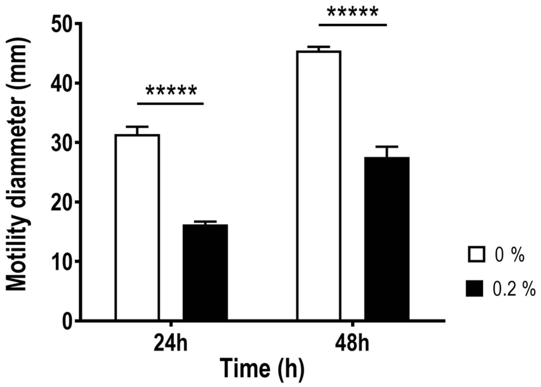
2.1. Effect of Bile Salts on the Growth, Aggregation and Motility of Aliarcobacter butzleri

To evaluate the effect of bile salts in the survival of A. butzleri, a range of physiological concentrations found in the gastrointestinal tract, from 0.2% to 2%, depending on circadian rhythm, diet and individual, and a concentration above this range (5%) were tested. The physiological range of concentrations of bile salts showed a low influence in A. butzleri Ab_2811 growth. However, the concentration above the physiological led to a reduction in survival to below the detection limit after 24 h of exposure (Figure 1). Thus, all subsequent analyses were performed with 0.2% of bile salts, a physiological concentration having no effect on bacterial growth when compared with growth in the absence of bile salts.
During the bacterial growth, formation of aggregates could be observed throughout the time of exposure to various concentrations of bile salts (Figure 2a); thus, the bacterial hydrophobicity was further evaluated using a salting-out method with ammonium sulphate. An increase in hydrophobicity of Ab_2811 in the presence of 0.2% of bile salts was observed, when compared to the bacteria incubated in the absence of the bile salts (Figure 2b). Considering this profile, prior to all subsequent assays the aggregates were previously dispersed by vortexing of the suspensions.
Regarding the effect of bile salts on the motility of A. butzleri Ab_2811, a significant difference was observed at 24 and 48 h after the incubation with and without bile salts (Figure 3). These results showed that the bacterial motility of the strain decreases when exposed to bile salts.

2.2. Impact of Bile Salts on Biofilm Formation of Aliarcobacter butzleri

The impact of bile salts on biofilm formation ability was assessed in 96-well plates and glass tubes (Figure 4). In the presence of bile salts, the biofilm-forming ability of the Ab_2811 strain significantly increased, in both experimental set-ups, when compared to the bacterial culture incubated without bile salts (Figure 4a). Additionally, an increased biofilm formation was measured after prolonged incubation time (Figure 4b). An incubation period of 5 days was chosen for the subsequent microscopy and evaluation of the biofilm thickness.
When Ab_2811 strain was exposed to bile salts, the formation of a pellicle in the interface air-medium was observed with both methods, microplate and glass tubes. Therefore, additional assays were performed to confirm and quantify this phenomenon. The phenotype change was confirmed by crystal violet staining of a coverslip vertically positioned during biofilm formation and the biofilm thickness corresponding to the pellicle was assessed. When observing the coverslip, a strong formation of biofilm at the interface air-medium was observed in the presence of bile salts, which was confirmed by confocal microscopy, where an increase in fluorescence was observed at the interface area in the presence of bile salts, when compared with the control (Figure 4c). An informatics analysis performed using COMSTAT software confirmed that, when considering the air-medium interface area, biofilm thickness was significantly higher in the presence of 0.2% of bile salts, when compared with the biofilm thickness in absence of bile salts (Figure 4d).

2.3. Effect of Bile Salts in the Expression of Five Virulence Genes of Aliarcobacter butzleri

We evaluated the relative expression of cadF, ciaB, cheV, flaA, and luxS genes of A. butzleri AB_2811 grown in presence or absence of bile salts by real-time PCR. A strong decrease in the expression of flaA gene was found, when the strain was exposed to 0.2% of bile salts, while an opposite trend was observed for luxS gene. On the other hand, no significant difference was observed for the cadF, ciaB and cheV genes in both tested conditions (Figure 5).

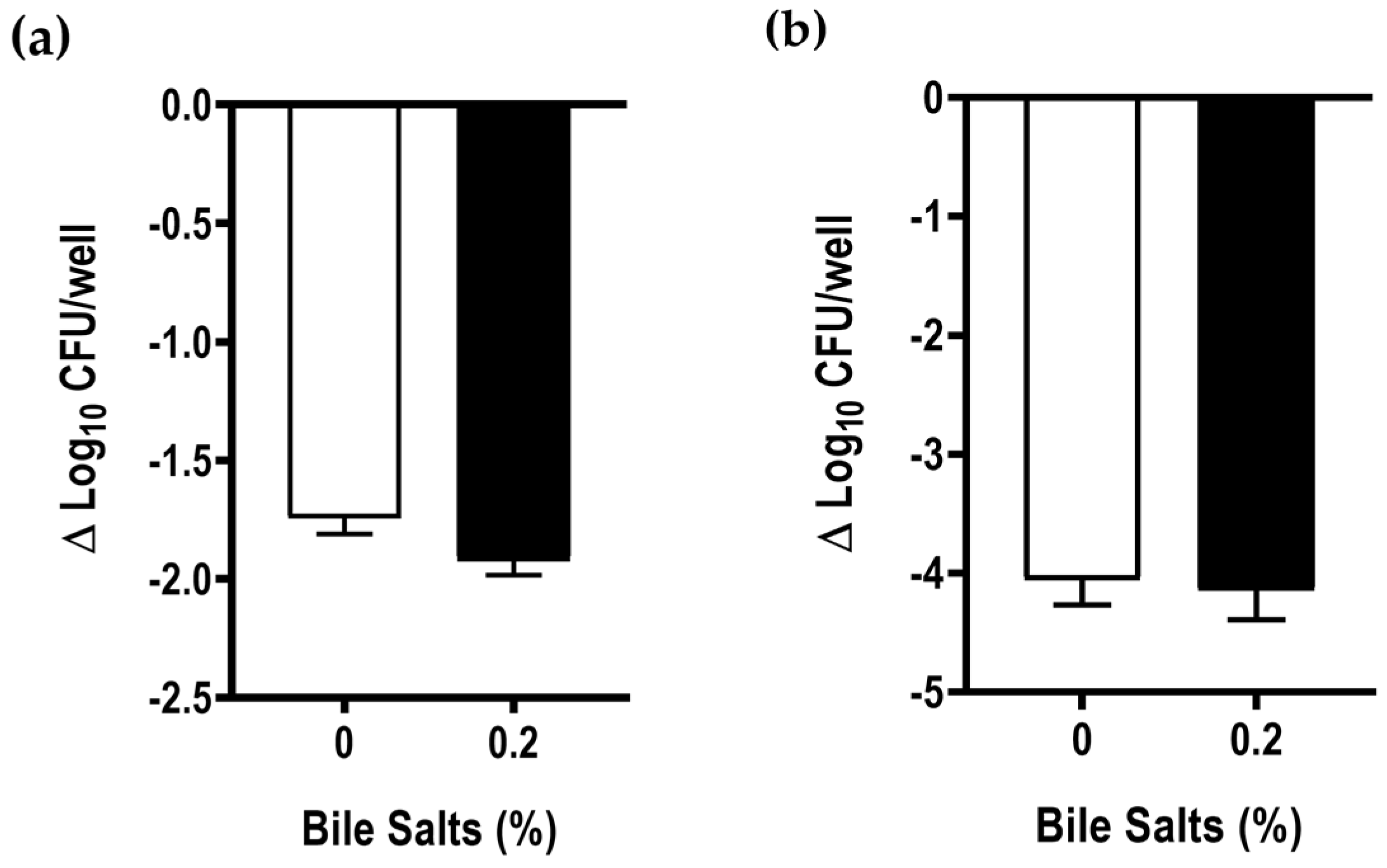
2.4. Effect of Bile Salts in the Adhesion and Invasion Abilities of Aliarcobacter butzleri to Caco-2 Cells

The capacity of A. butzleri to adhere and invade epithelial cells after exposure to bile salts was analyzed in Caco-2 cells. The results show that pre-exposure to 0.2% bile salts did not significantly influence the bacteria’s ability to adhere and invade the Caco-2 cell line (Figure 6).

3. Discussion

As an enteropathogen, A. butzleri must overcome numerous barriers to establish a successful infection, such as bile, which acts as harmful agent for bacteria outside the intestinal microbiota and could modulate several resistance and virulence mechanisms of the bacterium [4,7,16,17,23,35,36]. Several aspects of the virulence of this species are only now being elucidated, leaving a gap in our knowledge of how this bacterium might survive and migrate through the gastrointestinal tract. Thus, for the first time the virulence potential of A. butzleri considering its survival to bile salts, as well as the potential to modulate its virulence by these compounds was enlightened by this work.
Besides the digestive function of lipid absorption via micelles, bile salts are antibacterial compounds that may disrupt bacterial membranes, lead to DNA oxidative damage, denature proteins, or chelate iron and calcium [16,22,24]. Thus, firstly, the assessment of the planktonic growth of A. butzleri, in the absence and presence of physiological concentrations of bile salts and at higher concentration was performed. Although the bacterium was unable to survive at concentrations of 5% bile salts, it was able to survive at all concentrations tested in the range of physiological concentrations. This behavior has already been described for other microorganisms, such as S. flexneri, where a normal growth within the physiological range of bile salts was observed, but slowing and/or inhibition of growth occurred at concentrations above physiological levels [27]. Similarly, for C. jejuni and E. coli a decrease in bacterial growth can be associated with an increase in the concentration of bile or bile salt [28,37]. The presence of the bacterial wall provides a barrier to the absorption of different molecules; however, bile salts can enter the bacterial cell by diffusion or by passage through various existing porins, and consequently, some mechanisms are required to reduce its concentration within the cell, such as efflux pumps [16,24]. The presence of these efflux pumps in some bacteria can be correlated to the ability to survive in this condition. For example, the CmeABC multidrug efflux pump in C. jejuni or the AcrAB-TolC in E. coli and in S. enterica serovar Typhimurium have a clear role in bile salt resistance [38,39,40]. Previous work from our group showed that the use of the efflux pump inhibitor phenylalanine-arginine β-naphthylamide had influence on the resistance to sodium cholate, thus pointing to the role of efflux pumps in resistance to bile salts in A. butzleri [41]. The role of resistance-nodulation-cell division efflux pumps in bile salts resistance was also demonstrated by our group, where a knockdown of areG resulted in an 8-fold decrease in the minimum inhibitory concentration of the bile salt mix, highlighting the function of the AreGHI system in the A. butzleri response [42].
In the present work, following the growth analysis, a concentration of bile salts that did not affect bacterial growth was selected for the following assays (0.2%). The contact with bile salts led to the formation of aggregates, similarly to what was previously described for S. flexneri, E. coli and B. fragilis [27,28,30]. In fact, the observed aggregation correlated with a strong increase in the cellular hydrophobicity of the strain under study when previously exposed to 0.2% of bile salts, showing that bile salts play a role in altering the cellular hydrophobicity of A. butzleri Ab_2811. The formation of bacterial aggregates can be considered as a defense mechanism against biliary stress to which bacteria are subjected. Besides that, aggregation factors may also mediate binding to the host epithelium, and thus aid in other virulence mechanisms, such as biofilm development and, consequently, facilitate infection [28].
Besides the barrier role of bile salts, several enteric pathogens use these molecules as a signal to modulate virulence, regulating the colonization or maintenance of infection in the human gastrointestinal tract [16,19,23,24]. In fact, the resistance to bile, but also virulence modulation by bile may be critical to enteric pathogens’ success and survival [23]. Accordingly, considering bacterial motility, biofilm formation, host cell adhesion and invasion as mechanisms that play a role in the virulence of A. butzleri [4,7,9], these features were evaluated following exposure to bile salts. Motility is essential to bacteria’s ability to cause disease, influencing virulence mechanisms, such as biofilm formation, adhesion and invasion ability [7,9,43] that, consequently, modulating the pathogenicity of the bacterium. Regarding for A. butzleri motility, a strong decrease in the presence of 0.2% of bile salts was observed. This result is in line with data from literature for other enteropathogens, namely the significant reduction in motility in E. coli in the presence of glycol-conjugated cholate, a bile component [28], and in Bacillus cereus, where experiments in soft agar showed decreased motility in the presence of 0.005% bile salts [44]. In turn, the presence of bile acid deoxycholate had no effect in the bacterial motility of C. jejuni [25]. This decreased motility is likely associated with the decreased expression of A. butzleri flaA gene, similarly to what was described for S. enterica serovar Typhimurium that in response to the presence of bile showed a downregulation of several flagellar genes, such as flhC, flgC and fliC [45]. Different trends have been observed for other bacteria, such as C. jejuni, where an increased flaA gene expression was observed in the presence of bile salts [29,37,46]. As previously mentioned, motility is essential for host invasion, infection colonization and, for many bacteria, an important factor for biofilm formation [9,43]; indeed, in C. jejuni biofilm formation may be mediated through flagellar filament, and is induced by bile salts [47]. Induction of biofilm formation by bile and its components was also observed for several other enteric species, such as Vibrio cholerae, B. fragilis, S. enterica serovar Typhimurium and S. flexneri [27,30,48,49]. In line with this, exposure to bile salts also resulted in an increase in biofilm formation in A. butzleri. In addition, in the presence of 0.2% bile salts, a restructuring of the biofilm phenotype could be observed, changing from a predominantly superficial biofilm to a floating biofilm, where a pellicle at the air-medium interface is formed and can be correlated with an increase in bacterial cellular hydrophobicity. This pellicle, usually formed at the liquid–air interface of standing cultures has already been described for several bacteria and conditions [50].
Regarding the virulence, the relative expression of cadF, ciaB, cheV, flaA, and luxS genes of A. butzleri was evaluated, for Ab_2811 strain growing in the presence or absence of bile salts. Despite the association of cheV with chemotaxis activity and its role in different virulence mechanisms, such as biofilm formation and bacterial motility [23,51], the expression of this gene has not been changed following exposure of the bacteria to bile salts. This contrasts to the downregulation of the expression of the chemotaxis protein CheV found in C. jejuni in the presence of ox-bile [37]. In fact, both flaA and cheV gene expression have shown to be regulated by bile or bile salts in Campylobacter spp., which can influence the biofilm formation and bacterial motility [16,29,37,46]. The luxS gene encodes a protein that is responsible for production of autoinducers-2 that are important molecular communication signals between bacteria, for quorum sensing mechanism, and can be involved in bacterial motility and ability to form biofilm [52,53]. Nonetheless, the bacterial response to a luxS-deficient mutant may enhance the biofilm formation [54], or inhibit biofilm formation and decrease the bacterial motility, as described in C. jejuni [52,55]. In Salmonella spp. the presence of bile led to an increase in luxS gene expression associated with an increase in biofilm formation [53]. Although no significant difference was found, a trend towards increased expression of luxS gene in the presence of 0.2% bile salts compared with the control was observed in this work.
In the small intestine, bile salts are known to act as a signal to initiate microbial colonization [24]. Thus, the effect of previously exposed bacteria to bile salts was evaluated regarding its capacity to adhere to and/or invade into Caco-2 cells, since these are two steps required for successful colonization. Nevertheless, the results have shown no significant difference between bacteria exposed or not to 0.2% of bile salts, which is according with the study performed in C. jejuni, where the presence of the 0.1% bile acid deoxycholate did not affect the ability of the bacteria to adhere to INT-407 (human embryonic intestinal cells) epithelial cells [25]. Furthermore, when considering the expression of ciaB and cadF genes, which are homologues of the Campylobacter invasion antigen gene B and fibronectin-binding protein in C. jejuni, respectively, our results showed no influence of 0.2% of bile salts in the expression of these virulence genes. This finding is not in agreement with the study carried out in C. jejuni, where an increase in the expression of CiaB proteins was detected, when the bacteria were exposed to 0.05% sodium deoxycholate [29].
More studies will be needed in the future to elucidate the relationship between exposure to bile and the virulence mechanisms of A. butzleri. Accordingly, a transcriptomic or proteomic assessment of bile salt exposure at various time points could help to understand the impact of bile on the virulence features of A. butzleri. The use of these techniques has allowed for the evaluation of the effect of bile salts in the regulation of different cellular processes for several enteropathogens. For example, in Campylobacter spp., the presence of bile salts induced the expression of peritrichous pilus-like appendages, resulting in a highly aggregative phenotype [56,57]. Furthermore, in C. jejuni it was described that ox-bile can modulate the expression of proteins involved in chaperonin general stress responses, chemotaxis and motility, degradation of carbon compounds, protein translation and modification, and proton motive force ATP [37]. In V. cholerae, bile salts were shown to affect several virulence factors, such as increased motility due to increased expression of the flagellar genes, contrasting to a decreased production of the cholera toxin and toxin-coregulated pilus [58]. In Wolinella succinogenes, several strategies developed to tolerate bile were identified, namely several proteins involved in metabolic pathways that were associated with the amino acid metabolism and energy metabolism, providing protection to the bacteria under stress conditions [59]. Moreover, as the in vitro assays used do not represent the complexity found in the human gut, in the future, further studies that include other in vivo signals combined with bile should allow for deepening the response and survival of A. butzleri to the complex gastrointestinal tract.

4. Materials and Methods

4.1. Effect of Bile Salts on the Growth, Aggregation and Motility of Aliarcobacter butzleri

The strain used in this study was Aliarcobacter butzleri Ab_2811, which was isolated from the neck skin of a poultry carcass, in Portugal [60]. This strain was selected based on its use on previous studies [42,60], the available genome (Genbank accession number GCA_902500545.1), and being a food isolate it better mimics the infection transmitted to humans by the consumption of contaminated food. The strain was stored at −80 °C in Brain Heart Infusion (BHI, Liofilchem, Roseto d. Abruzzi, Italy) containing 20% (v/v) of glycerol (Labchem, Santo Antão do Tojal, Portugal). The strains were routinely cultured in Tryptic Soy Agar (TSA) (VWR, Leuven, Belgium) medium and incubated at 37 °C for 24 h, in microaerobic conditions (6% oxygen (O2), +/− 7.1% carbon dioxide (CO2) and 3.6% hydrogen (H2)). Before experiments, A. butzleri Ab_2811 was inoculated in TSA for 24 h at 37 °C under microaerobic conditions. Then, an overnight culture in 10 mL of Tryptic Soy Broth (TSB) (VWR, Leuven, Belgium) was prepared and incubated overnight, for approximately 16 h, at 37 °C and 100 rotations per minute (rpm), under microaerobic conditions. For all experiments, a negative control was performed consisting of TSB medium only, while the growth of the strain under study in TSB medium was considered the positive control. Bile salts (Sigma Aldrich, St. Louis, MO, USA), consisting of approximately of 1:1 mixture of sodium cholate and sodium deoxycholate, were used in this study.

4.2. Growth Curves

The growth of the strain in the presence of bile salts was performed according to the protocol previously described by Nickerson et al. (2017), with some modifications [27]. An overnight culture of A. butzleri was diluted 1:50 in TSB medium in the absence or in the presence of different concentrations of bile salts (0.2%, 0.5%, 1%, 2% and 5%). The cultures were grown with shaking at 250 rpm, 37 °C and under microaerobic conditions in glass tubes. Every two hours (0, 2, 4, 6, 8, 10, 12 and 24 h), aliquots were taken for viable counts (CFU/mL). All experiments were performed at least three independent times.

4.3. Hydrophobicity Test

The hydrophobicity of A. butzleri in the presence or absence of bile salts was measured by a salting-out method as described by Misawa and Blaser (2000), with some modifications [61]. After overnight culture, the strain was transferred to a new culture in glass tubes without or with 0.2% bile salts and the tubes were incubated for 6 h at 37 °C with shaking at 250 rpm, under microaerobic conditions. Subsequently, the incubated strains were resuspended in 2 mM sodium phosphate (Carlo Erba, Val-de-Reuil, France) at an optical density at 620 nm (OD620nm) of 1. Successive dilutions of ammonium sulfate (Labkem, Zelienople, PA, USA) at 4 M were carried out in 2 mM sodium phosphate in 96-well U-bottom plates (Thermo Scientific, Waltham, MA, USA). A volume of 25 μL of bacterial suspension was added to each well to a final volume of 50 μL. The plates were incubated at 25 °C (room temperature) overnight. After incubation, the minimum concentration of ammonium sulphate forming bacterial aggregation was determined by evaluation at naked eye, and the value was used for the hydrophobicity index. Hydrophobicity is inversely correlated with ammonium sulfate concentration causing bacterial aggregation. This test was performed at least three independent times.

4.4. Motility Assay

A. butzleri’s motility was evaluated as previously described by our research team [62]. Plates of TSB with 0.4% of agar (GRiSP, Porto, Portugal), with and without supplementation with 0.2% of bile salts were stabbed in the center with 5 μL of a bacterial suspension adjusted to 108 CFU/mL from an overnight culture. The halo of motility was measured at 24 h and 48 h after incubation at 37 °C in microaerobic conditions. This experiment was performed at least three independent times.

4.5. Biofilm Formation and Quantification Assay

The ability of A. butzleri to form biofilm was evaluated in the presence or absence of bile salts. Briefly, an overnight culture was centrifuged and resuspended to an OD620nm of 0.2 in TSB with or without 0.2% of bile salts. Then, 100 µL or 1 mL of bacterial suspension was transferred to a 96-well U-bottom plate (twenty wells) or sterile glass tubes (duplicates), respectively. TSB and TSB with 0.2% of bile salts were used as negative controls. After that, the 96-well U-bottom plate was incubated for 2 days, and the glass tubes incubated for 2, 5, and 7 days at 37 °C under microaerobic conditions. Biofilm formation was quantified using the crystal violet assay, following the protocol described by our research team [42]. After the medium was removed, the plate and tubes were incubated for 1 h at 55 °C. Subsequently, the biofilm was stained with crystal violet (Amresco, Solon, OH, USA) at 0.1% (w/v) in deionized water and incubated for 15 min at room temperature, followed by washing three times with distilled water and incubating again at 55 °C for 15 min. After drying, the biofilm was de-staining with a 30% (v/v) methanol (Fisher Chemical, Loughborough, UK) and 10% (v/v) acetic acid (Fisher Chemical, Loughborough, UK) solution. Then, 100 µL and 1 mL were transferred to a new plate of 96-well plate and 24-well plate (VWR, Radnor, PA, USA) (in the case of the tubes), respectively, and the absorbance at 570 nm was determined. The assay was performed at least three independent times.

4.6. Confocal Laser-Scanning Microscopy Imaging of Biofilms

The images of the biofilm of A. butzleri were acquired by the protocol previously described by Wang et al. (2020), with some modifications [63]. After overnight culture, a bacterial suspension with an OD620nm of 0.2 in a final volume of 10 mL TSB without or with 0.2% of bile salts was prepared in 50 mL conical tubes. Then, a sterile glass coverslip has been vertically added to the culture and subsequently incubated statically at 37 °C under microaerobic conditions for 5 days. After incubation, the glass coverslip with the biofilms was carefully removed with forceps and placed in sterile petri dishes, washed with ultra-pure water and stained with 200 μL of 0.01 mM SYTO 9 solution (Invitrogen, Molecular Probes, Eugene, OR, USA) and incubated for 30 min in the dark at room temperature. Finally, the glass coverslip was washed to remove excess of fluorochrome. Confocal microscopy images were obtained on a LSM 710 AxioObserver microscope using a 40× objective (Zeiss, Jena, Germany). The confocal microscope settings of excitation and emission of SYTO 9 were 488 and 535 nm, respectively. The stack images were obtained by scanning the biofilms along the Z-axis at 0.5 µm intervals. Three-dimensional (3D) reconstructed images were generated using Zen (3.5 blue edition) software. To quantify the thickness of the biofilms, confocal images were analyzed using COMSTAT software [64]. The assay was performed at least three independent times.

4.7. Relative Expression of Several Virulence Genes

The overnight culture was diluted to a new culture without or with 0.2% of bile salts and incubated at 37 °C with shaking at 250 rpm under microaerobic conditions in glass tubes. Samples were collected for total RNA extraction at the middle of the exponential phase (approximately 6 h). The aliquot was centrifuged, and the cells were washed twice, and total RNA was isolated using TripleXtractor reagent (GRiSP, Porto, Portugal) following the manufacturers’ instructions. After, DNase I (Thermo Scientific, Vilnius, Lithuania) treatment was performed, followed by cDNA synthesis. For each sample, 1 μg of total RNA was reverse transcribed using the Xpert cDNA Synthesis Mastermix kit (GRiSP, Porto, Portugal). RT-qPCR was performed with specific primers for the cadF, ciaB, cheV, flaA, luxS and 16S rRNA genes (Table 1). For a final volume of 10 μL, qPCR mixture was constituted by 5 μL of NZY qPCR green master mix (2×) (NZYTech Ltd., Lisbon, Portugal), 0.4 μM of specific primers and 1 μL of cDNA. The program used had three steps: first, 2 min at 95 °C, followed by 40 cycles of 5 s at 95 °C and 30 s at 60 °C, and finally 5 s in increasing gradient to 95 °C, in a CFX Real-Time PCR System (Bio-Rad, Hercules, CA, USA). For analysis of relative expression, the comparative threshold cycle (2−∆∆CT) method was applied, 16S rRNA was used for gene normalization [65].

4.8. Adhesion and Invasion Assays to Caco-2 Intestinal Epithelial Cells

The ability of A. butzleri to adhere and invade host cells was analyzed in Caco-2 cells, an epithelial cell line of human colorectal adenocarcinoma, based on the protocol previously described by our research team [42]. Caco-2 cells were grown in tissue culture flasks with Dulbecco’s modified Eagle medium (DMEM, Sigma-Aldrich, United States of America) supplemented with 10% fetal bovine serum (FBS, PAN-Biotech, Aidenbach, Germany), 1% non-essential amino acids (Lonza, Basel, Switzerland) and 100 µg/mL streptomycin and 100 U/mL penicillin solution (Sigma-Aldrich, St. Louis, MI, USA), and incubated at 37 °C in atmospheric of 5% CO2. For the assays, 24-well plates were seeded with 1 × 10⁵ cells/well of Caco-2 and incubated for 48 h in the same conditions. Bacterial suspensions were prepared after incubation for 6 h without or with 0.2% of bile salts. In the adhesion assay, the Caco-2 cells were infected with a multiplicity of infection (MOI) of about 100 for 3 h at 37 °C in a 5% CO2 atmosphere. After incubation, cells were washed three times with Phosphate-buffered saline (PBS, Lonza, Walkersville, ML, USA) and Caco-2 cells lysed with 1% Triton X-100 (VWR, Solon, OH, USA). For the invasion assay, a gentamicin protection assay was performed, where extracellular bacteria were killed by incubation with 125 μg/mL gentamicin (Sigma-Aldrich, St. Louis, MI, USA) for 1 h, after 3 h of infection. Then, the cells were washed to remove residual antibiotics and lysed with 1% Triton X-100. The adherent and invading bacteria were determined by successively diluting the lysates and plating on TSA plates by the drop-plate method. Each assay was performed at least three independent times, in triplicate.

4.9. Statistical Analysis

Data were presented with mean values ± standard deviation (SD) or standard error of the mean (SEM) according to the experiment. The statistical analysis of the data was analyzed with software GraphPad Prism (GraphPad Software version 8, USA), using the two-tailed Student’s t-test. Differences between the mean values to p < 0.05 were considered statistically significant.

5. Conclusions

As an adaptation mechanism to environmental/external stressors, A. butzleri should be able to induce changes in its physiological response to promote survival and proliferation in the host. This investigation provided for the first time information about the survival ability of A. butzleri in relation to bile salts, an enterohepatic stress factor, as well as changes in several virulence mechanisms. A. butzleri was able to survive bile salts at the physiological concentration found in the human gastrointestinal tract. In addition, exposure to bile salts has been found to modulate several bacterial virulence factors, such as bacterial motility, the ability to form biofilms and biofilm phenotype. Furthermore, a modification in bacterial hydrophobicity and an increase in the expression of the flaA gene in the presence of bile salts were observed. These findings suggest possible routes of adaptation of A. butzleri to this specific niche in the host environment and opens doors for new studies to be carried out on the influence of bile salts on the pathogenicity of A. butzleri.

Author Contributions

Conceptualization, S.F.; formal analysis, C.M.; investigation, C.M.; writing—original draft preparation, C.M.; writing—review and editing, C.J.M., R.B., M.O., F.D. and S.F.; supervision, R.B., M.O. and S.F.; funding acquisition, F.D., M.O. and S.F. All authors have read and agreed to the published version of the manuscript.

Funding

This research was developed within the scope of the CICS-UBI projects UIDB/00709/2020 and UIDP/00709/2020, funded by national funds through the Portuguese Foundation for Science and Technology/MCTES.

Institutional Review Board Statement

Not applicable.

Informed Consent Statement

Not applicable.

Data Availability Statement

Data is contained within the text.

Acknowledgments

Cristiana Mateus is recipient of a doctoral fellowship (UI/BD/151023/2021) under the scope of the CICS-UBI Programmatic Funding (UIDP/00709/2020). Susana Ferreira acknowledges UBI and FCT by the contract of Scientific Employment according to DL57/2016. The microscopy facility used in the development of this work is part of the PPBI-Portuguese Platform of BioImaging and is partially supported by the Project POCI-01-0145-FEDER-022122.

Conflicts of Interest

The authors declare no conflict of interest.

References

  1. Pérez-Cataluña, A.; Salas-Massó, N.; Diéguez, A.L.; Balboa, S.; Lema, A.; Romalde, J.L.; Figueras, M.J. Revisiting the taxonomy of the Genus Arcobacter: Getting order from the chaos. Front. Microbiol. 2018, 9, 2077. [Google Scholar] [CrossRef]
  2. Oren, A.; Garrity, G.M. List of new names and new combinations previously effectively, but not validly, published. Int. J. Syst. Evol. Microbiol. 2020, 70, 2960–2966. [Google Scholar] [CrossRef] [PubMed]
  3. Oren, A.; Garrity, G.M. List of new names and new combinations previously effectively, but not validly, published. Int. J. Syst. Evol. Microbiol. 2020, 70, 1–5. [Google Scholar] [CrossRef]
  4. Collado, L.; Figueras, M.J. Taxonomy, epidemiology, and clinical relevance of the Genus Arcobacter. Clin. Microbiol. Rev. 2011, 24, 174–192. [Google Scholar] [CrossRef]
  5. Figueras, M.J.; Levican, A.; Pujol, I.; Ballester, F.; Quilez, M.J.R.; Gomez-Bertomeu, F. A severe case of persistent diarrhoea associated with Arcobacter cryaerophilus but attributed to Campylobacter ap. and a review of the clinical incidence of Arcobacter spp. New Microbes New Infect. 2014, 2, 31–37. [Google Scholar] [CrossRef]
  6. Van den Abeele, A.M.; Vogelaers, D.; Van Hende, J.; Houf, K.; Van Den Abeele, A.; Vogelaers, D.; Van Hende, J.; Houf, K. Prevalence of Arcobacter species among humans, Belgium, 2008–2013. Emerg. Infect. Dis. 2014, 20, 1746–1749. [Google Scholar] [CrossRef]
  7. Ferreira, S.; Queiroz, J.A.; Oleastro, M.; Domingues, F.C. Insights in the pathogenesis and resistance of Arcobacter: A review. Crit. Rev. Microbiol. 2016, 42, 364–383. [Google Scholar] [CrossRef]
  8. Kerkhof, P.J.; Van den Abeele, A.M.; Strubbe, B.; Vogelaers, D.; Vandamme, P.; Houf, K. Diagnostic approach for detection and identification of emerging enteric pathogens revisited: The (Ali)Arcobacter lanthieri case. New Microbes New Infect. 2021, 39, 100829. [Google Scholar] [CrossRef]
  9. Chieffi, D.; Fanelli, F.; Fusco, V. Arcobacter Butzleri: Up-to-date taxonomy, ecology, and pathogenicity of an emerging pathogen. Compr. Rev. Food Sci. Food Saf. 2020, 19, 2071–2109. [Google Scholar] [CrossRef] [PubMed]
  10. Ferreira, S.; Oleastro, M.; Domingues, F. Current insights on Arcobacter butzleri in food chain. Curr. Opin. Food Sci. 2019, 26, 9–17. [Google Scholar] [CrossRef]
  11. Collado, L.; Gutiérrez, M.; González, M.; Fernández, H. Assessment of the prevalence and diversity of emergent Campylobacteria in human stool samples using a combination of traditional and molecular methods. Diagn. Microbiol. Infect. Dis. 2013, 75, 434–436. [Google Scholar] [CrossRef]
  12. Ferreira, S.; Júlio, C.; Queiroz, J.A.; Domingues, F.C.; Oleastro, M. Molecular diagnosis of Arcobacter and Campylobacter in diarrhoeal samples among portuguese patients. Diagn. Microbiol. Infect. Dis. 2014, 78, 220–225. [Google Scholar] [CrossRef] [PubMed]
  13. Ferreira, S.; Queiroz, J.A.; Oleastro, M.; Domingues, F.C. Genotypic and phenotypic features of Arcobacter butzleri pathogenicity. Microb. Pathog. 2014, 76, 19–25. [Google Scholar] [CrossRef]
  14. Bücker, R.; Troeger, H.; Kleer, J.; Fromm, M.; Schulzke, J.D. Arcobacter butzleri induces barrier dysfunction in intestinal HT-29/B6 cells. J. Infect. Dis. 2009, 200, 756–764. [Google Scholar] [CrossRef]
  15. Ducarmon, Q.R.; Zwittink, R.D.; Hornung, B.V.H.; van Schaik, W.; Young, V.B.; Kuijper, E.J. Gut microbiota and colonization resistance against bacterial enteric infection. Microbiol. Mol. Biol. Rev. 2019, 5, e00007-19. [Google Scholar] [CrossRef] [PubMed]
  16. Begley, M.; Gahan, C.G.M.; Hill, C. The interaction between bacteria and bile. FEMS Microbiol. Rev. 2005, 29, 625–651. [Google Scholar] [CrossRef]
  17. Flint, A.; Butcher, J.; Stintzi, A. Stress responses, adaptation, and virulence of bacterial pathogens during host gastrointestinal colonization. Microbiol. Spectr. 2016, 4, VMBF-0007-2015. [Google Scholar] [CrossRef] [PubMed]
  18. Joyce, S.A.; Gahan, C.G.M. Bile Acid modifications at the microbe-host interface: Potential for nutraceutical and pharmaceutical interventions in host health. Annu. Rev. Food Sci. Technol. 2016, 7, 313–333. [Google Scholar] [CrossRef] [PubMed]
  19. Long, S.L.; Gahan, C.G.M.; Joyce, S.A. Interactions between gut bacteria and bile in health and disease. Mol. Asp. Med. 2017, 56, 54–65. [Google Scholar] [CrossRef]
  20. Merritt, M.E.; Donaldson, J.R. Effect of bile salts on the DNA and membrane integrity of enteric bacteria. J. Med. Microbiol. 2009, 58, 1533–1541. [Google Scholar] [CrossRef]
  21. Reshetnyak, V.I. Physiological and molecular biochemical mechanisms of bile formation. World J. Gastroenterol. 2013, 19, 7341–7360. [Google Scholar] [CrossRef]
  22. Chiang, J.Y.L. Bile Acid metabolism and signaling. Compr. Physiol. 2013, 3, 1191–1212. [Google Scholar] [CrossRef] [PubMed]
  23. Sistrunk, J.R.; Nickerson, K.P.; Chanin, R.B.; Rasko, D.A.; Faherty, C.S. Survival of the fittest: How bacterial pathogens utilize bile to enhance infection. Clin. Microbiol. Rev. 2016, 29, 819–836. [Google Scholar] [CrossRef] [PubMed]
  24. Urdaneta, V.; Casadesús, J. Interactions between bacteria and bile salts in the gastrointestinal and hepatobiliary tracts. Front. Med. 2017, 4, 163. [Google Scholar] [CrossRef] [PubMed]
  25. Malik-Kale, P.; Parker, C.T.; Konkel, M.E. Culture of Campylobacter jejuni with sodium deoxycholate induces virulence gene expression. J. Bacteriol. 2008, 190, 2286–2297. [Google Scholar] [CrossRef]
  26. Fitzgerald, C.; Nachamkin, I. Campylobacter and Arcobacter. In Manual of Clinical Microbiology, 10th ed.; Versalovic, J., Carroll, K.C., Funke, G., Jorgensen, J.H., Landry, M.L., Warnock, D.W., Eds.; ASM Press: Washington, DC, USA, 2011; Volume 1, pp. 885–899. [Google Scholar] [CrossRef]
  27. Nickerson, K.P.; Chanin, R.B.; Sistrunk, J.R.; Rasko, D.A.; Fink, P.J.; Barry, E.M.; Nataro, J.P.; Faherty, C.S. Analysis of Shigella flexneri resistance, biofilm formation, and transcriptional profile in response to bile salts. Infect. Immun. 2017, 85, e01067-16. [Google Scholar] [CrossRef] [PubMed]
  28. Joffre, E.; Nicklasson, M.; Álvarez-Carretero, S.; Xiao, X.; Sun, L.; Nookaew, I.; Zhu, B.; Sjöling, Å. The Bile Salt Glycocholate induces global changes in gene and protein expression and activates virulence in enterotoxigenic Escherichia coli. Sci. Rep. 2019, 9, 108. [Google Scholar] [CrossRef]
  29. Rivera-Amill, V.; Kim, B.J.; Seshu, J.; Konkel, M.E. Secretion of the virulence-associated Campylobacter invasion antigens from Campylobacter jejuni requires a stimulatory signal. J. Infect. Dis. 2001, 183, 1607–1616. [Google Scholar] [CrossRef] [PubMed]
  30. Pumbwe, L.; Skilbeck, C.A.; Nakano, V.; Avila-Campos, M.J.; Piazza, R.M.F.; Wexler, H.M. Bile Salts enhance bacterial co-aggregation, bacterial-intestinal epithelial cell adhesion, biofilm formation and antimicrobial resistance of Bacteroides fragilis. Microb. Pathog. 2007, 43, 78–87. [Google Scholar] [CrossRef]
  31. Pace, J.L.; Chai, T.J.; Rossi, H.A.; Jiang, X. Effect of bile on Vibrio parahaemolyticus. Appl. Environ. Microbiol. 1997, 63, 2372–2377. [Google Scholar] [CrossRef] [PubMed]
  32. Prouty, A.M.; Gunn, J.S. Salmonella enterica serovar Typhimurium invasion is repressed in the presence of bile. Infect. Immun. 2000, 68, 6763–6769. [Google Scholar] [CrossRef] [PubMed]
  33. De Jesus, M.C.; Urban, A.A.; Marasigan, M.E.; Foster, D.E.B. Acid and Bile-Salt stress of enteropathogenic Escherichia coli enhances adhesion to epithelial cells and alters glycolipid receptor binding specificity. J. Infect. Dis. 2005, 192, 1430–1440. [Google Scholar] [CrossRef]
  34. Vandamme, P.; Vancanneyt, M.; Pot, B.; Mels, L.; Hoste, B.; Dewettinck, D.; Vlaes, L.; Van den Borre, C.; Higgins, R.; Hommez, J.; et al. Polyphasic taxonomic study of the emended Genus Arcobacter with Arcobacter butzleri comb. nov. and Arcobacter skirrowii sp. nov., an aerotolerant bacterium isolated from veterinary specimens. Int. J. Syst. Bacteriol. 1992, 42, 344–356. [Google Scholar] [CrossRef]
  35. Poole, K. Bacterial stress responses as determinants of antimicrobial resistance. J. Antimicrob. Chemother. 2012, 67, 2069–2089. [Google Scholar] [CrossRef]
  36. Kim, S.-H.; Chelliah, R.; Ramakrishnan, S.R.; Perumal, A.S.; Bang, W.S.; Rubab, M.; Daliri, E.B.M.; Barathikannan, K.; Elahi, F.; Park, E.; et al. Review on stress tolerance in Campylobacter jejuni. Front. Cell. Infect. Microbiol. 2021, 10, 1596570. [Google Scholar] [CrossRef] [PubMed]
  37. Fox, E.M.; Raftery, M.; Goodchild, A.; Mendz, G.L. Campylobacter jejuni response to ox-bile stress. FEMS Immunol. Med. Microbiol. 2007, 49, 165–172. [Google Scholar] [CrossRef]
  38. Lin, J.; Overbye Michel, L.; Zhang, Q. CmeABC functions as a multidrug efflux system in Campylobacter jejuni. Antimicrob. Agents Chemother. 2002, 46, 2124–2131. [Google Scholar] [CrossRef] [PubMed]
  39. Ma, D.; Cook, D.N.; Alberti, M.; Pon, N.G.; Nikaido, H.; Hearst, J.E. Genes acrA and acrB encode a stress-induced efflux system of Escherichia coli. Mol. Microbiol. 1995, 16, 45–55. [Google Scholar] [CrossRef] [PubMed]
  40. Buckley, A.M.; Webber, M.A.; Cooles, S.; Randall, L.P.; La Ragione, R.M.; Woodward, M.J.; Piddock, L.J.V. The AcrAB-TolC Efflux system of Salmonella enterica serovar Typhimurium plays a role in pathogenesis. Cell. Microbiol. 2006, 8, 847–856. [Google Scholar] [CrossRef]
  41. Ferreira, S.; Silva, A.L.; Tomás, J.; Mateus, C.; Domingues, F.; Oleastro, M. Characterization of AreABC, a RND-type efflux system involved in antimicrobial resistance of Aliarcobacter butzleri. Antimicrob. Agents Chemother. 2021, 65, e00729-21. [Google Scholar] [CrossRef]
  42. Mateus, C.; Nunes, A.R.; Oleastro, M.; Domingues, F.; Ferreira, S. RND Efflux systems contribute to resistance and virulence of Aliarcobacter butzleri. Antibiotics 2021, 10, 823. [Google Scholar] [CrossRef] [PubMed]
  43. Guttenplan, S.B.; Kearns, D.B. Regulation of flagellar motility during biofilm formation. Bone 2006, 23, 849–871. [Google Scholar] [CrossRef] [PubMed]
  44. Kristoffersen, S.M.; Ravnum, S.; Tourasse, N.J.; Økstad, O.A.; Kolstø, A.B.; Davies, W. Low concentrations of bile salts induce stress responses and reduce motility in Bacillus cereus ATCC 14570. J. Bacteriol. 2007, 189, 5302–5313. [Google Scholar] [CrossRef]
  45. Prouty, A.M.; Brodsky, I.E.; Manos, J.; Belas, R.; Falkow, S.; Gunn, J.S. Transcriptional regulation of Salmonella enterica serovar Typhimurium genes by bile. FEMS Immunol. Med. Microbiol. 2004, 41, 177–185. [Google Scholar] [CrossRef] [PubMed]
  46. Allen, K.J.; Griffiths, M.W. Effect of environmental and chemotactic stimuli on the activity of the Campylobacter jejuni FlaA Σ28 promoter. FEMS Microbiol. Lett. 2001, 205, 43–48. [Google Scholar] [CrossRef] [PubMed]
  47. Svensson, S.L.; Pryjma, M.; Gaynor, E.C. Flagella-mediated adhesion and extracellular DNA release contribute to biofilm formation and stress tolerance of Campylobacter jejuni. PLoS ONE 2014, 9, e106063. [Google Scholar] [CrossRef]
  48. Hung, D.T.; Zhu, J.; Sturtevant, D.; Mekalanos, J.J. Bile Acids stimulate biofilm formation in Vibrio cholerae. Mol. Microbiol. 2006, 59, 193–201. [Google Scholar] [CrossRef] [PubMed]
  49. Crawford, R.W.; Gibson, D.L.; Kay, W.W.; Gunn, J.S. Identification of a bile-induced exopolysaccharide required for Salmonella biofilm formation on gallstone surfaces. Infect. Immun. 2008, 76, 5341–5349. [Google Scholar] [CrossRef]
  50. Branda, S.S.; Vik, Å.; Friedman, L.; Kolter, R. Biofilms: The matrix revisited. Trends Microbiol. 2005, 13, 20–26. [Google Scholar] [CrossRef] [PubMed]
  51. Reuter, M.; Ultee, E.; Toseafa, Y.; Tan, A.; Arnoud, H.; Vliet, M. Van Inactivation of the core CheVAWY chemotaxis genes disrupts chemotactic motility and organised biofilm formation in Campylobacter jejuni. FEMS Microbiol. Lett. 2020, 367, fnaa198. [Google Scholar] [CrossRef] [PubMed]
  52. Plummer, P.J. LuxS and Quorum-Sensing in Campylobacter. Front. Cell. Infect. Microbiol. 2012, 2, 22. [Google Scholar] [CrossRef] [PubMed]
  53. Tsai, M.H.; Liang, Y.H.; Chen, C.L.; Chiu, C.H. Characterization of Salmonella resistance to bile during biofilm formation. J. Microbiol. Immunol. Infect. 2020, 53, 518–524. [Google Scholar] [CrossRef] [PubMed]
  54. Sela, S.; Frank, S.; Belausov, E.; Pinto, R. A mutation in the luxS gene influences Listeria monocytogenes biofilm formation. Appl. Environ. Microbiol. 2006, 72, 5653–5658. [Google Scholar] [CrossRef]
  55. Reeser, R.J.; Medler, R.T.; Billington, S.J.; Jost, B.H.; Joens, L.A. Characterization of Campylobacter jejuni biofilms under defined growth conditions. Appl. Environ. Microbiol. 2007, 73, 1908–1913. [Google Scholar] [CrossRef]
  56. Gunn, J.S. Mechanisms of bacterial resistance and response to bile. Microbes Infect. 2000, 2, 907–913. [Google Scholar] [CrossRef] [PubMed]
  57. Doig, P.; Yao, R.; Burr, D.H.; Guerry, P.; Trust, T.J. An environmentally regulated pilus-like appendage involved in Campylobacter pathogenesis. Mol. Microbiol. 1996, 20, 885–894. [Google Scholar] [CrossRef]
  58. Gupta, S.; Chowdhury, R. Bile affects production of virulence factors and motility of Vibrio cholerae. Infect. Immun. 1997, 65, 1131–1134. [Google Scholar] [CrossRef] [PubMed]
  59. Tan, B.M.; Tu, Q.V.; Kovach, Z.; Raftery, M.; Mendz, G.L. Wolinella succinogenes response to ox-bile stress. Antonie Van Leeuwenhoek 2007, 92, 319–330. [Google Scholar] [CrossRef] [PubMed]
  60. Ferreira, S.; Fraqueza, M.J.; Queiroz, J.A.; Domingues, F.C.; Oleastro, M. Genetic diversity, antibiotic resistance and biofilm-forming ability of Arcobacter butzleri isolated from poultry and environment from a Portuguese slaughterhouse. Int. J. Food Microbiol. 2013, 162, 82–88. [Google Scholar] [CrossRef]
  61. Misawa, N.; Blaser, M.J. Detection and characterization of autoagglutination activity by Campylobacter jejuni. Infect. Immun. 2000, 68, 6168–6175. [Google Scholar] [CrossRef]
  62. Martins, R.; Mateus, C.; Domingues, F.; Bücker, R.; Oleastro, M.; Ferreira, S. Effect of atmospheric conditions on pathogenic phenotypes of Arcobacter butzleri. Microorganisms 2022, 10, 2409. [Google Scholar] [CrossRef]
  63. Wang, F.; Deng, L.; Huang, F.; Wang, Z.; Lu, Q.; Xu, C. Flagellar motility is critical for Salmonella enterica serovar Typhimurium biofilm development. Front. Microbiol. 2020, 11, 1695. [Google Scholar] [CrossRef]
  64. Heydorn, A.; Nielsen, A.T.; Hentzer, M.; Sternberg, C.; Givskov, M.; Ersboll, B.K.; Molin, S. Quantification of biofilm structures by the novel computer program COMSTAT. Microbiology 2000, 146, 2395–2407. [Google Scholar] [CrossRef]
  65. Pfaffl, M.W. A new mathematical model for relative quantification in real-time RT-PCR. Nucleic Acids Res. 2001, 29, 16–21. [Google Scholar] [CrossRef] [PubMed]
  66. Medina, G.; Neves, P.; Flores-Martin, S.; Manosalva, C.; Andaur, M.; Otth, C.; Lincopan, N.; Fernández, H. Transcriptional analysis of flagellar and putative virulence genes of Arcobacter butzleri as an endocytobiont of Acanthamoeba castellanii. Arch. Microbiol. 2019, 201, 1075–1083. [Google Scholar] [CrossRef] [PubMed]
  67. Muyzer, G.; De Waal, E.C.; Uitterlinden, A.G. Profiling of complex microbial populations by denaturing gradient gel electrophoresis analysis of polymerase chain reaction-amplified genes coding for 16S rRNA. Appl. Environ. Microbiol. 1993, 59, 695–700. [Google Scholar] [CrossRef] [PubMed]
Figure 1. Effect of bile salts on the growth curve of Aliarcobacter butzleri AB_2811 by logarithmic counting CFU/mL in increasing concentrations of bile salts (0.2% to 5%). The graphic represents the mean ± SEM of at least three independent experiments.
Antibiotics 12 01387 g001
Figure 2. Effect of 0.2% of bile salts on cell hydrophobicity of Aliarcobacter butzleri Ab_2811. (a) Aggregation of Aliarcobacter butzleri Ab_2811 in absence (I) and presence (II) of 0.2% of bile salts (b) Cellular hydrophobicity by salting-out method. Each bar represents the mean ± SD of at least three independent experiments. **** p < 0.0001.
Antibiotics 12 01387 g002
Figure 3. Bacterial motility of Aliarcobacter butzleri Ab_2811 in presence and absence of 0.2% of bile salts. Data match the mean ± SD of at least three independent experiments. ***** p < 0.00001.
Antibiotics 12 01387 g003
Figure 4. Evaluation of biofilm formation by violet crystal staining of Aliarcobacter butzleri AB_2811 grown in the presence and absence of 0.2% of bile salts (a) in 96-well U-bottom plates with 2 days of incubation and (b) glass tubes at three time points (2, 5 and 7 days). (c) 3D z-stacks of biofilm of Aliarcobacter butzleri Ab_2811 grown on glass coverslips stained with SYTO 9 in absence (left) and presence (right) of bile salts and (d) mean thicknesses of the biofilm analyzed using COMSTAT software. Each bar represents the mean ± SD of at least three independent experiments. * p < 0.05; *** p < 0.001; **** p < 0.0001.
Antibiotics 12 01387 g004
Figure 5. Relative expression of cadF, ciaB, cheV, flaA and luxS genes of Aliarcobacter butzleri Ab_2811 in the presence and absence of 0.2% of bile salts. Data correspond to the mean ± SEM of at least three independent experiments. ** p < 0.01.
Antibiotics 12 01387 g005
Figure 6. (a) Adhesion and (b) invasion of Caco-2 cell line, after 6 h exposure of Aliarcobacter butzleri Ab_2811 to 0.2% of bile salts. Bacterial survival was determined by variation in the CFU/mL logarithm after 3 h of incubation with the Caco-2 cell line. Data correspond the mean ± SEM of at least three independent experiments.
Antibiotics 12 01387 g006
Table 1. Oligonucleotide sequences used in RT-qPCR.
PrimersTarget GeneSequenceReference
cadF_FcadF5′-CTCCAGTTGCTGCACCAAAA-3′This study
cadF_R5′-CCAATATTGTCAACTTTTGCACC-3′
ciaB_FciaB5′-TTGGCAAACTTCATGGACTGC-3′This study
ciaB_R5′-AGCAGTAATTCCTCCATGTCCT-3′
cheV_FcheV5′-TGAAGAGGTGGCGATAAATG-3′This study
cheV_R5′-GCTTTTAATCCAAGCCAAGC-3′
flaA_FflaA5′-AGTTGCACCAGCTGACATTT-3′[66]
flaA_R5′-AGTTGGTGAAGGAAGTTCCGA-3′
luxS_FluxS5′-GAGCACCTTTTTGCTGGATT-3′This study
luxS_R5′-TTCCAAGCAACTGCAACTTC-3′
P338_F16S rRNA5′-ACTCCTACGGGAGGCAGCAG-3′[67]
P518_R5′-ATTACCGCGGCTGCTGG-3′
Disclaimer/Publisher’s Note: The statements, opinions and data contained in all publications are solely those of the individual author(s) and contributor(s) and not of MDPI and/or the editor(s). MDPI and/or the editor(s) disclaim responsibility for any injury to people or property resulting from any ideas, methods, instructions or products referred to in the content.

