Article

Bioactive Plasma Coatings on Orthodontic Brackets: In Vitro Metal Ion Release and Cytotoxicity

by
Lasni Samalka Kumarasinghe
1,
Neethu Ninan
2,3,
Panthihage Ruvini Lakshika Dabare
2,
Alex Cavallaro
3,
Esma J. Doğramacı
1,
Giampiero Rossi-Fedele
1,
Craig Dreyer
1,
Krasimir Vasilev
2,3 and
Peter Zilm
1,*
1
Adelaide Dental School, University of Adelaide, Adelaide 5005, Australia
2
Academic Unit of STEM, University of South Australia, Mawson Lakes 5095, Australia
3
Future Industries Institute, University of South Australia, Mawson Lakes 5095, Australia
*
Author to whom correspondence should be addressed.
Coatings 2021, 11(7), 857; https://doi.org/10.3390/coatings11070857
Submission received: 23 June 2021 / Revised: 8 July 2021 / Accepted: 13 July 2021 / Published: 17 July 2021

Abstract

:
The metal ion release characteristics and biocompatibility of meta-based materials are key factors that influence their use in orthodontics. Although stainless steel-based alloys have gained much interest and use due to their mechanical properties and cost, they are prone to localised attack after prolonged exposure to the hostile oral environment. Metal ions may induce cellular toxicity at high dosages. To circumvent these issues, orthodontic brackets were coated with a functional nano-thin layer of plasma polymer and further immobilised with enantiomers of tryptophan. Analysis of the physicochemical properties confirmed the presence of functional coatings on the surface of the brackets. The quantification of metal ion release using mass spectrometry proved that plasma functionalisation could minimise metal ion release from orthodontic brackets. Furthermore, the biocompatibility of the brackets has been improved after functionalisation. These findings demonstrate that plasma polymer facilitated surface functionalisation of orthodontic brackets is a promising approach to reducing metal toxicity without impacting their bulk properties.

Graphical Abstract

1. Introduction

Orthodontic brackets are used in comprehensive orthodontic treatment during which they are semi-permanently bonded to teeth as part of a fixed appliance system to correct multiple malocclusion features simultaneously. They are typically made from alloys of metallic materials such as cobalt-chromium, stainless steel, and titanium [1]. Of these, stainless steel alloys are popular due to their low cost, ideal biomechanical behaviour, corrosion resistance, and bio-affinity [2]. However, they are subjected to localised attacks on prolonged applications due to hostile biologic effects [3]. As the oral environment is favourable for the biodegradation of metals owing to its enzymatic, thermal, microbiological and ionic properties, stainless steel alloys corrode and release metal ions that either accumulate in tissues surrounding the appliances or are transferred to distant parts of the body. Disseminated ions are found to inhibit the immune response by altering gene expression in human lymphocytes [4] and in animal models, which have been shown to induce toxicity in mouse testicular seminiferous epithelium [5]. Adverse reactions have been reported due to the metal ion release, including gingival hypertrophy, metal taste, gingivitis, glossitis, and erythema multiforme [6]. The risk of unwanted reactions in the oral tissues increases with elevated ion release rates. Thus, orthodontic brackets’ biocompatibility primarily focuses on the number of metal ions leached into the surrounding medium.
The design of orthodontic brackets and their surfaces have changed over time to improve tissue integration and long-term stability. Various treatments to modify their surface characteristics include blasting, grinding, acid etching, alkali etching, sol–gel, ion deposition, plasma spraying, and anodisation [7]. Among them, the deposition of functional coatings has recently gained interest. An ideal coating should have easy processability on any solid substrate, good stability to withstand the oral environment, and biocompatibility [8]. Nanoparticles coated orthodontic brackets were shown to impart an antibacterial effect, but the toxicity due to metal ion release is a serious issue to be addressed [9,10]. Plasma polymerisation is a substrate-independent technology used to deposit a nano-thin layer of polymer on the surface of any material without changing its bulk properties [11,12,13]. The unique features of this facile method are the absence of solvents or initiators, the minimal amount of monomer required, and the lack of liquid organic waste, making them environmentally friendly and cost-effective. Polyoxazoline (POX) based plasma polymerisation has attracted considerable attention due to unique properties such as low bio-fouling, biocompatibility, and stability [14]. Another important feature of POX is its ability to functionalise the surfaces by binding biologically active molecules such as antibodies, aptamers, and nanoparticles [15]. This will further enhance the biological or physicochemical characteristics of the substrate.
Amino acids are an example of such bioactive molecules composed of carboxyl groups (–COOH) and amino groups (–NH2) that intricate vital body functions, including homeostasis, regulation of gene expression, catalyse extracellular and intercellular reactions [16]. Owing to their unique properties, they have been explored for material surface modification to improve the biomaterials–tissue interface and tissue integration. Except for glycine, all other amino acids are stereoisomers with L and D mirror images of their structure. Tryptophan is the largest of all essential amino acids required for normal growth and the production of enzymes, neurotransmitters, proteins, and muscles. Tryptophan is used for the biofunctionalisation of implants and as an antimicrobial peptide [17,18].
To the best of current knowledge, there are no published reports of plasma polymer facilitated coating on orthodontic brackets to minimise metal biodegradation in the oral environment. Herein, we report a facile generation of plasma polymer coated orthodontic brackets functionalised with enantiomers of tryptophan. It is hypothesised that plasma coating and subsequent immobilisation of amino acids on orthodontic brackets can minimise metal ion release from brackets, thereby promoting biocompatibility. The morphological, chemical, and biological characterisation of functionalised brackets were evaluated in this study.State abbreviation is unnecessary for Australia

2. Materials and Methods

2.1. Materials

Damon Q self-ligating brackets were procured from Ormco Corporation (Orange, CA, USA). Glass coverslips were purchased from Pro Sci Tech (Kirwin, Australia). Each orthodontic bracket has a height of 0.123ʺ, width of 0.110ʺ and depth of 0.081ʺ. Silicon wafers were obtained from M.M.R.C Pty Ltd. (Malvern, Australia). 2-methyl- 2 oxazoline, L- and D- isomers of tryptophan, phosphate-buffered saline (PBS), Dulbecco’s Modified Eagle’s Medium (DMEM) were purchased from Sigma Aldrich (St Louis, MO, USA). Acetone and ethanol were obtained from Chem Supply (Adelaide, Australia). Penicillin and streptomycin were purchased from Thermo Fisher Scientific (Life Technologies Corporation, Grand Island, NY, USA). Fetal calf serum (FCS), trypsin, ethylene diamine tetra-acetic acid (EDTA) were purchased from Thermo Fischer Scientific (Life Technologies Corporation, Grand Island, NY, USA).

2.2. Methods

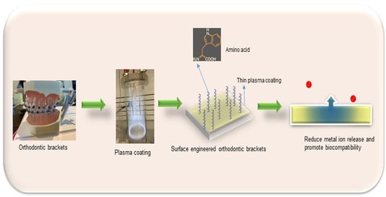
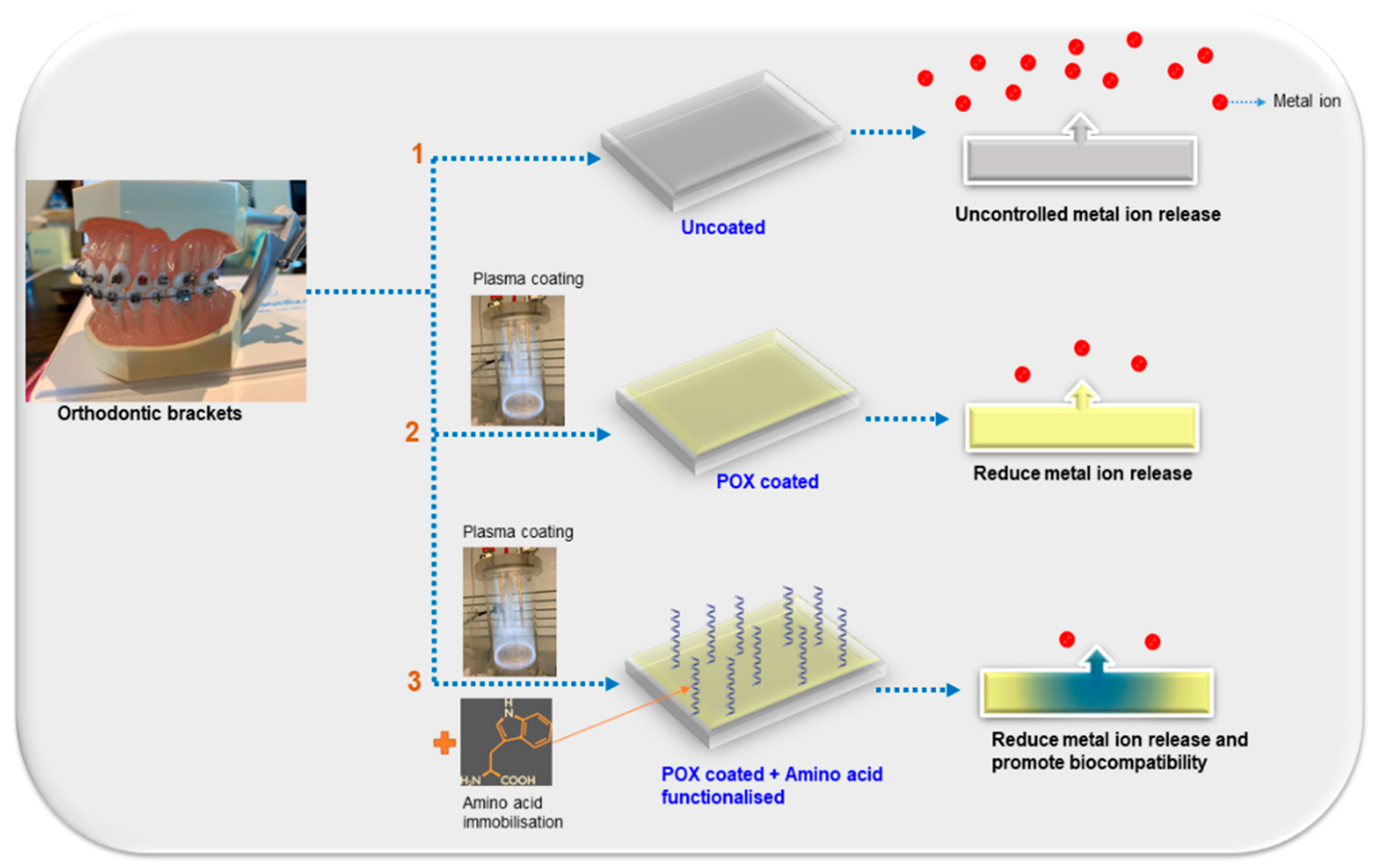
2.2.1. Plasma Coating of Orthodontic Brackets

Plasma polymerisation was used to deposit nanometre-thin polyoxazoline coatings on the surface of orthodontic brackets in a custom-built plasma reactor equipped with a 13.56 MHz plasma generator, designed by Vasilev et al. [14]. Orthodontic brackets were placed on a flat plate anode in the reactor chamber under a vacuum. The brackets were air cleaned in the chamber by exposure to air plasma for 5 min at a pressure of 1.3 × 10⁻¹ mbar and a power of 20 W. The deposition of polyoxazoline (POX) was then carried out at a pressure of 2.0 × 10⁻¹ mbar and an input power of 50 W for 2 min.
For further analysis of the coating, silicon wafers and glass coverslips were simultaneously coated with POX. The wafers were cleaned with acetone and ethanol, followed by cleaning for 5 min using air plasma. The deposition of POX was performed using the same parameters used for coating orthodontic brackets.

2.2.2. Surface Functionalisation of Orthodontic Brackets

The L- and D-isomers of the tryptophan were weighed and dissolved in ultrapure water with 18.2 MΩ, at a concentration of 25 mM. 96 POX coated orthodontic brackets were individually placed in each well of two 48-well plates and immersed in 1 mL each of either L- or D-tryptophan solutions for 24 h at room temperature. Following immobilisation, the samples were washed with Milli-Q water to remove any unbound amino acids, dried under nitrogen flow, and kept vacuum-sealed in new well plates. The samples used for analysis were uncoated orthodontic brackets (OB), brackets coated with POX alone (OB-POX), POX with L-Tryptophan (OB-POX/L-Trp), and POX with D-Tryptophan (OB-POX/D-Trp). All analyses were performed in triplicate.

2.2.3. Physico-Chemical Characterisation

A LEXT OLS5000 profilometer (Olympus Corporation, Tokyo, Japan) was used to assess the surface topography of OB, OB-POX, OB-POX/L-Trp, and OB-POX/D-Trp. For all samples, an area of ~360 µm, in the same location (on the bracket ligating gate), in a high magnification objective (100×) was used for the analysis.
A Variable Angle Spectroscopic Ellipsometer (VASE) (J.A. Woollam Co., Lincoln, NE, USA) was utilised to determine the thickness of the plasma coating before and after amino acid immobilisation. Following calibration, individual silicon wafers were mounted onto the ellipsometer for analysis of the thickness. Samples were measured at 5° angles between 65° and 75°, at wavelengths of 10 nm intervals between 250 and 1100 nm. Data were analysed using the WVASE software (Version 3.770, J.A. Woollam Co., Lincoln, NE, USA) and fitted using an established Cauchy model described previously [19].
The bulk chemical composition of the coated and uncoated orthodontic brackets was evaluated using a Niton XL3T Handheld XRF Analyser (Thermo Fischer Scientific, West Gosford, Australia). It was equipped with a high-resolution detector with a typical resolution of 145 eV that can screen heavy elements. Triplicates of samples were analysed, and values were plotted as ± SD.
XPS analysis was performed in triplicates using a Kratos Axis Ultra XPS spectrometer (Kratos Analytical, Manchester, UK) equipped with a monochromatic Al source operated at 15 keV and 15 mA to characterise the surface chemical composition of OB, OB-POX, OB-POX/L-Trp, and OB-POX/D-Trp. Survey spectra were obtained using 160 eV pass energy over a 0–1100 eV range in 0.5 eV steps. XPS data were analysed using Casa XPS software (Version 2.3.16, Casa Software Ltd., Teignmouth, UK), and all binding energies (BE) were corrected relative to the carbon C1s peak at 285.0 eV.
ToF-SIMS experiments were conducted using a PHI TRIFT V nanoTOF instrument (Physical Electronics Inc., Chanhassen, MN, USA) connected with a pulsed liquid metal Au+ primary ion gun (LMIG) (Physical Electronics Inc., Chanhassen, MN, USA), at 30 kV of energy. Experiments were performed under vacuum, and +SIMS spectra were collected from areas of 100 µm × 100 µm. Collected sample spectra and images were analysed using WincadenceN software V1.18.1 (Physical Electronics Inc., Chanhassen, MN, USA). The analysis was performed on L- and D-Trp powder to identify their characteristic peaks. Subsequently, ToF-SIMS was performed on OB, OB-POX, OB-POX/L-Trp, and OB-POX/D-Trp to determine if identical tryptophan peaks were detected on the surface of the coated brackets. Four spots on each sample were analysed.
The number of amino acids adsorbed on POX coated brackets was quantified using the Bradford assay kit (Thermo Fisher Scientific, Adelaide, Australia). 10 µL of each standard or test solution was transferred to a 96-well plate. To each of these wells, 300 µL of Bradford Reagent was added and mixed for 10 min. The plate was subsequently removed, and the absorbance was measured at 595 nm using a microplate reader (FLUOstar Optima, BMG Labtech, Mornington, Australia). A calibration curve for L-Trp and D-Trp was obtained in the concentration range from 3–50 mM. The amount of adsorbed amino acids on the brackets was calculated from the standard curve.
The brackets OB, OB-POX, OB-POX/L-Trp- and OB-POX/D-Trp were placed in 10 mL PBS solution for 7 days. The collected leachates were analysed using an Agilent 8900 Triple Quad ICP Mass spectrophotometer (Agilent, Santa Clara, CA, USA). Triplicate samples were analysed and quantified.
Cell viability assay was performed in triplicate to determine the toxicity of metal ion leachates from brackets when exposed to mammalian cells [20,21]. The brackets OB, OB-POX, OB-POX/L-Trp and OB-POX/D-Trp, were placed individually in 3 mL PBS for 7 days at room temperature to collect leachates. Human fibroblasts cells (HFFF2, CellBank, Westmead, Australia) were cultured in DMEM supplemented with 10% FCS, 1% penicillin (100 U·mL−1), and streptomycin (100 µg·mL−1). The cells were detached from the flasks using trypsin in EDTA media (0.25%) and then resuspended in DMEM. HFFF2 cells were seeded (1 × 10⁴ cells/well) in 96-well plates. The plate containing seeded cells was incubated for 24 h at 37 °C and 5% (v/v) CO2 atmosphere. After 24 h, the medium was replaced with fresh medium containing leachates (30 µL). Untreated wells with cells (no leachates) were maintained as a positive control. Fibroblasts were incubated for 24 h. 10% Resazurin in DMEM media was added to the wells and incubated for 4 h. During incubation, Resazurin was converted to resorufin by viable fibroblasts. The fluorescence intensity was measured at an excitation wavelength of 560 nm and emission at 590 nm using a microplate reader (FLUOstar Optima, BMG Labtech, Mornington, Australia) [22].
Statistical analysis was calculated using one-way ANOVA with Dunnett’s multiple comparisons test. Triplicates of samples were analysed, and data were presented as mean ± standard deviation.

3. Results and Discussion

Orthodontic brackets (OB) were coated with oxazoline (OB-POX) based coating deposited from plasma and further decorated with enantiomers of amino acids (OB-POX/L-Trp and OB-POX/D-Trp). In the first step, a unique configuration of plasma polymerisation was employed to allow partial retention of intact oxazoline functionalities on the surface of brackets and facilitate irreversible covalent binding of bioactive molecules [14]. In the next stage, plasma-coated brackets were separately immersed in either L- or D-tryptophan solutions to allow surface biofunctionalisation (Scheme 1). The capacity of the oxazoline functionality to covalently bind drugs, proteins, and antibodies is well documented [23]. Similar reactions have been reported on plasma polymerised oxazoline surfaces, where the oxazoline ring reacted with carboxylic functionalities present on the bioactive molecules to form an amide ester bond [15,24,25]. Here, the unique reactivity of POX with L- and D-Trp was explored to generate orthodontic brackets with different surface functionalities.

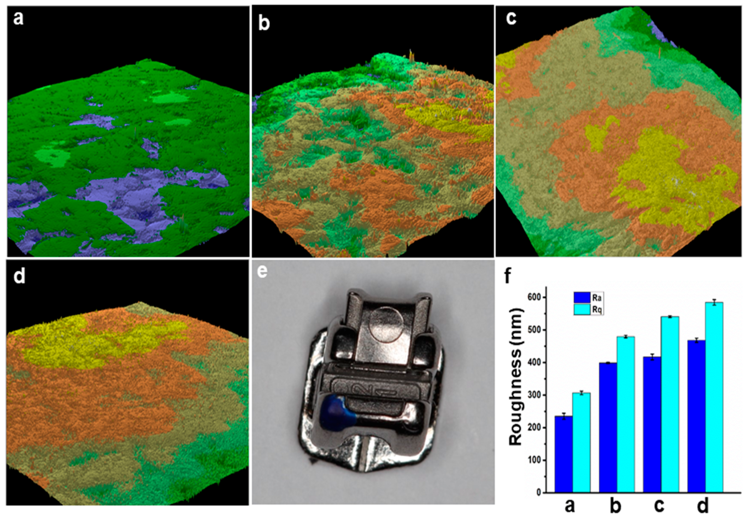
3.1. Surface Roughness

The 3D surface profilometry images provided an overview of the topography of OB, OB-POX, OB-POX/L-Trp, and OB-POX/D-Trp in a vertical orientation, using roughness descriptors such as average roughness (Ra) and the root mean squared roughness (Rs). The untreated bracket was relatively smooth with slight artefacts from the manufacturing machining process, but after plasma coating and attaching bioactive molecules, both Ra and Rb values substantially increased (Figure 1). OB-POX/D-Trp was found to have the highest roughness values with the Ra value of 468 ± 6 nm and Rq value of 584 ± 8 nm.

3.2. Bulk Chemistry

XRF was conducted to comprehend whether plasma polymerisation has any impact on the bulk chemistry of uncoated and coated orthodontic brackets. Figure 2 revealed that elemental analysis of OB, OB-POX, OB-POX/L-Trp and OB-POX/D-Trp consisted of approximately 60% Fe, 10% Si, 11% Cr, and trace amounts of Cu, Ni, Ti, and Ba. Based on published literature, it may be confirmed that their composition was similar to that of stainless steel [26] and was not affected by the plasma treatment. Nevertheless, the depth of penetration of X-rays in XRF spectroscopy was in the range of 0.003–3 mm and cannot determine the underlying surface chemistry of functionalised brackets. Hence, XPS and ToF-SIMS techniques were used for extensive investigation of surface functionalisation of the brackets.

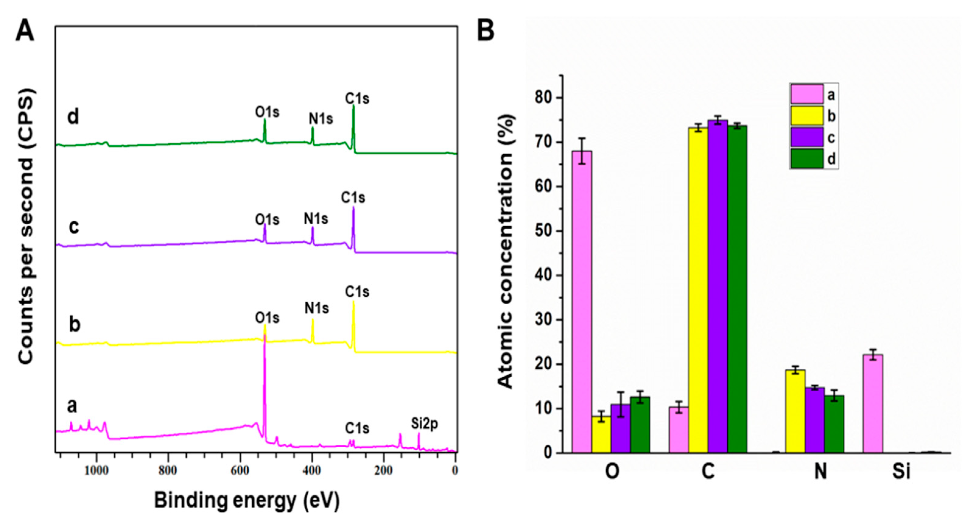
3.3. Surface Chemistry

The surface chemistry of uncoated and plasma-coated coverslips was evaluated using XPS. A typical survey spectrum of POX reveals oxygen, carbon, and nitrogen, arising from the oxazoline precursor structure (Figure 3). The absence of nitrogen in uncoated coverslips confirmed the successful deposition of POX over the surface. The silicon peak in uncoated coverslips was not visible in OB-POX, OB-POX/L-Trp and OB-POX/D-Trp. As the depth of penetration of X-rays in XPS was approximately 10 nm from the top surface, we expected that only the deposited plasma polymer would be detected and not the underlying silicon arising from the substrate. After coating again, the disappearance of a silicon peak confirmed that the surfaces were successfully modified with the plasma polymer. The N/C ratio of plasma functionalised samples was higher than uncoated coverslips due to the presence of nitrogen in oxazoline-derived coating and the core structure of the amino acids. Tryptophan is an α-amino acid composed of the α-carboxylic group, α-amino group, and an indole side chain. At the same time, the O/C ratio of OB-POX, OB-POX/L-Trp and OB-POX/D-Trp decreased compared to uncoated coverslips.
Although the proportion of C, O, N were comparable among the plasma coated samples, there was a slight increase in the O/C ratio from 0.11 for OB-POX, 0.148 for OB-POX/L-Trp, and 0.173 for POX/D- Trp. This might be due to the possible interaction of amino acids with the POX layer.

3.4. ToF-SIMS

ToF-SIMS is a highly sensitive analytic technique capable of detecting molecular structural fragments on the sample’s top surface (~2 nm). It was used to detect the presence of amino acids on the surface of plasma functionalised surfaces. There was no contamination (particularly silicones) in any of the samples. There was an increase in the total counts of C9H8N+ in the amino acid functionalised samples compared to uncoated samples. This correlates to a high-intensity fragment of tryptophan [27,28] which confirmed the successful immobilisation of amino acids on the surface of brackets (Figure 4).

3.5. Quantification of Adsorbed Amino Acids

A Bradford assay was used to quantify L- and D-Trp bound to POX coated orthodontic brackets. After 24 h of immobilisation of POX coated orthodontic brackets in 25 mM Trp, the amount of Trp present in the immobilisation media before and after bracket immersion was calculated from the calibration curve. In this assay, the change in the colour of the Coomassie G-250 dye from red to blue on binding with amino acids was quantified spectroscopically. The L- and D-Trp adsorbed on brackets were 0.0039 mM and 0.00686 mM, respectively (Figure 5). The results conveyed that adsorbed D-Trp on the brackets was higher than L-Trp. This agreed with the profilometry and XPS results, which confirmed that D-Trp had a higher POX affinity than L-Trp. Using ellipsometry, the thickness of the functionalised coating on OB-POX, OB-POX/L-Trp and OB-POX/D-Trp was 25.078 ± 1.2, 26.037 ± 2.54, and 30.7285 ± 1.01 nm, respectively. This again supported the observation that D-Trp has a higher binding affinity to POX.

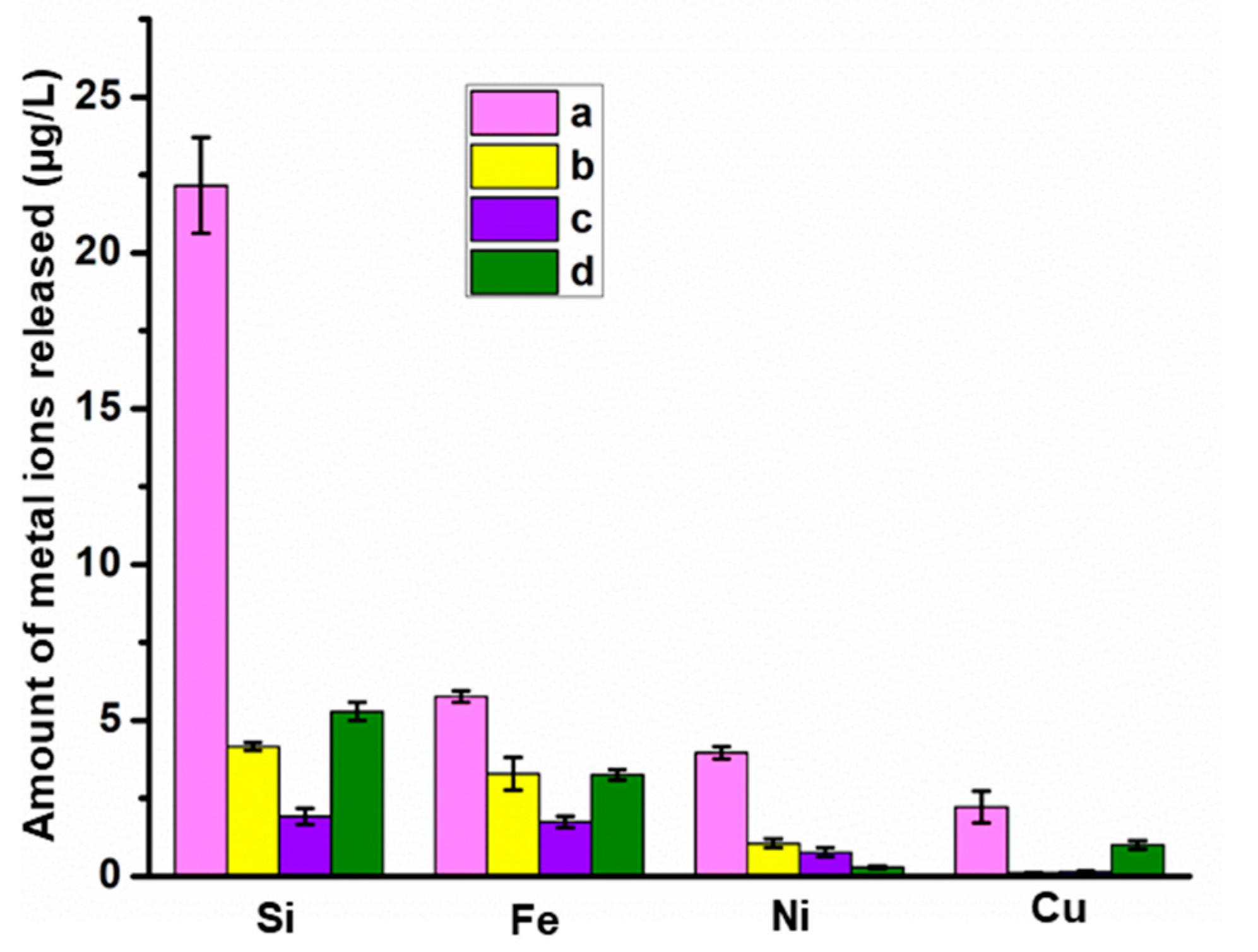
3.6. Metal Ion Release
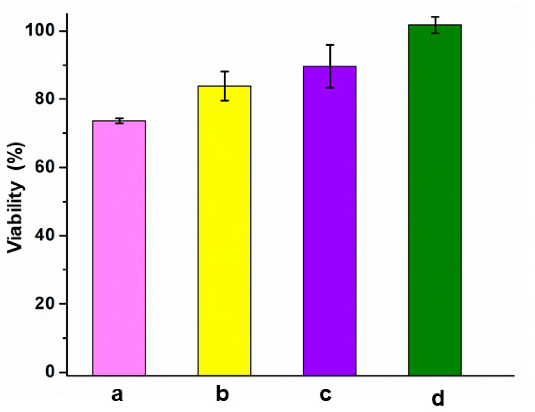
The results from quantitative ion release over seven days using ICP-MS are presented in Figure 6. Overall, the results showed that uncoated orthodontic brackets released the highest total amount of metal ions compared to other samples. There was a substantial decrease in silicon ions released after plasma coating. OB-POX/L-Trp and OB-POX/D-Trp showed reduced leaching of iron, nickel, and copper as well. Metal degradation will not only damage the material surface but also trigger adverse biological reactions [29]. The results revealed the unique feature of plasma coating to act as a metal ion release barrier function and control leaching.

3.7. Cytotoxicity

The cytotoxicity of the orthodontic brackets was evaluated using Resazurin assay on the HFFF2 cell line. The results conveyed that none of the materials can be rated toxic, which recommends a cut-off of 30% for cell viability reduction [30]. However, OB-POX/L-Trp and OB-POX/D-Trp exhibited higher cell viability compared to others (Figure 7). This may be because of reduced ion leaching from the metal brackets due to plasma coating and surface immobilisation of amino acids.

4. Conclusions

In the past decades, there has been substantial progress in using stainless-steel alloys for dental appliances. Nonetheless, when metal alloys are introduced in the oral environment, their stability will be impacted, leading to a greatly increased release of metal ions. The released metal ions may be harmful to human health and may eventually lead to severe complications. In the present study, plasma polymerisation was used to surface functionalise orthodontic brackets to minimise metal ion leaching. The surface properties of the brackets were successfully tailored via a thin plasma polymer film deposited from the vapour of 2-methyl-2-oxazoline. The brackets were further immobilised with enantiomers of tryptophan. The bulk composition of the brackets was least affected after plasma functionalisation. Detailed surface analysis showed that D- and L-tryptophan were successively immobilised on the surface of brackets. The results from quantitative ion-release studies confirmed that plasma functionalisation of orthodontic brackets minimises metal ion release. An analysis of primary human dermal fibroblasts showed no apparent reduction in cell viability caused by either plasma coating or immobilised amino acid functionalities. These results indicate that surface engineering of brackets using plasma polymerisation is a strategy to combat metal toxicity in orthodontics.

Author Contributions

Conceptualization, P.Z., K.V., and N.N.; methodology, L.S.K., N.N., P.R.L.D., and A.C.; software, P.R.L.D., A.C., and N.N.; validation, L.S.K., N.N., P.R.L.D., and A.C.; investigation, L.S.K.; data curation, L.S.K. and N.N.; writing—original draft preparation, N.N. and L.S.K.; writing—review and editing, G.R.-F., E.J.D., C.D., K.V., and P.Z.; supervision, G.R.-F., E.J.D., C.D., K.V., and P.Z.; funding acquisition, P.Z. and K.V. All authors have read and agreed to the published version of the manuscript.

Funding

K.V. thanks ARC for DP15104212, NHMRC for Fellowship APP1122825 and Project grant APP1032738, and the Alexander von Humboldt Foundation for the Fellowship for Experienced Researchers. L.S.K thanks the Adelaide Dental School for research support and the Australian Society of Orthodontists Foundation for Research and Education for further financial assistance.

Institutional Review Board Statement

Not applicable.

Informed Consent Statement

Not applicable.

Data Availability Statement

Data sharing is not applicable to this article.

Conflicts of Interest

The authors declare no conflict of interest.

References

  1. Ntasi, A.; Al Jabbari, Y.S.; Silikas, N.; Eliades, T.; Zinelis, S. Multitechnique characterization of conventional and experimental Ag-based brazing alloys for orthodontic applications. Dent. Mater. 2018, 34, e25–e35. [Google Scholar] [CrossRef]
  2. Redlich, M.; Katz, A.; Rapoport, L.; Wagner, H.; Feldman, Y.; Tenne, R. Improved orthodontic stainless steel wires coated with inorganic fullerene-like nanoparticles of WS2 impregnated in electroless nickel–phosphorous film. Dent. Mater. 2008, 24, 1640–1646. [Google Scholar] [CrossRef]
  3. Eliaz, N. Corrosion of Metallic Biomaterials: A Review. Materials 2019, 12, 407. [Google Scholar] [CrossRef] [Green Version]
  4. Tracana, R.B.; Sousa, J.; Carvalho, G.S. Mouse inflammatory response to stainless steel corrosion products. J. Mater. Sci. Mater. Electron. 1994, 5, 596–600. [Google Scholar] [CrossRef]
  5. Tracana, R.B.; Pereira, M.D.L.; Abreu, A.M.; Sousa, J.; Carvalho, G.S. Stainless steel corrosion products cause alterations on mouse spleen cellular populations. J. Mater. Sci. Mater. Electron. 1995, 6, 56–61. [Google Scholar] [CrossRef]
  6. Ortiz, A.J.; Fernández, E.; Vicente, A.; Calvo, J.L.; Ortiz, C. Metallic ions released from stainless steel, nickel-free, and tita-nium orthodontic alloys: Toxicity and DNA damage. Am. J. Orthod. Dentofacial. Orthop. 2011, 140, e115–e122. [Google Scholar] [CrossRef] [PubMed]
  7. Jemat, A.; Ghazali, M.J.; Razali, M.; Otsuka, Y. Surface Modifications and Their Effects on Titanium Dental Implants. BioMed Res. Int. 2015, 2015, 1–11. [Google Scholar] [CrossRef] [Green Version]
  8. Yang, Y.; Qi, P.; Ding, Y.; Maitz, M.F.; Yang, Z.; Tu, Q.; Xiong, K.; Leng, Y.; Huang, N. A biocompatible and functional adhesive amine-rich coating based on dopamine polymerization. J. Mater. Chem. B 2014, 3, 72–81. [Google Scholar] [CrossRef] [Green Version]
  9. Metin-Gürsoy, G.; Taner, L.; Akca, G. Nanosilver coated orthodontic brackets: In vivo antibacterial properties and ion re-lease. Eur. J. Orthod. 2016, 39, 9–16. [Google Scholar] [CrossRef] [PubMed] [Green Version]
  10. Ryu, H.-S.; Bae, I.-H.; Lee, K.-G.; Hwang, H.-S.; Lee, K.-H.; Koh, J.-T.; Cho, J.-H. Antibacterial effect of silver-platinum coating for orthodontic appliances. Angle Orthod. 2011, 82, 151–157. [Google Scholar] [CrossRef] [PubMed] [Green Version]
  11. Joseph, B.; Ninan, N.; Visalakshan, R.M.; Denoual, C.; Bright, R.; Kalarikkal, N.; Grohens, Y.; Vasilev, K.; Thomas, S. Insights into the biomechanical properties of plasma treated 3D printed PCL scaffolds decorated with gold nanoparticles. Compos. Sci. Technol. 2021, 202, 108544. [Google Scholar] [CrossRef]
  12. Forget, A.; Staehly, C.; Ninan, N.; Harding, F.J.; Vasilev, K.; Voelcker, N.H.; Blencowe, A. Oxygen-Releasing Coatings for Improved Tissue Preservation. ACS Biomater. Sci. Eng. 2017, 3, 2384–2390. [Google Scholar] [CrossRef]
  13. Gillam, T.; Goh, C.; Ninan, N.; Bilimoria, K.; Shirazi, H.; Saboohi, S.; Al-Bataineh, S.; Whittle, J.; Blencowe, A. Iodine complexed poly(vinyl pyrrolidone) plasma polymers as broad-spectrum antiseptic coatings. Appl. Surf. Sci. 2021, 537, 147866. [Google Scholar] [CrossRef]
  14. Macgregor-Ramiasa, M.N.; Cavallaro, A.; Vasilev, K. Properties and reactivity of polyoxazoline plasma polymer films. J. Mater. Chem. B 2015, 3, 6327–6337. [Google Scholar] [CrossRef]
  15. MacGregor, M.N.; Michelmore, A.; Shirazi, H.S.; Whittle, J.; Vasilev, K. Secrets of Plasma-Deposited Polyoxazoline Functionality Lie in the Plasma Phase. Chem. Mater. 2017, 29, 8047–8051. [Google Scholar] [CrossRef]
  16. Park, H.; Kim, K.M.; Lee, A.; Ham, S.; Nam, W.; Chin, J. Bioinspired Chemical Inversion of l-Amino Acids to d-Amino Acids. J. Am. Chem. Soc. 2007, 129, 1518–1519. [Google Scholar] [CrossRef] [PubMed]
  17. Yazici, H.; O’Neill, M.B.; Kacar, T.; Wilson, B.R.; Oren, E.E.; Sarikaya, M.; Tamerler, C. Engineered Chimeric Peptides as Antimicrobial Surface Coating Agents toward Infection-Free Implants. ACS Appl. Mater. Interfaces 2016, 8, 5070–5081. [Google Scholar] [CrossRef] [Green Version]
  18. Mishra, A.K.; Choi, J.; Moon, E.; Baek, K.-H. Tryptophan-Rich and Proline-Rich Antimicrobial Peptides. Molecules 2018, 23, 815. [Google Scholar] [CrossRef] [PubMed] [Green Version]
  19. Al-Bataineh, S.A.; Cavallaro, A.A.; Michelmore, A.; Macgregor, M.N.; Whittle, J.D.; Vasilev, K. Deposition of 2-oxazoline-based plasma polymer coatings using atmospheric pressure helium plasma jet. Plasma Process. Polym. 2019, 16, 1900104. [Google Scholar] [CrossRef]
  20. Ninan, N.; Albrecht, H.; Blencowe, A. Characterization of Nanomaterials; Woodhead Publishing: Cambridge, UK, 2018; pp. 129–166. [Google Scholar]
  21. Ninan, N.; Forget, A.; Shastri, V.P.; Voelcker, N.H.; Blencowe, A. Antibacterial and Anti-Inflammatory pH-Responsive Tannic Acid-Carboxylated Agarose Composite Hydrogels for Wound Healing. ACS Appl. Mater. Interfaces 2016, 8, 28511–28521. [Google Scholar] [CrossRef] [PubMed]
  22. Vega-Avila, E.; Pugsley, M.K. An overview of colorimetric assay methods used to assess survival or proliferation of mammalian cells. Proc. West. Pharmacol. Soc. 2011, 54, 10–14. [Google Scholar]
  23. De La Rosa, V.R. Poly(2-oxazoline)s as materials for biomedical applications. J. Mater. Sci. Mater. Med. 2014, 25, 1211–1225. [Google Scholar] [CrossRef] [PubMed]
  24. Chan, K.M.; Vasilev, K.; Shirazi, H.S.; McNicholas, K.; Li, J.; Gleadle, J.; MacGregor, M. Biosensor device for the pho-to-specific detection of immuno-captured bladder cancer cells using hexaminolevulinate: An ex-vivo study. Photodiagnosis Photodyn. Ther. 2019, 28, 238–247. [Google Scholar] [CrossRef]
  25. MacGregor, M.; Sinha, U.; Visalakshan, R.M.; Cavallaro, A.; Vasilev, K. Preserving the reactivity of coatings plasma depos-ited from oxazoline precursors − An in depth study. Plasma Process. Polym. 2019, 16, 1800130. [Google Scholar] [CrossRef]
  26. Barrea, R.A.; Mainardi, R.T. Standardless XRF analysis of stainless-steel samples. X-Ray Spectrom. 1998, 27, 111–116. [Google Scholar] [CrossRef]
  27. Henry, M.; Bertrand, P. Surface composition of insulin and albumin adsorbed on polymer substrates as revealed by multi-variate analysis of ToF-SIMS data. Surf. Interface Anal. 2009, 41, 105–113. [Google Scholar] [CrossRef]
  28. Tamulienė, J.; Romanova, L.G.; Vukstich, V.S.; Papp, A.V.; Snegursky, A.V. Electron-impact-induced tryptophan molecule fragmentation. Eur. Phys. J. D 2015, 69, 21. [Google Scholar] [CrossRef]
  29. Visalakshan, R.M.; Cavallaro, A.A.; MacGregor, M.N.; Lawrence, E.P.; Koynov, K.; Hayball, J.D.; Vasilev, K. Nanotopog-raphy-Induced Unfolding of Fibrinogen Modulates Leukocyte Binding and Activation. Adv. Funct. Mater. 2019, 29, 1807453. [Google Scholar] [CrossRef]
  30. Li, W.; Zhou, J.; Xu, Y. Study of the in vitro cytotoxicity testing of medical devices. Biomed. Rep. 2015, 3, 617–620. [Google Scholar] [CrossRef] [PubMed] [Green Version]
Scheme 1. Schematic illustration of the design of surface-functionalised orthodontic brackets involving plasma coating and subsequent immobilisation with enantiomers of amino acid to combat metal ion release.
Coatings 11 00857 sch001
Figure 1. 3D surface profiling of (a) OB (b) OB-POX (c) OB-POX/L-Trp (d) OB-POX/D-Trp, (e) Damon Q orthodontic bracket. (f) Comparison of surface roughness of a—OB, b—OB-POX, c—OB-POX/L-Trp, d—OB-POX/D-Trp, evaluated from surface profilometer.
Coatings 11 00857 g001
Figure 2. Bulk chemical characterisation of a—OB, b—OB-POX, c—OB-POX/L-Trp, d—OB-POX/D-Trp using XRF.
Coatings 11 00857 g002
Figure 3. (A) X-ray photoelectron spectroscopy survey spectra and (B) atomic concentration of elements such as O, C, N and Si of a—OB, b—OB-POX, c—OB-POX/L-Trp, d—OB-POX/D-Trp.
Coatings 11 00857 g003
Figure 4. Total counts per second (CPS) of C9H8N+ detected in a—OB, b—OB-POX, c—OB-POX/L-Trp d—OB-POX/D-Trp, using ToF-SIMS spectroscopy.
Coatings 11 00857 g004
Figure 5. (A) Quantification of adsorbed amino acids using a Bradford assay and (B) Thickness of plasma coating evaluated using ellipsometry for a—OB-POX/L-Trp, b—OB-POX/D-Trp, c—OB-POX.
Coatings 11 00857 g005
Figure 6. ICP MS analysis of metal ions leached of a—OB, b—OB-POX, c—OB-POX/L-Trp, d—OB-POX/D-Trp incubated in phosphate-buffered saline at pH 7.2 on day 7.
Coatings 11 00857 g006
Figure 7. Cell viability of the leachates collected from a—OB, b—OB-POX, c—OB-POX/L-Trp, d—OB-POX/D-Trp incubated in phosphate-buffered saline at pH 7.2 for seven days.
Coatings 11 00857 g007
Publisher’s Note: MDPI stays neutral with regard to jurisdictional claims in published maps and institutional affiliations.

