
Micro-Arc Oxidation in Titanium and Its Alloys: Development and Potential of Implants


1
School of Materials Science and Engineering, Beihang University, Beijing 100191, China
2
Zhongfa Aviation Institute, Beihang University, Hangzhou 311115, China
3
Institute of Translational Medicine, Shanghai University, Shanghai 200444, China
*
Authors to whom correspondence should be addressed.
Coatings 2023, 13(12), 2064; https://doi.org/10.3390/coatings13122064
Submission received: 15 November 2023
/
Revised: 5 December 2023
/
Accepted: 6 December 2023
/
Published: 9 December 2023
(This article belongs to the Special Issue Multifunctional Coatings on Medical Devices)
Abstract
:Titanium (Ti) and its alloys are widely recognized as preferred materials for bone implants due to their superior mechanical properties. However, their natural surface bio-inertness can hinder effective tissue integration. To address this challenge, micro-arc oxidation (MAO) has emerged as an innovative electrochemical surface modification technique. Its benefits range from operational simplicity and cost-effectiveness to environmental compatibility and scalability. Furthermore, the distinctive MAO process yields a porous topography that bestows versatile functionalities for biological applications, encompassing osteogenesis, antibacterial, and anti-inflammatory properties. In this review, we undertake an examination of the underlying mechanism governing the MAO process, scrutinize the multifaceted influence of various factors on coating performance, conduct an extensive analysis of the development of diverse biological functionalities conferred by MAO coatings, and discuss the practical application of MAO in implants. Finally, we provide insights into the limitations and potential pathways for further development of this technology in the field of bone implantation.
1. Introduction
The latest statistics show that by mid-century, the global population aged 65 or over is expected to more than double to a staggering 1.6 billion people [1]. An aging population means a larger population base of chronic diseases, and bone and joint diseases. In dentistry and orthopedics, implants, with titanium (Ti) and its alloys as the preferred choice among various materials, have become a common approach for treating conditions due to their exceptional biocompatibility and mechanical properties [2]. Despite the vast potential for Ti and its alloys, there are still limitations to surface bioactivity. They often exhibit biological inertness, limiting the close integration of implants with surrounding tissues. Furthermore, Ti alloys are susceptible to microorganisms, increasing the risk of postoperative infections, and threatening the prognosis of patients. To tackle these issues, apart from alloying [3,4,5], surface treatment proves to be an effective approach as well. Common methodologies encompass atmospheric plasma spraying [6], laser treatment [7], anodizing [8], micro-arc oxidation (MAO), and biomimetic deposition of apatite [9] or other biomaterials [10]. Among the various surface modifications available, MAO has garnered significant attention due to its simplicity of operation, cost-effectiveness, and applicability to complex devices. Through MAO, the surface of Ti-based implants can undergo alterations in microstructural features and chemical composition, rendering them more suitable for tissue adhesion and effectively reducing the risk of infections [11]. The specific impacts of MAO on the substrate surface can be found in Table 1.
In this review, we aim to provide an overview of MAO applied to the preparation of coatings on Ti and its alloys for biomedical applications over the past 5 years, especially orthopedic and dental implants. This review encompasses an analysis of the factors influencing the coating preparation process, and an examination of the biological functionalities of these coatings and their applications. Finally, we highlight the challenges and future directions for the application of MAO in metal implants and make a vision for the development of surface coatings for metal implants.
2. The Process of MAO
2.1. The Mechanism of MAO
MAO involves plasma discharge, but it differs from traditional plasma electrolytic discharge techniques like plasma cutting or welding. It is often confused with anodic oxidation (AO), but they do indeed represent different processes and technologies. MAO typically requires a higher voltage and shorter processing times, and the type of electrolyte used, as well as the characteristics of the resulting coating, are significantly different from AO [19].
At present, most researchers divide the MAO process into three, which are spark discharge, micro-arc discharge, and arc discharge. Some add the early short anodizing phase, for a total of four stages [11,20].
The formation mechanism of MAO coatings is essentially similar, regardless of the electrolyte or control mode used. Taking the constant current mode as an example, Figure 1 illustrates the changes during the MAO process. In the first stage, as the current increases, the voltage linearly increases, initiating the traditional anodic oxidation phase, involving only electrochemical reactions. At this stage, a porous insulating film with a columnar structure perpendicular to the substrate is formed on the metal substrate, accompanied by gas generation, serving as precursors for igniting plasma discharge [21].
With the increase in current, the voltage on the coating also rises, moving on to the second and third stages. Once the voltage exceeds the critical threshold, known as breakdown voltage, it leads to spark discharge. There are various areas of defects, stress concentrations, and uneven thickness on the coating, and these vulnerable areas are the first to break down. This is the micro-arc discharge stage. In the later stage, when the maximum thickness of the porous oxide film is approached, the oxide film grows slowly with the increase in time, which is the arc discharge stage [23].
The phenomenon of micro-arc discharge and arc discharge is very similar, with only a slight difference in energy level. Two models of arc discharge have been proposed: one involving dielectric breakdown of the oxide film [24], and the other involving gas breakdown within micro-pores [25]. Both models result in the generation of high-energy plasma, possibly coexisting. Optical Emission Spectroscopy (OES) and Intensified Charge Coupled Device (ICCD)-based spectral systems provide real-time imaging of the MAO discharge process (Figure 2). The bright spots in the figure represent sparks, and over time, the intensity and range of sparks increase, indicating a larger pore size formed on the surface of the oxide film [26].
Stojadinovic et al. [27] used OES-ICDD to record real-time discharges. Two types of plasma micro-discharge were observed during this process, with electron densities of Ne = 3.8 × 1015 cm−3 and Ne = 4.5 × 1016 cm−3, and electron temperatures ranging from Te = 3700 ± 500 K. Plasma discharge generates high temperatures, causing thermal decomposition of water molecules, producing more hydrogen and oxygen, further promoting plasma discharge. Oxides undergo melting ejections at high temperatures, encountering rapid solidification and recrystallization in contact with the cold electrolyte, depositing on the coating surface. A study [28] classified discharges into types a, b, and c, representing surface, internal, and intermediate discharges in coatings, leading to inward/outward growth of the coating. In this process, numerous white sparks scatter randomly and flicker on the workpiece surface [29]. As the discharge intensifies, the voltage increase rate slows down. With the melting and solidification of oxides, the thickness of coatings rapidly increases, and gas release and breakdown discharge generate pores. The number of sparks decreases, and the color changes from white to yellow. This marks the transition to the micro-arc discharge stage, a crucial phase in coating growth. As the reaction continues, the voltage stabilizes, entering the strong arc discharge stage. The spark discharge diameter gradually increases, turning orange-red, accompanied by a sharp noise. Intense discharge can cause spattering of coating materials and localized severe ablation characteristics.
Stress-induced cracking cannot be avoided during the rapid solidification process in contact with the cold electrolyte [30]. The resulting coating usually consists of a thinner internal dense layer and a thicker external porous layer, with thickness ranging from 1 to 100 μm [31]. To produce high-quality coatings, precise control of MAO process parameters is crucial, mainly including electrolytes, electrical parameters, and substrates.
2.2. Electrolyte
An electrolyte plays a pivotal role in the quality of MAO coatings. It directly influences the composition of coatings and exerts a significant impact on their growth mechanisms [31]. In the realm of MAO, electrolyte systems are traditionally classified by the anion type employed, including phosphate, silicate, borate, and aluminate. In recent years, organic phytic acid has gained recognition as an electrolyte for the MAO of biomedical alloys. Earlier practices involved acidic electrolytes [27], but recent mainstream electrolytes are typically alkaline with an ideal pH of around 13 [11]. The concentration, pH, electrical conductivity, additives, and temperature variations all influence coating formation.
In terms of electrolyte types, silicate and phosphate demonstrate better biocompatibility because Si and P elements promote osteogenesis [32]. When examining coating characteristics under similar conditions, it is observed that in the silicate electrolyte system, coatings grow more rapidly and to a greater thickness compared to other electrolyte systems. These coatings also exhibit increased roughness but relatively weaker adhesion to the metal substrates [33]. Consequently, such coatings often possess inferior wear resistance and corrosion resistance. The resulting bioceramic coatings primarily consist of rutile, anatase, and amorphous silica phases. In the phosphate electrolyte system, MAO coatings tend to be thinner but more compact, with lower roughness. They feature uniform, micrometer-sized pores, and exhibit strong adhesion to the substrate. These coatings consist primarily of anatase and rutile phases, providing excellent corrosion and wear resistance [30,34]. Silicate results in outward coating growth with poor adhesion due to oxide deposition, while phosphate promotes inward growth with strong adhesion linked to substrate oxidation. Combining silicate and phosphate achieves a balanced effect. The addition of calcium to the phosphate electrolyte can induce the formation of bone-like hydroxyapatite (HA), a biologically active material [32].
Due to the susceptibility of borates to hydrolysis, their solutions exhibit higher alkalinity and electrical conductivity. In the sodium tetraborate electrolyte, surfaces of Ti-based alloys undergo more intense discharges, resulting in the formation of a distinctive “channel-like” or “cortex-like” micro/nanostructure in the coatings (Figure 3). These coatings exhibit enhanced hydrophilicity and osseointegration. They are primarily composed of anatase, rutile TiO2, and amorphous boron oxide [35]. Wang et al. [36] used sodium tetraborate to create a micro/nanoscale porous structure on the surface of a Ti-20Zr-10Nb-4Ta alloy. Results indicated that discharges in sodium tetraborate electrolytes lasted longer, compared to phosphate electrolytes, resulting in a larger pore size and higher porosity in the coatings.
When utilizing aluminate electrolytes, the coatings tend to precipitate aluminum oxide (or crystalline aluminum titanate, TiAl2O5), which provides higher hardness and stability. Consequently, these coatings often display excellent resistance to wear and corrosion [37,38]. Jiang et al. [39] employed phosphate, silicate, and aluminate electrolytes to prepare MAO coatings. They found that coatings produced in aluminate electrolytes had the densest structure. It is imperative to acknowledge that aluminate, which exhibits relatively poor stability, is susceptible to hydrolysis, leading to the formation of aluminum hydroxide. The presence of aluminum ions may potentially have detrimental effects on the human body. Consequently, the use of aluminate electrolytes in surface modification for biomedical metals has seen a gradual decline. In recent years, organic phytic acid has begun to find applications in the electrolytes of MAO [40,41]. It is an organic molecule rich in phosphorus elements, resulting in MAO films composed of anatase, rutile, and Ti2O7. It imparts outstanding biocompatibility to the coatings.
The anions in the electrolyte are involved in the discharge and coating formation processes, while cations typically influence the conductivity through concentration [42,43]. Conductivity is generally in the range of 5–100 mS/cm for the MAO electrolyte [41]. During the process, the conductivity of the electrolyte also changes, and the aging of the electrolyte is a noteworthy issue [44]. The conductivity is critical, influencing current and voltage within the narrow pores, thereby affecting the initiation of plasma discharge [27]. In the same anion electrolyte system, increasing conductivity leads to thicker coatings and reduced defects [45].
Currently, the incorporation of nano- and micro-particles into coatings is recognized as a primary approach for enhancing coating properties and expanding the scope of their chemical composition [46]. So far, various nanoparticles (NPs) (5–100 nm) and microparticles (0.18–44 μm) have been employed. These enhancements encompass a wide range of properties, including corrosion resistance, mechanical strength, tribological performance, and biocompatibility. For instance, the incorporation of graphene oxide (GO), TiO2, and ZrO2 enhances the corrosion resistance and tribological characteristics of coatings, while the inclusion of HA improves the biocompatibility [47]. In terms of composition, some elements are beneficial for improving the biological activity of the material and further enhancing the osseointegration, such as P [48], Si [49], Ca [50], Zr [6], and Sr [32]. The addition of Ag, Cu, and Zn imparts antibacterial properties to the coatings [51]. Incorporating particles into coatings is complex due to their limited solubility in aqueous solutions. These particles may remain unchanged in size and shape or melt during micro-discharge, influencing the composition, microstructure, and other properties of coatings. Key factors affecting these reactions at the solid–liquid interface include zeta potential, melting point, and particle size [22].
Zeta potential signifies the electrical charge at the particle–fluid interface, with high, negative zeta potential being favorable in electrolytes for stability, resisting agglomeration, and settling [15]. Adjusting the pH of the electrolyte can shift the zeta potential toward negativity. By measuring the zeta potential, Necula et al. [52] established optimal conditions for Ag incorporation in the Keronite electrolyte. Melting point and particle size are two other critical factors affecting incorporation, because of the elevated local temperatures during the MAO treatment (~2700 °C) [53]. Particles with high melting points, such as CeO2 and SiC, tend to remain inert or partially reactive regardless of their size. On the other hand, particles with lower melting points are more prone to reactive incorporation, resulting in mixed-oxide coatings within the discharge channels during the MAO process. As an example, SiO2 particles with a low melting point have been successfully incorporated into MAO coatings. Nanometric particles (around 12 nm) showed active integration, while micrometric particles (1–5 μm) remained inert [54]. To mitigate additive agglomeration, various strategies are available, including the utilization of chelating agents and organic acids, such as nitrilotriacetic acid (C6H9NO6), phosphoric acid (H3PO4), phytic acid (C6H18O24P6), and disodium EDTA (Na2EDAT·2H2O). These approaches help create stable and chemically compatible solutions or suspensions [55].
Electrolyte temperature significantly impacts the discharge. There is a non-linear relationship between temperature and coating thickness. Initially, a higher temperature improves corrosion and wear resistance, peaking at 40 °C, but then it declines. Rapid temperature changes in the electrolyte pose challenges to process stability [56]. Electrolyte temperature fluctuations impact the properties of MAO coatings. Higher temperatures can lead to excessive discharging, causing uneven surface topography in the coating [11]. Coatings produced at 278 K have lower porosity than those at higher temperatures due to larger pores and increased pore density, resulting in reduced adhesion to substrates [57]. To obtain high-quality coatings, it is advisable to use a cooling system to keep the electrolyte temperature around 20 °C, with a maximum not exceeding 40 °C [58].
2.3. Electrical Parameters
In the MAO process, the performance of the coating is influenced not only by the electrolyte but also by various electrical parameters, including processing time, current mode, current density, and frequency.
In the standard power mode of the MAO process, different current modes can be employed, including direct current (DC) and alternating current (AC). Currently, the pulsed current is also widely used in MAO treatment, including both DC pulse power and AC pulse power, whether a unipolar or bipolar pulse [59]. The DC mode results in slower oxide growth and increased porosity, though it offers less control. Implementing pulsed DC may improve control over discharge duration and reduce energy consumption [60]. Compared to DC power, AC power can swiftly mitigate electrode polarization by intermittently interrupting the electric arc. It provides increased flexibility, improves process control efficiency, and enhances coating quality. Currently, the use of AC-pulsed bipolar MAO power sources is widespread due to their capacity to produce superior coatings. These coatings exhibit a greater thickness, higher micro-hardness, and enhanced adhesion to substrates [61]. This effectiveness is attributed to their ability to mitigate the impact of high-intensity plasma discharges and reduce the occurrence of high-temperature spikes [62]. In different power modes, constant current and constant voltage modes can be employed. A work [63] demonstrates that, when using the constant direct current mode, extending the duration at a low current density (2 A/dm²) helps prolong the micro-arc phase and results in the formation of a porous layer with fewer internal defects. The compact internal structure enhances the hardness and corrosion resistance. Conversely, with higher current density, the discharge enters the large arc phase earlier, leading to an increase in internal defects and a decrease in adhesion. Seo et al. [64] treated the surfaces of CP-Ti and Ti-6Al-4V ELI discs under pulsed DC with constant voltage conditions. Increasing voltage led to a larger grain size and surface pore size, and more rutile content, while anatase content decreased. A high voltage caused surface cracking. Both constant current and constant voltage modes have pros and cons. A constant current ensures uniform arc discharges but can generate excess heat, affecting coating uniformity. A constant voltage allows better control of arc discharges and reduces thermal effects, though it may slightly compromise coating uniformity.
Both unipolar and bipolar pulse modes introduce variable parameters like frequency, duty cycle, and anode-to-cathode current ratio, necessitating a deeper understanding of their impact on coatings. Previous research indicates that frequency plays a crucial role in coating performance. Lower frequencies result in rougher coatings with larger pores, making AC pulses more effective than DC pulses for coating formation. Higher frequencies lead to coatings with a higher density of finer pores [65]. Wang et al. [66] created Ca- and P-containing MAO films on Ti substrates at frequencies from 100 to 5000 Hz. Higher frequencies decrease crystallinity but increase Ca and P content. Films at higher frequencies have smaller pores on larger pore walls, improving film wettability.
Maintaining a constant voltage and frequency, an increased duty cycle lengthens the discharge period in a single-polarity pulse, resulting in thicker coatings, additional pores, and greater surface roughness [67,68]. However, excessively high duty cycles can damage the coating. Babaei et al. [69] prepared TiO2-ZrO2 composite coatings on CP-Ti, finding that different micro-discharge states influence the performance of coatings prepared under varying duty cycles. Higher duty cycles lead to an increased coating thickness and greater surface roughness. Pavarini et al. [70] created Cu and Zn co-doped MAO coatings with antimicrobial and osteogenic properties using pulsed DC in a sodium tetraborate electrolyte system. Higher frequencies (800 Hz) produced thinner, less uniform, and less porous coatings with smaller pores. Similar effects occurred with lower duty cycles (10%). Increasing the duty cycle and reducing the frequency resulted in a higher proportion of rutile in the oxide composition.
Pulsed bipolar MAO power sources are currently widely used for increasing coating thickness and ensuring strong adhesion to substrates. If the cathodic pulse is increased, or the ratio of cathodic current to anodic current (Ic/Ia) is increased [71], the residual discharge channels decrease, leading to higher coating density though the specific cathodic discharge mechanism remains to be elucidated [72]. Sun et al. [73] used α-Al2O3 to create a hard ceramic coating on a Ti-6Al-4V alloy. Lower Ic/Ia ratios led to thicker coatings, indicating higher anodic current density promotes growth. Conversely, higher Ic/Ia ratios resulted in thicker dense layers, reduced surface roughness, and increased coating hardness.
The duration of the MAO process is critical. In general, as the time is prolonged, the discharges become more intense, the number of micro-pores decreases, their size increases, and the coating thickness increases [29]. Wu et al. [74] found that as the processing time extended, the coating exhibited the mentioned phenomena, but some central pore sections closed and exhibited shrinkage. This is because the molten oxides flowing at the discharge locations sealed the pores.
MAO is an energy-intensive process, with actions like increasing current density, elevating voltage, reducing frequency, extending duty cycle, and prolonging processing time all equivalently increasing energy input over time [22]. Higher instant energy levels intensify discharges, resulting in more micro-pores in the coating, leading to increased roughness and a higher rutile phase content. Moderate energy increases create a complex porous structure, but excessive energy input may harm the coating. This can be addressed by raising the cathodic current, leading to the adoption of alternating current and pulse power sources. There is ongoing research into soft spark discharge states, which emit less heat, reducing excessive and damaging discharges [75]. This fosters uniform electrode reactions, lowers energy consumption, and promotes coatings rich in crystalline phases. However, soft spark discharge reduces surface porosity and roughness, and its potential benefits for enhancing bioactivity require further investigation [76].
The decision on when to use each mode or parameter requires a comprehensive evaluation of the desired coating characteristics, process control requirements, material and shape of the workpiece, energy efficiency, and application environment. The optimal choice often involves experimentation and optimization to meet specific project requirements.
2.4. Substrate
The composition, topography, and properties of the coating are significantly influenced by the composition and structure of the metal substrate. Additionally, pre-treatments conducted before MAO can also impact the grain size and surface topography of the substrate, subsequently affecting the topography and quality of the MAO coatings. Currently, commonly used Ti and its alloys in clinical applications include CP-Ti, Ti-6Al-4V, and Ti-6Al-7Nb, which have been the focus of substantial surface modification research. However, the rise of β-Ti alloys for hard tissue implants, owing to their superior biocompatibility and reduced elastic modulus for reduced “stress shielding”, has led to an increasing emphasis [77].
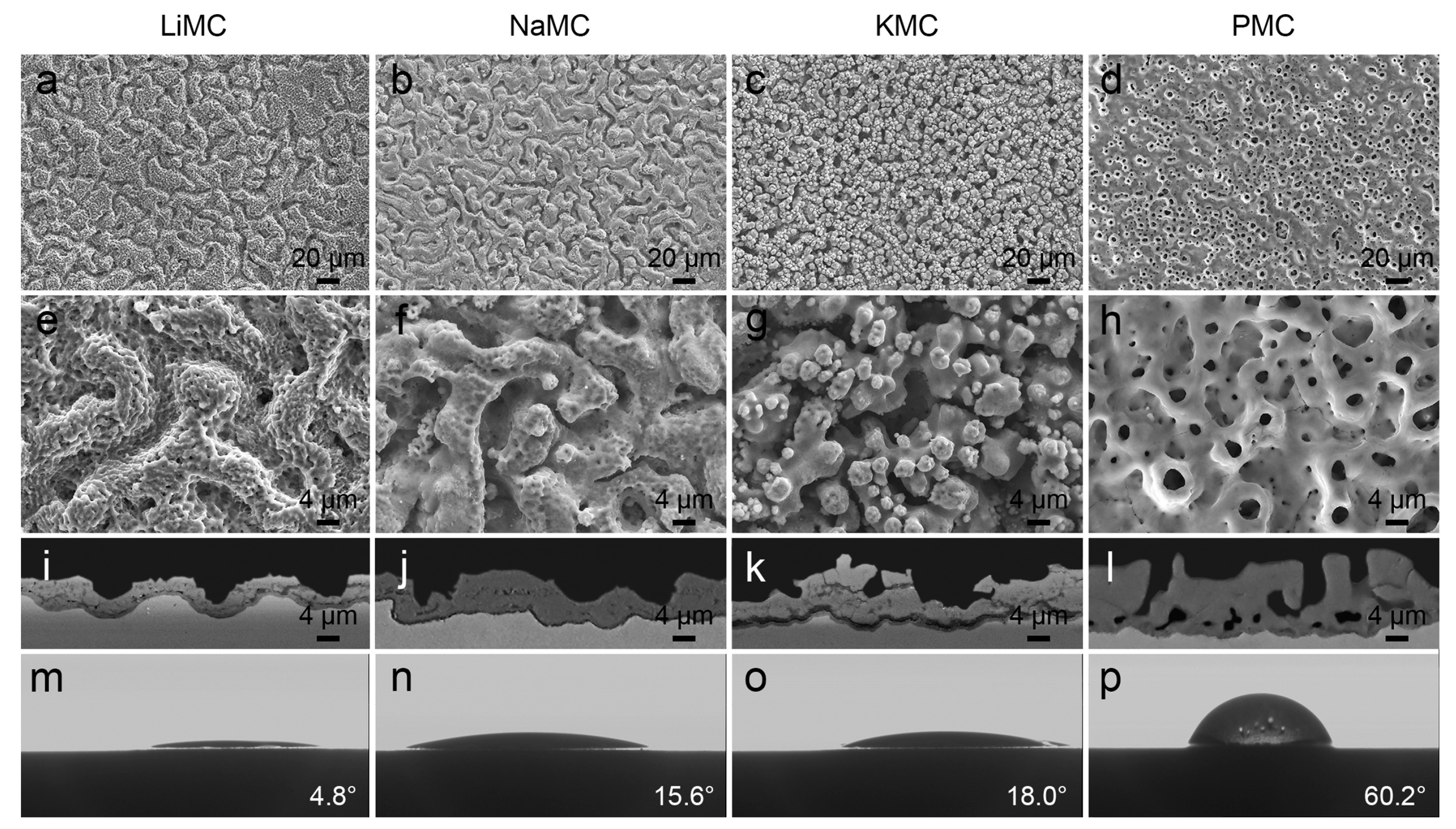
Chen et al. [78] utilized NaH2PO4 as an electrolyte for MAO treatment of a Ti-13Cr-3Al-1Fe alloy and found that the response to electrical parameters is akin to the trend observed in α and α + β alloys. Cimenoglu et al. [79] investigated MAO-treated Ti-6Al-4V and Ti-6Al-7Nb alloys using the same parameters. Both exhibited a similar thickness of approximately 10 μm. However, the coating on Ti-6Al-4V contained HA precipitates, while the Ti-6Al-7Nb coating had granular oxides with calcium titanate. In the study by Wang [80], an MAO process was employed under identical conditions on pure Ti, Ti-6Al-4V, and Ti-35Nb-2Ta-3Zr substrates. The results revealed distinct topographies of coatings on the three different substrates (Figure 4). Notably, the Ti-35Nb-2Ta-3Zr surface exhibited a porous topography with increased crystalline anatase content, which effectively enhanced the deposition of HA. Additionally, corrosion resistance and hydrophilicity tests indicated superior performance for the coating.
Material pretreatment processes alter the surface topography and grain size, consequently affecting the properties of coatings. Physical methods like surface shot peening, severe plastic deformation (SPD), and laser surface remelting (LSR) treatments are primarily considered. High-energy shot peening (HESP) enhances the wettability and surface-free energy of MAO coatings, significantly improving bioactivity on Ti-based substrates [49]. Equal channel angular pressing (ECAP) on Ti surfaces results in ultrafine grains and increases the thickness and porosity of MAO coatings. Furthermore, the dense microstructure enhances the corrosion resistance of MAO coatings [14]. LSR pretreatment, in combination with MAO processing, modulates coating topography. Increasing the energy density of LSR pretreatment initially decreases surface roughness before subsequently increasing it. This enhances coating densification and bond strength with the substrate, thereby improving corrosion resistance [81]. In conclusion, appropriate pretreatment can enhance the performance of MAO coatings. Additionally, subjecting alloy substrates to solution treatment, quenching, and varying aging treatments before MAO results in changes in coating pore size, surface roughness, and porosity with increasing aging time. This is likely due to enhanced substrate electrical conductivity from the aging treatment, leading to higher current density during the MAO process [82]. The comprehensive effects of electrolytes, electrical parameters, and substrates can be found in Table 2.
At present, additive manufacturing, also known as 3D printing, is progressively emerging as the preferred method for custom orthopedic implants. It allows the production of complex structures that surpass the capabilities of traditional techniques, all while preserving the physical and chemical advantages inherent to Ti and its alloys. Implants produced through this method offer an elevated level of patient-specific adaptability. In the context of employing MAO to enhance the surface bioactivity of 3D-printing Ti alloy implants, more detailed comparisons are available in other comprehensive reviews [2,83].
Obtaining the appropriate parameters requires multiple experiments. Researchers can start by choosing an experimentally suitable parameter range based on existing literature. They should then refine these parameters based on observed multifaceted effects of parameter changes on the coating. Due to the absence of a unified model, conducting multiple experiments is essential. The alteration of a single parameter in the MAO process can result in modifications across multiple coating properties, owing to the intricate interplay of various factors. Consequently, adjusting a specific parameter may necessitate the simultaneous coordination of other parameters to uphold the desired coating quality. This intricate balance poses a significant challenge in the production process of MAO coatings. Different applications of implants have varying requirements for the surface coating, leading to variations in the parameters used. Coating performances prepared under the same parameters with different devices may exhibit slight differences, making it challenging to define optimal parameters [22]. The parameters influencing the coatings are comprehensively addressed here to serve as a reference for subsequent researchers. After determining the parameters, real-time online monitoring systems can be introduced to track key parameters such as current, voltage, and electrolyte concentration. Through feedback control, maintaining parameters within a stable range ensures consistent and reproducible coating performance.

Table 2.
Factors affecting coating performance.
| Number | Substrate | Electrolyte | Electrical Regimes | Conclusion | Reference |
|---|---|---|---|---|---|
| 1 | Ti | Na3PO4, Na2SiO3, NaAlO2, composite electrolytes (Na3PO4 + Na2SiO3 + NaAlO2) | 0.1–0.3 A/cm2 current densities | The pores of coatings made in Na3PO4 and composite electrolytes are smaller and more uniform than those in other electrolytes. | [39] |
| 2 | Ti | KH2PO4, Ca(OH)2 or Ca(HCOO)2, Na2(EDTA) | Three different fixed set current densities: 50, 100, and 150 mA/cm2 | Porous coating promotes apatite formation and resists corrosion. | [55] |
| 3 | Ti | (CH3COO)2Ca·H2O and NaH2PO4·2H2O | Different voltages, currents, durations | HA-containing flower-like Structure Coatings have good biological activity. | [84] |
| 4 | Ti | Ca(OOCCH3)2, Ca(H2PO4)2, and Na2(EDTA) | Pulsed DC, duty cycle at 30%, 450 V, 5 min, 100–5000 Hz | Frequency affects the crystallinity, composition, topography, and wetting ability of the oxide film. | [66] |
| 5 | Ti | NaH2PO4, Na2ZrO3, and Na2SiO3 of different concentrations | Pulsed DC, 500 V, 1000 Hz, different duty cycle | Concentration and duty cycle affect topography and photocatalytic activity. | [69] |
| 6 | Ti (high-energy shot peening, HESP) | C3H7Na2O6P·5H2O, Ca (CH3COO)2·H2O, Na2SiO3·9H2O, and Cu(CH3COO)2· H2O | 480 V, 5 min | Porous antimicrobial coatings are prepared. | [49] |
| 7 | Ti (equal channel angular pressing, ECAP) | NaH2PO2 and (CH3COO)2Ca | 350 mA/cm2, 8 min | Porous microcrack coatings are prepared. | [14] |
| 8 | Ti | K3PO4 and KOH | Constant current density of 100 mA/cm2, frequency of 6 kHz, duty cycle at 50%, 6 min | Coating with pancake-like topography has corrosion resistance. | [46] |
| 9 | Ti | Different concentrations of KOH | 160 V, 1 min | 1 M KOH, Ca/P = 1.69. The higher the concentration of KOH, the stronger the corrosion resistance of the coating. | [42] |
| 10 | Ti | Na2B4O7·10H2O, Na2O2SiO2·2H2O, Cu(CH3COO)2, Zn(CH3COO)2, NaOH | AC, 300 V, different frequencies, and duty cycles | Duty cycle and frequency affect coating thickness, surface uniformity, and porosity. | [70] |
| 11 | Ti | Na2B4O7·10H2O | Pulsed DC, 465 V, 600 Hz, and 9% | “Cortex-like” micro/nanostructured coating has improved biocompatibility. | [35] |
| 12 | Ti, Nb, Mg, Al, Zr, Ta | NaOH and Na2SiO3 | Unipolar positive or bipolar working pulse | Soft sparks are more obvious on the surfaces of Mg, Al, Zr, and Ta, while Ti and Nb have only a small amount of spark softening. | [85] |
| 13 | Ti-6Al-4V | Ca(CH3COO)2, Ca(C3H7O6P), Mn(CH3COO)2·4H2O, Mg(CH3COO)2·4H2O, Sr(CH3COO)2·0.5H2O, Zn(CH3COO)2, Na2SiO3 | Pulsed DC, 280 V, 3 min | The coating surface is soaked in simulated body fluid (SBF) to generate bone-like apatite. | [32] |
| 14 | Ti-6Al-4V | Na3PO4, NaOH, Na2SiO3 | Fixed DC voltage (270 V), 5 min | Coatings prepared in silicate-based electrolytes achieved the most uniform structure, with lower porosity. | [33] |
| 15 | Ti-6Al-4V | NaAlO2, NaF, KOH, CuSO4, Na2(EDTA) | Constant current of 2 A/cm2 | It is porous at about 1μm. The addition of Cu ions makes the coating uniform and reduces the roughness. | [38] |
| 16 | Ti-6Al-4V | Na2HPO4, HA microns, and NPs | Constant potential of 250 V and then pulse unipolar or pulse bipolar constant current of 300 mA/cm2 | Micron and nanoscale HA have different effects on the microstructure of the coating, but both improve the scratch resistance and bioactivity of the coating. | [86] |
| 17 | Ti-6Al-4V | Na2AlO2, Na3PO4 | Bipolar pulse | With an appropriate increase in cathodic pulses, the coating density increases. | [72] |
| 18 | Ti-6Al-4V | Na2AlO2, Na3PO4 | Bipolar pulse | As the Ic/Ia ratio increases, the density and hardness of the coating increase, and the roughness decreases. | [73] |
| 19 | Ti-6Al-4V | Na2SiO3, NaH2PO2, Na2MoO4 | 520 V, 50 Hz, 10%, 30 min | Corrosion resistance is improved. | [81] |
| 20 | Ti-20Zr-10Nb-4Ta | Na2B4O7·10H2O | DC power, 465 V, 600 Hz, and 9% | Hierarchical porous coatings have good biocompatibility. | [36] |
| 21 | Ti-15V-3Al-3Cr-3Sn | Different electrolyte temperatures of 278–313 K, K2Al2O4, Na3PO4, NaOH | square waveform with 2.0 kA/m2 and −1.0 kA/m2 at 100 Hz | Wear resistance is improved at low temperatures. | [87] |
| 22 | Ti-13Cr-3Al-1Fe | NaH2PO4, pH = 9.2 | Different voltages and durations | As the discharge voltage and treatment time increase, the oxide layer thickens, and the pore size and surface roughness increase. | [78] |
| 23 | Ti6Al4V and Ti6Al7Nb | (CH3COO)2Ca·H2O and Na3PO4 | Bipolar power, +500 V and −83 V, 5 min | Two alloy coatings have different characteristics under the same conditions. | [79] |
| 24 | Ti-29Nb-13Ta-4.6Zr(hot forge) | CaOH, Na3PO4·12H2O, pH = 12.03, conductivity = 13.20 mS/cm | AC bipolar, different frequencies, and duty cycles | Frequency and duty cycle affect form and thickness, which in turn affects corrosion resistance. | [68] |
| 25 | Ti-25Ta-10Zr-15Nb and Ti-25Ta-20Zr30Nb | Ca(C2H3O2)2, Mg(C2H3O2)2, Glycerophosphate | 300 V, 2.5 A, 1 min | Both alloys exhibit porous structures with different pore sizes. | [88] |
| 26 | Ti, Ti-6Al-4V, and Ti-15V-3Al-3Cr-3Sn | K2Al2O4, Na3PO4, NaOH | Constant AC of 1.5 kA/m2 up to a maximum peak voltage of 400 V | The coating has excellent wear resistance. | [57] |
| 27 | Ti, Ti-6Al-4V, Ti35Nb-2Ta-3Zr | Na2SiO3·9H2O, (HOCH2CH2)3N, pH = 11.5 | Voltage of +400 V, −50 V, duty cycle at +16%, −10%, 500 Hz | The coating of Ti-35Nb-2Ta-3Zr is durable and has good hydrophilicity. | [80] |
3. Surface Biologic Function of MAO
3.1. Osteointegration
When an implant is exposed to the internal environment, it selectively adsorbs various proteins, depending on the physical and chemical characteristics of its surface. Cells initiate their connection with the implant by interacting with these unique proteins [89]. Surface parameters of the implant, including surface topography, chemical composition, and charge, all contribute to regulating specific interactions between cells and biomaterials.
On smooth surfaces, BMSCs are more prone to differentiate into fibroblasts rather than osteoblasts (OBs). Additionally, fibroblasts also adhere more easily to smooth surfaces. On the other hand, bone cells tend to adhere, proliferate, and differentiate more readily on surfaces with rough nanoscale patterns (Figure 5). Surfaces with moderate roughness and porosity provide a larger bonding area and establish a strong mechanical interlock, promoting a more stable implant–bone integration. Osteoblasts cultured on such rough surfaces demonstrate increased collagen production and enhanced calcification processes [90,91].
The ideal implant surface should mimic natural bone, featuring mineralized large pores and nanoscale components. Hierarchical structure, from the nanoscale (collagen molecules and minerals) to microscale (bone units), guides cells’ behavior [92]. Research suggests that multiscale topographical features on the implant surface are linked to increased osteoblast differentiation [93].
MAO can modify the surfaces of metal implants to give them specific characteristics. It can create various topological structures on the substrate, and incorporate substances for angiogenesis or osteointegration into the coatings.
It is widely acknowledged that a rough, porous surface contributes to improved osteogenic activity. Nevertheless, there is currently no consensus regarding the optimal specific values for roughness, pore size, and pore geometry [94]. Yamasaki et al. [95] highlighted that the interconnected micropores in the range of 2–10 μm can bestow osteoinductive properties upon the scaffold. Shalabi et al. [96] found that bone tissue bonded effectively to rough Ti surfaces within the 1 to 100 μm range, with higher surface roughness providing a greater surface area. Currently, the surface topographies generated through MAO exhibit diversity, encompassing porous [38], hierarchical porous [97,98], and “cortex-like” (or “worm-like”) [35] structures. Li et al. [98] employed a two-step MAO process to create pores of different sizes. In the first step, large pores (100–300 μm) were generated using NaNO3 and NaOH electrolytes. Subsequently, a second MAO treatment was conducted using a Na2B4O7 electrolyte, resulting in the formation of micro-pores (3–10 μm) as well as sub-micron/nano-pores (80–200 nm). This innovative tri-level structure not only enhances hydrophilicity but also holds the potential to improve the adhesion of osteoblasts to the implant and enhance implant fixation. Moreover, they refined the electrolyte composition by introducing CaO and SrO into Na2B4O7 [46]. Following a one-step MAO treatment, both Ca and Sr were integrated into the TiO2 hierarchical porous coating. This hierarchical structure exhibits a high level of porosity and superhydrophilicity, and the incorporated Ca and Sr have shown superior efficacy in promoting the proliferation of human-bone-marrow-derived mesenchymal stem cells (hBMSCs). This highlights that, beyond surface topography, the chemical composition of MAO coatings plays a crucial role in influencing the biological activity of the coatings.
Currently, a highly researched area revolves around the development of biomimetic calcium phosphate (CaP) coatings on surfaces using electrolytes containing both Ca and P. Researchers are dedicated to optimizing the parameters that influence coating formation during the MAO process, with a specific emphasis on the composition of the electrolyte. They aim to achieve a Ca/P ratio that closely matches the natural bone value of 1.67 [48].
Typically, immersion in SBF is employed to assess the ability to form HA on its surface, thus quantifying its bioactivity. The general formula of HA is Ca10(PO4)6(OH)2. Herein, Ca2+ can be completely or partially substituted by Sr2+, Si4+, Mg2+, Mn2+, and Zn2+, and OH− can be replaced by halide ions F−, Cl−, and Br−. Therefore, in addition to Ca and P, adding Si, Sr, Mn, Zn, and other elements to the electrolyte may also improve the osteogenic activity of the surface. Plekhova et al. [18] assessed the morphological and functional status of bone-marrow-derived mesenchymal stem cells (BMSCs) cultured on Ti-CaP coatings formed by MAO. There were no cytotoxic effects observed on the cultured cells. Notably, the high expression of receptors (CD90, CD29, and CD106) along with enhanced synthesis of osteocalcin and osteopontin, as well as observable changes in the surface structure of BMSCs adhered to the samples, collectively confirmed the osteoinductive properties of the calcium phosphate MAO coating. Park et al. [99] utilized Ti-6Al-4V ELI discs and subjected them to treatment with pulsed DC power for 3 min at 280 V in electrolytes containing bioactive ions such as Si, Zn, Mg, Mn, Sr, Ca, and P. The energy-dispersive X-ray spectroscopy (EDS) results revealed that the (Ca + Mg + Si + Mn + Zn + Sr)/P ratio was approximately 1.67. Some researchers have directly added HA to the electrolyte [100,101]. Zhou et al. [101] dispersed hydroxyapatite nanotubes (HNTs) in the MAO electrolyte and embedded HNTs into the Ti surface. When comparing three types of surfaces, Ti, MAO, and MAO-HNT, the HNT group outperformed the Ti and MAO groups in terms of angiogenesis, as evidenced by enhanced cell migration, tube formation, and vascular gene expression in HUVECs. Furthermore, concerning osteogenesis, the HNT group exhibited the highest levels of alkaline phosphatase (ALP) activity, collagen secretion, mineralized calcium nodules, and osteogenic gene expression in MC3T3-E1 cells, surpassing the other two groups. These findings suggest that HNT specimens can significantly promote angiogenesis and osteogenesis at both the cellular and molecular levels.
A good blood supply is essential for successful osseointegration. Studies have found that adulteration of appropriate amounts of Ca [101], Li [102], Se [103], Sr [104], Si [105], and Zn [106] elements can contribute to angiogenesis. Yu et al. [106] introduced zinc acetate into the electrolyte to produce coatings enriched with zinc. Co-cultivation of HUVECs with these coatings demonstrated excellent biocompatibility. This study explores the inherent connection between angiogenesis and osteogenesis, presenting evidence that Zn2+ promotes both processes.
MAO is a versatile technique that enhances the hardness, corrosion resistance, wear resistance, and biocompatibility of metal surfaces. This versatility makes it ideal for integration with other technologies to fine-tune surface properties for diverse application needs. The joint application of MAO and other surface modification technologies can incorporate functional elements into the MAO coating, such as hydrothermal treatment (HT) [107,108], dip coating [109,110], radiofrequency magnetron sputtering (RF-MS) [111], etc. Many research groups are exploring post-processing techniques using inorganic compounds to mimic the Ca/P ratio in a natural bone structure for enhanced bioactivity [50].
For instance, Song et al. [107] applied HT in two different solutions after MAO treatment, resulting in the formation of HA with a higher Ca/P ratio than the MAO-treated sample, facilitating the migration of Ca2+ and PO43− ions to the surface. MAO and HT treatments provide binding sites for other functional factors like osteogenic growth factors, and antibacterial and anti-inflammatory substances [112]. In a study [113], MAO was initially used to create a hierarchical micro/nano-topography on a substrate, followed by electrochemical reduction in an alkaline solution. The results demonstrated improved BMSC adhesion, proliferation, and up-regulation of osteogenesis-related gene expression, indicating the potential to enhance osteogenic processes.
Magnetron sputtering, when combined with MAO, deposits metal particles onto coatings, enhancing osteogenesis. Park et al. [114] used RF-MS to create a Mn coating on the MAO-treated Ti-29Nb-xHf alloy, highlighting the potential for introducing metal coatings to boost surface osteogenic properties.
The combination of multiple surface modification techniques offers a broader selection of surface ingredients, including growth factors, hormones, and proteins. Bone morphogenic proteins (BMPs), such as BMP-2 and BMP-7, are pivotal for bone cell proliferation, differentiation, and matrix synthesis. Fibroblast growth factors (FGFs) like FGF-2 and FGF-7 influence bone cell proliferation and differentiation, while insulin-like growth factors (IGFs) like IGF-I and IGF-II promote cell growth and protein synthesis, crucial for bone tissue growth and repair. Vascular Endothelial Growth Factor (VEGF), as a key growth factor, directly stimulates new blood vessel formation. These biological factors are typically incorporated onto material surfaces through gentle methods, such as the dip-coating method [115,116], immersion [110], and chemical deposition [109], to ensure the preservation of their activity.
Teng [117] employed a method combining MAO and Ca, P layers with BMP co-precipitation to treat 3D-printed porous Ti alloy implants, named MAO-CaP-BMP2. The porosity facilitates bone tissue and blood vessel ingrowth. The microporous dioxide layer, created by MAO treatment, serves as nucleation sites for concurrent deposition of the CaP layer and BMP-2. This microstructural arrangement allows the extended, controlled release of BMP-2 over 35 days. The modification of Ti alloy implants with MAO-CaP-BMP2 enhances their osteoinductive and osteoconductive properties, leading to superior osteogenic and angiogenic outcomes.
3.2. Antibacterial
Among common orthopedic infections, osteomyelitis stands out, often stemming from pathogens like Staphylococcus aureus (S. aureus), Escherichia coli (E. coli), and Pseudomonas aeruginosa (P. aeruginosa). Symptoms encompass a range of discomforts such as pain, swelling, pus formation, the presence of fistulas and/or sinus tracts, and wound reopening. The approach to treatment depends on various factors including the severity of infection, the responsible pathogen, and the overall health of patients. Treatment varies based on infection severity, pathogen, and patient health, typically involving debridement to remove infected bone tissue and implanted devices. Debridement surgery is complex and requires an extended recovery period. Dental-implant-associated microbial colonization may give rise to peri-implant mucositis or even peri-implantitis. Key pathogens involved include Porphyromonas gingivalis (P. gingivalis), Streptococcus sanguinis (S. sanguinis), Aggregatibacter actinomycetemcomitans (A. actinomycetemcomitans), etc. Peri-implantitis has a lower incidence compared to peri-implant issues in orthopedic implants, but it still demands attention as it could directly contribute to implant failure [118].
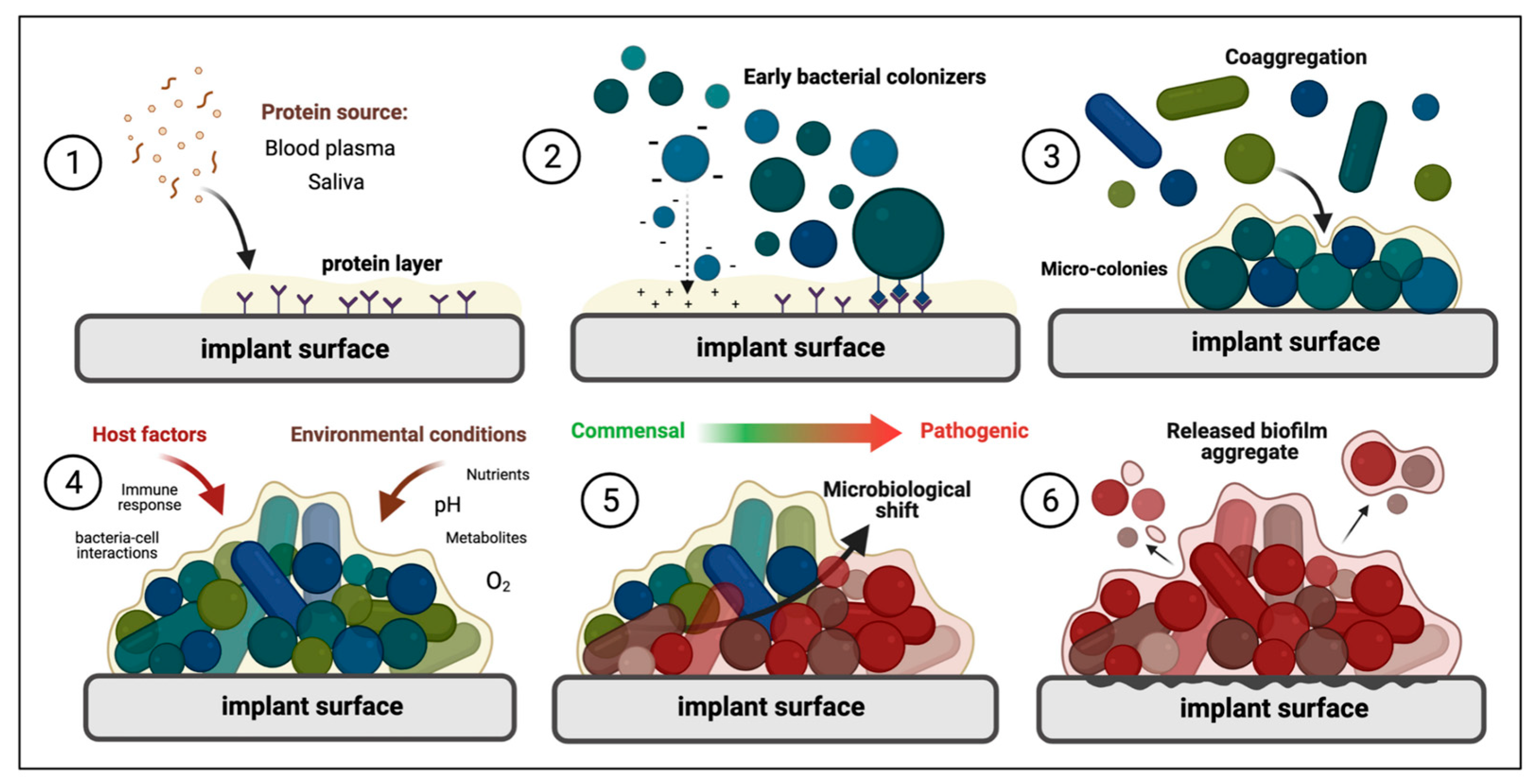
Microbial cells adhering to surfaces can come together to form highly structured microbial communities commonly known as “biofilms”. These biofilms often display drug resistance, complicating infection treatment. The process of biofilm formation involves various stages, including the adsorption of proteins, microbial adhesion, aggregation, biofilm maturation, and the diffusion of biofilm (Figure 6). Proteins adhere to surfaces, facilitating microbial attachment through electrostatic interactions and protein receptor recognition. As attachment progresses, biofilm formation occurs as microorganisms aggregate. Once established, biofilms involve intercellular communication and extracellular polymeric substance (EPS) secretion, transforming into a three-dimensional structure [119].
Antimicrobial implant materials must inhibit biofilm formation, particularly in the initial stages. Researchers and clinicians continue to seek effective prevention and treatment of implant-associated infections. In recent years, the development of surface antibacterial methods using MAO has been a research hotspot in preventing and managing biofilm growth on Ti-based implants [120].
Some researchers suggest that MAO-generated anatase TiO2 structures can reduce bacterial adhesion or confer antibacterial properties. For instance, Alipal et al. [121] found that circular TiO2 structures on MAO coatings exhibit improved biocompatibility and antibacterial properties compared to needle-like TiO2. Similarly, Jia et al. [122] proposed that the structure of TiO2 layers created by MAO can trap and kill bacteria on implant surfaces. However, conflicting views exist in the literature, with some researchers arguing that rough, porous MAO coatings increase microbial adhesion [123,124], while others contend there is no significant difference compared to untreated Ti [125]. Current research on MAO-based antibacterial coatings mainly focuses on incorporating antibacterial additives to alter surface chemistry and enhance functionality.
The main mechanisms to promote the killing of bacteria on the surface of antimicrobial implants are (i) the contact reaction of bacteria with the surface, causing the destruction of bacterial membranes or inhibiting the synthesis of their membranes; (ii) the gradual release of ions from the coating, resulting in electron transfer and cell membrane destruction; (iii) antimicrobial ions enter microbial cells, interrupt protein synthesis, block Adenosine Triphosphate (ATP) synthesis and deoxyribonucleic acid (DNA) replication, and promote apoptosis; and (iv) generation of ROS, which also induces apoptosis by denaturing proteins and damaging DNA [126]. Costa et al. [119] categorized antimicrobial agents into four primary groups: organic (biopolymers and bioactive agents), inorganic (metallic and semi-metallic elements), drugs (substances with pharmacological action), and biological (growth factors, peptides, and extracellular matrix proteins). Among these additives, people are more interested in inorganic elements, due to their stability and cost-effectiveness. The specific effects of elements on the biological function of the coating are shown in Table 3.
Since 2009, one-step MAO has been employed to create antimicrobial Ti surfaces by binding Ag, Cu, and Zn elements, as summarized in 35 articles by Shimabukuro up to 2020 [51]. The half-maximal inhibitory concentrations (IC50) of Ag+, Cu2+, and Zn2+ on MC3T3-E1 cells are 2.77, 15.9, and 90.0 μM, respectively [127]. Ag exhibits high toxicity, but controlled dosages do not negatively affect osteoblast activity. Researchers are increasingly interested in incorporating diverse elements (Ag + Cu [23], Ag + Zn [128,129,130,131], Cu + Zn [132,133]) into coatings through one- or two-step MAO processes to enhance antimicrobial effects or/and extend the duration of antimicrobial activity. Shimabukuro et al. [134,135] simulated the biodegradation behavior of Ti doped with Ag, Cu, and Zn by MAO soaked in physiological saline for 28 days to study the changes in surface composition and antibacterial effect. After 28 days, the antibacterial effect of the Ag-incorporated samples weakened, the antibacterial effect of Cu was maintained, and the antibacterial effect of Zn enhanced in the later period (Figure 7). In another study, Tsutsumi [136] employed a two-step MAO process to tailor surface coatings with Ag, with or without Zn, to assess the binding and ion release behavior of Ag and Zn in the resulting oxide layer. Over a 6-month immersion period, the release of Ag+ from the oxide layer indicated early antibacterial performance, while the release of Zn2+ signified later antibacterial effectiveness.
Research involving Mn on the surface of Ti-based implants shows promise, as it enhances osteoblast activity while also inhibiting the proliferation of Gram-negative E. coli and P. aeruginosa. Zhao et al. [137] employed MAO to create a Mn-TiO2 microporous coating. Their results revealed that the coating induced E. coli cell wall perforation, effectively inhibiting bacterial proliferation. Additionally, Bi compounds have shown efficacy in treating mucosal and dermal infections and are recognized as antimicrobial additives for calcium phosphate bone cement. Lin et al. [138] used MAO to develop a TiO2 coating containing the bismuth (Bi) element. The coating with 6.2% Bi displayed good biocompatibility with MG63 cells and demonstrated 1.5 times higher antibacterial efficacy against A. actinomycetemcomitans and 1.9 times higher efficacy against methicillin-resistant Staphylococcus aureus (MRSA) compared to the control group. MAO technology can also incorporate other non-metallic antibacterial ingredients into the coating, such as Ce [139], Se [103], I [140,141], B [142,143], and F [144,145,146].
MAO combined with other techniques can produce antimicrobial coatings that enhance effectiveness and minimize cytotoxicity. Arun et al. [111] employed a combination of MAO and RF-MS processes to create dual-phase coatings on Ti-6Al-4V. They conducted MAO in Ca/P electrolytes with varying Ag NP concentrations and applied an HA top layer using the RF-MS process. This approach generated a passivation layer with robust barrier properties. Ag contributed to antibacterial activity, while HA promoted cell proliferation and mitigated the minor adverse effect of Ag on cell viability. To enhance cell compatibility with antibacterial coatings or extend antibacterial substance release, polydopamine [147,148] and polylactic acid [149] can be loaded on the coating surface through secondary surface treatment.
Graphene and its derivatives (e.g., graphene oxide (GO) and redox graphene (rGO)) can be integrated into MAO coatings either directly through electrolytes or in combination with techniques like electrophoretic deposition (EPD), sol-gel, and dropping coating. This incorporation imparts favorable antibacterial and tribological properties [150]. For instance, Mazinani et al. [151] combined MAO and EPD to deposit GO sheets onto the MAO coating, resulting in significant antimicrobial properties against E. coli and S. aureus. Moreover, San et al. [152] created a suspension of rGO and Ag NPs, added it to the MAO electrolyte, and achieved a more effective coating against MRSA due to the generation of ROS and the “Nanoknife” structure of rGO.
Chitosan (CS) interacts with the anionic components on the microbial cell wall, resulting in cell wall damage and the killing of microorganisms, due to its cationic nature and polyamine structure. CS can be fixed on the MAO coating by dip coating [153]. For example, BMP-2-encapsulated CS gives the coating good osteogenic and antibacterial properties [115].
Incorporating drugs, such as antibiotics, is an effective means of rapid sterilization during early implantation stages. For example, Zhou et al. [154] used ciprofloxacin-loaded CS hydrogel to modify the coating, effectively sterilizing the sample with the bacteriostatic zone. Xu et al. [155] covalently grafted vancomycin to the MAO coating, creating Ti-@MV with significant antibacterial properties and enhanced osteogenic differentiation of BMSCs. In another study [156], octenidine (OCT) was loaded onto MAO-prepared coatings via electrophoresis, resulting in improved early antibacterial efficiency without compromising cell compatibility. This early-stage (4–6 h) intervention is crucial for preventing long-term bacterial infections. Octenidine, vancomycin, and heparin were among the early attempts at drug loading on polyethylene glycol MAO-treated surfaces. However, their initial release is too large, and it is easy for bacteria to develop drug resistance. Therefore, the use of antibiotics in implant surface modification is gradually decreasing [157]. In terms of biological compounds, antimicrobial peptides (AMPs) have become the main substances for functionalization in Ti surfaces through a multi-step process on MAO surfaces [147,158]. However, the main challenge with these antimicrobial surfaces is that cross-linking of peptides on the surfaces is difficult, resulting in less stable coatings, limited duration of antimicrobial activity, and higher manufacturing costs [159].
Phototherapy, including photothermal and photodynamic therapy, offers safe and controllable methods. Photothermal therapy employs agents that generate high temperatures under near-infrared light, effectively disrupting bacterial structures. In contrast, photodynamic therapy relies on photosensitizers absorbing specific light wavelengths to create reactive oxygen species (ROS), which disrupt bacterial DNA, proteins, and biomembranes. Hydrothermal treatment or sol-gel can be used to combine photothermal agents or photosensitizers into the coating [160]. Chai et al. [161] successfully prepared a TiO2/MoO2/chitosan coating on Ti surfaces using a composite process involving MAO, HT, and electrospinning. The MoO2 significantly improved the phototherapy performance. Both in vitro and in vivo experiments demonstrated excellent antibacterial capabilities against Streptococcus mutans (S. mutans) after 15 min of irradiation with near −808 nm wavelength infrared light, facilitated by the synergistic effects of a high temperature and ROS. Moreover, Li et al. [108] used MAO and HT techniques to form a β- FeOOH/Fe TiO2 heterojunction, which can also generate ROS under irradiation. In addition, ultraviolet rayscan remove surface carbon contaminants and increase the negative charge density on the surface of crystal TiO2, thereby reducing bacteria through electrostatic repulsion [125]. However, because UV rays have carcinogenic effects, they can be used as a pretreatment before implant placement.
3.3. Anti-Inflammatory
The biological properties of materials are initially evaluated through in vitro studies on osteoblast activity. In some cases, differences between in vivo and in vitro research results may stem from immune cell responses. Bone immunology emphasizes the role of immune cells in osteointegration, which involves the inflammatory response in the early and late stages after implantation [162].
The implantation of biomaterials is intricately tied to a continuous biological process (Figure 8). Shortly after implantation, proteins from the vascular system and interstitial fluid adhere to the surface of the biomaterial within minutes. Subsequently, various cells, including platelets, monocytes, and macrophages, infiltrate and adhere to the biomaterial. Within 1 to 5 days post-implantation, immune cells release cytokines on the biomaterial surface, initiating a pro-inflammatory response while recruiting bone repair cells (e.g., BMSCs and OBs) from distant sites to the inflammatory site [163]. This early-stage inflammation is crucial. The fate of the bone biomaterial, whether it becomes encapsulated by a fibrous capsule (indicating failure) or undergoes long-term replacement by new bone (the desired outcome), is determined by bone repair cell interactions with the inflammatory microenvironment and biomaterial properties in the following days. Acute inflammation typically subsides in less than 1 week, while a duration exceeding 3 weeks may lead to implant failure. The emerging field of bone immunology and immune modulation has shifted our expectations for bone biomaterials, emphasizing their role in bone immune modulation rather than just reducing inflammatory responses [164].
Recent advancements in Ti surface engineering focus on managing host and inflammatory responses. This approach leverages the benefits of physiological inflammation, such as cellular debris removal and infection prevention, while also mitigating the risks associated with severe inflammatory reactions. These risks include implant rejection, early or late failures, and impacts on implant functionality.
To modulate the inflammatory response of biomaterials, these strategies can be followed [164]:
- Adsorption of specific proteins: control the adsorption of plasma proteins on the implant surface to influence inflammatory responses.
- Macrophage polarization: M1 (pro-inflammatory) and M2 (anti-inflammatory) phenotypes can be induced. M1 macrophages clear debris and potential pathogens in the early stages of implantation, while M2 macrophages support inflammation resolution and tissue healing. The M1 phenotype is produced by M0 macrophages activated by the lipopolysaccharide (LPS) or interferon-γ (INF-γ). It secretes inflammatory mediators, including Interleukin-1β (IL-1β), IL-6, IL-12, IL-23, ROS, and Tumor Necrosis Factor-α (TNF-α). However, the M2 phenotype is induced by stimulation with IL-4, IL-10, and IL-1β. M2 macrophages release anti-inflammatory cytokines and growth factors, such as IL-10 and transforming growth factor-β (TGF-β).
- Surface topography: Surface roughness and texture can affect macrophage phenotype. Rough surfaces may favor M1 activation, while nano- or micron-scale directional textures promote an M2-like response. Hydrophilic surfaces can reduce pro-inflammatory cytokines.
- Biomimetic coating: modify the implant surface with biomimetic coatings like collagen, transmembrane molecular markers, mesenchymal stem cells, or biological peptides to reduce macrophage activation and pro-inflammatory factors.
- Local drug delivery: implement local drug delivery systems on the implant surface to create a localized anti-inflammatory environment with high drug concentration and minimal systemic side effects.
Local inflammatory reactions can result from debris accumulation caused by wear during implantation or long-term friction between the implant and surrounding bone tissue [165,166]. Furthermore, the corrosion of metals in the complex environment of the body can release metal ions, potentially triggering inflammation. MAO coatings offer excellent tribological properties and corrosion resistance. They can be modified to regulate host and inflammatory responses by adjusting the surface topography and composition of the implant. Li et al. [163] examined the in vitro immune response to Ca- and Si-doped MAO coatings. They found that the macrophage interaction with MAO-Ti favored the M1 phenotype, but collagen synthesis and matrix mineralization for osteocyte-like cell growth on the coating were favorable. This suggests that MAO-modified implant–bone interfaces support osteogenesis both before and after osteoblast recruitment to the biomaterial surface. Furthermore, Ti-Ta metal–metal composites with bioceramic coatings containing Ca and Si also demonstrate osteogenic, anti-inflammatory, and bone immunoregulatory properties [167].
Some metallic elements, such as Cu [168], Zn [169], Mg [170], Li [102], and Co [171], have been shown to modulate the immune environment at the implant surface. They can regulate macrophage polarization phenotypes, thus promoting either early pro-inflammatory and antimicrobial effects or long-term anti-inflammatory and pro-osteogenic effects of the implant. For instance, Li et al. [168] utilized MAO to create Cu-containing ceramic coatings on Ti surfaces (Cu-MAO). Macrophages on Cu-MAO displayed polarization toward the M1 phenotype, exhibiting high levels of inducible nitric oxide synthase (iNOS) expression, low arginase-1 (Arg1) expression, enhanced release of IL-6, and inhibited release of IL-4 and IL-10. This promoted a favorable inflammatory microenvironment for osteoblast-like cell differentiation and enhanced bactericidal abilities of the surface. Sun et al. [169] took a similar approach by developing Zn-doped MAO coatings on Ti surfaces, which exhibited excellent biocompatibility and osteogenic differentiation performance with BMSCs. In addition, evaluation of RAW264.7 cells’ survival on the surface and related pro-inflammatory markers revealed that this coating downregulated the expression of pro-inflammatory genes. Peng et al. [102] incorporated Lithium (Li) into Ti surface coatings via MAO. Low-dose Li was slowly released as ions, enhancing the recruitment of BMDMs while favoring M2 polarization and limiting M1 polarization. In vivo, implantation mimicking aseptic loosening revealed the ability of Li to mitigate inflammatory responses. Additionally, Li-doped MAO coatings supported healthy growth of both mouse embryonic cell lines (C3H10T1/2) and HUVECs in a macrophage-conditioned medium, highlighting their potential for osteogenic differentiation and angiogenesis. The molecular mechanism involved the regulation of cascade molecules in the PI3K/AKT signaling pathway for bone immunomodulation. Similarly, Yang et al. [171] prepared Co-doped MAO coatings on Ti implants with varying Co content. Their results showed that, in comparison to pure Ti samples, cobalt-loaded Ti exhibited immunomodulatory functions on macrophages, upregulating the expression of M1 genes and downregulating the expression of M2 genes. Notably, higher cobalt levels induced the polarization of macrophages into the M2 type.
Similar to antimicrobial properties, other surface modification techniques can incorporate a greater amount of anti-inflammatory components onto the surface of the MAO coating. Bai et al. [172] investigated the effects of MAO and steam heat treatment (SHT) on the preparation of nano-HA coatings on porous Ti surfaces. They found that Ti surfaces coated with HA NPs exhibited adjustable inflammatory responses, while HA nanorods negatively affected osteo-/angiogenesis and osteoimmunomodulation. Additionally, they [100] annealed the MAO-HA surfaces and observed that annealing temperature influenced surface topography, wettability, and chemical properties. In vitro experiments demonstrated that MAO coatings annealed at 650 °C promoted the proliferation and differentiation of osteoblasts and endothelial cells, while inhibiting the inflammatory response of macrophages.
Schmidlin et al. [173] found that ZrO2 particles induced lower expression of inflammatory factors compared to TiO2 particles at an equivalent dose. Zhu et al. [174] combined the MAO and sol-gel methods to fabricate a multifunctional composite coating on the surface of Ti, composed of SiO2 particles and zirconium hydrogen phosphate (ZrP). The ZrP coating exhibited superhydrophilicity and significantly reduced the release of pro-inflammatory cytokines (TNF-α, IL-6, and IL-1β). This coating also enhanced corrosion resistance and friction, resulting in reduced Ti ion/fragment release and minimized inflammatory responses. Wang et al. [175] prepared Sr-doped nanostructures on the Ti surface through MAO and electrochemical deposition, and subsequently incorporated silk fibroin protein-based nanocomposites (Ti-MAO/Sr/LBL/WNP) through a layer-by-layer self-assembly technique (LBL). This coating exhibited sustained release of wogonin and Sr ions for over 7 days, promoted the expression of anti-inflammatory factors (TGF-β1 and Arg-1) in M2 macrophages, and facilitated cell proliferation and osteogenic differentiation. Moreover, MAO coatings can be utilized to locally load bioactive substances and drugs, such as peptides that recruit immune-modulating cells (T cells, monocytes, mast cells, and neutrophils) [158].
MAO provides biofunctional coatings that interact with tissues over an extended period, involving multiple biological and biochemical processes [90]. This interaction is determined by the implant characteristics and the implantation environment. Evaluating the biocompatibility and long-term performance of the coatings is a task that requires various factors and methods. Biocompatibility testing includes both in vitro and in vivo assessments [109,113]. In vitro experiments can detect the effects of the coating on cell adhesion, proliferation, and differentiation. In vivo experiments assess the biocompatibility, tissue response, and implant stability within live animals. Assessing engineering parameters in the surrounding tissues, such as vascularization, bone formation, and healing, provides information on the coating impact on tissue reconstruction. Monitoring implant position and stability through imaging techniques such as X-rays, CT scans, and MRI is crucial for detecting issues like coating delamination, implant loosening, or displacement. Animal experiments observe the morphology, structure, chemical composition, and mechanical properties of the coating after a certain period of implantation, offering insights into the long-term stability of coatings in vivo. Evaluating long-term performance also involves considering inflammation and immune reactions around the implant, monitoring through tissue slices, inflammation levels, biomarker detection, and immune cell activity [168,172,175,176,177] (refer to the literature listed in Table 3). Integrating these assessments provides a more comprehensive understanding of the performance of MAO coatings during long-term use and their interaction with surrounding tissues. This multidisciplinary task typically requires collaboration among professionals in medicine, biomedical engineering, and materials science. Moreover, dental implants and hip stems featuring MAO coatings have been introduced as products and subjected to market testing over the long term, yet relevant literature reports are scarce [178,179].
Table 3.
Coatings contain different components with different biological functions and are validated in vitro and in vivo.
Table 3.
Coatings contain different components with different biological functions and are validated in vitro and in vivo.
| Number | Biologic Function | Adding Substances | Technique | In Vitro | In Vivo | Conclusion/Remark | Reference | |
|---|---|---|---|---|---|---|---|---|
| Cell | Bacterium | Animal Parts | ||||||
| 1 | Osteogenesis | Ca, Sr | MAO | hBMSCs | Simultaneously incorporating Ca and Sr demonstrated superior promotion of hBMSC proliferation. | [97] | ||
| 2 | Osteogenesis/Angiogenesis | Zn | MAO | HUVECs and BMSCs | In the Zn2+ environment, angiogenesis and osteogenesis mutually promote each other. | [106] | ||
| 3 | Osteogenesis/Angiogenesis | Hydroxyapatite nanotubes (HNTs) | MAO | HUVECs and MC3T3-E1 cells | HNT specimens promote both angiogenesis and osteogenesis on cellular and molecular levels. | [101] | ||
| 4 | Osteogenesis | B | MAO, hydrothermal treatment, and heat treatment | SaOS-2 cells | Nanorods inhibit SaOS-2 cell activity, whereas nanoparticles promote it. | [143] | ||
| 5 | Osteogenesis | Hierarchical coatings | MAO, electrochemical reduction | BMSCs | Beagle dogs, the shaft of the canine femur | The hierarchical coatings show higher osteogenesis rates compared to the ordinary MAO group. | [113] | |
| 6 | Osteogenesis | HA, BMP-2 | MAO, dip coating | MC3T3-E1 cells | Beagle femur | The interface bonding strength between HA/BMP-2 coating and surrounding new bone tissue is higher than that of Ca/PMAO coating. | [109] | |
| 7 | Osteogenesis/Angiogenesis | Ca, P, BMP-2 | 3D printing, sandblasting etching, MAO, electrochemical deposition | BMSCs | New Zealand White Rabbit Skull | MAO-CaP-BMP-2 is superior to the MAO and MAO-CaP groups in new bone formation. | [117] | |
| 8 | Osteogenesis/Antibacterial | Ca, P | MAO | hFOBs | E. coli and S. aureus | Volcanic-crater-like and needle-like CaP structures form at 350 V and 450 V, respectively. The former exhibits superior antibacterial performance and biocompatibility. | [121] | |
| 9 | Bioactivity/Antibacterial | Ca, P | MAO, UV catalysis | HGFs | S. sanguinis | Photofunctionalization reduces hydrocarbons and enhances surface protein adsorption. | [125] | |
| 10 | Osteogenesis/Antibacterial | Zn | MAO | MC3T3-E1 cells | E. coli | Incubation with salt solution converts Zn ions into zinc oxide, which helps with long-lasting antibacterial activity. | [134] | |
| 11 | Antibacterial/Osteogenesis/Angiogenesis | Sr, Zn | MAO | HUVECs, BMSC | MRSA and P. gingivalis | Rat femoral model | The surface osteogenesis of samples doped with Sr and Zn is superior to other groups. (No in vivo antibacterial test conducted.) | [104] |
| 12 | Antibacterial | Ag, Cu NPs | MAO | MC3T3-E1 cells | MRSA | Mouse femur ex vivo experiment | Ag and Cu ions synergistically kill bacteria, allowing a 10-fold reduction in Ag ion concentration with consistent antibacterial efficacy. | [124] |
| 13 | Osteogenesis/Antibacterial | Ag, Zn | 3D printing, MAO | MC3T3-E1 cells | MRSA | Mouse femur ex vivo experiment | The synergistic effect of Ag and Zn reduces the concentration of Ag+ by 120 times. | [128] |
| 14 | Osteogenesis/Antibacterial | Ag, Zn | MAO | MC3T3-E1 cells | S. aureus | Ag and ZnO synergy enhances antibacterial performance and promotes CaP phase formation. | [129] | |
| 15 | Osteogenesis/Antibacterial | Ag, Zn | MAO | MC3T3-E1 cells | S. aureus | Ag and Zn ion release is above the antibacterial threshold yet well below cytotoxic levels. | [130] | |
| 16 | Osteogenesis/Antibacterial | Ag, Zn | MAO | S. aureus | Ag and Zn have good synergistic antibacterial effects. | [131] | ||
| 17 | Osteogenesis, Antibacterial | Cu, Zn | MAO | MG63 | E. coli, S. aureus, and MRSA | Orthogonal experiments explore electrolyte effects on coatings, with phytic acid supplying the P element. | [132] | |
| 18 | Skin-integration/Antibacterial | Cu, Zn | MAO | Fibroblasts (L-929) | S. aureus | The synergistic effect of Cu and Zn facilitates skin integration and antibacterial activity. | [133] | |
| 19 | Osteogenesis, Anti-tumor/Antibacterial | Se | MAO | BMSCs, cancerous osteoblasts | S. aureus and E. coli | Se doping enhances osteogenic, anti-tumor, and antibacterial properties. | [103] | |
| 20 | Osteogenesis/Antibacterial | Mn | MAO | MC3T3-E1 cells | E. coli | Rabbit femur | The coating induces osteogenesis and promotes osseointegration. | [137] |
| 21 | Antibacterial | Bi | MAO | MG63 cells | A. actinomycetemcomitans, MRSA | Bismuth nitrate has excellent antibacterial activity compared to bismuth acetate, bismuth gallate, and silver nitrate. | [138] | |
| 22 | Osteogenesis/Antibacterial | Ce | MAO | BMSCs | P. gingivalis, S. aureus | Osteoporotic rat hind legs | Ce-TiO2 coating has excellent antibacterial and anti-inflammatory properties. | [139] |
| 23 | Antibacterial | I | MAO, HT, photocatalysis | BMSCs | S. aureus | Tibial Intramedullary Infection Model of Rats | Under NIR, the coating has good antibacterial and osteogenic properties. | [140] |
| 24 | Antibacterial | I | MAO, electrophoresis | BMSCs | S. aureus and E. coli | The rat osteomyelitis intramedullary nail model | Thirty days after implantation, excellent antimicrobial ability was verified. | [141] |
| 25 | Bioactivity/Antibacterial | B | MAO | ADSCs | S. aureus and P. aeruginosa | Add a small amount of sodium tetraborate to the Ca, P electrolyte system. | [142] | |
| 26 | Osteogenesis/Antibacterial | F | MAO | BMSCs | S. aureus and E. coli | Rabbit femur | Coatings with high F addition showed improved antibacterial and osteogenic abilities. | [144] |
| 27 | Antibacterial/Osteogenesis/Angiogenesis | Sr, Co, and F | MAO | BMSCs | S. aureus and E. coli | Rabbit femur | Sr, Co, and F co-doped coatings induce osteogenesis. | [145] |
| 28 | Osteogenesis/Antibacterial | Mn, F | MAO | BMSCs | S. aureus | Mn and F co-doped coatings show excellent wear and corrosion resistance, along with strong antibacterial properties. | [146] | |
| 29 | Osteogenesis/Antibacterial | Cu, BMP-2 | MAO, dip coating | MC3T3-E1 cells | E. coli, MRSA, Neurospora crassa, and Candida albicans | Mouse craniotomy model | The coating significantly promotes osseointegration. | [110] |
| 30 | Osteogenesis/Antibacterial | Ag, HA | MAO, RF-MS | MC3T3-E1 cells | E. coli | This coating exhibits strong biological activity and antibacterial properties. | [111] | |
| 31 | Bioactivity/Antibacterial | Ag NPs, polylactic acid (PLA) | MAO, electrospinning | MC3T3-E1 cells | S. aureus | PLA ultrafine fibers produced by electrospinning can control the release of silver ions. | [149] | |
| 32 | Osteogenesis/Antibacterial | AgNPs, polydopamine | MAO, dip coating | MG63 cells | S. aureus | New Zealand rabbit subdermal implantation | This coating exhibits strong biological activity and antibacterial properties. | [122] |
| 33 | Osteogenesis/Antibacterial | Polydopamine, cationic antimicrobial peptide LL-3, phospholipid | MAO, dip coating | BMSCs and OBs | S. aureus and E. coli | The coating exhibits good osteogenesis and antibacterial properties. | [147] | |
| 34 | Antibacterial | GO | MAO, EPD | S. aureus and E. coli | Achieves ~80% antibacterial activity against E. coli and 100% against S. aureus. | [151] | ||
| 35 | Antibacterial | rGO, Ag NPs | MAO | MC3T3-E1 cells | MRSA | The coating exhibits good osseogenesis and antibacterial properties. | [152] | |
| 36 | Osteogenesis/Antibacterial | HA, chitosan (CS) | MAO, dip coating | MC3T3-E1 cells | E. coli | Higher usage of CS results in decreased biological performance but improved antimicrobial performance. | [153] | |
| 37 | Osteogenesis/Antibacterial | HA, CS hydrogel containing ciprofloxacin | MAO, HT, chemical grafting | hBMSCs | S. aureus and E. coli | The coating exhibits good osseogenesis and antibacterial properties. | [154] | |
| 38 | Osteogenesis/Antibacterial | BMP-2/CS/HA | MAO, dip coating | MC3T3-E1 cells | E. coli | CS encapsulation sustains BMP-2 release with added antibacterial properties. | [115] | |
| 39 | Antibacterial | Vancomycin | MAO, dip coating | The rabbit osteomyelitis model (infection with MRSA) | In vivo studies demonstrate the potential of this coating to prevent MRSA infection. | [148] | ||
| 40 | Osteogenesis/Antibacterial | Vancomycin | MAO, dip coating, chemical grafting | BMSCs | S. aureus | Rat femur | Functional coatings prevent prosthesis infection and promote bone integration at the interface. | [155] |
| 41 | Antibacterial | Mesoporous silica NPs (MSNs), octenidine (OCT) | Electrophoretic-enhanced MAO | OBs | S. aureus and E. coli | The coating exhibits good osseogenesis and antibacterial properties. | [156] | |
| 42 | Bioactivity/Antibacterial | N, Bi | MAO, photocatalysis | HGFs | Streptococcus sanguinis and Actinomyces nasseri | The coating has bactericidal properties under visible light. | [123] | |
| 43 | Osteogenesis/Antibacterial | MoSe2, CS | MAO, electrospinning, photocatalysis | MC3T3-E1 cells | S. mutans | Rat tibia | Adding MoSe2 significantly enhances TiO2 coating photothermal and photodynamic capabilities. | [161] |
| 44 | Skin-integration/Antibacterial | β-FeOOH, Fe-TiO2 | MAO, HT, photocatalysis | Mouse fibroblasts (L-929) | S. aureus | Mouse skin infection model | The β-FeOOH/FeTiO2 heterojunction prevents bacterial infection under light irradiation. | [108] |
| 45 | Osteogenesis/Anti-inflammatory | Ca, Si | MAO | SaOS-2 cells | The coating inhibits inflammation and induces M2 macrophage polarization. | [167] | ||
| 46 | Antibacterial/Immunoregulation | Cu | MAO | RAW 264.7 macrophages, SaOS-2 cells | S. aureus | Cu boosts macrophage-driven osteogenesis and antibacterial activity in biomaterials. | [168] | |
| 47 | Osteogenesis/Anti-inflammatory | Zn | MAO | RAW264.7 macrophages, BMSCs | The coating shows good osteogenic and anti-inflammatory properties. | [169] | ||
| 48 | Osteogenesis/Anti-inflammatory | Mg | MAO | RAW 264.7 macrophages | Mg acts as an anti-inflammatory agent, inhibiting inflammation and promoting osteogenesis. | [170] | ||
| 49 | Anti-inflammatory | Co | MAO | RAW 264.7 macrophages | Mouse air chamber model | Cobalt-loaded Ti exhibits immune-regulatory effects on macrophages. | [171] | |
| 50 | Osteogenesis/Angiogenesis/Anti-inflammatory | Li | MAO | BMDMs, mouse embryonic cell line (C3H10T1/2), HUVEC | Mouse air-pouch model | Low Li doses effectively regulate immunity, and promote osteogenesis. | [102] | |
| 51 | Osteogenesis/Angiogenesis/Anti-inflammatory | HA | MAO, SHT | MC3T3-E1 cells, human umbilical vein fusion cells, RAW 264.7 cells | Rabbit femur | This coating promotes osteogenesis and angiogenesis, and induces M2 macrophage phenotype. | [172] | |
| 52 | Osteogenesis/Anti-inflammatory | HA | MAO, SHT | MC3T3-E1 cells, endothelial cells, RAW 264.7 cells | Rabbit femur | Nanoparticle-shaped HA is beneficial for osteogenesis, angiogenesis, and immune regulation, whereas nanorod-shaped HA is the opposite. | [173] | |
| 53 | Osteogenesis/Anti-inflammatory | SiO2, ZnPs | MAO, sol-gel | MC3T3-E1 cells | The coating shows good osteogenic and anti-inflammatory properties. | [175] | ||
| 54 | Osteogenesis/Anti-inflammatory | Sr, silk fibroin-based wogonin NPs | MAO, electrochemical deposition, LBL | RAW 264.7 cells, OBs | Osteoporotic rat femur | The coating shows good osteogenic and anti-inflammatory properties. | [176] | |
4. Applications and Challenges of MAO in Implants
4.1. Applications
MAO coatings, particularly with specific functional additives, possess remarkable biological functionalities including bone promotion, antimicrobial properties, and anti-inflammatory effects. Though clinical applications of Ti-based implants with MAO-modified functional coatings are limited, animal experiments have validated several implants with such coatings (Table 3). Presently, surface modification techniques like sandblasting, acid etching, and anodization, along with the introduction of bioactive materials such as calcium phosphate, hydroxyapatite, and calcium nanoparticles, are widely employed to optimize implant surface microstructure and topography. These modifications primarily aim to enhance biocompatibility and promote osseointegration. For example, MAO techniques have been utilized to create rough and porous surfaces on dental implants and femoral stems, facilitating improved tissue bonding [119].
TiUnite and TiUltra surfaces(Nobel Biocare, Zurich, Switzerland), along with the Ospol implant(OspolAB, Malmö, Sweden) and the M implant(Shinhung, Seoul, Republic of Korea), are all manufactured using the MAO process and are commercially available. The TiUnite surface series by Nobel BiocareTM, introduced in the early 2000s, has undergone continuous refinement and gained worldwide acceptance. These implants, containing 7% phosphorus in the form of titanium phosphate chemical bonds, are fabricated using an electrolyte mixture with phosphorus. A comprehensive review [178] encompassing prospective studies spanning from 2000 to 2016 on the clinical performance of implants featuring the TiUnite surface affirms the high survival rates and favorable maintenance of marginal bone associated with such implants. Incidences of peri-implantitis are notably lower for implants employing the TiUnite surface. Notably, implants with this surface consistently exhibit predictable treatment outcomes across diverse indications. In 2019, Nobel BiocareTM introduced the TiUltra surface series (Figure 9), which features a progressively rough and porous surface texture extending from the implant collar to its tip, exhibiting excellent hydrophilic properties [180]. Moreover, the M implants of ShinhungTM are produced using the MAO method with an electrolyte mixture containing magnesium, resulting in a TiO2 surface containing magnesium (≤9.3%) and phosphorus (≤3%) [181]. Similarly, OspolTM implants are created using the MAO method with an electrolyte mixture containing Ca, leading to the incorporation of calcium ions (less than 11%) within the TiO2 in the form of calcium titanium oxide bonds [182].
Femoral shafts treated with sandblasting and MAO coating are commercially available. For example, the Korean BencoxTM hip system (Corentec, Seoul, Republic of Korea) has cementless sandblasted femoral stems with MAO coating on the surface (Figure 10). Made from Ti-6Al-4V alloy, it is a double-wedge straight tapered stem with a rectangular cross-section. The coating method of MAO coating is as follows: After Ti plating, the sample is electrochemically oxidized using the MAO process. The sample is subjected to MAO treatment with a DC pulse power supply in an aqueous electrolyte containing Ca and P.
In a study by Lim et al. [179], in a follow-up period of at least 5 years, the MAO-coated sandblasted surface did not demonstrate superiority over primary total hip arthroplasty (THA) with the same femoral component design. While MAO is theoretically known to promote bone growth through calcium and phosphorus binding to the titanium alloy surface, forming thick oxides and nano-porous coatings, its clinical utility has not been confirmed. In a study with 10 years of follow up [183], 309 cases of THA (involving 256 patients) were performed using the non-cemented BencoxTM hip joint system. A Kaplan–Meier survival analysis indicated a 10-year survival rate of 97.4% when using revision for any reason as the endpoint, and a survival rate of 98.7% when using aseptic loosening revision as the endpoint. While there was a decrease in the proportion of cases with bone resorption in the follow up, the absence of a control group precludes concluding the ability of MAO coatings to resist bone resorption, despite various studies suggesting their potential for preventing bone resorption. In summary, after a follow-up period of at least 10 years, the outcomes of non-cemented sandblasted and MAO-coated THA tapered wedge stems are satisfactory.
The MAO coating cannot only promote bone integration but also improve the wear and corrosion resistance of implants. Khanna et al. [184] performed cold spraying to deposit an Al metal layer onto a Ti-6Al-4V alloy substrate, followed by heat treatment to enhance adhesion. They then conducted MAO treatment to form a dense α-Al2O3 layer on the substrate surface, with a Vickers hardness matching that of sintered alumina used for femoral heads (Figure 11).
4.2. Challenges
Although MAO coatings have undergone extensive research and led to the emergence of related products, there are still challenges and limitations in the production and application:
- Despite considerable progress in studying the discharge process of MAO, some micro-scale mechanisms remain unclear, such as cathodic discharge and soft spark discharge. This uncertainty affects the control of the microstructure of MAO coatings [22].
- In the coating manufacturing process, establishing the electrolyte composition and electrical parameters still necessitates multiple experiments. Defining the optimal parameters remains challenging. Minor losses of electrolytes during usage and the settling of particulate or colloidal electrolytes can also impact coating performance [44,54].
- The impact of coating morphology on cell bioactivity remains inconclusive. While some studies suggest that moderate roughness aids in cell adhesion and proliferation, and porous surfaces facilitate cell osteogenic differentiation, there is still debate about the optimal pore size. Different cell types may have varied requirements for morphology [90,91]. Additionally, due to cracks and interconnected pores, their durability and wear resistance require attention [20].
- There is a substantial amount of research on biologically functional coatings; the availability of implants in the market is relatively limited. Successfully translating these technologies into commercial products may face greater challenges [119].
5. Conclusions and Prospects
5.1. Conclusions
The main focus of this paper revolves around a series of factors influencing the performance of MAO coatings and the biological functions of these coatings under various treatment conditions. As a result, the following conclusions are drawn:
- The discharge process is pivotal, influenced by the electrolyte, electrical parameters, and substrate. The electrolyte affects the composition and structure of coatings. Electrical parameters impact coating performance by altering discharge energy. Additionally, the substrate and pre-treatment also contribute.
- MAO transforms implant surfaces, creating varied topologies and infusing substances for osteointegration. It provides multiple strategies to combat surface infections by adding antibacterial agents. Recent surface engineering maximizes inflammation benefits while minimizing risks, with MAO coatings showing considerable potential. The biocompatibility and long-term stability of coatings require more attention.
- Presently, MAO is being clinically applied to promote bone integration, especially in the field of dentistry. Despite some progress in the MAO bioactive coatings on Ti and its alloys, challenges still exist in the production and application of coatings.
5.2. Prospects
The progress and challenges of MAO coatings suggest considerable research and application potential for implant surfaces. The following outlines prospects:
- Certain micro-mechanisms in the MAO process need more study. Collaborative efforts across disciplines will enhance the understanding of discharge, electrolyte behavior, and coating growth complexities. Real-time detection, high-resolution characterization, and numerical simulation may unveil the link between microstructures and performance, paving the way for tailored and controllable coatings.
- Developments in electrolytes will prioritize optimizing formulations, extending lifespan, and emphasizing sustainability by using renewable or recyclable components. Equipment improvements will focus on enhancing power supply design for increased efficiency and stability. Orthogonal experiments can be used to validate new formulations and equipment designs under various conditions. Standardized and automated manufacturing will enhance production efficiency and reduce waste, contributing to the sustainability and cost-effectiveness of MAO technology.
- The integration of biology, materials science, and engineering will be employed to design a controllable coating. Comprehensive experiments are imperative to unveil the influence of morphology on cellular behavior. Incorporating nanoparticles into the electrolyte or using other techniques to repair defects in MAO coatings could potentially enhance wear resistance and durability.
- Optimizing antimicrobial and anti-inflammatory components in coatings is crucial. Researchers need to delve into understanding mechanisms of action to reduce concentrations while maintaining efficiency and minimizing adverse effects. Avoiding overuse aims to slow bacterial resistance. When designing coatings, selecting friendly components and comprehensive biocompatibility testing will ensure coatings do not trigger allergic or inflammatory responses.
- MAO coatings have vast market opportunities. However, successful market entry requires meeting strict approval and regulatory standards. Key factors include in-depth research on biocompatibility, drug release kinetics, and rigorous safety testing. The evaluation methods and standards for coatings also need to be further developed. Collaboration, innovation, and a continued focus on product safety will drive the successful commercialization of coatings in the future.
Author Contributions
X.M. formulated the work plan, wrote the paper, and designed the manuscript layout. Y.W. adjusted the framework and revised the paper. Z.Z. provided important information and made some additions. Y.L. provided critical comments and revised further. All authors have read and agreed to the published version of the manuscript.
Funding
This work was supported by the National Key Research and Development Program of China (No. 2018YFC1106600).
Data Availability Statement
No new data were created or analyzed in this study. Data sharing is not applicable to this article.
Conflicts of Interest
The authors declare no conflict of interest.
References
- Vereinte, N. (Ed.) Leaving No One Behind in an Ageing World; World social report; United Nations: New York, NY, USA, 2023; ISBN 978-92-1-130458-9. [Google Scholar]
- Jing, Z.; Zhang, T.; Xiu, P.; Cai, H.; Wei, Q.; Fan, D.; Lin, X.; Song, C.; Liu, Z. Functionalization of 3D-printed titanium alloy orthopedic implants: A literature review. Biomed. Mater. 2020, 15, 052003. [Google Scholar] [CrossRef]
- Jimenez-Marcos, C.; Mirza-Rosca, J.C.; Baltatu, M.S.; Vizureanu, P. Experimental Research on New Developed Titanium Alloys for Biomedical Applications. Bioengineering 2022, 9, 686. [Google Scholar] [CrossRef] [PubMed]
- Zhang, Z.; He, D.; Zheng, Y.; Wu, Y.; Li, Q.; Gong, H.; Ma, X.; Li, Y. Microstructure and mechanical properties of hot-extruded Mg–2Zn-xGa (x = 1, 3, 5 and 7 wt.%) alloys. Mater. Sci. Eng. A 2022, 859, 144208. [Google Scholar] [CrossRef]
- Li, Q.; Ma, X.; Xiong, C.; Qu, W.; Li, Y. Effects of annealing temperature on microstructures and shape memory effect of Ti-19Zr-11Nb-2Ta alloy sheets. J. Alloys Compd. 2022, 897, 162728. [Google Scholar] [CrossRef]
- Istrate, B.; Mareci, D.; Munteanu, C.; Stanciu, S.; Luca, D.; Crimu, C.I.; Kamel, E. In vitro electrochemical properties of biodegradable ZrO2-CaO coated MgCa alloy using atmospheric plasma spraying. J. Optoelectron. Adv. Mater. 2015, 17, 1186–1192. [Google Scholar]
- Xue, X.; Ma, C.; An, H.; Li, Y.; Guan, Y. Corrosion resistance and cytocompatibility of Ti-20Zr-10Nb-4Ta alloy surface modified by a focused fiber laser. Sci. China Mater. 2018, 61, 516–524. [Google Scholar] [CrossRef]
- Wu, Y.; Li, Q.; Xu, B.; Fu, H.; Li, Y. Nano-hydroxyapatite coated TiO2 nanotubes on Ti-19Zr-10Nb-1Fe alloy promotes osteogenesis in vitro. Colloids Surf. B Biointerfaces 2021, 207, 112019. [Google Scholar] [CrossRef] [PubMed]
- Di Pompo, G.; Liguori, A.; Carlini, M.; Avnet, S.; Boi, M.; Baldini, N.; Focarete, M.L.; Bianchi, M.; Gualandi, C.; Graziani, G. Electrospun fibers coated with nanostructured biomimetic hydroxyapatite: A new platform for regeneration at the bone interfaces. Biomater. Adv. 2023, 144, 213231. [Google Scholar] [CrossRef]
- Nijhuis, A.W.G.; Nejadnik, M.R.; Nudelman, F.; Walboomers, X.F.; Te Riet, J.; Habibovic, P.; Tahmasebi Birgani, Z.; Li, Y.; Bomans, P.H.H.; Jansen, J.A.; et al. Enzymatic pH control for biomimetic deposition of calcium phosphate coatings. Acta Biomater. 2014, 10, 931–939. [Google Scholar] [CrossRef]
- Li, G.; Ma, F.; Liu, P.; Qi, S.; Li, W.; Zhang, K.; Chen, X. Review of micro-arc oxidation of titanium alloys: Mechanism, properties and applications. J. Alloys Compd. 2023, 948, 169773. [Google Scholar] [CrossRef]
- Yu, S.; Yu, Z.; Wang, G.; Han, J.; Ma, X.; Dargusch, M.S. Biocompatibility and osteoconduction of active porous calcium–phosphate films on a novel Ti–3Zr–2Sn–3Mo–25Nb biomedical alloy. Colloids Surf. B Biointerfaces 2011, 85, 103–115. [Google Scholar] [CrossRef] [PubMed]
- De Lima, G.G.; De Souza, G.B.; Lepienski, C.M.; Kuromoto, N.K. Mechanical properties of anodic titanium films containing ions of Ca and P submitted to heat and hydrothermal treatment. J. Mech. Behav. Biomed. Mater. 2016, 64, 18–30. [Google Scholar] [CrossRef] [PubMed]
- Reshadi, F.; Faraji, G.; Baniassadi, M.; Tajeddini, M. Surface modification of severe plastically deformed ultrafine grained pure titanium by plasma electrolytic oxidation. Surf. Coat. Technol. 2017, 316, 113–121. [Google Scholar] [CrossRef]
- Chen, L.; Wei, K.; Qu, Y.; Li, T.; Chang, B.; Liao, B.; Xue, W. Characterization of plasma electrolytic oxidation film on biomedical high niobium-containing β-titanium alloy. Surf. Coat. Technol. 2018, 352, 295–301. [Google Scholar] [CrossRef]
- Lederer, S.; Arat, S.; Fuerbeth, W. Influence of Process Parameters on the Tribological Behavior of PEO Coatings on CP-Titanium 4+ Alloys for Biomedical Applications. Materials 2021, 14, 5364. [Google Scholar] [CrossRef] [PubMed]
- Lederer, S.; Sankaran, S.; Smith, T.; Fürbeth, W. Formation of bioactive hydroxyapatite-containing titania coatings on CP-Ti 4+ alloy generated by plasma electrolytic oxidation. Surf. Coat. Technol. 2019, 363, 66–74. [Google Scholar] [CrossRef]
- Plekhova, N.G.; Lyapun, I.N.; Drobot, E.I.; Shevchuk, D.V.; Sinebryukhov, S.L.; Mashtalyar, D.V.; Gnedenkov, S.V. Functional State of Mesenchymal Stem Cells upon Exposure to Bioactive Coatings on Titanium Alloys. Bull. Exp. Biol. Med. 2020, 169, 147–156. [Google Scholar] [CrossRef]
- Makurat-Kasprolewicz, B.; Ossowska, A. Recent advances in electrochemically surface treated titanium and its alloys for biomedical applications: A review of anodic and plasma electrolytic oxidation methods. Mater. Today Commun. 2023, 34, 105425. [Google Scholar] [CrossRef]
- Aliofkhazraei, M.; Macdonald, D.D.; Matykina, E.; Parfenov, E.V.; Egorkin, V.S.; Curran, J.A.; Troughton, S.C.; Sinebryukhov, S.L.; Gnedenkov, S.V.; Lampke, T.; et al. Review of plasma electrolytic oxidation of titanium substrates: Mechanism, properties, applications and limitations. Appl. Surf. Sci. Adv. 2021, 5, 100121. [Google Scholar] [CrossRef]
- Jiang, B.L.; Wang, Y.M. Plasma electrolytic oxidation treatment of aluminium and titanium alloys. In Surface Engineering of Light Alloys; Elsevier: Amsterdam, The Netherlands, 2010; pp. 110–154. ISBN 978-1-84569-537-8. [Google Scholar]
- Kaseem, M.; Fatimah, S.; Nashrah, N.; Ko, Y.G. Recent progress in surface modification of metals coated by plasma electrolytic oxidation: Principle, structure, and performance. Progress Mater. Sci. 2021, 117, 100735. [Google Scholar] [CrossRef]
- Gao, A.; Hang, R.; Bai, L.; Tang, B.; Chu, P.K. Electrochemical surface engineering of titanium-based alloys for biomedical application. Electrochim. Acta 2018, 271, 699–718. [Google Scholar] [CrossRef]
- Albella, J.M.; Montero, I.; Martinez-Duart, J.M. A theory of avalanche breakdown during anodic oxidation. Electrochim. Acta 1987, 32, 255–258. [Google Scholar] [CrossRef]
- Yerokhin, A.L.; Snizhko, L.O.; Gurevina, N.L.; Leyland, A.; Pilkington, A.; Matthews, A. Discharge characterization in plasma electrolytic oxidation of aluminium. J. Phys. D Appl. Phys. 2003, 36, 2110–2120. [Google Scholar] [CrossRef]
- Stojadinović, S.; Vasilić, R.; Petković, M.; Kasalica, B.; Belča, I.; Žekić, A.; Zeković, L. Characterization of the plasma electrolytic oxidation of titanium in sodium metasilicate. Appl. Surf. Sci. 2013, 265, 226–233. [Google Scholar] [CrossRef]
- Stojadinović, S.; Vasilić, R.; Petković, M.; Zeković, L. Plasma electrolytic oxidation of titanium in heteropolytungstate acids. Surf. Coat. Technol. 2011, 206, 575–581. [Google Scholar] [CrossRef]
- Hussein, R.O.; Northwood, D.O.; Nie, X. Processing-Microstructure Relationships in the Plasma Electrolytic Oxidation (PEO) Coating of a Magnesium Alloy. Mater. Sci. Appl. 2014, 5, 124–139. [Google Scholar] [CrossRef]
- Wang, Y.; Yu, H.; Chen, C.; Zhao, Z. Review of the biocompatibility of micro-arc oxidation coated titanium alloys. Mater. Des. 2015, 85, 640–652. [Google Scholar] [CrossRef]
- Wang, J.; Pan, Y.; Feng, R.; Cui, H.; Gong, B.; Zhang, L.; Gao, Z.; Cui, X.; Zhang, H.; Jia, Z. Effect of electrolyte composition on the microstructure and bio-corrosion behavior of micro-arc oxidized coatings on biomedical Ti6Al4V alloy. J. Mater. Res. Technol. 2020, 9, 1477–1490. [Google Scholar] [CrossRef]
- Liu, W.; Liu, S.; Wang, L. Surface Modification of Biomedical Titanium Alloy: Micromorphology, Microstructure Evolution and Biomedical Applications. Coatings 2019, 9, 249. [Google Scholar] [CrossRef]
- Lim, B.-S.; Lim, S.-G.; Choe, H.-C. Precipitation of bone-like apatite on plasma electrolytic oxidized Ti-6Al-4V alloy. Thin Solid. Film. 2022, 746, 139136. [Google Scholar] [CrossRef]
- Rakhadilov, B.; Baizhan, D. Creation of Bioceramic Coatings on the Surface of Ti–6Al–4V Alloy by Plasma Electrolytic Oxidation Followed by Gas Detonation Spraying. Coatings 2021, 11, 1433. [Google Scholar] [CrossRef]
- Li, Q.; Yang, W.; Liu, C.; Wang, D.; Liang, J. Correlations between the growth mechanism and properties of micro-arc oxidation coatings on titanium alloy: Effects of electrolytes. Surf. Coat. Technol. 2017, 316, 162–170. [Google Scholar] [CrossRef]
- Li, Y. Formation and in vitro/in vivo performance of “cortex-like” micro/nano-structured TiO2 coatings on titanium by micro-arc oxidation. Mater. Sci. 2018, 14, 90–103. [Google Scholar]
- Wang, W.; Zheng, X.; Yu, F.; Li, Y.; Xue, X.; Qi, M.; Li, Y. Formation and cytocompatibility of a hierarchical porous coating on Ti-20Zr-10Nb-4Ta alloy by micro-arc oxidation. Surf. Coat. Technol. 2020, 404, 126471. [Google Scholar] [CrossRef]
- Wu, T.; Blawert, C.; Serdechnova, M.; Karlova, P.; Dovzhenko, G.; Florian Wieland, D.C.; Stojadinovic, S.; Vasilic, R.; Mojsilovic, K.; Zheludkevich, M.L. Formation of plasma electrolytic oxidation coatings on pure niobium in different electrolytes. Appl. Surf. Sci. 2022, 573, 151629. [Google Scholar] [CrossRef]
- Yu, S.-R.; Yao, Q.; Chu, H.-C. Morphology and phase composition of MAO ceramic coating containing Cu on Ti6Al4V alloy. Rare Met. 2017, 36, 671–675. [Google Scholar] [CrossRef]
- Jiang, H.; Shao, Z.; Benqin, J. Effect of Electrolyte Composition on Photocatalytic Activity and Corrosion Resistance of Micro-Arc Oxidation Coating on Pure Titanium. Procedia Earth Planet. Sci. 2011, 2, 156–161. [Google Scholar] [CrossRef]
- Zhang, R.; Zhong, S.; Zeng, L.; Li, H.; Zhao, R.; Zhang, S.; Duan, X.; Huang, J.; Zhao, Y. Novel Mg-Incorporated Micro-Arc Oxidation Coatings for Orthopedic Implants Application. Materials 2021, 14, 5710. [Google Scholar] [CrossRef] [PubMed]
- Li, G.; Wang, Y.; Qiao, L.; Zhao, R.; Zhang, S.; Zhang, R.; Chen, C.; Li, X.; Zhao, Y. Preparation and formation mechanism of copper incorporated micro-arc oxidation coatings developed on Ti-6Al-4V alloys. Surf. Coat. Technol. 2019, 375, 74–85. [Google Scholar] [CrossRef]
- Tkachuk, O.V.; Pohrelyuk, I.M.; Proskurnyak, R.V.; Danyliak, M.-O.M.; Vynar, V.A. Influence of Concentration of Potassium Hydroxide in Electrolyte on Formation of Hydroxyapatite Coatings on Titanium. JOM 2023, 75, 5088–5095. [Google Scholar] [CrossRef]
- Lakrat, M.; Mejdoubi, E.M.; Ozdemir, F.; Santos, C. Effect of sodium silicate concentration on the physico-chemical properties of dual-setting bone-like apatite cements. Mater. Today Commun. 2022, 31, 103421. [Google Scholar] [CrossRef]
- Martin, J.; Leone, P.; Nominé, A.; Veys-Renaux, D.; Henrion, G.; Belmonte, T. Influence of electrolyte ageing on the Plasma Electrolytic Oxidation of aluminium. Surf. Coat. Technol. 2015, 269, 36–46. [Google Scholar] [CrossRef]
- Simchen, F.; Sieber, M.; Lampke, T. Electrolyte influence on ignition of plasma electrolytic oxidation processes on light metals. Surf. Coat. Technol. 2017, 315, 205–213. [Google Scholar] [CrossRef]
- Bordbar-Khiabani, A.; Ebrahimi, S.; Yarmand, B. Highly corrosion protection properties of plasma electrolytic oxidized titanium using rGO nanosheets. Appl. Surf. Sci. 2019, 486, 153–165. [Google Scholar] [CrossRef]
- Fattah-alhosseini, A.; Molaei, M.; Babaei, K. The effects of nano- and micro-particles on properties of plasma electrolytic oxidation (PEO) coatings applied on titanium substrates: A review. Surf. Interfaces 2020, 21, 100659. [Google Scholar] [CrossRef]
- Su, Y.; Cockerill, I.; Zheng, Y.; Tang, L.; Qin, Y.-X.; Zhu, D. Biofunctionalization of metallic implants by calcium phosphate coatings. Bioact. Mater. 2019, 4, 196–206. [Google Scholar] [CrossRef] [PubMed]
- Shen, X.; Hu, W.; Ping, L.; Liu, C.; Yao, L.; Deng, Z.; Wu, G. Antibacterial and Osteogenic Functionalization of Titanium with Silicon/Copper-Doped High-Energy Shot Peening-Assisted Micro-Arc Oxidation Technique. Front. Bioeng. Biotechnol. 2020, 8, 573464. [Google Scholar] [CrossRef]
- Qadir, M.; Li, Y.; Munir, K.; Wen, C. Calcium Phosphate-Based Composite Coating by Micro-Arc Oxidation (MAO) for Biomedical Application: A Review. Crit. Rev. Solid. State Mater. Sci. 2018, 43, 392–416. [Google Scholar] [CrossRef]
- Shimabukuro, M. Antibacterial Property and Biocompatibility of Silver, Copper, and Zinc in Titanium Dioxide Layers Incorporated by One-Step Micro-Arc Oxidation: A Review. Antibiotics 2020, 9, 716. [Google Scholar] [CrossRef]
- Necula, B.S.; Fratila-Apachitei, L.E.; Berkani, A.; Apachitei, I.; Duszczyk, J. Enrichment of anodic MgO layers with Ag nanoparticles for biomedical applications. J. Mater. Sci Mater. Med. 2009, 20, 339–345. [Google Scholar] [CrossRef]
- Yerokhin, A.L.; Nie, X.; Leyland, A.; Matthews, A.; Dowey, S.J. Plasma electrolysis for surface engineering. Surf. Coat. Technol. 1999, 122, 73–93. [Google Scholar] [CrossRef]
- Lu, X.; Blawert, C.; Zheludkevich, M.L.; Kainer, K.U. Insights into plasma electrolytic oxidation treatment with particle addition. Corros. Sci. 2015, 101, 201–207. [Google Scholar] [CrossRef]
- Kyrylenko, S.; Warchoł, F.; Oleshko, O.; Husak, Y.; Kazek-Kęsik, A.; Korniienko, V.; Deineka, V.; Sowa, M.; Maciej, A.; Michalska, J.; et al. Effects of the sources of calcium and phosphorus on the structural and functional properties of ceramic coatings on titanium dental implants produced by plasma electrolytic oxidation. Mater. Sci. Eng. C 2021, 119, 111607. [Google Scholar] [CrossRef] [PubMed]
- Wang, J.; Lu, H.; Sun, Z.; Xu, G.; Bai, Z.; Peng, Z. Effect of ultra accurate control of electrolyte temperature on the performance of micro arc oxidation ceramic coatings. Ceram. Int. 2023, 49, 33236–33246. [Google Scholar] [CrossRef]
- Nakajima, M.; Miura, Y.; Fushimi, K.; Habazaki, H. Spark anodizing behaviour of titanium and its alloys in alkaline aluminate electrolyte. Corros. Sci. 2009, 51, 1534–1539. [Google Scholar] [CrossRef]
- O’Hara, M.; Troughton, S.C.; Francis, R.; Clyne, T.W. The incorporation of particles suspended in the electrolyte into plasma electrolytic oxidation coatings on Ti and Al substrates. Surf. Coat. Technol. 2020, 385, 125354. [Google Scholar] [CrossRef]
- Sikdar, S.; Menezes, P.V.; Maccione, R.; Jacob, T.; Menezes, P.L. Plasma Electrolytic Oxidation (PEO) Process—Processing, Properties, and Applications. Nanomaterials 2021, 11, 1375. [Google Scholar] [CrossRef]
- Famiyeh, L.; Huang, X. Plasma Electrolytic Oxidation Coatings on Aluminum Alloys: Microstructures, Properties, and Applications. Mod. Concepts Mater. Sci. 2019, 2, 526. [Google Scholar] [CrossRef]
- Pesode, P. Surface modification of titanium and titanium alloy by plasma electrolytic oxidation process for biomedical applications: A review. Mater. Today 2021, 46, 594–602. [Google Scholar] [CrossRef]
- Hussein, R.O.; Zhang, P.; Nie, X.; Xia, Y.; Northwood, D.O. The effect of current mode and discharge type on the corrosion resistance of plasma electrolytic oxidation (PEO) coated magnesium alloy AJ62. Surf. Coat. Technol. 2011, 206, 1990–1997. [Google Scholar] [CrossRef]
- Zhao, Z.; Pan, Q.; Yan, J.; Ye, J.; Liu, Y. Direct current micro-arc oxidation coatings on Al-Zn-Mg-Mn-Zr extruded alloy with tunable structures and properties templated by discharge stages. Vacuum 2018, 150, 155–165. [Google Scholar] [CrossRef]
- Seo, C.-S.; Lee, S.-R.; Choe, H.-C. Phenomena of nano-particle formation in the plasma electrolytic oxidation process according to applied potentials. Appl. Nanosci. 2022, 12, 3337–3342. [Google Scholar] [CrossRef]
- Lv, G.-H.; Chen, H.; Gu, W.-C.; Li, L.; Niu, E.-W.; Zhang, X.-H.; Yang, S.-Z. Effects of current frequency on the structural characteristics and corrosion property of ceramic coatings formed on magnesium alloy by PEO technology. J. Mater. Process. Technol. 2008, 208, 9–13. [Google Scholar] [CrossRef]
- Wang, Y.; Wang, L.; Zheng, H.; Du, C.; Ning, C.; Shi, Z.; Xu, C. Effect of frequency on the structure and cell response of Ca- and P-containing MAO films. Appl. Surf. Sci. 2010, 256, 2018–2024. [Google Scholar] [CrossRef]
- Dehnavi, V.; Luan, B.L.; Shoesmith, D.W.; Liu, X.Y.; Rohani, S. Effect of duty cycle and applied current frequency on plasma electrolytic oxidation (PEO) coating growth behavior. Surf. Coat. Technol. 2013, 226, 100–107. [Google Scholar] [CrossRef]
- Songur, F.; Dikici, B.; Niinomi, M.; Arslan, E. The plasma electrolytic oxidation (PEO) coatings to enhance in-vitro corrosion resistance of Ti–29Nb–13Ta–4.6Zr alloys: The combined effect of duty cycle and the deposition frequency. Surf. Coat. Technol. 2019, 374, 345–354. [Google Scholar] [CrossRef]
- Babaei, M.; Dehghanian, C.; Taheri, P.; Babaei, M. Effect of duty cycle and electrolyte additive on photocatalytic performance of TiO2-ZrO2 composite layers prepared on CP Ti by micro arc oxidation method. Surf. Coat. Technol. 2016, 307, 554–564. [Google Scholar] [CrossRef]
- Pavarini, M.; Moscatelli, M.; Candiani, G.; Tarsini, P.; Cochis, A.; Rimondini, L.; Najmi, Z.; Rocchetti, V.; De Giglio, E.; Cometa, S.; et al. Influence of frequency and duty cycle on the properties of antibacterial borate-based PEO coatings on titanium for bone-contact applications. Appl. Surf. Sci. 2021, 567, 150811. [Google Scholar] [CrossRef]
- Sun, X.; Jiang, Z.; Yao, Z.; Zhang, X. The effects of anodic and cathodic processes on the characteristics of ceramic coatings formed on titanium alloy through the MAO coating technology. Appl. Surf. Sci. 2005, 252, 441–447. [Google Scholar] [CrossRef]
- Yao, Z.; Liu, Y.; Xu, Y.; Jiang, Z.; Wang, F. Effects of cathode pulse at high frequency on structure and composition of Al2TiO5 ceramic coatings on Ti alloy by plasma electrolytic oxidation. Mater. Chem. Phys. 2011, 126, 227–231. [Google Scholar] [CrossRef]
- Sun, X.; Jiang, Z.; Xin, S.; Yao, Z. Composition and mechanical properties of hard ceramic coating containing α-Al2O3 produced by microarc oxidation on Ti–6Al–4V alloy. Thin Solid. Film. 2005, 471, 194–199. [Google Scholar] [CrossRef]
- Wu, Y.; Yang, Z.; Wang, R.; Wu, G.; Chen, D.; Wang, D.; Liu, X.; Li, D.; Guo, C.; Yu, S.; et al. An investigation of microstructure evolution for plasma electrolytic oxidation (PEO) coated Al in an alkaline silicate electrolyte. Surf. Coat. Technol. 2018, 351, 136–152. [Google Scholar] [CrossRef]
- Tang, Q.; Qiu, T.; Ni, P.; Zhai, D.; Shen, J. Soft Sparking Discharge Mechanism of Micro-Arc Oxidation Occurring on Titanium Alloys in Different Electrolytes. Coatings 2022, 15, 1191. [Google Scholar] [CrossRef]
- Zhai, D.; Tang, Q.; Ni, P.; Li, X.; Shen, J. Growth pattern of soft-spark micro-arc oxide coating on titanium alloy in silicon anion electrolyte. Surf. Coat. Technol. 2023, 473, 130030. [Google Scholar] [CrossRef]
- Xue, X.; Lu, L.; He, D.; Guan, Y.; Li, Y. Antibacterial properties and cytocompatibility of Ti-20Zr-10Nb-4Ta alloy surface with Ag microparticles by laser treatment. Surf. Coat. Technol. 2021, 425, 127716. [Google Scholar] [CrossRef]
- Chen, H.-T.; Hsiao, C.-H.; Long, H.-Y.; Chung, C.-J.; Tang, C.-H.; Chen, K.-C.; He, J.-L. Micro-arc oxidation of β-titanium alloy: Structural characterization and osteoblast compatibility. Surf. Coat. Technol. 2009, 204, 1126–1131. [Google Scholar] [CrossRef]
- Cimenoglu, H.; Gunyuz, M.; Kose, G.T.; Baydogan, M.; Uğurlu, F.; Sener, C. Micro-arc oxidation of Ti6Al4V and Ti6Al7Nb alloys for biomedical applications. Mater. Charact. 2011, 62, 304–311. [Google Scholar] [CrossRef]
- Wang, C.; Ma, F.; Liu, P.; Chen, J.; Liu, X.; Zhang, K.; Li, W.; Han, Q. The influence of alloy elements in Ti6Al4V and Ti35Nb2Ta3Zr on the structure, morphology and properties of MAO coatings. Vacuum 2018, 157, 229–236. [Google Scholar] [CrossRef]
- Gu, Y.; Chen, L.; Yue, W.; Chen, P.; Chen, F.; Ning, C. Corrosion behavior and mechanism of MAO coated Ti6Al4V with a grain-fined surface layer. J. Alloys Compd. 2016, 664, 770–776. [Google Scholar] [CrossRef]
- Qi, X.; Jiang, B.; Song, R. Effects of ageing treatment on corrosion behavior of 7075 aluminum alloy coated by micro arc oxidation (MAO). Corros. Sci. 2022, 199, 110164. [Google Scholar] [CrossRef]
- Bose, S.; Robertson, S.F.; Bandyopadhyay, A. Surface modification of biomaterials and biomedical devices using additive manufacturing. Acta Biomater. 2018, 66, 6–22. [Google Scholar] [CrossRef] [PubMed]
- Chen, K.-T.; Huang, J.-W.; Lin, W.-T.; Kuo, T.-Y.; Chien, C.-S.; Chang, C.-P.; Lin, Y.-D. Effects of Micro-Arc Oxidation Discharge Parameters on Formation and Biomedical Properties of Hydroxyapatite-Containing Flower-like Structure Coatings. Materials 2023, 16, 57. [Google Scholar] [CrossRef] [PubMed]
- Rogov, A.B.; Huang, Y.; Shore, D.; Matthews, A.; Yerokhin, A. Toward rational design of ceramic coatings generated on valve metals by plasma electrolytic oxidation: The role of cathodic polarisation. Ceram. Int. 2021, 47, 34137–34158. [Google Scholar] [CrossRef]
- Yeung, W.K.; Sukhorukova, I.V.; Shtansky, D.V.; Levashov, E.A.; Zhitnyak, I.Y.; Gloushankova, N.A.; Kiryukhantsev-Korneev, P.V.; Petrzhik, M.I.; Matthews, A.; Yerokhin, A. Characteristics and in vitro response of thin hydroxyapatite–titania films produced by plasma electrolytic oxidation of Ti alloys in electrolytes with particle additions. RSC Adv. 2016, 6, 12688–12698. [Google Scholar] [CrossRef] [PubMed]
- Habazaki, H.; Tsunekawa, S.; Tsuji, E.; Nakayama, T. Formation and characterization of wear-resistant PEO coatings formed on β-titanium alloy at different electrolyte temperatures. Appl. Surf. Sci. 2012, 259, 711–718. [Google Scholar] [CrossRef]
- Kuroda, P.A.B.; Grandini, C.R.; Afonso, C.R.M. Surface Characterization of New β Ti-25Ta-Zr-Nb Alloys Modified by Micro-Arc Oxidation. Materials 2023, 16, 2352. [Google Scholar] [CrossRef] [PubMed]
- Kim, Y.; Meade, S.M.; Chen, K.; Feng, H.; Rayyan, J.; Hess-Dunning, A.; Ereifej, E.S. Nano-Architectural Approaches for Improved Intracortical Interface Technologies. Front. Neurosci. 2018, 12, 456. [Google Scholar] [CrossRef]
- Stich, T.; Alagboso, F.; Křenek, T.; Kovářík, T.; Alt, V.; Docheva, D. Implant-bone-interface: Reviewing the impact of titanium surface modifications on osteogenic processes in vitro and in vivo. Bioeng. Transl. Med. 2022, 7, e10239. [Google Scholar] [CrossRef]
- Balshe, A.A.; Assad, D.A.; Eckert, S.E.; Koka, S.; Weaver, A.L. A Retrospective Study of the Survival of Smooth- and Rough-Surface Dental Implants. Int. J. Oral Maxillofac. Implant 2009, 24, 1113–1118. [Google Scholar]
- Shah, F.A.; Thomsen, P.; Palmquist, A. Osseointegration and current interpretations of the bone-implant interface. Acta Biomater. 2019, 84, 1–15. [Google Scholar] [CrossRef]
- Hanawa, T. Titanium–Tissue Interface Reaction and Its Control with Surface Treatment. Front. Bioeng. Biotechnol. 2019, 7, 170. [Google Scholar] [CrossRef]
- Perez, R.A.; Mestres, G. Role of pore size and morphology in musculo-skeletal tissue regeneration. Mater. Sci. Eng. C 2016, 61, 922–939. [Google Scholar] [CrossRef]
- Yamasaki, H.; Sakai, H. Osteogenic response to porous hydroxyapatite ceramics under the skin of dogs. Biomaterials 1992, 13, 308–312. [Google Scholar] [CrossRef] [PubMed]
- Shalabi, M.M.; Gortemaker, A.; Hof, M.A.V.; Jansen, J.A.; Creugers, N.H.J. Implant Surface Roughness and Bone Healing: A Systematic Review. J. Dent. Res. 2006, 85, 496–500. [Google Scholar] [CrossRef] [PubMed]
- Li, Y.; Wang, W.; Yu, F.; Wang, D.; Guan, S.; Li, Y.; Qi, M. Characterization and cytocompatibility of hierarchical porous TiO2 coatings incorporated with calcium and strontium by one-step micro-arc oxidation. Mater. Sci. Eng. C 2020, 109, 110610. [Google Scholar] [CrossRef] [PubMed]
- Li, Y.; Wang, W.; Duan, J.; Qi, M. A super-hydrophilic coating with a macro/micro/nano triple hierarchical structure on titanium by two-step micro-arc oxidation treatment for biomedical applications. Surf. Coat. Technol. 2017, 311, 1–9. [Google Scholar] [CrossRef]
- Park, M.-G.; Choe, H.-C. Corrosion behaviors of bioactive element coatings on PEO-treated Ti-6Al-4V alloys. Surf. Coat. Technol. 2019, 376, 44–51. [Google Scholar] [CrossRef]
- Bai, L.; Du, Z.; Du, J.; Yao, W.; Zhang, J.; Weng, Z.; Liu, S.; Zhao, Y.; Liu, Y.; Zhang, X.; et al. A multifaceted coating on titanium dictates osteoimmunomodulation and osteo/angio-genesis towards ameliorative osseointegration. Biomaterials 2018, 162, 154–169. [Google Scholar] [CrossRef]
- Zhou, Z.; Cai, K.; Shen, J.; Cai, L.; Dai, B.; Wang, Z.; Ma, P.; Liu, J.; Shen, X. Fabrication and biological assessment of halloysite-doped micro/nano structures on titanium surface. Ceram. Int. 2023, 49, 8886–8896. [Google Scholar] [CrossRef]
- Peng, F.; Qiu, L.; Yao, M.; Liu, L.; Zheng, Y.; Wu, S.; Ruan, Q.; Liu, X.; Zhang, Y.; Li, M.; et al. A lithium-doped surface inspires immunomodulatory functions for enhanced osteointegration through PI3K/AKT signaling axis regulation. Biomater. Sci. 2021, 9, 8202–8220. [Google Scholar] [CrossRef]
- Zhou, J.; Wang, X. The osteogenic, anti-oncogenic and antibacterial activities of selenium-doped titanium dioxide coatings on titanium. Surf. Coat. Technol. 2020, 403, 126408. [Google Scholar] [CrossRef]
- Yan, R.; Li, J.; Wu, Q.; Zhang, X.; Hu, L.; Deng, Y.; Jiang, R.; Wen, J.; Jiang, X. Trace Element-Augmented Titanium Implant with Targeted Angiogenesis and Enhanced Osseointegration in Osteoporotic Rats. Front. Chem. 2022, 10, 839062. [Google Scholar] [CrossRef] [PubMed]
- Lin, K.; Wang, X.; Zhang, N.; Shen, Y. Strontium (Sr) strengthens the silicon (Si) upon osteoblast proliferation, osteogenic differentiation and angiogenic factor expression. J. Mater. Chem. B 2016, 4, 3632–3638. [Google Scholar] [CrossRef] [PubMed]
- Yu, L.; Yin, Y.; Guo, Z.; Fei, Y.; Wen, X.; Wang, J.; Sun, H.; Hu, J.; Jin, S. A functional study of zinc–titanium coatings and exploration of the intrinsic correlation between angiogenesis and osteogenesis. J. Mater. Chem. B 2023, 11, 3236–3251. [Google Scholar] [CrossRef] [PubMed]
- Song, H.-J.; Shin, K.-H.; Kook, M.-S.; Oh, H.-K.; Park, Y.-J. Effects of the electric conditions of AC-type microarc oxidation and hydrothermal treatment solution on the characteristics of hydroxyapatite formed on titanium. Surf. Coat. Technol. 2010, 204, 2273–2278. [Google Scholar] [CrossRef]
- Li, K.; Xue, Y.; Zhang, L.; Han, Y. β-FeOOH/Fe-TiO2 heterojunctions on Ti for bacteria inactivation under light irradiation and biosealing. Biomater. Sci. 2020, 8, 6004–6016. [Google Scholar] [CrossRef]
- Yu, S.; Yu, Z.; Guo, D.; Zhu, H.; Zhang, M.; Han, J.; Yu, Z.; Cao, Y.; Wang, G. Enhanced bioactivity and interfacial bonding strength of Ti3Zr2Sn3Mo25Nb alloy through graded porosity and surface bioactivation. J. Mater. Sci. Technol. 2022, 100, 137–149. [Google Scholar] [CrossRef]
- Popova, A.D.; Sheveyko, A.N.; Kuptsov, K.A.; Advakhova, D.Y.; Karyagina, A.S.; Gromov, A.V.; Krivozubov, M.S.; Orlova, P.A.; Volkov, A.V.; Slukin, P.V.; et al. Osteoconductive, Osteogenic, and Antipathogenic Plasma Electrolytic Oxidation Coatings on Titanium Implants with BMP-2. ACS Appl. Mater. Interfaces 2023, 15, 37274–37289. [Google Scholar] [CrossRef]
- Arun, S.; Ahn, S.-G.; Choe, H.-C. Surface characteristics of HA-coated and PEO-treated Ti-6Al-4V alloy in solution containing Ag nanoparticles. Surf. Interfaces 2023, 39, 102932. [Google Scholar] [CrossRef]
- Liu, F.; Wang, F.; Shimizu, T.; Igarashi, K.; Zhao, L. Formation of hydroxyapatite on Ti–6Al–4V alloy by microarc oxidation and hydrothermal treatment. Surf. Coat. Technol. 2005, 199, 220–224. [Google Scholar] [CrossRef]
- Li, G.; Cao, H.; Zhang, W.; Ding, X.; Yang, G.; Qiao, Y.; Liu, X.; Jiang, X. Enhanced Osseointegration of Hierarchical Micro/Nanotopographic Titanium Fabricated by Microarc Oxidation and Electrochemical Treatment. ACS Appl. Mater. Interfaces 2016, 8, 3840–3852. [Google Scholar] [CrossRef] [PubMed]
- Park, S.-Y.; Choe, H.-C. Mn-coatings on the micro-pore formed Ti-29Nb-xHf alloys by RF-magnetron sputtering for dental applications. Appl. Surf. Sci. 2018, 432, 278–284. [Google Scholar] [CrossRef]
- Wang, X.; Li, B.; Liu, S.; Zhang, C.; Hao, J. Antibacterial and Biological Properties of a Micro-structured BMP-2/Chitosan/Hydroxyapatite Hybrid Coating on Ti Surface. J. Hard Tissue Biol. 2019, 28, 303–314. [Google Scholar] [CrossRef]
- Wang, X.; Li, B.; Zhang, C. Preparation of BMP-2/chitosan/hydroxyapatite antibacterial bio-composite coatings on titanium surfaces for bone tissue engineering. Biomed. Microdevices 2019, 21, 89. [Google Scholar] [CrossRef] [PubMed]
- Teng, F.-Y.; Tai, I.-C.; Ho, M.-L.; Wang, J.-W.; Weng, L.W.; Wang, Y.J.; Wang, M.-W.; Tseng, C.-C. Controlled release of BMP-2 from titanium with electrodeposition modification enhancing critical size bone formation. Mater. Sci. Eng. C 2019, 105, 109879. [Google Scholar] [CrossRef] [PubMed]
- Belibasakis, G.N.; Manoil, D. Microbial Community-Driven Etiopathogenesis of Peri-Implantitis. J. Dent. Res. 2021, 100, 21–28. [Google Scholar] [CrossRef] [PubMed]
- Costa, R.C.; Nagay, B.E.; Dini, C.; Borges, M.H.R.; Miranda, L.F.B.; Cordeiro, J.M.; Souza, J.G.S.; Sukotjo, C.; Cruz, N.C.; Barão, V.A.R. The race for the optimal antimicrobial surface: Perspectives and challenges related to plasma electrolytic oxidation coating for titanium-based implants. Adv. Colloid Interface Sci. 2023, 311, 102805. [Google Scholar] [CrossRef] [PubMed]
- Wu, Y.; Deng, Z.; Wang, X.; Chen, A.; Li, Y. Synergistic antibacterial photocatalytic and photothermal properties over bowl-shaped TiO2 nanostructures on Ti-19Zr-10Nb-1Fe alloy. Regen. Biomater. 2022, 9, rbac025. [Google Scholar] [CrossRef]
- Alipal, J.; Saidin, S.; Lo, A.Z.K.; Koshy, P.; Abdullah, H.Z.; Idris, M.I.; Lee, T.C. In vitro surface efficacy of CaP-based anodised titanium for bone implants. Surf. Interfaces 2023, 39, 102872. [Google Scholar] [CrossRef]
- Jia, Z.; Xiu, P.; Li, M.; Xu, X.; Shi, Y.; Cheng, Y.; Wei, S.; Zheng, Y.; Xi, T.; Cai, H.; et al. Bioinspired anchoring AgNPs onto micro-nanoporous TiO2 orthopedic coatings: Trap-killing of bacteria, surface-regulated osteoblast functions and host responses. Biomaterials 2016, 75, 203–222. [Google Scholar] [CrossRef]
- Nagay, B.E.; Dini, C.; Cordeiro, J.M.; Ricomini-Filho, A.P.; de Avila, E.D.; Rangel, E.C.; da Cruz, N.C.; Barão, V.A.R. Visible-Light-Induced Photocatalytic and Antibacterial Activity of TiO2 Codoped with Nitrogen and Bismuth: New Perspectives to Control Implant-Biofilm-Related Diseases. ACS Appl. Mater. Interfaces 2019, 11, 18186–18202. [Google Scholar] [CrossRef] [PubMed]
- van Hengel, I.A.J.; Tierolf, M.W.A.M.; Valerio, V.P.M.; Minneboo, M.; Fluit, A.C.; Fratila-Apachitei, L.E.; Apachitei, I.; Zadpoor, A.A. Self-defending additively manufactured bone implants bearing silver and copper nanoparticles. J. Mater. Chem. B 2020, 8, 1589–1602. [Google Scholar] [CrossRef] [PubMed]
- Dini, C.; Nagay, B.E.; Cordeiro, J.M.; da Cruz, N.C.; Rangel, E.C.; Ricomini-Filho, A.P.; de Avila, E.D.; Barão, V.A.R. UV-photofunctionalization of a biomimetic coating for dental implants application. Mater. Sci. Eng. C 2020, 110, 110657. [Google Scholar] [CrossRef] [PubMed]
- Rizwan, M.; Alias, R.; Zaidi, U.Z.; Mahmoodian, R.; Hamdi, M. Surface modification of valve metals using plasma electrolytic oxidation for antibacterial applications: A review. J. Biomed. Mater. Res. Part. A 2018, 106, 590–605. [Google Scholar] [CrossRef] [PubMed]
- Yamamoto, A.; Honma, R.; Sumita, M. Cytotoxicity evaluation of 43 metal salts using murine fibroblasts and osteoblastic cells. J. Biomed. Mater. Res. 1998, 39, 331–340. [Google Scholar] [CrossRef]
- van Hengel, I.A.J.; Putra, N.E.; Tierolf, M.W.A.M.; Minneboo, M.; Fluit, A.C.; Fratila-Apachitei, L.E.; Apachitei, I.; Zadpoor, A.A. Biofunctionalization of selective laser melted porous titanium using silver and zinc nanoparticles to prevent infections by antibiotic-resistant bacteria. Acta Biomater. 2020, 107, 325–337. [Google Scholar] [CrossRef] [PubMed]
- Lv, Y.; Zhang, T.; Zhang, X.; Fu, S.; Yang, L.; Dong, Z.; Ma, Y.; Zhang, E. The synergistic effect of Ag and ZnO on the microstructure, corrosion resistance and in vitro biological performance of titania coating. Surf. Coat. Technol. 2021, 426, 127798. [Google Scholar] [CrossRef]
- Zhang, L.; Gao, Q.; Han, Y. Zn and Ag Co-doped Anti-microbial TiO2 Coatings on Ti by Micro-arc Oxidation. J. Mater. Sci. Technol. 2016, 32, 919–924. [Google Scholar] [CrossRef]
- Lv, Y.; Cai, G.; Zhang, X.; Fu, S.; Zhang, E.; Yang, L.; Xiao, J.; Dong, Z. Microstructural characterization and in vitro biological performances of Ag, Zn co-incorporated TiO2 coating. Ceram. Int. 2020, 46, 29160–29172. [Google Scholar] [CrossRef]
- Wang, Y.; Zhao, S.; Li, G.; Zhang, S.; Zhao, R.; Dong, A.; Zhang, R. Preparation and in vitro antibacterial properties of anodic coatings co-doped with Cu, Zn, and P on a Ti–6Al–4V alloy. Mater. Chem. Phys. 2020, 241, 122360. [Google Scholar] [CrossRef]
- Zhang, L.; Guo, J.; Yan, T.; Han, Y. Fibroblast responses and antibacterial activity of Cu and Zn co-doped TiO2 for percutaneous implants. Appl. Surf. Sci. 2018, 434, 633–642. [Google Scholar] [CrossRef]
- Shimabukuro, M.; Tsutsumi, Y.; Nozaki, K.; Chen, P.; Yamada, R.; Ashida, M.; Doi, H.; Nagai, A.; Hanawa, T. Chemical and Biological Roles of Zinc in a Porous Titanium Dioxide Layer Formed by Micro-Arc Oxidation. Coatings 2019, 9, 705. [Google Scholar] [CrossRef]
- Shimabukuro, M.; Hiji, A.; Manaka, T.; Nozaki, K.; Chen, P.; Ashida, M.; Tsutsumi, Y.; Nagai, A.; Hanawa, T. Time-Transient Effects of Silver and Copper in the Porous Titanium Dioxide Layer on Antibacterial Properties. J. Funct. Biomater. 2020, 11, 44. [Google Scholar] [CrossRef] [PubMed]
- Tsutsumi, H.; Tsutsumi, Y.; Shimabukuro, M.; Manaka, T.; Chen, P.; Ashida, M.; Ishikawa, K.; Katayama, H.; Hanawa, T. Investigation of the Long-Term Antibacterial Properties of Titanium by Two-Step Micro-Arc Oxidation Treatment. Coatings 2021, 11, 798. [Google Scholar] [CrossRef]
- Zhao, Q.-M.; Sun, Y.-Y.; Wu, C.-S.; Yang, J.; Bao, G.-F.; Cui, Z.-M. Enhanced osteogenic activity and antibacterial ability of manganese–titanium dioxide microporous coating on titanium surfaces. Nanotoxicology 2020, 14, 289–309. [Google Scholar] [CrossRef] [PubMed]
- Lin, D.-J.; Tsai, M.-T.; Shieh, T.-M.; Huang, H.-L.; Hsu, J.-T.; Ko, Y.-C.; Fuh, L.-J. In vitro antibacterial activity and cytocompatibility of bismuth doped micro-arc oxidized titanium. J. Biomater. Appl. 2013, 27, 553–563. [Google Scholar] [CrossRef] [PubMed]
- Qi, S.; Kang, B.; Yao, C.; Lan, D.; Chen, X.; Ma, F.; Liu, P.; Liu, Y. Cerium doped TiO2 coating with superior antibacterial property and biocompatibility prepared by micro-arc oxidation. Mater. Des. 2023, 234, 112312. [Google Scholar] [CrossRef]
- Teng, W.; Zhang, Z.; Wang, Y.; Ye, Y.; Yinwang, E.; Liu, A.; Zhou, X.; Xu, J.; Zhou, C.; Sun, H.; et al. Iodine Immobilized Metal–Organic Framework for NIR-Triggered Antibacterial Therapy on Orthopedic Implants. Small 2021, 17, 2102315. [Google Scholar] [CrossRef] [PubMed]
- Wang, Y.; Teng, W.; Zhang, Z.; Ma, S.; Jin, Z.; Zhou, X.; Ye, Y.; Zhang, C.; Gou, Z.; Yu, X.; et al. Remote Eradication of Bacteria on Orthopedic Implants via Delayed Delivery of Polycaprolactone Stabilized Polyvinylpyrrolidone Iodine. JFB 2022, 13, 195. [Google Scholar] [CrossRef]
- Sopchenski, L.; Cogo, S.; Dias-Ntipanyj, M.F.; Elifio-Espósito, S.; Popat, K.C.; Soares, P. Bioactive and antibacterial boron doped TiO2 coating obtained by PEO. Appl. Surf. Sci. 2018, 458, 49–58. [Google Scholar] [CrossRef]
- Ying, D.; Ouyang, Z.; Liu, T.; Liu, X.; Huang, Q. Boron-containing micro/nano-structured TiO2/bioceramics coatings with modulatory effects on SaOS-2 cell response. Mater. Lett. 2018, 228, 29–32. [Google Scholar] [CrossRef]
- Zhou, J.; Li, B.; Han, Y. F-doped TiO2 microporous coating on titanium with enhanced antibacterial and osteogenic activities. Sci. Rep. 2018, 8, 17858. [Google Scholar] [CrossRef]
- Zhou, J.; Zhao, L. Multifunction Sr, Co and F co-doped microporous coating on titanium of antibacterial, angiogenic and osteogenic activities. Sci. Rep. 2016, 6, 29069. [Google Scholar] [CrossRef] [PubMed]
- Zhao, Q.; Wu, J.; Zhang, S.; Ni, X.; Wang, B.; Lu, K.; Zhang, P.; Xu, R. Preparation and properties of composite manganese/fluorine coatings on metallic titanium. RSC Adv. 2023, 13, 14863–14877. [Google Scholar] [CrossRef] [PubMed]
- He, Y.; Zhang, Y.; Shen, X.; Tao, B.; Liu, J.; Yuan, Z.; Cai, K. The fabrication and in vitro properties of antibacterial polydopamine-LL-37-POPC coatings on micro-arc oxidized titanium. Colloids Surf. B Biointerfaces 2018, 170, 54–63. [Google Scholar] [CrossRef]
- Zhang, H.; Wang, G.; Liu, P.; Tong, D.; Ding, C.; Zhang, Z.; Xie, Y.; Tang, H.; Ji, F. Vancomycin-loaded titanium coatings with an interconnected micro-patterned structure for prophylaxis of infections: An in vivo study. RSC Adv. 2018, 8, 9223–9231. [Google Scholar] [CrossRef]
- Zaniolo, K.; Biaggio, S.; Cirelli, J.; Cominotte, M.; Bocchi, N.; Rocha-Filho, R.; De Souza, C. Production and Characterization of Bioactive and Antimicrobial Titanium Oxide Surfaces with Silver Nanoparticles and a Poly (lactic Acid) Microfiber Coating. J. Braz. Chem. Soc. 2023, e-20230134. [Google Scholar] [CrossRef]
- Fattah-alhosseini, A.; Molaei, M.; Nouri, M.; Babaei, K. Review of the role of graphene and its derivatives in enhancing the performance of plasma electrolytic oxidation coatings on titanium and its alloys. Appl. Surf. Sci. Adv. 2021, 6, 100140. [Google Scholar] [CrossRef]
- Mazinani, A.; Nine, M.J.; Chiesa, R.; Candiani, G.; Tarsini, P.; Tung, T.T.; Losic, D. Graphene oxide (GO) decorated on multi-structured porous titania fabricated by plasma electrolytic oxidation (PEO) for enhanced antibacterial performance. Mater. Des. 2021, 200, 109443. [Google Scholar] [CrossRef]
- San, H.; Paresoglou, M.; Minneboo, M.; van Hengel, I.A.J.; Yilmaz, A.; Gonzalez-Garcia, Y.; Fluit, A.C.; Hagedoorn, P.-L.; Fratila-Apachitei, L.E.; Apachitei, I.; et al. Fighting Antibiotic-Resistant Bacterial Infections by Surface Biofunctionalization of 3D-Printed Porous Titanium Implants with Reduced Graphene Oxide and Silver Nanoparticles. Int. J. Mol. Sci. 2022, 23, 9204. [Google Scholar] [CrossRef]
- Li, B.; Xia, X.; Guo, M.; Jiang, Y.; Li, Y.; Zhang, Z.; Liu, S.; Li, H.; Liang, C.; Wang, H. Biological and antibacterial properties of the micro-nanostructured hydroxyapatite/chitosan coating on titanium. Sci. Rep. 2019, 9, 14052. [Google Scholar] [CrossRef] [PubMed]
- Zhou, R.; Zhou, Y.; Cheng, J.; Cao, J.; Li, M.; Yu, H.; Wei, D.; Li, B.; Wang, Y.; Zhou, Y. Surface configuration of microarc oxidized Ti with regionally loaded chitosan hydrogel containing ciprofloxacin for improving biological performance. Mater. Today Bio 2022, 16, 100380. [Google Scholar] [CrossRef]
- Xu, X.; Xu, H.; Chai, Q.; Li, Z.; Man, Z.; Li, W. Novel functionalized Ti6Al4V scaffold for preventing infection and promoting rapid osseointegration. Mater. Des. 2023, 226, 111612. [Google Scholar] [CrossRef]
- Xu, G.; Shen, X.; Dai, L.; Ran, Q.; Ma, P.; Cai, K. Reduced bacteria adhesion on octenidine loaded mesoporous silica nanoparticles coating on titanium substrates. Mater. Sci. Eng. C 2017, 70, 386–395. [Google Scholar] [CrossRef]
- Souza, J.G.S.; Bertolini, M.M.; Costa, R.C.; Nagay, B.E.; Dongari-Bagtzoglou, A.; Barão, V.A.R. Targeting implant-associated infections: Titanium surface loaded with antimicrobial. iScience 2021, 24, 102008. [Google Scholar] [CrossRef]
- Pinto, I.B.; Dos Santos Machado, L.; Meneguetti, B.T.; Nogueira, M.L.; Espínola Carvalho, C.M.; Roel, A.R.; Franco, O.L. Utilization of antimicrobial peptides, analogues and mimics in creating antimicrobial surfaces and bio-materials. Biochem. Eng. J. 2019, 150, 107237. [Google Scholar] [CrossRef]
- Costa, R.C.; Nagay, B.E.; Bertolini, M.; Costa-Oliveira, B.E.; Sampaio, A.A.; Retamal-Valdes, B.; Shibli, J.A.; Feres, M.; Barão, V.A.R.; Souza, J.G.S. Fitting pieces into the puzzle: The impact of titanium-based dental implant surface modifications on bacterial accumulation and polymicrobial infections. Adv. Colloid Interface Sci. 2021, 298, 102551. [Google Scholar] [CrossRef]
- Li, T.; Zhao, Y.; Chen, M. Study on Enhancing the Corrosion Resistance and Photo-Thermal Antibacterial Properties of the Micro-Arc Oxidation Coating Fabricated on Medical Magnesium Alloy. Int. J. Mol. Sci. 2022, 23, 10708. [Google Scholar] [CrossRef] [PubMed]
- Chai, M.; An, M.; Zhang, X. Construction of a TiO2/MoSe2/CHI coating on dental implants for combating Streptococcus mutans infection. Mater. Sci. Eng. C 2021, 129, 112416. [Google Scholar] [CrossRef] [PubMed]
- Chen, Z.; Yuen, J.; Crawford, R.; Chang, J.; Wu, C.; Xiao, Y. The effect of osteoimmunomodulation on the osteogenic effects of cobalt incorporated β-tricalcium phosphate. Biomaterials 2015, 61, 126–138. [Google Scholar] [CrossRef] [PubMed]
- Li, X.; Huang, Q.; Hu, X.; Wu, D.; Li, N.; Liu, Y.; Li, Q.; Wu, H. Evaluating the osteoimmunomodulatory properties of micro-arc oxidized titanium surface at two different biological stages using an optimized in vitro cell culture strategy. Mater. Sci. Eng. C 2020, 110, 110722. [Google Scholar] [CrossRef] [PubMed]
- Spriano, S.; Yamaguchi, S.; Baino, F.; Ferraris, S. A critical review of multifunctional titanium surfaces: New frontiers for improving osseointegration and host response, avoiding bacteria contamination. Acta Biomater. 2018, 79, 1–22. [Google Scholar] [CrossRef]
- Mokarram, N.; Bellamkonda, R.V. A Perspective on Immunomodulation and Tissue Repair. Ann. Biomed. Eng. 2014, 42, 338–351. [Google Scholar] [CrossRef]
- Li, H.-F.; Huang, J.-Y.; Lin, G.-C.; Wang, P.-Y. Recent advances in tribological and wear properties of biomedical metallic materials. Rare Met. 2021, 40, 3091–3106. [Google Scholar] [CrossRef]
- Huang, Q.; Li, X.; Elkhooly, T.A.; Xu, S.; Liu, X.; Feng, Q.; Wu, H.; Liu, Y. The osteogenic, inflammatory and osteo-immunomodulatory performances of biomedical Ti-Ta metal–metal composite with Ca- and Si-containing bioceramic coatings. Colloids Surf. B Biointerfaces 2018, 169, 49–59. [Google Scholar] [CrossRef]
- Huang, Q.; Li, X.; Elkhooly, T.A.; Liu, X.; Zhang, R.; Wu, H.; Feng, Q.; Liu, Y. The Cu-containing TiO2 coatings with modulatory effects on macrophage polarization and bactericidal capacity prepared by micro-arc oxidation on titanium substrates. Colloids Surf. B Biointerfaces 2018, 170, 242–250. [Google Scholar] [CrossRef]
- Sun, H.; Yang, Y.; Yu, L.; Liu, K.; Fei, Y.; Guo, C.; Zhou, Y.; Hu, J.; Shi, L.; Ji, H. Inhibition of Inflammatory Response and Promotion of Osteogenic Activity of Zinc-Doped Micro-Arc Titanium Oxide Coatings. ACS Omega 2022, 7, 14920–14932. [Google Scholar] [CrossRef] [PubMed]
- Li, X.; Huang, Q.; Liu, L.; Zhu, W.; Elkhooly, T.A.; Liu, Y.; Feng, Q.; Li, Q.; Zhou, S.; Liu, Y.; et al. Reduced inflammatory response by incorporating magnesium into porous TiO2 coating on titanium substrate. Colloids Surf. B Biointerfaces 2018, 171, 276–284. [Google Scholar] [CrossRef]
- Yang, X.; Zhang, C.; Zhang, T.; Xiao, J. Cobalt-doped Ti surface promotes immunomodulation. Biomed. Mater. 2022, 17, 025003. [Google Scholar] [CrossRef] [PubMed]
- Bai, L.; Liu, Y.; Du, Z.; Weng, Z.; Yao, W.; Zhang, X.; Huang, X.; Yao, X.; Crawford, R.; Hang, R.; et al. Differential effect of hydroxyapatite nano-particle versus nano-rod decorated titanium micro-surface on osseointegration. Acta Biomater. 2018, 76, 344–358. [Google Scholar] [CrossRef] [PubMed]
- Ramenzoni, L.L.; Flückiger, L.B.; Attin, T.; Schmidlin, P.R. Effect of Titanium and Zirconium Oxide Microparticles on Pro-Inflammatory Response in Human Macrophages under Induced Sterile Inflammation: An In Vitro Study. Materials 2021, 14, 4166. [Google Scholar] [CrossRef] [PubMed]
- Zhu, Y.; Shen, Y.; Xiang, Y.; Fang, K.; Xu, K.; Ma, P.; Cai, C.; Ma, J.; Shen, X. Combined application of silica particles and zirconium hydrogen phosphate coating to improve the friction resistance and osteogenic/anti-inflammatory properties of micro-arc oxidation-treated titanium. Surf. Coat. Technol. 2022, 451, 129037. [Google Scholar] [CrossRef]
- Wang, D.; Chen, M.-W.; Wei, Y.-J.; Geng, W.-B.; Hu, Y.; Luo, Z.; Cai, K.-Y. Construction of Wogonin Nanoparticle-Containing Strontium-Doped Nanoporous Structure on Titanium Surface to Promote Osteoporosis Fracture Repair. Adv. Healthc. Mater. 2022, 11, 2201405. [Google Scholar] [CrossRef] [PubMed]
- Wang, X.; Mei, L.; Jiang, X.; Jin, M.; Xu, Y.; Li, J.; Li, X.; Meng, Z.; Zhu, J.; Wu, F. Hydroxyapatite-Coated Titanium by Micro-Arc Oxidation and Steam–Hydrothermal Treatment Promotes Osseointegration. Front. Bioeng. Biotechnol. 2021, 9, 625877. [Google Scholar] [CrossRef] [PubMed]
- Trindade, R.; Albrektsson, T.; Tengvall, P.; Wennerberg, A. Foreign Body Reaction to Biomaterials: On Mechanisms for Buildup and Breakdown of Osseointegration. Clin. Implant Dent. Relat. Res. 2016, 18, 192–203. [Google Scholar] [CrossRef] [PubMed]
- Karl, M.; Albrektsson, T. Clinical Performance of Dental Implants with a Moderately Rough (TiUnite) Surface: A Meta-Analysis of Prospective Clinical Studies. Int. J. Oral. Maxillofac. Implants 2017, 32, 717–734. [Google Scholar] [CrossRef] [PubMed]
- Lim, Y.W.; Kwon, S.Y.; Sun, D.H.; Kim, Y.S. The Otto Aufranc Award: Enhanced Biocompatibility of Stainless Steel Implants by Titanium Coating and Microarc Oxidation. Clin. Orthop. Relat. Res. 2011, 469, 330–338. [Google Scholar] [CrossRef]
- Milleret, V.; Lienemann, P.S.; Gasser, A.; Bauer, S.; Ehrbar, M.; Wennerberg, A. Rational design and in vitro characterization of novel dental implant and abutment surfaces for balancing clinical and biological needs. Clin. Implant Dent. Relat. Res. 2019, 21, 15–24. [Google Scholar] [CrossRef]
- Pang, K.-M.; Lee, J.-W.; Lee, J.-Y.; Lee, J.-B.; Kim, S.-M.; Kim, M.-J.; Lee, J.-H. Clinical outcomes of magnesium-incorporated oxidised implants: A randomised double-blind clinical trial. Clin. Oral. Implants Res. 2014, 25, 616–621. [Google Scholar] [CrossRef]
- Kang, B.-S. On the Bone Tissue Response to Surface Chemistry Modifications of Titanium Implants; Department of Biomaterials, Institute of Clinical Sciences, Sahlgrenska Academy at University of Gothenburg: Göteborg, Sweden, 2011; ISBN 978-91-628-8325-6. [Google Scholar]
- Lim, Y.-W.; Song, J.-H.; Kwon, S.-Y.; Kim, Y.-S.; Byun, Y.-S.; Lee, S.-W. Minimum 10-year follow-up of micro-arc oxidation coating on a cementless grit-blasted tapered-wedge stem of total hip arthroplasty: A multicentre study. HIP Int. 2022, 32, 501–509. [Google Scholar] [CrossRef]
- Khanna, R.; Kokubo, T.; Matsushita, T.; Takadama, H. Fabrication of dense α-alumina layer on Ti-6Al-4V alloy hybrid for bearing surfaces of artificial hip joint. Mater. Sci. Eng. C 2016, 69, 1229–1239. [Google Scholar] [CrossRef] [PubMed]
Figure 1.
Voltage–time curve and various stages of the MAO process [22].
Figure 1.
Voltage–time curve and various stages of the MAO process [22].

Figure 2.
Micro-discharge phenomena at various stages of the MAO process on the same sample: (a) 15 s; (b) 30 s; (c) 150 s; (d) 300 s; (e) 600 s; (f) 1200 s [26].
Figure 2.
Micro-discharge phenomena at various stages of the MAO process on the same sample: (a) 15 s; (b) 30 s; (c) 150 s; (d) 300 s; (e) 600 s; (f) 1200 s [26].

Figure 3.
The electrolytes used on the surface SEM (a–d) are Li2B4O7, Na2B4O7, K2B4O7, and Na2HPO4, respectively. From top to bottom are the corresponding enlarged view (e–h), the cross-sectional view (i–l), and the water contact angle (m–p). LiMC, NaMC, and KMC are “cortex-like”, and PMC is “volcanic crater-like” [35].

Figure 4.
Surface and cross-sectional SEM of MAO coatings on three substrates (Ti: (a,b); Ti-6Al-4V: (c,d); Ti-35Nb-2Ta-3Zr: (e,f)) under the same conditions [80].
Figure 4.
Surface and cross-sectional SEM of MAO coatings on three substrates (Ti: (a,b); Ti-6Al-4V: (c,d); Ti-35Nb-2Ta-3Zr: (e,f)) under the same conditions [80].

Figure 5.
A diagram illustrates the interaction between cells and materials on smooth or textured surfaces; ① surface structure influences protein adsorption, ② proteins impact cell attraction, and attachment, and ③ proliferation, differentiation, and maturation [90].
Figure 5.
A diagram illustrates the interaction between cells and materials on smooth or textured surfaces; ① surface structure influences protein adsorption, ② proteins impact cell attraction, and attachment, and ③ proliferation, differentiation, and maturation [90].

Figure 6.
Microbial biofilm formation on implant surfaces comprises five key stages: ① Protein adsorption, ② Microbial adhesion, ③ Coaggregation, ④ Mature biofilm, ⑤ and ⑥ Dispersal. This complex process can result in infection, tissue damage, and implant surface degradation [119].
Figure 6.
Microbial biofilm formation on implant surfaces comprises five key stages: ① Protein adsorption, ② Microbial adhesion, ③ Coaggregation, ④ Mature biofilm, ⑤ and ⑥ Dispersal. This complex process can result in infection, tissue damage, and implant surface degradation [119].

Figure 7.
The changes in the concentrations of Ag, Cu, and Zn in the oxide layer (a) and their antimicrobial effectiveness (b) after 28 days of cultivation in a saline solution. *: Significant difference between specimens (p < 0.05), n.s.: No significant difference [51].
Figure 7.
The changes in the concentrations of Ag, Cu, and Zn in the oxide layer (a) and their antimicrobial effectiveness (b) after 28 days of cultivation in a saline solution. *: Significant difference between specimens (p < 0.05), n.s.: No significant difference [51].

Figure 8.
Three key stages involve interactions between host immune cells, bone cells, and biomaterials in bone regeneration: early stage (thrombosis and acute inflammation), bone formation (chronic inflammation and new bone initiation), and bone remodeling [162].
Figure 8.
Three key stages involve interactions between host immune cells, bone cells, and biomaterials in bone regeneration: early stage (thrombosis and acute inflammation), bone formation (chronic inflammation and new bone initiation), and bone remodeling [162].

Figure 9.
Implants with TiUltra surfaces [180]. (A) Microscopic analysis of the implant system’s four regions: abutment (B–D), implant collar (E–G), transition zone (H–J), and apex (K–M). This includes an overview (B,E,H,K), high-magnification scanning electron micrographs of each region (C,F,I,L), and 3D surface profile reconstructions using white-light interferometry (D,G,J,M).

Figure 10.
The BencoxTM stem is a collarless cementless bi-tapered rectangular Ti stem with an MAO-coated sandblasted surface [183].
Figure 10.
The BencoxTM stem is a collarless cementless bi-tapered rectangular Ti stem with an MAO-coated sandblasted surface [183].

Figure 11.
Ceramic/metal hybrid artificial hip joint cross-sectional design schematic, with alumina layers formed on Ti alloy for both the cup and head components [184].
Figure 11.
Ceramic/metal hybrid artificial hip joint cross-sectional design schematic, with alumina layers formed on Ti alloy for both the cup and head components [184].

Table 1.
A comparison table with the improved properties of MAO coatings on titanium alloys and uncoated ones.
Table 1.
A comparison table with the improved properties of MAO coatings on titanium alloys and uncoated ones.
| Performance | Uncoated Ti Alloy | Coated Ti Alloy | Reference |
|---|---|---|---|
| Topography | Smooth or machined | Rough and porous | [12,13] |
| Chemical composition | Ti alloy | Ceramics dominated by TiO2 | [12,13] |
| Hardness | Relatively not hard | Relatively hard | [13,14] |
| Wear resistance | Relatively not wear-resistant | Relatively wear-resistant | [15,16] |
| Corrosion resistance | Corrosion-resistant | More corrosion-resistant | [12,14,17] |
| Bioactivity | Biological inert | Biological activity | [12,18] |
Disclaimer/Publisher’s Note: The statements, opinions and data contained in all publications are solely those of the individual author(s) and contributor(s) and not of MDPI and/or the editor(s). MDPI and/or the editor(s) disclaim responsibility for any injury to people or property resulting from any ideas, methods, instructions or products referred to in the content. |
© 2023 by the authors. Licensee MDPI, Basel, Switzerland. This article is an open access article distributed under the terms and conditions of the Creative Commons Attribution (CC BY) license (https://creativecommons.org/licenses/by/4.0/).
Share and Cite
MDPI and ACS Style
Ming, X.; Wu, Y.; Zhang, Z.; Li, Y. Micro-Arc Oxidation in Titanium and Its Alloys: Development and Potential of Implants. Coatings 2023, 13, 2064. https://doi.org/10.3390/coatings13122064
AMA Style
Ming X, Wu Y, Zhang Z, Li Y. Micro-Arc Oxidation in Titanium and Its Alloys: Development and Potential of Implants. Coatings. 2023; 13(12):2064. https://doi.org/10.3390/coatings13122064
Chicago/Turabian StyleMing, Xinwei, Yan Wu, Ziyue Zhang, and Yan Li. 2023. "Micro-Arc Oxidation in Titanium and Its Alloys: Development and Potential of Implants" Coatings 13, no. 12: 2064. https://doi.org/10.3390/coatings13122064
Note that from the first issue of 2016, this journal uses article numbers instead of page numbers. See further details here.