Article

Feasibility of Coloring Bamboo with the Application of Natural and Extracted Fungal Pigments

Department of Wood Science & Engineering, 119 Richardson Hall, Oregon State University, Corvallis, OR 97331, USA
*
Author to whom correspondence should be addressed.
Coatings 2016, 6(3), 37; https://doi.org/10.3390/coatings6030037
Submission received: 18 July 2016 / Revised: 15 August 2016 / Accepted: 18 August 2016 / Published: 30 August 2016

Abstract

:
Fungal pigments, specifically those generated from spalting fungi, are being developed for broader use in the wood and textile industry, and due to their coloration properties, may also be useful as aesthetic bamboo dyes. This paper evaluates the potential use of fungal pigments in bamboo (Phyllostachys spp.), and compares the difference between natural spalting and the direct application of extracted fungal pigments of three known spalting fungi: Scytalidium cuboideum, Scytalidium ganodermophthorum, and Chlorociboria aeruginosa. Bamboo was significantly spalted by S. cuboideum under live inoculation, while the other two fungi did not colonize. For the direct application of fungal pigments, bamboo did not develop internal pigmentation with any pigment, but did develop visible surface color for S. cuboideium and C. aeruginosa. Light microscopy and scanning electron microscopy confirmed the presence of hyphae in bamboo vessels and parenchyma. An HPLC analysis for simple sugars showed the presence of glucose but no sucrose. Results indicate that the extracted pigments of the aforementioned fungi are ideal for the surface treatment of bamboo, while only direct inoculation of S. cuboideum is appropriate for internal coloration.

1. Introduction

Bamboo holds a growing presence in the lumber market [1] due to its fast growth rate and the low cost of procurement [2,3]. Some of the most common bamboo products on the market today include tables, flooring, chairs, decorative bowls, and kitchenware. These items are in direct competition with dicot wood products, and have also bled into the traditional textile markets. The most widely used bamboo genus is Phyllostachys, also known as timber bamboo.
Despite its current popularity, bamboo use in the aforementioned contexts is not recent, recorded as far back as the second century in China for buildings, furniture, and decorative works [4]. Current uses are expanding worldwide, especially in the field of decorative design. In addition to use for its natural aesthetic, there is also a history of altering the surface of bamboo to create different visual styles. Traditional effects include carbonization, a method where the bamboo culm is exposed to high temperatures to give a darker color to the material [5]. The current style trends towards the application of varnishes, anilines, and other finishes to alter bamboo’s natural color [6]. Most of these finishes contain inorganic pigments that are composed of heavy metals such as lead, chromium, and copper, which come from non-renewable sources [7]. Many of these inorganic pigments have been determined to be toxic, especially the ones containing lead.
Spalting is the coloration of wood by fungi [8], and it has been used since the 15th century on decorative wood intarsia and marquetry [9]. The past decade has seen significant advances in the development of this ancient coloring technique into commercial colorants, with a special focus on the genera Chlorociboria and Scytalidium [10,11,12,13,14,15,16,17,18,19,20]. One of these advances is the ability to extract the fungal pigments and apply them directly to the wood without requiring the inoculation of live fungi [18,19,20].
Chlorociboria spp. have been used since the 1400s in spalting applications due to their ability to generate a blue-green color on wood [9,21]. This color is called xylindein, a secondary metabolite produced by the fungi which diffuses into wood [22,23]. This metabolite has been widely studied, and there have been trials to synthesize it under laboratory conditions [24].
The genus Scytalidium has two widely used species for spalting, Scytalidium cuboideum and Scytalidium ganodermophthorum. The first one produces red pigmentation on wood, while the second one produces yellow pigmentation [25,26]. For both species, the color is produced by quinones [27].
Due to the renewable nature of spalting fungal pigments, experiments have been done to test their suitability for pressure treatment, submersion, and dripping applications [19,20,28]. Currently, there are no tests for the suitability of fungal pigments, either extracted or naturally grown, on monocots, specifically bamboo. There has been research on the decay ability of some fungi on bamboo, and bamboo’s susceptibility to blue stain fungi; however, none of these studies looked at decorative effects and were only focused on the physical property loss [29,30,31,32].
The purpose of this research was to test the suitability of three common spalting fungi to color bamboo, specifically for decorative purposes, as well as to compare and visualize if there were differences between natural spalting (direct fungal inoculation) versus extracted spalting (applying extracted pigments directly). Results from this study will help to determine the capabilities of spalting bamboo. The use of fungal pigments for coloring bamboo represents a chance to replace traditional, non-renewable pigments with sustainable pigments for the finishing industry. In addition, by doing this replacement, toxic metals that are hazardous for humans—especially the workers that use these materials—will be reduced.

2. Materials and Methods

Two tests were run: A natural spalting test consisting of inoculation with live fungal cultures, and a dripping test where the extracted fungal pigment was applied directly to the test blocks.
Three known spalting fungi were used: Scytalidium cuboideum UAMH 4801, isolated from treated red oak lumber, location unknown, Scytalidium ganodermophthorum UAMH 10320, isolated from oak wood logs in Gyeonggi province, South Korea, and Chlorociboria aeruginosa UAMH 11657, isolated from a decaying hardwood log in Haliburton, ON, Canada. For the dripping test, the fungi were cultured on malt agar plates with chips of white-rotted maple [13] and 2% malt agar, as previously used by Robinson et al. [8].
Both tests were done on carbonized commercial bamboo (Phyllostachys spp.). Blocks were conditioned to at 8% moisture content (MC) and had a specific gravity (SG) of 0.6, and a density (D) of 0.65 cm3/g.

2.1. Natural Spalting

Before the beginning of the tests, a curcuma test was done to the bamboo samples to ensure there were no boron treatments (bamboo shipped into the United States is often subject to boron treatments to limit pest distribution) on the samples that could affect the development of the fungi [29]. No boron was found.
Fourteen-millimeter cubes of bamboo (Phyllostachys spp.) were used for the experiment, with sugar maple (Acer saccharum Marsh, SG = 0.64, D = 0.67 cm3/g at 8% MC) used as a control to verify the conditions of the experiment [33]. Both species were inoculated with one of the above fungal species using the protocol established by Robinson et al. [8].
Mason jars (250 mL) were filled with 15 g of medium-grade vermiculite with 50 mL of distilled water [29,33,34]. Three blocks were placed in each jar. Three jars were inoculated per fungal species per week (n = 9). The jars were placed in an incubator at 19 ± 2 °C, 80% ± 5% RH conditions for 4, 6, 8, 10, 12, 14, and 16 weeks. Three jars per fungus were pulled each week (nine replicates per fungus per week). Once pulled, blocks were removed and cleaned with a soft-bristled brush to remove mycelia and vermiculite. Blocks were then dried at 102 °C for 24 h, after which the external cross-section surface was color read using a Konica Minolta Chroma Meter CR-5 color reader together with the computer program Spectramagic NX, Color Data Software. This method is used to determine the color difference (∆E) in the CIE L * a * b color space, where the original color of the material (in this case, bamboo) is compared with the resulting color after treatment.
Δ E = Δ L 2 + Δ a 2 + Δ b 2
This is one of the current methods used for the determination of color variation for spalted wood [15].
After finishing the external cross-section color readings, the blocks were scanned on an Epson Perfection V370 Photo scanner on the same plane. After finishing the evaluation of the external face, the blocks were cut in the radial direction with a Grizzly G8976 bandsaw, and one internal face was scanned again to determine internal color coverage. The block images were analyzed with ImageJ 64, (National Institute of Health, Bethesda, MA, USA) following the protocol established by Robinson [35] to determine the amount of area pigmented on the inner and outer face of the maple and bamboo blocks.

2.2. Dripping Test

Scytalidium cuboideum, S. ganodermophthorum, and C. aeruginosa were grown on malt agar plates with white-rotted maple wood chips following the protocol established in Robinson et al. [13]. Pigments were extracted with dichloromethane (DCM) following the methodology of Robinson et al. [18]. The pigments were standardized to the following CIE L * a * b values: C. aeruginosa = 82.28, = −11.06, = −5.40; S. cuboideum = 82.32, = 26.84, = 13.19; and S. ganodermophthorum = 95.46, = −3.00, = 8.15 with a range of ±2.0, which were used previously by Robinson el al. [19].
For the pigment application by drops on the cross-section surface of the blocks, 5.75-inch disposable borosilicate glass Pasteur pipettes with reusable 2 mL latex bulbs were used. The average volume of each drop was of 0.0165 mL [19]. One, five, ten, forty, and sixty drops of the solubilized extracted pigments were applied. In addition, tests with paused applications were done: 10 drops of pure DCM with 10 drops of pigment solution with a 10-min pause between (priming), 28 drops of solution and a repetition of 28 more drops after 24 h, and 50 drops of solution with a repetition of the same amount after 24 h on the same surface. The blocks were left under a fume hood for 24 h to allow the dichloromethane to evaporate, following the protocol of Robinson et al. [19].
After 24 h, the cross-section was color read utilizing a Konica Minolta Chroma Meter CR-5 paired with the Spectramagic NX, Color Data Software. The blocks were then cut on the radial direction with a Grizzly G8976 band saw, and one internal face was scanned on the aforementioned Epson scanner [19]. The internal percentage of color on the blocks was analyzed with ImageJ 64 utilizing the threshold color tool to determine the pigmented surface area following the protocol in Robinson et al. [35].

2.3. Microscopy

After cutting the blocks, a microscopy analysis was performed on random specimens from week 14 and 16 for all of the fungi, to see if there was fungal growth inside them. For this, a microtome (Spencer Buffalo) was used to obtain slides of 12 μm. Then, the slides were observed with a Nikon Eclipse Ni-U microscope, and pictures were taken with a Nikon DS-Ri 2 camera. The images were taken with the Nikon NIS Elements software. Samples from bamboo blocks containing S. cuboideum from week 14 and 16 were analyzed using a FEI Quanta 600 scanning electron microscope. The samples were sputter-coated with gold/palladium (Au/Pd) alloy before being analyzed.

2.4. High Performance Liquid Chromatography (HPLC)

Five samples of unspalted bamboo were kiln dried at 40 °C for 24 h and then ground and passed through a 20-mesh screen on a Wiley mill. Zero-point-five grams of bamboo dust was weighted and placed into 20 mL centrifuge tubes. The tubes were then filled with 10 mL of distilled water and placed on a sonicator Solid Tech™ (Solid Tech, China, Model# UC-ST130400) for 30 min and then placed in a water bath at 70 °C for 60 min. After this, 1.5 mL was removed from the tubes and placed in a 2 mL microcentifuge tube. The tubes were then centrifuged, and the water decanted for free sugar analysis. The resulting product was used for ion chromatography with a Dionex™ (Dionex Corporation, Sunnyvale, CA, USA) GP50 system, using an ED40 detector. The analytical column used was a Dionex™ CarboPac™ (Dionex Corporation, Sunnyvale, CA, USA) MA-1 IC. The mobile phase was 375 mM NaOH at a flow of 0.4 mL/min with an injection loop of 10 μL.

2.5. Data Analysis

Data from the dripping and the natural spalting tests were analyzed separately. A two-way ANOVA followed by Tukey HSD test was performed on SAS version 9.4 for each test. For the test of natural spalting, the independent variables were fungus and week, and the dependent variable was percentage of coverage. For the color difference evaluation, the independent variables were treatment and color; the dependent variable was the color difference (∆E). This statistical analysis was applied on the natural spalting and dripping test.

3. Results

3.1. Natural Spalting

3.1.1. External and Internal Percentage of Area Coverage

There was a significant interaction between incubation time and fungus (p = 0.0003). Scytalidium cuboideum had the highest percentage of external area coverage for all the incubation times overall, with the highest value for the exterior spalting in week 10 (65.8%), and in week 14 (57.51%) for the internal area. Scytalidium ganodermophthorum and C. aeruginosa during weeks 6, 8, 10, and 14 did not differ significantly from each other. In week 16, S. cuboideum showed a significant reduction of external red pigment, as shown in Table 1.
From week 6 until week 14, S. cuboideum had an internal coverage greater than 30% (Table 1), which is considered a successful spalting rate [35]. In the 16th week, the pigmented area reduced to 15.65%, which is statistically similar to week four. Scytalidium ganodermophthorum and C. aeruginosa showed statistically different results compared to S. cuboideum. Both fungi had values between 2.91% to 0%, respectively. Chlorociboria aeruginosa had the lowest area coverage, and was not significantly different from S. ganodermophthorum for all of the treatment weeks.

3.1.2. Color Analysis

For the color variation of the bamboo with natural spalting, there was a significant interaction (p < 0.0001) between weeks of incubation and the fungus for the external face of the bamboo blocks. On the cross-section, S. cuboideum had the highest color coverage for weeks 6 to 14, although there was no significant difference between S. cuboideum and the other two fungi for week 6. The second highest categories were S. cuboideum for week 4 and S. ganodermophthorum for week 14. Neither were significantly different from each other, but were significantly different from S. cuboideum in weeks 6 to 14, as shown in Table 2.

3.1.3. HPLC

The total sugar concentration of the uninoculated bamboo samples was 0.1746 g/100 mL. From there, 0.0878 g/100 mL corresponded to glucose, 0.0806 g/110 mL to fructose, and 0.0062 g/100 mL to unknown sugars. No sucrose was detected in the samples.

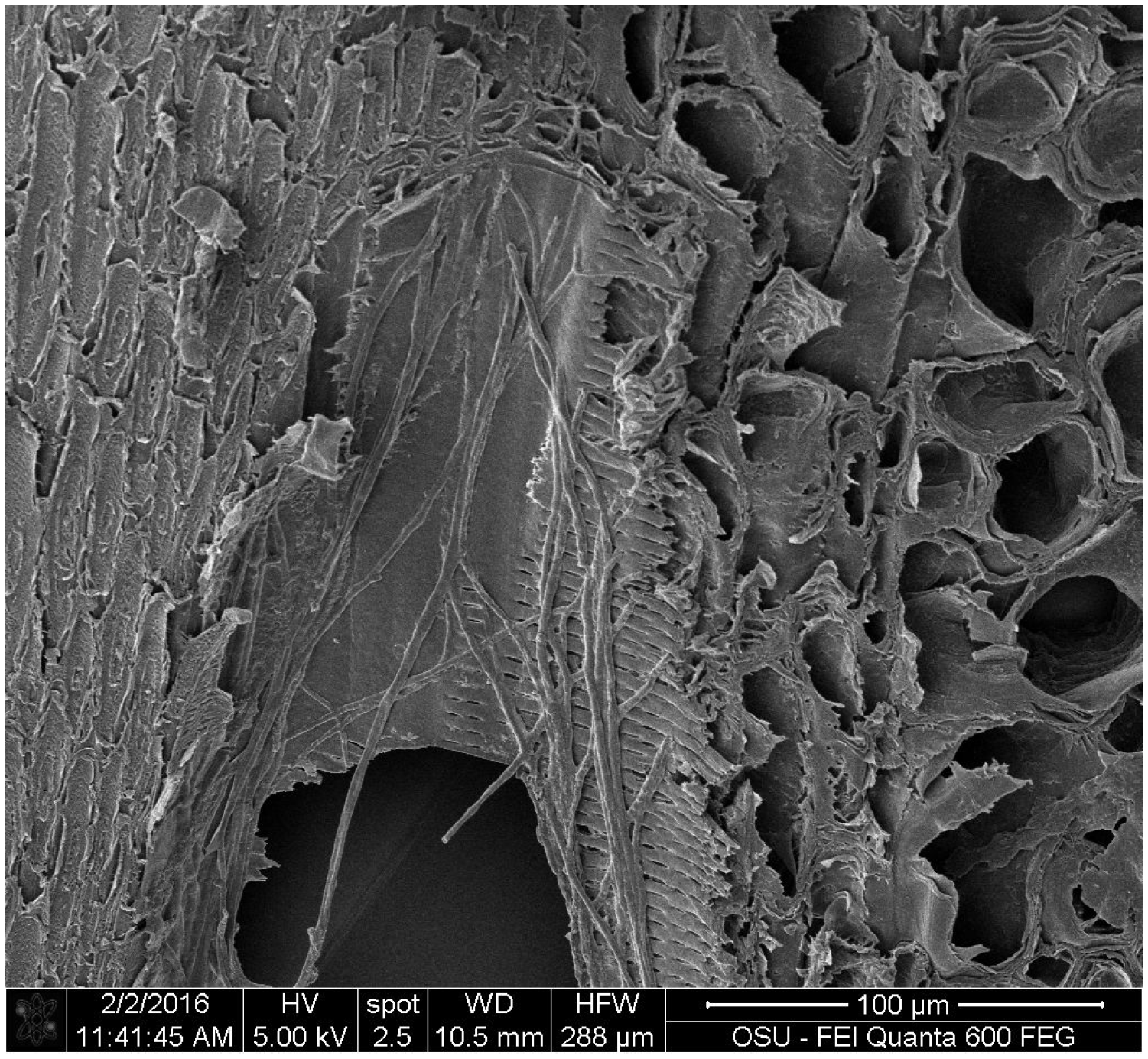
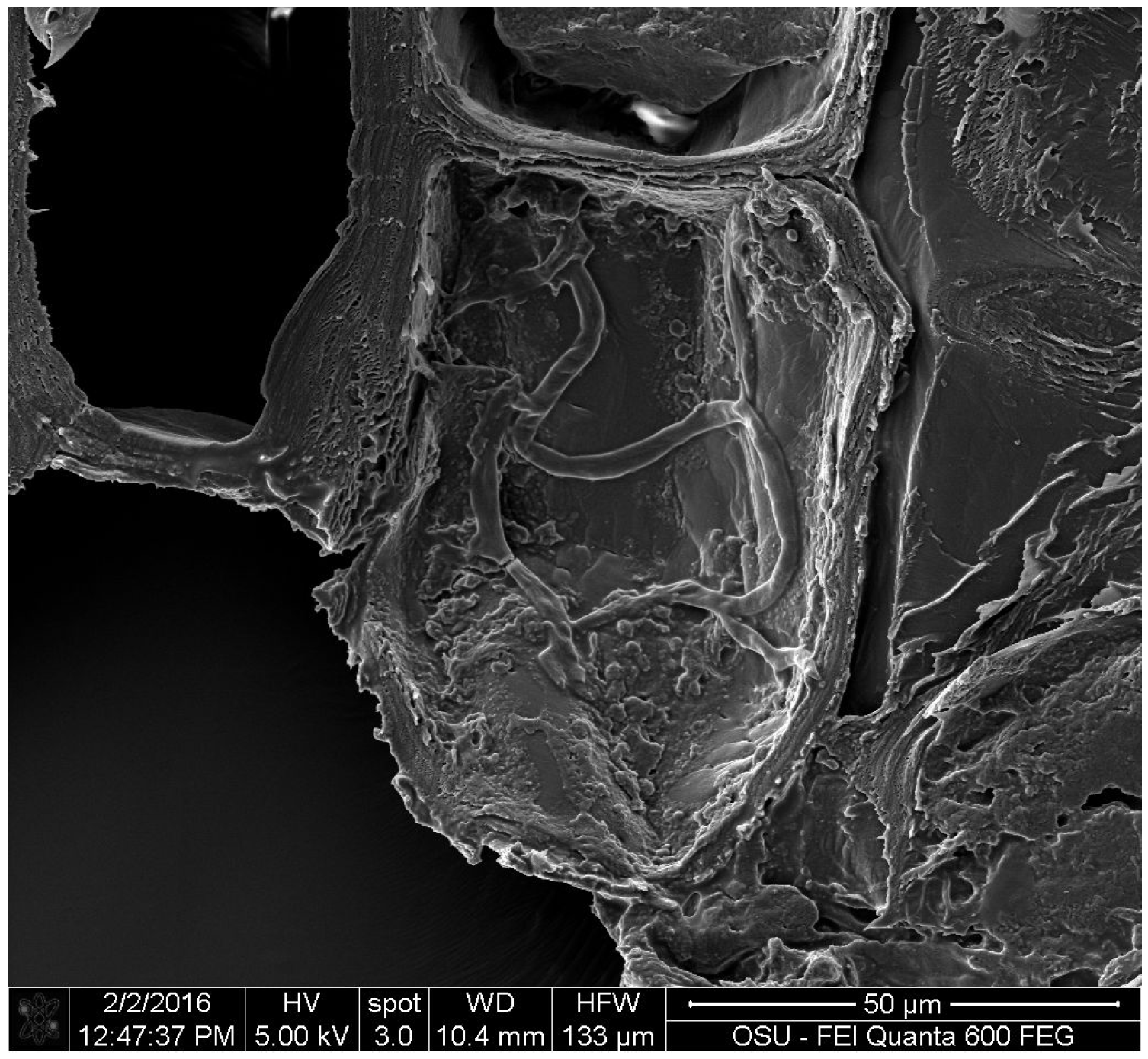
3.1.4. Scanning Electron Microscopy

The images obtained by electron microscopy showed that the hyphae of S. cuboideum developed in the vessels (Figure 1) and parenchyma cells (Figure 2) of bamboo. Hyphae were able to spread through the vessel pits to the surrounding parenchyma cells (Figure 3).

3.2. Dripping Test

3.2.1. Percentage of Area Coverage

No internal color was detected with any of the applied fungal pigments. No area percentage was calculated for external color, because the dripping test covered 100% of the block area on every replicate (Figure 4).

3.2.2. Color Analysis

There was a significant interaction between the colors and the treatments at p < 0.0001. The highest amount of internal color variation was with S. cuboideum applied with 28 drops, a pause of 24 h, and an additional application of 28 drops. This was followed by the treatment of 60 drops and 50 drops, a pause of 24 h, and the application of additional 50 drops. In general, the extracted pigments from S. cuboideum showed the highest color variation when applied on bamboo, as shown in Table 3.

4. Discussion

4.1. Natural Spalting

For the natural spalting test, S. cuboideum had the highest pigmentation rate, with an external pigmented area of 33.25% in four weeks. This result can be compared with a previous test performed by Robinson [26,34], where S. cuboideum obtained a similar result in sugar maple from six to ten weeks. Interestingly, incubation conditions between this experiment and the previous cited were different—the bamboo was run at 19 ± 2 °C, while the experiment run by Robinson was at 27 ± 2 °C. The selection of a lower temperature was due to the inclusion of C. aeruginosa, which prefers lower growth temperatures (21 ± 2 °C) [13]. Based on this observation, it is possible that with a higher temperature, S. cuboideum would have produced more pigment in a shorter time frame.
For bamboo, only S. cuboideum pigmented the material, while S. ganodermophthorum and C. aeruginosa did not produce pigment or even develop mycelium in bamboo. A previous study of wood sugar content and pigment production by spalting fungi found that most of the tested fungi produced more pigment on woods high in sucrose (sugar maple, Acer saccharum, in particular) [36]. Interestingly, S. cuboideum produced the most pigment in the aforementioned study on tree of heaven (TOH, Ailanthus altissima), which had very low sucrose content, but high glucose content. The faster development of S. cuboideum seen in the bamboo study could be due to the abundance of glucose in the tested bamboo.
Most of the aforementioned sugars accumulate in bamboo parenchyma cells [37]. SEM results determined that these cells were where most of the hyphae of S. cuboideum developed, and that they used the vessel structures for mobility. Higher availability of parenchyma tissue with higher amounts of glucose may have encouraged the development of S. cuboideum pigment in bamboo.
Noting the sugar preference of S. ganodermophthorum and C. aeruginosa [25] for sucrose instead of glucose, the failure of these two fungi to produce internal pigment on bamboo test blocks was not unexpected. HPLC results on the bamboo confirmed the findings of Fengei and Shao [37], in which bamboo contained a higher amount of glucose and low to non-existent levels of sucrose, which did not allow the aforementioned fungi to develop pigmentation. In addition, the lack of sucrose in the commercial bamboo could be due to the carbonization process that was applied to the raw material.
It is also possible that the anatomical differences between bamboo and wood affected the pigment production of the fungi, as well as the visibility of the produced color. In the SEM images, the hyphae only concentrated in the vessels and parenchyma cells, and did not expand to the fiber cells. This meant that the only visible color change was in the areas closer to the vascular bundles. This can affect the visibility of the pigments, where areas with a higher concentration of vascular bundles will have a more visible concentration of pigments, compared to areas with fewer bundles. This is correlated with the findings by Robinson et al. [20], where the authors found that the dispersion of the fungal pigments into small dots (or zones) did not create a visibly detectible color change.
In general, S. cuboideum developed the most significant amount of pigment in bamboo, testing the highest of the three fungi in internal and external pigmentation with natural inoculation.

4.2. Dripping Test

For the dripping test, the highest surface color variation came from the red pigment paired with the 28 drops —24 h—28 drops treatment, followed by the 60 and 50 drops —24 h—50 drops treatments. The color variation could be related to the color difference that exists between the pigment and the natural color of bamboo. For the red pigment, a high variation was reported because the CIE L * a * b color of bamboo tends more toward the yellow color space, with CIE L * a * b values = 67.72, = 5.09, = 13.64 (making a greater contrast with the pigment from a red color space). The yellow tendency of bamboo could potentially mask the color effects of the yellow-producing S. ganodermophthorum. Similar results to the yellow pigment were obtained with the blue-green pigment from C. aeruginosa. It is possible that the results for the green pigment are also affected by the color of bamboo, due to the proximity of the bamboo “a” value to the blue spectra. In the samples, it was possible to observe a visible color change on the surface of the bamboo blocks with higher pigment concentration compared with the yellow pigment.
On the internal face, the highest variation of color came from the application of 60 drops for all of the three tested pigments; however, there was no perceivable color coverage inside the blocks. A similar effect was observed in a previous study by Robinson et al. [18,20] on wood, where the authors postulated that there might be a significant internal color change; however, this color change is not visible to the naked eye. For a significant color change to be perceivable to the human eye, the value of the ∆E must be higher than one [20]. Because of the lack of a perceivable color change, the fungal pigments might not be suitable for their use for internal pigmentation on bamboo.
The pigment concentration used was standardized from previous wood experiments. It is possible that higher pigments concentrations would have shown a higher color variation in the external and internal face of the bamboo blocks. Anatomical differences may also be at play here, where the vascular bundles in bamboo may limit the pigment distribution within the material, whereas a wider scattering of vascular elements in wood can permit a more even distribution of the pigments, allowing them to have a more visible internal pigmentation (as with cottonwood, Populus trichocarpa (Torr. And A. Gray)) [19].

5. Conclusions

Scytalidium cuboideum spalted bamboo thoroughly with live inoculation, with blocks showing external and internal pigmentation from week four, with increasing pigment until week 14. Scytalidium ganodermophthorum and C. aeruginosa showed significantly lower pigmentation, comparatively. In the dripping test, the red pigment (S. cuboideum) showed the highest color variation in bamboo with the treatments of 28 drops—24 h—28 drops, 50 drops—24 h—50 drops and 60 drops, but no internal pigmentation was detected. Therefore, no internal spalting was achieved. Both methods showed promising results with S. cuboideum, however the natural method was successful in spalting the bamboo internally and externally, whereas the dripping method showed results in coloring the external surface only.
The results obtained with the natural spalting test indicate that bamboo can be a suitable material for spalting with S. cuboideum, allowing the possibility to modify the color of the material for decorative uses through live fungal inoculation. In terms of the application of the pigments through dripping, the red and green pigments show the potential to modify the surface color of bamboo; however, this method does not generate internal spalting. These results indicate a potential for spalting fungal pigments as surface stains of bamboo, and in the future may be applicable for large-scale commercial bamboo coloration use as a surface dye, replacing the synthetics currently on the market.

Author Contributions

Sarath M. Vega Gutierrez and Sara C. Robinson conceived and designed the experiments; Sarath M. Vega Gutierrez, Patricia T. Vega Gutierrez, Auna Godinez, Lauren Pittis, Megan Huber and Savannah Stanton performed the experiments; Sarath M. Vega Gutierrez analyzed the data; Sarath M. Vega Gutierrez wrote the paper.

Conflicts of Interest

The authors declare no conflict of interest.

References

  1. Hunter, I.R. Bamboo resources, uses and trade: The future? J. Bamboo Rattan 2003, 2, 319–326. [Google Scholar]
  2. Farrely, D. The Book of Bamboo; Sierra Club Books: San Francisco, CA, USA, 1984. [Google Scholar]
  3. Wang, X.; Li, X.; Ren, H. Variation of microfibril angle and density in moso bamboo (Phyllostachys pubescens). J. Trop. For. Sci. 2010, 22, 88–96. [Google Scholar]
  4. Walkling, G. Antique Bamboo Furniture; Bell and Ryman: London, UK, 1979. [Google Scholar]
  5. Lee, A.W.C.; Liu, Y. Selected physical properties of commercial bamboo flooring. For. Prod. J. 2003, 53, 23–26. [Google Scholar]
  6. Terada, T. Acoustical Investigation of the Japanese Bamboo Pipe, Syakuhati; Imperial University: London, UK, 1907. [Google Scholar]
  7. Holley, C.D.; Ladd, E.F. Mixed Paints, Color Pigments and Varnishes, 1st ed.; J.W. Sons: London, UK, 1908. [Google Scholar]
  8. Robinson, S.C.; Richter, D.L.; Laks, P.E. Colonization of sugar maple by spalting fungi. For. Prod. J. 2007, 57, 24–32. [Google Scholar]
  9. Blanchette, R.A.; Wilmering, A.M.; Baumeister, M. The use of green-stained wood caused by the fungus Chlorociboria in Intarsia masterpieces from the 15th century. Holzforschung 1992, 46, 225–232. [Google Scholar]
  10. Fenwick, G.A. Chlorociboria aeruginascens in laboratory culture. Mycologist 1993, 7, 172–175. [Google Scholar]
  11. Maeda, M.; Yamauchi, T.; Oshima, K.; Shimomura, M.; Miyauchi, S.; Mukae, K.; Sakaki, T.; Shibata, M.; Wakamatsu, K. Extraction of xylindein from Chlorociboria aeruginosa complex and its biological characteristics. Bull. Nagaoka Univ. Technol. 2003, 25, 105–111. [Google Scholar]
  12. Robinson, S.C. Developing fungal pigments for “painting” vascular plants. Appl. Microbiol. Biotechnol. 2012, 93, 1389–1394. [Google Scholar] [PubMed]
  13. Robinson, S.C.; Tudor, D.; Snider, H.; Cooper, P.A. Stimulating growth and xylindein production of Chlorociboria aeruginascens in agar-based systems. AMB Express 2012, 2, 1–7. [Google Scholar]
  14. Robinson, S.C.; Tudor, D.; Cooper, P.A. Utilizing pigment-producing fungi to add commercial value to American beech (Fagus grandifolia). Appl. Microbiol. Biotechnol. 2012, 93, 1041–1048. [Google Scholar] [CrossRef] [PubMed]
  15. Tudor, D.; Robinson, S.C.; Cooper, P.A. The influence of pH on pigment formation by lignicolous fungi. Int. Biodeterior. Biodegrad. 2013, 80, 22–28. [Google Scholar] [CrossRef]
  16. Cease, K.R.; Blanchette, R.A. Interactions between Scytalidium species and brown- or white-rot basidiomycetes in birch wood. Wood Sci. Technol. 1989, 23, 151–170. [Google Scholar]
  17. Robinson, S.C.; Tudor, D.; MacDonald, G.; Mansourian, Y.; Cooper, P.A. Repurposing mountain pine beetle blue wood for art through additional fungal colonization. Int. Biodeterior. Biodegrad. 2013, 85, 372–374. [Google Scholar] [CrossRef]
  18. Robinson, S.C.; Hinsch, E.; Weber, G.; Freitas, S. Method of extraction and resolubilisation of pigments from Chlorociboria aeruginosa and Scytalidium cuboideum, two prolific spalting fungi. Color. Technol. 2014, 130, 221–225. [Google Scholar] [CrossRef]
  19. Robinson, S.C.; Weber, G.; Hinsch, E.; Gutierrez, S.M.V.; Pittis, L.; Freitas, S. Utilizing Extracted Fungal Pigments for Wood Spalting: A Comparison of Induced Fungal Pigmentation to Fungal Dyeing. J. Coat. 2014, 2014. [Google Scholar] [CrossRef]
  20. Robinson, S.C.; Hinsch, E.; Weber, G.; Leipus, K.; Cerney, D. Wood Colorization through Pressure Treating: The Potential of Extracted Colorants from Spalting Fungi as a Replacement for Woodworkers’ Aniline Dyes. Materials 2014, 7, 5427–5437. [Google Scholar] [CrossRef]
  21. Otterstedt, A. Investigating green Marquetry on bowed-string instruments. The leaves be greene. Galpin Soc. J. 2001, 54, 330–338. [Google Scholar] [CrossRef]
  22. Blackburn, G.M.; Blackburn, G.M.; Ekong, D.E.U.; Neilson, A.H.; Todd, L. Xylindein. Chimia 1965, 19, 208–212. [Google Scholar]
  23. Edwards, R.L.; Kale, N. The structure of Xylindein. Tetrahedron 1965, 21, 2095–2107. [Google Scholar] [CrossRef]
  24. Donner, C.D.; Cuzzupea, A.N.; Falzona, C.L.; Gill, M. Investigations towards the synthesis of xylindein, a blue-green pigment from the fungus Chlorociboria aeruginosa. Tetrahedron 2012, 68, 2799–2805. [Google Scholar] [CrossRef]
  25. Robinson, S.C.; Tudor, D.; Zhang, W.R.; Ng, S.; Cooper, P.A. Ability of three yellow pigment producing fungi to colour wood under controlled conditions. Int. Wood Prod. J. 2014, 5, 103–107. [Google Scholar] [CrossRef]
  26. Robinson, S.C.; Tudor, D.; Cooper, P.A. Feasibility of using red pigment producing fungi to stain wood for decorative applications. Can. J. For. Res. 2011, 41, 1722–1728. [Google Scholar] [CrossRef]
  27. Hinsch, E.M.; Weber, G.; Chen, H.-L.; Robinson, S.C. Colorfastness of Extracted Wood-Staining Fungal Pigments on Fabrics: A new potential for textile dyes. J. Text. Appar. Technol. Manag. 2015, 9, 1. [Google Scholar]
  28. Weber, G.; Chen, H.; Hinsch, E.; Freitas, S.; Robinson, S. Pigments extracted from the wood-staining fungi Chlorociboria aeruginosa, Scytalidium cuboideum, and S. ganodermophthorum show potential for use as textile dyes. Color. Technol. 2014, 130, 445–452. [Google Scholar] [CrossRef]
  29. Wei, D.S.; Schmidt, O.; Liese, W. Method to test fungal degradation of bamboo and wood using vermiculite as reservoir for moisture and nutrients. Maderas Cienc. Y Tecnol. 2013, 15, 349–356. [Google Scholar] [CrossRef]
  30. Schmidt, O.; Wei, D.S.; Tang, T.K.H.; Liese, W. Bamboo and fungi. J. Bamboo Rattan 2013, 12, 1–14. [Google Scholar]
  31. Wei, D.; Schmidt, O.; Liese, W. Durability test of bamboo against fungi according to EN standards. Eur. J. Wood Wood Prod. 2013, 71, 551–556. [Google Scholar] [CrossRef]
  32. Kim, J.J.; Lee, S.-S.; Ra, J.-B.; Lee, H.; Huh, N.; Kim, G.-Y. Fungi associated with bamboo and their decay capabilities. Holzforschung 2011, 65, 271–275. [Google Scholar] [CrossRef]
  33. Robinson, S.C.; Tudor, D.; Cooper, P.A. Promoting fungal pigment formation in wood by utilizing a modified decay jar method. Wood Sci. Technol. 2011, 46, 841–849. [Google Scholar] [CrossRef]
  34. Robinson, S.C.; Richter, D.L.; Laks, P.E. Effects of substrate on laboratory spalting of sugar maple. Holzforschung 2009, 63, 491–495. [Google Scholar] [CrossRef]
  35. Robinson, S.C.; Laks, P.E.; Turnquist, E.J. A Method for Digital Color Analysis of Spalted Wood Using Scion Image Software. Materials 2009, 2, 62–75. [Google Scholar] [CrossRef]
  36. Robinson, S.C.; Tudor, D.; Cooper, P.A. Wood preference of spalting fungi in urban hardwood species. Int. Biodeterior. Biodegrad. 2011, 65, 1145–1149. [Google Scholar] [CrossRef]
  37. Fengel, D.; Shao, X. A chemical and ultrastructural study of the bamboo species Phyllostachys makinoi Hay. Wood Sci. Technol. 1984, 18, 103–112. [Google Scholar]
Figure 1. S. cuboideum hyphae developing inside a bamboo vessel on week 16.
Coatings 06 00037 g001
Figure 2. S. cuboideum hyphae developing inside a parenchyma cell on week 16.
Coatings 06 00037 g002
Figure 3. S. cuboideum hyphae developing across the vessel pits.
Coatings 06 00037 g003
Figure 4. External face of the bamboo blocks after the dripping treatments. Y = yellow pigment; B = blue pigment; and R = red pigment; DCM: dichloromethane.
Coatings 06 00037 g004
Table 1. Mean area coverage of the external and internal surfaces of the bamboo blocks with natural spalting. Different letters classify the data as statistically different within each pigment color.
MaterialWeekFungusMean % Exterior SpaltStandard DeviationMean % Internal SpaltStandard Deviation
Bamboo4C. aeruginosa5.06 (D)3.270 (C)0
S. cuboideum33.25 (BC)24.5518.49 (BC)17.83
S. ganodermophthorum5.94 (D)14.820.97 (C)2.90
Bamboo6C. aeruginosa9.06 (CD)3.140 (C)0
S. cuboideum59.72 (A)27.9440.41 (AB)22.89
S. ganodermophthorum16.22 (CD)2.981.22 (C)2.68
Bamboo8C. aeruginosa10.58 (CD)5.130 (C)0
S. cuboideum57.71 (AB)17.6034.88 (AB)24.08
S. ganodermophthorum10.35 (CD)2.851.01 (C)3.04
Bamboo10C. aeruginosa12.66 (CD)6.660 (C)0
S. cuboideum65.8 (A)26.5233.39 (AB)36.38
S. ganodermophthorum12.96 (CD)4.500 (C)0
Bamboo12C. aeruginosa11.11 (CD)3.560.22 (C)0.46
S. cuboideum49.08 (AB)27.1536.38 (AB)25.58
S. ganodermophthorum9.82 (CD)5.532.28 (C)2.79
Bamboo14C. aeruginosa9.05 (CD)3.632.91 (C)4.91
S. cuboideum48.77 (AB)28.3257.51 (A)31.67
S. ganodermophthorum11.93 (CD)3.302.75 (C)6.76
Bamboo16C. aeruginosa4.01 (D)3.510 (C)0
S. cuboideum23.28 (CD)18.8915.65 (BC)17.84
S. ganodermophthorum7.42 (D)5.281.22 (C)2.42
Table 2. Mean ΔE of the external surfaces of the bamboo blocks for the natural spalting test. Different letters classify the data as statistically different within each pigment color. DF = 7.
MaterialWeekFungusMean ∆E ExteriorStandard Deviation
Bamboo4C. aeruginosa2.22 (C)1.29
S. cuboideum7.33 (AB)2.16
S. ganodermophthorum2.59 (C)1.30
Bamboo6C. aeruginosa8.35 (A)0.70
S. cuboideum8.57 (A)1.67
S. ganodermophthorum7.97 (A)0.55
Bamboo8C. aeruginosa2.33 (C)1.03
S. cuboideum8.89 (A)2.05
S. ganodermophthorum2.66 (C)1.03
Bamboo10C. aeruginosa3.62 (C)1.45
S. cuboideum8.36 (A)2.30
S. ganodermophthorum3.06 (C)1.28
Bamboo12C. aeruginosa3.90 (C)0.89
S. cuboideum8.33 (A)2.44
S. ganodermophthorum3.94 (C)1.33
Bamboo14C. aeruginosa3.78 (C)1.28
S. cuboideum9.57 (A)2.68
S. ganodermophthorum4.62 (BC)0.72
Bamboo16C. aeruginosa3.09 (C)1.33
S. cuboideum4.47 (C)1.62
S. ganodermophthorum3.19 (C)1.92
Table 3. Mean ΔE of the internal and external surfaces of the bamboo blocks for the dripping test. Different letters classify the data as statistically different within each pigment color. DF = 7.
MaterialTreatment (Drops)ColorMean ΔE ExteriorStandard DeviationMean ΔE InteriorStandard Deviation
Bamboo1Blue5.69 (FGHI)0.356.95 (B)0.94
Red5.30 (HI)0.267.38 (B)0.79
Yellow5.55 (GHI)0.516.32 (B)0.86
Bamboo5Blue5.16 (I)0.436.91 (B)0.64
Red6.17 (DEFG)0.637.06 (B)0.85
Yellow5.51 (GHI)0.736.62 (B)0.70
Bamboo10Blue5.97 (DEFGHI)0.716.64 (B)0.85
Red6.70 (BCD)0.266.45 (B)0.73
Yellow6.28 (DEFG)0.497.15 (B)0.45
BambooDCM pause 10Blue5.57 (GHI)0.444.35 (C)0.54
Red6.16 (DEFGH)0.564.56 (C)0.74
Yellow6.34 (DEFG)0.524.28 (C)0.61
Bamboo40Blue5.79 (EFGHI)0.327.16 (B)0.67
Red6.79 (BCD)0.4411.82 (A)0.54
Yellow6.23 (DEFG)0.484.23 (C)0.72
Bamboo28 pause 28Blue6.09 (DEFGH)0.494.27 (C)0.74
Red7.74 (A)0.284.53 (C)0.81
Yellow6.29 (DEFG)0.264.81 (C)0.48
Bamboo50 pause 50Blue5.75 (EFGHI)0.576.61 (B)0.89
Red7.42 (ABC)0.536.25 (B)0.61
Yellow6.59 (CDE)0.476.70 (B)0.77
Bamboo60Blue6.06 (DEFGH)0.5311.26 (A)0.74
Red7.48 (AB)0.5311.31 (A)0.47
Yellow6.54 (DEF)0.5611.52 (A)0.53

